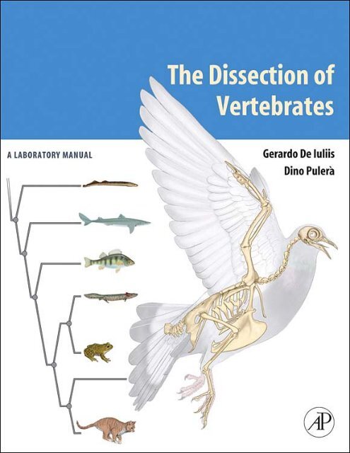
The Dissection of Vertebrates A Laboratory Manual
The Dissection of Vertebrates A Laboratory Manual
The Dissection of Vertebrates A Laboratory Manual

Create successful ePaper yourself
Turn your PDF publications into a flip-book with our unique Google optimized e-Paper software.
<strong>The</strong> <strong>Dissection</strong><br />
<strong>of</strong> <strong>Vertebrates</strong><br />
A <strong>Laboratory</strong> <strong>Manual</strong>
<strong>The</strong> <strong>Dissection</strong><br />
<strong>of</strong> <strong>Vertebrates</strong><br />
A <strong>Laboratory</strong> <strong>Manual</strong><br />
Gerardo De Iuliis, PhD<br />
University <strong>of</strong> Toronto<br />
and George Brown College <strong>of</strong> Applied Arts and Technology<br />
Dino Pulerà, MScBMC, CMI<br />
AMSTERDAM • BOSTON • HEIDELBERG • LONDON • NEW YORK • OXFORD<br />
PARIS • SAN DIEGO • SAN FRANCISCO • SINGAPORE • SYDNEY • TOKYO<br />
Academic Press is an imprint <strong>of</strong> Elsevier
Acquisitions Editor: Tamsin Kent<br />
Marketing Manager: Trevor Daul<br />
Project Manager: Jeff Freeland<br />
Cover Design Direction: Cate Rickard Barr<br />
Text Design: Julio Esperas<br />
Composition: SNP Best-set Typesetter Ltd., Hong Kong<br />
Printer: Hing Yip Printing Co., Ltd.<br />
Academic Press is an imprint <strong>of</strong> Elsevier<br />
30 Corporate Drive, Suite 400, Burlington, MA 01803, USA<br />
525 B Street, Suite 1900, San Diego, California 92101-4495, USA<br />
84 <strong>The</strong>obald’s Road, London WC1X 8RR, UK<br />
This book is printed on acid-free paper.<br />
Copyright © 2007, Elsevier Inc. All rights reserved.<br />
Exceptions:<br />
(a) Except as otherwise provided in Paragraph 2(b) below, the Author hereby grants and transfers to the Publisher the Work<br />
(including any prior unpublished versions <strong>of</strong> the Work) and all rights in the Work, including the entire copyright (and any<br />
renewals and extensions <strong>of</strong> the copyright) throughout the world, and all rights under copyright, including the exclusive<br />
right to publish, perform, reproduce, distribute, and sell the Work and to create derivative works, in all forms or media<br />
now known or later developed, in all languages, throughout the world, and the right to license or authorize others to do<br />
all <strong>of</strong> the foregoing.<br />
(b) With respect to original illustrations created by the Author for the Work (herein the “Illustrations”), copyright for which<br />
is retained by the Author, the Author grants to the Publisher the non-exclusive right to publish the Illustrations in all<br />
editions and versions <strong>of</strong> the Work, including derivative works based on the Work, for sales throughout the world in all<br />
forms or media now known or hereafter developed, and in all languages. <strong>The</strong> Author further agrees that the Publisher<br />
may grant to third parties permission to include the Illustrations in other works, and any copies <strong>of</strong> display <strong>of</strong> the<br />
Illustrations, will include proper credit to the Work as the sources <strong>of</strong> first publication <strong>of</strong> the Illustrations. However, the<br />
Author further agrees that the Author will not permit publication <strong>of</strong> the Illustrations in any competing works.<br />
No part <strong>of</strong> this publication may be reproduced or transmitted in any form or by any means, electronic or mechanical,<br />
including photocopy, recording, or any information storage and retrieval system, without permission in writing from the<br />
publisher.<br />
Permissions may be sought directly from Elsevier’s Science & Technology Rights Department in Oxford, UK: phone:<br />
(+44) 1865 843830, fax: (+44) 1865 853333, E-mail: permissions@elsevier.com. You may also complete your request on-line<br />
via the Elsevier homepage (http://elsevier.com), by selecting “Support & Contact” then “Copyright and Permission” and then<br />
“Obtaining Permissions.”<br />
Library <strong>of</strong> Congress Cataloging-in-Publication Data<br />
Application submitted<br />
British Library Cataloguing-in-Publication Data<br />
A catalogue record for this book is available from the British Library.<br />
ISBN 13: 978-0-12-088776-7<br />
ISBN 10: 0-12-088776-2<br />
For information on all Academic Press Publications<br />
visit our Web site at www.books.elsevier.com<br />
Printed in the United States <strong>of</strong> America<br />
06 07 08 09 10 8 7 6 5 4 3 2 1<br />
Working together to grow<br />
libraries in developing countries<br />
www.elsevier.com | www.bookaid.org | www.sabre.org
This book is for our spouses,<br />
Virginia and Cinzia,<br />
and children,<br />
Daniel, <strong>The</strong>odore, and Jacob,<br />
who are our loves and our lives<br />
With deep respect, admiration, and gratitude, we dedicate this book to three teachers at the<br />
University <strong>of</strong> Toronto that early on in our academic careers instilled in us a passion for<br />
anatomy, paleontology, and art, as well as the intellectual discipline required to make<br />
them our careers. <strong>The</strong>ir contributions are to be seen throughout the following pages.<br />
Charles S. (Rufus) Churcher<br />
Stephen G. Gilbert<br />
Thomas S. Parsons
To our readers:<br />
Despite our best efforts, there are bound to be errors that escaped our<br />
notice, and we would appreciate being informed <strong>of</strong> these. We encourage<br />
you to contact us directly with your comments, suggestions, and<br />
possible ideas <strong>of</strong> text and illustrations for future editions <strong>of</strong> this book.<br />
We look forward to hearing from you.<br />
Sincerely,<br />
Dr. Gerry De Iuliis<br />
gerry.deiuliis@utoronto.ca<br />
Dino Pulerà<br />
dino.pulera@utoronto.ca
Contents Guide xi<br />
Preface xiii<br />
Acknowledgments xvii<br />
Introduction xix<br />
C HAPTER 1<br />
THE CRANIATA AND VERTEBRATA<br />
Phylogeny and Classification 1<br />
Vertebrate Relatives 4<br />
Craniates and <strong>Vertebrates</strong><br />
Early Stages in the Evolution<br />
7<br />
<strong>of</strong> <strong>Vertebrates</strong> 8<br />
Vertebrata 8<br />
C HAPTER 2<br />
THE LAMPREY<br />
Introduction 19<br />
Section I—Skeleton 19<br />
Section II—External Anatomy<br />
Section III—Pleuroperitoneal Cavity<br />
20<br />
and Viscera 21<br />
Section IV—Sagittal Section 22<br />
Key Terms: Lamprey 26<br />
C HAPTER 3<br />
THE SHARK<br />
Introduction 27<br />
Section I—Skeleton 27<br />
Chondrocranium 28<br />
Splanchnocranium 28<br />
CONTENTS<br />
Vertebrae and Fins 31<br />
Key Terms: Skeleton 33<br />
Section II—External Anatomy 35<br />
Key Terms: External Anatomy 37<br />
Section III—Muscular System 39<br />
Trunk and Appendicular Muscles<br />
Muscles <strong>of</strong> the Head and<br />
39<br />
Branchial Region 40<br />
Branchiomeric Musculature 40<br />
Hypobranchial Musculature 43<br />
Key Terms: Muscular System 45<br />
Section IV—Digestive and<br />
Respiratory Systems 45<br />
Key Terms: Digestive and<br />
Respiratory Systems 50<br />
Section V—Cardiovascular System 51<br />
Heart and Arterial Circulation 51<br />
Heart 51<br />
Arteries <strong>of</strong> the Branchial Region 51<br />
Branches <strong>of</strong> the Dorsal Aorta 52<br />
Venous Circulation 55<br />
Hepatic Portal System 55<br />
Renal Portal System 58<br />
Systemic Veins 58<br />
Key Terms: Cardiovascular System 59<br />
Section VI—Urogenital System 59<br />
Male Urogenital System 62<br />
Key Terms: Male Urogenital System 62<br />
Female Reproductive System 62<br />
Key Terms: Female Reproductive System 63<br />
Section VII—Sensory Organs 63<br />
Ampullae <strong>of</strong> Lorenzini 63<br />
Lateral Line System 63<br />
Nose 63<br />
Eye 63<br />
vii
Ear 67<br />
Key Terms: Sensory Organs 68<br />
Section VIII—Brain and Cranial Nerves 70<br />
Brain<br />
Cranial Nerves (CNN 0, I–X, and<br />
71<br />
Lateral Line Nerves) 73<br />
Ventral View <strong>of</strong> the Brain 75<br />
Sagittal Section <strong>of</strong> the Brain 76<br />
Key Terms: Brain and Cranial Nerves 76<br />
C HAPTER 4<br />
THE PERCH<br />
Introduction 77<br />
Section I—Skeleton 77<br />
Skull 77<br />
Key Terms: Skull 79<br />
Postcranial Skeleton 80<br />
Key Terms: Postcranial Skeleton 80<br />
Section II—External Anatomy 81<br />
Key Terms: External Anatomy<br />
Section III—Mouth, Oral Cavity,<br />
82<br />
and Pharynx<br />
Key Terms: Mouth, Oral Cavity,<br />
82<br />
and Pharynx<br />
Section IV—Pleuroperitoneal Cavity<br />
83<br />
and Viscera<br />
Key Terms: Pleuroperitoneal Cavity<br />
83<br />
and Viscera 87<br />
C HAPTER 5<br />
THE MUDPUPPY<br />
Introduction 89<br />
Section I—Skeleton 89<br />
Cranial Skeleton 89<br />
Skull 89<br />
Mandible 90<br />
Hyoid Apparatus 91<br />
Key Terms: Cranial Skeleton 91<br />
Postcranial Skeleton 92<br />
Axial Skeleton 92<br />
Appendicular Skeleton 92<br />
Key Terms: Postcranial Skeleton 92<br />
viii CONTENTS<br />
Section II—External Anatomy 94<br />
Key Terms: External Anatomy 95<br />
Section III—Mouth, Oral Cavity,<br />
and Pharynx 95<br />
Key Terms: Mouth, Oral Cavity,<br />
and Pharynx 96<br />
Section IV—Pleuroperitoneal Cavity<br />
and Viscera 96<br />
Key Terms: Pleuroperitoneal Cavity<br />
and Viscera 98<br />
Section V—Urogenital System 98<br />
Male Urogenital System 102<br />
Female Urogenital System 102<br />
Key Terms: Urogenital System 102<br />
Section VI—Cardiovascular System 102<br />
Heart 102<br />
Venous System 102<br />
Arterial System 105<br />
Key Terms: Cardiovascular System 107<br />
C HAPTER 6<br />
THE FROG<br />
Introduction 113<br />
Section I—Skeleton 113<br />
Skull, Mandible, and Hyoid Apparatus<br />
Key Terms: Skull, Mandible, and<br />
113<br />
Hyoid Apparatus 116<br />
Postcranial Skeleton 117<br />
Key Terms: Postcranial Skeleton 118<br />
Section II—External Anatomy 118<br />
Key Terms: External Anatomy<br />
Section III—Mouth, Oral Cavity,<br />
120<br />
and Pharynx<br />
Key Terms: Mouth, Oral Cavity,<br />
120<br />
and Pharynx<br />
Section IV—Pleuroperitoneal Cavity,<br />
120<br />
Viscera, and Urogenital System<br />
Key Terms: Pleuroperitoneal Cavity,<br />
120<br />
Viscera, and Urogenital System 125<br />
Section V—Cardiovascular System 125<br />
Key Terms: Cardiovascular System 130
C HAPTER 7<br />
THE CAT<br />
Introduction 131<br />
Section I—Skeleton 132<br />
Cranial Skeleton 132<br />
Skull 132<br />
Mandible 138<br />
Hyoid Apparatus 139<br />
Key Terms: Cranial Skeleton 139<br />
Postcranial Skeleton 140<br />
Vertebral Column 140<br />
Cervical Vertebrae 140<br />
Thoracic Vertebrae 141<br />
Lumbar Vertebrae 142<br />
Sacral Vertebrae 142<br />
Caudal Vertebrae 143<br />
Ribs 144<br />
Sternum 144<br />
Forelimb 144<br />
Scapula 144<br />
Clavicle 145<br />
Humerus 145<br />
Ulna 146<br />
Radius 146<br />
Manus 147<br />
Hind Limb 147<br />
Pelvis 147<br />
Femur 147<br />
Patella 148<br />
Tibia 148<br />
Fibula 149<br />
Pes 149<br />
Key Terms: Postcranial Skeleton 150<br />
Section II—External Anatomy 151<br />
Key Terms: External Anatomy 152<br />
Section III—Muscular System 153<br />
Muscle Terminology 153<br />
Connective Tissue and Fiber Direction 153<br />
Key Terms: Muscular System 153<br />
Subdivision <strong>of</strong> the Musculature 153<br />
Skinning the Cat 153<br />
Appendicular Musculature 158<br />
Muscles <strong>of</strong> the Forelimb 158<br />
Superficial Forelimb Muscles: Lateral View 158<br />
Superficial Forelimb Muscles: Ventral View 160<br />
Deep Forelimb Muscles: Lateral View<br />
Deep Forelimb Muscles: Lateral View<br />
160<br />
with Forelimb Abducted 163<br />
Key Terms: Muscles <strong>of</strong> the Forelimb 165<br />
Muscles <strong>of</strong> the Hind Limb 165<br />
Superficial Hind Limb Muscles: Lateral View 165<br />
Superficial Hind Limb Muscles: Medial View 166<br />
Deep Hind Limb Muscles: Lateral View 168<br />
Deep Hind Limb Muscles: Medial View 171<br />
Key Terms: Muscles <strong>of</strong> the Hind Limb 173<br />
Muscles <strong>of</strong> the Head and Trunk 174<br />
Muscles <strong>of</strong> the Trunk 174<br />
Muscles <strong>of</strong> the Back and Neck 174<br />
Muscles <strong>of</strong> the Throat and Jaw 178<br />
Key Terms: Muscles <strong>of</strong> the Head and Trunk 180<br />
Section IV—Digestive and<br />
Respiratory Systems 180<br />
Salivary Glands 180<br />
Oral Cavity and Pharynx 180<br />
Pericardial Cavity 186<br />
Abdominopelvic Cavity 186<br />
Key Terms: Digestive and Respiratory Systems 194<br />
Section V—Cardiovascular System 194<br />
Heart 194<br />
Vessels 195<br />
Main Vessels Associated with the Heart 195<br />
Vessels Anterior to the Diaphragm 195<br />
Vessels Posterior to the Diaphragm 201<br />
Vessels Associated with the Viscera 205<br />
Key Terms: Cardiovascular System 207<br />
Section VI—Urogenital System 212<br />
Excretory System 212<br />
Key Terms: Excretory System 214<br />
Male Reproductive System 214<br />
Opening the Pelvic Canal 216<br />
Male Reproductive System, Continued 216<br />
Key Terms: Male Reproductive System 216<br />
Female Reproductive System 216<br />
Key Terms: Female Reproductive System 217<br />
Section VII—Brain and Cranial Nerves 218<br />
Meninges 218<br />
Telencephalon 218<br />
Diencephalon 220<br />
Mesencephalon 222<br />
Metencephalon 222<br />
Myelencephalon 222<br />
Sagittal Section <strong>of</strong> the Brain 222<br />
Cranial Nerves 224<br />
Key Terms: Brain and Cranial Nerves 224<br />
CONTENTS ix
C HAPTER 8<br />
THE PIGEON<br />
Introduction 227<br />
Section I—Skeleton 227<br />
Skull, Mandible, and Hyoid Apparatus 227<br />
Postcranial Skeleton 229<br />
Vertebrae 229<br />
Ribs 230<br />
Sternum 231<br />
Pectoral Girdle and Forelimb 231<br />
Pelvic Girdle and Hind Limb 233<br />
Key Terms: Skeleton 233<br />
x CONTENTS<br />
Section II—External Anatomy 234<br />
Key Terms: External Anatomy 234<br />
Section III—Musculature 236<br />
Key Terms: Musculature 241<br />
Section IV—Body Cavity, Viscera,<br />
and Vessels 241<br />
Key Terms: Body Cavity, Viscera,<br />
and Vessels 250<br />
Selected References 251<br />
Index 253
CONTENTS GUIDE<br />
C HAPTER 1 THE CRANIATA AND VERTEBRATA 1<br />
C HAPTER 2 THE LAMPREY 19<br />
C HAPTER 3 THE SHARK 27<br />
C HAPTER 4 THE PERCH 77<br />
C HAPTER 5 THE MUDPUPPY 89<br />
C HAPTER 6 THE FROG 113<br />
C HAPTER 7 THE CAT 131<br />
C HAPTER 8 THE PIGEON 227<br />
xi
<strong>The</strong> past two decades have witnessed a rediscovery<br />
among researchers <strong>of</strong> the value <strong>of</strong> comparative vertebrate<br />
anatomy. In large part this has been due to the<br />
establishment <strong>of</strong> phylogenetic systematics and the<br />
renewed awareness <strong>of</strong> the vast contribution that morphology<br />
can make to our understanding <strong>of</strong> the history<br />
<strong>of</strong> vertebrates. However, the study <strong>of</strong> anatomy at the<br />
introductory and intermediate college levels has suffered,<br />
as both its stature and perceived importance have<br />
diminished. <strong>The</strong>re are several reasons for this. Certainly,<br />
and regrettably, the trend at most major academic institutions<br />
has followed a path away from whole organism<br />
biology, as genetics and molecular biology have, for<br />
good reason, become popular. At the same time, there<br />
has been increased resistance from some quarters to the<br />
use <strong>of</strong> animals in various scientific endeavors. Further,<br />
easily accessible computer s<strong>of</strong>tware has been developed<br />
that allows convenient visual journeys through vertebrate<br />
bodies without the effort, expense, and “mess” <strong>of</strong><br />
actual dissection.<br />
<strong>The</strong> study <strong>of</strong> anatomy and morphology has much to<br />
<strong>of</strong>fer the student wishing to pursue a career in biological<br />
or medical fields. Proper training in vertebrate<br />
anatomy must include a practical component that<br />
involves dissection in addition to lectures. No other<br />
method, regardless <strong>of</strong> how intricate in presentation and<br />
scope, can replace the actual hands-on experience. It is<br />
only through a careful, patient, and repeated practical<br />
approach that one gains the expertise and practice<br />
required for understanding the spatial relationships that<br />
are essential to learning how a vertebrate body is constructed,<br />
how its component structures are related to<br />
each other, and how form and function interact.<br />
<strong>The</strong>re are those who would suggest that such a course<br />
<strong>of</strong> study is unnecessary and that anatomy can be learned<br />
solely through texts or s<strong>of</strong>tware. While such materials<br />
(this text among them) may prove to be invaluable as<br />
aids or tools for learning, we ought not to substitute<br />
these adjuncts for the means through which we must<br />
come to know the vertebrate body. To do so would be<br />
akin to preparing for an acting career by watching films,<br />
rather than through rehearsing and acting workshops.<br />
Few <strong>of</strong> us would feel comfortable with mechanics<br />
PREFACE<br />
trained solely through the Internet, trust a surgeon who<br />
has learned the craft strictly through instructional<br />
videos, or fly with a pilot who has only flown missions<br />
on a flight simulator. It is not because such instructional<br />
methods are not useful that we would be suspicious.<br />
Rather, we recognize that, for fields whose subject<br />
matter includes components arranged in complex<br />
spatial relationships, these media are meant to be used<br />
as tools that supplement and guide the trainee through<br />
a methodical, first-hand experience with the subject<br />
matter itself. <strong>The</strong> debate on the value <strong>of</strong> dissection is<br />
particularly lively for human medical anatomy (see, for<br />
example, Elizondo-Omaña et al., 2005; Pawlina and<br />
Lachman, 2004; and Rizzolo, 2002). Many researchers<br />
are clearly in favor <strong>of</strong> dissection, but also see the need<br />
to incorporate the advanced imaging technologies currently<br />
available. <strong>The</strong> same logic should apply for any<br />
vertebrate, but similarly advanced technologies are<br />
unlikely to be applied to a broad range <strong>of</strong> vertebrates in<br />
the foreseeable future.<br />
<strong>The</strong> central theme <strong>of</strong> most previous dissection manuals<br />
has been the structural changes in vertebrates through<br />
their evolution from fish to mammals, with the ultimate<br />
goal being to place mammalian anatomy in context.<br />
This is certainly a necessary prerequisite for one interested<br />
principally in mammalian systematics or medicine.<br />
However, not all students or instructors are interested<br />
primarily in mammals. Two <strong>of</strong> the important lessons<br />
emphasized by phylogenetics are that all living vertebrates<br />
are as evolved as mammals, and that their<br />
anatomy has as much to tell us about evolution, function,<br />
and morphology. Indeed, a common complaint<br />
among academic faculty is that comparative vertebrate<br />
anatomy courses have become courses on the anatomy<br />
<strong>of</strong> the cat. Be that as it may, it is important to remember<br />
that negative perceptions can <strong>of</strong>ten be detrimental<br />
to the well-being <strong>of</strong> a field <strong>of</strong> study and may sway<br />
departmental decisions on whether the continuation <strong>of</strong><br />
some courses is worth the effort and expense. It is up<br />
to those <strong>of</strong> us who teach comparative anatomy to<br />
push forward and maintain its vigor and centrality, in<br />
part by relating its wide applicability to related fields,<br />
such as systematics, evolutionary biology, paleontology,<br />
paleobiology, and functional morphology; and as a<br />
xiii
prerequisite for higher-level zoology courses, such as<br />
mammalogy, herpetology, ornithology, and vertebrate<br />
paleontology.<br />
Our format and coverage is aimed at striking a balance<br />
between presenting an evolutionary sequence to<br />
“higher” vertebrates and treating the anatomy <strong>of</strong> each<br />
representative vertebrate as inherently important. <strong>The</strong><br />
sequence <strong>of</strong> vertebrates is similar to those presented by<br />
other authors, but we emphasize throughout that the<br />
living vertebrates are not and cannot be used as intermediates.<br />
For this reason, we provide discussions <strong>of</strong> the<br />
important features <strong>of</strong> each group based on the derived<br />
features that diagnose a particular phylogenetic grouping.<br />
We thus do not treat vertebrates by traditional<br />
grouping methods; we would rather, from the beginning,<br />
present the student with information that reflects<br />
our formal thinking and classification.<br />
<strong>The</strong> main goal <strong>of</strong> this text is to provide today’s visually<br />
oriented student population with a manual that links<br />
succinct and pedagogically effective textual direction<br />
with relevant, high-quality, accurate, and attractive<br />
visual references to promote efficient learning <strong>of</strong> the<br />
complex, spatially abstract subject matter in the limited<br />
time available in a laboratory setting. Thus, a critical<br />
feature <strong>of</strong> <strong>The</strong> <strong>Dissection</strong> <strong>of</strong> <strong>Vertebrates</strong> is the inclusion<br />
<strong>of</strong> numerous high-quality, didactic, color illustrations.<br />
Each depicts the vertebrate approximately as it would<br />
appear in a particular stage <strong>of</strong> dissection, rather than<br />
presenting an idealized figure or photographs, as is the<br />
case for most other manuals. This in itself facilitates the<br />
use <strong>of</strong> these illustrations, both in learning and later<br />
during recall for studying purposes. Photographs are<br />
used sparingly. We have chosen illustration over photography<br />
in the vast majority <strong>of</strong> cases because illustration<br />
is the method that affords the most control in<br />
communicating the pertinent features <strong>of</strong> a particular<br />
dissection. Photographs are indiscriminate, whereas<br />
illustration in combination with color allows minimization<br />
<strong>of</strong> unnecessary and distracting background<br />
anatomical detail, while still maintaining it. Indeed, we<br />
have taken great care to ensure that the background<br />
anatomy in the illustrations is accurate. This is important<br />
because it gives the user (instructor and student) a<br />
context for the anatomical structures under study.<br />
Although students aspiring to careers in systematics,<br />
vertebrate paleontology, or functional morphology are<br />
the primary intended audience <strong>of</strong> this manual, <strong>The</strong> <strong>Dissection</strong><br />
<strong>of</strong> <strong>Vertebrates</strong> is sufficiently flexible in scope and<br />
organization that it may be used in any course on vertebrate<br />
anatomy. We present a wide-ranging and encompassing<br />
reference manual that will both help students<br />
learn the basic anatomy <strong>of</strong> vertebrates and function as<br />
a guide once they are ready to venture into the primary<br />
literature.<br />
xiv PREFACE<br />
<strong>The</strong> <strong>Dissection</strong> <strong>of</strong> <strong>Vertebrates</strong> presents dissection<br />
instructions on more vertebrates than is normally the<br />
case. <strong>The</strong> primary focus is on the shark, mudpuppy, and<br />
cat, as is usual, but it also provides detailed information<br />
on vertebrates either not usually considered or treated<br />
very superficially by most other manuals that include<br />
multiple vertebrates. It is ironic that the two most speciose<br />
groups <strong>of</strong> vertebrates, the birds and ray-finned<br />
fishes, are not adequately covered (if covered at all) in<br />
other dissection manuals. We hope that by providing<br />
reasonably detailed guides for these vertebrates, instructors<br />
will feel more inclined to include these readily available<br />
and inexpensive vertebrates in their courses.<br />
This manual is organized by vertebrate. <strong>The</strong> anatomy<br />
<strong>of</strong> each is then presented systemically. This approach<br />
allows all the information on a particular vertebrate to<br />
be studied at one time and in sequence. We believe,<br />
based on years <strong>of</strong> instruction, that this method provides<br />
a more straightforward integration <strong>of</strong> the systems. <strong>The</strong><br />
inclusion <strong>of</strong> many vertebrates and the organization by<br />
vertebrate makes <strong>The</strong> <strong>Dissection</strong> <strong>of</strong> <strong>Vertebrates</strong> more<br />
flexible for use in a broad-based full or half-year course<br />
at the introductory college level, and allows more convenient<br />
organization <strong>of</strong> course content, depending on<br />
time and availability <strong>of</strong> specimens and the instructor’s<br />
preferences.<br />
At the same time, we omit many topics that are <strong>of</strong>ten<br />
covered in most other manuals. Sections on vertebrates<br />
or structures that students are unlikely ever to dissect at<br />
the intended level <strong>of</strong> study are not included. Instead, we<br />
have focused the material on examples that are likely to<br />
be encountered in an introductory lab course, leaving<br />
those topics best presented in texts that accompany the<br />
lecture portion <strong>of</strong> a course.<br />
Much <strong>of</strong> the required background information is presented<br />
in the Introduction and Chapter 1. This includes<br />
sections on planes <strong>of</strong> dissection and orientational terminology<br />
(see below), as well as an introduction to vertebrates<br />
and their relatives (Chapter 1). We suggest that<br />
these sections be included as part <strong>of</strong> the assigned readings<br />
for a particular laboratory for each vertebrate. This<br />
method will expose students repeatedly to the broad<br />
evolutionary development <strong>of</strong> each system. Terms that<br />
are required learning are in boldface print throughout<br />
the manual. Bold-faced terms are listed in a Key Terms<br />
section (which also provides common synonyms in<br />
parentheses) following each major component. Students<br />
will know at a glance the structures for which they are<br />
responsible. We suggest that students use this section as<br />
a key to learning the structures by writing a short<br />
description for each. <strong>The</strong> Key Terms sections also allow<br />
instructors to adapt this manual to their personal preferences<br />
in running their course. Structures that are not<br />
required can be identified and crossed out, so that
students know they are not responsible for them. This<br />
method effectively allows an instructor to limit the<br />
detail <strong>of</strong> the dissection.<br />
We believe that the concise presentation <strong>of</strong> dissection<br />
instructions combined with minimal background information<br />
results in a straightforward text that will<br />
facilitate and focus the student’s learning <strong>of</strong> anatomy<br />
in laboratory. In contrast to most other manuals, much<br />
<strong>of</strong> the background material presented in lecture is<br />
omitted here, so <strong>The</strong> <strong>Dissection</strong> <strong>of</strong> <strong>Vertebrates</strong> is less<br />
cumbersome to use even though it covers more vertebrates<br />
than do other manuals. All the information is<br />
relevant for laboratory purposes. This should facilitate<br />
matters for the instructor as well. Among other<br />
things, it will allow a clear answer to the <strong>of</strong>ten-asked<br />
question, “What am I responsible for reading?” <strong>The</strong><br />
response can, without too much exaggeration, be, “All<br />
<strong>of</strong> it.”<br />
PREFACE xv
Many colleagues, students, friends, and members <strong>of</strong> our<br />
families have contributed to the production <strong>of</strong> this<br />
book, from carrying out simple tasks to pro<strong>of</strong>reading<br />
to providing emotional encouragement and support. We<br />
are grateful to them all, although we can directly<br />
acknowledge only a few <strong>of</strong> them here: Thomas Carr<br />
(Carthage College) for preparing selected dissections<br />
and providing valuable input on text and illustrations;<br />
Hans-Dieter Sues (Smithsonian Institution), Jeff<br />
Thomason (University <strong>of</strong> Guelph), and Sergio F. Vizcaíno<br />
(Museo de La Plata) for providing particularly comprehensive<br />
reviews <strong>of</strong> earlier versions <strong>of</strong> the manuscript that<br />
greatly im-proved the final product; Rob Baker and Jim<br />
Thomson (University <strong>of</strong> Toronto) and Rivie Seaberg<br />
(George Brown College <strong>of</strong> Applied Arts and Technology)<br />
for academic and institutional support; Stephen Mader<br />
(Artery Studios Inc.) for his encouragement and support;<br />
Peter von Bitter, Kathy David, Brian Iwama, and Peter<br />
Reali for help with photography; Celestino De Iuliis for<br />
reading earlier drafts <strong>of</strong> the manuscript; Marco Zimmer-<br />
De Iuliis for expert preparation <strong>of</strong> specimens; Sandra<br />
Reali for logistic support; Kevin Seymour for access to<br />
the skeletal collections <strong>of</strong> the Royal Ontario Museum;<br />
Skulls Unlimited (www.skullsunlimited.com) for providing<br />
skeletons for study and photography; Barry<br />
Bruce (CSIRO Division <strong>of</strong> Marine Research, Tasmania),<br />
Mark McGrouther and Elizabeth Cameron (Australian<br />
ACKNOWLEDGMENTS<br />
Museum) and Andrew and Silvy Fox (Rodney Fox Shark<br />
Museum, Australia) for providing shark dissection<br />
photos; a special thank you to Steven E. Campana<br />
(Bedford Institute <strong>of</strong> Oceanography, Canada; www.<br />
marinebiodiversity.ca/shark/english/index.htm) for also<br />
providing digital and labeled shark dissection photos, and<br />
very kindly allowing us to reprint the SEM photo <strong>of</strong> the<br />
spiny dogfish skin (Figure 3.12(g)). We thank several<br />
anonymous reviewers for their helpful comments,<br />
suggestions, and corrections. <strong>The</strong> efforts <strong>of</strong> Timothy<br />
Rowe (<strong>The</strong> University <strong>of</strong> Texas at Austin) on the<br />
Digital Morphology web site (www.Digimorph.org)<br />
are greatly appreciated. Although we did not make<br />
direct use <strong>of</strong> Digimorph images, several <strong>of</strong> them were<br />
extremely useful in the interpretation <strong>of</strong> anatomical<br />
features. We thank our editors Tamsin Kent, Nancy<br />
Maragioglio, and Kelly Sonnack and Jeff Freeland <strong>of</strong><br />
Elsevier for their patience, guidance, and keeping us on<br />
target. We are also indebted to David Cella (formerly <strong>of</strong><br />
Elsevier) for initial consideration <strong>of</strong> our proposal and recognizing<br />
the potential for this book and to Stephen G.<br />
Gilbert for showing us how to get started on creating our<br />
own book, and for his encouragement, support, advice,<br />
and continued inspiration. Lastly, we thank Virginia and<br />
Cinzia for being there beside us every step <strong>of</strong> the way in<br />
seeing this book through to the end—it has been a long<br />
and challenging journey.<br />
xvii
<strong>The</strong> study <strong>of</strong> vertebrate anatomy is an interesting and<br />
valid field <strong>of</strong> study for gaining insight into the structure<br />
and function <strong>of</strong> vertebrates. But why should this be<br />
important? Of the numerous reasons, we mention only<br />
a few.<br />
• It provides us with knowledge <strong>of</strong> the structures <strong>of</strong><br />
different organisms and the great variety <strong>of</strong> form<br />
among vertebrates.<br />
• It allows us to examine how the form <strong>of</strong> these<br />
structures is related to their function and thus<br />
how morphology is suited to a particular mode <strong>of</strong><br />
life.<br />
• <strong>The</strong> characteristics or features <strong>of</strong> vertebrates<br />
preserve information on their ancestry: <strong>The</strong> features<br />
are modified and passed on through the course <strong>of</strong><br />
generations, and we may use such knowledge to<br />
discover the genealogical relationships among<br />
vertebrates.<br />
• Comparative anatomical studies help us to<br />
understand how the major transitions in vertebrate<br />
design might have occurred. S<strong>of</strong>t tissues do not<br />
fossilize, meaning that (with rare exceptions) only<br />
transformations <strong>of</strong> the hard parts <strong>of</strong> the vertebrate<br />
body are preserved in the fossil record. For<br />
other parts <strong>of</strong> the body, we must rely on a sequence<br />
<strong>of</strong> living forms. <strong>The</strong>re are problems with<br />
this approach, but if we begin with a robust<br />
phylogenetic hypothesis and keep in mind that<br />
the living members <strong>of</strong> some groups are highly<br />
derived, then we may be confident in this method<br />
as a reasonable approach for deducing the major<br />
steps in the evolution <strong>of</strong> different vertebrate<br />
groups.<br />
We will consider all <strong>of</strong> these aspects in the following<br />
course on comparative vertebrate anatomy. Before<br />
beginning this study, however, there are several important<br />
terms that unambiguously describe position and<br />
direction. <strong>The</strong>se indispensable terms greatly facilitate<br />
INTRODUCTION<br />
navigating through the complex three-dimensional<br />
structure <strong>of</strong> vertebrate bodies.<br />
DIRECTIONAL TERMINOLOGY AND PLANES<br />
OF SECTION<br />
As with all advanced fields <strong>of</strong> research, anatomical<br />
study requires the use <strong>of</strong> specialized terminology. Such<br />
terminology includes not only special words for the<br />
anatomical structures themselves and concepts or<br />
processes (such as homology, for example) but also<br />
terms to designate unambiguously the orientation and<br />
direction <strong>of</strong> structures <strong>of</strong> the vertebrate body. <strong>The</strong>se<br />
terms may at first seem superfluous, but that is because<br />
most people have never dealt with anatomy in a comprehensive<br />
and detailed manner. It is perfectly adequate<br />
in everyday life to say that the stomach is lower than<br />
the heart or the appendix is in the lower right part <strong>of</strong><br />
the belly. But this is not anatomy. You will quickly come<br />
to realize the importance <strong>of</strong> the terms presented in this<br />
section, and you are urged to learn and become familiar<br />
with them.<br />
<strong>The</strong>re are two main sets <strong>of</strong> terms. One is used in medicine<br />
and by some anthropologists, the other by comparative<br />
anatomists, paleontologists, and veterinarians.<br />
To compound the problem, various synonyms exist for<br />
some terms in each set. <strong>The</strong>se circumstances may be<br />
cause for confusion, but we may simplify matters by<br />
adhering to one set <strong>of</strong> terms. As we are studying comparative<br />
anatomy, we will use the system commonly<br />
used for nonhuman vertebrates.<br />
Unlike humans, the vast majority <strong>of</strong> vertebrates go<br />
through life with the long axis <strong>of</strong> the body oriented horizontally<br />
or parallel to the substrate. It is with reference<br />
to this position that the main directional terms are<br />
defined. Most <strong>of</strong> these terms are coupled; that is, there<br />
are two terms that describe opposite directions along a<br />
single axis. Refer to Figure 1 while reading through the<br />
xix
Anterior<br />
Proximal Proximal<br />
Distal<br />
following explanations. Anterior and posterior refer to<br />
the horizontal longitudinal axis and respectively designate<br />
the directions toward the head and tail. Synonyms<br />
for these terms that you may encounter are cranial or<br />
rostral for anterior, and caudal for posterior. <strong>The</strong> vertical<br />
direction toward the belly or the ground is ventral;<br />
toward the back or up is dorsal. Medial refers to the<br />
horizontal direction toward the sagittal midline (see<br />
below) <strong>of</strong> the body, whereas lateral refers to the directions<br />
away from the midline. <strong>The</strong>se are the main terms,<br />
but there is another set that is useful. Proximal and<br />
distal are terms usually used with a particular reference.<br />
At times this reference may be the trunk <strong>of</strong> the body; at<br />
other times a particular structure, such as the heart, may<br />
be the reference point. Proximal designates a position<br />
closer to the trunk or structure <strong>of</strong> reference, and distal<br />
furthest from the trunk or structure <strong>of</strong> reference. Thus,<br />
for example, the fingers (phalanges) are distal to the<br />
upper arm (brachium); and the proximal end <strong>of</strong> the<br />
brachium is that end closest to the trunk. If the reference<br />
point is another structure, say the heart, then the<br />
Dorsal<br />
Ventral<br />
xx INTRODUCTION<br />
Medial Medial Lateral<br />
Lateral<br />
FIGURE 1 Directional terms and main planes or sections through the body shown on a horse.<br />
Posterior<br />
proximal part <strong>of</strong> a blood vessel is the part closest to the<br />
heart, and the distal end is that part furthest away.<br />
Combinations <strong>of</strong> these terms may be used, and indeed<br />
are used <strong>of</strong>ten in this manual, to describe directions that<br />
are oblique to the main axes. For example, anterolateral<br />
combines anterior and lateral, and indicates a<br />
simultaneous direction toward the head and to the side.<br />
Thus, taking the umbilicus (navel or bellybutton) as a<br />
reference, we may describe the shoulder as anterolateral<br />
to the umbilicus. Figure 2 provides examples <strong>of</strong> these<br />
terms.<br />
<strong>Dissection</strong> <strong>of</strong>ten involves cutting the body in various<br />
planes to obtain internal or sectional views, which are<br />
extremely useful for comprehending the spatial arrangement<br />
<strong>of</strong> structures. <strong>The</strong>re are three main sections or<br />
planes that pass through the body (Figure 1). <strong>The</strong> sagittal<br />
section is vertical and lies in the midline longitudinal<br />
<strong>of</strong> the body. It separates the body into right and left<br />
halves. Sections that are parallel to and on one side <strong>of</strong>
Anterior<br />
Anterodorsal<br />
(a)<br />
(b)<br />
Posteromedial<br />
Lateral<br />
Dorsal<br />
Ventral<br />
Anterior<br />
Medial<br />
Posterior<br />
Posteroventral<br />
Anterolateral<br />
Lateral<br />
Posterior<br />
FIGURE 2 (a, b) Combined directional terms shown on a cat.<br />
the sagittal plane are termed parasagittal. A second<br />
major section is in the transverse plane, which is also<br />
vertical but is perpendicular to the sagittal plane. A<br />
transverse section cuts across the main longitudinal axis<br />
and subdivides the body into anterior and posterior<br />
parts. <strong>The</strong> last major section is in the frontal plane,<br />
which is horizontal and perpendicular to the sagittal<br />
and transverse planes. A frontal section separates the<br />
body into dorsal and ventral parts.<br />
KEY TERMS: INTRODUCTION<br />
anterior (cranial,<br />
rostral)<br />
distal<br />
dorsal<br />
frontal<br />
lateral<br />
medial<br />
parasagittal<br />
posterior<br />
proximal<br />
sagittal<br />
transverse<br />
ventral<br />
INTRODUCTION xxi
<strong>The</strong> vertebrates or Vertebrata (see below) form an<br />
ancient group with a history spanning some 545 million<br />
years. On the one hand, they include the organisms most<br />
familiar to us, such as fish, birds, cats and dogs, as well<br />
as other humans; on the other, few people are aware <strong>of</strong><br />
the great diversity in their form, structure, and habits.<br />
Indeed, they include some <strong>of</strong> the largest and more<br />
complex organisms ever evolved. But vertebrates are<br />
part <strong>of</strong> a larger grouping <strong>of</strong> animals, and to understand<br />
their history and the development <strong>of</strong> their structure,<br />
they must be placed in phylogenetic context.<br />
In discussing vertebrates, several other groups <strong>of</strong> organisms<br />
are usually considered. A group <strong>of</strong> organisms is<br />
referred to as a taxon (plur., taxa). <strong>The</strong> taxa related to<br />
vertebrates include the Echinodermata (sand dollars, sea<br />
lilies, starfish, sea cucumbers, urchins), Hemichordata<br />
(acorn worms and pterobranchs), Urochordata (tunicates<br />
or sea squirts), and Cephalochordata (amphioxus).<br />
<strong>The</strong>se are the typical nonvertebrate (or “invertebrate”)<br />
relatives <strong>of</strong> the group we are mainly interested in. <strong>The</strong><br />
vertebrates themselves, or Vertebrata, are included in a<br />
larger taxon termed the Craniata. Within the Craniata<br />
and Vertebrata are many taxa. <strong>The</strong>se taxa and the relationships<br />
among them (see Figure 1.1) are briefly outlined<br />
below to provide an organizational framework for<br />
undertaking the dissection <strong>of</strong> the vertebrates discussed<br />
in this manual. Before this, however, it is necessary to<br />
present an explanation <strong>of</strong> several important terms used<br />
in discussions <strong>of</strong> phylogeny.<br />
PHYLOGENY AND CLASSIFICATION<br />
For most <strong>of</strong> the past 250 years, the classification <strong>of</strong><br />
organisms has followed the Linnean system, which uses<br />
ranks to designate levels <strong>of</strong> organization <strong>of</strong> the organisms<br />
being classified. Most readers will be familiar with<br />
the main formal Linnean ranks, ordered hierarchically<br />
from most to least inclusive: Kingdom, Phylum, Class,<br />
Order, Family, Genus, and Species. Researchers have<br />
differed in assigning rank to the vertebrates and their<br />
relatives. For example, some authors have recognized<br />
three phyla: Phylum Echinodermata, Phylum Hemichordata,<br />
and Phylum Chordata. Others consider the<br />
C HAPTER 1<br />
THE CRANIATA AND VERTEBRATA<br />
Urochordata and Cephalochordata as phyla on their<br />
own, separate from the Chordata. Still others have<br />
viewed the Urochordata as a separate phylum, but the<br />
Cephalochordata as a subphylum <strong>of</strong> the Phylum Chordata.<br />
If you find this confusing, you’re not alone! <strong>The</strong><br />
different designations did—or at any rate were meant<br />
to—have some grounding in biological reality. <strong>The</strong>y<br />
reflected a particular researcher’s perception <strong>of</strong> the magnitude<br />
<strong>of</strong> the difference in the levels <strong>of</strong> organization (a<br />
quality that may be referred to as a grade) among the<br />
taxa under consideration. Thus, if a taxon was considered<br />
a phylum, it mainly implied that its members made<br />
their living in a very different way than if they were<br />
considered only a subphylum <strong>of</strong> a larger taxon. As you<br />
have probably already realized, researchers’ perceptions<br />
along these lines are subjective.<br />
In recent years, however, the formal Linnean ranking<br />
system has fallen increasingly into disuse as systematists<br />
have become aware that there is no intrinsic or “special”<br />
value <strong>of</strong> any particular taxon that would justify its<br />
recognition as a higher or lower rank, compared to<br />
other taxa. In other words, there is no special reason for<br />
“elevating” birds or Aves to the rank <strong>of</strong> Class, equal and<br />
thereby excluded from, the Class Reptilia. In fact, it is<br />
improper to do so, because the birds are properly part<br />
<strong>of</strong> the taxon named Reptilia. Here, formal ranks are not<br />
used, and taxa are referred to simply by their name.<br />
Formal names are applied to natural or monophyletic<br />
groups. A monophyletic group includes an ancestor and<br />
all <strong>of</strong> its descendants (provided that the phylogeny has<br />
been carefully reconstructed). Such groups are termed<br />
clades. Clades are recognized based on common ancestry.<br />
If two taxa are in a clade, it is because they are<br />
linked by a common ancestor. Biologists infer such<br />
ancestral relationships through the presence <strong>of</strong> shared<br />
derived characters or synapomorphies (see below). If<br />
two (or more) taxa share a character that is exclusive<br />
to them, then we assume that they share this feature<br />
because they have inherited it from a common ancestor,<br />
rather than each having evolved the character independently,<br />
and so infer that the taxa are descendants <strong>of</strong><br />
the same ancestor (which we are not able to actually<br />
recognize, and thus refer to as hypothetical). This, <strong>of</strong><br />
1
PROTOSTOMATA<br />
ECHINODERMATA<br />
ENTEROPNEUSTA<br />
PHARYNGOTREMATA<br />
HEMICHORDATA CHORDATA<br />
DEUTEROSTOMATA<br />
PTEROBRANCHIA<br />
• first embryonic opening becomes anus;<br />
second opening becomes mouth<br />
• basally, mesoderm forms bilaterally as<br />
out-pocketing <strong>of</strong> embryonic gut; ceolom,<br />
which develops within mesoderm, thus<br />
originally has connection with gut<br />
• basally, a ciliated looped band is present on<br />
surface <strong>of</strong> larva<br />
DEUTEROSTOMATA<br />
UROCHORDATA<br />
PHARYNGOTREMATA<br />
• pharyngeal skeleton<br />
• pharyngeal slits<br />
• blood vascular system<br />
SOMITICHORDATA<br />
CEPHALOCHORDATA<br />
CHORDATA<br />
• pharyngeal slits<br />
• endostyle<br />
• dorsal, hollow nerve cord<br />
• notochord<br />
• postanal tail<br />
CRANIATA<br />
FIGURE 1.1 Cladogram showing phylogeny <strong>of</strong> the Deuterostomata. Some synapomorphies <strong>of</strong> the main groups are provided in the boxes below the cladogram.
course, is the idealized situation. In reality, biologists use<br />
many characters in trying to reconstruct phylogeny.<br />
<strong>The</strong> practice is complicated by the fact that organisms<br />
can and do evolve very similar characters independently<br />
<strong>of</strong> each other, an occurrence referred to as homoplasy.<br />
In reconstructing phylogeny, a researcher considers the<br />
totality <strong>of</strong> evidence. It is rare that only a single character<br />
can be used to reconstruct phylogeny.<br />
<strong>The</strong> pattern <strong>of</strong> relationships among taxa is depicted<br />
visually by a cladogram, which is essentially a diagram<br />
<strong>of</strong> nodes and branches, with the nodes representing<br />
ancestors and the branches that diverge from a node<br />
representing the descendant taxa <strong>of</strong> the ancestor. <strong>The</strong><br />
node, then, may be thought <strong>of</strong> as representing the hypothetical<br />
ancestor <strong>of</strong> the two taxa that diverge from it.<br />
<strong>The</strong> pattern <strong>of</strong> branching represents the pattern <strong>of</strong> relationship.<br />
Examine the cladogram in Figure 1.1. Note the<br />
node from which the Hemichordata and Chordata<br />
diverge. This node represents the ancestor species that<br />
split to produce two lineages, one that evolved into the<br />
Chordata and the other into the Hemichordata. <strong>The</strong> two<br />
branches that diverge from this ancestor represent the<br />
evolutionary paths to the divergent taxa.<br />
Only the branching pattern is <strong>of</strong> concern. <strong>The</strong> length <strong>of</strong><br />
the branches is immaterial in terms <strong>of</strong> absolute time, but<br />
relative time is implied by branching sequence. Clearly,<br />
the divergence <strong>of</strong> the Cephalochordata and Craniata<br />
occurred after the divergence <strong>of</strong> the Hemichordata and<br />
Somitichordata.<br />
Informal names, set between quotation marks, are used<br />
to designate a group <strong>of</strong> organisms that do not descend<br />
from the same common ancestor but that do possess (or<br />
lack) some <strong>of</strong> the features <strong>of</strong> the taxon in which we are<br />
interested. Many <strong>of</strong> these terms were considered formal<br />
names in earlier classifications. For example, the term<br />
“protochordates” is commonly used to refer to the<br />
hemichordates, urochordates, and cephalochordates.<br />
Grouping them together is a shorthand way <strong>of</strong> referring<br />
to them as close relatives <strong>of</strong> chordates (no quotation<br />
marks here, so this is the vernacular form <strong>of</strong> the formal<br />
name Chordata), and that they lack various characters<br />
that chordates possess. We must be clear that informal<br />
groups, though convenient, do not reflect phylogeny;<br />
they are not monophyletic.<br />
In discussing how biologists reconstruct phylogeny, the<br />
nature <strong>of</strong> the similarity among organisms must be considered,<br />
because it is necessary to differentiate between<br />
those similarities that are useful in reconstructing phylogeny<br />
and those that are not. One kind, termed plesiomorphic,<br />
refers to similarity based on the presence <strong>of</strong><br />
primitive or ancestral conditions or states. Consider the<br />
Vertebrata, in which the presence <strong>of</strong> vertebrae is an<br />
ancestral feature—in other words, it was present in the<br />
common ancestor <strong>of</strong> all vertebrates. <strong>The</strong>se structures<br />
may inform us that all vertebrates share a common<br />
ancestor, but their presence per se cannot be used to<br />
decipher the relationships among vertebrates. As a<br />
practical example, let us consider a turtle, a bird, and a<br />
mammal. All possess vertebrae, and are therefore vertebrates,<br />
but the presence <strong>of</strong> these structures does not<br />
allow us to say which two <strong>of</strong> these forms are more<br />
closely related to each other than either would be to the<br />
third. This similarity, therefore, is due to the retention<br />
<strong>of</strong> a trait that is ancestral for vertebrates. When an<br />
ancestral character it shared by various forms, it is<br />
described as symplesiomorphic.<br />
A second kind <strong>of</strong> similarity is due to the inheritance <strong>of</strong> a<br />
modified character state. Such modification is considered<br />
derived or apomorphic. When organisms share a derived<br />
trait, it is described as synapomorphic. Synapomorphies<br />
do indicate phylogenetic relationship. In the most basic<br />
sense, sharing a derived trait is a shorthand way <strong>of</strong> saying<br />
that the organisms under consideration possess a modified<br />
trait because it was inherited from an ancestor that<br />
first acquired or evolved the modification. An assortment<br />
<strong>of</strong> organisms united by synapomorphies forms a natural<br />
group or clade; that is, the clade is a real entity in evolutionary<br />
terms. It means that all the organisms included<br />
within the clade were ultimately derived from the same<br />
ancestor. All vertebrates that possess jaws do so because<br />
this character was inherited from an ancestor that had<br />
evolved jaws as a modification <strong>of</strong> the mandibular arch<br />
(see below). If we wish to understand the relationships<br />
among a lamprey, a fish, and a dog, the presence <strong>of</strong> jaws<br />
is a character state that indicates that the dog and fish are<br />
more closely related to each other than either is to a<br />
lamprey. When two groups are each other’s closest relatives,<br />
they are said to be sister groups.<br />
A natural or monophyletic group may be recognized<br />
formally by a name. <strong>The</strong> only restriction imposed is that<br />
a monophyletic group include the ancestor and all<br />
descendants <strong>of</strong> the ancestor, even though the latter<br />
cannot be identified. A monophyletic group may also be<br />
termed a clade, from which is derived the alternate term<br />
cladistics for phylogenetic systematics. Cladistics is the<br />
methodology that recognizes shared derived traits as<br />
the only valid indicators for inferring phylogenetic<br />
relationships.<br />
<strong>The</strong> third type <strong>of</strong> similarity is termed homoplasic and<br />
results from morphologically similar solutions to particular<br />
selection pressures. For example, the fusiform body<br />
shape <strong>of</strong> fishes and <strong>of</strong> dolphins, which are mammals, is<br />
not due to inheritance from a common ancestor, but to<br />
selection pressure to adopt a form suitable for moving<br />
efficiently through water. Such similarity does not indicate<br />
phylogenetic relationship, although in some cases the<br />
similarity may be so pr<strong>of</strong>ound that it may lead us inaccu-<br />
PHYLOGENY AND CLASSIFICATION 3
ately to attribute its cause to phylogenetic proximity. <strong>The</strong><br />
reliable method <strong>of</strong> recognizing homoplasy is to identify it<br />
as similarity in different monophyletic groups, following,<br />
<strong>of</strong> course, a phylogenetic analysis.<br />
In addition to clades or monophyletic groups, we may<br />
speak <strong>of</strong> grades, which are not natural groups. A grade<br />
recognizes a group <strong>of</strong> organisms based on a shared level<br />
<strong>of</strong> organization or complexity. A new grade may be<br />
achieved through the accumulation <strong>of</strong> a number <strong>of</strong><br />
derived characters so that a new “mode <strong>of</strong> living” is made<br />
possible. In the past, some groups were formally recognized,<br />
but they were united essentially because their<br />
members shared a particular grade <strong>of</strong> evolution. We now<br />
recognize such groups as artificial rather than natural.<br />
Probably the most familiar example is the case <strong>of</strong> the<br />
Reptilia. Formerly the Reptilia included living and fossil<br />
crocodiles, turtles, snakes, and lizards, as well as their<br />
extinct relatives, such as dinosaurs, pterosaurs, and plesiosaurs.<br />
<strong>The</strong> Class Reptilia was given a rank equivalent<br />
to that <strong>of</strong> the Aves (birds) and Mammalia (mammals),<br />
even though the ancestors (and early relatives) <strong>of</strong> these<br />
two groups were considered reptiles. As so defined,<br />
however, the Reptilia is not a natural group because it<br />
does not include all <strong>of</strong> its descendants, as the birds and<br />
mammals are excluded and each belong to a group <strong>of</strong><br />
equal rank. Current usage <strong>of</strong> Reptilia varies. As its traditional<br />
concept is so embedded in our thinking, some<br />
authors have preferred to abandon it entirely for formal<br />
purposes but retain it in its colloquial sense. In this latter<br />
meaning, reptile represents a grade that includes coldblooded<br />
amniote tetrapods or land-dwelling vertebrates,<br />
with scales (lacking hair or feathers); that is, the features<br />
we usually associate with living reptiles such as crocodiles,<br />
snakes, and lizards. Other authors redefine Reptilia<br />
as a formal group that includes the typical reptiles<br />
and birds. <strong>The</strong> more primitive fossil allies <strong>of</strong> the<br />
mammals, termed mammal-like reptiles, are excluded<br />
from the Reptilia and properly united with their mammalian<br />
descendants in the Synapsida.<br />
<strong>The</strong> discussion given here provides the basic background<br />
information required to interpret cladograms<br />
and how they are constructed. For more detailed discussions<br />
on cladistics and classification, consult a text<br />
in comparative anatomy that provides more detailed<br />
explanations <strong>of</strong> these concepts. Liem et al. (2002)<br />
provide a particularly thorough discussion.<br />
VERTEBRATE RELATIVES<br />
All the taxa mentioned so far belong to the Deuterostomata,<br />
a major clade <strong>of</strong> coelomate triploblastic metazoans,<br />
multicellular animals that possess three primary<br />
body layers (ectoderm, mesoderm, and endoderm) and<br />
4 CHAPTER 1 THE CRANIATA AND VERTEBRATA<br />
have a true body cavity that houses the viscera. <strong>The</strong><br />
other major clade is the Protostomata, which includes<br />
annelids, arthropods, mollusks, and various other<br />
smaller groups.<br />
<strong>The</strong> synapomorphies (shared derived characters) <strong>of</strong><br />
deuterostomes, at least among basal members, that<br />
indicate they are a clade are mainly similarities <strong>of</strong> early<br />
embryonic development. <strong>The</strong>y include type <strong>of</strong> cleavage<br />
<strong>of</strong> the fertilized egg, pattern <strong>of</strong> mouth and anus formation,<br />
and formation <strong>of</strong> the mesoderm and coelom (body<br />
cavity). <strong>The</strong> clades within the Deuterostomata share<br />
these features, but several <strong>of</strong> them have become modified<br />
in some advanced members.<br />
Next, we must consider the pattern <strong>of</strong> relationships, or<br />
phylogeny, among deuterostomes. For the most part,<br />
the phylogeny outlined here follows the traditionally<br />
recognized scheme based primarily on morphology. Be<br />
aware, however, that several recent analyses based<br />
mainly on mitochondrial or ribosomal gene sequences<br />
do not corroborate this scheme. Such discrepancies are<br />
noted appropriately below.<br />
To review, the main clades <strong>of</strong> deuterostomes are the<br />
echinoderms, hemichordates, urochordates, cephalochordates,<br />
and craniates (Figure 1.1). It may seem surprising<br />
that the echinoderms, seemingly so different<br />
from what we usually think <strong>of</strong> as vertebrates, are closely<br />
related to vertebrates and included with them in the<br />
Deuterostomata. As noted above, however, they are<br />
clearly united by strong similarities in early developmental<br />
patterns.<br />
One group traditionally considered important in vertebrate<br />
history is the Chordata, which includes the Urochordata,<br />
Cephalochordata, and Craniata. One reason<br />
the Chordata has been considered particularly important<br />
is that there are several easily recognizable characters<br />
that are clearly shared by chordates. Without<br />
belaboring the point, such distinctions as “important”<br />
or “major” <strong>of</strong>ten imply a status that may not be justified.<br />
<strong>The</strong>re is no real reason why the chordates should<br />
be considered more “important” than the next most<br />
inclusive group, for example. It is more a matter <strong>of</strong> convenience<br />
and tradition and, perhaps, that we have only<br />
recently begun to fully comprehend that all branches in<br />
the tree <strong>of</strong> life may be considered equally important.<br />
At any rate, beginning with the Chordata is convenient.<br />
<strong>The</strong> chordates are united by the presence <strong>of</strong> the following<br />
synapomorphies: pharyngeal slits; an endostyle; a<br />
dorsal, hollow nerve cord; a notochord; and a postanal<br />
tail. <strong>The</strong>se features are present at some point during the<br />
lives <strong>of</strong> all chordates, although they may be expressed<br />
to varying degrees and restricted to part <strong>of</strong> the life cycle<br />
in different vertebrate groups, or modified in advanced
members. Humans, for example, do not possess a tail,<br />
notochord, or pharyngeal slits, but pharyngeal pouches,<br />
a notochord, and a tail are transient features that are<br />
present during embryonic development. <strong>The</strong> endostyle<br />
is represented by its homologue, the thyroid gland.<br />
Pharyngeal slits are bilateral apertures that connect the<br />
pharynx (essentially the “neck” <strong>of</strong> the animal), which is<br />
the anterior part <strong>of</strong> the gut, with the outside. In forms<br />
that are familiar to us, such as fish, the slits are part <strong>of</strong><br />
the respiratory system: <strong>The</strong> gills reside in the walls <strong>of</strong><br />
the slits and perform gaseous exchange as water passes<br />
over them. In some fishes, like sharks, the slits open individually<br />
onto the surface <strong>of</strong> the body; in most other<br />
fishes, the slits open into a common chamber that then<br />
leads out to the surface <strong>of</strong> the body by a common<br />
opening. Originally the slits did not function in respiration.<br />
Ancestral vertebrates were suspension or filter<br />
feeders (as are urochordates and cephalochordates<br />
still), and the slits were the means for allowing water to<br />
exit the oral cavity and pharynx. As water passed out<br />
<strong>of</strong> the pharynx through the slits, food particles were<br />
filtered out and directed toward the digestive system.<br />
<strong>The</strong> endostyle, a midventral groove (on the floor <strong>of</strong> the<br />
pharynx), has ciliated cells that secrete mucus, which is<br />
spread around the walls <strong>of</strong> the pharynx. Food particles<br />
suspended in the water are trapped by the mucus, and<br />
the water then leaves the pharynx through the slits. <strong>The</strong><br />
mucus and entrapped food particles are then passed<br />
back into the digestive system. <strong>The</strong> slits and endostyle<br />
were thus originally part <strong>of</strong> the feeding mechanism.<br />
<strong>The</strong> notochord is a relatively thin rod-like structure<br />
running dorsally along the length <strong>of</strong> the trunk and tail<br />
in less derived chordates. It is an important support<br />
structure, and the name Chordata is derived from<br />
notochord. It is a hydrostatic structure, consisting <strong>of</strong> a<br />
fibrous sheath that encloses a fluid-filled central core. It<br />
is flexible along its length, but, as it is filled with fluid,<br />
cannot easily be compressed anteroposteriorly (or telescoped).<br />
<strong>The</strong> notochord provides support for the body<br />
and allows the side-to-side locomotory movements<br />
characteristic <strong>of</strong> primitive vertebrates. In advanced vertebrates,<br />
the notochord is largely replaced functionally<br />
by the bone <strong>of</strong> the spinal column. It is present embryologically,<br />
and in adult humans, notochordal tissue may<br />
persist as part <strong>of</strong> the intervertebral disks that lie between<br />
adjacent vertebrae.<br />
<strong>The</strong> presence <strong>of</strong> a tubular nerve cord enclosing a fluidfilled<br />
central canal occurs only in chordates. <strong>The</strong>re are<br />
additional distinctive features about the chordate nerve<br />
cord. It is formed by an embryological process called<br />
invagination, a rolling and sinking into the body <strong>of</strong> ectodermal<br />
tissue. Further, it is dorsal to the digestive tract,<br />
whereas in most nonchordates the nerve cord is solid<br />
and ventral in position.<br />
<strong>The</strong> postanal tail is a continuation past the anus <strong>of</strong> the<br />
trunk musculature and notochord. This extension is an<br />
important development that allows the locomotion particular<br />
to vertebrates. Many chordates do not possess<br />
a postanal tail as adults, humans being an obvious<br />
example. However, a tail is present in nearly all chordate<br />
larvae.<br />
<strong>The</strong> Cephalochordata is usually considered the sister<br />
group to the craniates, a phylogenetic arrangement<br />
reflecting the idea that vertebrates and cephalochordates<br />
share a common ancestor. All five chordate characters<br />
are clearly present during the life <strong>of</strong> a cephalochordate.<br />
<strong>The</strong> name Cephalochordata is derived from the presence<br />
<strong>of</strong> a notochord extending from the tail nearly to the tip<br />
<strong>of</strong> the head (from the ancient Greek kephalos, head).<br />
<strong>The</strong> commonly studied cephalochordate is Branchiostoma.<br />
Cephalochordate species commonly are referred<br />
to as amphioxus (which means sharp at both ends) or<br />
lancelet (little spear). Given the fact that these little creatures<br />
essentially lack a head and so are pointed at both<br />
ends, amphioxus is an especially appropriate designation.<br />
Although amphioxus has a fish-like body (see<br />
below), it is not an active swimmer as an adult. Instead,<br />
it burrows into the substrate, usually just out from<br />
sandy beaches, and assumes a position with only its<br />
mouth exposed. Its filter-feeding lifestyle is similar to<br />
that described above for ancestral vertebrates. Intake <strong>of</strong><br />
water and its movement through the pharynx is accomplished<br />
by ciliary action. <strong>The</strong> pharynx has numerous<br />
slits that collectively empty into a surrounding chamber,<br />
the atrium, before leaving the body though a common<br />
opening. <strong>The</strong> endostyle secretes mucus, which traps<br />
food particles suspended in the water.<br />
<strong>The</strong> taxon including the Cephalochordata and Craniata<br />
is termed the Somitichordata (Figures 1.1, 1.2). Although<br />
there are several differences between these sister groups,<br />
we are interested in their synapomorphies, for these features<br />
provide evidence <strong>of</strong> their shared ancestry. Among<br />
these characters are similarity in development <strong>of</strong> mesoderm,<br />
including the hypomere (or lateral plate mesoderm)<br />
and mesodermal somites, which develop into<br />
myomeres (segmented blocks <strong>of</strong> trunk musculature,<br />
which in amphioxus extends through to the anterior tip<br />
<strong>of</strong> the body); arrangement <strong>of</strong> the circulatory system,<br />
with dorsal and ventral aortae; and segmentally<br />
arranged spinal nerves. Some researchers also recognize<br />
the retention <strong>of</strong> larval features—the notochord, the<br />
nerve cord, and the postanal tail—as synapomorphies.<br />
Others, however, consider these retention <strong>of</strong> ancestral<br />
features rather than a novel development, and that the<br />
loss <strong>of</strong> these features in the Urochordata, the sister<br />
group <strong>of</strong> the somitichordates (see below), is derived.<br />
<strong>The</strong> Urochordates are the next most related group,<br />
sharing a common ancestor with the Somitichordata.<br />
VERTEBRATE RELATIVES 5
UROCHORDATA<br />
SOMITICHORDATA<br />
CEPHALOCHORDATA<br />
• hypomere (or lateral plate mesoderm)<br />
• mesodermal somites<br />
• myomeres (segmented blocks <strong>of</strong> trunk<br />
musculature in trunk and tail)<br />
• blood circulation through gills from ventral aorta<br />
to dorsal aorta<br />
• segmentally arranged spinal nerves<br />
CHORDATA<br />
CRANIATA<br />
SOMITICHORDATA<br />
CRANIATA<br />
MIXINOIDEA VERTEBRATA<br />
• anterior enlargement <strong>of</strong> nervous system, forming<br />
tripartite brain<br />
• enlargement <strong>of</strong> specialized sensory organs: nose,<br />
eyes, ears<br />
• braincase protects and supports brain and sense<br />
organs<br />
• neural crest<br />
• neurogenic placodes<br />
• muscular action moves water through pharynx<br />
PETROMYZONTOIDEA GNATHOSTOMATA<br />
VERTEBRATA<br />
• vertebrae or their rudimentary precursors<br />
• two semicircular ducts in inner ear<br />
• musculature associated with fins<br />
• pineal eye<br />
• hypoglossal nerve<br />
FIGURE 1.2 Cladogram showing phylogeny <strong>of</strong> the Chordata. Some synapomorphies <strong>of</strong> the main groups are provided in the boxes below the<br />
cladogram.
Urochordates are characterized by sea squirts or tunicates,<br />
which are sessile, sac-like organisms as adults. In<br />
the larval stage, however, all five chordate characters are<br />
present. Predictably, the three characters lost in adults—<br />
the tail, notochord, and nerve cord—are used in locomotion<br />
by the free-swimming larva as it searches for a<br />
suitable place to anchor itself to metamorphose into the<br />
adult form. During this transformation, the tail is<br />
absorbed, along with the nerve cord and notochord, <strong>of</strong><br />
which only small remnants remain in the adult. <strong>The</strong><br />
name Urochordata is derived from the fact that the<br />
notochord is present in the tail (from the ancient Greek<br />
uron, tail). Conversely, the pharyngeal region expands<br />
dramatically into a barrel-shaped structure with numerous<br />
slits. Water and suspended food particles are drawn<br />
into this “barrel,” which is lined with mucus from the<br />
endostyle. Food particles are trapped by the mucus, and<br />
water leaves through the slits into the atrium, the<br />
chamber surrounding the pharynx.<br />
This arrangement is the more commonly accepted phylogenetic<br />
scheme. Some, however, reverse the positions<br />
<strong>of</strong> the Urochordata and Cephalochordata, with the<br />
former considered the sister group to the Craniata. One<br />
recent study removed cephalochordates from chordata,<br />
and considered them as a sister group to the Echinodermata.<br />
Compare, for example, Beaster-Jones et al.<br />
(2006) with Delsuc et al. (2006).<br />
<strong>The</strong> phylogenetic position <strong>of</strong> the Hemichordata is particularly<br />
uncertain. <strong>The</strong>y were traditionally grouped with<br />
the chordates, as is done here, but this arrangement is far<br />
from stable. Molecular evidence has been mounting over<br />
the last decade that points to a monophyletic relationship<br />
<strong>of</strong> hemichordates with echinoderms; as well, morphological<br />
evidence suggests monophyly. Hemichordates comprise<br />
two clades, the Enteropneusta (acorn worms) and<br />
the Pterobranchia, both <strong>of</strong> which are marine animals. <strong>The</strong><br />
acorn worms are reasonably diversified and well known,<br />
but the pterobranchs are not as well understood. Some<br />
but not all pterobranchs have a single pair <strong>of</strong> pharyngeal<br />
apertures, whereas all acorn worms have several such<br />
openings. <strong>The</strong> presence <strong>of</strong> these slits and embryonic<br />
invagination <strong>of</strong> the nerve cord are about the only definitive<br />
evidence <strong>of</strong> a relationship with the chordates. On the<br />
other hand, evidence suggests that echinoderms originally<br />
had slits as well, although no living echinoderm possesses<br />
them. If this is true, then a monophyletic relationship<br />
between Hemichordata and Chordata becomes tenuous<br />
indeed.<br />
CRANIATES AND VERTEBRATES<br />
Difference <strong>of</strong> opinion exists in precisely which chordates<br />
are to be regarded formally as Vertebrata (Figure 1.2).<br />
In part this is because one <strong>of</strong> the main characters <strong>of</strong> ver-<br />
tebrates is, <strong>of</strong> course, the presence <strong>of</strong> vertebrae, a repeating<br />
series <strong>of</strong> articulating cartilaginous or bony elements<br />
forming the spinal column, which provides support for<br />
the body, muscular attachment, and protection for the<br />
nerve or spinal cord. Vertebrae form around the notochord<br />
during embryonic development and enclose the<br />
spinal cord.<br />
However, not all chordates traditionally included in the<br />
Vertebrata have complete vertebrae, as just noted above;<br />
and some have no trace <strong>of</strong> vertebrae at all. In large part,<br />
which chordates are actually recognized as vertebrates<br />
depends largely on the relationships <strong>of</strong> the most basal<br />
or primitive living vertebrates, the hagfishes (Myxinoidea)<br />
and lampreys (Petromyzontoidea), both to each<br />
other and to unquestioned vertebrates. <strong>The</strong> hagfishes<br />
and lampreys are clearly more advanced than cephalochordates,<br />
sharing various characteristics with unquestioned<br />
vertebrates (see below). However, they are<br />
undoubtedly less derived than the latter in the absence<br />
<strong>of</strong> jaws, the feature to which they owe their designation<br />
as “agnathans” (from the ancient Greek a-, without;<br />
and gnathos, jaw). <strong>The</strong>ir mouths are circular, so they<br />
are also known as “cyclostomes” (round mouth). <strong>The</strong><br />
undoubted vertebrates, united by the fact that they<br />
possess jaws, are grouped together as the Gnathostomata<br />
(jaw-mouthed). Traditionally, the hagfish and<br />
lamprey were considered to be each other’s closest relative,<br />
and so grouped in the “Cyclostomata” as a formal<br />
taxon. As well, several groups <strong>of</strong> jawless extinct forms<br />
were considered more closely related to cyclostomes<br />
than to gnathostomes, and the whole lot <strong>of</strong> these jawless<br />
forms were included in the “Agnatha,” again,<br />
as a formal taxon. At this stage <strong>of</strong> research, the<br />
“cyclostomes” (and other “agnathans”) were usually all<br />
included in the Vertebrata.<br />
“Agnathans” diversified into many different forms early<br />
in craniate history, but only two forms, the hagfish mentioned<br />
above and the lamprey, represent the jawless<br />
condition among living craniates. Many <strong>of</strong> the early<br />
agnathans were excessively bony, but most <strong>of</strong> this bone<br />
was dermal and formed shields or plates that covered<br />
and protected the body. <strong>The</strong>se forms are informally<br />
termed “ostracoderms,” and are not considered in the<br />
phylogenies presented here.<br />
About 20 years ago, morphological analyses began to<br />
suggest that lampreys (and some extinct “agnathans”)<br />
are more closely related to gnathostomes than to hagfishes.<br />
Following cladistic procedure, the lampreys were<br />
then grouped together with the gnathostomes. Because<br />
lampreys possess rudimentary vertebrae (or, at least,<br />
precursors <strong>of</strong> true vertebrae), termed arcualia, which<br />
are essentially cartilaginous blocks on either side <strong>of</strong> the<br />
spinal cord, most researchers began to restrict the Vertebrata<br />
to the lamprey + gnathostome assemblage, with<br />
CRANIATES AND VERTEBRATES 7
the hagfishes considered the sister group to this Vertebrata.<br />
An important set <strong>of</strong> features shared by both hagfishes<br />
and Vertebrata (lampreys + gnathostomes) is the<br />
development <strong>of</strong> a true head (see below), and so the term<br />
Craniata was applied to this clade. We note in passing<br />
that some researchers have continued to consider the<br />
Vertebrata as including the hagfishes (while recognizing<br />
the sister group relationship between lampreys and<br />
gnathostomes), and so consider Craniata and Vertebrata<br />
as synonyms. Most recently, however, molecular studies<br />
have not corroborated the morphological evidence.<br />
Instead, these studies suggest that the hagfishes and<br />
lampreys may indeed be each other’s closest relative, in<br />
which case the “Cyclostomata” would be monophyletic.<br />
<strong>The</strong> scheme followed here is based mainly on morphological<br />
evidence and recognizes the Craniata and Vertebrata<br />
as successive clades (Figure 1.2).<br />
As noted above, a major feature <strong>of</strong> craniates is the development<br />
<strong>of</strong> a “true” head. A distinct anterior enlargement<br />
<strong>of</strong> the nervous system, forming a brain, and <strong>of</strong> sensory<br />
organs occurs in craniates. <strong>The</strong> brain <strong>of</strong> craniates is<br />
tripartite, with three main primary subdivisions; and<br />
the specialized sense organs—eyes, ears, and nose—are<br />
complex. <strong>The</strong>se structures are protected and supported<br />
by a bony or cartilaginous cranium or braincase. Closely<br />
associated with a head is the neural crest, a unique feature<br />
<strong>of</strong> craniates. This comprises embryonic tissue formed <strong>of</strong><br />
cells assembled near the developing neural tube that<br />
migrate through the embryo to give rise to a great variety<br />
<strong>of</strong> structures. In the head region, neural crest cells initiate<br />
and largely form the vast array <strong>of</strong> cranial structures characteristic<br />
<strong>of</strong> craniates. Another unique feature associated<br />
with the head are neurogenic placodes. Placodes are<br />
thickenings <strong>of</strong> the ectoderm occurring early in embryonic<br />
development that differentiate and help form a variety <strong>of</strong><br />
structures <strong>of</strong> the body. Neurogenic placodes occur only<br />
on the head and are involved in forming sensory receptors<br />
and neurons, and contribute to the cranial nerves.<br />
Thus, the presence <strong>of</strong> the neural crest and neurogenic placodes,<br />
transitory though they may be, are distinguishing<br />
feature <strong>of</strong> craniates. Pharyngeal slits are still present,<br />
<strong>of</strong> course, but in craniates they are associated with gills<br />
and are thus used for respiration rather than feeding. Yet<br />
another innovation <strong>of</strong> craniates is that moving water into<br />
the pharynx and out through the pharyngeal slits is<br />
accomplished by muscular, rather than ciliary, action (see<br />
Figure 1.2).<br />
EARLY STAGES IN THE EVOLUTION<br />
OF VERTEBRATES<br />
Before continuing with the phylogenetic story <strong>of</strong> vertebrates,<br />
it is useful to examine the feeding habits <strong>of</strong> the<br />
earliest vertebrates, as evolutionary innovations <strong>of</strong> the<br />
feeding apparatus reflect the major transitions in verte-<br />
8 CHAPTER 1 THE CRANIATA AND VERTEBRATA<br />
brate evolution. It is generally hypothesized that the<br />
earliest step in becoming a vertebrate occurred in<br />
creatures considered to represent a prevertebrate stage,<br />
although such forms may have qualified as true vertebrates.<br />
<strong>The</strong>ir precise phylogenetic position, in any event,<br />
is not <strong>of</strong> concern here, because we are interesting in<br />
feeding mode. <strong>The</strong> prevertebrate was probably a suspension<br />
or filter feeder (that is, it filtered food material<br />
that was suspended in water) and used ciliary action to<br />
generate a current <strong>of</strong> water into its mouth and out <strong>of</strong><br />
the pharyngeal slits; in other words, a creature very<br />
much like amphioxus. It did not have jaws, and its<br />
pharyngeal bars were probably collagenous. <strong>The</strong> ciliary<br />
pump imposed limits on size, as it restricted the<br />
amount and type <strong>of</strong> food the animal was capable <strong>of</strong><br />
ingesting.<br />
<strong>The</strong> next step involved a change from the ciliary pump<br />
with collagenous bars to a muscular pump with cartilaginous<br />
bars. This is the agnathan stage. <strong>The</strong> combination<br />
<strong>of</strong> these characteristics meant that the intake <strong>of</strong><br />
water, and thus food, was controlled by active expansion<br />
and compression <strong>of</strong> the pharyngeal region, which<br />
allowed a diversification in size and type <strong>of</strong> food, and<br />
thus <strong>of</strong> the vertebrates themselves. <strong>The</strong> cartilaginous,<br />
rather than collagenous, bars were instrumental in this.<br />
Musculature could be used to compress the pharynx,<br />
including the bars, but once the muscles relaxed, the cartilaginous<br />
bars could spring back into shape, expanding<br />
the pharynx.<br />
<strong>The</strong> third level <strong>of</strong> development is the gnathostome stage,<br />
which involved the development <strong>of</strong> jaws. Jaws conferred<br />
the ability to grasp prey and close the mouth to prevent<br />
its escape; hence, to seek and capture food. <strong>The</strong>se features<br />
set the stage for the predaceous, active lifestyle <strong>of</strong><br />
vertebrates, in sharp contrast to the sedentary lifestyles<br />
<strong>of</strong> protochordates.<br />
VERTEBRATA<br />
As noted above, the lampreys, or Petromyzontoidea,<br />
represent the most basal living vertebrates. Several<br />
important synapomorphies mark this group. All vertebrates<br />
have at least two semicircular ducts in the inner<br />
ear, structures concerned with improving balance and<br />
position <strong>of</strong> the organism (the single duct in hagfishes<br />
possibly represents a secondary simplification <strong>of</strong> the two<br />
present in vertebrates, rather than being the primitive<br />
condition). Also, vertebrates have musculature associated<br />
with the fins, allowing better control <strong>of</strong> the fins and<br />
thus <strong>of</strong> their locomotion through water.<br />
Several extinct early vertebrates have extensive fossils<br />
records, and were clearly the dominant forms during<br />
early vertebrate history. Several <strong>of</strong> these groups are rec-
ognized as being more closely related to more derived<br />
vertebrates. One particularly interesting group are the<br />
conodonts, which for nearly 200 years were considered<br />
as “invertebrates.” Recent fossil evidence clearly indicates<br />
that not only are they vertebrates, but according<br />
to some authors, they are more derived than lampreys.<br />
Several other extinct groups possessed excessive dermal<br />
bone arranged as protective broad plates or shields, particularly<br />
around the head, and so are termed “ostracoderms.”<br />
<strong>The</strong>se, mentioned only in passing here and<br />
excluded from Figure 1.2, include Heterostraci, Anaspida,<br />
Osteostraci, and various others. A textbook will<br />
provide further discussion <strong>of</strong> their anatomy and phylogenetic<br />
relationships.<br />
<strong>The</strong> remaining vertebrates form the clade Gnathostomata<br />
(Figure 1.3; ancient Greek, gnathos, jaw, and<br />
stoma, mouth). As their name implies, gnathostomes<br />
have jaws, structures that are modifications <strong>of</strong> an anterior<br />
or mandibular pharyngeal arch (see below). <strong>The</strong>ir<br />
development was a significant evolutionary advancement,<br />
perhaps the most important in vertebrate history,<br />
because jaws controlled by muscles allow animals to<br />
grasp objects firmly. <strong>The</strong> development <strong>of</strong> teeth confer a<br />
more certain hold and further allow the reduction <strong>of</strong><br />
food to smaller pieces. <strong>The</strong>se abilities allowed the<br />
exploitation <strong>of</strong> many feeding opportunities. A second<br />
innovation was necessary before vertebrates could fully<br />
exploit potential new food sources, because the mouth,<br />
hence body, must be guided toward an object. <strong>The</strong><br />
control <strong>of</strong> the body in three dimensions is allowed by<br />
the presence <strong>of</strong> paired fins with internal skeletal and<br />
muscular support that permitted control <strong>of</strong> the body in<br />
locomotion. A horizontal semicircular duct is added, so<br />
gnathostomes have three semicircular ducts. Other<br />
synapomorphies include the presence <strong>of</strong> five pharyngeal<br />
slits and jointed visceral arches. In gnathostomes, the<br />
arches are embedded deep in the body, adjacent to the<br />
pharyngeal wall, whereas in “agnathans,” they are not<br />
articulated structures and lie superficially, just beneath<br />
the skin toward the outside <strong>of</strong> the body. <strong>The</strong> traditional<br />
hypothesis on jaw origins considers jaws as an anterior<br />
visceral arch (located close to the original mouth) that<br />
was modified to form upper and lower jaws. This arch<br />
is termed the mandibular arch.<br />
<strong>The</strong> familiar group <strong>of</strong> vertebrates that possess these features,<br />
at least initially, are the fishes, which are also the<br />
earliest and in many ways among the most primitive<br />
gnathostomes. Most people know what a fish is, but few<br />
recognize that not all fishes are the same with respect to<br />
their relationships to other vertebrates. Although they<br />
were once all included as “Pisces,” they do not form a<br />
natural group because some possess features that indicate<br />
a common ancestry with tetrapods. <strong>The</strong>refore, if<br />
“Pisces” were to be retained as a formal term, then the<br />
tetrapods would have to be included in the taxon, but<br />
it would then be equivalent to Gnathostomata (however,<br />
see below).<br />
Fish, by and large, all have a similar way <strong>of</strong> getting on<br />
in the world, and it should thus be clear that our everyday<br />
concept <strong>of</strong> fish represents a grade rather than a<br />
clade. That they are fish conveys the general idea that<br />
locomotion is accomplished essentially through lateral<br />
undulations <strong>of</strong> the trunk and tail with guidance supplied<br />
by paired pectoral and pelvic fins; gas exchange occurs<br />
primarily through gills located in the walls <strong>of</strong> pharyngeal<br />
slits; the heart is a simple, tubular, single-barrelled<br />
pump; and so on. However, as some fish are more<br />
closely related to other types <strong>of</strong> vertebrates, including<br />
birds and mammals, our classification must reflect this.<br />
<strong>The</strong> most primitive fishes are the extinct Placodermi.<br />
Placoderms (from the ancient Greek placo, plate, and<br />
derma, skin) had several large plates covering the head<br />
and anterior part <strong>of</strong> the trunk. <strong>The</strong>se armored head and<br />
trunk sections were linked by a movable joint. Placoderms<br />
were generally small (though some were giants,<br />
reaching 6 m in length) and possessed extensive dermal<br />
coverings, as did the “ostracoderms,” but they share<br />
numerous features that unite them with all other<br />
gnathostomes, including jaws and paired fins. <strong>The</strong>re is,<br />
however, considerable question as to the homology <strong>of</strong><br />
their jaws with those <strong>of</strong> other jaw-bearing vertebrates.<br />
Various differences in the masticatory apparatus, such<br />
as an internal rather than external position for the jaw<br />
muscles, may indicate that jaws were independently<br />
derived in placoderms and other gnathostomes. If so,<br />
the immediate common ancestor <strong>of</strong> these two groups<br />
may have lacked functional jaws. Placoderms had<br />
numerous shared derived characters that indicate that<br />
they form a monophyletic group, such as a unique joint<br />
in the neck that allowed the head to be lifted and a distinctive<br />
upper jaw articulation. Most authors consider<br />
placoderms as the sister group to all remaining gnathostomes,<br />
as is done here, but some regard them as united<br />
with the cartilaginous fishes, the Chondrichthyes (see<br />
below), in a monophyletic group.<br />
In the remaining gnathostomes, the Eugnathostomata,<br />
the second visceral arch is modified into a hyomandibula,<br />
a supporting element for the jaw (Figure 1.3). In addition,<br />
eugnathostomes possess true teeth. <strong>The</strong>y include the<br />
remaining fishes and the terrestrial vertebrates. Among<br />
the fishes, three major radiations may be recognized, the<br />
most primitive being the Chondrichthyes (Figure 1.3).<br />
This clade includes the sharks (Elasmobranchii) and chimaeras<br />
(Holocephali), and is united by various derived<br />
features, such as placoid scales, a cartilaginous skeleton<br />
with prismatic calcification, an endolymphatic duct connecting<br />
the inner ear with the exterior, and the presence<br />
<strong>of</strong> claspers in males. Despite these and other specializations,<br />
chondrichthyeans retain numerous plesiomorphic<br />
VERTEBRATA 9
PETROMYZONTOIDEA<br />
PLACODERMI<br />
GNATHOSTOMATA EUGNATHOSTOMATA<br />
• jaws formed from anterior mandibular arch<br />
• paired nasal openings<br />
• five gill slits<br />
• paired pectoral and pelvic appendages<br />
• three semicircular canals in inner ear<br />
• part <strong>of</strong> second visceral arch forms<br />
hyomandibula to support jaws<br />
• true teeth<br />
GNATHOSTOMA<br />
CHONDRICHTHYES<br />
Elasmobranchii Holocephalia<br />
EUGNATHOSTOMATA<br />
CHONDRICHTHYES<br />
• loss <strong>of</strong> bone in internal skeleton<br />
• prismatic calcification <strong>of</strong> cartilage<br />
• placoid scales<br />
• endolymphatic duct, connects inner ear to<br />
external body surface<br />
TELEOSTOMI<br />
Acanthodii Osteichthyes<br />
TELEOSTOMI<br />
• bony operculum covers pharyngeal slits<br />
externally<br />
• gill not supported by interbranchial septum<br />
• branchiostegal rays<br />
FIGURE 1.3 Cladogram showing phylogeny <strong>of</strong> the Vertebrata. Some synapomorphies <strong>of</strong> the main groups are provided in the boxes below the cladogram.
features in their basic anatomy. <strong>The</strong> near absence <strong>of</strong><br />
bone, however, is not primitive, but a secondary loss. <strong>The</strong><br />
combination <strong>of</strong> this condition and <strong>of</strong> ancestral features<br />
is a main reason why the shark is used so extensively<br />
for dissection: <strong>The</strong> primitive features allow an understanding<br />
<strong>of</strong> the basic vertebrate systems, and the specialized<br />
absence <strong>of</strong> bone facilitates the dissection <strong>of</strong> these<br />
systems.<br />
<strong>The</strong> Elasmobranchii includes the sharks and rays. Traditionally<br />
these were considered monophyletic groups,<br />
with the Squalomorpha including sharks and Batoidea<br />
including skates and rays. However, the former may not<br />
be monophyletic, with some being more closely related<br />
to batoids than to other sharks. Elasmobranchs (from<br />
the ancient Greek elasmos, thin plate, and branchia,<br />
gills) have partitions between the pharyngeal slits that<br />
bear the gills. <strong>The</strong> holocephalians (chimaeras or ratfishes)<br />
differ in having a fleshy operculum covering the<br />
slits. Also, the upper jaw is fused to the braincase, a<br />
feature from which the group gets its name (holocephalian:<br />
from the ancient Greek, holos, whole, and<br />
kephale, head), whereas the upper jaw is separate from<br />
the braincase.<br />
<strong>The</strong> Teleostomi includes the bony fishes and tetrapods<br />
(Figure 1.3). <strong>The</strong> bony fishes, as their name implies,<br />
retained and improved on a bony skeleton. <strong>The</strong> acanthodians<br />
are a relatively minor group <strong>of</strong> very early,<br />
extinct bony fishes, characterized by long stout spines<br />
associated with their paired fins, <strong>of</strong> which more than<br />
two were <strong>of</strong>ten present. <strong>The</strong> other two major radiations<br />
<strong>of</strong> bony fishes form a clade (including the tetrapods)<br />
termed the Osteichthyes, and include the Actinopterygii<br />
and Sarcopterygii (Figures 1.3, 1.4). A lung or air sac is<br />
considered a primitive trait for this group. From it a<br />
swim bladder evolved in some derived bony fishes.<br />
<strong>The</strong> Actinopterygii (Figure 1.4) or ray-finned fishes are<br />
the most diverse and numerous vertebrates (about half<br />
<strong>of</strong> all living vertebrates are actinopterygians) and<br />
inhabit nearly all aquatic habitats. <strong>The</strong>ir fins are<br />
supported internally by lepidotrichia or rays and are<br />
controlled by muscles that lie within the body wall.<br />
Actinopterygians include a staggering diversity <strong>of</strong> fossil<br />
and living forms, and only the most general <strong>of</strong> evolutionary<br />
outlines <strong>of</strong> living actinopterygians is possible<br />
here, with additional detail provided in Figure 1.3.<br />
Several major groupings may be recognized. <strong>The</strong> most<br />
basal is the Polypteriformes (= Cladista), which include<br />
Polypterus (bichir). <strong>The</strong>y retain several <strong>of</strong> the early features<br />
<strong>of</strong> early actinopterygians, such as ganoid scales,<br />
well-ossified skeleton, and paired ventral lungs (air sacs)<br />
connected to the pharynx for aerial respiration. Many<br />
students are surprised that lungs would be important in<br />
fishes, but aerial respiration is so important that bichirs<br />
drown if deprived <strong>of</strong> it.<br />
<strong>The</strong> Actinopteri includes more derived actinopterygians,<br />
with the Chondrostei—sturgeons and paddlefishes, as<br />
well as extinct relatives—representing the basal forms<br />
<strong>of</strong> this clade. <strong>The</strong> Neopterygii are the sister group to<br />
chondrosteans. <strong>The</strong> Lepisosteidae (gars) are basal<br />
neopterygians. <strong>The</strong> sister clade to lepisosteids is composed<br />
<strong>of</strong> a relatively small group, Amiidae, which<br />
includes the bowfins, and the Teleostei, which includes<br />
the most advanced actinopterygians.<br />
Earlier classifications recognized three groups <strong>of</strong><br />
actinopterygians, the chondrosteans, “holosteans,” and<br />
teleosteans, as reflecting a sequence <strong>of</strong> primitive,<br />
intermediate, and advanced actinopterygians. <strong>The</strong>se categories<br />
were more properly evolutionary grades, with<br />
“holosteans” including the lepisosteids and amiids (see<br />
Figure 1.4), and are useful in following, in broad<br />
outline, some <strong>of</strong> the main trends in actinopterygian evolution.<br />
<strong>The</strong>se include changes in the feeding apparatus,<br />
fin form and position, and body shape.<br />
<strong>The</strong> feeding apparatus <strong>of</strong> bony fishes is structured so<br />
that the lower jaw was primitively capable only <strong>of</strong><br />
simple lowering and closing. In this system, the upper<br />
jaw was fused to the braincase. <strong>The</strong> upper and lower<br />
jaws were long, with the articulation far back under the<br />
skull, permitting a wide gape. <strong>The</strong>se features are<br />
reflected by the orientation <strong>of</strong> the hyomandibula, which<br />
sloped posteroventrally. <strong>The</strong> feeding apparatus underwent<br />
modifications, resulting in a complex kinetic<br />
system in which the jaws are protruded and allow inertial<br />
suction feeding. <strong>The</strong> main anatomical changes are<br />
that the jaws shortened, so the hyomandibula swung<br />
forward to assume, in advanced teleosts, an anteroventral<br />
orientation. <strong>The</strong> maxilla, a bone <strong>of</strong> the upper jaw,<br />
was freed from the jaw margin and functions as a<br />
lever in participating in movements <strong>of</strong> the premaxilla,<br />
the most anterior element <strong>of</strong> the upper jaw. Inertial<br />
suction feeding opened up numerous opportunities and<br />
is one <strong>of</strong> the main features cited in the success <strong>of</strong><br />
actinopterygians. Associated changes occurred in the<br />
position and form <strong>of</strong> the fins and <strong>of</strong> the body. Primitively,<br />
the tail was heterocercal and the paired fins were<br />
in positions similar to that in the sharks: relatively<br />
vental, with the pectoral fins lying anteriorly and pelvic<br />
fins posteriorly. Also, the body is, again as in sharks,<br />
fusiform, or torpedo-shaped. <strong>The</strong>se features make for<br />
fast swimming. In teleosts, the pectoral fins are moved<br />
dorsally and the pelvic fins anteriorly. <strong>The</strong> tail is homocercal<br />
(superficially symmetrical) and the body laterally<br />
compressed. <strong>The</strong>se changes allowed for different swimming<br />
styles, with considerably more precision control<br />
(for example, the dorsal position <strong>of</strong> the pectoral fins<br />
allows them to function as “brakes”). <strong>The</strong>se changes<br />
were also instrumental in the great radiation <strong>of</strong><br />
actinopterygians.<br />
VERTEBRATA 11
<strong>The</strong> sister group <strong>of</strong> the Actinopterygii is the Sarcopterygii,<br />
the second group <strong>of</strong> bony fish (but including<br />
tetrapods), which possess paired fins with internal skeletal<br />
support and muscles, and so are known as the lobefinned<br />
fish (Figure 1.5). As fish, sarcopterygians were<br />
never as diverse or successful as the actinopterygians.<br />
As a clade, however, sarcopterygians are extremely successful,<br />
owing to the radiation <strong>of</strong> tetrapods. <strong>The</strong> fleshy<br />
fins <strong>of</strong> sarcopterygian fishes were not used for walking<br />
on terrestrial environments, but for maneuvering in<br />
shallow waters. Interestingly, a group <strong>of</strong> living sarcopterygian<br />
fishes, ceolocanths (see below), swim by<br />
moving their fins the same way a terrestrial vertebrate<br />
uses its limbs to move on land.<br />
Other sarcopterygian synapomorphies are provided in<br />
Figure 1.4. <strong>The</strong> most basal clade is the Actinista or<br />
coelocanths, represented only by two living species. Its<br />
sister group, the Rhipidistia, includes the Dipnoi and<br />
Choanata (Figure 1.5). Dipnoans are the lungfishes, <strong>of</strong><br />
which only three genera survive. <strong>The</strong> choanates include<br />
several fossil groups, which are not considered here, and<br />
the Stegocephali, all marked by, among other features,<br />
true internal nostrils. Prestegocephalian choanates<br />
include several fish groups. <strong>The</strong> sister group to stegocephalians<br />
are the Elpistostegidae, which are notable for<br />
an elongated humerus and loss <strong>of</strong> the dorsal and anal<br />
fins. Among the earliest stegocephalians are the Acanthostegidae<br />
and Ichthyostegidae, which possessed fully<br />
developed limbs with more than five digits (fingers or<br />
toes). Our most recent understanding <strong>of</strong> these groups<br />
indicated that limbs first evolved in vertebrates that<br />
lived almost entirely in an aquatic environment. It is<br />
thought that the limbs were useful particularly in<br />
shallow areas, helping maintain the animal’s position so<br />
it could wait for prey and allowing it easy access to air.<br />
Perhaps these vertebrates could also clamber out <strong>of</strong> the<br />
water to escape predators. After all, the terrestrial environment<br />
would at that time have been relatively free <strong>of</strong><br />
predators and competition.<br />
A short digression on other phylogenetic schemes is<br />
warranted at this point. <strong>The</strong> phylogenetic arrangement<br />
<strong>of</strong> vertebrates just presented is the conventionally<br />
accepted hypothesis. Several recent molecular analyses,<br />
however, are challenging this view. One analysis indicates<br />
that the Chondrichthyes do not occupy a basal<br />
position among gnathostomes. Some authors place<br />
Chondrichthyes and Placodermi in a single group as the<br />
sister clade to Teleostomi, thereby eliminating the Eugnathostomata<br />
as a clade. Another study suggests that,<br />
among living gnathostomes, all fishes do indeed form a<br />
monophyletic group, to the exclusion <strong>of</strong> the tetrapods,<br />
and thus that the Pisces is a natural group. <strong>The</strong> interested<br />
student is urged to consult the Selected References<br />
for further information (e.g., Arnason et al., 2004;<br />
Martin, 2001).<br />
12 CHAPTER 1 THE CRANIATA AND VERTEBRATA<br />
We may return to our discussion <strong>of</strong> stegocephalians<br />
(Figures 1.5, 1.6). Omitting several fossil groups for<br />
simplicity, we recognize a major stegocephalian clade,<br />
the Tetrapoda, defined as the clade that includes the last<br />
common ancestor <strong>of</strong> living amphibians and amniotes<br />
(see below). Tetrapods were and have remained mainly<br />
amphibious or terrestrial, although advanced members<br />
<strong>of</strong> several lineages have reverted to a mainly or entirely<br />
aquatic existence (e.g., the extinct ichthyosaurs among<br />
reptiles and the whales among mammals), and others<br />
are capable <strong>of</strong> flight (e.g., birds and the extinct<br />
pterosaurs among reptiles and bats among mammals).<br />
<strong>The</strong>y share (together with several groups omitted here)<br />
five or fewer digits. Two tetrapod clades, the Amphibia<br />
and the Amniota, may be recognized.<br />
Amphibians (from the ancient Greek amphi, both, and<br />
bios, life) are so called owing to the duality <strong>of</strong> their<br />
lifestyle—<strong>of</strong>ten a larval aquatic stage and a terrestrial<br />
adult stage are present. Amphibians are <strong>of</strong>ten viewed<br />
as intermediate between fishes and amniotes, mainly<br />
because their reproductive strategy is still tied to an<br />
aquatic environment (though this is not true <strong>of</strong> all<br />
amphibians), whereas amniotes’ reproduction is more<br />
nearly independent <strong>of</strong> water. This general impression is<br />
true in the sense that amphibians do tend to retain an<br />
ancestral reproductive strategy. From this it is a small<br />
step to the view that all amphibians, including the living<br />
forms, are therefore primitive tetrapods. However, this<br />
is both misleading and incorrect. On the one hand, it is<br />
wrong to think <strong>of</strong> any living organism as primitive. A<br />
creature may retain ancestral or primitive features, but<br />
that does not make the creature itself primitive. Each<br />
living organism is the product <strong>of</strong> long evolutionary<br />
history and is a mosaic <strong>of</strong> both ancestral (primitive) and<br />
advanced (derived) features. For example, humans<br />
retain bone, an ancestral vertebrate character, whereas<br />
sharks are derived (advanced) in the loss <strong>of</strong> bone. <strong>The</strong><br />
presence <strong>of</strong> this ancestral feature does not make humans<br />
“more primitive” or “less advanced” than sharks. Using<br />
the same logic, frogs are not more primitive than<br />
humans just because they retain an ancestral reproductive<br />
strategy.<br />
<strong>The</strong> second misconception is that living amphibians are<br />
representative <strong>of</strong> the lifestyle <strong>of</strong> the earliest stegocephalians.<br />
In some ways these early forms were intermediate<br />
between fishes and more derived terrestrial<br />
vertebrates, and in the past we have lumped these forms<br />
together with living amphibians. But we must be careful.<br />
Living amphibians, while retaining an ancestral reproductive<br />
mode, are clearly very specialized. <strong>The</strong>y are not<br />
like the early stegocephalians, and in fact are highly<br />
derived vertebrates.<br />
<strong>The</strong> Amphibia includes several fossil groups as well as<br />
the living forms. <strong>The</strong> latter form the clade Lissamphibia,
ACANTHODII<br />
SARCOPTERYGII<br />
TELEOSTOMI<br />
OSTEICHTHYES<br />
ACTINOPTERYGII<br />
POLYPTERIFORMES ACTINOPTERI<br />
CHONDROSTEI<br />
LEPISOSTEIFORMES<br />
NEOPTERYGII<br />
AMIIDAE<br />
OSTEICHTHYES SARCOPTERYGII<br />
• lung (or swim<br />
bladder) formed as<br />
outpocketing <strong>of</strong> gut<br />
• unique pattern <strong>of</strong><br />
dermal head bones,<br />
forming large plates<br />
• dermal bones<br />
forming margin <strong>of</strong><br />
mouth, bearing<br />
rooted teeth<br />
• bony fin rays (or<br />
lepidotrichia)<br />
• muscular paired fins<br />
• paired fins each with<br />
single basal skeletal<br />
element<br />
• scales covered with<br />
cosmine (primitively)<br />
• enamel on teeth<br />
TELEOSTI<br />
ACTINOPTERYGII<br />
• fin skeletal elements<br />
and musculature<br />
mainly within body<br />
wall<br />
• scales covered with<br />
ganoine (primitively)<br />
• single dorsal fin<br />
(primitively)<br />
FIGURE 1.4 Cladogram showing phylogeny <strong>of</strong> the Teleostomi. Some synapomorphies <strong>of</strong> the main groups are provided in the boxes below the cladogram.
ACTINOPTERYGII<br />
ACTINISTA<br />
DIPNOI<br />
RHIPIDISTIA CHOANATA<br />
• partial ventricular septum in heart<br />
• heart with differentiated pulmonary and<br />
systemic circulation<br />
• infolding <strong>of</strong> tooth enamel (i.e.,<br />
labyrinthodont teeth)<br />
• true internal nostrils (choanae); single<br />
external nostril on each side <strong>of</strong> head<br />
• loss <strong>of</strong> dorsal and anal fins<br />
• head flattened<br />
• ribs enlarged<br />
SARCOPTERYGII<br />
RHIPIDISTIA<br />
ELPISTOSTEGIDAE<br />
CHOANATA<br />
STEGOCEPHALI<br />
STEGOCEPHALIA<br />
• dactylous paired appendages (i.e., with<br />
carpals/tarsals and digits)<br />
• zygapophyseal articulations between<br />
vertebrae<br />
• loss <strong>of</strong> connection between skull and<br />
pectoral girdle<br />
• pelvic girdle attached to specialized<br />
vertebra <strong>of</strong> vertebral column<br />
FIGURE 1.5 Cladogram showing phylogeny <strong>of</strong> the Sarcopterygii. Some synapomorphies <strong>of</strong> the main groups are provided in the boxes below<br />
the cladogram.
ELPISTOSTEGIDAE<br />
TETRAPODA<br />
• notochord not part <strong>of</strong> braincase<br />
• occipital condyles<br />
AMPHIBIA<br />
STEGOCEPHALI<br />
AMPHIBIA<br />
• manus with four digits<br />
• exoccipitals <strong>of</strong> braincase attached to skull<br />
ro<strong>of</strong><br />
TETRAPODA<br />
Synapsida<br />
AMNIOTA<br />
AMNIOTA<br />
Reptilia<br />
• amniotic membrane<br />
• three ossifications in shoulder girdle<br />
• ankle with distinct astragalus<br />
FIGURE 1.6 Cladogram showing phylogeny <strong>of</strong> the Choanata. Some synapomorphies <strong>of</strong> the main groups are provided<br />
in the boxes below the cladogram. Dashed gray lines denote a paraphyletic group.<br />
a term referring to their nature <strong>of</strong> their skin (lissos,<br />
smooth). <strong>The</strong> three lissamphibian groups are quite distinct.<br />
<strong>The</strong>y are the Salienta (frogs), Urodela (salamanders),<br />
and Gymnophiona (caecilians). Frogs and<br />
salamanders are reasonably familiar vertebrates, and<br />
are considered sister groups. Frogs are highly specialized<br />
for saltational locomotion, whereas salamanders<br />
retain a more general body form and locomotion.<br />
Caecilians are specialized in being legless burrowers or<br />
swimmers.<br />
<strong>The</strong> Amniota includes the remaining tetrapods (Figures<br />
1.6, 1.7). <strong>The</strong> main innovation <strong>of</strong> amniotes is the amniotic<br />
egg. Amniote embryos develop within extraembryonic<br />
membranes that are usually encased in a calcareous<br />
or leathery egg (the term “anamniotes” refers to vertebrates<br />
whose eggs do not have extraembryonic mem-<br />
branes). <strong>The</strong> membranes provide the embryo with a<br />
“watery” environment that is protected against desiccation,<br />
and thus amniotes’ reproduction has become<br />
essentially independent <strong>of</strong> an aqueous environment in<br />
which vertebrates ancestrally reproduced (however, a<br />
relatively moist environment is still essential).<br />
<strong>The</strong> amniotes (Figure 1.7) include two great lineages,<br />
the Reptilia (ignoring some fossil members) and Synapsida,<br />
which have followed independent evolutionary<br />
paths since the early history <strong>of</strong> amniotes. <strong>The</strong> synapsids<br />
include mammal-like reptiles and mammals. <strong>The</strong> latter,<br />
the Mammalia, are the living synapsids. One mammalian<br />
group is the Monotremata, a relatively small<br />
clade, including the echidnas and platypus, that retains<br />
the ancestral reproductive strategy <strong>of</strong> laying eggs.<br />
<strong>The</strong> other group, the <strong>The</strong>ria, includes the marsupials<br />
VERTEBRATA 15
AMPHIBIA<br />
REPTILIA<br />
AMNIOTA<br />
TESTUDINES SAURIA<br />
"NONMAMMALIAN"<br />
SYNAPSIDS<br />
MAMMALIA<br />
REPTILIA<br />
SYNAPSIDA<br />
Lepidosauromorpha Archosauromorpha<br />
Monotremata <strong>The</strong>ria<br />
• aglandular skin<br />
• large post-temporal (i.e., on<br />
occiput) fenestra present<br />
SYNAPSIDA<br />
• lower temporal fenestra<br />
FIGURE 1.7 Cladogram showing phylogeny <strong>of</strong> the Tetrapoda. Some synapomorphies <strong>of</strong> the main groups are provided in the boxes below the cladogram. Dashed gray lines<br />
denote a paraphyletic group.
(Marsupialia) and the placental mammals (Eutheria).<br />
<strong>The</strong>se mammals have evolved reproductive modes<br />
where embryos are retained in and nourished by the<br />
mother’s body.<br />
<strong>The</strong> Reptilia includes the typical living and fossil reptiles,<br />
such as turtles, lizards, snakes, and crocodiles,<br />
along with other familiar and mainly extinct groups,<br />
such as dinosaurs (which includes the birds), pterosaurs,<br />
and ichthyosaurs. Several groups may be recognized.<br />
<strong>The</strong> turtles (Testudines) apparently are the sister group<br />
to all other reptiles (though recent molecular evidence<br />
contradicts this, suggesting turtles may be more closely<br />
related to dinosaurs and crocodiles than was previously<br />
thought). <strong>The</strong> remaining living reptiles are usually<br />
placed in the Sauria, which includes Archosauromorpha<br />
(crocodiles and birds) and Lepidosauromorpha (lizards<br />
and snakes).<br />
VERTEBRATA 17
This page intentionally left blank
INTRODUCTION<br />
As noted in Chapter 1, the earliest craniates lacked jaws,<br />
and so are termed “agnathans.” Numerous extinct<br />
“agnathans” are known from the fossil record and most<br />
possessed a covering armor <strong>of</strong> dermal bone, from which<br />
is derived the term “ostracoderms.” Living craniates<br />
retaining the absence <strong>of</strong> jaws are the hagfishes (Myxinoidea)<br />
and lampreys (Petromyzontoidea). A characteristic<br />
feature <strong>of</strong> these craniates is an anterior, rounded,<br />
sucker-like structure used to attach themselves to the<br />
body <strong>of</strong> their prey, from which the term “cyclostome”<br />
is derived. <strong>The</strong> relationship between these “agnathans”<br />
is not settled. <strong>The</strong>y were for many years considered<br />
to be each other’s closest relatives and classified as<br />
“Cyclostomata.” About two decades ago, morphologists<br />
began to note that some characteristics <strong>of</strong> lampreys<br />
indicated they were more closely related to the jawed<br />
vertebrates and classified with them in the Vertebrata.<br />
Most recently, molecular evidence is again suggesting a<br />
close relationship between “cyclostomes.”<br />
Of the approximately 50 lamprey species, the marine<br />
lamprey, Petromyzon marinus, <strong>of</strong> the Atlantic Ocean<br />
and North American Great Lakes, is the most commonly<br />
studied “cyclostome.” Hagfishes are about as<br />
diverse, with 60 species generally recognized, and are<br />
exclusively marine—indeed, they are the only vertebrates<br />
having their body fluids isosmotic with seawater.<br />
While lampreys and hagfishes share several similarities,<br />
such as an eel-like body shape and a sucker-like mouth,<br />
there are important differences between them. Lampreys<br />
are generally parasitic, attaching themselves to their<br />
prey and relying mainly on a liquid diet, while hagfishes<br />
are scavengers and tear <strong>of</strong>f pieces <strong>of</strong> dead or dying prey.<br />
<strong>The</strong>se differences are reflected in several innovative<br />
modifications. <strong>The</strong> lamprey, for example, has a subdivided<br />
pharynx, with the ventral part forming a respiratory<br />
tube that can be isolated from the mouth by a valve<br />
termed the velum. This ensures that its liquid diet<br />
neither escapes from the pharyngeal slits nor interferes<br />
with gas exchange through the gills. While the tube is<br />
isolated, the lamprey continues to ventilate its gills by<br />
pumping water in and out <strong>of</strong> the pharyngeal slits. <strong>The</strong><br />
hagfish, on the other hand, has a more substantial diet<br />
C HAPTER 2<br />
THE LAMPREY<br />
and does not require a respiratory tube that can be isolated.<br />
It ventilates its gills by having a nasal opening that<br />
continues past the nasal sac to communicate with the<br />
pharynx. A velum is also present in the hagfish. With<br />
the velum closed, muscular action compresses the<br />
pharynx and water moves over the gills.<br />
SECTION I—SKELETON<br />
<strong>The</strong> entirely cartilaginous skeleton <strong>of</strong> the lamprey is<br />
not particularly well developed (Figure 2.1). Study the<br />
skeletal elements in prepared specimens, which are<br />
usually embedded in acrylic. <strong>The</strong> head skeleton is<br />
complex and quite unlike that <strong>of</strong> other vertebrates.<br />
Endochondral elements forming a chondrocranium<br />
include large cartilages that partially enclose the brain<br />
and sense organs (nasal capsule, otic capsule), and<br />
others that extend anteriorly to support the annular cartilage,<br />
the ring-like structure that is the main skeletal<br />
element <strong>of</strong> the oral funnel (see below). Several <strong>of</strong> these<br />
cartilages are labeled in Figure 2.1. <strong>The</strong> median lingual<br />
cartilage, supporting the rasping tongue, extends posteriorly<br />
from the annular cartilage, ventral to the<br />
chondrocranium.<br />
Extending posterior and connected to the chondrocranium<br />
is a network <strong>of</strong> cartilages forming the branchial<br />
basket, which supports the pharyngeal region. <strong>The</strong><br />
somewhat hemispherical cartilage at the posterior end<br />
<strong>of</strong> the branchial basket is the pericardial cartilage, which<br />
lies on the posterior wall <strong>of</strong> the pericardial cavity, the<br />
space that contains the heart. <strong>The</strong>re are three main differences<br />
between this pharyngeal skeletal support and<br />
that found in more derived fishes. One is that the structure<br />
is a connected network, rather than relatively separate<br />
and articulated arches. Another difference is the<br />
connection to the pericardial cartilage. Yet another is<br />
that the branchial basket lies superficially, lateral to the<br />
gills and thus just beneath the skin. In contrast, in other<br />
fishes the branchial arches lie more deeply, medial to<br />
the gills, and thus in the medial wall <strong>of</strong> the pharynx.<br />
Because the branchial basket lies just under the skin, it<br />
may be observed by carefully skinning one side <strong>of</strong> the<br />
head <strong>of</strong> your specimen. If you wish to do this, postpone<br />
19
Anterior<br />
dorsal<br />
cartilage<br />
Annular<br />
cartilage<br />
Posterior<br />
dorsal<br />
cartilage<br />
Auditory capsule<br />
Nasal capsule<br />
your dissection until you have studied the remaining<br />
lamprey structures described below.<br />
<strong>The</strong> large notochord is the axial support <strong>of</strong> the body. It<br />
is an elongated rod extending from beneath the posterior<br />
half <strong>of</strong> the brain to the tip <strong>of</strong> the tail. <strong>The</strong> vertebrae<br />
are represented by small, cartilaginous structures,<br />
termed arcualia, that lie dorsal to the notochord on<br />
either side <strong>of</strong> the spinal cord (see below). <strong>The</strong>y are<br />
usually embedded in connective tissue or lost in prepared<br />
specimens, and are not particularly evident.<br />
Indeed, they are difficult to find, but may occasionally<br />
be observed in cross sections.<br />
SECTION II—EXTERNAL ANATOMY<br />
Lingual<br />
cartilage<br />
Arcualia<br />
FIGURE 2.1 Skeletal elements <strong>of</strong> the lamprey in left lateral view.<br />
Naris<br />
Pineal organ<br />
Eye<br />
Lateral line pores<br />
Buccal funnel<br />
External pharyngeal slits<br />
Myomeres<br />
Trunk<br />
FIGURE 2.2 External features <strong>of</strong> the lamprey in left lateral view.<br />
<strong>The</strong> body <strong>of</strong> the lamprey is elongated and cylindrical,<br />
and covered by smooth, scaleless skin (Figure 2.2). <strong>The</strong><br />
head extends posteriorly to include the slanted row <strong>of</strong><br />
seven rounded or oval apertures, the external pharyngeal<br />
slits, which lie posterior to each <strong>of</strong> the laterally<br />
placed, lidless eyes. <strong>The</strong> eyes, <strong>of</strong> moderate size, are<br />
covered by transparent skin. In preservatives, however,<br />
this skin turns opaque. Anteriorly the oral funnel forms<br />
a wide, sucking disk that attaches to the body <strong>of</strong> the<br />
lamprey’s prey (Figures 2.1, 2.3). <strong>The</strong> funnel’s margin<br />
20 CHAPTER 2 THE LAMPREY<br />
Branchial basket<br />
Pharyngeal slit<br />
Anterior dorsal fin Posterior dorsal fin<br />
Teeth<br />
Mouth<br />
Cloacal aperture<br />
Teeth on<br />
tongue<br />
Oral disk<br />
Tongue<br />
Buccal<br />
papillae<br />
Notochord<br />
Pericardial<br />
cartilage<br />
Caudal fin<br />
FIGURE 2.3 Anterior view <strong>of</strong> buccal funnel <strong>of</strong> the lamprey<br />
showing mouth and oral disk with teeth.<br />
bears small, s<strong>of</strong>t projections, the buccal papillae, which<br />
are primarily sensory structures. <strong>The</strong> funnel’s interior<br />
surface is termed the oral disk, and it is lined with<br />
numerous horny teeth, which are cornified epidermal<br />
derivatives and thus not homologous with the teeth <strong>of</strong><br />
more derived vertebrates. Remove a tooth with forceps<br />
Tail
Liver Intestine Follicles<br />
Ovary<br />
to observe a replacement tooth immediately underneath<br />
it. At the pit <strong>of</strong> the funnel lies the rasping tongue, which<br />
also bears horny teeth and, like them, is not homologous<br />
with the structure <strong>of</strong> the same name in more<br />
derived vertebrates. <strong>The</strong> tongue is used to abrade the<br />
skin <strong>of</strong> prey, so that its blood and body fluids may be<br />
ingested. Dorsal to the tongue is the mouth. <strong>The</strong><br />
lamprey, being an “agnathan,” lacks jaws.<br />
A single, median naris lies middorsally between the eyes<br />
(Figures 2.1, 2.8). A lighter patch <strong>of</strong> skin immediately<br />
posterior to the nostril denotes the position <strong>of</strong> the pineal<br />
complex, which lies just below the skin and functions<br />
as a photoreceptor that detects changes in light. A<br />
lateral line system, which functions in detecting vibrations<br />
in the water, is present in the lamprey, but not<br />
typically conspicuous. Usually, however, pores for the<br />
canals are noticeable in the head region. <strong>The</strong> pores are<br />
arranged in rows. One occurs just posterior to each eye,<br />
and several others may be found between the eye and<br />
the anterior end <strong>of</strong> the oral funnel.<br />
<strong>The</strong> trunk extends from the head to the cloaca, a rather<br />
shallow midventral depression (Figure 2.4). <strong>The</strong> intestine<br />
opens into the cloaca through a slit-like anus. Posterior<br />
to the latter, there is a small urogenital papilla<br />
with a terminal genital pore through which gametes and<br />
excretory products leave the body. <strong>The</strong> tail extends posteriorly<br />
to the tip <strong>of</strong> the body and becomes laterally<br />
compressed. <strong>The</strong> lamprey has no trace <strong>of</strong> paired fins, but<br />
there are three median fins (Figure 2.2), the anterior<br />
dorsal and posterior dorsal fins and the caudal fin. <strong>The</strong><br />
fins are supported by fin rays, which can be seen if the<br />
Cloaca<br />
Urogenital<br />
papilla<br />
FIGURE 2.4 Ventral view <strong>of</strong> the lamprey, with pleuroperitoneal cavity exposed (top). Detail <strong>of</strong> anterior end <strong>of</strong><br />
pleuroperitoneal cavity and contained structures (left). Detail <strong>of</strong> cloacal region (right).<br />
specimen is held against the light. Examine the lateral<br />
surface <strong>of</strong> the trunk or tail to observe the outline <strong>of</strong> the<br />
segmented series <strong>of</strong> muscular blocks, the myomeres.<br />
<strong>The</strong>se are composed <strong>of</strong> longitudinal fibers extending<br />
between successive connective tissue partitions, the<br />
myosepta. Contraction <strong>of</strong> the myomeres on one side <strong>of</strong><br />
the body flexes the body toward that side, and alternating<br />
contraction <strong>of</strong> the myomeres on opposite sides<br />
<strong>of</strong> the body produces the characteristic side-to-side<br />
swimming motion <strong>of</strong> fishes.<br />
SECTION III—PLEUROPERITONEAL<br />
CAVITY AND VISCERA<br />
Open the pleuroperitoneal cavity by making an incision<br />
through the midventral body wall extending from about<br />
1 cm posterior to the last pharyngeal slit to just anterior<br />
to the cloaca. Make several vertical cuts on one side <strong>of</strong><br />
the body from the ventral incision so the body wall may<br />
be reflected. <strong>The</strong> wall, or portions <strong>of</strong> it, may be removed<br />
to facilitate exposing the contents <strong>of</strong> the cavity, but be<br />
careful not to injure the underlying organs.<br />
<strong>The</strong> most notable feature <strong>of</strong> the lamprey viscera, as with<br />
much <strong>of</strong> the lamprey’s anatomy, is its relative simplicity.<br />
This is <strong>of</strong>ten manifest in the apparent absence <strong>of</strong> structures,<br />
as noted below. Keep in mind, however, that an<br />
absence or apparent lack <strong>of</strong> complexity in a particular<br />
structure does not necessarily indicate primitiveness.<br />
On opening the cavity, the gonad may be the first structure<br />
visible (Figures 2.4, 2.5). It is normally <strong>of</strong> moder-<br />
SECTION III—PLEUROPERITONEAL CAVITY AND VISCERA 21
Ovary<br />
Mesentery<br />
Kidney<br />
FIGURE 2.5 Ventral view <strong>of</strong> pleuroperitoneal cavity <strong>of</strong> the<br />
lamprey, showing detail <strong>of</strong> gonad, kidney, and mesentery.<br />
ate size but in breeding season it may be huge, occupying<br />
much <strong>of</strong> the pleuroperitoneal cavity. <strong>The</strong> ovary and<br />
testis are, secondarily, single median structures, supported<br />
by a mesentery from the middorsal line (Figure<br />
2.5), and lying dorsal to the liver and intestine. <strong>The</strong><br />
ovary and testis are difficult to distinguish from each<br />
other. Just before breeding, however, the ovary contains<br />
many follicles, giving it a granular appearance compared<br />
to the testis. Genital ducts are absent in the<br />
lamprey, and gametes are shed into the coelom. <strong>The</strong>y<br />
must make their way to the posterior end <strong>of</strong> the pleuroperitoneal<br />
cavity, where they exit by way <strong>of</strong> genital<br />
pores to enter the urogenital sinus (the single median<br />
cavity within the urogenital papilla). You may, after<br />
removing the gonad (see below) gently probe the posterior<br />
end <strong>of</strong> the pleuroperitoneal cavity, on either side <strong>of</strong><br />
the urogenital sinus, to locate the genital pores; they are,<br />
however, difficult to find.<br />
<strong>The</strong> liver (Figure 2.4) is the large, greenish organ at the<br />
anterior end <strong>of</strong> the pleuroperitoneal cavity. If the ovary<br />
is very large, it may cover the posterior end <strong>of</strong> the liver.<br />
A gall bladder and bile duct are present in the larval<br />
stage, but absent in adult lampreys. <strong>The</strong> digestive system<br />
is relatively simple, and consists mainly <strong>of</strong> a long,<br />
narrow, tubular intestine (though it may be distended in<br />
some specimens) extending the length <strong>of</strong> the pleuroperitoneal<br />
cavity. <strong>The</strong>re are none <strong>of</strong> the bends and<br />
folds so common in more derived vertebrates. <strong>The</strong> anterior<br />
part <strong>of</strong> the intestine extends from the “esophagus”<br />
(see below and Figure 2.8) and is dorsal to the liver, but<br />
more posteriorly it is easily seen in ventral view. Note<br />
the lack <strong>of</strong> any real regional specialization along the<br />
digestive tract. <strong>The</strong>re is no stomach, for example. Given<br />
the mainly liquid diet <strong>of</strong> lampreys, there is no need for<br />
a separate stomach to temporarily store food and “feed”<br />
it to the intestine. Nor is there a distinct pylorus. A terminal<br />
swelling, sometimes present, may be recognized<br />
as a rectum. Make a longitudinal slit along a portion <strong>of</strong><br />
22 CHAPTER 2 THE LAMPREY<br />
Longitudinal<br />
ridges<br />
Typhlosole<br />
FIGURE 2.6 Intestine <strong>of</strong> the lamprey, cut to show its internal<br />
structure.<br />
the intestine and spread it open. Numerous longitudinal<br />
folds are present on its interior surface to increase<br />
surface area (Figure 2.6). One <strong>of</strong> these, the typhlosole,<br />
is notably larger than the others. It is also termed a<br />
spiral valve but its relationship to the spiral valve <strong>of</strong><br />
other vertebrates is ambiguous. Also note the virtual<br />
absence <strong>of</strong> either dorsal or ventral mesenteries. A few<br />
small mesenteric sheets <strong>of</strong> dorsal mesentery, carrying<br />
blood vessels to the gut, are present near the posterior<br />
end <strong>of</strong> the intestine.<br />
A spleen is absent, as is a distinct pancreas. Tissue performing<br />
pancreatic functions is present, however, and<br />
scattered through some viscera. Exocrine pancreas is<br />
present in parts <strong>of</strong> the intestinal wall and islet tissue<br />
(endocrine) occurs in the liver, but they are not visible<br />
grossly.<br />
A long, thin, ribbon-like kidney lies on either side <strong>of</strong> the<br />
middorsal line and extends for much <strong>of</strong> the length <strong>of</strong> the<br />
pleuroperitoneal cavity (Figure 2.7). <strong>The</strong> archinephric<br />
duct, which drains the kidney, lies along its free lateral<br />
margin. Make a transverse cut through the kidney and<br />
examine the cut section to observe the duct. Posteriorly<br />
the archinephric ducts enter the urogenital sinus, but it is<br />
impractical to attempt tracing them.<br />
SECTION IV—SAGITTAL SECTION<br />
A sagittal section <strong>of</strong> the head and anterior part <strong>of</strong> the<br />
trunk reveals a number <strong>of</strong> interesting features, and, in<br />
particular, allows the observation <strong>of</strong> the extreme spe-
Direction<br />
<strong>of</strong> view<br />
Line <strong>of</strong><br />
section<br />
cializations <strong>of</strong> the lamprey’s digestive and respiratory<br />
systems. Using a large, sharp scalpel, make a clean sagittal<br />
section through the head <strong>of</strong> the lamprey. Maintain<br />
your cut as close to the sagittal plane as possible and<br />
extend the section about 10 cm posterior to the level <strong>of</strong><br />
the last external pharyngeal opening. Compare your<br />
specimen to Figure 2.8. If your section is not quite sagittal,<br />
use a new scalpel blade and carefully shave tissue<br />
from the larger half until you reach midsagittally.<br />
Examine the anterior part <strong>of</strong> the head to reidentify<br />
structures already noted, such as the annular cartilage,<br />
oral funnel, horny teeth, mouth, and tongue. Trace posteriorly<br />
from the mouth, over the anterodorsal surface<br />
<strong>of</strong> the tongue, into the oral cavity, which has a short<br />
anterodorsal extension, the oral cecum. Posteriorly the<br />
oral cavity passes into the “esophagus” dorsally and the<br />
respiratory tube or “pharynx” ventrally. <strong>The</strong> terms set<br />
in quotation marks denote subdivisions <strong>of</strong> the embryonic<br />
pharynx, and so are not homologous with the<br />
esophagus and pharynx <strong>of</strong> other vertebrates (although,<br />
<strong>of</strong> course the “pharynx” is partly homologous with the<br />
pharynx). This anatomical condition in the lamprey<br />
reflects its highly specialized feeding mode. It would<br />
make little sense to feed on fluids if they could easily<br />
escape through pharyngeal openings. During metamorphosis<br />
from the larval stage, a horizontal partition<br />
develops that subdivides the originally single pharynx in<br />
dorsal and posterior portions. <strong>The</strong> dorsal subdivision,<br />
as indicated, is the “esophagus” and serves to conduct<br />
Hemopoietic tissue<br />
Spinal cord<br />
Notochord<br />
Posterior cardinal v.<br />
Dorsal aorta<br />
Kidney<br />
Archinephric duct<br />
(pin inside duct)<br />
Ovary and intestine<br />
removed for clarity<br />
FIGURE 2.7 Cross section through body <strong>of</strong> the lamprey, seen in anterior view. <strong>The</strong> left and right views are <strong>of</strong><br />
the same section, seen at slightly different angles.<br />
food to the intestine. It is thin-walled and usually collapsed,<br />
thus difficult to identify. <strong>The</strong> respiratory tube is<br />
the ventral subdivision. It ends blindly posteriorly. A<br />
valve, the velum, can close its anterior opening into the<br />
oral cavity, thereby isolating the tube. This system is<br />
necessary because the lamprey spends much <strong>of</strong> its time<br />
with the oral funnel attached to its prey, so that the<br />
mouth cannot function in ventilating the gills. With the<br />
respiratory system isolated from the ingestion mechanism,<br />
food is passed back into the intestine. <strong>The</strong> lamprey<br />
continues to ventilate its gills with respiratory water currents<br />
going both in and out through the gill openings.<br />
<strong>The</strong> tongue is supported by the large lingual cartilage.<br />
Note the complex <strong>of</strong> muscles arranged around the<br />
lingual cartilage. This musculature is responsible for<br />
working the tongue. Protractor muscles extend anteriorly<br />
ventral to the cartilage, whereas the retractor<br />
muscles extend posteriorly from it.<br />
<strong>The</strong> small brain lies dorsally. It is not particularly similar<br />
to that <strong>of</strong> more derived vertebrates, but it is tripartite,<br />
having the same major subdivisions. Trace it posteriorly<br />
as it passes into the spinal cord. Note the large notochord,<br />
which begins ventral to the posterior half <strong>of</strong> the<br />
brain and extends posteriorly, ventral to the spinal cord,<br />
for the rest <strong>of</strong> the lamprey. Note the slender sections <strong>of</strong><br />
the cartilaginous elements that contribute to the chondrocranium.<br />
<strong>The</strong>re are not true vertebrae, but cartilaginous<br />
blocks termed arcualia, on either side <strong>of</strong> the spinal<br />
SECTION IV—SAGITTAL SECTION 23
Pineal organ<br />
Olfactory sac<br />
Naris<br />
Oral cecum<br />
Cranial cartilage<br />
Tongue<br />
Annular<br />
cartilage<br />
Annular<br />
muscle<br />
Mouth<br />
Horny teeth<br />
Buccal funnel<br />
Annular muscle<br />
Buccal papillae<br />
Brain<br />
Hypophyseal<br />
pouch<br />
Spinal cord<br />
Velum<br />
Protractor<br />
musculature<br />
Lingual cartilage<br />
Pharynx<br />
cord. As noted above, however, these are difficult to<br />
find, and are lost in most prepared specimens.<br />
Locate the naris. It opens into a short tube that leads<br />
to the dark-walled olfactory sac, directly anterior to the<br />
brain. <strong>The</strong> sac’s interior surface has numerous folds to<br />
increase surface area. <strong>The</strong> tube continues past the<br />
opening <strong>of</strong> the olfactory sac as the hypophyseal pouch,<br />
which ends blindly ventral to the brain and anterior end<br />
<strong>of</strong> the notochord. Dorsal to the nasal sac and anterior<br />
end <strong>of</strong> the brain is the pineal eye complex, a region specialized<br />
primarily for light detection.<br />
<strong>The</strong> heart lies in the pericardial cavity, posterior to the<br />
respiratory tube. Posteriorly it is separated from the<br />
pleuroperitoneal cavity by the transverse septum, which<br />
is stiffened by the pericardial cartilage. <strong>The</strong> heart has<br />
three chambers, in contrast to the four present in more<br />
derived vertebrates. <strong>The</strong>se are, in order <strong>of</strong> blood flow,<br />
the sinus venosus, atrium, and ventricle. <strong>The</strong> spatial<br />
positions <strong>of</strong> these chambers do not follow the sequential<br />
posterior-to-anterior progression typical <strong>of</strong> the heart<br />
<strong>of</strong> most fishes. Instead, the sinus venosus is a tubular<br />
structure, oriented dorsoventrally, that lies between the<br />
atrium and ventricle. <strong>The</strong> atrium mainly occupies the<br />
left side <strong>of</strong> the pericardial cavity and the ventricle mainly<br />
occupies the right side. <strong>The</strong> depiction <strong>of</strong> the heart in<br />
24 CHAPTER 2 THE LAMPREY<br />
Notochord<br />
Myomeres<br />
Dorsal aorta<br />
Esophagus Myosepta<br />
Respiratory tube<br />
Internal gill slit<br />
Branchial basket<br />
cartilages<br />
Ventral aorta<br />
Inferior jugular v.<br />
Retractor<br />
musculature<br />
Ovary<br />
Intestine<br />
Pericardial<br />
cartilage<br />
Sinus<br />
venosus<br />
Hemopoietic tissue<br />
FIGURE 2.8 Schematic sagittal section through head and pharyngeal region <strong>of</strong> the lamprey. <strong>The</strong> vessels, esophagus,<br />
respiratory tube, and parts <strong>of</strong> the heart are shown with portions cut away, rather than in sagittal section.<br />
Liver<br />
Atria<br />
Ventricle<br />
sagittal section (Figure 2.8) is somewhat idealized, as<br />
structures such as the intestine and inferior jugular vein<br />
are shown in their entirety, rather than as sectioned, as<br />
they would be in a true sagittal section. Compare these<br />
structures with Figure 2.9, which shows the relationships<br />
<strong>of</strong> these and other structures in transverse view.<br />
<strong>The</strong> flow <strong>of</strong> blood passes forward from the ventricle<br />
through the ventral aorta, which sends out seven afferent<br />
branchial arteries to the capillaries in the septa<br />
between the pharyngeal slits. <strong>The</strong> blood is recollected<br />
by the efferent branchial arteries, which channel it into<br />
the dorsal aorta for distribution to the body. It is easiest<br />
to observe the dorsal aorta in a transverse section. It is<br />
a median structure extending just ventral to the notochord<br />
(Figures 2.7, 2.9). In the tail, it is known as the<br />
caudal artery.<br />
Blood from the head returns to the heart mainly through<br />
the paired anterior cardinal veins dorsally and the<br />
median inferior jugular vein ventrally. Much <strong>of</strong> the posterior<br />
part <strong>of</strong> the body is drained by the paired posterior<br />
cardinal veins, as well as the hepatic portal vein.<br />
<strong>The</strong> latter is difficult to identify and not considered<br />
further here except to note that it comprises a system <strong>of</strong><br />
veins that drains blood from the intestine and conducts<br />
it to the heart. Dorsal to the heart, the right anterior
View <strong>of</strong><br />
section <strong>of</strong> (b)<br />
View <strong>of</strong><br />
section <strong>of</strong> (a)<br />
Epaxial musculature<br />
Hemopoietic tissue<br />
Spinal cord<br />
Notochord<br />
Cardinal vein<br />
Ovary<br />
Dorsal aorta<br />
Intestine<br />
Atrium<br />
Sinoatrial aperture<br />
Sinus venosus<br />
Liver<br />
Ventricle<br />
Pericardial<br />
cartilage<br />
Anterior wall<br />
<strong>of</strong> sinus venosus<br />
(a) (b)<br />
Epaxial musculature<br />
Hemopoietic tissue<br />
Spinal cord<br />
Notochord<br />
Cardinal vein<br />
Ovary<br />
Dorsal aorta<br />
Intestine<br />
Atrium<br />
Sinoatrial aperture<br />
Sinus venosus<br />
Liver<br />
Ventricle<br />
Pericardial<br />
cartilage<br />
Anterior wall<br />
<strong>of</strong> sinus venosus<br />
Left Right<br />
Right<br />
Left<br />
FIGURE 2.9 Cross section through body <strong>of</strong> the lamprey to show structure <strong>of</strong> the heart. A shows the anterior<br />
portion <strong>of</strong> the section in posterior view. B shows the posterior section in anterior view. Interpretative illustrations<br />
are below each photograph.<br />
SECTION IV—SAGITTAL SECTION 25
and posterior cardinal veins join to form a right<br />
common cardinal vein, which then enters the dorsal end<br />
<strong>of</strong> the sinus venosus (Figure 2.9). <strong>The</strong> left anterior and<br />
posterior cardinal veins join the right common cardinal<br />
vein. Note that this condition is distinct from that<br />
present in most jawed fishes, such as the shark, where<br />
the anterior and posterior cardinal veins <strong>of</strong> each side <strong>of</strong><br />
the body unite to form their own common cardinal vein<br />
that enters the sinus venosus separately. <strong>The</strong> inferior<br />
jugular vein is a median structure and enters the ventral<br />
end <strong>of</strong> the sinus venosus. For most <strong>of</strong> its length, it is a<br />
median structure, but near the sinus venosus it veers<br />
slightly to the left.<br />
In the tail, the caudal vein accompanies the caudal<br />
artery. <strong>The</strong> lamprey does not have a renal portal system,<br />
as do jawed fishes, so the caudal vein does not bifurcate<br />
into renal portal veins that then enter the kidneys.<br />
Instead the caudal vein bifurcates into the right and left<br />
posterior cardinal veins (Figure 2.7), which receive<br />
blood from the kidneys.<br />
KEY TERMS: LAMPREY<br />
afferent branchial<br />
arteries<br />
annular cartilage<br />
anterior cardinal veins<br />
anterior dorsal fin<br />
anus<br />
archinephric duct<br />
arcualia<br />
atrium<br />
brain<br />
branchial basket<br />
buccal papillae<br />
caudal artery<br />
caudal fin<br />
caudal vein<br />
chondrocranium<br />
26 CHAPTER 2 THE LAMPREY<br />
cloaca<br />
coelom<br />
dorsal aorta<br />
efferent branchial<br />
arteries<br />
“esophagus”<br />
external pharyngeal<br />
slits<br />
eyes<br />
fin rays<br />
genital ducts<br />
genital pore<br />
gonad<br />
head<br />
heart<br />
hepatic portal vein<br />
horny teeth<br />
hypophyseal pouch<br />
(nasohypophyseal<br />
pouch)<br />
inferior jugular vein<br />
intestine<br />
kidney<br />
lateral line system<br />
lingual cartilage (piston<br />
cartilage)<br />
liver<br />
mouth<br />
myomere<br />
myoseptum (plur.,<br />
myosepta)<br />
naris (nostril)<br />
notochord<br />
olfactory sac<br />
oral cecum<br />
oral funnel<br />
ovary<br />
pericardial cartilage<br />
pineal eye complex<br />
pleuroperitoneal cavity<br />
posterior cardinal veins<br />
posterior dorsal fin<br />
respiratory tube<br />
(“pharynx”)<br />
sinus venosus<br />
spinal cord<br />
tail<br />
testis<br />
tongue (piston)<br />
transverse septum<br />
typhlosole<br />
urogenital papilla<br />
velum<br />
ventral aorta<br />
ventricle
INTRODUCTION<br />
<strong>The</strong> spiny dogfish shark, Squalus acanthias, belongs to<br />
the Chondrichthyes, which first appeared in the Silurian<br />
Period and are among the earliest to branch <strong>of</strong>f from<br />
the rest <strong>of</strong> the gnathostomes (jawed vertebrates). Living<br />
chondrichthyeans comprise the sharks and rays (Elasmobranchii,<br />
meaning plate-gilled) and the chimaeras<br />
(Holocephali, meaning whole or entire head). Among<br />
the specialized features that unite these groups are<br />
unique perichondral and endochondral mineralization,<br />
distinctive placoid scales, an inner ear that opens externally<br />
through the endolymphatic duct, pelvic claspers<br />
in males (an adaptation for the internal fertilization<br />
practiced by modern chondrichthyeans), and a cartilaginous<br />
skeleton. As the earliest agnathan vertebrates<br />
were bony, the cartilaginous condition <strong>of</strong> the chondrichthyean<br />
skeleton is apparently derived. In addition<br />
to these specialized features, they retain numerous<br />
ancestral characteristics, which is one reason why they<br />
are ideally suited as subjects for the study <strong>of</strong> a basic<br />
vertebrate. Chondrichthyeans lack the swim bladder or<br />
lung that evolved early among the bony fishes, and<br />
modern sharks all possess a large, oil-filled liver that<br />
dramatically reduces their specific gravity.<br />
Modern chondrichthyeans are a fairly large and diverse<br />
group due mainly to the success <strong>of</strong> the sharks or<br />
Neoselachii, which are arranged in two groups,<br />
although it is not clear that they are sister groups.<br />
Sharks have fusiform bodies with well-developed paired<br />
fins and a powerful heterocercal tail. Five to seven<br />
branchial slits and, usually, a spiracle are present. A row<br />
<strong>of</strong> serrated, triangular, and pointed teeth commonly line<br />
each <strong>of</strong> the upper and lower jaws and are followed by<br />
rows <strong>of</strong> teeth that can rapidly replace broken or worn<br />
teeth. Efficient swimmers with a well-armed mouth,<br />
sharks are formidable slashing predators. However, not<br />
all sharks fit this mold. Indeed, the largest <strong>of</strong> them, the<br />
basking and whale sharks, feed by straining food from<br />
the water.<br />
<strong>The</strong> skates and rays are closely related to the sharks<br />
and are placed in the Batoidea. <strong>The</strong>y are dorsoventrally<br />
flattened, largely due to greatly expanded pectoral fins.<br />
C HAPTER 3<br />
THE SHARK<br />
<strong>The</strong> branchial slits open ventrally (the group is thus also<br />
known as Hypotremata, meaning under slits) and the<br />
prominent spiracle dorsally. <strong>The</strong> tail and caudal fin are<br />
generally reduced and <strong>of</strong>ten whip-like. Locomotion is<br />
accomplished through wave-like flapping <strong>of</strong> the fins<br />
rather than lateral undulations <strong>of</strong> the trunk and tail.<br />
Some rays can generate electric shocks, produced by<br />
modified muscles, to repel an attack or capture prey.<br />
<strong>The</strong> flattened condition <strong>of</strong> the body is typical <strong>of</strong> bottomdwelling<br />
forms. <strong>The</strong> teeth are modified into flattened<br />
plates for crushing bottom-dwelling small vertebrates,<br />
molluscs, and crustaceans. Like the largest sharks, however,<br />
the largest rays, such as the manta ray, tend to<br />
strain food from the water.<br />
Holocephali, the other main chondrichthyean group,<br />
includes the chimaeras. Holocephalans are so called<br />
because they possess an operculum that covers the<br />
gills (thus there is a single opening on either side <strong>of</strong> the<br />
head, as occurs in bony fishes) and gives the head an<br />
undivided appearance. <strong>The</strong> peculiar globular head with<br />
dental plates and long, thin tail have inspired their alternate<br />
designation as ratfish. Additional features <strong>of</strong> chimaeras<br />
are that the upper jaw is fused to the braincase,<br />
a spiracle is present only as a transitory structure during<br />
ontogeny, the anterior vertebrae form a synarcual that<br />
articulates with an anterior dorsal fin, and the male has,<br />
in addition to pelvic claspers, a cephalic clasper. <strong>The</strong>y<br />
also lack scales except for the modified spine at the base<br />
<strong>of</strong> the dorsal fin.<br />
SECTION I—SKELETON<br />
Study a prepared specimen <strong>of</strong> the dogfish skeleton. It<br />
may be immersed in fluid, in a sealed glass jar, or set in<br />
clear acrylic blocks and available only for visual inspection.<br />
Separate specimens <strong>of</strong> the head skeleton may be<br />
available for closer inspection. Place such specimens in a<br />
tray and cover them with preservative fluid. As they are<br />
delicate and easily broken, handle them cautiously. Segments<br />
<strong>of</strong> the vertebral column and the fins are <strong>of</strong>ten set<br />
in acrylic blocks. Although these are not easily broken,<br />
handle them carefully. Do not, for example, slide them<br />
across a table or poke them with needles or pens.<br />
27
Examine a specimen <strong>of</strong> a dogfish skeleton (Figure 3.1).<br />
Anteriorly, the skeleton <strong>of</strong> the head includes the<br />
chondrocranium and splanchnocranium. <strong>The</strong> vertebral<br />
column extends from the chondrocranium to the tail<br />
and supports the anterior dorsal fin, posterior dorsal fin,<br />
and caudal fin. <strong>The</strong> pectoral girdle, supporting the pectoral<br />
fins, lies just posterior to the splanchnocranium;<br />
the pelvic girdle, supporting the pelvic fins, lies further<br />
posteriorly. Note that the pectoral and pelvic girdles are<br />
isolated components, without direct attachment to the<br />
rest <strong>of</strong> the skeleton.<br />
Chondrocranium<br />
<strong>The</strong> chondrocranium is the large single element <strong>of</strong> the<br />
head skeleton (Figure 3.2). It surrounds and provides<br />
support for the brain and sense organs. <strong>The</strong> scoop-like<br />
rostrum projects anteriorly and contains the precerebral<br />
cavity. <strong>The</strong> cavity communicates posteriorly with the<br />
cranial cavity by way <strong>of</strong> the precerebral fenestra. <strong>The</strong><br />
rostrum is supported ventrally by the rostral carina.<br />
Paired rostral fenestrae lie on either side <strong>of</strong> the carina.<br />
<strong>The</strong> paired nasal capsules lie laterally to the base <strong>of</strong> the<br />
rostrum. Paired openings, the nares, may be preserved<br />
on the ventral surface <strong>of</strong> each capsule.<br />
Posterior to the capsules lies the optic region <strong>of</strong> the<br />
chondrocranium. An orbit, the space occupied in life by<br />
the eyeball, lies on either side <strong>of</strong> this region. <strong>The</strong> antorbital<br />
shelf forms the anterior orbital wall. <strong>The</strong> orbit is<br />
defined dorsally by the antorbital process, supraorbital<br />
crest, and postorbital process. Posteriorly and ventrally<br />
the orbit expands into paired basitrabecular processes.<br />
Various openings into the chondrocranium are present<br />
in this region. On the dorsal surface between the antorbital<br />
processes is the median epiphyseal foramen.<br />
Several smaller openings, the superficial ophthalmic<br />
foramina, pierce each supraorbital crest. <strong>The</strong> medial<br />
wall <strong>of</strong> the orbit bears numerous openings, the largest<br />
being the optic foramen lying ventrally in the central<br />
part <strong>of</strong> the orbit. Another large opening, the trigemin<strong>of</strong>acial<br />
foramen, is located posteroventrally in<br />
the orbit. <strong>The</strong> optic pedicle, which supports the eyeball,<br />
takes root from the orbit just anterior to the<br />
trigemin<strong>of</strong>acial foramen. Other openings such as the<br />
trochlear, abducens, and oculomotor foramina may be<br />
identified.<br />
Behind the orbits is the squared otic region, which<br />
contains the otic capsules that house the inner ears.<br />
<strong>The</strong> basal plate is the wide, flattened ventral part. <strong>The</strong><br />
carotid foramen lies at the very anterior end <strong>of</strong> the basal<br />
plate. On the dorsal surface, near the junction <strong>of</strong> the<br />
postorbital processes and otic regions, the chondrocranium<br />
bears a large, median depression, the endolymphatic<br />
fossa. Within the fossa are two pairs <strong>of</strong> openings.<br />
<strong>The</strong> smaller, anterior two are the endolymphatic foram-<br />
28 CHAPTER 3 THE SHARK<br />
ina; the posterior pair the perilymphatic foramina. <strong>The</strong><br />
hyomandibular foramen pierces the chondrocranium at<br />
the anteroventral part <strong>of</strong> the otic region.<br />
<strong>The</strong> occipital region forms the posterior part <strong>of</strong> the<br />
chondrocranium. <strong>The</strong> large median opening is the<br />
foramen magnum. An occipital condyle lies on either<br />
side and just below it. <strong>The</strong> paired condyles articulate<br />
with the first vertebra. A vagus foramen lies lateral to<br />
each condyle. A glossopharyngeal foramen lies at each<br />
ventrolateral corner <strong>of</strong> the occipital region.<br />
Splanchnocranium<br />
<strong>The</strong> splanchnocranium includes the seven visceral<br />
arches: the mandibular arch, hyoid arch, and five<br />
branchial arches (Figures 3.3 and 3.4). Each arch comprises<br />
various segments. <strong>The</strong> mandibular and hyoid<br />
arches are highly modified for their use as jaw elements.<br />
<strong>The</strong> branchial arches support the interbranchial<br />
septa (see Figure 3.20) and lie between successive gill<br />
slits.<br />
<strong>The</strong> mandibular arch, the largest <strong>of</strong> the arches, forms<br />
the jaws. Paired palatoquadrate cartilages fuse anteriorly<br />
to form the upper jaw, which articulates with<br />
the chondrocranium. <strong>The</strong> palatoquadrate cartilage<br />
bears two prominent dorsal projections. Anteriorly, the<br />
orbital process contacts the medial wall <strong>of</strong> the orbit.<br />
Posteriorly, the adductor mandibulae process serves<br />
for the attachment <strong>of</strong> the jaw-closing musculature, the<br />
adductor mandibulae, which is considered later. <strong>The</strong><br />
ventral half <strong>of</strong> the mandibular arch forms the lower<br />
jaws, which consists <strong>of</strong> paired Meckel’s cartilages<br />
fused anteriorly. <strong>The</strong> slender labial cartilage, supporting<br />
the labial folds (see page 37), is attached to<br />
Meckel’s cartilage, but may not be preserved in your<br />
specimen.<br />
<strong>The</strong> hyoid arch is modified to support the jaws (or<br />
mandibular arch). <strong>The</strong> dorsal segment <strong>of</strong> the hyoid arch,<br />
on each side, is the hyomandibular, which abuts against<br />
the otic capsule. A ceratohyal articulates with the other<br />
end <strong>of</strong> the hyomandibular. <strong>The</strong> ceratohyals from either<br />
side articulate ventrally with the median basihyal. Ligamentous<br />
attachments exist between the mandibular<br />
arch and hyoid arch, which, through articulation with<br />
the otic regions, acts as a support or suspensor <strong>of</strong> the<br />
jaw.<br />
<strong>The</strong> remaining arches are branchial arches. Each arch is<br />
formed, in dorsal to ventral order, from paired pharyngobranchials,<br />
epibranchials, and ceratobranchials. Ventrally<br />
the arches are completed by three paired<br />
hypobranchials and two median, unpaired basibranchials.<br />
<strong>The</strong> pharyngobranchials <strong>of</strong> visceral arches 6<br />
and 7 are fused.
Chondrocranium<br />
Pectoral girdle<br />
Splanchnocranium<br />
FIGURE 3.1 Skeleton <strong>of</strong> the shark in left lateral view.<br />
Vertebral column<br />
Rib<br />
Pectoral fin<br />
Anterior<br />
dorsal fin<br />
Pelvic girdle<br />
Pelvic fin<br />
Posterior<br />
dorsal fin<br />
Caudal fin
Precerebral cavity<br />
Precerebral fenestra<br />
Rostral fenestra<br />
Epiphyseal foramen<br />
Superficial ophthalmic<br />
foramina (V–VIII)<br />
Endolymphatic<br />
foramen<br />
Endolymphatic<br />
fossa<br />
Otic capsule<br />
Glossopharyngeal (IX)<br />
foramen<br />
Vagus (X) foramen<br />
Foramen magnum<br />
(c)<br />
Rostrum<br />
Nasal<br />
capsule<br />
Rostral carina<br />
Naris<br />
(a)<br />
Supraorbital crest<br />
Antorbital process<br />
Antorbital shelf<br />
Orbit<br />
Optic (II)<br />
foramen<br />
Trochlear (IV) foramen<br />
Rostrum<br />
Nasal capsule<br />
Supraorbital<br />
crest<br />
Postorbital<br />
process<br />
Perilymphatic<br />
foramen<br />
Occipital condyle<br />
30 CHAPTER 3 THE SHARK<br />
(b)<br />
Superficial ophthalmic foramina<br />
Postorbital<br />
process<br />
Rostral carina<br />
Rostral fenestra<br />
Antorbital process<br />
Antorbital shelf<br />
Optic pedicle<br />
Basitrabecular process<br />
Carotid foramen<br />
Basal plate<br />
Otic capsule<br />
Notochord<br />
Oculomotor (III)<br />
foramen<br />
Otic capsule<br />
Glossopharyngeal (IX)<br />
foramen<br />
Hyomandibular (VII) foramen<br />
Trigemin<strong>of</strong>acial (V) foramen<br />
Optic<br />
Abducens (VI) foramen<br />
pedicle Basitrabecular<br />
process<br />
FIGURE 3.2 Chondrocranium <strong>of</strong> the shark. a. Dorsal view. b. Ventral view. c. Left lateral view.
Adductor mandibulae process<br />
Orbital process<br />
Palatoquadrate cartilage<br />
Labial cartilage<br />
Meckel's cartilage<br />
FIGURE 3.3 Visceral arches <strong>of</strong> the shark in left lateral view.<br />
Ceratohyal<br />
Basibranchials<br />
Branchial arch 4<br />
(= Visceral arch 6)<br />
Epibranchial<br />
Ceratohyal<br />
Basihyal<br />
Hyomandibular<br />
Gill raker<br />
Basihyal<br />
Ceratobranchial<br />
Hypobranchials<br />
Gill ray<br />
Gill raker<br />
Pharyngobranchial<br />
Pharyngobranchial<br />
Epibranchial<br />
Ceratobranchial<br />
FIGURE 3.4 Hyoid arch and branchial arches <strong>of</strong> the shark in ventral view. Note that mandibular arch, illustrated<br />
in Figure 3.3, is absent here.<br />
Vertebrae and Fins<br />
<strong>The</strong>re are two vertebral types: trunk and caudal (Figures<br />
3.5 and 3.6). Sections <strong>of</strong> these types are usually set in<br />
acrylic blocks for examination. Although the vertebral<br />
column has a dorsal passageway in both the trunk and<br />
tail, a ventral passageway is present only in the tail.<br />
Each segment <strong>of</strong> the vertebral column is formed largely<br />
from an hourglass-shaped vertebral body or centrum.<br />
Notochordal tissue can be observed within the centrum<br />
in sagittal section. Dorsal to each centrum is a triangular<br />
neural plate, the base <strong>of</strong> which sits on the centrum.<br />
<strong>The</strong> tips <strong>of</strong> the neural plates bear a low ridge termed<br />
the neural spine. Between successive neural plates are<br />
SECTION I—SKELETON 31
Neural canal<br />
Centrum<br />
Notochord<br />
Dorsal intercalary plate<br />
Nerve foramen<br />
Neural plate<br />
Spinal cord<br />
Centrum<br />
Basapophysis<br />
FIGURE 3.5 Anterolateral view <strong>of</strong> sagittally sectioned and whole articulated trunk vertebrae <strong>of</strong> the shark.<br />
Spinal cord<br />
Notochord<br />
Spinal cord<br />
Notochord<br />
Caudal a.<br />
Hemal plate<br />
Caudal v.<br />
(a) Trunk vertebra,<br />
anterior view<br />
(b) Caudal vertebra,<br />
anterior view<br />
Dorsal<br />
intercalary<br />
plate<br />
Neural canal<br />
Centrum<br />
Neural spine<br />
Neural arch<br />
Neural canal<br />
Centrum<br />
Hemal arch<br />
Hemal canal<br />
Hemal spine<br />
FIGURE 3.6 Vertebrae <strong>of</strong> the shark in anterolateral view. a. Trunk vertebra. b. Caudal vertebra.<br />
Basapophysis
Fin spine<br />
Basal<br />
pterygiophore<br />
Vertebral<br />
column<br />
Ceratotrichia<br />
Radial<br />
pterygiophores<br />
FIGURE 3.7 Skeletal elements <strong>of</strong> the anterior dorsal fin <strong>of</strong> the<br />
shark in left lateral view.<br />
intercalary plates, also triangular but inverted so that<br />
neural and intercalary plates together form the neural<br />
arch. <strong>The</strong> arch helps form a passageway, the neural<br />
canal (for passage <strong>of</strong> the spinal cord), above the centra.<br />
Each plate is pierced by a foramen for the root <strong>of</strong> a<br />
spinal nerve. A basapophysis projects, on either side,<br />
from the ventrolateral surfaces <strong>of</strong> the centra <strong>of</strong> the trunk<br />
vertebrae. A slender rib (Figure 3.1) attaches to the<br />
basapophysis.<br />
Caudal vertebrae also bear a vertebral arch. In addition,<br />
the ventral surface <strong>of</strong> each centrum bears, on either side,<br />
a plate <strong>of</strong> cartilage that forms with its opposite number<br />
the hemal arch. <strong>The</strong> hemal canal, for the caudal artery<br />
and vein, passes within the arch. A thin horizontal partition,<br />
the hemal plate, may separate the passage for the<br />
artery dorsally and the vein ventrally. A hemal spine<br />
extends ventrally from the hemal arch.<br />
<strong>The</strong> vertebral column helps support the anterior and<br />
posterior dorsal fins, and the caudal fin. <strong>The</strong> anterior<br />
(Figures 3.1 and 3.7) and posterior (Figure 3.1) dorsal<br />
fins are similar in structure. Each has a large proximal<br />
basal pterygiophore, to which the fin spine is anchored<br />
anteriorly. More distally are a series <strong>of</strong> radial pterygiophores<br />
and, finally, the ceratotrichia, which are fibrous<br />
dermal rays, rather than cartilaginous elements. <strong>The</strong><br />
caudal fin (Figure 3.8) is <strong>of</strong> the heterocercal type—asymmetric,<br />
with the vertebral axis curving into the dorsal<br />
half <strong>of</strong> the fin. It is supported proximally by the hemal<br />
arches and radial pterygiophores.<br />
<strong>The</strong> paired appendages, the pectoral and pelvic fins, are<br />
supported by cartilaginous girdles. <strong>The</strong> pectoral girdle<br />
(Figure 3.9) includes a stout, U-shaped cartilage to<br />
which the fins attach on either side. <strong>The</strong> portion<br />
between the fins is the coracoid bar, and the part extending<br />
dorsally past the attachment <strong>of</strong> each fin is the scapular<br />
process. <strong>The</strong> area that articulates with the fin is the<br />
glenoid surface. A separate slender element, the suprascapular<br />
cartilage, attaches dorsally to each scapular<br />
process. <strong>The</strong> pectoral fin (Figures 3.9 and 3.10) has<br />
proximal basal and radial pterygiophores, followed by<br />
ceratotrichia. Three basals are recognized: the proptery-<br />
Ceratotrichia<br />
Radial<br />
pterygiophore<br />
Hemal arch<br />
Vertebral<br />
column<br />
FIGURE 3.8 Skeletal elements <strong>of</strong> the caudal fin <strong>of</strong> the shark in<br />
left lateral view.<br />
gium, mesopterygium, and metapterygium, in anterior<br />
to posterior order. <strong>The</strong> glenoid surface <strong>of</strong> the fin is borne<br />
mainly by the mesopterygium. <strong>The</strong> radials <strong>of</strong> the pectoral<br />
fin are rod-like structures, more regularly shaped<br />
than in the median dorsal fins, and are arranged in rows.<br />
<strong>The</strong> pelvic girdle (Figure 3.11) consists <strong>of</strong> a single<br />
element, the puboischiadic bar. <strong>The</strong> pelvic fins articulate<br />
on either end <strong>of</strong> the bar at the acetabular surfaces.<br />
An iliac process lies near each acetabular surface. <strong>The</strong><br />
pelvic fin <strong>of</strong> the female and male are similar in bearing<br />
a short propterygium and a stout, elongated metapterygium<br />
that extends posteriorly (Figure 3.11). A row <strong>of</strong><br />
cylindrical radial pterygiophores articulates mainly with<br />
the metapterygium and supports the ceratotrichia. <strong>The</strong><br />
pelvic fin <strong>of</strong> the male differs in bearing a clasper, formed<br />
from modified radials, that functions in the intromission<br />
<strong>of</strong> sperm into the cloaca <strong>of</strong> the female. <strong>The</strong> clasper<br />
extends posteriorly from the metapterygium and bears<br />
a hook and spine distally (Figure 3.11a).<br />
KEY TERMS: SKELETON<br />
abducens foramen<br />
acetabular surfaces<br />
adductor mandibulae<br />
process<br />
anterior dorsal fin<br />
antorbital process<br />
antorbital shelf<br />
basal plate<br />
basal pterygiophore<br />
basapophysis<br />
basibranchials<br />
basihyal<br />
basitrabecular processes<br />
branchial arches<br />
caudal fin<br />
centrum (vertebral<br />
body)<br />
ceratobranchials<br />
ceratohyal<br />
ceratotrichia<br />
chondrocranium<br />
clasper<br />
coracoid bar<br />
endolymphatic foramina<br />
(sing., foramen)<br />
SECTION I—SKELETON 33
Posterior<br />
Anterior<br />
Ceratotrichia<br />
Radial<br />
pterygiophores<br />
Glenoid surface<br />
Mesopterygium<br />
Propterygium<br />
Anatomical position and relationship<br />
between pectoral fin and girdle in situ<br />
Suprascapular cartilage<br />
Dorsal<br />
Scapular process<br />
Coracoid bar<br />
Ventral<br />
Mesopterygium<br />
Metapterygium<br />
34 CHAPTER 3 THE SHARK<br />
Propterygium<br />
FIGURE 3.9 Pectoral girdle and skeletal elements <strong>of</strong> pectoral fins <strong>of</strong> the shark in anterior view.<br />
Glenoid surface<br />
Radial<br />
pterygiophores<br />
Leading<br />
edge<br />
<strong>of</strong> fin<br />
Propterygium<br />
Mesopterygium<br />
Metapterygium<br />
Basal pterygiophores<br />
Ceratotrichia<br />
FIGURE 3.10 Skeletal elements <strong>of</strong> the left pectoral fin <strong>of</strong> the<br />
shark in dorsal view.<br />
endolymphatic fossa<br />
(plur., fossae)<br />
epibranchials<br />
epiphyseal foramen<br />
fin spine<br />
foramen magnum<br />
glenoid surface<br />
glossopharyngeal<br />
foramen<br />
hemal arch<br />
hemal canal<br />
hemal plate<br />
hemal spine<br />
hook <strong>of</strong> clasper<br />
hyoid arch<br />
hyomandibular<br />
hyomandibular foramen<br />
hypobranchials<br />
iliac process<br />
intercalary plates<br />
(interneural arch)<br />
labial cartilage<br />
Pectoral fin rotated in position<br />
<strong>of</strong>ten seen when studied<br />
Ceratotrichia<br />
mandibular arch<br />
Meckel’s cartilage<br />
(mandibular cartilage)<br />
mesopterygium<br />
metapterygium<br />
nares (sing., naris)<br />
nasal capsules<br />
neural canal (vertebral<br />
canal)<br />
neural plate<br />
neural spine<br />
occipital condyle<br />
oculomotor foramina<br />
(sing., foramen)<br />
optic foramen<br />
optic pedicle<br />
orbital process<br />
otic capsules<br />
palatoquadrate<br />
cartilages<br />
pectoral fins<br />
pectoral girdle
pelvic fins<br />
pelvic girdle<br />
perilymphatic foramina<br />
pharyngobranchials<br />
posterior dorsal fin<br />
postorbital process<br />
precerebral cavity<br />
precerebral fenestra<br />
propterygium<br />
puboischiadic bar<br />
radial pterygiophores<br />
rib<br />
rostral carina<br />
rostral fenestra (plur.,<br />
fenestrae)<br />
(a) Male pelvic fin<br />
ventral view<br />
Iliac process<br />
Puboischiadic bar<br />
Acetabular surface<br />
Propterygium<br />
Metapterygium<br />
Radial pterygiophore<br />
Clasper<br />
Ceratotrichia<br />
Spine <strong>of</strong> clasper<br />
Hook <strong>of</strong> clasper<br />
(b) Female pelvic fin<br />
ventral view<br />
FIGURE 3.11 Skeletal elements <strong>of</strong> the pelvic fin <strong>of</strong> the shark in dorsal view. a. Left fin <strong>of</strong> the male. b. Right fin<br />
<strong>of</strong> the female.<br />
rostrum<br />
scapular process<br />
spine <strong>of</strong> clasper<br />
splanchnocranium<br />
superficial ophthalmic<br />
foramina<br />
supraorbital crest<br />
suprascapular cartilage<br />
trigemin<strong>of</strong>acial foramen<br />
trochlear foramen<br />
vagus foramen<br />
vertebral arch<br />
vertebral column<br />
SECTION II—EXTERNAL ANATOMY<br />
<strong>The</strong> integument <strong>of</strong> sharks is relatively thin. It is subdivided<br />
into dermis and epidermis, but the latter is not<br />
keratinized and comprises a layer <strong>of</strong> live cells, which,<br />
in life, is covered by mucus. As noted earlier, bone is<br />
almost entirely absent in sharks, and so they lack extensive<br />
bony coverings. However, placoid scales or denticles<br />
are embedded in the skin (Figure 3.12a). A scale<br />
is built like a tooth, formed from dentine, covered by<br />
enamel, and containing a pulp cavity (Figure 3.12b).<br />
Such scales, which reduce the drag <strong>of</strong> water passing over<br />
the skin during swimming, are uniformly distributed<br />
over the skin and can be seen under low magnification,<br />
but their presence can be felt by running your hand over<br />
the skin. <strong>The</strong> integument also contains various specialized<br />
cells, such as melanophores that control pigmentation<br />
<strong>of</strong> the skin and secretory cells, but these cannot be<br />
seen grossly.<br />
SECTION II—EXTERNAL ANATOMY 35
(a) (b)<br />
(e)<br />
Epidermis<br />
Placoid<br />
scales<br />
Placoid scales<br />
Placoid scales<br />
Epidermis<br />
36 CHAPTER 3 THE SHARK<br />
(c)<br />
Spine <strong>of</strong> scale<br />
Enamel<br />
Dentine<br />
Pulp cavity<br />
Base <strong>of</strong> scale<br />
Dermis<br />
Melanophores<br />
Placoid<br />
scales<br />
(f) (g)<br />
(d)<br />
Melanophore<br />
Dermis<br />
Placoid scales<br />
FIGURE 3.12 Skin <strong>of</strong> the shark. a.–d. Successive blowups <strong>of</strong> the surface <strong>of</strong> the skin to show structure <strong>of</strong> scales.<br />
e.–f. Vertical section through skin and scales. g. SEM image <strong>of</strong> skin showing scales. Source: Dr Steven Campana,<br />
Bedford Institute <strong>of</strong> Oceanography, Canada.
<strong>The</strong> body regions (Figure 3.13) <strong>of</strong> the shark, and fishes in<br />
general, are not as neatly differentiated as in tetrapods.<br />
This is due to the elongated and fusiform body, which<br />
facilitates movement through the water. <strong>The</strong> head<br />
extends posteriorly to the end <strong>of</strong> the pharyngeal slits and<br />
includes the laterally placed eyes. <strong>The</strong> nares (sing. naris)<br />
lie ventrolaterally on the tapered snout. Examine a naris<br />
closely, and note that it is incompletely subdivided by a<br />
flap <strong>of</strong> skin into incurrent and excurrent apertures (see<br />
Figure 3.18). <strong>The</strong> naris leads into the blind-ended olfactory<br />
sac (see Figure 3.41). <strong>The</strong> ventrally located mouth is<br />
supported by upper and lower jaws that have rows <strong>of</strong><br />
sharp teeth. A labial pocket lies on either side <strong>of</strong> the<br />
mouth (Figures 3.13 and 3.18). Mouth and pocket are<br />
separated by a flap, the labial fold, which is supported<br />
by the labial cartilage. <strong>The</strong> spiracle is a relatively large<br />
opening into the pharynx and lies directly posterior to<br />
each eye. Its anterior wall contains a fold <strong>of</strong> tissue, the<br />
spiracular valve, that can be folded over the opening to<br />
close the spiracle. <strong>The</strong> valve bears a pseudobranch, a<br />
reduced gill, on its posterior surface. A row <strong>of</strong> five<br />
pharyngeal slits, separated by four interbranchial septa<br />
(Figure 3.13), lie posterolaterally on either side and lead<br />
into the pharynx. <strong>The</strong> slits are the means by which water<br />
leaves the pharynx as it passes over the respiratory<br />
structures or gills. Manipulate the interbranchial septa<br />
and note that on its anterior and posterior surfaces each<br />
carries gill lamellae (see Figure 3.23), which are injected<br />
with red latex. Note that lamellae are present on the<br />
anterior wall <strong>of</strong> the first slit, but not on the posterior<br />
wall <strong>of</strong> the last slit.<br />
<strong>The</strong> trunk follows the head and extends posteriorly<br />
to the midventrally located cloaca (Figure 3.14), a large<br />
chamber into which the urinary, reproductive, and<br />
digestive tracts exit. Pull the pelvic fins laterally to<br />
expose the cloaca more clearly. In the male (Figure<br />
3.14a), the genital ducts and urinary ducts open at the<br />
tip <strong>of</strong> the urogenital papilla, a large cone-like structure<br />
lying in the cloaca. A similar structure occurs in females<br />
(Figure 3.14b), but as only the urinary ducts open at its<br />
tip, it is termed the urinary papilla. <strong>The</strong> anus, the posterior<br />
opening <strong>of</strong> the digestive tract, opens into the<br />
cloaca anterior to the papilla; you will see this presently.<br />
Locate the abdominal pores, one each on the posterolateral<br />
side <strong>of</strong> the cloaca (Figure 3.33). <strong>The</strong> pores extend<br />
into the pleuroperitoneal cavity and may allow removal<br />
<strong>of</strong> excess fluid from the cavity.<br />
<strong>The</strong> median anterior dorsal fin (Figure 3.13) lies middorsally<br />
on the trunk and carries a sharp spine anteriorly.<br />
<strong>The</strong> paired large pectoral fins lie anteriorly, just<br />
behind the gill slits, while the paired pelvic fins lie at the<br />
end <strong>of</strong> the trunk, one on either side <strong>of</strong> the cloaca. In the<br />
male an elongated, cylindrical, and dorsally grooved<br />
clasper, a copulatory organ, lies between the cloaca and<br />
pelvic fin (Figure 3.14a).<br />
<strong>The</strong> tail (Figure 3.13) extends posteriorly from the<br />
cloaca and carries the well-developed caudal fin. Lateral<br />
undulations <strong>of</strong> the tail and caudal fin produce the<br />
propulsive force that moves the shark forward. <strong>The</strong> posterior<br />
dorsal fin, another medial fin, lies at the anterior<br />
part <strong>of</strong> the tail and also has an anterior spine. <strong>The</strong> spiny<br />
dogfish, the common name <strong>of</strong> Squalus acanthias, is<br />
derived from the presence <strong>of</strong> the spines on the anterior<br />
and posterior dorsal fins.<br />
Examine the lateral surface <strong>of</strong> the body, just dorsal to the<br />
midlateral plane, and note the faint, pale stripe extending<br />
anteroposteriorly. It marks the position <strong>of</strong> the lateral<br />
line canal (Figure 3.13), a cutaneous tube that contains<br />
sensory nerve endings and opens to the surface by way<br />
<strong>of</strong> tiny pores. <strong>The</strong> lateral line canal is the predominant<br />
part <strong>of</strong> the lateral line system, a system <strong>of</strong> sensory cells<br />
responsive to pressure changes caused by vibrations and<br />
movements in the water. <strong>The</strong> distribution <strong>of</strong> this system<br />
into the head and other parts <strong>of</strong> the body will be considered<br />
later. <strong>The</strong> lateral line system is modified to provide<br />
two other functions. <strong>The</strong> balancing apparatus is contained<br />
within the otic capsule <strong>of</strong> the chondrocranium. It<br />
is mentioned here because this system is connected to<br />
the surface by a pair <strong>of</strong> endolymphatic ducts (recall the<br />
endolymphatic foramina, page 28) that open through<br />
the endolymphatic pores (Figure 3.15). <strong>The</strong>se lie middorsally<br />
on the head between the levels <strong>of</strong> the eyes and<br />
spiracles. <strong>The</strong> second modification consists <strong>of</strong> the ampullae<br />
<strong>of</strong> Lorenzini, which function in electroreception. <strong>The</strong><br />
ampullae open in numerous pores on the dorsal and<br />
lateral surface <strong>of</strong> the head and may be noted by gently<br />
squeezing these regions to extrude a gel-like substance.<br />
KEY TERMS: EXTERNAL ANATOMY<br />
abdominal pores<br />
naris (plur., nares)<br />
ampullae <strong>of</strong> Lorenzini olfactory sac<br />
anterior dorsal fin<br />
pectoral fins<br />
anus<br />
pelvic fins<br />
caudal fin<br />
pharyngeal slits<br />
clasper<br />
placoid scales<br />
cloaca<br />
posterior dorsal fin<br />
endolymphatic pores pseudobranch<br />
eyes<br />
snout<br />
gill lamellae<br />
spiracle<br />
head<br />
spiracular valve<br />
interbranchial septa tail<br />
labial fold<br />
trunk<br />
labial pocket<br />
urinary papilla<br />
lateral line canal<br />
mouth<br />
urogenital papilla<br />
SECTION II—EXTERNAL ANATOMY 37
Interbranchial septum<br />
Spiracle<br />
Spiracular valve<br />
Eye<br />
Naris<br />
Mouth<br />
Labial pocket<br />
Pharyngeal slits<br />
Head Trunk Tail<br />
Spine<br />
FIGURE 3.13 External features <strong>of</strong> the shark in left lateral view.<br />
Pectoral fin<br />
Anterior dorsal fin<br />
Lateral line<br />
Spine<br />
Pelvic fin<br />
Caudal fin<br />
Posterior dorsal fin
Urogenital papilla<br />
Urinary papilla<br />
Cloaca<br />
Abdominal pore<br />
Clasper<br />
(a) (b)<br />
FIGURE 3.14 Ventral view <strong>of</strong> posterior portion <strong>of</strong> the shark to show cloaca and pelvic fin. a. Right side <strong>of</strong> the<br />
male. b. Left side <strong>of</strong> the female.<br />
SECTION III—MUSCULAR SYSTEM<br />
Study <strong>of</strong> the musculature requires that the skin be<br />
removed. <strong>The</strong>re are various techniques that allow efficient<br />
skinning. One <strong>of</strong> these is that a scalpel, in most<br />
cases, be used as little as possible. Remember that dissection<br />
does not have as its goal the frequent use <strong>of</strong> a<br />
scalpel, as many students would like to believe. Instead,<br />
one may recognize skill in dissection by selection <strong>of</strong> the<br />
proper implement for the task at hand. <strong>The</strong> scalpel has<br />
limited application. It may be used to cut the skin and<br />
then to help reflect it. In the latter procedure, the scalpel<br />
should be used in a sideways scraping motion. Turning<br />
the blade axially 180° and using its point as a pick can<br />
also be very effective. Its cutting edge should rarely be<br />
used in separating skin from underlying body structures.<br />
Trunk and Appendicular Muscles<br />
<strong>The</strong> trunk musculature is considered first for various<br />
reasons: It is relatively easy to examine as an introduction<br />
to the musculature; it comprises the bulk <strong>of</strong> the<br />
musculature <strong>of</strong> the body; and the procedure will give<br />
you practice in skinning a region where mistakes and<br />
inexperience will not produce serious damage.<br />
Peel back a strip <strong>of</strong> skin, about 10 cm in width, from<br />
one side <strong>of</strong> the trunk region just posterior to the anterior<br />
dorsal fin (Figure 3.16, flap 1). Cut with a scalpel<br />
three sides <strong>of</strong> a rectangular section <strong>of</strong> skin. Begin the<br />
incisions middorsally and continue ventrally down the<br />
body to the midventral plane. <strong>The</strong> skin is thin, so cut<br />
carefully to avoid damaging underlying structures. Do<br />
not cut the fourth side. This will produce a flap <strong>of</strong> skin<br />
that can be wrapped around exposed tissues at the end<br />
<strong>of</strong> a lab period to help protect them. After cutting the<br />
edges <strong>of</strong> the flap, lift one corner and begin to pull the<br />
skin away from the trunk musculature. As the skin<br />
begins to pull away, scrape the connective tissue<br />
between skin and muscles. Use the non-cutting edge <strong>of</strong><br />
your scalpel, as described above. Skin will come <strong>of</strong>f<br />
easily in some spots but will stick in others because<br />
muscle fibers attach directly to the dermis. Patiently<br />
scrape the muscle fibers from the inside <strong>of</strong> the skin<br />
toward the body with a scalpel. In some areas, a probe<br />
or the ends <strong>of</strong> narrow forceps will suffice.<br />
SECTION III—MUSCULAR SYSTEM 39
Shark head,<br />
dorsal view<br />
(a) Pores <strong>of</strong> ampullae <strong>of</strong> Lorenzini<br />
(b) Endolymphatic pores<br />
FIGURE 3.15 Enlarged views <strong>of</strong> the surface <strong>of</strong> the skin <strong>of</strong> the<br />
shark. a. Ampullae <strong>of</strong> Lorenzini. b. Endolymphatic pores.<br />
Once flap 1 is reflected, notice that the trunk musculature<br />
is divided into the dorsal epaxial musculature and<br />
the ventral hypaxial musculature by the horizontal<br />
skeletogenous septum, a connective sheet lying in the<br />
frontal plane (Figure 3.17). Each <strong>of</strong> these primary divisions<br />
consists <strong>of</strong> longitudinally segmented, Z-shaped<br />
myomeres, separated by connective tissue sheets termed<br />
myosepta. <strong>The</strong> linea alba is connective tissue separating<br />
left and right myomeres midventrally (Figures 3.18 and<br />
3.19). <strong>The</strong> myomeres are complex internally, extending<br />
further anteriorly and posteriorly than their margins at<br />
the surface <strong>of</strong> the body.<br />
To examine the appendicular muscles, skin portions<br />
from the dorsal and ventral surfaces <strong>of</strong> a pectoral fin<br />
and note the musculature revealed. <strong>The</strong> pectoral abductor<br />
lies dorsally (Figure 3.17) and the pectoral adductor<br />
lies ventrally (Figure 3.18). Abductor and adductor are<br />
terms that describe the actions <strong>of</strong> muscles. Abductor<br />
muscles pull a structure away from the midventral line;<br />
adductor muscles pull toward the midventral line. <strong>The</strong><br />
pelvic adductor and abductor may be found associated<br />
with the pelvic fin. However, be careful if you decide to<br />
remove the skin from the ventral surface <strong>of</strong> the pelvic<br />
40 CHAPTER 3 THE SHARK<br />
flap 2<br />
fin <strong>of</strong> a male. <strong>The</strong> siphon (see page 62; Figure 3.33), a<br />
structure associated with the reproductive system, lies<br />
on the ventral surface <strong>of</strong> the fin musculature.<br />
Muscles <strong>of</strong> the Head and<br />
Branchial Region<br />
flap 1<br />
FIGURE 3.16 Sketch <strong>of</strong> the shark in left lateral view showing<br />
guidelines for cutting the skin.<br />
Examine next the musculature <strong>of</strong> the head and branchial<br />
region. Ideally, the head would be skinned from snout<br />
to the pectoral girdle, but this is not necessary. Instead,<br />
skin only half the head (as in Figure 3.16, flap 2). Make<br />
an incision through the skin middorsally between the<br />
eyes that extends posteriorly to the level <strong>of</strong> the pectoral<br />
fin. Continue the incision ventrally to the dorsal edge <strong>of</strong><br />
the fifth pharyngeal slit. <strong>The</strong> skin may be removed from<br />
around the pharyngeal slits, but this is difficult and timeconsuming,<br />
as the skin adheres tightly here. This effort<br />
can be avoided without missing much anatomical detail<br />
by leaving a rectangular flap around the slits, as follows<br />
(Figure 3.16, flap 2). From the fifth slit, cut anteriorly<br />
to the first slit, ventrally along the anterior margin <strong>of</strong><br />
this slit, and then posteriorly to the ventral end <strong>of</strong> the<br />
fifth slit. Continue the incision ventrally around the<br />
body to about 2 cm past the midventral line. Do not cut<br />
midventrally. Return to the beginning <strong>of</strong> the incision<br />
between the eyes and cut posteroventrally around the<br />
eye to the angle <strong>of</strong> the mouth. Proceed along the edge<br />
<strong>of</strong> the jaw until you reach ventrally approximately to<br />
the same level as the posterior cut. Separate the skin<br />
from the body using the methods described above for<br />
the axial musculature.<br />
Branchiomeric Musculature<br />
On exposing the musculature, view the shark in lateral<br />
view (Figures 3.17 and 3.37) and locate the spiracle.<br />
<strong>The</strong>re are two muscles anterior to it, but careful dissection<br />
is required to separate them, so proceed cautiously.<br />
<strong>The</strong> small spiracularis is on the anterior wall <strong>of</strong> the<br />
spiracular valve. <strong>The</strong> larger levator palatoquadrati lies<br />
anterior to it. <strong>The</strong> muscles pass from the otic capsule to<br />
the upper jaw and serve to lift or stabilize it. <strong>The</strong> levator<br />
hyomandibulae lies posterior to the spiracle (Figures
Levator hyomandibulae m.<br />
Afferent spiracular a.<br />
Spiracle<br />
Levator palatoquadrati m.<br />
Spiracularis m.<br />
Hyomandibular n.<br />
Meckel's cartilage<br />
Adductor mandibulae m.<br />
Intermandibularis m.<br />
Dorsal superficial constrictor m.<br />
Epibranchial m.<br />
Cucullaris m.<br />
Dorsal hyoid constrictor m.<br />
Ventral hyoid constrictor m.<br />
Pharyngeal slit<br />
Ventral superficial constrictor m.<br />
Raphe<br />
Scapular process<br />
Pectoral abductor m.<br />
Horizontal skeletogenous<br />
septum<br />
Myoseptum<br />
Myomere<br />
FIGURE 3.17 Head, branchial region, and anterior part <strong>of</strong> the trunk <strong>of</strong> the shark in left lateral view, skinned to<br />
reveal musculature.<br />
3.17 and 3.37). It extends between the otic capsule<br />
and hyomandibular and compresses the pharyngeal<br />
pouches. Ventral to these muscles and just posterior to<br />
the angle <strong>of</strong> the mouth is the large adductor mandibulae,<br />
which extends between upper and lower jaws and<br />
closes the mouth (Figures 3.17 and 3.18). Two other<br />
structures may be noted briefly at this time. <strong>The</strong> conspicuous<br />
hyomandibular nerve passes across the posterior<br />
part <strong>of</strong> the levator hyomandibulae toward the<br />
spiracle. <strong>The</strong> afferent spiracular artery passes deep to<br />
the nerve, and should be injected with red latex.<br />
Posterior to the muscles just discussed are the five superficial<br />
constrictors, comprising a block <strong>of</strong> muscles that<br />
surrounds the pharyngeal slits (Figures 3.17–3.19). <strong>The</strong><br />
constrictors are subdivided into dorsal and ventral portions.<br />
<strong>The</strong>y are arranged sequentially from front to back<br />
and separated by vertical connective tissue partitions<br />
termed raphes, which look like white lines on the muscle<br />
surface. <strong>The</strong> first constrictor, more complex than the<br />
others, includes the dorsal and ventral hyoid constrictors,<br />
which lie between the adductor mandibulae and<br />
the first pharyngeal slit. Raphes extending dorsally and<br />
ventrally from the first slit separate the hyoid constric-<br />
tors from the first <strong>of</strong> four superficial constrictors. <strong>The</strong><br />
dorsal and ventral superficial constrictors, properly constrictors<br />
3–6, are similar in form and extend between<br />
the raphes associated with each slit. <strong>The</strong> constrictors,<br />
which compress the branchial region, extend medially.<br />
Superficially, however, they overlap one another so that<br />
only their outermost portions are visible. <strong>The</strong> triangular<br />
cucullaris lies dorsal to the constrictors. Dorsal to<br />
the cucullaris are the dorsal longitudinal bundles <strong>of</strong><br />
the epibranchial muscles, which attach anteriorly to the<br />
back <strong>of</strong> the chondrocranium and represent the anterior<br />
continuation <strong>of</strong> the epaxial musculature into the head.<br />
<strong>The</strong> cucullaris arises from fascia covering the longitudinal<br />
bundle and inserts on the epibranchial <strong>of</strong> each<br />
branchial arch and the scapular process <strong>of</strong> the pectoral<br />
girdle.<br />
Examine the ventral surface <strong>of</strong> the shark (Figure 3.18).<br />
<strong>The</strong> intermandibularis lies between the Meckel’s cartilages.<br />
Composed <strong>of</strong> left and right halves separated midventrally<br />
by a raphe, the fibers <strong>of</strong> the intermandibularis<br />
run posteromedially from mandibular Meckel’s cartilage<br />
toward the midline. <strong>The</strong> muscle is some 2 mm<br />
thick, but posteriorly it may be lifted from the underly-<br />
SECTION III—MUSCULAR SYSTEM 41<br />
Epaxial musculature<br />
Hypaxial musculature
Ampullae <strong>of</strong> Lorenzini<br />
Labial fold<br />
Labial pocket<br />
Meckel's cartilage<br />
Labial cartilage<br />
Adductor mandibulae m.<br />
Intermandibularis m.<br />
Coracomandibular m.<br />
Coracoarcual m.<br />
Ventral superficial<br />
constrictor m.<br />
Linea alba<br />
Pectoral<br />
adductor m.<br />
ing muscle. Carefully cut through the intermandibularis<br />
parallel to and about 2–3 mm to one side <strong>of</strong> the raphe.<br />
A second, thin, and less extensive muscular sheet lies<br />
deep to it. This is the interhyoideus which, as its name<br />
implies, extends between the cartilages <strong>of</strong> the hyoid<br />
arch, specifically the ceratohyals. <strong>The</strong> muscle adheres<br />
tightly to the intermandibularis. It is difficult to separate<br />
them, but attempt to do so from the anterior end<br />
<strong>of</strong> the interhyoideus. <strong>The</strong> fibers <strong>of</strong> the interhyoideus are<br />
less obliquely oriented and do not extend as far anteriorly<br />
as the intermandibularis. <strong>The</strong> hyomandibular nerve<br />
may be seen lying ventrolaterally on the interhyoideus<br />
42 CHAPTER 3 THE SHARK<br />
Excurrent aperature<br />
<strong>of</strong> naris<br />
Incurrent aperature<br />
<strong>of</strong> naris<br />
Intermandibularis m.<br />
(cut and reflected)<br />
Hyomandibular n.<br />
Interhyoideus m.<br />
Coracohyoid m.<br />
Ventral hyoid<br />
constrictor m.<br />
Coracoid bar<br />
Ventral longitudinal<br />
bundles<br />
(hypaxial musculature)<br />
FIGURE 3.18 Branchial region and anterior part <strong>of</strong> the trunk <strong>of</strong> the shark in ventral view, skinned to reveal<br />
musculature.<br />
(Figure 3.18). If it becomes too difficult and time-consuming<br />
to separate the muscles, you may instead view<br />
the interhyoideus by cutting through it as well and<br />
reflecting the sheets (Figure 3.19).<br />
<strong>The</strong> muscles discussed in this section are part <strong>of</strong> the<br />
branchiomeric musculature (there are others, but they<br />
lie more deeply and will be studied shortly). Now that<br />
their distribution has been noted, their relationships<br />
may be summarized. <strong>The</strong> muscles are each associated<br />
with particular visceral arches. Presumably in early<br />
vertebrates there were at least seven such arches, each
Pores <strong>of</strong><br />
ampullae <strong>of</strong><br />
Lorenzini<br />
Interhyoideus m.<br />
Intermandibularis m.<br />
Meckel's cartilage<br />
Coracomandibular m.<br />
(reflected)<br />
Thyroid gland<br />
Coracohyoid m.<br />
Ceratobranchial<br />
cartilage<br />
Afferent<br />
branchial a.<br />
supporting a muscular septum that lay between pharyngeal<br />
slits. <strong>The</strong> musculature was probably a repeating<br />
series <strong>of</strong> units, one for each arch, with each in the series<br />
resembling the relatively simple arrangement <strong>of</strong> a typical<br />
branchial arch in the shark. During the evolution <strong>of</strong><br />
jawed vertebrates, the two most anterior arches were<br />
modified into the mandibular arch, which forms the<br />
upper and lower jaws, and behind it, the hyoid arch,<br />
which supports the jaws. <strong>The</strong> musculature associated<br />
with these arches was thus modified to meet the new<br />
functions <strong>of</strong> these arches. In the shark, the muscles <strong>of</strong><br />
the mandibular arch are the levator palatoquadrati,<br />
spiracularis, adductor mandibulae, and intermandibularis.<br />
<strong>The</strong> levator hyomandibulae, hyoid constrictors,<br />
and interhyoideus belong to the hyoid arch. Each <strong>of</strong><br />
the branchial arches, except for the last, has the typical<br />
constrictor setup that has just been described. <strong>The</strong> constrictors<br />
are differentiated into various parts; only the<br />
more superficial portions have been examined so far.<br />
<strong>The</strong> deeper portions require further dissection. Insert<br />
scissors into the third slit and cut vertically through the<br />
dorsal and ventral superficial constrictors so that the<br />
slit may be spread open. You will thus be able to view<br />
the interbranchial septa <strong>of</strong> the third and fourth<br />
branchial arches. Compare your specimen with Figures<br />
3.20 and 3.23. Note that most <strong>of</strong> each septum is covered<br />
by gill lamellae. This portion <strong>of</strong> the septum is formed<br />
by the interbranchial muscle. Its fibers are circularly<br />
Coracomandibular m. (cut)<br />
Coracoarcual mm.<br />
Ventral<br />
superficial<br />
constrictor m.<br />
FIGURE 3.19 Branchial and hypobranchial musculature <strong>of</strong> the shark in ventrolateral view.<br />
Coracoid<br />
bar Linea alba<br />
Coracobranchial mm.<br />
Pectoral<br />
adductor m.<br />
Ventral longitudinal<br />
bundles<br />
arranged. Carefully remove, by scraping, the lamellae<br />
from one septum to see them. <strong>The</strong> interbranchial is a<br />
deep portion <strong>of</strong> the constrictor musculature, and the<br />
superficial branchial constrictor lies lateral to it. Next<br />
snip frontally and completely through the middle <strong>of</strong> a<br />
septum and locate the cartilaginous arch. <strong>The</strong> small circular<br />
section <strong>of</strong> muscle that has been cut is the branchial<br />
adductor, a short muscle extending between the ceratobranchial<br />
and epibranchial that represents a deep derivative<br />
<strong>of</strong> the constrictor sheet.<br />
Using a probe, separate the epibranchial musculature<br />
from the cucullaris and levator hyomandibulae and<br />
push these regions apart to expose the anterior cardinal<br />
sinus, which is part <strong>of</strong> the cardiovascular system (Figure<br />
3.21). On the medial side <strong>of</strong> the sinus lie the dorsal surfaces<br />
<strong>of</strong> the branchial arches. Pick away connective<br />
tissue to expose the muscles, but do not injure the nerves<br />
that lie in this region. Find two sets <strong>of</strong> small, thin<br />
muscles. <strong>The</strong> strap-like and elongated dorsal interarcuals<br />
lie between successive pharyngobranchial cartilages<br />
<strong>of</strong> branchial arches 1–4. Lateroventral to these lie<br />
the shorter and wider lateral interarcuals, which run<br />
between the epibranchial and pharyngobranchial cartilages<br />
<strong>of</strong> each <strong>of</strong> branchial arches 1–4.<br />
Hypobranchial Musculature<br />
Return to the ventral surface (Figures 3.18 and 3.19). If<br />
you have not already cut through the interhyoideus, do<br />
SECTION III—MUSCULAR SYSTEM 43
Gill ray<br />
Superficial<br />
constrictor m.<br />
Pottrematic<br />
gill lamellae<br />
Interbranchial<br />
septum<br />
External gill slit<br />
Internal<br />
pharyngeal slit<br />
Posttrematic a.<br />
FIGURE 3.20 Section through branchial septa <strong>of</strong> the shark.<br />
so now and reflect it. <strong>The</strong> muscles visible between the<br />
hypaxial muscles and the lower jaw constitute nearly all<br />
<strong>of</strong> the hypobranchial musculature. <strong>The</strong>y may be divided<br />
into a prehyoid group, including the coracomandibular,<br />
and posthyoid group (see below). <strong>The</strong> coracomandibular<br />
is the long, midventral, and nearly cylindrical muscle<br />
exposed on reflection <strong>of</strong> the intermandibularis and<br />
interhyoideus. It extends between the lower jaw and the<br />
muscles posterior to it, which attach to the coracoid bar.<br />
Cut the coracomandibular near its posterior end, reflect<br />
it, and note the dark, pinkish thyroid gland deep to the<br />
muscle’s anterior end (Figure 3.19).<br />
<strong>The</strong> remaining muscles belong to the posthyoid group.<br />
Deep to the coracomandibular lie the elongated, paired<br />
coracohyoids (Figures 3.18 and 3.19), which insert anteriorly<br />
on the basihyals. Posteriorly the coracohyoids<br />
are continuous with the broader, nearly triangular<br />
coracoarcuals, which lie medially between the ventral<br />
superficial constrictors and anterior to the hypaxial<br />
44 CHAPTER 3 THE SHARK<br />
Gill rakers<br />
Branchial<br />
adductor m.<br />
Gill arch<br />
(epibranchial cartilage)<br />
Interbranchial m.<br />
Pretrematic gill lamellae<br />
Cross trunk<br />
Afferent branchial a.<br />
Pretrematic a.<br />
musculature. Cut the ventral superficial branchial contrictor<br />
by snipping, on the same side that is skinned,<br />
from the last branchial slit to (but NOT through) the<br />
posterolateral end <strong>of</strong> the coracoarcual. <strong>The</strong>n dissect<br />
forward between the coracoarcual and ventral superficial<br />
constrictor to separate these muscular regions.<br />
Spread them apart to reveal the coracobranchials (Figure<br />
3.19), a series <strong>of</strong> five muscles that fan out from the coracoid<br />
bar, the coracoarcuals and the walls <strong>of</strong> the pericardial<br />
cavity to the ceratobranchial and basibranchial<br />
cartilages. Do not attempt to dissect these muscles at this<br />
point; doing so would require the removal and destruction<br />
<strong>of</strong> structures that have yet to be seen.<br />
<strong>The</strong> names <strong>of</strong> the hypobranchial muscles contain the<br />
common component “coraco-” that refers to the coracoid<br />
bar, but only the coracoarcuals and parts <strong>of</strong> the<br />
coracobranchials attach directly to this structure.<br />
<strong>The</strong> others gain an indirect attachment by way <strong>of</strong> the<br />
coracoarcuals.
Spinal<br />
column<br />
Epibranchial<br />
cartilages<br />
Posterior end<br />
<strong>of</strong> cucullaris m.<br />
Efferent branchial aa.<br />
Dorsal interarcual mm.<br />
Lateral<br />
interarcual mm.<br />
Epibranchial<br />
musculature<br />
Pharyngobranchial<br />
cartilages<br />
FIGURE 3.21 Blowup <strong>of</strong> branchiomeric and epibranchial musculature <strong>of</strong> the shark in right lateral view. <strong>The</strong> epibranchial<br />
musculature has been dissected to reveal the interarcual muscles.<br />
KEY TERMS: MUSCULAR SYSTEM raphe<br />
ventral hyoid<br />
adductor mandibulae horizontal skeletogenous siphon<br />
constrictors<br />
afferent spiracular<br />
artery<br />
septum<br />
hyomandibular nerve<br />
spiracularis<br />
ventral superficial<br />
constrictor<br />
branchial adductor hypaxial musculature<br />
coracoarcuals<br />
interbranchial<br />
coracobranchials<br />
coracohyoids<br />
interhyoideus<br />
intermandibularis<br />
SECTION IV—DIGESTIVE AND<br />
RESPIRATORY SYSTEMS<br />
coracomandibular<br />
lateral interarcuals<br />
cucullaris<br />
dorsal hyoid<br />
constrictors<br />
levator hyomandibulae<br />
levator palatoquadrati<br />
linea alba<br />
Anteriorly the digestive system includes the mouth, oral<br />
cavity, and pharynx, but these structures also function<br />
in the respiratory system. To observe them, make a<br />
frontal cut, using scissors, through the gills on the side<br />
dorsal interarcuals<br />
dorsal superficial<br />
constrictor<br />
epaxial musculature<br />
epibranchial<br />
myomeres<br />
myosepta<br />
pectoral abductor<br />
pectoral adductor<br />
<strong>of</strong> the head that was skinned for the muscles. Insert the<br />
scissors into the angle <strong>of</strong> the mouth and cut posteriorly<br />
through the gills. Stop once you have cut into the last<br />
branchial slit. Swing open the floor <strong>of</strong> the mouth as far<br />
as you can. Refer to Figure 3.22, but do not make a<br />
transverse cut through the ventral body wall at this time.<br />
SECTION IV—DIGESTIVE AND RESPIRATORY SYSTEMS 45
Oral cavity<br />
Hemibranch,<br />
with lamellae<br />
Gill raker<br />
on one side only Branchial<br />
adductor m., cut<br />
Pharynx<br />
Pectoral girdle, cut<br />
Esophagus<br />
Stomach<br />
<strong>The</strong> mouth is the opening into the oral cavity. <strong>The</strong> oral<br />
cavity is bounded anteriorly by the teeth. Its floor is<br />
formed by the primary tongue, which is not a true<br />
tongue as occurs in tetrapods. <strong>The</strong> boundary between<br />
the oral cavity and pharynx is not clearly defined in the<br />
adult. <strong>The</strong> pharynx is considered the region into which<br />
the pharyngeal slits lead. Posteriorly it narrows into the<br />
esophagus, which leads to the stomach. Observe the five<br />
internal pharyngeal slits. Gill rakers are the finger-like<br />
structures projecting across the slit that help keep food<br />
from escaping through the slits or damaging the gills,<br />
which are the organs <strong>of</strong> respiration (i.e., across which<br />
gas exchange occurs). <strong>The</strong> spiracle, a smaller and dorsally<br />
placed opening, lies in front <strong>of</strong> the first pharyngeal<br />
slit. <strong>The</strong> spiracle, along with the mouth, allows water<br />
to enter the pharynx, from which it passes out through<br />
46 CHAPTER 3 THE SHARK<br />
Spiracles<br />
Primary tongue<br />
Branchial arches, cut<br />
Holobranchs, each with<br />
lamellae on both sides<br />
Internal pharyngeal slit<br />
Papillae<br />
Rugae<br />
FIGURE 3.22 Anterior portion <strong>of</strong> the shark in ventral view. <strong>The</strong> right side visceral arches have been cut through<br />
to reflect the floor <strong>of</strong> the oral cavity and pharynx. <strong>The</strong> esophagus and stomach have also been cut and reflected.<br />
the pharyngeal slits. <strong>The</strong> spiracle is larger and more<br />
important as a passageway for water in bottomdwelling<br />
skates and rays.<br />
Examine the branchial region along the cut you made<br />
to open the mouth. Note the four interbranchial septa,<br />
the partitions that separate the five pharyngeal slits. <strong>The</strong><br />
anterior and posterior surfaces <strong>of</strong> each septum bear a<br />
gill (Figures 3.20, 3.22), which is made <strong>of</strong> parallel,<br />
ridge-like primary gill lamellae. A septum that has<br />
lamellae on both anterior and posterior surfaces is<br />
termed a holobranch. A hemibranch has lamellae on<br />
only one side, as occurs with the first gill, in which<br />
lamellae are present on the posterior wall. Keep in mind<br />
that holobranch and hemibranch are used with reference<br />
to lamellae or septa and not to pharyngeal slits.
Collector<br />
loop<br />
Pretrematic a.<br />
Posttrematic a.<br />
Primary gill lamellae<br />
Interbranchial septa<br />
Reexamine a septum in section (Figures 3.20 and 3.22).<br />
It consists <strong>of</strong> a medial cartilaginous support, the<br />
branchial arch. Review the cartilages <strong>of</strong> the arch if necessary.<br />
A branchial adductor muscle lies medial to the<br />
arch. Extending laterally is a muscular wall, the interbranchial<br />
muscle, which supports lamellae on its anterior<br />
and posterior surfaces. Cartilaginous gill rays (see<br />
Figure 3.20) extend from the arch into and help support<br />
the interbranchial muscle. Further laterally, the septum<br />
is completed by the superficial constrictor muscle. A pretrematic<br />
artery lies at the base <strong>of</strong> the lamellae on the<br />
posterior surface <strong>of</strong> the septum, and a posttrematic<br />
artery lies anteriorly. <strong>The</strong>se names are used with reference<br />
to a pharyngeal slit, trema being the ancient Greek<br />
word for hole or slit. Probe the septum for cross trunks<br />
linking these arteries. An afferent branchial artery lies<br />
near the middle <strong>of</strong> the septum between the pretrematic<br />
and posttrematic arteries.<br />
<strong>The</strong> celom or body cavity includes the pericardial cavity<br />
anteriorly and the pleuroperitoneal cavity posteriorly.<br />
<strong>The</strong>y are separated by the transverse septum, lying very<br />
near the pectoral girdle. <strong>The</strong> pericardial cavity, which<br />
contains the heart, will be considered below together<br />
Efferent branchial a.<br />
More dorsal primary<br />
gill lamellae cut and<br />
retracted to reveal<br />
efferent branchial a.<br />
Gill raker<br />
External openings<br />
<strong>of</strong> pharyngeal pouches<br />
FIGURE 3.23 Branchial region <strong>of</strong> the shark in dorsolateral view, with blowup showing detail <strong>of</strong> pretrematic and<br />
posttrematic surfaces <strong>of</strong> successive interbranchial septa.<br />
with the cardiovascular system. <strong>The</strong> pleuroperitoneal<br />
cavity contains the viscera, mainly digestive organs, and<br />
various other structures.<br />
To gain access to this cavity, make a longitudinal cut,<br />
slightly to one side <strong>of</strong> the midline, from the posterior<br />
side <strong>of</strong> the pectoral girdle to the base <strong>of</strong> the tail. In doing<br />
so, cut through the puboischiadic bar and to one side<br />
<strong>of</strong> the cloaca. <strong>The</strong>n make two transverse cuts on either<br />
side to produce four flaps as shown in Figure 3.24.<br />
Note that you have cut through various layers to open<br />
the pleuroperitoneal cavity: skin, musculature, and<br />
parietal peritoneum, the epithelium that lines the body<br />
cavity (Figure 3.24). <strong>The</strong> epithelium covering the organs<br />
within the cavity is the visceral peritoneum. <strong>The</strong> organs<br />
are suspended from the body wall or connected to each<br />
other via membranes termed mesenteries. Any membrane<br />
in the peritoneal cavity that supports an organ<br />
may be called a mesentery. One mesentery is simply<br />
called the mesentery, and you will meet that below.<br />
First, examine the viscera. <strong>The</strong> most conspicuous is the<br />
liver, which occupies most <strong>of</strong> the anterior part <strong>of</strong> the<br />
SECTION IV—DIGESTIVE AND RESPIRATORY SYSTEMS 47
Falciform ligament<br />
Liver, right lobe<br />
Liver, median lobe<br />
Gallbladder<br />
Celiac a.<br />
Gastrohepatoduodenal<br />
ligament,<br />
enveloping bile<br />
duct and vessels<br />
Parietal peritoneum,<br />
lining internal surface<br />
<strong>of</strong> body wall<br />
Bile duct Body <strong>of</strong> stomach<br />
Hepatoduodenal<br />
ligament<br />
Hepatogastric<br />
ligament<br />
Ventral lobe<br />
<strong>of</strong> pancreas<br />
Visceral peritoneum,<br />
covering surface <strong>of</strong><br />
organs in pleuroperitoneal<br />
cavity<br />
Mesentery<br />
Anterior<br />
mesenteric a.<br />
Valvular intestine<br />
Puboischiadic bar<br />
Cloaca<br />
FIGURE 3.24 Pleuroperitoneal cavity <strong>of</strong> the shark in ventral view, showing viscera and vessels.<br />
48 CHAPTER 3 THE SHARK<br />
Liver, left lobe<br />
Duodenum<br />
Pylorus<br />
Isthmus<br />
<strong>of</strong> pancreas<br />
Dorsal lobe<br />
<strong>of</strong> pancreas<br />
Gastrosplenic<br />
ligament<br />
Pyloric region<br />
<strong>of</strong> stomach<br />
Mesentery<br />
Spleen<br />
Dorsal aorta<br />
Posterior<br />
mesenteric a.<br />
Digitiform gland<br />
Mesorectum<br />
Rectum<br />
Urinary papilla<br />
Abdominal pore<br />
Pelvic fin
cavity. Right and left lobes extend posteriorly on either<br />
side. Do not cut <strong>of</strong>f these lobes (otherwise, you will have<br />
to contend with a continuous leakage <strong>of</strong> the oil present<br />
in the liver that reduces the shark’s specific gravity). A<br />
small, median lobe extends for a short distance between<br />
them and contains an elongated gall bladder.<br />
Spread the lobes <strong>of</strong> the liver to expose the viscera<br />
completely. Entering the cavity dorsal to the liver is the<br />
esophagus. It passes posteriorly into the stomach, a<br />
large J-shaped organ. <strong>The</strong>re is no external distinction<br />
between the stomach and esophagus. Internally,<br />
however, the esophagus bears finger-like projections or<br />
papillae, whereas the stomach bears longitudinal ridges<br />
termed rugae (Figure 3.22). Slit open a portion <strong>of</strong> the<br />
esophagus and stomach to observe these structures.<br />
Note the possible presence <strong>of</strong> stomach contents.<br />
<strong>The</strong> main part <strong>of</strong> the stomach is the body. <strong>The</strong> smaller,<br />
narrower, posterior part is the pyloric region. It constricts<br />
at the pylorus, which marks the separation<br />
between the stomach and intestine. Two organs lie near<br />
the junction <strong>of</strong> the stomach and intestine. <strong>The</strong> triangular<br />
spleen is the large, dark-colored organ at the posterior<br />
end <strong>of</strong> the stomach. It is not part <strong>of</strong> the digestive<br />
system, but belongs to the cardiovascular system. <strong>The</strong><br />
pancreas consists <strong>of</strong> two parts linked by a narrow<br />
isthmus. A flattened, oval ventral lobe (Figure 3.24) lies<br />
on the anteroventral surface <strong>of</strong> the intestine. <strong>The</strong><br />
narrow, elongated dorsal lobe extends posteriorly.<br />
<strong>The</strong> intestine <strong>of</strong> the shark is subdivided into a short duodenum,<br />
a valvular intestine, and a narrow colon. <strong>The</strong><br />
valvular intestine bears a spiral valve, an internal subdivision<br />
that increases the effective length <strong>of</strong> the intestine.<br />
Slit open the intestine to see the valve (Figure 3.25).<br />
<strong>The</strong> colon, lacking a spiral valve, extends from the<br />
valvular intestine. It is joined by the salt-excreting digitiform<br />
gland, before continuing into the cloaca as the<br />
rectum.<br />
Other structures in the pleuroperitoneal cavity should<br />
be noted only briefly at this time. <strong>The</strong> gonads, either<br />
paired testes or ovaries, lie anteriorly, dorsal to the liver<br />
(see Figures 3.33 and 3.34). <strong>The</strong> kidneys extend longitudinally<br />
along the dorsal wall <strong>of</strong> the cavity on either<br />
side <strong>of</strong> the sagittal plane as two narrow strips. <strong>The</strong>se<br />
organs, as well as various ducts associated with the urogenital<br />
system, will be considered later.<br />
Next, consider the mesenteries. Pull the digestive tract<br />
ventrally and to the right. Note the dorsal mesentery,<br />
the thin, translucent sheet that suspends the gut middorsally<br />
(Figure 3.26). It is subdivided into various<br />
parts. <strong>The</strong> mesogaster (or greater omentum) extends to<br />
the esophagus and stomach. <strong>The</strong> mesentery (in the<br />
limited sense <strong>of</strong> this word; in the general sense, mesen-<br />
Pyloric sphincter<br />
Duodenum<br />
Valvular<br />
intestine<br />
Rectum<br />
Stomach<br />
Spiral valve<br />
Rugae <strong>of</strong><br />
stomach<br />
Greater curvature<br />
<strong>of</strong> stomach<br />
FIGURE 3.25 Cutaway view <strong>of</strong> the valvular intestine and posterior<br />
end <strong>of</strong> stomach <strong>of</strong> the shark in ventral view, revealing<br />
the structure <strong>of</strong> the spiral valve.<br />
tery can refer to any membrane that supports viscera)<br />
supports the anterior part <strong>of</strong> the intestine. Gently probe<br />
the mesentery and note that it arises from the mesogaster.<br />
Pull the digitiform gland ventrally to observe the<br />
mesorectum. Finally, gently pull the spleen posteriorly<br />
and note that it is attached to the stomach via the<br />
gastrosplenic ligament.<br />
In the embryo a complete ventral mesentery is also<br />
present, but only portions <strong>of</strong> this membrane remain in<br />
the adult. Spread the median and right lobes <strong>of</strong> the liver.<br />
A ribbon-like strand, the gastrohepatoduodenal ligament<br />
(or lesser omentum) will be seen extending from<br />
the liver toward the gut. Near the liver it is a single<br />
bundle, carrying the bile duct and supporting blood<br />
vessels between the liver and gut. As it approaches the<br />
pyloric region, it divides into a hepatoduodenal ligament<br />
that carries the bile duct to the duodenum, and a<br />
hepatogastric ligament that passes into the adjacent portions<br />
<strong>of</strong> the body and pyloric region <strong>of</strong> the stomach.<br />
Look between these ligaments and note that the pancreas<br />
is supported by a part <strong>of</strong> the dorsal mesentery. <strong>The</strong><br />
falciform ligament extends between anteroventral<br />
surface <strong>of</strong> the liver and the mid-ventral body wall. This<br />
SECTION IV—DIGESTIVE AND RESPIRATORY SYSTEMS 49
Pylorus<br />
Stomach<br />
Line <strong>of</strong> attachment<br />
<strong>of</strong> mesentery to the<br />
mesogaster<br />
short mesentery also supports the common openings<br />
<strong>of</strong> the oviducts, described later. Other mesenteries<br />
supporting the reproductive tract are discussed with the<br />
urogenital system.<br />
Liver, right lobe<br />
50 CHAPTER 3 THE SHARK<br />
gills<br />
gonads<br />
hemibranch<br />
hepatoduodenal<br />
ligament<br />
hepatogastric ligament<br />
holobranch<br />
interbranchial muscle<br />
interbranchial septa<br />
isthmus <strong>of</strong> pancreas<br />
kidneys<br />
left lobe <strong>of</strong> liver<br />
liver<br />
median lobe <strong>of</strong> liver<br />
mesenteries—general<br />
sense<br />
Pancreas,<br />
ventral lobe<br />
Mesentery<br />
Valvular<br />
intestine<br />
Mesogaster<br />
FIGURE 3.26 Pleuroperitoneal cavity <strong>of</strong> the shark in right lateral view, with intestine pulled ventrally and liver<br />
reflected to expose mesenteries.<br />
KEY TERMS: DIGESTIVE AND RESPIRATORY SYSTEMS<br />
afferent branchial artery duodenum<br />
bile duct<br />
esophagus<br />
body <strong>of</strong> stomach<br />
falciform ligament<br />
branchial adductor gall bladder<br />
muscle<br />
gastrohepatoduodenal<br />
celom<br />
ligament (lesser<br />
colon<br />
omentum)<br />
cross trunks between<br />
pretrematic and<br />
gastrosplenic ligament<br />
(lienogastric ligament)<br />
posttrematic arteries gill rakers<br />
digitiform gland (rectal<br />
gland)<br />
gill rays<br />
mesentery—specific<br />
sense<br />
mesogaster (greater<br />
omentum)<br />
mesorectum<br />
ovaries<br />
pancreas, dorsal lobe<br />
pancreas, ventral lobe<br />
papillae (sing., papilla)<br />
parietal peritoneum<br />
pericardial cavity<br />
pleuroperitoneal cavity<br />
posttrematic artery<br />
pretrematic artery<br />
primary gill lamellae<br />
primary tongue
pyloric region <strong>of</strong><br />
stomach<br />
pylorus<br />
rectum<br />
right lobe <strong>of</strong> liver<br />
rugae (sing., ruga)<br />
spiracle<br />
spiral valve<br />
SECTION V—CARDIOVASCULAR SYSTEM<br />
Heart and Arterial Circulation<br />
Heart<br />
spleen<br />
stomach<br />
teeth<br />
testes (sing., testis)<br />
transverse septum<br />
valvular intestine<br />
visceral peritoneum<br />
<strong>The</strong> pericardial cavity contains the heart, the muscular<br />
pump that drives the blood around the cardiovascular<br />
system. To expose the pericardial cavity, continue<br />
the incision into the pleuroperitoneal cavity forward<br />
through the coracoid bar and the hypobranchial musculature.<br />
Spread the flaps to reveal the heart (Figure 3.27).<br />
Note the parietal pericardium, the shiny epithelium<br />
lining the cavity, and the visceral pericardium, covering<br />
the heart. <strong>The</strong> transverse septum separating the pleuroperitoneal<br />
and pericardial cavities is incomplete; they<br />
communicate through a small opening, the pericardioperitoneal<br />
canal, that lies ventral to the esophagus.<br />
<strong>The</strong> ventricle is the most conspicuous structure <strong>of</strong> the<br />
heart. It is a large, oval, muscular chamber; a coronary<br />
artery, supplying the heart, will be seen on its surface.<br />
Lift the ventricle’s posterior end to expose the sinus<br />
venosus and atrium. <strong>The</strong> sinus venosus is the thin, triangular,<br />
posterior, sac-like chamber attached to the<br />
transverse septum. It receives venous blood from the<br />
body and passes it anteriorly into the atrium. Anteriorly<br />
the ventricle narrows into the fourth chamber <strong>of</strong> the<br />
heart, the muscular, tube-like conus arteriosus, through<br />
which blood leaves the heart.<br />
Arteries <strong>of</strong> the Branchial Region<br />
Once blood leaves the heart, it enters the ventral aorta<br />
and passes through the afferent branchial arteries on its<br />
way to being aerated in the gills. To trace this arterial<br />
route, continue dissecting forward from the conus arteriosus.<br />
<strong>The</strong> ventral aorta gives <strong>of</strong>f five pairs <strong>of</strong> afferent<br />
branchial arteries. <strong>The</strong> most posterior are the 4th and<br />
5th afferent branchial arteries, which may arise separately<br />
or by a short common trunk. <strong>The</strong> 3rd afferent<br />
branchials emerge further anteriorly. Trace the ventral<br />
aorta anteriorly to its bifurcation. Each <strong>of</strong> the divisions<br />
divides again into the 1st and 2nd afferent branchial<br />
arteries. Follow the arteries as far as possible without<br />
causing damage into the interbranchial septa. <strong>The</strong> arter-<br />
ies pass between bundles <strong>of</strong> the coracobranchial muscles<br />
(Figures 3.19 and 3.27).<br />
Blood passing through the afferent branchial arteries<br />
enters the gill lamellae, where gas exchange occurs. <strong>The</strong><br />
aerated blood is then collected again so it may be sent<br />
to the rest <strong>of</strong> the body. To view the system that recollects<br />
the blood, delicately remove the membrane lining<br />
the ro<strong>of</strong> <strong>of</strong> the oral cavity and pharynx and the area<br />
around the uncut internal branchial openings (Figures<br />
3.28–3.30). Aerated blood is initially collected from the<br />
lamellae into the pretrematic and posttrematic arteries.<br />
<strong>The</strong>se arteries join dorsally and ventrally to form a complete<br />
collector loop around a pharyngeal slit (Figure<br />
3.23). Note that there are only four collector loops, as<br />
the last slit has only a pretrematic artery. From the<br />
dorsal end <strong>of</strong> each loop, an efferent branchial artery<br />
carries blood away from a gill (Figure 3.28). <strong>The</strong> four<br />
pairs <strong>of</strong> arteries pass posteromedially and empty into the<br />
dorsal aorta on the ro<strong>of</strong> <strong>of</strong> the pharynx, which passes<br />
posteriorly to supply most <strong>of</strong> the body with aerated<br />
blood. Its branches are considered below.<br />
Examine first the vessels that supply the head. A<br />
hyoidean artery arises from the anterodorsal part <strong>of</strong> the<br />
first collector loop, just anterior to the first efferent<br />
branchial artery, and extends anteriorly. Paired dorsal<br />
aortae arise from the first efferent branchial arteries.<br />
<strong>The</strong>y pass anteriorly and then veer laterally and join a<br />
hyoidean artery. <strong>The</strong> union <strong>of</strong> the hyoidean and a paired<br />
dorsal aorta forms the internal carotid artery. <strong>The</strong> internal<br />
carotid extends anteriorly a short distance and, near<br />
the level <strong>of</strong> the spiracle, gives rise to the stapedial artery,<br />
which extends anterolaterally a short distance before<br />
passing into the chondrocranium. Past the origin <strong>of</strong> the<br />
stapedial artery, the internal carotids from either side<br />
extend anteromedially and meet middorsally to form a<br />
single vessel that enters the chondrocranium.<br />
<strong>The</strong> afferent spiracular artery arises from near the<br />
middle <strong>of</strong> the pretrematic artery <strong>of</strong> the first loop. Do not<br />
dissect for it here. It was noted earlier (page 41; Figure<br />
3.17) as it passed deep to the hyomandibular nerve on<br />
the levator hyomandibulae muscle. Return to the dissection<br />
<strong>of</strong> the muscles, and trace the afferent spiracular<br />
artery anteriorly into the pseudobranch <strong>of</strong> the spiracle<br />
and posteriorly into the collector loop. <strong>The</strong> efferent<br />
spiracular artery collects blood from the pseudobranch.<br />
Find it on the ro<strong>of</strong> <strong>of</strong> the oral cavity. It extends anteromedially,<br />
passing ventral to the stapedial artery, and<br />
enters the chondrocranium. <strong>The</strong> lower jaw is supplied<br />
by the external carotid artery, which arises from<br />
the anteromedial corner <strong>of</strong> the first collector loop<br />
(Figure 3.30).<br />
<strong>The</strong> hypobranchial artery (Figure 3.30) usually arises<br />
from the ventral end <strong>of</strong> the second collector loop, but<br />
SECTION V—CARDIOVASCULAR SYSTEM 51
Left coracohyoid m. (cut)<br />
Right coracohyoid m.<br />
Ventral aorta<br />
Conus arteriosus<br />
Coronary a.<br />
Atrium<br />
Ventricle<br />
Sinus venosus<br />
Coracoid bar (cut)<br />
Falciform ligament<br />
Liver<br />
branches from the other loops may contribute to it. <strong>The</strong><br />
hypobranchial passes posteriorly to the conus arteriosus,<br />
where it divides into the coronary and the pericardial<br />
arteries. <strong>The</strong> former is clearly observed on the conus<br />
arteriosus and ventricle (Figure 3.27), the latter on the<br />
wall <strong>of</strong> the pericardial cavity. <strong>The</strong> narrow, sinuous<br />
pharyngoesophageal artery arises from the second efferent<br />
branchial artery and extends posteriorly to give <strong>of</strong>f<br />
branches to the pharynx and esophagus (Figure 3.28).<br />
<strong>The</strong> rest <strong>of</strong> the blood flow passing back to the body goes<br />
through the dorsal aorta, which gives <strong>of</strong>f various large<br />
branches.<br />
Branches <strong>of</strong> the Dorsal Aorta<br />
<strong>The</strong> paired subclavian arteries are the first major<br />
branches <strong>of</strong> the dorsal aorta (Figures 3.28 and 3.29).<br />
<strong>The</strong>se arise usually between the 3rd and 4th efferent<br />
brachial arteries. Initially they pass posterolaterally. At<br />
the pectoral girdle each subclavian veers lateroventrally,<br />
passing deep to the posterior cardinal sinus, along the<br />
52 CHAPTER 3 THE SHARK<br />
Coracomandibular<br />
m. (cut and reflected)<br />
Meckel's cartilage<br />
Afferent branchial a.<br />
Gill lamellae<br />
Coracobranchial mm.<br />
Parietal pericardium,<br />
lining internal surface<br />
<strong>of</strong> pericardial cavity<br />
Ventral superficial constrictor m.<br />
Visceral pericardium, covering<br />
the surface <strong>of</strong> the heart<br />
Transverse septum<br />
FIGURE 3.27 Head and branchial region <strong>of</strong> the shark in ventral view, with the hypobranchial musculature dissected<br />
and reflected to expose the heart and ventral aorta.<br />
scapular process. Follow the artery on the side for which<br />
the sinus was dissected. It gives rise to two main<br />
branches, the brachial and anterior ventrolateral arteries,<br />
but it is difficult to neatly dissect the origins <strong>of</strong> these<br />
vessels. <strong>The</strong> brachial supplies the pectoral fin (do not<br />
confuse its spelling with branchial: brachial = arm,<br />
branchial = gill). To find it, pull the pectoral fin away<br />
from the body and cut through the skin between the<br />
body and posteromedial surface <strong>of</strong> the fin. This will free<br />
the fin from the body. Picking away the connective tissue<br />
from the medial surface <strong>of</strong> the fin will soon reveal the<br />
brachial artery as it passes along the medial cartilaginous<br />
fin support. Trace the artery back toward the subclavian<br />
to note the position <strong>of</strong> its origin. After the origin<br />
<strong>of</strong> the brachial, the anterior ventrolateral artery continues,<br />
on the inside <strong>of</strong> the body wall, passing first slightly<br />
anteriorly and then curving markedly posteriorly, about<br />
midway between the lateral and midventral lines.<br />
Follow it as it continues back, giving <strong>of</strong>f branches that<br />
supply the myomeres, and eventually anastomoses with
Internal carotid a.<br />
Labial cartilage<br />
Stapedial a.<br />
Efferent spiracular a.<br />
Hyoidean a.<br />
Spiracle<br />
Gill raker<br />
Pharyngobranchial<br />
cartilage<br />
Efferent branchial aa.<br />
the posterior ventrolateral artery (a branch <strong>of</strong> the iliac<br />
arteries; see page 55).<br />
After giving <strong>of</strong>f the subclavian arteries, the dorsal aorta<br />
continues posteriorly into the pleuroperitoneal cavity.<br />
Return to this cavity to examine the following vessels<br />
(Figures 3.29 and 3.31). <strong>The</strong> first branch <strong>of</strong> the dorsal<br />
aorta in the cavity is the celiac artery, a large, unpaired<br />
vessel that continues posteriorly along the right side <strong>of</strong><br />
the stomach. Near its origin, it gives rise to a pair <strong>of</strong> tes-<br />
Labial pocket<br />
Stapedial a.<br />
Paired dorsal<br />
aortae<br />
Adductor<br />
mandibulae m.<br />
Hyoid arch<br />
Basal plate <strong>of</strong><br />
chondrocranium<br />
Basitrabecular process<br />
Dorsal interarcual m.<br />
Pharyngoesophageal a.<br />
Subclavian a.<br />
Dorsal aorta<br />
Pectoral girdle (cut)<br />
Celiac artery<br />
Celom<br />
FIGURE 3.28 Ro<strong>of</strong> <strong>of</strong> the oral cavity and pharynx in the shark in ventral view, showing the pattern <strong>of</strong> the arterial<br />
circulation. <strong>The</strong> left side visceral arches have been cut. <strong>The</strong> floor <strong>of</strong> the oral cavity and <strong>of</strong> the pharynx have<br />
been swung open.<br />
ticular (in males) or ovarian (in females) arteries (Figure<br />
3.29) to supply the gonads. <strong>The</strong> celiac artery continues<br />
to the anterior tip <strong>of</strong> the dorsal lobe <strong>of</strong> the pancreas,<br />
where it divides into the pancreaticomesenteric and<br />
gastrohepatic arteries. <strong>The</strong> latter is a very short branch<br />
that subdivides almost immediately into a small hepatic<br />
artery and a larger gastric artery. <strong>The</strong> hepatic artery<br />
turns anteriorly toward the liver, accompanied by the<br />
hepatic portal vein (see below) and the anterior part <strong>of</strong><br />
the bile duct. <strong>The</strong> gastric artery passes to the stomach,<br />
SECTION V—CARDIOVASCULAR SYSTEM 53
1<br />
2<br />
3<br />
4<br />
5<br />
6<br />
Anterior intestinal a.<br />
Anterior mesenteric a.<br />
Anterior ventrolateral a.<br />
Brachial a.<br />
Celiac a.<br />
Caudal a.<br />
7 Dorsal aorta<br />
8 Dorsal gastric a.<br />
9<br />
Femoral a.<br />
10 Gastric a.<br />
11 Gastroheptic a.<br />
12 Gastrosplenic a.<br />
13 Gonadial a. (ovarian or testicular)<br />
14 Hepatic a.<br />
15 Iliac a.<br />
16 Intersegmental aa.<br />
17 Pancreaticomesenteric a.<br />
18 Posterior intestinal a.<br />
19 Posterior mesenteric a.<br />
20 Posterior ventrolateral a.<br />
21 Renal aa.<br />
22 Subclavian a.<br />
23 Ventral gastric a.<br />
See Figure 3.30 for cranial<br />
and branchial arteries<br />
4<br />
4<br />
22<br />
3<br />
5<br />
22<br />
13<br />
3<br />
5<br />
14<br />
8<br />
23<br />
7 10<br />
FIGURE 3.29 Schematic illustration showing the pattern <strong>of</strong> the arterial system <strong>of</strong> the shark superimposed on ventral (top figure) and left lateral (bottom figure)<br />
views <strong>of</strong> the body outline.<br />
14<br />
23<br />
11<br />
8<br />
11<br />
17<br />
10<br />
7<br />
16<br />
21<br />
2<br />
2<br />
1<br />
12<br />
1<br />
12<br />
18<br />
18<br />
19<br />
19<br />
20<br />
20<br />
9<br />
9<br />
15<br />
15<br />
6
Internal<br />
carotid a.<br />
Paired dorsal aortae<br />
Hyoidean a.<br />
Spiracular a.<br />
Ventral aorta<br />
First right afferent<br />
branchial a.<br />
External<br />
carotid a.<br />
Left afferent<br />
branchial aa.<br />
where it branches into the dorsal and ventral gastric<br />
arteries to the dorsal and ventral parts <strong>of</strong> the stomach,<br />
respectively. Return to the pancreaticomesenteric artery<br />
and trace it posteriorly as it passes dorsal to the pylorus<br />
and onto the ventral side <strong>of</strong> the intestine as the anterior<br />
intestinal artery. Before doing so it gives <strong>of</strong>f several<br />
smaller branches near the pylorus (which you do not<br />
need to name).<br />
Trace the dorsal aorta further posteriorly. At about the<br />
level <strong>of</strong> the spleen, two arteries arise close together. <strong>The</strong><br />
anterior one is the anterior mesenteric artery, which<br />
extends onto the intestine as the posterior intestinal<br />
artery. <strong>The</strong> posterior artery is the gastrosplenic artery,<br />
which mainly supplies the spleen and the posterior part<br />
<strong>of</strong> the stomach. Note that the anterior mesenteric and<br />
gastrosplenic arteries lie in the posterior edge <strong>of</strong> the<br />
greater omentum. Sometimes these arteries come <strong>of</strong>f in<br />
reverse order (i.e., the gastrosplenic is the more anterior);<br />
trace them to identify the arteries in your specimen.<br />
After a short distance the dorsal aorta gives <strong>of</strong>f a<br />
posterior mesenteric artery, which passes along the anterior<br />
edge <strong>of</strong> the mesorectum and onto the digitiform<br />
gland.<br />
<strong>The</strong> final branches <strong>of</strong> the dorsal aorta are the paired iliac<br />
arteries and the caudal artery (Figure 3.29). At about<br />
Pretrematic<br />
branch<br />
Intertrematic<br />
branch<br />
Hypobranchial a.<br />
the level <strong>of</strong> the cloaca, the iliac arteries arise and pass<br />
posterolaterally dorsal to the kidneys. <strong>The</strong>y emerge<br />
from under the kidneys and extend toward the pelvic<br />
fins. Before entering the fin, each iliac branches into the<br />
femoral artery, which enters the fin, and the posterior<br />
ventrolateral artery, which turns anteriorly along the<br />
body wall and eventually unites with the anterior ventrolateral<br />
artery, noted above. <strong>The</strong> caudal artery is the<br />
posterior continuation <strong>of</strong> the dorsal aorta into the tail.<br />
Finally, the dorsal aorta gives rise to numerous intersegmental<br />
arteries (Figures 3.29, 3.34) to the axial musculature.<br />
Free the lateral margin <strong>of</strong> a kidney and lift it<br />
to observe these arteries.<br />
Venous Circulation<br />
Post-trematic<br />
branch<br />
Ventricle<br />
Efferent<br />
branchial a.<br />
Subclavian a.<br />
Gill slits<br />
Coronary a.<br />
Sinus<br />
venosus<br />
Atrium<br />
Dorsal aorta<br />
Celiac a.<br />
FIGURE 3.30 Schematic illustration showing the pattern <strong>of</strong> the arterial system in the head and branchial region<br />
<strong>of</strong> the shark superimposed on ventral (top figure) and left lateral (bottom figure) views <strong>of</strong> the head.<br />
Hepatic Portal System<br />
<strong>The</strong> hepatic portal system is the venous system that<br />
returns blood from the alimentary tract to the liver<br />
(where raw nutrients in blood are processed before the<br />
blood returns to the heart). Essentially, it drains the<br />
structures supplied ultimately by the celiac (except for<br />
the gonads), anterior mesenteric, gastrosplenic, and<br />
posterior mesenteric arteries. Thus, the branches <strong>of</strong><br />
the hepatic portal system closely follow many <strong>of</strong> the<br />
branches <strong>of</strong> these arteries, and it is convenient to study<br />
SECTION V—CARDIOVASCULAR SYSTEM 55
Falciform ligament<br />
Liver, right lobe<br />
Liver, median lobe<br />
Gallbladder<br />
Celiac a.<br />
Anterior<br />
ventrolateral a.<br />
Gastrohepatoduodenal<br />
ligament,<br />
enveloping bile<br />
duct and vessels<br />
Bile duct Body <strong>of</strong> stomach<br />
Hepatic a.<br />
Hepatoduodenal<br />
ligament<br />
Pancreaticomesenteric<br />
a. & v.<br />
Hepatogastric<br />
ligament<br />
Pancreas,<br />
ventral lobe<br />
Anterior<br />
lienogastric v.<br />
Anterior intestinal v.<br />
Posterior ventrolateral a.<br />
Mesentery<br />
Anterior mesenteric a.<br />
Valvular intestine<br />
Posterior intestinal v.<br />
Puboischiadic bar<br />
FIGURE 3.31 Pleuroperitoneal cavity <strong>of</strong> the shark in ventral view, showing the pattern <strong>of</strong> the arteries and veins.<br />
them following your identification <strong>of</strong> the arteries. In<br />
some specimens the hepatic portal system is injected<br />
with yellow latex, which greatly facilitates its study.<br />
You may follow its branches in an uninjected specimen,<br />
but also briefly examine a shark that has the system<br />
injected.<br />
<strong>The</strong> main vessel <strong>of</strong> the hepatic portal system is the<br />
hepatic portal vein (Figure 3.32), a large vein that lies<br />
in the lesser omentum alongside the hepatic artery and<br />
56 CHAPTER 3 THE SHARK<br />
Hepatic sinus,<br />
inside liver<br />
Liver, left lobe<br />
Gastric a. & v.<br />
Lateral<br />
abdominal v.<br />
Duodenum<br />
Pylorus<br />
Isthmus <strong>of</strong><br />
pancreas (cut)<br />
Pyloric region<br />
<strong>of</strong> stomach<br />
Gastrosplenic<br />
ligament<br />
Spleen<br />
Lienomesenteric v.<br />
Posterior<br />
lienogastric v.<br />
Gastrosplenic a.<br />
Dorsal aorta<br />
Posterior<br />
mesenteric a.<br />
Digitiform gland<br />
Mesorectum<br />
Rectum<br />
Pelvic fin<br />
anterior part <strong>of</strong> the bile duct. <strong>The</strong> hepatic portal vein<br />
is formed by the confluence <strong>of</strong> three main vessels, the<br />
gastric, pancreaticomesenteric, and lienomesenteric<br />
veins. <strong>The</strong>y unite to form the hepatic portal near the<br />
anterior tip <strong>of</strong> the dorsal lobe <strong>of</strong> the pancreas. Recall<br />
that the celiac artery splits into its branches very near<br />
this point as well. Occasionally, the gastric and lienomesenteric<br />
veins join to form a very short vessel that<br />
then unites with the pancreaticomesenteric to form the<br />
hepatic portal vein.
1 Afferent renal vv.<br />
2 Anterior cardinal sinus<br />
3 Anterior intestinal v.<br />
4 Anterior lienogastric v.<br />
5 Atrium<br />
6 Brachial v.<br />
7 Caudal v.<br />
8 Cloacal v.<br />
9 Common cardinal v.<br />
10 Conus arteriosus<br />
11 Efferent renal vv.<br />
12 Femoral v.<br />
13 Gastric v.<br />
14 Genital sinus<br />
15 Hepatic v.<br />
16 Hepatic portal v.<br />
17 Hepatic sinus<br />
18 Hyoidean sinus<br />
19 Inferior jugular v.<br />
20 Lateral abdominal v.<br />
21 Lienomesenteric v.<br />
22 Orbital sinus<br />
23 Pancreaticomesenteric v.<br />
24 Posterior cardinal sinus<br />
25 Posterior cardinal v.<br />
26 Posterior intestinal v.<br />
27 Posterior lienogastric v.<br />
28 Renal portal v.<br />
29 Sinus venosus<br />
30 Subclavian v.<br />
31 Subscapular v.<br />
32 Ventral aorta<br />
33 Ventricle<br />
22<br />
18<br />
32 10<br />
19<br />
2<br />
32 10<br />
5<br />
9<br />
33<br />
30<br />
29<br />
30<br />
15<br />
31<br />
6<br />
6<br />
24<br />
17<br />
17<br />
14<br />
20<br />
16<br />
25<br />
20<br />
13<br />
16<br />
13<br />
11<br />
21 4<br />
27<br />
FIGURE 3.32 Schematic illustration showing the pattern <strong>of</strong> the venous system in the shark superimposed on ventral (top figure) and left lateral (bottom figure)<br />
views <strong>of</strong> the body outline. In the top figure, the hepatic portal system has been moved outside the body outline for clarity. <strong>The</strong> stars indicate the position <strong>of</strong> the<br />
hepatic portal system.<br />
23<br />
3<br />
1<br />
21<br />
23<br />
3<br />
4<br />
26<br />
28<br />
26<br />
8<br />
8<br />
12<br />
12<br />
27<br />
7
<strong>The</strong> gastric vein accompanies the gastric artery onto the<br />
dorsal and ventral surfaces <strong>of</strong> the stomach. Follow the<br />
lienomesenteric vein as it runs along the dorsal lobe <strong>of</strong><br />
the pancreas. Rotate the spleen toward the left to<br />
observe that the vein is formed by the confluence, near<br />
the posterior end <strong>of</strong> the dorsal lobe, <strong>of</strong> the posterior<br />
lienogastric vein and the posterior intestinal vein (Figure<br />
3.32). <strong>The</strong> former comes from the spleen and posterior<br />
part <strong>of</strong> the stomach (i.e., in parallel with the lienogastric<br />
artery), the latter from the posterior part <strong>of</strong> the intestine<br />
(recall that this region is supplied by the anterior<br />
mesenteric artery, which becomes the posterior intestinal<br />
artery). <strong>The</strong> pancreaticomesenteric vein accompanies<br />
the pancreaticomesentric artery to the beginning <strong>of</strong><br />
the intestine. Here it is formed by various tributaries.<br />
Among these are the anterior intestinal and the anterior<br />
lienogastric veins (Figure 3.32). As you might expect,<br />
the former vein extends parallel to the anterior intestinal<br />
artery. It is larger than the anterior lienogastric vein,<br />
which comes from the spleen and adjacent regions <strong>of</strong><br />
the pyloric region <strong>of</strong> the stomach.<br />
Renal Portal System<br />
Blood from the tail passes through the kidneys through<br />
the renal portal system before returning, through the<br />
posterior cardinal veins and sinus, to the heart. <strong>The</strong><br />
main vessels <strong>of</strong> the renal portal system are the caudal<br />
vein and the renal portal veins. <strong>The</strong> latter arise through<br />
bifurcation <strong>of</strong> the caudal vein (Figure 3.32). <strong>The</strong> caudal<br />
vein passes anteriorly through the hemal arches <strong>of</strong><br />
the vertebrae. Make a partial transverse section—deep<br />
enough to cut through the vertebrae—<strong>of</strong> the tail just<br />
posterior to the level <strong>of</strong> the cloaca. Note the caudal<br />
artery, which will be injected, lying in the dorsal part <strong>of</strong><br />
the hemal arch. <strong>The</strong> caudal vein, which should not be<br />
injected, lies ventral to the artery (see Figure 3.6). Make<br />
additional partial sections, spaced about 1 cm apart,<br />
anterior to the first. As these sections are anterior to the<br />
cloaca, they will be at the level <strong>of</strong> the posterior end <strong>of</strong><br />
the body cavity. Thus, cut deeply enough to go through<br />
the caudal artery and vein, but avoid cutting the<br />
kidneys, which lie ventral to and on either side <strong>of</strong> the<br />
vertebrae. You will thus be able to observe, near the posterior<br />
end <strong>of</strong> the kidneys, the bifurcation <strong>of</strong> the caudal<br />
vein into left and right renal portal veins. <strong>The</strong> latter continue<br />
anteriorly along the dorsolateral margins <strong>of</strong> the<br />
kidneys. Afferent renal veins, which you will not be able<br />
to observe (but see Figure 3.32), branch from the renal<br />
portal veins and carry blood to the sinuses <strong>of</strong> the<br />
kidneys.<br />
Systemic Veins<br />
<strong>The</strong> systemic veins (Figure 3.32) are those that drain<br />
most <strong>of</strong> the body other than the viscera. Each side <strong>of</strong><br />
the head and branchial region are drained mainly by the<br />
anterior cardinal sinus and inferior jugular vein. <strong>The</strong><br />
58 CHAPTER 3 THE SHARK<br />
anterior cardinal sinus is a relatively large space (compared<br />
with the diameter <strong>of</strong> most veins) lying dorsal to<br />
the pharyngobranchial cartilages. It was exposed during<br />
the dissection <strong>of</strong> the musculature to find the dorsal and<br />
lateral interarcual muscles (page 43). <strong>The</strong> sinus receives<br />
vessels that drain the eye, brain, and head, and leads<br />
blood posteriorly toward the heart. <strong>The</strong> orbital sinus,<br />
for example, surrounds the eye, but it is impractical to<br />
attempt to find it. <strong>The</strong> inferior jugular vein is a thin<br />
vessel draining the floor <strong>of</strong> the branchial region and you<br />
will not see it. <strong>The</strong> anterior cardinal sinus and inferior<br />
jugular vein on each side <strong>of</strong> the head are connected via<br />
the hyoidean sinus. Pass a probe into the hyoidean sinus<br />
to determine its course. It lies along the posterior surface<br />
<strong>of</strong> the hyoid arch, and so it was sectioned during the cut<br />
made to open the oral cavity and pharynx.<br />
Return to the heart, and slit the ventral wall <strong>of</strong> the sinus<br />
venosus from side to side. Gently probe its walls and<br />
note that there are a number <strong>of</strong> openings that lead into<br />
it. Probing its posterior wall just to either side <strong>of</strong> the<br />
sagittal plane will lead into the opening <strong>of</strong> a hepatic<br />
vein and then into a hepatic sinus, which collects blood<br />
from the liver (Figures 3.31 and 3.32). Each posterolateral<br />
corner <strong>of</strong> the sinus venosus leads to a short though<br />
large vessel, the common cardinal sinus, which receives<br />
the rest <strong>of</strong> the main vessels leading blood back to the<br />
heart (Figure 3.32). Pass a blunt, curved probe along the<br />
posterior wall <strong>of</strong> the common cardinal sinus. It should<br />
lead, without much effort, into the posterior cardinal<br />
sinus (described below). <strong>The</strong> anterior cardinal sinus<br />
enters the common cardinal at about the same level, but<br />
it is much more difficult to probe for by this method.<br />
Here, merely note that the tip <strong>of</strong> the probe veers anteriorly<br />
and dorsally.<br />
<strong>The</strong> other vessels that enter the common cardinal are<br />
the inferior jugular vein and the subclavian vein (Figure<br />
3.32). Gently probing the anterior wall <strong>of</strong> the common<br />
cardinal will lead into the inferior jugular vein. You<br />
may follow its course by palpating for the probe in the<br />
hypobranchial region. <strong>The</strong> subclavian vein enters the<br />
common cardinal just lateral to the entrance <strong>of</strong> the inferior<br />
jugular.<br />
Return to the pleuroperitoneal cavity. Lift the gonad<br />
and anterior part <strong>of</strong> the liver so that the ro<strong>of</strong> <strong>of</strong> the<br />
cavity may be observed. Lying dorsolateral to the esophagus<br />
is the posterior cardinal sinus (noted above), a<br />
large space bounded by a thin-walled membrane. It is<br />
filled with blue latex and therefore appears bluish. Note<br />
that the sinus curves toward the dorsal midsagittal line.<br />
Slit open the sinus and carefully remove the latex. Probe<br />
gently to verify that this sinus is continuous with the<br />
sinus from the other side <strong>of</strong> the body. Veins from the<br />
gonads and esophagus empty into the sinus, but it is<br />
impractical to trace them.
Follow the sinus posteriorly, moving the viscera to one<br />
side as you do so (Figure 3.32). <strong>The</strong> sinus narrows into<br />
a posterior cardinal vein that lies laterally to the dorsal<br />
aorta. <strong>The</strong> posterior cardinal veins appear as thin,<br />
translucent vessels because they are probably not<br />
injected, and so may be difficult to discern. Do not<br />
confuse them with the oviducts <strong>of</strong> a female or the<br />
archinephric duct <strong>of</strong> immature males. <strong>The</strong>se thin, flattened,<br />
and straight structures are easily identified<br />
because they continue anteriorly to lie on the posterior<br />
cardinal sinus. Note that the veins lie along the medial<br />
margins <strong>of</strong> the kidneys. <strong>The</strong>y receive efferent renal veins<br />
that collect blood from the kidneys and the segmentally<br />
arranged intersegmental veins that drain blood from<br />
the body wall. <strong>The</strong>se veins, however, may not be easily<br />
observable.<br />
A lateral abdominal vein extends along the inside <strong>of</strong> the<br />
ventrolateral body wall on each side (Figures 3.31 and<br />
3.32). Trace one <strong>of</strong> these veins posteriorly. It is formed,<br />
at about the level <strong>of</strong> the cloaca, by the confluence <strong>of</strong><br />
the cloacal vein (from the cloaca) and the femoral vein<br />
(from the pelvic fin). Trace the lateral abdominal<br />
forward to the pectoral girdle. Here, you will observe a<br />
number <strong>of</strong> veins coming together. You will probably<br />
see a conspicuous subscapular vein (Figure 3.31), which<br />
essentially runs parallel to the subclavian artery (but<br />
does not share its name). Find the brachial vein on the<br />
medial surface <strong>of</strong> the fin, in company with the brachial<br />
artery. It will probably be uninjected. Trace the brachial<br />
vein toward the body. It unites with the lateral abdominal<br />
to form the subclavian vein, already noted as<br />
entering the common cardinal sinus. <strong>The</strong> subscapular<br />
vein actually joins the brachial vein, but the brachial,<br />
subscapular, and lateral abdominal veins all come<br />
together very near each other in forming the subclavian<br />
vein, the vessel that continues the blood’s journey<br />
toward the heart. Follow the subclavian vein as it arches<br />
dorsomedially.<br />
KEY TERMS: CARDIOVASCULAR SYSTEM<br />
afferent branchial<br />
arteries<br />
afferent renal veins<br />
afferent spiracular<br />
artery<br />
anterior cardinal sinus<br />
anterior intestinal artery<br />
anterior intestinal vein<br />
anterior lienogastric<br />
vein<br />
anterior mesenteric<br />
artery<br />
anterior ventrolateral<br />
artery (anterior<br />
epigastric artery)<br />
atrium<br />
brachial artery<br />
brachial vein<br />
caudal artery<br />
caudal vein<br />
celiac artery<br />
cloacal vein<br />
collector loop <strong>of</strong> gill<br />
common cardinal sinus<br />
conus arteriosus<br />
coronary artery<br />
dorsal aorta<br />
dorsal gastric artery<br />
efferent branchial artery<br />
efferent renal veins<br />
efferent spiracular artery<br />
external carotid artery<br />
femoral artery<br />
femoral vein<br />
gastric artery<br />
gastric vein<br />
gastrohepatic artery<br />
gastrosplenic artery<br />
hepatic artery<br />
hepatic sinus<br />
hepatic portal vein<br />
hepatic vein<br />
hyoidean artery<br />
hyoidean sinus<br />
hypobranchial artery<br />
(commissural artery)<br />
iliac arteries<br />
inferior jugular vein<br />
internal carotid artery<br />
intersegmental arteries<br />
intersegmental veins<br />
lateral abdominal vein<br />
lienomesenteric vein<br />
orbital sinus<br />
ovarian artery<br />
SECTION VI—UROGENITAL SYSTEM<br />
paired dorsal aortae<br />
pancreaticomesenteric<br />
artery<br />
pancreaticomesenteric<br />
vein<br />
parietal pericardium<br />
pericardial artery<br />
pericardioperitoneal<br />
canal<br />
pharyngoesophageal<br />
artery<br />
posterior cardinal vein<br />
posterior intestinal<br />
artery<br />
posterior intestinal vein<br />
posterior lienogastric<br />
vein<br />
posterior mesenteric<br />
artery<br />
posterior ventrolateral<br />
artery (posterior<br />
epigastric artery)<br />
renal portal vein<br />
sinus venosus<br />
stapedial artery<br />
subclavian arteries<br />
subclavian vein<br />
subscapular vein<br />
testicular artery<br />
transverse septum<br />
ventral aorta<br />
ventral gastric artery<br />
ventricle<br />
visceral pericardium<br />
As various ducts <strong>of</strong> the excretory and reproductive<br />
systems become associated during embryonic development,<br />
it is convenient to discuss these systems together.<br />
<strong>The</strong> kidneys and gonads, noted earlier (page 49), are the<br />
main organs <strong>of</strong> the excretory and reproductive systems,<br />
respectively. Manipulate the digestive tract and liver to<br />
observe them. <strong>The</strong> paired kidneys are long, narrow<br />
structures on either side <strong>of</strong> the dorsal aorta on the ro<strong>of</strong><br />
<strong>of</strong> the body cavity (Figures 3.33 and 3.34). <strong>The</strong>y extend<br />
nearly the length <strong>of</strong> the cavity. As they lie dorsal to the<br />
SECTION VI—UROGENITAL SYSTEM 59
Liver (cut)<br />
Esophagus<br />
Epididymis<br />
Leydig's gland<br />
Kidney<br />
Caudal ligament<br />
Probe in siphon<br />
Seminal vesicle<br />
Sperm sac<br />
Siphon<br />
Pelvic fin, skinned<br />
Probe exiting clasper<br />
60 CHAPTER 3 THE SHARK<br />
Mesorchium<br />
Stomach<br />
Right testis<br />
Left testis<br />
Dorsal aorta<br />
Archinephric duct<br />
Anterior<br />
mesenteric a.<br />
Gastrosplenic a.<br />
Posterior<br />
mesenteric a.<br />
Seminal<br />
vesicle (cut)<br />
Seminal<br />
vesicle (cut)<br />
Cloaca Rectum<br />
Digitiform gland<br />
Kidney<br />
Accessory urinary<br />
duct (on dorsomedial<br />
surface<br />
<strong>of</strong> kidney)<br />
Valvular intestine<br />
Urogenital papilla<br />
Abdominal pore<br />
Pelvic fin<br />
Clasper<br />
FIGURE 3.33 Pleuroperitoneal cavity in ventral view, showing the urogenital system <strong>of</strong> the male shark. Much<br />
<strong>of</strong> the viscera has been removed. <strong>The</strong> right testis is reflected to the left. <strong>The</strong> anterior end <strong>of</strong> the right siphon<br />
has been cut away. <strong>The</strong> bristle inserted into the siphon shows the path leading out <strong>of</strong> the siphon on the clasper.
Falciform ligament<br />
Ostium tubae<br />
Liver (cut)<br />
Left ovary<br />
Mesovarium<br />
Right ovary<br />
Celiac a.<br />
Oviduct (right)<br />
Dorsal aorta<br />
Anterior mesenteric a.<br />
Gastrosplenic a.<br />
Posterior mesenteric a.<br />
Kidney<br />
Caudal ligament<br />
Uterus (right)<br />
Cloaca<br />
Oviduct (left)<br />
Esophagus<br />
Nidamental gland<br />
Stomach<br />
Posterior<br />
cardinal v.<br />
Mesotubarium<br />
Digitiform gland<br />
Valvular intestine<br />
Kidney<br />
Archinephric duct<br />
Rectum<br />
Urinary papilla<br />
Abdominal pore<br />
Pelvic fin<br />
FIGURE 3.34 Pleuroperitoneal cavity in ventral view, showing the urogenital system <strong>of</strong> the female shark. Much<br />
<strong>of</strong> the viscera has been removed. <strong>The</strong> left ovary is reflected to the right.<br />
SECTION VI—UROGENITAL SYSTEM 61
parietal peritoneum lining the pleuroperitoneal cavity,<br />
the kidneys are retroperitoneal. <strong>The</strong> paired, elongated<br />
gonads, testes (sing., testis) in the male and ovaries<br />
(sing., ovary) in the female, lie dorsal to the liver in<br />
the pleuroperitoneal cavity. Note the caudal ligament<br />
between the posterior ends <strong>of</strong> the kidneys. It arises from<br />
the vertebrae and passes to the tail. <strong>The</strong> posterior<br />
mesenteric artery pierces the ligament on its way from<br />
the dorsal aorta to the digitiform gland.<br />
Male Urogenital System<br />
<strong>The</strong> kidney <strong>of</strong> the male (Figure 3.33) has two distinct<br />
functional parts. <strong>The</strong> anterior part is related to sperm<br />
transport. It includes tubules that help transport sperm<br />
in the most anterior part <strong>of</strong> the kidney, <strong>of</strong>ten referred to<br />
as the epididymis, and tubules that form secretions that<br />
help transport sperm. <strong>The</strong> latter tubules lie in approximately<br />
the middle third <strong>of</strong> the kidney, a region known<br />
as Leydig’s gland. <strong>The</strong> posterior thickened end <strong>of</strong> the<br />
kidney functions in excretion.<br />
Each testis is supported by the mesorchium, a mesentery<br />
that suspends the organ from the middorsal wall <strong>of</strong><br />
the body cavity. Within the anterior part <strong>of</strong> the mesorchium<br />
are small tubules (efferent ductules, which may<br />
be observed as strand-like structures) that extend from<br />
the testis to tubules in the epididymis <strong>of</strong> the kidney,<br />
which in turn are connected to the archinephric duct.<br />
Sperm take this route from the testes to reach the<br />
archinephric duct (originally a duct <strong>of</strong> the kidney) and<br />
then to be transported posteriorly for eventual release<br />
from the body (see below). In a mature male the<br />
archinephric duct is tightly convoluted and embedded<br />
in the ventral surface <strong>of</strong> the kidney. In an immature male<br />
it is nearly straight and resembles the oviduct <strong>of</strong> the<br />
female (see below).<br />
As the archinephric duct approaches the cloaca, it<br />
straightens and expands into the seminal vesicle. Trace<br />
it posteriorly into the sperm sac, a small, anteriorly<br />
blindly ending pouch that lies on the ventral surface <strong>of</strong><br />
the seminal vesicle. Free the anterior end <strong>of</strong> the sperm<br />
sac from the seminal vesicle and slit the sperm sac ventrally.<br />
Probe its ro<strong>of</strong> and note the entrance <strong>of</strong> the<br />
seminal vesicle. Probe the posterior part <strong>of</strong> the sperm<br />
sac. Left and right sperm sacs unite to form the urogenital<br />
sinus, a median space that continues posteriorly<br />
through the urogenital papilla, which is used for the<br />
passage <strong>of</strong> urine and sperm.<br />
As noted above, urine is produced almost entirely (and<br />
probably exclusively in the adult male) by the posterior<br />
part <strong>of</strong> the kidney. However, it does not enter the<br />
archinephric duct or seminal vesicle. Instead, a thin, delicate<br />
accessory urinary duct carries urine to the urogenital<br />
sinus. <strong>The</strong> entrance <strong>of</strong> the duct is posterior to<br />
62 CHAPTER 3 THE SHARK<br />
the entrance <strong>of</strong> the seminal vesicle into the sperm sac,<br />
but is difficult to locate. <strong>The</strong> accessory urinary duct<br />
extends along the medial margin <strong>of</strong> the kidney. Expose<br />
it by carefully lifting up the medial border <strong>of</strong> the kidney<br />
and delicately dissecting along its dorsomedial surface.<br />
<strong>The</strong> remaining reproductive structures <strong>of</strong> the male are<br />
associated with the pelvic fin. <strong>The</strong> clasper is an intromittent<br />
organ inserted into the cloaca and oviduct <strong>of</strong> the<br />
female during copulation. A groove lies along the medial<br />
side <strong>of</strong> the clasper. Sperm is released from the urogenital<br />
papilla and travels along the groove to the cloaca <strong>of</strong><br />
the female. Associated with each clasper is the elongated,<br />
sac-like siphon, which lies just below the skin on<br />
the ventral surface <strong>of</strong> the pelvic fin. Expose the siphon<br />
by removing the skin, and then cut open the siphon.<br />
Using a thin probe, search its posteromedial region for<br />
a passageway. Follow it as it passes along the groove <strong>of</strong><br />
the clasper. <strong>The</strong> siphon expels fluid through this route<br />
that contributes to seminal fluid.<br />
KEY TERMS: MALE UROGENITAL SYSTEM<br />
accessory urinary duct mesorchium<br />
archinephric duct<br />
ovary (plur., ovaries)<br />
caudal ligament<br />
seminal vesicle<br />
clasper<br />
siphon<br />
digitiform gland (rectal sperm sac<br />
gland)<br />
testis (plur., testes)<br />
efferent ductules<br />
urogenital papilla<br />
epididymis<br />
kidneys<br />
Leydig’s gland<br />
urogenital sinus<br />
Female Reproductive System<br />
<strong>The</strong> ovaries are suspended from the ro<strong>of</strong> <strong>of</strong> the anterior<br />
pleuroperitoneal cavity by the mesovarium (Figure<br />
3.34). <strong>The</strong> ovaries are smooth and oval in immature<br />
females, but in mature females their surfaces have<br />
swellings that represent ova in various stages <strong>of</strong> development.<br />
An oviduct lies on the ventral surface <strong>of</strong> each<br />
kidney. Each is a narrow tube unsupported by a mesentery<br />
in immature specimens, but more conspicuous and<br />
supported by a mesotubarium in mature individuals.<br />
Trace an oviduct anteriorly. It passes dorsal to the ovary<br />
and then curves ventrally around the anterior surface <strong>of</strong><br />
the liver. <strong>The</strong> left and right oviducts pass within the falcifom<br />
ligament and join to form a common opening, the<br />
ostium tubae, in the free, posterior edge <strong>of</strong> the ligament.<br />
It is conspicuous in mature females and can easily be<br />
probed, but may be difficult to find in immature females.<br />
Note that there is no direct connection between the<br />
ovaries and the ostium: Eggs are shed into the pleu-
operitoneal cavity and must make their way to the<br />
ostium, where they can enter an oviduct.<br />
Trace the oviducts posteriorly and note that each eventually<br />
enters the cloaca through an opening on either<br />
side <strong>of</strong> the urinary papilla. An oviduct has two enlarged<br />
regions. <strong>The</strong> first, appearing as a slight anterior swelling<br />
dorsal to the ovary, is the nidamental gland, which<br />
secretes a thin membrane around groups <strong>of</strong> eggs as they<br />
pass through the oviduct. It is also the location where<br />
eggs are fertilized. <strong>The</strong> second enlargement, the uterus,<br />
occurs posteriorly. <strong>The</strong> uterus is greatly enlarged in<br />
pregnant specimens because it contains the developing<br />
pups. If you have a gravid female, cut open one <strong>of</strong> the<br />
uteri to observe the developing embryos. If not, observe<br />
embryos from another specimen. Depending on the<br />
stage <strong>of</strong> gestation, the pups will vary in length. <strong>The</strong>y are<br />
nourished mainly with yolk, much <strong>of</strong> it stored in the<br />
external yolk sac, which enters the embryo via a stalk.<br />
Cut open the pleuroperitoneal cavity and observe that<br />
the stalk is continuous with the internal yolk sac.<br />
<strong>The</strong> anterior part <strong>of</strong> the female kidney is greatly reduced<br />
and, unlike in the male, the kidney is not involved in<br />
the reproductive system. However, an archinephric duct,<br />
which is formed from the kidney, is present. In the<br />
female it is uncoiled, much smaller than in the male, and<br />
transports urine. Accessory urinary ducts are not<br />
present in female Squalus, but may be present in females<br />
<strong>of</strong> other sharks.<br />
KEY TERMS: FEMALE REPRODUCTIVE SYSTEM<br />
external yolk sac<br />
internal yolk sac<br />
mesotubarium<br />
mesovarium<br />
nidamental gland (shell<br />
gland)<br />
SECTION VII—SENSORY ORGANS<br />
Ampullae <strong>of</strong> Lorenzini<br />
ostium tubae<br />
oviduct<br />
urinary papilla<br />
uterus<br />
<strong>The</strong> ampullae <strong>of</strong> Lorenzini (Figures 3.15 and 3.35) are<br />
a modified part <strong>of</strong> the lateral line system (see below) and<br />
primarily sensitive to electrical fields (they can help a<br />
shark sense prey by detecting the electrical fields generated<br />
by activities <strong>of</strong> the prey). <strong>The</strong>y form a series <strong>of</strong><br />
tube-like structures just beneath and parallel to the skin.<br />
<strong>The</strong> ampullae are concentrated on the head, particularly<br />
on the ventral and dorsal surfaces <strong>of</strong> the snout and posterior<br />
to the eye. Squeeze the snout. Thick fluid emerges<br />
from the ampullae through pores in the skin. Remove a<br />
portion <strong>of</strong> skin from the snout and top <strong>of</strong> the head<br />
between the eyes and observe the pattern formed by the<br />
ampullae.<br />
Lateral Line System<br />
<strong>The</strong> lateral line system is composed <strong>of</strong> a series <strong>of</strong> canals<br />
and sensory receptors that provide sensory information<br />
by detecting disturbances in water. <strong>The</strong> position <strong>of</strong><br />
the lateral line canal, extending midlaterally along the<br />
length <strong>of</strong> the body and into the tail, was noted earlier<br />
(page 37). <strong>The</strong> canal lies within the skin. Make a cut in<br />
the skin, perpendicular to the canal, and examine the<br />
cut surface. <strong>The</strong> canal appears as a small hole. <strong>The</strong> canal<br />
leads to various other canals in the head. If you skinned<br />
the head to find the ampullae <strong>of</strong> Lorenzini, you should<br />
be able to observe, about midway between the eye and<br />
sagittal plane, a groove for one <strong>of</strong> these canals (the<br />
supraorbital) heading toward the snout. Another canal<br />
(the infraorbital) passes down behind the eye and then<br />
forward toward the snout. Trace the canals on the head,<br />
which are named in Figure 3.35, if time permits.<br />
Nose<br />
<strong>The</strong> olfactory sacs function in olfaction (Figures 3.36,<br />
3.41–3.44). Reexamine the nares and note that each is<br />
subdivided into lateral incurrent and medial excurrent<br />
apertures that direct the flow <strong>of</strong> water into and out <strong>of</strong><br />
the olfactory sac. Skin the region adjacent to one <strong>of</strong> the<br />
nares. Carefully pick away and remove the cartilaginous<br />
nasal capsule to expose the olfactory sac. It is a delicate,<br />
light-colored, spherical structure. Cut into the sac and<br />
note within it the parallel folds, or lamellae, on which<br />
the sensory receptors lie. <strong>The</strong> sac has no connection with<br />
the oral cavity.<br />
Eye<br />
Lift the edge <strong>of</strong> an eyelid and note its s<strong>of</strong>t and thin inner<br />
surface, which consists <strong>of</strong> a portion <strong>of</strong> s<strong>of</strong>t, modified<br />
skin. <strong>The</strong> skin folds over and fuses onto the surface <strong>of</strong><br />
the eyeball as the transparent conjunctiva. Begin the<br />
exposure <strong>of</strong> the eye by removing the s<strong>of</strong>t tissue <strong>of</strong> the<br />
upper eyelid. <strong>The</strong>n cut two or three very thin slices from<br />
the top <strong>of</strong> the supraorbital crest, holding the scalpel horizontal.<br />
This will clearly reveal the semicircular margin<br />
<strong>of</strong> the orbit, the space housing the eyeball and various<br />
other structures. Continue to remove the crest. Make a<br />
few vertical slices and follow the semicircular margin as<br />
you make your way toward (but not to) the medial wall<br />
<strong>of</strong> the orbit. Stop after you have removed a thickness <strong>of</strong><br />
about 3 mm <strong>of</strong> cartilage. <strong>The</strong> eyeball is covered by a<br />
gelatinous connective tissue. Carefully remove the latter.<br />
Observe the eyeball’s medial surface as well as some <strong>of</strong><br />
the extrinsic muscles <strong>of</strong> the eyeball that extend between<br />
it and the orbital wall. Identify the superficial ophthalmic<br />
nerve, a strand about one-third the width <strong>of</strong> the<br />
SECTION VII—SENSORY ORGANS 63
Supraorbital canal<br />
passes onto ventral<br />
surface <strong>of</strong> snout (see (b))<br />
Canals leading<br />
to ampullae <strong>of</strong><br />
Lorenzini<br />
Supraorbital<br />
canal<br />
Spiracle<br />
Infraorbital<br />
canal<br />
Endolymphatic ducts<br />
Supratemporal canal<br />
Lateral line<br />
Pores <strong>of</strong><br />
ampullae <strong>of</strong><br />
Lorenzini<br />
(a) (b)<br />
muscles, which passes anteroposteriorly along the dorsomedial<br />
wall <strong>of</strong> the orbit. Once the nerve is located,<br />
continue to remove cartilage to expose the orbit more<br />
completely, but do not cut into the otic capsule. Remove<br />
any other connective tissue, but avoid injuring the<br />
nerves in the orbit. You now have a dorsal view <strong>of</strong> the<br />
orbit and its contents (Figure 3.36).<br />
<strong>The</strong> two groups <strong>of</strong> extrinsic eye muscles are the obliques<br />
and rectus muscles. <strong>The</strong>re are two obliques, which pass<br />
posterolaterally from the anteromedial wall <strong>of</strong> the orbit.<br />
In dorsal view the dorsal oblique muscle is clearly<br />
evident. To find the ventral oblique muscle, push the<br />
lateral part <strong>of</strong> the eyeball posteriorly and peer below the<br />
dorsal oblique.<br />
<strong>The</strong> four rectus muscles radiate toward the eyeball from<br />
the posteromedial wall <strong>of</strong> the orbit. Three are clearly<br />
evident in dorsal view. <strong>The</strong> lateral rectus muscle extends<br />
almost directly laterally and attaches to the posterior<br />
surface <strong>of</strong> the eyeball. <strong>The</strong> medial rectus muscle passes<br />
Ampullae <strong>of</strong><br />
Lorenzini<br />
64 CHAPTER 3 THE SHARK<br />
Naris<br />
Pores <strong>of</strong> ampullae <strong>of</strong> Lorenzini<br />
Continuation <strong>of</strong><br />
supraorbital canal<br />
from dorsal surface<br />
Supraorbital<br />
canal<br />
Infraorbital canal<br />
FIGURE 3.35 Head <strong>of</strong> the shark partially skinned in (a) dorsal and (b) ventral views to reveal the lateral line system.<br />
Hyomandibular<br />
canal<br />
anteriorly and attaches to the medial surface <strong>of</strong> the<br />
eyeball. <strong>The</strong> dorsal rectus muscle extends between the<br />
lateral and medial rectus muscles and attaches to the top<br />
<strong>of</strong> the eyeball. <strong>The</strong> fourth muscle, the ventral rectus<br />
muscle, will be seen more completely once the eyeball<br />
has been removed. For now, probe gently between the<br />
dorsal and lateral rectus muscles to find the ventral<br />
rectus. Avoid injuring the thin nerves that wind around<br />
the muscles.<br />
By probing between the medial and dorsal rectus<br />
muscles, observe how the optic pedicle provides support<br />
for the eyeball (Figure 3.37). Tug the eyeball laterally<br />
and identify the deep ophthalmic nerve, a thin, whitish<br />
strand passing anteroposteriorly through the orbit and<br />
adhering to the medial surface <strong>of</strong> the eyeball. Using<br />
needle and forceps, separate the nerve from the eyeball.<br />
Next, look deep within the orbit, where the ventral<br />
oblique and medial rectus muscles converge toward the<br />
eyeball. Note the thick optic nerve extending from the<br />
medial wall <strong>of</strong> the orbit laterally to the eyeball.
Olfactory sac<br />
Olfactory bulb<br />
Olfactory tract<br />
Superficial<br />
ophthalmic n.<br />
Optic n.<br />
Trochlear n.<br />
Dorsal<br />
oblique m.<br />
Deep ophthalmic n.<br />
Medial rectus m.<br />
Dorsal rectus m.<br />
Lateral rectus m.<br />
Oculomotor n.<br />
Hyomandibular n.<br />
Remove the eyeball by cutting the oblique and rectus<br />
muscles near their insertions. Be careful not to injure the<br />
thin, whitish strands that pass to or near the muscles.<br />
<strong>The</strong>se are nerves that must still be identified. Separate<br />
the eyeball from the optic pedicle. Also, cut through the<br />
optic nerve and the tissue between the eyeball and lower<br />
eyelid. Finally, pick away connective tissue ventrally in<br />
the orbit.<br />
Observe the orbit in lateral view and review the structures<br />
already identified: oblique and rectus muscles,<br />
superficial and deep ophthalmic nerves, optic nerve and<br />
optic pedicle (Figure 3.37). Lift the ventral rectus and<br />
observe the thick infraorbital nerve passing anteriorly<br />
and slightly ventrally (Figures 3.36 and 3.37). It is<br />
nearly as wide as any <strong>of</strong> the extrinsic muscles. Carefully<br />
picking away tissue where the nerve meets the<br />
anteroventral orbital wall reveals that the infraorbital<br />
Infraorbital n.<br />
Dorsal<br />
oblique m. (cut)<br />
Ventral oblique m.<br />
Medial rectus m. (cut)<br />
Optic n.<br />
Dorsal<br />
oblique m. (cut)<br />
Dorsal<br />
rectus m. (cut)<br />
Orbital process <strong>of</strong><br />
palatoquadrate cartilage<br />
Lateral rectus m. (cut)<br />
Preorbitalis m.<br />
Mandibular n.<br />
Spiracle<br />
Ventral rectus m.<br />
FIGURE 3.36 Head <strong>of</strong> the shark in dorsal view showing the chondrocranium shaved down to reveal the brain,<br />
sensory organs, and nerves.<br />
nerve subdivides. As the nerve passes through the orbit,<br />
it crosses over the preorbitalis muscle (Figure 3.41). <strong>The</strong><br />
preorbitalis muscle is about twice as wide as the extrinsic<br />
muscles and passes anteromedially across the floor<br />
<strong>of</strong> the orbit.<br />
Trace the path <strong>of</strong> the superficial ophthalmic nerve and<br />
note the orbital process <strong>of</strong> the palatoquadrate (Figure<br />
3.37; see also page 28 and Figure 3.3) that lies medial<br />
to the posterior half <strong>of</strong> the nerve. Next, follow the<br />
course <strong>of</strong> the deep ophthalmic nerve. It passes forward<br />
in the orbit, ventral to the dorsal rectus and dorsal<br />
oblique muscles. Lift the dorsal oblique muscle and<br />
observe the deep ophthalmic nerve and stalk <strong>of</strong> the optic<br />
pedicle. Just ventral to the deep ophthalmic is the oculomotor<br />
nerve. <strong>The</strong> latter passes almost immediately<br />
ventrally and around the margin <strong>of</strong> the ventral rectus.<br />
<strong>The</strong> lateral rectus muscle is innervated by the abducens<br />
SECTION VII—SENSORY ORGANS 65
Antorbital<br />
process (cut)<br />
Dorsal oblique m.<br />
Deep<br />
ophthalmic n.<br />
Medial rectus m.<br />
Optic n. (cut)<br />
Ventral oblique m.<br />
Maxillary n.<br />
Buccal n.<br />
Infraorbital n.<br />
Orbital process <strong>of</strong><br />
palatoquadrate cartilage<br />
Superficial<br />
ophthalmic n.<br />
Dorsal rectus m.<br />
Trochlear n.<br />
Ventral rectus m.<br />
Optic pedicle<br />
Preorbitalis m.<br />
nerve, which you will probably not see now (see page<br />
74). Gently tug the dorsal oblique muscle anteriorly and<br />
note the thin strand-like trochlear nerve passing to it.<br />
Lastly, identify the mandibular nerve. It lies on the posterior<br />
wall <strong>of</strong> the orbit, almost directly posterior to the<br />
lateral rectus muscle, and extends laterally.<br />
Examine the eyeball removed earlier (Figure 3.38) and<br />
note the insertions <strong>of</strong> the extrinsic muscles. <strong>The</strong> outer<br />
surface <strong>of</strong> the eye is the fibrous tunic. Its lateral portion<br />
is modified into the transparent cornea. <strong>The</strong> remainder<br />
is the mainly cartilaginous sclera, which helps support<br />
the eyeball. <strong>The</strong> pigmented structure visible through the<br />
cornea is the iris, which has a circular opening, the<br />
pupil, at its center, through which light enters the eye.<br />
Section the outer eyeball transversely and refer to Figure<br />
3.39 to help identify the following structures. <strong>The</strong> lens<br />
is the hard spherical structure. Carefully cut through the<br />
pigmented tissue, the ciliary body (see below), that<br />
attaches to the lens, but only cut around half <strong>of</strong> the circumference<br />
<strong>of</strong> the lens so that it remains attached to the<br />
66 CHAPTER 3 THE SHARK<br />
Lateral rectus m.<br />
Postorbital process<br />
<strong>of</strong> chondrocranium<br />
Levator<br />
palatoquadrati m.<br />
Spiracularis m.<br />
Mandibular n.<br />
Palatoquadrate cartilage<br />
Oculomotor n.<br />
Spiracle Levator<br />
hyomandibulae<br />
m.<br />
Hyomandibular<br />
trunk <strong>of</strong> facial n.<br />
Afferent<br />
spiracular a.<br />
Adductor<br />
mandibulae m.<br />
FIGURE 3.37 Head <strong>of</strong> the shark in left lateral view. <strong>The</strong> head has been skinned and the left eyeball removed to<br />
show muscles and nerves in the orbit.<br />
half <strong>of</strong> the eyeball that you will examine. <strong>The</strong> vascular<br />
tunic is the dark pigmented layer internal to the sclera,<br />
and most <strong>of</strong> it consists <strong>of</strong> the choroid. <strong>The</strong> medial wall<br />
<strong>of</strong> the eyeball, where the optic pedicle attaches, contains<br />
the suprachoroidea, a vascular tissue between the<br />
choroid and sclera.<br />
<strong>The</strong> iris, noted above, is a modified part <strong>of</strong> the vascular<br />
tunic. It extends between the cornea and the lens; the<br />
pupil is the opening in the iris. <strong>The</strong> iris contains some<br />
<strong>of</strong> the eye’s intrinsic musculature. <strong>The</strong>se smooth muscles<br />
act to control the size <strong>of</strong> the pupil, thus regulating the<br />
amount <strong>of</strong> light that enters the eye. Another modified<br />
part <strong>of</strong> the vascular tunic forms the ciliary body, which<br />
holds the lens in place. It is the tissue you cut through<br />
to free the lens from one half <strong>of</strong> the eyeball. <strong>The</strong> ciliary<br />
body also contains some <strong>of</strong> the eyeball’s intrinsic musculature<br />
that here helps control the shape <strong>of</strong> the lens.<br />
<strong>The</strong> light-colored tissue internal to the choroid is the<br />
retina, the light-sensitive layer that contains the photoreceptors<br />
responsible for absorbing light. Note that it
Dorsal rectus m. Dorsal oblique m.<br />
Lateral rectus m.<br />
Pupil (observed<br />
through cornea)<br />
Ventral rectus m.<br />
Dorsal oblique m.<br />
Medial rectus m.<br />
Location <strong>of</strong><br />
optic pedicle<br />
Ventral oblique m.<br />
is an incomplete layer, extending to about the base <strong>of</strong><br />
the ciliary body. It is not tightly interconnected with the<br />
choroid, and when the eye is cut open the retina tends<br />
to become detached. <strong>The</strong> large cavity <strong>of</strong> the eye medial<br />
to the lens is the vitreous chamber. It is filled by a gelatinous<br />
mass, the vitreous humor, which helps maintain<br />
the eyeball’s shape and holds the retina in place. <strong>The</strong><br />
smaller cavity lateral to the lens is subdivided by the iris<br />
into the anterior chamber and the posterior chamber,<br />
and filled with the watery aqueous humor.<br />
Ear<br />
Lateral view <strong>of</strong> left eyeball<br />
Medial view <strong>of</strong> left eyeball<br />
Iris (observed<br />
through cornea)<br />
Medial rectus m.<br />
Ventral oblique m.<br />
Dorsal rectus m.<br />
Optic n.<br />
Lateral rectus m.<br />
Ventral rectus m.<br />
FIGURE 3.38 Eyeball <strong>of</strong> the shark in lateral and medial views.<br />
<strong>The</strong> paired inner ears <strong>of</strong> the dogfish are organs <strong>of</strong><br />
balance or equilibrium and are embedded in the otic<br />
capsules <strong>of</strong> the chondrocranium. Each ear consists <strong>of</strong> a<br />
series <strong>of</strong> ducts and sacs, collectively termed the membranous<br />
labyrinth. <strong>The</strong> ducts and sacs are suspended<br />
in a series <strong>of</strong> canals and chambers, the cartilaginous<br />
labyrinth, within the otic capsule. <strong>The</strong> membranous<br />
labyrinth is filled with a fluid termed endolymph. Movement<br />
<strong>of</strong> the endolymph within the canals leads to perception<br />
<strong>of</strong> the orientation and position <strong>of</strong> the body.<br />
<strong>The</strong> membranous labyrinth is formed <strong>of</strong> three thin,<br />
semicircular ducts and the sacculus (Figures 3.40 and<br />
3.41). <strong>The</strong> anterior and posterior semicircular ducts are<br />
vertically oriented. <strong>The</strong> third duct, the lateral semicircular<br />
duct, lies mainly horizontally. <strong>The</strong> sacculus, a<br />
large, triangular, sac-like structure, extends between<br />
Vitreous<br />
chamber<br />
Iris<br />
Posterior<br />
chamber<br />
Pupil<br />
Cornea<br />
Anterior<br />
chamber<br />
Lens<br />
Ciliary<br />
body<br />
Retina<br />
Sclera<br />
Choroid<br />
Choroid<br />
Sclera<br />
Suprachoroidea<br />
Optic<br />
pedicle<br />
Optic<br />
nerve<br />
FIGURE 3.39 3-D cutaway illustration <strong>of</strong> the left eyeball <strong>of</strong> the<br />
shark in posterolateral view revealing internal structures.<br />
the vertical ducts and medial to the lateral duct. <strong>The</strong><br />
endolymphatic duct extends dorsally from the sacculus<br />
and reaches the exterior surface <strong>of</strong> the head through<br />
the endolymphatic pores. Two cylindrical chambers, the<br />
anterior and posterior utriculi, are closely associated<br />
with the sacculus. Each utriculus communicates with the<br />
sacculus by way <strong>of</strong> small openings. <strong>The</strong> ends <strong>of</strong> each<br />
duct attach to one <strong>of</strong> the utriculi. Those <strong>of</strong> the anterior<br />
and lateral semicircular ducts attach to the anterior<br />
utriculus, those <strong>of</strong> the posterior semicircular duct to the<br />
posterior utriculus.<br />
<strong>The</strong> ear may be examined by shaving away the cartilage<br />
<strong>of</strong> the otic capsule. Before beginning, study a preparation<br />
<strong>of</strong> the ears. Such preparations usually have the<br />
chondrocranium embedded in an acrylic block. <strong>The</strong><br />
form and position <strong>of</strong> the canals and chambers, injected<br />
with red latex, are clearly discernable.<br />
Start your dissection by removing skin and musculature<br />
from around an otic capsule. Using a fresh scalpel blade,<br />
shave away thin slices <strong>of</strong> cartilage from one <strong>of</strong> the otic<br />
capsules, beginning dorsally. <strong>The</strong> slices should be thin<br />
enough so that you clearly see the blade through the cartilage.<br />
As you progress ventrally, you will probably need<br />
to remove more musculature laterally, as well as the<br />
spiracle. <strong>The</strong> dorsal portions <strong>of</strong> the anterior and posterior<br />
semicircular canals will become apparent through<br />
the cartilage. Carefully continue to remove cartilage<br />
to cut into the canals. <strong>The</strong> thin, tube-like semicircular<br />
ducts <strong>of</strong> the membranous labyrinth will then be<br />
exposed. As you proceed ventrally, you will also<br />
SECTION VII—SENSORY ORGANS 67
Anterior<br />
utriculus<br />
Membranous<br />
labyrinth found<br />
within chondrocranium<br />
uncover the large central cavity housing the sacculus.<br />
<strong>The</strong> cavity lies medial to the lateral semicircular canal<br />
and increases in size ventrally. Careful dissection will<br />
reveal the sacculus, but it is <strong>of</strong>ten collapsed in the floor<br />
<strong>of</strong> the cavity. <strong>The</strong> position <strong>of</strong> the lagena, a posteroventral<br />
extension <strong>of</strong> the sacculus, may be discerned by<br />
probing the floor <strong>of</strong> the sacculus.<br />
KEY TERMS: SENSORY ORGANS<br />
abducens nerve<br />
ampullae <strong>of</strong> Lorenzini<br />
anterior chamber<br />
anterior semicircular<br />
duct<br />
anterior utriculus<br />
aqueous humor<br />
Dorsal<br />
Endolymphatic<br />
pore<br />
Dorsal<br />
Ampullae<br />
Anterior semicircular duct<br />
Horizontal semicircular duct<br />
Posterior semicircular duct<br />
Endolymphatic<br />
pore<br />
Skin<br />
(dorsal surface)<br />
Endolymphatic duct<br />
Posterior<br />
utriculus<br />
68 CHAPTER 3 THE SHARK<br />
Ampullae<br />
Anterior<br />
Sacculus<br />
Posterior<br />
Anterior<br />
Lagena<br />
Statoacoustic nerve (CN VIII)<br />
dorsal rectus muscle<br />
endolymphatic duct<br />
eyeball<br />
fibrous tunic<br />
infraorbital nerve<br />
iris<br />
lagena<br />
lateral line canal<br />
lateral rectus muscle<br />
lateral semicircular duct<br />
lens<br />
mandibular nerve<br />
medial rectus muscle<br />
oculomotor nerve<br />
Anterior semicircular duct<br />
Horizontal semicircular duct<br />
Posterior semicircular duct<br />
Lateral view Medial view<br />
FIGURE 3.40 Membranous labyrinth <strong>of</strong> the inner ear <strong>of</strong> the shark in lateral and medial views.<br />
choroid<br />
ciliary body<br />
conjunctiva<br />
cornea<br />
deep ophthalmic nerve<br />
dorsal oblique muscle<br />
Sacculus<br />
Anterior<br />
utriculus<br />
olfactory sacs<br />
optic nerve<br />
optic pedicle<br />
orbit<br />
posterior chamber<br />
posterior semicircular<br />
duct<br />
posterior utriculus<br />
preorbitalis muscle<br />
pupil<br />
retina<br />
sacculus<br />
sclera
Chondrocranium (cut)<br />
Olfactory sac<br />
Olfactory bulb<br />
Olfactory tract<br />
Olfactory lobe<br />
Cerebral hemisphere<br />
Optic n. (II)<br />
Tela choroidea<br />
Optic lobe<br />
Optic pedicle<br />
Cerebellum<br />
Preorbitalis m.<br />
Oculomotor n. (III)<br />
Auricle<br />
Levator<br />
palatoquadrati m.<br />
Ampullae<br />
Anterior<br />
semicircular duct<br />
Spiracle<br />
Medulla oblongata<br />
Sacculus<br />
Horizontal<br />
semicircular duct<br />
Levator<br />
hyomandibulae m.<br />
Posterior<br />
semicircular duct<br />
Vagus n. (X) &<br />
postotic lateral line nn.<br />
Anterior<br />
cardinal sinus<br />
Occipital nn.<br />
Anterior spinal nn.<br />
Hypobranchial n.<br />
Visceral branch<br />
<strong>of</strong> vagus n.<br />
Lateral branch<br />
<strong>of</strong> posterior<br />
lateral line n.<br />
Spinal cord<br />
Pectoral girdle<br />
Ovary<br />
Superficial ophthalmic n.<br />
Medial rectus m.<br />
Dorsal oblique m.<br />
Trochlear n. (IV)<br />
Eyeball<br />
Dorsal rectus m.<br />
Optic process<br />
<strong>of</strong> palatoquadrate<br />
Infraorbital n.<br />
Lateral rectus m.<br />
Deep ophthalmic n.<br />
Palatoquadrate<br />
Mandibular n.<br />
Adductor mandibulae m.<br />
Trigeminal n. (V) & preotic<br />
lateral line nn.<br />
Facial n. (VII)<br />
Palatine branch<br />
Geniculate ganglion<br />
Statoacoustic n. (VIII)<br />
Hyomandibular n.<br />
Floor <strong>of</strong> otic capsule<br />
Glossopharyngeal n. (IX)<br />
Petrosal ganglion<br />
Pretrematic n.<br />
Posttrematic n.<br />
Branchial n.<br />
Anterior cardinal sinus<br />
Efferent branchial a.<br />
Visceral branch<br />
<strong>of</strong> vagus, reflected<br />
Third pharyngeal pouch<br />
Fifth branchial arch<br />
Intestinal branch <strong>of</strong> vagus<br />
(reflected)<br />
Hypobranchial n. (cut)<br />
Lateral branch <strong>of</strong><br />
posterior lateral line n.<br />
(cut & reflected)<br />
Left internal vagus (X)<br />
Vertebral column (cut)<br />
Esophagus<br />
This section has<br />
been removed<br />
FIGURE 3.41 Head and branchial region <strong>of</strong> the shark in dorsal view. Cartilage and s<strong>of</strong>t tissue have been shaved<br />
down to reveal brain, nerves, and sensory organs. <strong>The</strong> right side auditory region has been shaved down farther<br />
ventrally than the left side. Left eyeball has been removed.<br />
SECTION VII—SENSORY ORGANS 69
superficial ophthalmic<br />
nerve<br />
suprachoroidea<br />
trochlear nerve<br />
vascular tunic<br />
ventral oblique muscle<br />
ventral rectus muscle<br />
vitreous chamber<br />
vitreous humor<br />
TABLE 3.1 <strong>The</strong> cranial nerves and their branches <strong>of</strong> the shark.<br />
Preotic lateral<br />
line nerves<br />
Postotic lateral<br />
line nerves<br />
70 CHAPTER 3 THE SHARK<br />
SECTION VIII—BRAIN AND<br />
CRANIAL NERVES<br />
<strong>The</strong> brain and cranial nerves (Figures 3.41–3.45; Table<br />
3.1) should be exposed by removing thin slices <strong>of</strong> cartilage<br />
from the ro<strong>of</strong> <strong>of</strong> the chondrocranium, as was done<br />
NUMBER NERVE BRANCHES NERVE FORMED<br />
CN 0<br />
CN I<br />
CN II<br />
CN III<br />
CN IV<br />
CN V<br />
ADLLN<br />
OLLN<br />
AVLLN<br />
CN VI<br />
CN VII<br />
CN VIII<br />
CN IX<br />
MLLN<br />
STLLN<br />
PLLN<br />
CN X<br />
Terminal n.<br />
Olfactory n.<br />
Optic n.<br />
Oculomotor n.<br />
Trochlear n.<br />
Trigeminal n.<br />
Anterodorsal lateral line n.<br />
Otic lateral line n.<br />
Anteroventral lateral line n.<br />
Abducens n.<br />
Facial n.<br />
Statoacoustic n.<br />
Glossopharyngeal n.<br />
Middle lateral line n.<br />
Supratemporal lateral line n.<br />
Posterior lateral line n.<br />
Vagus n.<br />
Occipital nn.<br />
Anterior spinal nn.<br />
Various branches, mainly to<br />
the ventral rectus, and dorsal,<br />
ventral, and medial oblique<br />
muscles<br />
Superficial ophthalmic branch<br />
Deep ophthalmic branch<br />
Mandibular branch<br />
Maxillary branch<br />
Buccal branch<br />
Superficial ophthalmic branch<br />
Hyomandibular branch<br />
Palatine branch<br />
Pretrematic branch<br />
Posttrematic branch<br />
Pharyngeal branch<br />
Lateral branch<br />
Dorsal branch (not discussed in text)<br />
Ventral branch (not discussed in text)<br />
Visceral branch:<br />
four branchial branches,<br />
each with pretrematic, posttrematic,<br />
and pharyngeal branches<br />
Intestinal branch<br />
Accessory branch (not discussed in text)<br />
Infraorbital n. Superficial<br />
ophthalmic n.<br />
Hyomandibular n.<br />
Hypobranchial n.
Telencephalon<br />
Diencephalon<br />
Mesencephalon<br />
Metencephalon<br />
Myelencephalon<br />
(a) (b)<br />
FIGURE 3.42 Brain <strong>of</strong> the shark in (a) dorsal and (b) ventral views, with regions <strong>of</strong> the brain color-coded.<br />
for the ear. Earlier dissections <strong>of</strong> the ear and eye probably<br />
destroyed some <strong>of</strong> the cranial nerves that must still<br />
be identified, so look for these structures mainly on the<br />
intact side <strong>of</strong> the head. Work carefully in removing the<br />
cartilage forming the dorsal ro<strong>of</strong> <strong>of</strong> the orbit to avoid<br />
injuring the superficial ophthalmic nerve. <strong>The</strong> nerve was<br />
noted on page 63, but its connections to the rest <strong>of</strong> the<br />
nervous system may now be followed.<br />
<strong>The</strong> brain sits in the large cranial cavity within the posterior<br />
part <strong>of</strong> the chondrocranium. Begin exposing<br />
the brain posteriorly, between the otic capsules. As you<br />
work your way forward, peer into the anterior part <strong>of</strong><br />
the cranial cavity and you may be able to see the thin,<br />
strand-like epiphysis (Figures 3.43–3.45) extending<br />
from the brain dorsally to the epiphyseal foramen (see<br />
page 28) in the ro<strong>of</strong> <strong>of</strong> the chrondrodranium. Continue<br />
to shave the ro<strong>of</strong> and as much <strong>of</strong> the lateral walls <strong>of</strong> the<br />
cavity as you can without injuring the nerves. When you<br />
have finished opening the cranial cavity, the dorsal<br />
surface <strong>of</strong> the brain will be revealed. Work carefully, as<br />
the brain is easily scrambled by poking with sharp<br />
instruments. Also remove thin frontal slices, mainly <strong>of</strong><br />
muscle tissue, posterior to the chondrocranium until<br />
you expose the vertebral column. Shave down the neural<br />
arch dorsally and laterally. Stop when you can clearly<br />
see, through the cartilage, the spinal nerves extending<br />
laterally from the spinal cord.<br />
Brain<br />
<strong>The</strong> brain is subdivided into the following five regions,<br />
in anterior to posterior order: telencephalon, diencephalon,<br />
mesencephalon, metencephalon, and myelencephalon<br />
(Figure 3.42). <strong>The</strong> telencephalon includes the<br />
olfactory bulbs anteriorly (Figure 3.43). <strong>The</strong> olfactory<br />
sacs (see page 63) lie anterior to the bulbs. <strong>The</strong> bulbs<br />
narrow into the olfactory tracts, which in turn extend<br />
into the cerebral hemispheres. Together these constitute<br />
the cerebrum. <strong>The</strong> diencephalon lies posterior to the<br />
SECTION VIII—BRAIN AND CRANIAL NERVES 71
Olfactory sac<br />
Olfactory bulb<br />
Terminal n. (0)<br />
Olfactory tract<br />
Cerebral hemisphere<br />
Tela choroidea<br />
Optic lobe<br />
Epiphysis<br />
Optic n. (II)<br />
Trochlear n. (IV)<br />
Third ventricle<br />
Maxillary n. (V)<br />
(tela choroidea<br />
removed)<br />
Buccal n. (VII)<br />
Oculomotor n. (III)<br />
Thalamus<br />
Infraorbital n.<br />
Cerebellum Auricle <strong>of</strong> cerebellum<br />
Acousticolateral<br />
area<br />
Visceral<br />
sensory column<br />
Somatic<br />
motor column<br />
Fourth<br />
ventricle<br />
Medulla<br />
oblongata<br />
Superficial ophthalmic n.<br />
Deep ophthalmic n.<br />
Mandibular n.<br />
Trigeminal n. (V) &<br />
preotic lateral line nn.<br />
Palatine branch<br />
Facial n. (VII)<br />
Geniculate ganglion<br />
Hyomandibular n.<br />
Statoacoustic n. (VIII)<br />
Glossopharyngeal n. (IX)<br />
Petrosal ganglion<br />
Occipital nn.<br />
Vagus n. (X) &<br />
postotic lateral line nn.<br />
Hypobranchial n.<br />
Spinal n.<br />
(a) Dorsal view Spinal cord<br />
(b) Ventral view<br />
FIGURE 3.43 Brain and cranial nerves <strong>of</strong> the shark in (a) dorsal and (b) ventral views.<br />
cerebral hemispheres. Its ro<strong>of</strong> is the epithalamus, its<br />
sides the thalamus, and its floor the hypothalamus. <strong>The</strong><br />
epiphysis attaches posteriorly on the epithalamus. Most<br />
<strong>of</strong> the epithalamaus is formed by a thin, vascular tela<br />
choroidea. Anteriorly the tela choroidea attaches to the<br />
cerebrum. Carefully remove the tela choroidea to reveal<br />
the third ventricle (Figures 3.43 and 3.45), the cavity <strong>of</strong><br />
the diencephalon that contains various vascular structures<br />
such as the choroid plexus, which is an extension<br />
<strong>of</strong> the tela choroidea and is involved in production <strong>of</strong><br />
cerebrospinal fluid. Anteriorly the third ventricle leads,<br />
through the foramen <strong>of</strong> Monro, into left and right<br />
lateral ventricles, which lie respectively within the left<br />
and right cerebral hemispheres.<br />
<strong>The</strong> mesencephalon includes a pair <strong>of</strong> rounded optic<br />
lobes. <strong>The</strong> main structure <strong>of</strong> the metencephalon is the<br />
72 CHAPTER 3 THE SHARK<br />
Optic chiasm<br />
Inferior lobe<br />
Hypophysis<br />
Vascular<br />
sac<br />
Medulla<br />
Infundibulum<br />
Facial n.<br />
(VII)<br />
Statoacoustic n.<br />
(VIII)<br />
Abducens n. (VI)<br />
large cerebellum, which partially overhangs the optic<br />
lobes anteriorly and the myelencephalon posteriorly.<br />
<strong>The</strong> auricles <strong>of</strong> the cerebellum project anterolaterally<br />
from the posterior end <strong>of</strong> the metencephalon. <strong>The</strong> myelencephalon<br />
includes nearly all <strong>of</strong> the medulla oblongata,<br />
the posterior part <strong>of</strong> the brain than narrows into<br />
the spinal cord. <strong>The</strong> ro<strong>of</strong> <strong>of</strong> the medulla oblongata is<br />
covered by a tela choroidea, which extends anteriorly<br />
to cover the ro<strong>of</strong> <strong>of</strong> each auricle as well. Removing the<br />
tela choroidea reveals the fourth ventricle, the cavity <strong>of</strong><br />
the medulla. <strong>The</strong> internal floor and sides <strong>of</strong> the medulla<br />
have various longitudinal ridges and grooves. <strong>The</strong> two<br />
large midventral ridges, one on either side <strong>of</strong> the midventral<br />
groove, are the somatic motor columns. Lateral<br />
to each column is a deep longitudinal depression. A<br />
second, large pair <strong>of</strong> ridges extends longitudinally on<br />
the wall <strong>of</strong> the medulla lateral to these depressions.
Cerebral hemisphere<br />
Terminal n. (0)<br />
Olfactory sac<br />
Olfactory bulb<br />
Olfactory tract<br />
Epiphysis<br />
Trochlear n. (IV)<br />
Optic n. (II)<br />
Optic chiasm<br />
Inferior lobe<br />
<strong>The</strong>se are the visceral sensory columns. Deep within the<br />
depression (thus between the somatic motor column and<br />
visceral sensory column) is a much smaller column, the<br />
visceral motor column. Finally, there is a large longitudinal<br />
ridge dorsal to each visceral sensory column. This<br />
is the somatic sensory column, and its surface has the<br />
form <strong>of</strong> small, bead-like swellings. Anteriorly the somatic<br />
sensory column becomes enlarged and forms the<br />
acousticolateral area.<br />
Cranial Nerves (CNN 0, I–X, and Lateral<br />
Line Nerves)<br />
This section mainly examines the cranial nerves, but the<br />
occipital and the anterior spinal nerves must also be considered<br />
to properly comprehend the pattern and distribution<br />
<strong>of</strong> the nerves arising from the brain. Many <strong>of</strong> the<br />
nerves attach to the ventral surface <strong>of</strong> the brain, but their<br />
proximal ends will be examined shortly. Thus, unless<br />
Optic lobe<br />
FIGURE 3.44 Brain and cranial nerves <strong>of</strong> the shark in left lateral view.<br />
Terminal nerve<br />
Olfactory bulb<br />
Olfactory sac<br />
Olfactory tract<br />
Cerebral<br />
hemisphere<br />
Optic ventricle<br />
Optic lobe<br />
Posterior commissure<br />
Tela choroidea<br />
Choroid plexus in<br />
foramen <strong>of</strong> Monro<br />
Epiphysis<br />
Lateral<br />
ventricle<br />
Optic nerve (II)<br />
Third ventricle<br />
Optic chiasm<br />
Hypophysis<br />
FIGURE 3.45 Right half <strong>of</strong> the brain <strong>of</strong> the shark, showing sagittal surface.<br />
Cerebellum<br />
Auricle <strong>of</strong><br />
cerebellum<br />
Mammilary body<br />
Trigeminal (V) & preotic<br />
lateral line nn.<br />
Vagus (X) & postotic<br />
lateral line nn.<br />
Glossopharyngeal n. (IX)<br />
Statoacoustic n. (VIII)<br />
Facial n. (VII)<br />
Abducens n. (VI)<br />
Oculomotor n. (III)<br />
Vascular sac<br />
Cerebral aqueduct<br />
Cerebellar ventricle<br />
Cerebellum<br />
Acousticolateral area<br />
Fourth ventricle<br />
Occipital nn.<br />
Somatic motor column<br />
Tela choroidea<br />
Visceral sensory column<br />
Central canal<br />
Abducens nerve (VI)<br />
Vascular sac<br />
Spinal cord<br />
otherwise instructed, do not jump ahead and study the<br />
other structures <strong>of</strong> the brain. Remove as much cartilage<br />
as possible from the lateral wall <strong>of</strong> the cranial cavity and<br />
<strong>of</strong> the neural arch without damaging the nerves. Also,<br />
clean the orbit <strong>of</strong> connective tissue, as was done for the<br />
other eye during examination <strong>of</strong> the orbit.<br />
<strong>The</strong> cranial nerves <strong>of</strong> vertebrates were first studied in<br />
humans, and the sequence and names <strong>of</strong> the nerves were<br />
based on the pattern in humans. Twelve nerves were<br />
initially recognized and they were given both name and<br />
Roman numeral designations (see Table 7.5). A small<br />
anterior nerve, the terminal nerve, was discovered later<br />
to be present in almost all vertebrates (but not birds).<br />
As it is anterior to CN I, it was designated as “0”<br />
(zero—which is not a Roman numeral!). <strong>The</strong>se cranial<br />
nerves and their designations became commonly<br />
accepted, as their pattern is generally applicable to all<br />
amniotes.<br />
SECTION VIII—BRAIN AND CRANIAL NERVES 73
This sequence, however, does not apply equally well to<br />
anamniotes, in which the final two nerves, the accessory<br />
(CN XI) and hypoglossal nerves (CN XII), are not definitively<br />
recognized. Further, many anamniotes also have<br />
a set <strong>of</strong> six additional cranial nerves associated with<br />
the lateral line and electroreceptor organs. As these six<br />
nerves, termed lateral line nerves, are lacking in<br />
amniotes, they were never numbered and were considered<br />
to be components <strong>of</strong> some <strong>of</strong> the conventionally<br />
recognized cranial nerves. However, the lateral line<br />
nerves have their own roots and they are currently considered<br />
separate nerves, even though their separate<br />
emergence from the brain is difficult to identify grossly.<br />
<strong>The</strong>y enter the brain very near cranial nerves VII and X<br />
and so may be referred to as either preotic or postotic<br />
lateral line nerves. More distally, the fibers <strong>of</strong> some do<br />
accompany those <strong>of</strong> other cranial nerves (see Table 3.1).<br />
For these reasons, they will not, with one exception, be<br />
considered in detail.<br />
<strong>The</strong> most anterior cranial nerve is the terminal nerve<br />
(CN 0), a thin nerve passing along the medial side <strong>of</strong> the<br />
olfactory tract. It is <strong>of</strong>ten difficult to find, but look for it<br />
where the olfactory tract meets the cerebrum. <strong>The</strong> olfactory<br />
nerve (CN I) is formed by many fine fibers that pass<br />
from the olfactory sac into the olfactory bulb, but they<br />
will not be seen grossly. <strong>The</strong> optic nerve (CN II) has<br />
already been seen (see page 64). Trace it from its attachment<br />
to the medial surface <strong>of</strong> the eyeball into the cranial<br />
cavity and then to its attachment on the diencephalon.<br />
<strong>The</strong> oculomotor nerve (CN III) arises from the ventral<br />
surface <strong>of</strong> the mesencephalon and branches out to innervate<br />
four eye muscles (ventral oblique, and dorsal,<br />
ventral, and medial rectus muscles). Examine the ventral<br />
oblique and find the branch <strong>of</strong> the oculomotor that<br />
passes to it, and then trace the nerve posteriorly (see<br />
Figure 3.37). <strong>The</strong> nerve passes ventral to the ventral<br />
rectus muscle, then curves dorsally and gives <strong>of</strong>f a<br />
branch to the ventral rectus muscle. It continues dorsally,<br />
crosses anterior to the base <strong>of</strong> the lateral rectus<br />
muscle and dorsal to the base <strong>of</strong> the dorsal rectus<br />
muscle, and passes medially. It gives <strong>of</strong>f branches to the<br />
dorsal and medial rectus muscles and then enters the<br />
chondrocranium. <strong>The</strong> oculomotor nerve can be seen<br />
leaving the brain by pushing the brain gently to the side.<br />
<strong>The</strong> slender trochlear nerve (CN IV) extends anterolaterally<br />
from the dorsal surface <strong>of</strong> the mesencephalon. It<br />
crosses over the optic lobe, and passes to innervate the<br />
dorsal oblique muscle.<br />
<strong>The</strong> abducens nerve (CN VI; this is discussed here, out<br />
<strong>of</strong> sequence, so that the nerves discussed in the next<br />
paragraph can be treated together) arises from the<br />
ventral surface <strong>of</strong> the medulla. Its origin will be seen<br />
later. <strong>The</strong> aducens passes anterolaterally to innervate the<br />
lateral rectus muscle. It can be found on the ventral<br />
surface <strong>of</strong> this muscle.<br />
74 CHAPTER 3 THE SHARK<br />
<strong>The</strong> next three nerves, the trigeminal (CN V), facial (CN<br />
VII), and statoacoustic (CN VIII) nerves share a close<br />
origin from the surface <strong>of</strong> the brain. In addition, the<br />
roots <strong>of</strong> the preotic lateral line nerves arise from<br />
between the roots <strong>of</strong> the trigeminal and facial nerves.<br />
All <strong>of</strong> these nerves arise in this sequence from the anterior<br />
part <strong>of</strong> the medulla, beginning just behind the auricles<br />
<strong>of</strong> the cerebellum. However, they emerge so closely<br />
together that it is difficult to identify them separately.<br />
<strong>The</strong>ir peripheral distributions, however, can be readily<br />
traced. <strong>The</strong> preotic lateral line nerves include the anterodorsal<br />
lateral line nerve (ADLLN), the anteroventral<br />
lateral line nerve (AVLLN), and the otic lateral line<br />
nerve (OLLN).<br />
Some branches <strong>of</strong> the trigeminal, facial, and lateral line<br />
nerves merge to form larger nerves or trunks. Although<br />
some branches <strong>of</strong> the trigeminal, facia, and lateral line<br />
nerves were observed during dissection <strong>of</strong> the orbit, use<br />
the following description to review them carefully and<br />
ascertain their pattern <strong>of</strong> distribution.<br />
<strong>The</strong> trigeminal* is a large nerve that divides into four<br />
branches on emerging from the cranial cavity into the<br />
orbit. <strong>The</strong>se are the superficial ophthalmic, deep ophthalmic,<br />
mandibular, and maxillary branches. <strong>The</strong> first<br />
branch is accompanied by the superficial ophthalmic<br />
branch <strong>of</strong> the ADLLN, the second branch by the buccal<br />
branch <strong>of</strong> the ADLLN (Table 3.1). <strong>The</strong> superficial ophthalmic<br />
branch is the most dorsal and passes just ventral<br />
to the dorsal margin <strong>of</strong> the orbit. Anteriorly, the infraorbital<br />
divides into separate maxillary (medially) and<br />
buccal (laterally) nerves. <strong>The</strong> deep ophthalmic branch<br />
passes through the orbit but adheres to the dorsomedial<br />
surface <strong>of</strong> the eyeball. <strong>The</strong> mandibular branch extends<br />
laterally along the posterior wall <strong>of</strong> the orbit, almost<br />
directly posterior to the lateral rectus muscle. <strong>The</strong> maxillary<br />
branch contributes to the formation <strong>of</strong> the infraorbital<br />
nerve, the large nerve passing along the floor <strong>of</strong><br />
the orbit and crossing the preorbitalis muscle.<br />
<strong>The</strong> facial nerve has two branches. One is the<br />
hyomandibular branch, which forms the hyomandibular<br />
nerve with the AVLLN. <strong>The</strong> hyomandibular nerve<br />
was observed as it passed toward the spiracle on the<br />
external surface <strong>of</strong> the levator hyomandibulae muscle<br />
(page 41). Trace it now from the brain, carefully cutting<br />
away portions <strong>of</strong> the musculature, ear, and spiracle.<br />
Near its origin, the hyomandibular branch bears a<br />
swelling, the geniculate ganglion. <strong>The</strong> second branch <strong>of</strong><br />
the facial is the palatine branch, which participates in<br />
the innervation <strong>of</strong> the lining <strong>of</strong> the oral cavity.<br />
*<strong>The</strong> Trigeminal nerve (CN V) is termed the trigeminal because in<br />
amniotes it has three branches. <strong>The</strong>se branches are named but also<br />
designated as V1, V2, and V3. <strong>The</strong> trigeminal <strong>of</strong> anamniotes, however,<br />
has four branches.
<strong>The</strong> statoacoustic nerve is a short nerve that innervates<br />
the ear. Remove cartilage <strong>of</strong> the otic capsule to see<br />
branches passing, in particular, to the ampullae <strong>of</strong> the<br />
semicircular ducts and the sacculus.<br />
<strong>The</strong> glossopharyngeal nerve (CN IX) arises posterior<br />
to the statoacoustic nerve. It extends posterolaterally<br />
through the floor <strong>of</strong> the otic capsule to the first pharyngeal<br />
pouch. Pick away the cartilage <strong>of</strong> the otic<br />
capsule to follow the nerve. Note the swelling, the petrosal<br />
ganglion, along the nerve just before it emerges<br />
from the capsule. Very near the dorsal margin <strong>of</strong> the<br />
pharyngeal pouch, the glossopharyngeal divides into<br />
pretrematic and posttrematic branches (remember that<br />
trema refers to the slit, or opening, <strong>of</strong> the pouch).<br />
<strong>The</strong> three postotic lateral line nerves are the middle<br />
lateral line nerve (MLLN), the supratemporal lateral<br />
line nerve (STLLN), and the posterior lateral line nerve<br />
(PLLN). <strong>The</strong>ir roots emerge from the brain between the<br />
glossopharyngeal (CN IX) and vagus (CN X) nerves.<br />
<strong>The</strong> peripheral distribution <strong>of</strong> the PLLN will be traced<br />
below.<br />
<strong>The</strong> vagus nerve (CN X) mainly innervates the remaining<br />
pharyngeal pouches and the viscera. Note the series <strong>of</strong><br />
fan-like rootlets emerging from the medulla just posterior<br />
to the glossopharyngeal nerve. <strong>The</strong>se include the<br />
roots <strong>of</strong> the lateral line nerves, more anteriorly, and <strong>of</strong><br />
the vagus, more posteriorly. However, it is not practical<br />
to attempt to separate them. <strong>The</strong> roots <strong>of</strong> the vagus and<br />
PLLN merge and pass posterolaterally through the otic<br />
capsule. Follow them posteriorly, removing cartilage and<br />
s<strong>of</strong>t tissue as required. <strong>The</strong> vagus and PLLN separate just<br />
medial to the first pharyngeal pouch. <strong>The</strong> PLLN lies<br />
medial to the vagus and curves posteromedially as it<br />
extends between the epaxial and hypaxial musculature<br />
to innervate the lateral line canal in the trunk.<br />
<strong>The</strong> vagus has two main parts, the visceral and intestinal<br />
branches. <strong>The</strong> visceral branch may be observed as it<br />
passes over the pharyngeal pouches and gives rise to<br />
four branchial branches, one each to all but the most<br />
anterior pharyngeal pouch (which is innervated by the<br />
glossopharyngeal nerve). Reflect the visceral branch <strong>of</strong><br />
the vagus and follow the branchial branches laterally<br />
(Figure 3.4). <strong>The</strong>y lie along the floor <strong>of</strong> the anterior<br />
cardinal sinus (see pages 43 and 58). Like the glossopharyngeal,<br />
each branchial branch sudivides into<br />
pretrematic, posttrematic, and pharyngeal branches.<br />
<strong>The</strong> intestinal branch continues posteriorly after the last<br />
<strong>of</strong> the branchial branches. Medial to the last branchial<br />
pouch, it turns sharply medially. Dissect carefully here,<br />
as other nerves cross it dorsally (see below). <strong>The</strong> intestinal<br />
then continues ventrally into the pleuroperitoneal<br />
cavity, passing initially along the esophagus. This<br />
portion may also be seen by turning your specimen on<br />
its dorsal surface, slitting open the posterior cardinal<br />
sinus, and examining the dorsomedial wall <strong>of</strong> the sinus.<br />
Return to the nerves crossing the intestinal branch <strong>of</strong><br />
the vagus, noted in the preceding paragraph. <strong>The</strong> large<br />
nerve is the hypobranchial nerve, which passes ventrally<br />
to innervate the hypobranchial musculature. A smaller<br />
nerve, a spinal nerve, passes just posterior to the hypobranchial<br />
in this region. Trace the hypobranchial anteromedially<br />
as it passes deep to the PLLN. Note that the<br />
hypobranchial becomes gradually thinner as you trace<br />
it toward the brain. It initially arises from the brain as<br />
two or three occipital roots (see below).<br />
<strong>The</strong> nerves immediately posterior to the vagus <strong>of</strong> the<br />
shark, and most anamniotes, are not directly comparable<br />
to those <strong>of</strong> more derived vertebrates. In the shark<br />
these nerves merge from roots that arise from the transition<br />
between the medulla and spinal cord. <strong>The</strong>y are<br />
thus formed from roots that are occipital and spinal. As<br />
a result, they are not entirely within the chondrocranium<br />
and thus are not “cranial.” <strong>The</strong>re is ambiguity<br />
because the posterior end <strong>of</strong> the cranium is phyogenetically<br />
variable among anamniotes. However, they are in<br />
part homologous with cranial nerves <strong>of</strong> higher vertebrates<br />
and so are considered here.<br />
Usually, the first two slender roots that emerge posterior<br />
to the vagus unite to form the occipital nerve, which<br />
then partially merges with the vagus nerve. <strong>The</strong> occipital<br />
nerve continues posteriorly and receives contributions<br />
from the first few spinal nerves, which arise<br />
posterior to the occipital nerve. <strong>The</strong> union <strong>of</strong> the occipital<br />
and spinal nerves is the hypobranchial nerve, as<br />
noted above. Each spinal nerve results from the union<br />
<strong>of</strong> a dorsal root and a spinal root that arise from the<br />
spinal cord. <strong>The</strong> roots unite a short distance from the<br />
spinal cord, and the dorsal root bears a swelling, or ganglion.<br />
In amniotes the transition between the head and<br />
the trunk becomes fixed and the occipital nerves clearly<br />
arise from the brain within the skull as a “cranial” nerve<br />
termed the hypoglossal nerve (see page 222). Thus, the<br />
hypobranchial nerve <strong>of</strong> the shark (and other anamniotes)<br />
is homologous with the hypoglossal (CN XII) <strong>of</strong><br />
amniotes. As noted above, the accessory nerve (CN XI)<br />
<strong>of</strong> higher vertebrates (mainly derived from the vagus),<br />
is not represented as a distinct nerve in the shark.<br />
Ventral View <strong>of</strong> the Brain<br />
When you are familiar with the cranial nerves, the brain<br />
may be removed from the chondrocranium. Cut the<br />
olfactory tracts and then across the spinal cord just posterior<br />
to the medulla. Lift the anterior part <strong>of</strong> the brain,<br />
and locate and cut the optic nerves. Continue to cut each<br />
<strong>of</strong> the cranial nerves, leaving as long a stump as possible.<br />
Lift the brain laterally and note the ventral extension, the<br />
SECTION VIII—BRAIN AND CRANIAL NERVES 75
hypophysis, just behind the optic nerves. Cartilage posterior<br />
to the hypophysis will have to be removed in order<br />
to free the brain entirely without damage.<br />
Examine the ventral surface <strong>of</strong> the brain, noting its<br />
regions as described above (page 70) and the cranial<br />
nerves, particularly those that arise from ventral<br />
surface—the optic, oculomotor, and abducens. <strong>The</strong><br />
optic nerves converge toward the anterior part <strong>of</strong> the<br />
hypothalamus and form the optic chiasm, where<br />
the optic nerves cross over to the opposite side <strong>of</strong> the<br />
brain (Figures 3.43 and 3.44). <strong>The</strong> rest <strong>of</strong> the hypothalamus<br />
is formed mainly by the infundibulum, which<br />
bears several important structures. <strong>The</strong> paired inferior<br />
lobes <strong>of</strong> the infundibulum lie just posterior to the optic<br />
chiasm. Posterior to each inferior lobe, the infundibulum<br />
continues as a vascular sac. Between the left and<br />
right inferior lobes and vascular sacs lies the hypophysis,<br />
noted above. <strong>The</strong> hypophysis is usually torn during<br />
removal <strong>of</strong> the brain. If this occurs in your specimen,<br />
examine another student’s specimen.<br />
Sagittal Section <strong>of</strong> the Brain<br />
<strong>The</strong> brain and spinal cord are hollow, with various connected<br />
cavities or ventricles in the brain itself, and a<br />
narrow central canal in the spinal cord. In life, the ventricles<br />
and central canal are filled with cerebrospinal<br />
fluid. Some <strong>of</strong> the ventricles have already been noted<br />
(see page 71), but to examine all the ventricles and their<br />
relationships to each other, cut the brain in half by<br />
making a midsagittal section using a new scalpel blade.<br />
Observe one <strong>of</strong> the halves in sagittal view and briefly<br />
review the regions <strong>of</strong> the brain before studying the<br />
ventricles (Figure 3.45).<br />
<strong>The</strong> third ventricle, noted above (page 71), is the cavity<br />
<strong>of</strong> the diencephalon and communicates anteriorly with<br />
each <strong>of</strong> the lateral ventricles (one each in the cerebral<br />
hemispheres) through the foramen <strong>of</strong> Munro. <strong>The</strong> third<br />
ventricle extends into the optic lobes as the optic ventricle,<br />
as well as into the hypophysis. <strong>The</strong> fourth ventricle<br />
is the cavity <strong>of</strong> the medulla, and continues into the<br />
cerebellum as the cerebellar ventricle. <strong>The</strong> third and<br />
fourth ventricles are connected by a narrow canal, the<br />
cerebral aqueduct.<br />
KEY TERMS: BRAIN AND CRANIAL NERVES<br />
abducens nerve (CN VI) anterodorsal lateral line<br />
accessory nerve (CN XI, nerve (ADLLN)<br />
not recognized in anteroventral lateral line<br />
shark) (spinal<br />
nerve (AVLLN)<br />
accessory)<br />
acousticolateral area<br />
auricles <strong>of</strong> cerebellum<br />
76 CHAPTER 3 THE SHARK<br />
branchial branches <strong>of</strong><br />
visceral branch <strong>of</strong><br />
vagus nerve<br />
buccal branch <strong>of</strong> facial<br />
nerve<br />
central canal<br />
cerebellar ventricle<br />
cerebellum<br />
cerebral aqueduct<br />
(aqueduct <strong>of</strong> Sylvius)<br />
cerebral hemispheres<br />
cerebrum<br />
choroid plexus<br />
deep ophthalmic branch<br />
<strong>of</strong> facial nerve<br />
(pr<strong>of</strong>undus nerve)<br />
diencephalon<br />
epiphysis<br />
epithalamus<br />
facial nerve (CN VII)<br />
foramen <strong>of</strong> Monro<br />
(interventricular<br />
foramen)<br />
fourth ventricle<br />
geniculate ganglion<br />
glossopharyngeal nerve<br />
(CN IX)<br />
hyomandibular branch<br />
<strong>of</strong> facial nerve<br />
hypobranchial nerve<br />
hypoglossal nerve (CN<br />
XII, not recognized in<br />
shark)<br />
hypophysis<br />
hypothalamus<br />
infraorbital nerve<br />
infundibulum<br />
lateral ventricles<br />
middle lateral line nerve<br />
(MLLN)<br />
medulla oblongata<br />
mesencephalon<br />
metencephalon<br />
myelencephalon<br />
occipital nerve<br />
oculomotor nerve (CN<br />
III)<br />
olfactory bulbs<br />
olfactory nerve (CN I)<br />
olfactory tracts<br />
optic chiasm<br />
optic lobes<br />
optic nerve (CN II)<br />
optic ventricle<br />
otic lateral line nerve<br />
(OLLN)<br />
palatine branch <strong>of</strong> facial<br />
nerve<br />
petrosal ganglion<br />
posterior lateral line<br />
nerve (PLLN)<br />
somatic motor columns<br />
somatic sensory column<br />
spinal nerves<br />
statoacoustic nerve<br />
(CN VIII)<br />
(vestibulocochlear,<br />
octaval)<br />
superficial ophthalmic<br />
branch <strong>of</strong> ADLLN<br />
superficial ophthalmic<br />
branch <strong>of</strong> trigeminal<br />
nerve<br />
superficial ophthalmic<br />
nerve<br />
supratemporal lateral<br />
line nerve (STLLN)<br />
tela choroidea<br />
telencephalon<br />
terminal nerve<br />
thalamus<br />
third ventricle<br />
trigeminal nerve (CN V)<br />
trochlear nerve (CN IV)<br />
vagus nerve (CN X)<br />
vascular sac<br />
visceral branch <strong>of</strong> vagus<br />
nerve<br />
visceral motor column<br />
visceral sensory columns
INTRODUCTION<br />
<strong>The</strong> yellow perch, Perca flavens, is an actinopterygian.<br />
Besides the sharks and their relatives, there are two<br />
groups <strong>of</strong> living fish-like vertebrates, the Actinopterygii,<br />
the ray-finned fishes, and the Sarcopterygii, which<br />
includes the lobe-finned fishes and their tetrapod relatives.<br />
<strong>The</strong> latter is a relatively small group, at least in<br />
terms <strong>of</strong> their fish-like forms, and includes the coelacanths<br />
and lungfishes, barely a handful <strong>of</strong> species, which<br />
is a rather dismal record as far as fish go. Conversely,<br />
the actinopterygians are a huge success story, both in<br />
terms <strong>of</strong> diversity and numbers, with about 25,000<br />
species known (although this number varies depending<br />
on author). <strong>The</strong> fins <strong>of</strong> actinopterygians are supported<br />
by slender, rod-like rays or lepidotrichia radiating from<br />
the base <strong>of</strong> the fin; the musculature controlling the fin<br />
are within the body wall.<br />
<strong>The</strong> actinopterygians are subdivided into two broad<br />
groups, the “palaeoniscids”, a small, paraphyletic<br />
grouping <strong>of</strong> some 40 species that includes basal forms<br />
such as bichirs (Polypteriformes) and sturgeons and<br />
paddlefishes (Acipenseriformes), and the Neopterygii, a<br />
clade that includes, clearly, the great majority <strong>of</strong> rayfinned<br />
fishes. In older classifications, the basal forms<br />
were considered chondrosteans, representative <strong>of</strong> a<br />
more primitive stage <strong>of</strong> ray-finned fishes. Neopterygians<br />
include several basal forms, such as gars and bowfins,<br />
that were grouped as “holosteans”, intermediate<br />
between the chondrosteans and the advanced ray-finned<br />
fishes, the Teleostei.<br />
Teleosts have undergone extensive radiations to produce<br />
fishes that have invaded nearly every aquatic niche.<br />
<strong>The</strong>re are far too many groups to discuss here, but we<br />
may mention the Elopomorpha (eels and tarpons), the<br />
commercially important Clupeomorpha (anchovies and<br />
herrings), and the Paracanthopterygii, another commercially<br />
important group that includes codfishes. <strong>The</strong><br />
perch belongs to the Perciformes, a member <strong>of</strong> the Acanthopterygii,<br />
the spiny-finned fishes. Another interesting<br />
group is the Ostariophysi, which includes catfishes,<br />
characins, and minnows. <strong>The</strong>se fish share a unique<br />
sound detection system, the Weberian apparatus, which<br />
C HAPTER 4<br />
THE PERCH<br />
involves the swim bladder and modified elements <strong>of</strong> the<br />
first few trunk vertebrae. <strong>The</strong> vertebrae act as a conduction<br />
system between the swim bladder and the inner<br />
ear at the back <strong>of</strong> the skull. Vibrations <strong>of</strong> the swim<br />
bladder caused by sound waves are transmitted by the<br />
bony elements to a posterior extension <strong>of</strong> the ear’s<br />
membranous labyrinth, stimulating the auditory center<br />
<strong>of</strong> the brain.<br />
SECTION I—SKELETON<br />
Skull<br />
Examine a mounted skeleton <strong>of</strong> the perch and differentiate<br />
the head, trunk, and tail regions (Figure 4.1).<br />
Unlike the Chondrichthyes, the Actinopterygii have<br />
well-developed bony skeletons. This is especially evident<br />
in the head, where numerous dermal bony elements (as<br />
well as endochondral elements) produce a complex<br />
skull. It is difficult and impractical to attempt identification<br />
<strong>of</strong> all the bones <strong>of</strong> the skull unless detailed preparations<br />
<strong>of</strong> skulls in various stages <strong>of</strong> disarticulation are<br />
available. Instead it is more productive to focus on<br />
various features that are particular to bony fishes, such<br />
as the bones <strong>of</strong> the jaw mechanism and opercular<br />
region. Several other easily identifiable bones are<br />
included in this discussion for context.<br />
Begin by locating the opercular bones on the pharyngeal<br />
region that cover the gills (Figures 4.1 and 4.2). <strong>The</strong><br />
operculum is the large, triangular bony plate. Several<br />
bones surrounding the operculum can be easily identified.<br />
<strong>The</strong> L-shaped preoperculum lies anterior to<br />
the operculum; the suboperculum posteroventral to the<br />
operculum. Ventral to the preoperculum is the interoperculum.<br />
<strong>The</strong> four branchial arches that support<br />
the gills lie deep to the operculuar bones. Peer beneath<br />
the operculars to observe the arches.<br />
Next, locate the large, circular orbit that houses the<br />
eyeball. Some <strong>of</strong> the deeper bones <strong>of</strong> the skull, such as<br />
the parasphenoid, are visible within the orbit. A series<br />
<strong>of</strong> bones surround and help form the orbit. Several are<br />
large and easily identifiable, such as the frontal, which<br />
77
Cleithrum<br />
Operculum<br />
Orbit<br />
Premaxilla<br />
Dentary<br />
Maxilla<br />
Branchiostegal rays<br />
Procoracoid<br />
Scapula<br />
Radials<br />
Basipterygium<br />
Anterior dorsal fin<br />
Fin spine<br />
Pterygiophores<br />
Interneural<br />
FIGURE 4.1 Skeleton <strong>of</strong> the perch in left lateral view.<br />
Dorsal rib<br />
Pectoral fin<br />
Pelvic fin<br />
Ventral ribs<br />
Vertebrae<br />
Fin rays<br />
Posterior dorsal fin<br />
Caudal fin<br />
Uroneural<br />
Neural spine<br />
Epural<br />
Hemal<br />
spine<br />
Anal fin<br />
Hypural
Nasal<br />
Mesopterygoid<br />
Lacrimal<br />
Premaxilla<br />
Pterygoid<br />
Maxilla<br />
Pro-otic<br />
Dentary<br />
Metapterygoid<br />
Articular<br />
Hyomandibular<br />
Quadrate<br />
Frontal<br />
Alisphenoid<br />
Infraorbitals<br />
Prefrontal<br />
Parasphenoid<br />
Symplectic<br />
Angular<br />
Sphenotic<br />
Parietal<br />
Orbit<br />
Supraoccipital<br />
Branchiostegal<br />
rays<br />
Cleithrum<br />
forms much <strong>of</strong> the skull ro<strong>of</strong>, and the lacrimal, which<br />
forms the anteroventral margin <strong>of</strong> the orbit. <strong>The</strong> maxilla<br />
is the slender, edentulous bone articulating with the<br />
lacrimal. Its widened posterior end extends laterally and<br />
is embedded in s<strong>of</strong>t tissue. Note that it does not form<br />
part <strong>of</strong> the margin <strong>of</strong> the mouth. <strong>The</strong> premaxilla, which<br />
bears teeth on its anteroventral surface, is the most<br />
anterior bone <strong>of</strong> the upper jaw. It articulates with the<br />
premaxilla from the opposite side, and the nasal<br />
and maxilla posteriorly. A main feature <strong>of</strong> advanced<br />
actinopterygians is their ability to protrude the premaxilla<br />
during opening <strong>of</strong> the mouth. <strong>The</strong> maxilla acts<br />
as a lever for the jaw muscles in helping to protrude the<br />
premaxilla forward.<br />
A series <strong>of</strong> three main bones, lying between the orbit<br />
and preoperculum, extend anteroventrally. <strong>The</strong>se are, in<br />
dorsal to ventral order, the hyomandibular, metapterygoid,<br />
and quadrate. <strong>The</strong>se bones contribute to the suspensorium,<br />
the apparatus that supports the jaws on the<br />
rest <strong>of</strong> the skull. <strong>The</strong> hyomandibular and metapterygoid<br />
support the quadrate, which forms the articulation with<br />
Basipterygium<br />
FIGURE 4.2 Skull and branchial skeleton <strong>of</strong> the perch in left lateral view.<br />
the lower jaw. <strong>The</strong> lower jaw is formed by three bones.<br />
<strong>The</strong> articular forms most <strong>of</strong> the posterior end <strong>of</strong> the<br />
lower jaw and articulates with the quadrate <strong>of</strong> the upper<br />
jaw. <strong>The</strong> dentary, which bears teeth, may be seen anterior<br />
to it. <strong>The</strong> angular is a small bone posteroventrally<br />
on the lower jaw.<br />
KEY TERMS: SKULL<br />
Posttemporal<br />
Epiotic<br />
Pterotic<br />
Supracleithrum<br />
Operculum<br />
Preoperculum<br />
Cleithrum<br />
Suboperculum<br />
Interoperculum<br />
Scapula<br />
Postcleithrum<br />
Radials<br />
Procoracoid<br />
Pelvic fin<br />
angular<br />
articular<br />
branchial arches<br />
dentary<br />
frontal<br />
head<br />
hyomandibular<br />
interoperculum<br />
lacrimal<br />
maxilla<br />
Pectoral fin<br />
metapterygoid<br />
nasal<br />
operculum<br />
orbit<br />
premaxilla<br />
preoperculum<br />
quadrate<br />
tail<br />
trunk<br />
suboperculum<br />
Fin rays<br />
Fin rays<br />
SECTION I—SKELETON 79
Postcranial Skeleton<br />
<strong>The</strong> vertebral column includes trunk vertebrae anteriorly<br />
and caudal vertebrae posteriorly (Figure 4.1). <strong>The</strong><br />
main part <strong>of</strong> a vertebra is the centrum. All the vertebrae<br />
bear elongated neural spines dorsally. <strong>The</strong> caudal vertebrae<br />
also bear elongated hemal spines ventrally. <strong>The</strong><br />
trunk vertebrae bear ribs. <strong>The</strong>re are two types <strong>of</strong> ribs in<br />
the perch, the dorsal and the ventral ribs. <strong>The</strong> ventral<br />
ribs are much more prominent and are usually present<br />
in prepared specimens. <strong>The</strong>se curved structures, which<br />
form in the myosepta adjacent to the body cavity<br />
(celom), extend ventrally. More delicate dorsal ribs<br />
extend laterally. <strong>The</strong>y are attached ligamentously to the<br />
posterior surface <strong>of</strong> the more anterior ventral ribs, and<br />
are <strong>of</strong>ten missing in prepared specimens.<br />
Examine the anterior and posterior dorsal fins on the<br />
middorsal line (Figure 4.1). <strong>The</strong> fins are supported by<br />
thin, elongated fin rays. <strong>The</strong>se rays may be ossified, as<br />
are those supporting the anterior dorsal fin, and usually<br />
termed spines. Other fin rays, termed s<strong>of</strong>t fin rays, are<br />
flexible and unossified and may branch distally. Each fin<br />
ray is supported at its base by a pterygiophore, a ventrally<br />
tapered bony element. <strong>The</strong> tip <strong>of</strong> a pterygiophore<br />
extends ventrally into the connective tissue between two<br />
neural spines. Note that the pterygiophore series is<br />
continuous—they extend all the way along the dorsum<br />
beneath and between the anterior and posterior dorsal<br />
fins. <strong>The</strong> separation between these fins, in fact, is due to<br />
reduction <strong>of</strong> the spines between them. <strong>The</strong> posterior<br />
dorsal fin is structured similarly to the anterior dorsal<br />
fin, except that only the first two fin rays are spines, the<br />
remainder being s<strong>of</strong>t fin rays.<br />
<strong>The</strong> anal fin lies along the midventral line, opposite the<br />
position <strong>of</strong> the posterior dorsal fin. As in the latter, all<br />
but the first two fin rays are s<strong>of</strong>t rays. Pterygiophores<br />
support the fin rays, essentially as in the dorsal fins, but<br />
taper dorsally. <strong>The</strong> anterior few (usually two) pterygiophores<br />
fuse into a large element that extends dorsally to<br />
attach to one or two ventral ribs, which here are quite<br />
reduced in size. It is posterior to this point <strong>of</strong> attachment<br />
that the series <strong>of</strong> hemal spines begins.<br />
<strong>The</strong> caudal fin is supported by s<strong>of</strong>t fin rays, which are<br />
arranged to form a homocercal tail. This type <strong>of</strong> fin is<br />
superficially symmetrical with about equal numbers <strong>of</strong><br />
fin rays dorsal and ventral to the longitudinal axis<br />
extending posteriorly from the vertebral column.<br />
However, the body axis itself turns abruptly dorsally, so<br />
it is not symmetrical internally, although this is not easy<br />
to detect with the naked eye (see below). This upward<br />
turn is easily noted in less derived teleosts, such as sturgeons.<br />
It may still be appreciated in advanced forms<br />
such as the perch by the orientation <strong>of</strong> the uroneurals,<br />
the last few neural spines, and the position <strong>of</strong> the hypu-<br />
80 CHAPTER 4 THE PERCH<br />
rals immediately posterior to them. <strong>The</strong> hypurals, also<br />
present ventral to the uroneurals, are the flattened hemal<br />
spines <strong>of</strong> the last few caudal vertebrae and provide most<br />
<strong>of</strong> the support for the fin rays <strong>of</strong> the caudal fin. Several<br />
neural spines, termed epurals and unattached to vertebrae,<br />
provide some support for the dorsal part <strong>of</strong> the<br />
fin.<br />
Lastly, examine the paired fins (Figures 4.1 and 4.2).<br />
<strong>The</strong> cleithrum is the main supporting element <strong>of</strong> the pectoral<br />
girdle. It is a large, dorsoventrally elongated bone<br />
that lies mainly deep to the operculum and suboperculum,<br />
and extends to the ventral midline to articulate<br />
with the cleithrum from the other side <strong>of</strong> the body. Dorsally<br />
it articulates with the supracleithrum, which, in<br />
turn, articulates with the posttemporal. <strong>The</strong> latter is<br />
attached to the posterior end <strong>of</strong> the skull. This chain <strong>of</strong><br />
connections links the head and shoulder girdle. <strong>The</strong><br />
scapula and procoracoid are the large skeletal elements<br />
<strong>of</strong> the shoulder girdle that directly support the fin. <strong>The</strong><br />
scapula lies dorsal to the procoracoid. <strong>The</strong>ir anterior<br />
ends lie deep to and are covered by the cleithrum. <strong>The</strong><br />
postcleithrum is an elongated triangular bone that<br />
extends dorsoventrally. Its widened end lies deep to the<br />
cleithrium. It tapers ventrally, passes medial to the fin,<br />
and extends toward the pelvic fin. <strong>The</strong> radials, <strong>of</strong> which<br />
there are usually four, extend from the scapula or procoracoid<br />
and distally articulate with the s<strong>of</strong>t fin rays<br />
supporting the fin. Movement is possible proximally<br />
(between the scapula or procoracoid and radials) and<br />
distally (between the radials and fin rays).<br />
<strong>The</strong> pelvic girdle is formed by paired basipterygia. Each<br />
is a triangular plate <strong>of</strong> bone oriented anteroposteriorly<br />
and with base located posteriorly. As it passes anteriorly,<br />
the basipterygium tapers and passes dorsal to the<br />
articulation between the right and left cleithra. <strong>The</strong> fin<br />
rays <strong>of</strong> each fin attach directly to the posterior end <strong>of</strong> a<br />
basipterygium, and, except for the medial ray, are s<strong>of</strong>t<br />
fin rays.<br />
KEY TERMS: POSTCRANIAL SKELETON<br />
anal fin<br />
anterior dorsal fin<br />
basipterygium (plur.,<br />
basipterygia)<br />
caudal fin<br />
caudal vertebrae<br />
centrum<br />
cleithrum<br />
dorsal ribs<br />
epurals<br />
fin rays<br />
hemal spines<br />
hypurals<br />
neural spines<br />
postcleithrum<br />
posterior dorsal fin<br />
posttemporal
procoracoid<br />
pterygiophore<br />
radials<br />
scapula<br />
SECTION II—EXTERNAL ANATOMY<br />
<strong>The</strong> external anatomy <strong>of</strong> the perch (Figure 4.3) is similar<br />
in several aspects to that <strong>of</strong> the dogfish shark. <strong>The</strong> body,<br />
which may be subdivided into head, trunk, and tail<br />
regions, is generally streamlined, not surprising in a<br />
swimming fish, and there are several fins. <strong>The</strong> constricted<br />
region connecting the trunk and tail regions is<br />
the caudal peduncle. Several differences are immediately<br />
apparent, however. <strong>The</strong> skin, for example, has numerous<br />
scales. Also, there is only a single opening on each<br />
side <strong>of</strong> the body for the exit <strong>of</strong> water from the pharynx,<br />
and the positions <strong>of</strong> the paired fins are quite different.<br />
Examine the head (Figure 4.3). On each side it bears a<br />
large eye, lacking lids. Posterior to it, the preopercular<br />
region, containing the bones that help support the jaws,<br />
and the opercular region, containing the opercular<br />
bones covering the gills, are easily recognizable. <strong>The</strong><br />
large mouth is terminal in the perch but may be slightly<br />
dorsal or ventral in other teleosts. Note that the maxilla,<br />
a bone <strong>of</strong> the upper jaw, is free posteriorly, embedded<br />
in a fold <strong>of</strong> skin, and lacks teeth (see also Figure 4.1).<br />
<strong>The</strong> premaxilla is recognizable also, at the anterior end<br />
<strong>of</strong> the upper jaw, and can slide back and forth, thus<br />
allowing the perch (and most other teleosts) to protrude<br />
its jaws. Teeth are present on the premaxilla, as well as<br />
Opercular<br />
region<br />
Preopercular<br />
region<br />
Naris<br />
Mandible<br />
Maxilla<br />
Anterior dorsal fin<br />
(Spiny-rayed dorsal fin)<br />
Brachiostegal rays<br />
supracleithrum<br />
trunk vertebrae<br />
uroneurals<br />
ventral ribs<br />
FIGURE 4.3 External features <strong>of</strong> the perch in left lateral view.<br />
Pectoral fin<br />
Pelvic fin<br />
on the lower jaw or mandible. A naris can be found<br />
anterior to each eye. On each side <strong>of</strong> the head there are<br />
two nostrils, one anterior and the other posterior,<br />
opening into the nasal cavities. Water enters the nasal<br />
cavity through the anterior nostril and exits through the<br />
posterior nostril.<br />
On the trunk and caudal peduncle, the prominent lateral<br />
line forms a distinct ridge along the scales. Other canals<br />
occur on the head but they are much less conspicuous.<br />
Using forceps, pull out one <strong>of</strong> the scales from the trunk<br />
(Figure 4.4). Most <strong>of</strong> the scale is embedded in the skin,<br />
and only a small posterior portion is exposed. This posterior<br />
end, termed the bony portion (though it is distinct<br />
from true bone), has numerous small tooth-like projections<br />
called ctenii. This type <strong>of</strong> scale is termed ctenoid<br />
(comb-like), based on the structure <strong>of</strong> its posterior end<br />
(other teleosts may have circular or cycloid scales<br />
because they lack ctenii; some lack scales). <strong>The</strong> embedded<br />
portion <strong>of</strong> the scale is made <strong>of</strong> fibrous connective<br />
tissue. Scales grow as the fish age. <strong>The</strong> concentric<br />
growth rings on the embedded portion <strong>of</strong> the scale can<br />
be used to age an individual.<br />
<strong>The</strong>re are four median fins, the anterior dorsal fin, posterior<br />
dorsal fin, anal fin, and caudal fin (Figure 4.3).<br />
Identify the anterior and posterior dorsal fins along the<br />
dorsal midline. <strong>The</strong> anterior dorsal fin is larger and is<br />
supported by ossified fin rays, as noted above. Most <strong>of</strong><br />
the supporting fin rays in the posterior dorsal fin are<br />
unossified and flexible. <strong>The</strong> anal fin is on the ventral<br />
midline, just anterior to the tail, and is supported mainly<br />
by s<strong>of</strong>t fin rays. <strong>The</strong> homocercal caudal fin is superficially<br />
symmetrical. Note the paired fins, the pectoral and<br />
Lateral line<br />
Posterior dorsal fin<br />
(S<strong>of</strong>t-rayed dorsal fin)<br />
Caudal peduncle<br />
Anal fin<br />
Caudal fin<br />
SECTION II—EXTERNAL ANATOMY 81
Anterior<br />
margin<br />
Growth rings<br />
Embedded Exposed<br />
Posterior<br />
margin<br />
(bearing<br />
small ctenii)<br />
FIGURE 4.4 Detail <strong>of</strong> scale <strong>of</strong> the perch in lateral view.<br />
pelvic fins. Although some teleosts (for example, the<br />
catfish) have these fins in positions comparable to those<br />
in the shark, in the perch the pectoral fin is displaced<br />
dorsally and the pelvic is displaced anteriorly.<br />
Lastly, examine the posterior openings <strong>of</strong> the urogenital<br />
and digestive tracts. Unlike the shark, the perch does<br />
not have a cloaca. Instead, the digestive tract has a<br />
separate opening, an anus, the large, circular opening<br />
anterior to the anal fin. <strong>The</strong> urogenital aperture is<br />
considerably smaller and less evident, and lies immediately<br />
posterior to the anus. In some females (see below),<br />
however, the urogenital opening may be as large as and<br />
even larger than the anus.<br />
KEY TERMS: EXTERNAL ANATOMY<br />
anal fin<br />
mouth<br />
anterior dorsal fin<br />
naris (nostril)<br />
anus<br />
pectoral fin<br />
caudal fin<br />
pelvic fin<br />
caudal peduncle<br />
posterior dorsal fin<br />
ctenoid<br />
premaxilla<br />
eye<br />
scales<br />
head<br />
teeth<br />
lateral line<br />
trunk<br />
mandible<br />
maxilla<br />
urogenital aperture<br />
82 CHAPTER 4 THE PERCH<br />
Pharynx<br />
Gill arch<br />
Gill rakers<br />
Gills<br />
FIGURE 4.5 Gills <strong>of</strong> the perch in left lateral view.<br />
SECTION III—MOUTH, ORAL CAVITY,<br />
AND PHARYNX<br />
Boundary<br />
<strong>of</strong> operculum<br />
Gill filaments<br />
Examine the mouth, which forms the anterior opening<br />
<strong>of</strong> the digestive tract. It is terminal, a position common<br />
in fish that swim to overtake their prey. Note the posterior<br />
end <strong>of</strong> the maxilla. Embedded in s<strong>of</strong>t tissue, it is<br />
free to move laterally, an important feature during<br />
expansion <strong>of</strong> the oral cavity during feeding. Note the<br />
marginal series <strong>of</strong> teeth in the upper and lower jaws. In<br />
addition to the marginal series, there are palatal teeth<br />
in the ro<strong>of</strong> <strong>of</strong> the oral cavity, and pharyngeal teeth, both<br />
upper and lower, in the posterior part <strong>of</strong> the pharynx.<br />
Postpone identifying them until the pharynx is opened<br />
(see below).<br />
Expose the oral cavity and pharynx by removing the<br />
opecular bones as follows. Lift the free, posterior end<br />
<strong>of</strong> the operculum. Insert one blade <strong>of</strong> a stout pair <strong>of</strong> scissors<br />
beneath the surface <strong>of</strong> the operculum, at approximately<br />
the midheight <strong>of</strong> the posterior margin. Keeping<br />
the blade close to the deep surface <strong>of</strong> the operculum, cut<br />
through the bones, heading toward and through the<br />
angle <strong>of</strong> the mouth. Spread the flaps to observe the gills,<br />
each composed <strong>of</strong> numerous gill filaments, which are<br />
involved with respiration (or gas exchange) with the<br />
water flowing over them. <strong>The</strong>n cut away, bit by bit, the<br />
opercular flaps covering the gill until you have exposed<br />
the region, as shown in Figure 4.5. <strong>The</strong> most anterior<br />
<strong>of</strong> the four branchial arches, and the gill it supports,<br />
should be plainly visible. Manipulate the arches and<br />
gills to identify the remaining three arches. Gill rakers<br />
should be plainly visible on the first arch. <strong>The</strong> rakers are<br />
projections that extend inward across the pharyngeal<br />
slit. <strong>The</strong>y help in feeding, preventing prey (and other
debris) from passing through the pharyngeal openings<br />
and escaping.<br />
In respiration, water passes through the mouth and oral<br />
cavity into the pharynx. It passes over the gills and<br />
into the opercular chamber (which corresponds to the<br />
parabranchial chambers seen in the shark), the space<br />
between the gills and operculum, and then leaves posteriorly<br />
through the opening at the posterior end <strong>of</strong> the<br />
operculum. <strong>The</strong> floor <strong>of</strong> the opercular chamber is supported<br />
by the branchiostegal rays.<br />
<strong>The</strong>re are five passages or slits through the pharynx:<br />
Three are between the four brachial arches, one is anterior<br />
to the first arch, and another is posterior to the last<br />
arch. Each arch bears a double set <strong>of</strong> filaments. This is<br />
similar to the condition in the holobranch <strong>of</strong> the shark,<br />
in which gill lamellae are present on the anterior and<br />
posterior surfaces <strong>of</strong> the interbranchial septum (see page<br />
46). In the holobranch <strong>of</strong> the perch, however, the<br />
septum is absent, so the gill filaments are positioned<br />
almost side by side. Still, they correspond to the anterior<br />
and posterior sets <strong>of</strong> lamellae <strong>of</strong> the shark, with the<br />
anterior filaments being posttrematic (“after the slit”)<br />
and the posterior being pretrematic (“before the slit”).<br />
<strong>The</strong> perch thus has four holobranchs. Unlike in the<br />
shark, there is no hemibranch (and, <strong>of</strong> course, no<br />
pseudobranch, as the spiracle is absent).<br />
KEY TERMS: MOUTH, ORAL CAVITY, AND PHARYNX<br />
branchial arches<br />
oral cavity<br />
branchiostegal rays palatal teeth<br />
gill filaments<br />
pharyngeal teeth<br />
gill rakers<br />
pharynx<br />
gills<br />
posttrematic<br />
holobranch<br />
pretrematic<br />
mouth<br />
teeth<br />
SECTION IV—PLEUROPERITONEAL<br />
CAVITY AND VISCERA<br />
To expose the pleuroperitoneal cavity and viscera, make<br />
an incision along the midventral line, proceeding as<br />
follows. In a female with an enlarged urogenital<br />
opening, insert a scissor blade into the anterolateral<br />
margin <strong>of</strong> the aperture. Make a small incision, approximately<br />
0.5 cm, in this direction, and then turn back<br />
anteromedially toward the midline, anterior to the anus.<br />
This will avoid injuring the digestive tract. In a specimen<br />
with a small urogenital aperture, proceed as just<br />
described, but begin by inserting the scissor blade into<br />
the anus. Continue to cut anteriorly along the midline,<br />
keeping close to the deep surface <strong>of</strong> the body wall, past<br />
the pelvic girdle. Turn your incision dorsally to pass posterior<br />
to the attachment <strong>of</strong> the pectoral fin on the body.<br />
Cut dorsally past the pectoral fin approximately to the<br />
level posterior to the eye.<br />
Return to the posterior end <strong>of</strong> the midventral incision.<br />
Cut dorsally for about 1 cm, then reflect the flap <strong>of</strong> body<br />
wall and examine the dorsal part <strong>of</strong> the cavity. You<br />
should see a dark, membranous sac, the swim bladder.<br />
Probe it gently. Resume cutting through the body wall<br />
until you reach the level just ventral to the swim bladder<br />
(this point will be ventral to the position <strong>of</strong> the dorsalmost<br />
point <strong>of</strong> the anterior vertical incision). <strong>The</strong>n cut<br />
anteriorly, more or less parallel to the swim bladder, to<br />
join the anterior vertical incision. Your incision should<br />
veer slightly dorsally as you cut. In addition to the musculature<br />
<strong>of</strong> the body wall, you will also cut through the<br />
ventral ribs. Once finished, remove the section <strong>of</strong> body<br />
wall and examine the underlying structures. <strong>The</strong> viscera<br />
are covered by visceral peritoneum, whereas the cavity<br />
itself is lined by parietal peritoneum.<br />
Many female specimens will possess a very large ovary<br />
that seems to fill most <strong>of</strong> the pleuroperitoneal cavity<br />
(Figure 4.6). In such specimens you will probably only<br />
be able to see the darkly colored liver at the very anterior<br />
end <strong>of</strong> the cavity. Find the narrow small intestine<br />
ventral to the ovary. It is normally midventral, but may<br />
be displaced to one side by a massive ovary. Note the<br />
swim bladder dorsal to the ovary. In specimens with<br />
smaller gonads, several other visceral structures may be<br />
observed without further dissection. <strong>The</strong> small intestine<br />
is easily identifiable on the floor <strong>of</strong> the cavity. Posterior<br />
to the liver, you should observe the short, thick stomach,<br />
and perhaps the spleen and gall bladder if you have<br />
opened the right side <strong>of</strong> the cavity.<br />
Once you have identified these structures, remove more<br />
<strong>of</strong> the body wall, preferably in small pieces, as follows.<br />
Remove the wall anterior to the liver, keeping close to<br />
the deep surface <strong>of</strong> the wall and continuously checking<br />
that you are not destroying underlying structures.<br />
Immediately anterior to the liver is the transverse<br />
septum, a thin membrane that separates the pleuroperitoneal<br />
cavity and the pericardial cavity. Cut through<br />
the transverse septum along its attachment to the<br />
body wall to expose the heart within the pericardial<br />
cavity.<br />
At this stage, continue the midventral incision anteriorly<br />
to the posterior margin <strong>of</strong> the mouth, as shown in<br />
Figures 4.8 and 4.9. <strong>The</strong> musculature is thicker here, so<br />
cut carefully to avoid damaging the heart. You may wish<br />
to use a scalpel. Also cut away the lateral portions <strong>of</strong><br />
the branchial arches to expose the pharynx. <strong>The</strong>n<br />
remove more <strong>of</strong> the lateral body wall dorsal to the liver.<br />
This will expose a small, dark, lobulated mass, the head<br />
SECTION IV—PLEUROPERITONEAL CAVITY AND VISCERA 83
Pharynx<br />
Heart<br />
Head kidney<br />
Liver<br />
Esophagus<br />
Transverse<br />
septum<br />
Bulbus<br />
arteriosus<br />
Atrium<br />
Ventricle<br />
Sinus<br />
venosus<br />
kidney, lying immediately dorsal to the liver. Once you<br />
have exposed the head kidney, use a scalpel to cut a<br />
parasagittal section through the musculature dorsal to<br />
the swim bladder. This will allow you to expose the<br />
kidneys, which lie against the dorsal wall <strong>of</strong> the cavity,<br />
dorsal to the swim bladder. Be careful in using the<br />
scalpel. It is worth removing a row or two <strong>of</strong> scales<br />
along the path you intend to cut. If this method proves<br />
too awkward, find the kidneys by removing the swim<br />
bladder, but do so after you have examined the remaining<br />
structures described below.<br />
<strong>The</strong> preparation described above is time-consuming, but<br />
it reveals the pattern, context, and arrangement <strong>of</strong> the<br />
various systems and their structures in a single view.<br />
Although you can begin with any <strong>of</strong> the structures, it is<br />
best to examine the gonads first, because in many specimens<br />
they will be so large that they will have to be<br />
removed.<br />
<strong>The</strong> ovary <strong>of</strong> the female will vary considerably in size<br />
with the reproductive cycle <strong>of</strong> the fish, and may be<br />
massive, filled with eggs (Figure 4.6). <strong>The</strong> ovary <strong>of</strong> the<br />
perch is secondarily fused into a single structure<br />
(although this is not true <strong>of</strong> most teleosts, which retain<br />
paired ovaries) that is enveloped during embryonic<br />
development by bilateral peritoneal folds. This envelopment<br />
continues posteriorly and meets a funnel-like<br />
internal elongation <strong>of</strong> the urogenital aperture, lying just<br />
posterior to the anus. This combination (i.e., <strong>of</strong> the peritoneum<br />
and internal elongation <strong>of</strong> the aperture) forms<br />
84 CHAPTER 4 THE PERCH<br />
Swim bladder<br />
Epaxial muscles<br />
Kidney<br />
Small intestine<br />
Eggs<br />
(ovary)<br />
FIGURE 4.6 Left lateral view <strong>of</strong> the perch with body wall cut away to reveal pharynx and pleuroperitoneal<br />
cavity.<br />
an ovarian duct for passage <strong>of</strong> the eggs, although it is<br />
not comparable to that (usually considered a Mullerian<br />
duct) <strong>of</strong> other vertebrates. Gently tugging the posterior<br />
end <strong>of</strong> the ovary will pull taut the funnel-shaped posterior<br />
end <strong>of</strong> the ovarian duct and make it easier to distinguish.<br />
In other teleosts the ovarian duct is formed<br />
differently. For example, a common pattern is that the<br />
ovary contains an internal cavity, formed by envelopment<br />
<strong>of</strong> a small part <strong>of</strong> the celomic cavity during embryonic<br />
development. <strong>The</strong> eggs are shed into this cavity, the<br />
lining <strong>of</strong> which extends posteriorly to form an ovarian<br />
duct. In most teleosts, therefore, the eggs are released<br />
directly into a tube, the ovarian duct, rather than into<br />
the pleuroperitoneal cavity, as occurs in almost all other<br />
vertebrates.<br />
Cut transversely through the ovary, approximately 3 cm<br />
from its posterior margin, and carefully remove the<br />
anterior portion. This will leave a cone-shaped posterior<br />
end in place. Gently reflect it ventrally and delicately<br />
dissect between the ovary and swim bladder,<br />
now clearly visible, to expose the small, light-colored,<br />
elongated, and oval urinary bladder (Figure 4.7). <strong>The</strong><br />
bladder continues posteroventrally into the urinary<br />
opening <strong>of</strong> the urogenital pore, but postpone tracing<br />
it.<br />
<strong>The</strong> paired testes <strong>of</strong> the male perch are lobulated, lightcolored,<br />
and posteriorly tapered structures (Figure 4.8).<br />
Each testis has its own duct, the testicular duct, that<br />
carries only sperm. <strong>The</strong> testicular duct is a specializa-
tion <strong>of</strong> teleosts (although not all teleosts, such as<br />
salmonids, possess a duct for the testis; instead, sperm<br />
are released by the testes into the body cavity and leave<br />
the body through pores) and is not comparable to the<br />
archinephric duct observed in sharks. It is fairly small<br />
and thus difficult to find without a dissecting micro-<br />
Epaxial<br />
muscles<br />
Kidney<br />
Urinary<br />
bladder<br />
Archinephric<br />
duct<br />
Ovarian<br />
duct<br />
Urogenital<br />
aperture<br />
Anus<br />
Intestine<br />
Swim<br />
bladder<br />
Ovary<br />
FIGURE 4.7 Schematic illustration showing the urogenital<br />
structures <strong>of</strong> the female perch in right lateral view.<br />
Epaxial<br />
muscles<br />
Swim<br />
bladder<br />
Kidney<br />
Testes<br />
Urinary<br />
bladder<br />
Archinephric<br />
duct<br />
Rectum<br />
Anus<br />
Urogenital<br />
aperture<br />
Small intestine<br />
scope. Right and left testicular ducts unite near the posterior<br />
end <strong>of</strong> the testes into a single duct that leads out<br />
<strong>of</strong> the body through the urogenital pore just posterior<br />
to the anus. <strong>The</strong> opening <strong>of</strong> the duct can be distinguished<br />
from that <strong>of</strong> the archinephric duct (see below)<br />
with a magnifying glass. <strong>The</strong> urinary bladder lies dorsal<br />
to the posterior end <strong>of</strong> the testes.<br />
Turn your attention to the anterior end <strong>of</strong> the animal<br />
(Figures 4.8 and 4.9). Identify the oral cavity—look<br />
now for the various teeth described above—and follow<br />
it posteriorly into the pharynx. <strong>The</strong> pharynx leads into<br />
the wide, short, and straight esophagus that passes posteriorly<br />
into the stomach. <strong>The</strong> stomach is “T”-shaped,<br />
with a broad horizontal portion and a short, vertical,<br />
pyloric portion forming the stem <strong>of</strong> the “T”. <strong>The</strong> coiled<br />
intestine follows the pyloric portion. Note the three<br />
finger-like projections, the pyloric ceca, at the anterior<br />
end <strong>of</strong> the intestine. <strong>The</strong>se are typically present in<br />
teleosts, though their number varies. <strong>The</strong> anterior part<br />
<strong>of</strong> the intestine, the duodenum, is somewhat wider than<br />
the remaining distal portion. Although its terminal<br />
portion may be referred to as a rectum, it is not sharply<br />
demarcated from the rest <strong>of</strong> the intestine.<br />
Note various structures associated with the digestive<br />
tract. <strong>The</strong> large, massive liver has already been noted.<br />
It may be necessary to remove part <strong>of</strong> it, as shown in<br />
Figures 4.8 and 4.9, in order for the stomach and other<br />
organs to be seen properly. <strong>The</strong> gall bladder is a small,<br />
elongated sac. <strong>The</strong> bile duct leads to the duodenum, but<br />
Spleen<br />
Stomach<br />
Gallbladder<br />
Liver (cut)<br />
Head kidney<br />
Esophagus<br />
Transverse septum<br />
Pharynx<br />
Duodenum<br />
Pyloric ceca<br />
Atrium<br />
Bulbus<br />
arteriosus<br />
Ventricle<br />
FIGURE 4.8 Cutaway view <strong>of</strong> the male perch in right lateral view, to reveal structures <strong>of</strong> the pharynx and pleuroperitoneal<br />
cavity.<br />
Heart<br />
SECTION IV—PLEUROPERITONEAL CAVITY AND VISCERA 85
Pharynx<br />
Heart<br />
Duodenum<br />
Esophagus<br />
Head kidney<br />
Liver (cut)<br />
Transverse<br />
septum<br />
Bulbus<br />
arteriosus<br />
Atrium<br />
Sinus<br />
venosus (cut)<br />
Ventricle<br />
Pyloric<br />
ceca<br />
Spleen<br />
Stomach<br />
Small intestine<br />
it is difficult to find. <strong>The</strong> pancreas cannot be seen<br />
grossly. Bits <strong>of</strong> pancreatic tissue are scattered throughout<br />
the mesentery, <strong>of</strong>ten embedded in the fatty tissue<br />
there (Figure 4.9). <strong>The</strong> spleen, not properly an organ <strong>of</strong><br />
the digestive system but concerned with production <strong>of</strong><br />
blood cells, is an elongated, dark-colored structure near<br />
the posterior end <strong>of</strong> the stomach.<br />
<strong>The</strong> swim bladder is the large, hollow sac lying, as noted<br />
above, dorsally in the body cavity. It is not enclosed by<br />
the peritoneum, and so is retroperitoneal in position. It<br />
is a hydrostatic organ used to control buoyancy. Its<br />
inflation decreases the fish’s density, thus increasing<br />
buoyancy. Its deflation has the opposite effect. <strong>The</strong><br />
bladder develops as an outgrowth <strong>of</strong> the anterior part<br />
<strong>of</strong> the digestive tract, and in many teleosts it retains an<br />
open duct connection to the esophagus, a condition<br />
termed physostomus. In the perch, however, the connection<br />
between the bladder and gut is lost, the physoclistous<br />
condition.<br />
<strong>The</strong> kidneys lie dorsal to the swim bladder and are thus<br />
also retroperitoneal. <strong>The</strong>y are long, narrow, ribbon-like<br />
structures with somewhat scalloped lateral margins<br />
lying on either side <strong>of</strong> the dorsal midline <strong>of</strong> the body<br />
cavity. Posteriorly, the kidneys curve ventrally, following<br />
the surface <strong>of</strong> the body cavity (Figure 4.7). Each<br />
86 CHAPTER 4 THE PERCH<br />
Swim bladder<br />
Fat<br />
Mesentry<br />
FIGURE 4.9 Cutaway view <strong>of</strong> the female perch in left lateral view, to reveal structures <strong>of</strong> the pharynx and pleuroperitoneal<br />
cavity. Ovary has been removed.<br />
Epaxial<br />
muscles<br />
Kidney<br />
Urinary<br />
bladder<br />
Archinephric<br />
duct<br />
Urogenital<br />
aperture<br />
Anus<br />
Rectum<br />
kidney is drained by an archinephric duct (in the males <strong>of</strong><br />
some species it may also receive sperm, but the more<br />
common condition is that represented by the perch,<br />
in which a separate testicular duct serves for sperm<br />
passage). <strong>The</strong> right and left ducts enter the urinary<br />
bladder. Urine exits the body through a single duct<br />
leading to the urinary opening <strong>of</strong> the urogenital aperture.<br />
<strong>Dissection</strong> <strong>of</strong> this region to reveal the ducts is difficult<br />
without a microscope and considerable patience. Figure<br />
4.7 indicates the structures and their relationships.<br />
Finally, examine the heart, which has already been<br />
exposed and noted. As in the shark, the heart is an Sshaped,<br />
four-chambered structure that receives venous<br />
blood posteriorly and pumps it anteriorly into the gills<br />
(Figures 4.8 and 4.9). <strong>The</strong> most posterior chamber is the<br />
sinus venosus, which directs blood into the atrium lying<br />
immediately anterior to it. From the atrium, blood<br />
enters the ventricle, which lies ventrally. <strong>The</strong> ventricle<br />
pumps blood through the fourth chamber, the bulbus<br />
arteriosus, which leads into the ventral aorta. Afferent<br />
branchial arteries branch <strong>of</strong>f the ventral aorta, leading<br />
blood through the gills. Efferent branchial arteries<br />
recollect the blood into the dorsal aorta, which distributes<br />
it to the various parts <strong>of</strong> the body. Unless you have<br />
an injected specimen, the vessels will be difficult to<br />
follow.
KEY TERMS: PLEUROPERITONEAL CAVITY AND VISCERA<br />
archinephric duct<br />
head kidney<br />
(Wolffian duct)<br />
heart<br />
atrium<br />
kidneys<br />
bulbus arteriosus<br />
liver<br />
duodenum<br />
ovarian duct<br />
esophagus<br />
gall bladder<br />
ovary<br />
pancreas<br />
parietal peritoneum<br />
pyloric cecum (plur.,<br />
ceca)<br />
rectum<br />
sinus venosus<br />
small intestine<br />
spleen<br />
stomach<br />
swim bladder<br />
testes<br />
testicular duct<br />
transverse septum<br />
urinary bladder<br />
ventricle<br />
visceral peritoneum<br />
SECTION IV—PLEUROPERITONEAL CAVITY AND VISCERA 87
This page intentionally left blank
INTRODUCTION<br />
<strong>The</strong> common mudpuppy, Necturus maculosus, is a<br />
member <strong>of</strong> the Caudata, which together with Anura<br />
(frogs and toads) and Gymnophiona (caecilians or<br />
apodans) form the Lissamphibia. <strong>The</strong> Caudata, including<br />
the salamanders and newts, are the least specialized<br />
amphibians in body form and locomotion. <strong>The</strong> body<br />
is elongated and stout, with well-developed axial<br />
musculature and tail. Salamanders use their limbs in<br />
combination with the side-to-side body undulations<br />
characteristic <strong>of</strong> fish, and thus probably resemble the<br />
earliest land vertebrates in locomotion. In contrast,<br />
frogs (see Chapter 6) are characterized by a shortened<br />
body and specialized saltatory locomotion, while<br />
gympnophionans are limbless burrowers.<br />
<strong>The</strong>re are several clades <strong>of</strong> salamanders, <strong>of</strong> which the<br />
Plethiodontidae includes by far the most species.<br />
Salamanders range from being fully aquatic to fully terrestrial.<br />
Members <strong>of</strong> several families do not metamorphose.<br />
Necturus is commonly included with Proteus in<br />
the Proteidae, a small group <strong>of</strong> fully aquatic, neotenic<br />
(i.e., retaining juvenile features) salamanders that retain<br />
their larval, filamentous external gills, two pairs <strong>of</strong><br />
pharyngeal slits, and caudal fins as adults. Necturus<br />
includes six species. N. maculosus is apparently closely<br />
related to N. lewisi, the Red River mudpuppy. <strong>The</strong><br />
remaining species <strong>of</strong> Necturus are commonly referred to<br />
as waterdogs.<br />
SECTION I—SKELETON<br />
Cranial Skeleton<br />
Skull<br />
Examine the skeleton <strong>of</strong> the head on a mounted specimen<br />
(Figure 5.1) to identify the skull, mandible, and<br />
hyoid apparatus, but examine these cranial skeletal<br />
components on isolated specimens as well. <strong>The</strong> skull<br />
(Figure 5.2) is formed from a dermal skull ro<strong>of</strong>, chondrocranium<br />
(Figure 5.3), and palatal complex, itself<br />
formed from dermal bones and a remnant <strong>of</strong> the<br />
splanchnocranium (see below). Other parts <strong>of</strong> the<br />
C HAPTER 5<br />
THE MUDPUPPY<br />
splanchnocranium form the mandible (Figure 5.3) and<br />
hyoid apparatus (see below; Figure 5.4).<br />
<strong>The</strong> dermal skull ro<strong>of</strong> covers the brain and major sense<br />
organs dorsally. <strong>The</strong> ventral surface, or underside, <strong>of</strong> the<br />
skull is formed mainly by the palatal complex. Between<br />
the dermal ro<strong>of</strong> and palatal complex is the chondrocranium,<br />
which remains mainly cartilaginous and has only<br />
small exposures on the outside <strong>of</strong> the skull. In the shark<br />
(page 28), the chondrocranium forms a nearly complete<br />
enclosing and supportive structure for the brain and<br />
major sense organs. <strong>The</strong> chondrocranium is highly<br />
developed in the shark and other chondrichthyeans, but<br />
this is necessary because <strong>of</strong> the near-absence <strong>of</strong> bone in<br />
these vertebrates. In most other vertebrates, bone covers<br />
the brain dorsally and forms the floor <strong>of</strong> the skull, so<br />
the chondrocranium is a relatively minor structure. This<br />
is particularly true in Necturus, in which the chondrocranium<br />
(Figure 5.3) retains larval characteristics, as do<br />
many other parts <strong>of</strong> the body, and is even less developed<br />
than in most vertebrates.<br />
Examine an isolated skull. In dorsal view (Figure 5.2a)<br />
the paired premaxillae are seen anteriorly, followed by<br />
the paired frontals and parietals. Note the long slender<br />
anterior extension <strong>of</strong> the parietal that runs along the<br />
lateral margin <strong>of</strong> the frontal. <strong>The</strong> cartilaginous antorbital<br />
processes, which are part <strong>of</strong> the chondrocranium<br />
(Figure 5.3), project laterally near the anterior end <strong>of</strong><br />
the parietal bones, but these delicate elements are <strong>of</strong>ten<br />
missing in prepared specimens. <strong>The</strong> vomer forms the<br />
anterolateral margin <strong>of</strong> the skull and, posterior to the<br />
antorbital process, the pterygoid forms the central<br />
portion <strong>of</strong> the lateral margin.<br />
<strong>The</strong> otic capsules lie at the posterolateral corners <strong>of</strong> the<br />
skull (Figures 5.2a–c). <strong>The</strong> paired bones that contribute<br />
to each capsule are the prootics and the opisthotics. <strong>The</strong><br />
prootic lies lateral to the parietal near the suture <strong>of</strong> the<br />
parietals and frontals, and the opisthotic lies posterior<br />
to the parietal. A cartilaginous region, containing the<br />
oval window, lies between the prootic and opisthotic.<br />
<strong>The</strong> squamosal extends anterolaterally from the<br />
opisthotic, forming the posterolateral margin <strong>of</strong> the<br />
skull. <strong>The</strong> quadrate articulates with the anterior end <strong>of</strong><br />
89
Procoracoid cartilage<br />
First trunk vertebra<br />
Cervical vertebra<br />
Skull<br />
Phalanges<br />
Radius<br />
Humerus<br />
Ulna<br />
Carpals<br />
FIGURE 5.1 Skeleton <strong>of</strong> the mudpuppy in dorsal view.<br />
Coracoid cartilage<br />
the squamosal and inclines anteroventrally (Figure<br />
5.2b). It contacts the pterygoid anteriorly and forms the<br />
jaw joint through an articulation with the mandible.<br />
<strong>The</strong> quadrate cartilage forms a slender bridge extending<br />
from the anterior end <strong>of</strong> the quadrate toward the lateral<br />
margin <strong>of</strong> the parietal.<br />
Examine the skull in lateral view (Figure 5.2c) and reidentify<br />
the elements described above. Just anterior to<br />
the opisthotic, and thus essentially covering the cartilaginous<br />
region between the prootic and opisthotic, is<br />
the disc-shaped columella. <strong>The</strong> columella covers the<br />
oval window, mentioned above. A small stylus projects<br />
from its lateral surface. <strong>The</strong> columella, with its stylus,<br />
is a small delicate bone that transmits vibrations to the<br />
inner ear. It may be missing in your specimen, in which<br />
case the oval window should be clearly visible. <strong>The</strong><br />
foramen magnum (Figures 5.2a, b) is the large opening<br />
on the posterior surface <strong>of</strong> the skull through which the<br />
spinal cord passes. <strong>The</strong> exoccipitals, each bearing an<br />
occipital condyle, form the skull lateral and ventral to<br />
the foramen magnum.<br />
Examine the skull in ventral view (Figure 5.2b) and<br />
identify the premaxillae, vomers, and pterygoids. Note<br />
that the premaxillae and pterygoids each bear a short<br />
row <strong>of</strong> relatively large anterior teeth, whereas the<br />
vomers bear a longer row <strong>of</strong> teeth. Close inspection<br />
reveals a row <strong>of</strong> smaller teeth lying along the lingual<br />
margins <strong>of</strong> the larger teeth. <strong>The</strong> large, triangular, and<br />
plate-like parasphenoid forms nearly all <strong>of</strong> the rest <strong>of</strong><br />
the skull’s ventral surface. A portion <strong>of</strong> the cartilaginous<br />
ethmoid plate is visible anterior to the parasphenoid.<br />
<strong>The</strong> chondrocranium should be studied in a separate<br />
preparation (Figure 5.3), usually set in an acrylic block,<br />
even though many <strong>of</strong> its elements have already been<br />
90 CHAPTER 5 THE MUDPUPPY<br />
Tarsals<br />
Sacral vertebra<br />
Seventeenth<br />
trunk vertebra<br />
Tibia<br />
Femur<br />
Fibula<br />
First caudal<br />
vertebra<br />
Phalanges<br />
observed. <strong>The</strong> posterior half <strong>of</strong> the chondrocranium<br />
consists <strong>of</strong> the otic capsules, with its cartilaginous<br />
portion set between the bony prootic and opisthotic,<br />
and the exoccipitals. Left and right otic capsules are<br />
connected dorsally by a delicate cartilaginous bridge,<br />
the synotic tectum. <strong>The</strong> basal plate forms a similar<br />
bridge between left and right exoccipitals. A parachordal<br />
plate extends anteriorly from each otic capsule.<br />
Trabeculae are the slender rod-like elements extending<br />
anteriorly from the parachordals. <strong>The</strong> antorbital cartilages<br />
(noted above) project laterally from each trabecula.<br />
Beyond the antorbital cartilages the trabeculae<br />
converge to form the ethmoid plate (noted above) and<br />
then send a pair <strong>of</strong> trabecular horns anteriorly between<br />
the nasal capsules, which are extremely delicate and<br />
usually not preserved.<br />
Just anterior and laterally to each otic capsule, many<br />
preparations include the bony and cartilaginous components<br />
<strong>of</strong> the quadrate. <strong>The</strong>se have already been<br />
observed, but remember that they are part <strong>of</strong> the palatal<br />
complex rather than the chondrocranium. <strong>The</strong>y are in<br />
part homologous to the palatoquadrate cartilage, and<br />
thus belong to the mandibular arch, which is part <strong>of</strong> the<br />
splanchnocranium.<br />
Mandible<br />
In lateral view, most <strong>of</strong> the mandible is formed by the<br />
large dentary bone, which bears a long row <strong>of</strong> marginal<br />
teeth (Figures 5.2c, d). Posterior to these teeth is a short<br />
tooth row borne by the splenial bone. <strong>The</strong> latter is<br />
barely visible in lateral view, but has a wider exposure<br />
in medial view. <strong>The</strong> dentary is also exposed in medial<br />
view, forming a small part <strong>of</strong> the anterior end <strong>of</strong> the<br />
mandible. Medially, however, the mandible is composed<br />
primarily by the angular bone. <strong>The</strong> articulation with the<br />
upper jaw is formed by Meckel’s cartilage.
(a) Skull, dorsal view<br />
Frontal<br />
Vomer<br />
Premaxilla<br />
Dentary<br />
Hyoid Apparatus<br />
Antorbital<br />
cartilage<br />
Coronoid<br />
Orbit<br />
Pterygoid<br />
<strong>The</strong> hyoid apparatus (Figure 5.4) is large and composed<br />
<strong>of</strong> the hyoid arch and parts <strong>of</strong> the first three branchial<br />
arches. <strong>The</strong> hyoid arch is the largest and most anterior,<br />
and supports the tongue. It is composed on each side by<br />
the small hypohyal and, more laterally, the larger ceratohyal.<br />
<strong>The</strong> median basibranchial 1 extends posteriorly<br />
from the hypohyals to the base <strong>of</strong> the first branchial<br />
arch, which is also composed on each side by two large<br />
elements, ceratobrachial 1 and epibranchial 1. <strong>The</strong> more<br />
slender basibranchial 2 extends posteriorly from the left<br />
and right ceratobranchial 1. Ceratobranchial 2 is a tiny,<br />
nodular element at the base <strong>of</strong> epibranchial 2. Epi-<br />
Premaxilla<br />
Vomer<br />
Frontal<br />
Antorbital<br />
process<br />
Pterygoid<br />
Parasphenoid<br />
Quadrate<br />
Parietal<br />
Quadrate<br />
cartilage<br />
Prootic<br />
Squamosal<br />
Exoccipital<br />
Opisthotic<br />
Occipital condyle<br />
Foramen<br />
magnum<br />
Parietal Prootic<br />
Squamosal<br />
Quadrate cartilage<br />
Quadrate<br />
Prearticular<br />
(b) Skull, ventral view<br />
Opisthotic<br />
Occipital condyle<br />
Exoccipital<br />
Collumella<br />
Meckel's<br />
cartilage<br />
Angular<br />
Splenial<br />
Dentary<br />
(c) Skull, left lateral view (d) Mandible, left medial view<br />
branchial 3 lies posterior to epibranchial 2. <strong>The</strong> remaining<br />
branchial arches are apparently represented by tiny<br />
cartilaginous elements, but these are rarely preserved in<br />
prepared specimens.<br />
KEY TERMS: CRANIAL SKELETON<br />
angular<br />
ceratohyal<br />
antorbital processes columella<br />
basal plate<br />
dentary<br />
basibranchials<br />
epibranchials<br />
ceratobranchials<br />
ethmoid plate<br />
Ethmoid plate<br />
Meckel's<br />
cartilage<br />
FIGURE 5.2 Skull and mandible <strong>of</strong> the mudpuppy: (a) skull in dorsal view; (b) skull in ventral view; (c) skull<br />
and mandible in left lateral view; (d) left mandible in medial view.<br />
Prootic<br />
foramen<br />
Teeth<br />
SECTION I—SKELETON 91
Trabecular<br />
horn<br />
Antorbital<br />
process<br />
Trabecular<br />
cartilage<br />
Foramen<br />
magnum<br />
exoccipitals<br />
foramen magnum<br />
frontals<br />
hyoid apparatus<br />
hypohyal<br />
mandible<br />
Meckel’s cartilage<br />
(mandibular<br />
cartilage)<br />
occipital condyle<br />
opisthotics<br />
parachordal plate<br />
parasphenoid<br />
parietals<br />
Ethmoid<br />
plate<br />
Fenestra<br />
communis<br />
Quadrate<br />
Quadrate<br />
cartilage<br />
Prootic<br />
Opisthotic<br />
Exoccipital Basal plate<br />
FIGURE 5.3 Chondrocranium <strong>of</strong> the mudpuppy in dorsal view.<br />
Postcranial Skeleton<br />
premaxillae<br />
prootics<br />
pterygoid<br />
quadrate<br />
quadrate cartilage<br />
skull<br />
splenial<br />
squamosal<br />
stylus<br />
synotic tectum<br />
trabeculae<br />
trabecular horns<br />
vomer<br />
Axial Skeleton<br />
<strong>The</strong> axial skeleton consists <strong>of</strong> four types <strong>of</strong> vertebrae<br />
(Figure 5.1). Most anteriorly is the single cervical vertebra<br />
that articulates with the occipital condyles <strong>of</strong> the<br />
skull and with the first <strong>of</strong> the long series <strong>of</strong> trunk vertebrae<br />
(Figures 5.1 and 5.5). <strong>The</strong> latter bear transverse<br />
processes, which extend from the centrum and articulate<br />
with small, Y-shaped, posterolaterally directed ribs.<br />
<strong>The</strong> neural canal passes dorsal to the centrum. <strong>The</strong><br />
neural arch forms the ro<strong>of</strong> <strong>of</strong> the canal and bears a<br />
neural process. Prezygapophyses and postzygapophyses<br />
are present for articulation with the preceding vertebra<br />
and succeeding vertebra, respectively. A single sacral<br />
vertebra (Figures 5.1 and 5.8) articulates with the pelvic<br />
92 CHAPTER 5 THE MUDPUPPY<br />
girdle by way <strong>of</strong> its ribs. <strong>The</strong> caudal vertebrae (Figure<br />
5.6), each bearing a hemal arch, follow the sacral vertebra.<br />
Most lack ribs, but bear transverse processes.<br />
Appendicular Skeleton<br />
<strong>The</strong> pectoral girdle (Figure 5.7) is composed <strong>of</strong> left and<br />
right halves that overlap ventrally but do not fuse. <strong>The</strong><br />
ossified scapula is a short bone that dorsally bears the<br />
suprascapular cartilage. <strong>The</strong> glenoid fossa, the depression<br />
that articulates with the forelimb, is ventral to the<br />
scapula. <strong>The</strong> slender, elongated procoracoid cartilage<br />
projects anteriorly in front <strong>of</strong> the glenoid fossa. <strong>The</strong><br />
coracoid cartilage forms the broad, ventral, plate-like<br />
part <strong>of</strong> the girdle.<br />
As is typical <strong>of</strong> tetrapods, the forelimb consists <strong>of</strong> the<br />
brachium, antebrachium, and manus in proximal to<br />
distal order. <strong>The</strong> brachium is supported by the humerus.<br />
In the anterbrachium the radius is the anteromedial<br />
bone, and the ulna is the posterolateral bone. <strong>The</strong><br />
manus includes six cartilaginous carpals, followed distally<br />
by four digits. <strong>The</strong> most proximal segment <strong>of</strong> each<br />
digit is a metacarpal, and the more distal elements are<br />
phalanges.<br />
<strong>The</strong> pelvic girdle (Figure 5.8), like the pectoral, is mainly<br />
cartilaginous. <strong>The</strong> ilium is ossified and extends ventrally<br />
from its articulation with the rib <strong>of</strong> the sacral vertebra<br />
toward the acetabulum, the depression that articulates<br />
with the hindlimb. <strong>The</strong> girdle is formed ventrally by the<br />
expansive puboischiadic plate. Examine the plate in<br />
ventral view. <strong>The</strong> elongated, triangular pubic cartilage<br />
is the anterior portion. <strong>The</strong> posterior portion is the<br />
ischiadic cartilage, which contains a pair <strong>of</strong> ossifications<br />
termed ischia. Note the obturator foramen just anterior<br />
to each acetabulum.<br />
<strong>The</strong> hindlimb is also formed <strong>of</strong> three segments, the<br />
thigh, crus, and pes. <strong>The</strong> femur is the single bone <strong>of</strong> the<br />
thigh. <strong>The</strong> tibia and fibula lie anteriorly and posteriorly,<br />
respectively, in the crus. <strong>The</strong> pes is formed proximally<br />
by six cartilaginous tarsals and distally by four ossified<br />
digits. <strong>The</strong> proximal bone <strong>of</strong> each digit is a metatarsal,<br />
while the distal elements are phalanges.<br />
KEY TERMS: POSTCRANIAL SKELETON<br />
acetabulum<br />
glenoid fossa<br />
carpals<br />
hemal arch<br />
caudal vertebrae<br />
humerus<br />
centrum (vertebral body) ilium<br />
cervical vertebra<br />
ischium (pl., ischia)<br />
coracoid cartilage<br />
ischiadic cartilage<br />
femur<br />
metacarpal<br />
fibula<br />
metatarsal
neural arch (vertebral<br />
arch)<br />
neural canal (vertebral<br />
canal)<br />
neural process (spinous<br />
process)<br />
obturator foramen<br />
(a)<br />
(b)<br />
Hypohyal<br />
Basibranchial<br />
Ceratobranchial<br />
Ceratohyal<br />
pubic cartilage<br />
puboischiadic plate<br />
radius<br />
ribs<br />
sacral vertebra<br />
scapula<br />
Basibranchial<br />
Hypohyal<br />
Ceratohyal<br />
Basibranchial<br />
Ceratobranchial<br />
Basibranchial<br />
Ceratobranchial<br />
Epibranchials<br />
Epibranchials<br />
FIGURE 5.4 Skull, mandible, and hyoid apparatus <strong>of</strong> the mudpuppy in (a) left lateral and (b) ventral views.<br />
pectoral girdle<br />
pelvic girdle<br />
phalanges<br />
postzygapophyses<br />
prezygapophyses<br />
procoracoid cartilage<br />
suprascapular cartilage<br />
tarsals<br />
tibia<br />
transverse processes<br />
trunk vertebrae<br />
ulna<br />
SECTION I—SKELETON 93
Neural canal<br />
Prezygapophysis<br />
Vertebral arch<br />
Transverse process<br />
Rib<br />
Postzygopophysis<br />
Spinous process<br />
(a) Trunk vertebra dorsal view<br />
Prezygapophysis<br />
Vertebral arch<br />
Transverse process<br />
Rib<br />
Postzygopophysis<br />
Spinous process<br />
(b) Trunk vertebra ventral view<br />
Spinous process<br />
Postzygopophysis<br />
Rib<br />
(c) Trunk vertebra right lateral view<br />
Prezygapophysis<br />
Rib<br />
Neural canal<br />
Centrum<br />
(d) Trunk vertebra anterior view<br />
Spinous process<br />
Rib<br />
Transverse process<br />
Postzygopophysis<br />
Neural canal<br />
(e) Trunk vertebra posterior view<br />
Dorsal<br />
Ventral<br />
Dorsal<br />
Ventral<br />
SECTION II—EXTERNAL ANATOMY<br />
Anterior<br />
Posterior<br />
Anterior<br />
Posterior<br />
Vertebral arch<br />
Prezygapophysis<br />
Centrum<br />
Transverse<br />
process<br />
Dorsal<br />
Ventral<br />
FIGURE 5.5 Trunk vertebra <strong>of</strong> the mudpuppy in (a) dorsal,<br />
(b) ventral, (c) right lateral, (d) anterior, and (e) posterior<br />
views.<br />
Perhaps the mudpuppy’s (Figure 5.9) most striking<br />
larval feature is the presence <strong>of</strong> three pairs <strong>of</strong> external<br />
gills, located just posterior to the flattened head. Also<br />
retained are two pairs <strong>of</strong> pharyngeal slits, one between<br />
the first and second gills, the other between the second<br />
and third gills. <strong>The</strong> slits may be probed but will be<br />
observed later (Figure 5.10). Note the prominent transverse<br />
gular fold.<br />
Neural canal<br />
Prezygapophysis<br />
Postzygopophysis<br />
Hemal arch<br />
Centrum<br />
Hemal arch<br />
94 CHAPTER 5 THE MUDPUPPY<br />
Vertebral arch<br />
Transverse process<br />
Spinous process<br />
a) Caudal vertebra dorsal view<br />
Prezygapophysis<br />
Transverse process<br />
Postzygopophysis<br />
Spinous process<br />
b) Caudal vertebra ventral view<br />
Spinous process<br />
Postzygopophysis<br />
c) Caudal vertebra posterior view<br />
Ventral<br />
Vertebral canal<br />
Transverse<br />
process<br />
Anterior<br />
Posterior<br />
Anterior<br />
Posterior<br />
FIGURE 5.6 Caudal vertebra <strong>of</strong> the mudpuppy in (a) dorsal,<br />
(b) ventral, and (c) posterior views.<br />
<strong>The</strong> mouth is large and bounded by lips. <strong>The</strong> lidless eyes<br />
are small, as are the widely separated nares, which communicate<br />
with the oral cavity (see below). A short neck<br />
is present between the head and the long trunk. Posteriorly,<br />
the large, flattened tail bears a small, marginal<br />
fin, which, however, lacks the supporting rays present<br />
in fishes.<br />
Paired pectoral and pelvic limbs are small, but bear the<br />
three segments typical <strong>of</strong> terrestrial tetrapods. <strong>The</strong><br />
cloaca, marking the posterior end <strong>of</strong> the trunk, lies ventrally<br />
between the pelvic limbs. In males the cloacal<br />
aperture is surrounded by small projections or papillae.<br />
<strong>The</strong> region around the cloaca is swollen due to the presence<br />
<strong>of</strong> the cloacal gland. Lastly, note the smooth, scaleless<br />
skin, which has an important respiratory function.<br />
A lateral line system is present as well, but is not<br />
obvious.
Ulna<br />
Phalanges<br />
Metacarpals<br />
Carpals<br />
Radius<br />
Humerus<br />
(a) Pectoral girdle dorsal view<br />
Suprascapular<br />
cartilage<br />
Scapula<br />
Humerus<br />
Anterior<br />
Scapula<br />
Coracoid<br />
cartilage<br />
Posterior<br />
(b) Pectoral girdle left dorsolateral view<br />
Procoracoid<br />
cartilage<br />
Procoracoid<br />
cartilage<br />
Coracoid<br />
cartilage<br />
FIGURE 5.7 Pectoral girdle and forelimbs <strong>of</strong> the mudpuppy in<br />
(a) dorsal and (b) dorsolateral views.<br />
KEY TERMS: EXTERNAL ANATOMY<br />
cloaca<br />
nares<br />
cloacal gland<br />
neck<br />
external gills<br />
papillae<br />
eyes<br />
pectoral limbs<br />
fin<br />
pelvic limbs<br />
gular fold<br />
pharyngeal slits<br />
head<br />
tail<br />
lips<br />
mouth<br />
trunk<br />
SECTION III—MOUTH, ORAL CAVITY,<br />
AND PHARYNX<br />
Open the oral cavity and pharynx by cutting from<br />
each corner <strong>of</strong> the mouth posteriorly through the gills.<br />
Swing open the lower jaw, as shown in Figure 5.10,<br />
to expose the oral cavity and pharynx. <strong>The</strong>se regions<br />
are not distinctly demarcated. Note the teeth, arranged<br />
in V-shaped rows in the anterior part <strong>of</strong> the oral cavity.<br />
In the upper jaw, the most anterior row contains<br />
premaxillary teeth. Just behind these teeth are the<br />
vomerine teeth medially and the pterygoid teeth laterally.<br />
An internal choana, the internal opening <strong>of</strong> the<br />
nasal passage, may be seen on each side <strong>of</strong> the ro<strong>of</strong> <strong>of</strong><br />
Pubic cartilage<br />
Sacral vertebra<br />
Sacral rib<br />
Pubic cartilage<br />
Sacral vertebra<br />
Femur<br />
Sacral rib<br />
Ilium<br />
Ischium<br />
Tibia<br />
Ilium<br />
Tarsals<br />
Sacral vertebra<br />
Pubic cartilage<br />
Metatarsals<br />
Ischiadic cartilage<br />
Ischium<br />
Ischiadic cartilage<br />
Phalanges<br />
Posterior<br />
(a) Pelvic girdle dorsal view<br />
(b) Pelvic girdle dorsal view (closeup)<br />
(c) Pelvic girdle ventral view (closeup)<br />
Posterior<br />
Dorsal<br />
Sacral<br />
vertebra<br />
Femur<br />
Femur<br />
SECTION III—MOUTH, ORAL CAVITY, AND PHARYNX 95<br />
Ilium<br />
Anterior<br />
(d) Pelvic girdle right lateral view<br />
Ventral<br />
Anterior<br />
Tibia<br />
Posterior<br />
Sacral rib<br />
Pubic cartilage<br />
Fibula<br />
Puboischiadic plate<br />
Anterior<br />
Posterior<br />
Femur<br />
Ilium<br />
Anterior<br />
FIGURE 5.8 Pelvic girdle and hind limbs <strong>of</strong> the mudpuppy in<br />
(a) dorsal view, (b) closeup <strong>of</strong> pelvic girdle in dorsal view,<br />
(c) closeup <strong>of</strong> pelvic girdle in ventral view, and (d) right lateral<br />
view.
Mouth Head<br />
Eye<br />
Neck<br />
Lips<br />
Gular fold<br />
Pectoral limb<br />
External gills<br />
the oral cavity, just lateral to the pterygoid teeth. In<br />
the lower jaw there is a single row <strong>of</strong> teeth. Most<br />
<strong>of</strong> them are dentary teeth; a few splenial teeth lie near<br />
the posterior end <strong>of</strong> the row. <strong>The</strong> tongue, supported<br />
by the hyoid arch, lies behind these teeth. Identify<br />
the sectioned surfaces <strong>of</strong> the hyoid arch and mandibular<br />
arch. <strong>The</strong> entrances <strong>of</strong> the pharyngeal slits into<br />
the pharynx may be noted on each side. Probe to<br />
verify that there are two slits. Midventrally on the floor<br />
<strong>of</strong> the pharynx, between the entrances to the slits, is the<br />
glottis, a small, slit-like opening into the respiratory<br />
system.<br />
Trunk<br />
FIGURE 5.9 External features <strong>of</strong> the mudpuppy in left lateral view.<br />
Internal<br />
choana<br />
Premaxillary<br />
teeth<br />
Vomerine<br />
teeth<br />
Pterygoid<br />
teeth<br />
Ro<strong>of</strong> <strong>of</strong><br />
oral cavity<br />
Glottis<br />
Pharyngeal<br />
slit<br />
Tongue<br />
FIGURE 5.10 Oral cavity and pharynx <strong>of</strong> the mudpuppy.<br />
96 CHAPTER 5 THE MUDPUPPY<br />
Pelvic limb<br />
Tail Fin<br />
KEY TERMS: MOUTH, ORAL CAVITY, AND PHARYNX<br />
dentary teeth<br />
pharynx<br />
glottis<br />
premaxillary teeth<br />
hyoid arch<br />
pterygoid teeth<br />
internal choana<br />
splenial teeth<br />
mandibular arch<br />
tongue<br />
oral cavity<br />
vomerine teeth<br />
SECTION IV—PLEUROPERITONEAL<br />
CAVITY AND VISCERA<br />
<strong>Dissection</strong> <strong>of</strong> the pleuroperitoneal cavity in Necturus<br />
allows examination <strong>of</strong> both digestive and urogenital<br />
structures. Many vessels will also be noted at this time.<br />
Enter the pleuroperitoneal cavity by making a longitudinal<br />
incision about 0.5 cm to the left <strong>of</strong> the midventral<br />
line. If the vessels <strong>of</strong> your specimen have been injected,<br />
its abdominal wall will have been cut. If so, continue<br />
along this cut to expose the cavity. Cut anteriorly until<br />
you reach the level <strong>of</strong> the posterior margin <strong>of</strong> the forelimbs,<br />
at which point you will have reached the posterior<br />
margin <strong>of</strong> the coracoid cartilage. Cut posteriorly to<br />
the posterior margin <strong>of</strong> the hindlimbs, cutting through<br />
the puboischiadic plate.<br />
Gently spread the abdominal wall (Figure 5.11). <strong>The</strong><br />
large, elongated, dark structure midventrally is the liver.<br />
Lift the cut edge <strong>of</strong> the body wall to observe the falciform<br />
ligament extending between the wall and the<br />
ventral surface <strong>of</strong> the liver. <strong>The</strong> ligament is much larger<br />
than in the other vertebrates considered here. Note the<br />
ventral abdominal vein passing along the midventral<br />
abdominal wall and toward the liver through the free<br />
posterior margin <strong>of</strong> the ligament. Examine the posteroventral<br />
part <strong>of</strong> the cavity to observe the thin-walled
Liver<br />
Falciform ligament Stomach<br />
Right oviduct<br />
Ventral abdominal v.<br />
Vesicular v.<br />
Median ligament<br />
Urinary bladder<br />
Cloacal gland<br />
Cloacal opening<br />
FIGURE 5.11 Pleuroperitoneal cavity <strong>of</strong> the mudpuppy in ventral view.<br />
Gular fold<br />
Left lung<br />
Pyloric sphincter<br />
Duodenum<br />
Ovary<br />
Small intestine<br />
Large intestine<br />
Left oviduct<br />
SECTION IV—PLEUROPERITONEAL CAVITY AND VISCERA 97
urinary bladder. It is supported from the midventral<br />
abdominal wall by the median ligament. A vesicular<br />
vein from the bladder passes in the anterior free margin<br />
<strong>of</strong> this ligament to join the ventral abdominal vein.<br />
Carefully cut through the falciform ligament, and make<br />
two transverse cuts through the body wall, one on either<br />
side, to create four flaps that can be spread apart.<br />
(Much <strong>of</strong> the ventral body wall has been removed in<br />
Figures 5.12–5.14, but you need not do so to observe<br />
the following structures.) <strong>The</strong> long, light-colored,<br />
tubular stomach (Figure 5.11) lies dorsal and slightly to<br />
the left <strong>of</strong> the liver. Lift the stomach to see that it is supported<br />
by the greater omentum. Spread apart the liver<br />
and stomach. <strong>The</strong> gastrohepatic ligament is the small,<br />
triangular sheet stretching between their anterior portions<br />
(Figure 5.12).<br />
<strong>The</strong> elongated spleen hangs from the posterior left side<br />
<strong>of</strong> the stomach (Figures 5.11 and 5.12). <strong>The</strong> gastrosplenic<br />
ligament extends between these organs (Figure<br />
5.12). <strong>The</strong> stomach ends abruptly at the pyloric sphincter,<br />
a marked constriction beyond which the digestive<br />
tract continues as the long, coiled small intestine, followed<br />
by the short, straight large intestine. <strong>The</strong> mesentery<br />
supports the small intestine. Spread the coils <strong>of</strong> the<br />
small intestine to observe it. <strong>The</strong> mesorectum supports<br />
the large intestine (Figure 5.12).<br />
<strong>The</strong> first loop <strong>of</strong> the small intestine is the duodenum<br />
(Figures 5.11 and 5.12). <strong>The</strong> hepatoduodenal ligament<br />
extends between the duodenum and the dorsal surface<br />
<strong>of</strong> the liver (Figure 5.12). Several organs and vessels lie<br />
in this region, but may be difficult to discern. Examine<br />
this region in several views as follows, referring to<br />
Figures 5.12–5.14. <strong>The</strong> pancreas lies along the duodenum<br />
(and partly within the hepatoduodenal ligament).<br />
To help identify it, pull the stomach to the left and<br />
reflect the liver to the right to expose its dorsal surface<br />
(Figure 5.12). <strong>The</strong> pancreas is irregular, but note that<br />
part <strong>of</strong> it extends anteriorly toward the spleen. <strong>The</strong> gastrosplenic<br />
vein lies partly embedded within this lobe and<br />
extends toward the spleen.<br />
Observe the long, thin transparent left lung lying dorsal<br />
to the stomach. <strong>The</strong> right lung is similar in form. Look<br />
between the stomach and the liver. You should discern<br />
the right and left lungs, apparently lying side by<br />
side. For now, distinguish between them but do not<br />
tear through any mesenteries. <strong>The</strong> liver is supported<br />
anterodorsally by the hepatocavopulmonary ligament.<br />
<strong>The</strong> posterior vena cava is the large vessel passing<br />
through the posterior end <strong>of</strong> this ligament (Figure 5.12).<br />
Note the large hepatic portal vein embedded in the<br />
dorsal part <strong>of</strong> the liver.<br />
98 CHAPTER 5 THE MUDPUPPY<br />
Next, reflect the stomach to the right, so that it lies on<br />
the liver’s dorsal surface, as shown in Figure 5.13. Note<br />
the relationships among the liver, stomach, spleen, and<br />
pancreas, as well as their associated mesenteries. Lift the<br />
left lung. It is supported by a narrow pulmonary ligament,<br />
which is connected to the greater omentum.<br />
Next, let the viscera fall back in place, and then reflect<br />
the liver to the left, as shown in Figure 5.14. Examine<br />
the posterodorsal part <strong>of</strong> the liver for the gall bladder,<br />
a thin, translucent, greenish sac. Gently lift it and<br />
examine the region where it attaches to the liver. <strong>The</strong><br />
cystic duct leaves the gall bladder but is joined by several<br />
(and usually fairly narrow) hepatic ducts from the liver<br />
to form the common bile duct, which passes partly<br />
through the substance <strong>of</strong> the pancreas to reach the duodenum.<br />
<strong>The</strong> bile duct is similar to that <strong>of</strong> the shark and<br />
cat, but is much shorter. Note again the positions <strong>of</strong> the<br />
posterior vena cava and gastrosplenic vein. Examine the<br />
right lung. Its pulmonary ligament, wider than that<br />
<strong>of</strong> the left lung, is part <strong>of</strong> the hepatocavopulmonary<br />
ligament.<br />
KEY TERMS: PLEUROPERITONEAL CAVITY AND VISCERA<br />
common bile duct<br />
liver<br />
cystic duct<br />
lung<br />
duodenum<br />
median ligament<br />
falciform ligament<br />
mesentery<br />
gall bladder<br />
mesorectum<br />
gastrosplenic vein<br />
pancreas<br />
gastrohepatic ligament posterior vena cava<br />
gastrosplenic ligament pulmonary ligament<br />
greater omentum<br />
pyloric sphincter<br />
hepatic ducts<br />
small intestine<br />
hepatic portal vein<br />
spleen<br />
hepatocavopulmonary stomach<br />
ligament<br />
urinary bladder<br />
hepatoduodenal<br />
ligament<br />
large intestine<br />
ventral abdominal vein<br />
vesicular vein<br />
SECTION V—UROGENITAL SYSTEM<br />
Urinary and reproductive structures should be examined<br />
next, but do not injure mesenteries and vessels in doing<br />
so. Postpone detailed tracing <strong>of</strong> the structures until the<br />
vessels have been studied. Lift the coils <strong>of</strong> the small<br />
intestine in the posterior part <strong>of</strong> the peritoneal cavity to<br />
locate one <strong>of</strong> the paired gonads.
Liver<br />
Hepatic portal v.<br />
Aorta<br />
Hepatocavopulmonary<br />
ligament<br />
Right lung<br />
(dorsal to ligament)<br />
Hepatoduodenal<br />
ligament<br />
Pancreas<br />
Duodenum<br />
Gallbladder<br />
Mesenteric v.<br />
Mesenteric aa.<br />
Mesentery<br />
Small intestine<br />
Right oviduct<br />
Large intestine<br />
Urinary bladder<br />
Cloacal opening<br />
Gular fold<br />
Gastrohepatic ligament<br />
Greater omentum<br />
Gastric a.<br />
Stomach<br />
Gastrosplenic<br />
ligament<br />
Spleen<br />
Gastrosplenic v.<br />
Pyloric sphincter<br />
Left lung<br />
Posterior vena cava<br />
Follicles in ovary<br />
Mesorectum<br />
Left oviduct<br />
FIGURE 5.12 Pleuroperitoneal cavity <strong>of</strong> the mudpuppy in ventral view, with the liver reflected to the right and<br />
stomach pulled to the left.<br />
SECTION V—UROGENITAL SYSTEM 99
Stomach<br />
Liver<br />
Hepatic portal v.<br />
Pancreas<br />
Mesenteric aa.<br />
Mesenteric v.<br />
Ventral abdominal v.<br />
Median ligament <strong>of</strong> bladder<br />
Oviducts<br />
Urinary bladder<br />
Cloacal<br />
opening<br />
100 CHAPTER 5 THE MUDPUPPY<br />
Gastric a.<br />
Left oviduct<br />
Spleen<br />
Gastrosplenic ligament<br />
Pulmonary a.<br />
Gastrosplenic v.<br />
Posterior vena cava<br />
Left lung<br />
Right lung (dorsal to<br />
mesentery)<br />
Posterior vena cava<br />
Ovary<br />
Small intestine<br />
Large intestine<br />
FIGURE 5.13 Pleuroperitoneal cavity <strong>of</strong> the mudpuppy in ventral view, with the liver and stomach reflected to<br />
the right.
Right oviduct<br />
Right lung<br />
Hepatocavopulmonary<br />
ligament<br />
Gastrosplenic v.<br />
Hepatic portal v.<br />
Posterior<br />
vena cava<br />
Pancreaticoduodenal a.<br />
Left lung<br />
Ovary<br />
Small intestine<br />
Large intestine<br />
Urinary bladder<br />
Posterior vena cava<br />
Liver<br />
Spleen observed through<br />
hepatocavopulmonary<br />
ligament<br />
Mesentery<br />
Stomach<br />
Hepatic ducts<br />
Cystic ducts<br />
Gallbladder<br />
Common bile duct<br />
Pancreas<br />
Mesenteric v.<br />
Liver<br />
Left oviduct<br />
Cloacal opening<br />
FIGURE 5.14 Pleuroperitoneal cavity <strong>of</strong> the mudpuppy in ventral view, with the posterior end <strong>of</strong> the liver reflected<br />
to the left.<br />
SECTION V—UROGENITAL SYSTEM 101
Male Urogenital System<br />
In males the testis is an elongated organ posteriorly<br />
in the pleuroperitoneal cavity lying ventral to the kidney<br />
(Figure 5.15). It is supported by the mesorchium. <strong>The</strong><br />
kidney, longer than the testis, is considerably wider posteriorly<br />
than anteriorly. Its narrow anterior part is<br />
genital in function, while the wider posterior portion is<br />
urinary.<br />
<strong>The</strong> archinephric duct is a longitudinal tube that runs<br />
along the lateral margin <strong>of</strong> the kidney. <strong>The</strong> portion<br />
along the genital part <strong>of</strong> the kidney is coiled, but its<br />
more posterior part, lying along the urinary portion <strong>of</strong><br />
the kidney, is straight. Numerous small collecting<br />
tubules may be observed leading to the duct from the<br />
kidney. Trace the duct posteriorly to its entrance into<br />
the cloaca. In males the duct carries sperm from the<br />
testis, as well as urine from the urinary portion <strong>of</strong> the<br />
kidney. Sperm reaches the duct by way <strong>of</strong> ductules efferentes,<br />
which lead into the genital portion <strong>of</strong> the kidney<br />
and thence to the archinephric duct. <strong>The</strong> vestigial<br />
oviduct is a dark thread-like structure along the lateral<br />
edge <strong>of</strong> the archinephric duct. It has no connection with<br />
the latter and continues anteriorly on its own. <strong>The</strong><br />
margins <strong>of</strong> the cloaca bear numerous cloacal papillae.<br />
Skin the region on one side <strong>of</strong> the cloaca to expose<br />
the cloacal gland, which consists <strong>of</strong> numerous tiny<br />
tubules and is involved in clumping sperm to form<br />
spermatophores.<br />
Female Urogenital System<br />
In females the elongated ovary, supported by the mesovarium,<br />
may be quite large (Figure 5.16). <strong>The</strong> presence<br />
<strong>of</strong> numerous eggs within follicles gives the ovary a lobulated<br />
or granular appearance, in contrast to the more<br />
regular surface <strong>of</strong> the testes. <strong>The</strong> follicles and eggs vary<br />
in size depending on their stage <strong>of</strong> maturity, being quite<br />
large in some specimens (Figure 5.12) and smaller in<br />
others (Figures 5.14 and 5.16). <strong>The</strong> archinephric duct<br />
lies along the lateral margin <strong>of</strong> the kidney but is straight<br />
and narrower than in the male. In the female it carries<br />
only urine from the kidney. Follow it posteriorly to its<br />
entrance into the cloaca.<br />
<strong>The</strong> oviduct, supported by the mesotubarium, is the<br />
long, prominent, and convoluted tube lying between the<br />
ovary and kidney and extending nearly the length <strong>of</strong><br />
the peritoneal cavity. At its anterior end is the open,<br />
funnel-shaped ostium, into which the eggs pass after<br />
they have been released into the coelom by the ovary.<br />
Follow the oviduct posteriorly to its entrance into the<br />
cloaca. Cloacal glands and papillae are absent in the<br />
female.<br />
SECTION VI—CARDIOVASCULAR SYSTEM<br />
Heart<br />
102 CHAPTER 5 THE MUDPUPPY<br />
KEY TERMS: UROGENITAL SYSTEM<br />
archinephric duct<br />
cloacal gland<br />
cloacal papillae<br />
kidney<br />
mesorchium<br />
mesotubarium<br />
<strong>The</strong> pericardial cavity lies just anterior to the liver.<br />
It is enclosed by the pericardial sac and contains the<br />
heart (Figure 5.17). Continue the midventral incision <strong>of</strong><br />
the abdominal wall anteriorly, cutting through the<br />
coracoid cartilage, to expose the pericardial cavity. In<br />
doing so you will also cut through the transverse<br />
septum, the partition separating the pericardial and<br />
pleuroperitoneal cavities. Do not injure the posterior<br />
vena cava, which passes through the septum to reach<br />
the pericardial cavity. Carefully remove the musculature<br />
ventral to the cavity. <strong>The</strong> cavity is lined by parietal pericardium,<br />
while the heart itself is covered by visceral<br />
pericardium.<br />
<strong>The</strong> largest and most conspicuous part <strong>of</strong> the heart<br />
is the ventricle, which occupies the posteroventral<br />
part <strong>of</strong> the pericardial cavity (Figure 5.18). Lift the posterior<br />
end <strong>of</strong> the ventricle to observe the sinus venosus<br />
(Figure 5.19). <strong>The</strong> atrium lies anterodorsal to the ventricle<br />
and is partially divided into left and right atria.<br />
<strong>The</strong> conus arteriosus is the narrow tube extending anteriorly<br />
between the atria from the right side <strong>of</strong> the ventricle.<br />
It continues anteriorly as the wider bulbus<br />
arteriosus.<br />
Venous System<br />
mesovarium<br />
ostium<br />
ovary<br />
oviduct<br />
testis<br />
Trace the posterior vena cava as it passes from the liver<br />
through the transverse septum. On entering the pericardial<br />
cavity, it subdivides into left and right hepatic<br />
sinuses, large vessels that extend anterolaterally into<br />
the sinus venosus (Figures 5.18 and 5.20). Probe just<br />
anterolateral to the entrance <strong>of</strong> the hepatic sinus on<br />
one side to observe the common cardinal vein, which<br />
enters the posterolateral end <strong>of</strong> the sinus venosus. <strong>The</strong><br />
common cardinal vein mainly receives vessels that drain<br />
the head and forelimbs, such as the jugular and lingual<br />
veins as well as the subclavian vein, which is formed by<br />
the union <strong>of</strong> the brachial and cutaneous veins. It also
Liver (cut)<br />
Posterior<br />
cardinal vein<br />
Dorsal aorta<br />
Spermatic arteries<br />
Right archinephric duct<br />
Renal veins<br />
Kidney<br />
Renal arteries<br />
Large intestine (cut)<br />
Mesonephric duct<br />
Cloacal gland<br />
Cloacal opening<br />
Cloacal<br />
papillae<br />
Stomach (cut)<br />
Homolog <strong>of</strong> oviduct<br />
Efferent ductule<br />
Mesorchium<br />
Left testis<br />
(right testis has<br />
been removed)<br />
Collecting tubules<br />
Urinary bladder<br />
FIGURE 5.15 Pleuroperitoneal cavity <strong>of</strong> the male mudpuppy in ventral view, showing the urogenital system.<br />
Much <strong>of</strong> the viscera has been removed.<br />
SECTION VI—CARDIOVASCULAR SYSTEM 103
Right lung (reflected)<br />
Stomach (cut)<br />
Dorsal aorta<br />
Right oviduct<br />
Archinephric duct<br />
Kidney<br />
Renal arteries<br />
Cloacal opening<br />
104 CHAPTER 5 THE MUDPUPPY<br />
Hepatic sinus (cut)<br />
Left lung (reflected)<br />
Posterior cardinal vein<br />
Left ostium <strong>of</strong> oviduct<br />
Ovarian arteries<br />
Left ovary<br />
Mesovarium<br />
Renal veins<br />
Adrenal glands<br />
Large intestine (cut)<br />
Urinary bladder<br />
FIGURE 5.16 Pleuroperitoneal cavity <strong>of</strong> the female mudpuppy in ventral view, showing the urogenital system.<br />
Much <strong>of</strong> the viscera has been removed.
Bulbus<br />
arteriosus<br />
Left atrium<br />
Right atrium<br />
Ventricle<br />
Pericardial<br />
sac (cut and<br />
reflected)<br />
FIGURE 5.17 Pericardial cavity <strong>of</strong> the mudpuppy in ventral<br />
view, opened to reveal the heart.<br />
receives the lateral vein, which extends along the lateral<br />
side <strong>of</strong> the trunk, and the posterior cardinal vein<br />
(Figures 5.18 and 5.20).<br />
Various veins within the peritoneal cavity have already<br />
been identified. <strong>The</strong> posterior vena cava was identified<br />
first as it passed to the liver through the hepatocavopulmonary<br />
ligament and then through the transverse<br />
septum. Reidentify this vein. Follow it as it passes<br />
through the liver, from which it receives several hepatic<br />
veins. Posteriorly, it passes middorsally in the pleuroperitoneal<br />
cavity, extending between the kidneys (Figure<br />
5.20). It receives numerous efferent renal veins from the<br />
kidneys, as well as testicular or ovarian veins from the<br />
gonads (see also Figure 5.20).<br />
A renal portal vein extends longitudinally along the dorsolateral<br />
margin <strong>of</strong> each kidney. Follow it posteriorly.<br />
At about the level <strong>of</strong> the hind limbs, each renal portal<br />
vein receives a common iliac vein. Posterior to the<br />
kidneys, the left and right renal portal veins form by the<br />
bifurcation <strong>of</strong> the median caudal vein from the tail.<br />
Anterior to the kidneys, the renal portal veins become<br />
the posterior cardinal veins. In this region the posterior<br />
cardinals anastomose with the posterior vena cava via<br />
the converging subcardinal veins; anteriorly, they drain<br />
into the common cardinal sinus.<br />
<strong>The</strong> ventral abdominal vein was observed passing<br />
through the free posterior margin <strong>of</strong> the falciform ligament<br />
(Figure 5.11). Return to this vessel. As you will<br />
see presently, its anterior end joins the hepatic portal<br />
vein. For now, follow the ventral abdominal vein posteriorly<br />
along the midventral abdominal wall. As noted<br />
above, it receives the vesicular vein from the urinary<br />
bladder. <strong>The</strong> ventral abdominal vein is formed, farther<br />
posteriorly, by the union <strong>of</strong> left and right pelvic veins.<br />
Follow one <strong>of</strong> the pelvic veins posterolaterally. At the<br />
level <strong>of</strong> the hind limbs, it receives the femoral vein. At<br />
this point, note that the common iliac vein, observed<br />
above as it entered the renal portal vein, joins the pelvic<br />
and femoral veins. <strong>The</strong> iliac is thus a short vein running<br />
between the renal portal vein and the union <strong>of</strong> the pelvic<br />
and femoral veins.<br />
Observe the mesentery by spreading the coils <strong>of</strong> the<br />
small intestine. <strong>The</strong> mesenteric vein runs within the<br />
mesentery (Figure 5.18). Follow it as it passes anteriorly<br />
toward the pancreas. Next, locate the gastrosplenic<br />
vein, identified above as extending between the pancreas<br />
and spleen. <strong>The</strong> union <strong>of</strong> these veins, within the pancreas,<br />
forms the hepatic portal vein, which runs along<br />
the dorsal surface <strong>of</strong> the liver before entering this organ<br />
(Figure 5.20). <strong>The</strong> ventral abdominal vein joins the<br />
hepatic portal vein a short distance anterior to the union<br />
<strong>of</strong> the mesenteric and gastrosplenic veins.<br />
Arterial System<br />
Return to the bulbus arteriosus. As it leaves the front<br />
<strong>of</strong> the pericardial cavity, it bifurcates into left and<br />
right arteries (Figure 5.21). Each <strong>of</strong> these arches laterally<br />
and subdivides into two branches. <strong>The</strong>y are probably<br />
not injected, but usually have remnants <strong>of</strong> blood so<br />
that they can, with diligence, be followed. <strong>The</strong> most<br />
anterior branch is the first afferent branchial artery,<br />
which follows the first branchial arch and enters the first<br />
external gill. <strong>The</strong> second branch bifurcates into the<br />
second and third afferent branchial arteries. Follow<br />
them, respectively, into the second and third external<br />
gills. <strong>The</strong> external carotid artery, which should be<br />
injected, is a narrow vessel running anteromedially<br />
along the anterior margin <strong>of</strong> the first afferent branchial<br />
artery.<br />
Remove the lining on the ro<strong>of</strong> <strong>of</strong> the oral cavity and<br />
pharynx. Medial to the external gills are the left<br />
and right radices (sing., radix) <strong>of</strong> the dorsal aorta<br />
(Figure 5.22). Each radix arches toward the dorsal<br />
midline and joins its fellow to form the dorsal aorta,<br />
which continues middorsally along the trunk and tail.<br />
Follow the radix laterally. Its first branch is the vertebral<br />
artery, which passes anteriorly and almost immediately<br />
enters the musculature. Further laterally, a short<br />
SECTION VI—CARDIOVASCULAR SYSTEM 105
External carotid a.<br />
Left branch <strong>of</strong><br />
ventral aorta<br />
Bulbus arteriosus<br />
Conus arteriosus<br />
Lingual v.<br />
Common cardinal v.<br />
Jugular v.<br />
Pulmonary v.<br />
Lateral v.<br />
Pulmonary a.<br />
Right lung<br />
Stomach<br />
Dorsal aorta<br />
Mesenteric aa.<br />
Mesentery<br />
Kidney<br />
Renal portal v.<br />
Pelvic v.<br />
Femoral v.<br />
Common iliac v.<br />
Oviducts (cut)<br />
106 CHAPTER 5 THE MUDPUPPY<br />
Afferent branchial<br />
aa.<br />
Gill slit<br />
Left atrium<br />
Ventricle<br />
Subclavian vein<br />
Left lung<br />
Hepatic sinuses<br />
Falciform ligament (cut)<br />
Liver<br />
Gastric a.<br />
Posterior vena cava<br />
(within liver)<br />
Spleen<br />
Posterior vena cava<br />
Gastrosplenic v.<br />
Mesentery<br />
Celiacomesenteric a.<br />
Pancreas<br />
Mesenteric v.<br />
Small intestine<br />
Large intestine (cut)<br />
Renal vv.<br />
Large intestine (cut)<br />
Urinary bladder Cloacal opening<br />
FIGURE 5.18 Pleuroperitoneal cavity <strong>of</strong> the mudpuppy in ventral view, revealing heart and vessels. Liver is pulled<br />
to the left.
Bulbus<br />
arteriosus<br />
Left atrium<br />
(lifted)<br />
Sinus venosus<br />
Ventricle<br />
(reflected)<br />
Left<br />
hepatic<br />
sinus<br />
FIGURE 5.19 Pericardial cavity <strong>of</strong> the mudpuppy in ventral<br />
view, exposing the heart. <strong>The</strong> ventricle and atrium are lifted<br />
and moved aside to reveal the sinus venosus.<br />
trunk, the carotid duct, enters the anterior surface <strong>of</strong> the<br />
radix. <strong>The</strong> duct connects to the following two arteries.<br />
<strong>The</strong> first efferent branchial artery extends medially from<br />
the first external gill. <strong>The</strong> internal carotid artery extends<br />
anteromedially from the trunk, and helps supply the<br />
facial region and brain. <strong>The</strong> external carotid artery,<br />
observed in connection with the afferent branchial arteries,<br />
helps supply the floor <strong>of</strong> the mouth and pharynx. It<br />
arises from the first efferent branchial artery just after<br />
the latter emerges from the gill. Return to the radix and<br />
continue to follow it laterally. <strong>The</strong> pulmonary artery<br />
arises from its posterior surface and passes to the lung.<br />
Beyond the pulmonary artery, the second and third<br />
efferent branchial arteries, which emerge from the<br />
second and third gills, respectively, unite to form the<br />
radix.<br />
As in the other vertebrates so far studied, the dorsal<br />
aorta supplies the structures <strong>of</strong> the trunk by paired and<br />
unpaired branches. Its most anterior paired vessels are<br />
the subclavian arteries. Each subclavian extends almost<br />
directly laterally, giving <strong>of</strong>f several small arteries before<br />
continuing into the forelimb as the brachial artery. <strong>The</strong><br />
next few posterior arteries are single median arteries<br />
that mainly supply the viscera (Figure 5.23). <strong>The</strong> most<br />
anterior <strong>of</strong> these, the gastric artery, arises near the anterior<br />
end <strong>of</strong> the stomach and passes to this organ. <strong>The</strong><br />
larger celiacomesenteric artery arises near the posterior<br />
end <strong>of</strong> the stomach and soon subdivides into various<br />
branches that primarily supply the spleen, stomach,<br />
duodenum, and liver. <strong>The</strong> dorsal aorta then gives <strong>of</strong>f<br />
several unpaired mesenteric arteries that pass through<br />
the mesentery and supply the intestines. In addition, the<br />
aorta gives <strong>of</strong>f paired testicular or ovarian arteries to<br />
the testes or ovaries that pass through the mesorchium<br />
or mesovarium, respectively, and paired renal arteries<br />
that pass into the kidneys. Dorsal to the posterior part<br />
<strong>of</strong> the kidneys, large, paired iliac arteries extend laterally.<br />
Each branches into three vessels, an anterior epigastric<br />
artery (onto the body wall), a lateral femoral<br />
artery (into the hind limb), and a posterior hypogastric<br />
artery (to the cloaca and urinary bladder). <strong>The</strong> small,<br />
paired cloacal arteries are the last branches <strong>of</strong> the<br />
aorta, which then continues into the tail as the caudal<br />
artery.<br />
KEY TERMS: CARDIOVASCULAR SYSTEM<br />
afferent branchial<br />
femoral vein<br />
arteries<br />
gastric artery<br />
brachial artery<br />
gastrosplenic vein<br />
brachial vein<br />
heart<br />
bulbus arteriosus<br />
hepatic portal vein<br />
carotid duct<br />
hepatic sinuses<br />
caudal artery<br />
hepatic veins<br />
caudal vein<br />
hypogastric artery<br />
celiacomesenteric artery iliac arteries<br />
cloacal arteries<br />
internal carotid artery<br />
common cardinal vein jugular vein<br />
common iliac vein lateral vein<br />
conus arteriosus<br />
left atrium<br />
cutaneous vein<br />
lingual vein<br />
dorsal aorta<br />
mesenteric arteries<br />
efferent branchial<br />
arteries<br />
efferent renal veins<br />
epigastric artery<br />
external carotid artery<br />
femoral artery<br />
mesenteric vein<br />
ovarian arteries<br />
ovarian vein<br />
parietal pericardium<br />
pelvic veins<br />
Continues<br />
SECTION VI—CARDIOVASCULAR SYSTEM 107
108 CHAPTER 5 THE MUDPUPPY<br />
Internal jugular v.<br />
External jugular v.<br />
Lingual v.<br />
Anterior cardinal v.<br />
Common cardinal v.<br />
Subclavian v.<br />
Cutaneous v.<br />
Brachial v.<br />
Hepatic sinus<br />
Lateral v.<br />
Posterior cardinal v.<br />
Posterior vena cava<br />
Subcardinal v.<br />
Gastric v.<br />
Splenic v.<br />
Hepatic portal v.<br />
Gastrosplenic v.<br />
Mesenteric v.<br />
Ventral abdominal v.<br />
Renal portal v.<br />
Pelvic v.<br />
Common iliac v.<br />
Femoral v.<br />
Afferent renal v.<br />
Efferent renal v.<br />
Caudal v.<br />
FIGURE 5.20 Schematic illustration showing the pattern <strong>of</strong> the venous system in the mudpuppy superimposed<br />
on a ventral view <strong>of</strong> the body outline.
Bulbus<br />
arteriosus<br />
Right<br />
atrium<br />
Conus<br />
arteriosus<br />
Ventricle<br />
Posterior<br />
vena cava<br />
FIGURE 5.21 Ventral view <strong>of</strong> the heart and ventral aorta in the mudpuppy.<br />
Internal carotid a.<br />
First efferent<br />
branchial a.<br />
Gill arch (cut)<br />
Second efferent<br />
branchial a.<br />
Third efferent<br />
branchial a.<br />
Pulmonary a.<br />
Subclavian a.<br />
Brachial a.<br />
Dorsal aorta<br />
Gastric a.<br />
Left branch <strong>of</strong><br />
ventral aorta<br />
External<br />
carotid a.<br />
Afferent<br />
brachial aa.<br />
Left atrium<br />
Common<br />
cardinal v.<br />
Left lung<br />
Subclavian a.<br />
Carotid duct<br />
Vertebral a.<br />
Left radix <strong>of</strong><br />
dorsal aorta<br />
Esophagus<br />
Stomach<br />
FIGURE 5.22 Ventral view <strong>of</strong> the dorsal aorta and its branches in the mudpuppy. <strong>The</strong> right side <strong>of</strong> the mouth<br />
has been cut and the lower jaw swung open.<br />
SECTION VI—CARDIOVASCULAR SYSTEM 109
Right lung<br />
Stomach<br />
Dorsal aorta<br />
Right ovary<br />
Mesenteric aa.<br />
Mesovarium<br />
Oviducts<br />
Urinary bladder<br />
Cloacal opening<br />
110 CHAPTER 5 THE MUDPUPPY<br />
Left lung<br />
Gastric a.<br />
Gastrosplenic a.<br />
Gastrosplenic ligament<br />
Posterior cardinal v.<br />
Spleen<br />
Mesotubarium<br />
Gastrosplenic v.<br />
Pancreas<br />
Ovarian a. Left ovary<br />
Celiacomesenteric a.<br />
Mesenteric v.<br />
Small intestine<br />
Large intestine<br />
FIGURE 5.23 Pleuroperitoneal cavity <strong>of</strong> the mudpuppy in ventral view, showing the pattern <strong>of</strong> the arterial system<br />
posterior to the heart.
pericardial cavity<br />
posterior cardinal vein<br />
pulmonary artery<br />
radices (sing., radix)<br />
renal arteries<br />
renal portal vein<br />
right atrium<br />
sinus venosus<br />
subcardinal veins<br />
subclavian artery<br />
subclavian vein<br />
testicular artery<br />
testicular vein<br />
transverse septum<br />
ventral abdominal vein<br />
ventricle<br />
vertebral artery<br />
vesicular vein<br />
visceral pericardium<br />
SECTION VI—CARDIOVASCULAR SYSTEM 111
This page intentionally left blank
INTRODUCTION<br />
<strong>The</strong> North American bullfrog, Rana catesbeiana, is a<br />
member <strong>of</strong> the Anura, which together with the Caudata<br />
(salamanders and newts) and the Gymnophiona (caecilians<br />
or apodans) form the Lissamphibia, the only living<br />
group <strong>of</strong> amphibians. Frogs have a long fossil history,<br />
with the Early Triassic Triadobatrachus massinoti<br />
recognized as the earliest frog. This primitive frog is<br />
included with the Anura in the Salienta (although some<br />
authorities consider the Salienta and Anura as equivalent).<br />
As with lissamphibians generally, anurans tend<br />
to have permeable, scaleless skin, kept moist by numerous<br />
mucous glands, that allows for considerable<br />
cutaneous respiration. Frogs have highly specialized<br />
locomotory features that make them instantly<br />
recognizable. Most obvious perhaps are that the body<br />
is rigid, short, and wide, the hind limbs are long and,<br />
familiarly though not exclusively, used for jumping,<br />
and the tail is absent. It is to the last feature that<br />
the group owes its name: Anuran is derived from the<br />
ancient Greek words meaning “without” (an) and “tail”<br />
(oura).<br />
<strong>The</strong>se specializations, among others, provide ample<br />
pro<strong>of</strong> <strong>of</strong> the risks involved in viewing living vertebrates<br />
as primitive or somehow intermediate between other<br />
vertebrate grades. <strong>The</strong>y (and the other lissamphibians)<br />
are in fact highly derived tetrapods. Anurans are the<br />
most successful lissamphibians, including more than<br />
4,000 species, living on all continents except Antarctica.<br />
<strong>The</strong>y have diversified into numerous and markedly different<br />
ecological types, even within families.<br />
Anurans generally have an aquatic tadpole or larval<br />
stage and undergo metamorphosis to produce the radically<br />
different adult form, but different reproductive<br />
strategies have evolved. Some, such as some members<br />
<strong>of</strong> the Pipidae, a group <strong>of</strong> specialized aquatic frogs,<br />
produce eggs that develop directly into juvenile frogs,<br />
whereas other pipids have aquatic larvae. Some species<br />
<strong>of</strong> Nectophrynoides (Bufonidae, the true toads) are<br />
viviparous, and in Gastrotheca (Hylidae) the juvenile<br />
frogs develop directly in pouches in the female’s skin. In<br />
the species Rhinoderma darwini (Rhinodermatidae) the<br />
C HAPTER 6<br />
THE FROG<br />
tadpoles complete their development in the vocal sacs<br />
<strong>of</strong> the male.<br />
Long hind limbs used for jumping is the stereotyped frog<br />
locomotory behavior. Again, considerable specialization<br />
exists among anurans in this regard. For example,<br />
pipids are specialized aquatic frogs with webbed feet<br />
used for propulsion through water. Several clades (e.g.,<br />
Centrolenidae, Hylidae, Rhacophoridae) include arboreal<br />
frogs, which can move by quadrupedal walking or<br />
climbing as well as by leaping. <strong>The</strong> Hemisotidae include<br />
burrowing frogs that dig headforemost, a behavior<br />
reflected by their heavily ossified skulls. Several terrestrial<br />
frogs tend to hop or walk rather than jump, such<br />
as the Bufonidae, which tend to have heavy or robust<br />
bodies with relatively short legs.<br />
Although frogs are commonly used in vertebrate dissection<br />
courses, it is worth remembering that, as vertebrates,<br />
they are neither primitive nor typical—they are<br />
just readily available. However, their frequent use, particularly<br />
for some species, has been a factor in their<br />
decline in many areas. Rana catesbeiana belongs to the<br />
Ranidae, although the systematics <strong>of</strong> this group are not<br />
resolved and it may be paraphyletic. R. catesbeiana is a<br />
native North American frog with a fairly wide natural<br />
distribution and has been introduced in Asia, South<br />
America, and parts <strong>of</strong> Europe. Bullfrogs vary considerably<br />
in size, but length tends to be between 10–17 cm,<br />
although many will be 20 cm in length. <strong>The</strong>y live in<br />
water, so are found near lakes, ponds, and rivers.<br />
SECTION I—SKELETON<br />
Skull, Mandible, and Hyoid Apparatus<br />
<strong>The</strong> skeleton <strong>of</strong> anurans demonstrates quite dramatically<br />
the misleading assumption that many beginning<br />
students have about amphibians—that they are intermediate<br />
between fishes and higher tetrapods, and so are<br />
simpler versions <strong>of</strong> reptiles and mammals. One glance<br />
at the highly specialized skeleton <strong>of</strong> a frog should suffice<br />
to dispel such views. <strong>The</strong> skeleton described here is<br />
<strong>of</strong> Rana catesbeiana, but its features apply to anurans<br />
generally.<br />
113
Phalanges<br />
Metacarpals<br />
Carpals<br />
Prepollex<br />
Radio-ulna<br />
Humerus<br />
Suprascapula<br />
Rib<br />
Sacral vertebra<br />
Ilium<br />
Urostyle<br />
Femur<br />
Tibio-fibula<br />
Pubis<br />
Ischium<br />
Fibulare<br />
Tibiale<br />
Prehallux<br />
FIGURE 6.1 Dorsal view <strong>of</strong> the skeleton <strong>of</strong> the frog.<br />
Living amphibians generally tend to have specialized<br />
and reduced (and in some cases largely cartilaginous)<br />
skeletons, and that <strong>of</strong> frogs is no exception (Figure 6.1).<br />
<strong>The</strong>re has indeed been considerable loss <strong>of</strong> bone<br />
and decrease in ossification over basal tetrapods (as well<br />
as higher tetrapods), which is clearly evident in the<br />
broad, flattened, and fenestrated skull (Figures 6.1 and<br />
6.2). Two particularly large openings are the orbits dorsally<br />
and the interpterygoid vacuities ventrally on the<br />
palate.<br />
Examine the skull in dorsal view (Figure 6.2a). Its<br />
margin is approximately parabolic. On each side, this<br />
margin is composed <strong>of</strong> the small median premaxilla, the<br />
long maxilla, and the shorter quadratojugal, in anterior<br />
to posterior order. Ventrally, the premaxilla and maxilla<br />
bear a single row <strong>of</strong> small teeth, the premaxillary and<br />
maxillary teeth, respectively (Figures 6.2b–d). <strong>The</strong><br />
114 CHAPTER 6 THE FROG<br />
Skull<br />
Dorsal<br />
vertebrae<br />
Pelvis<br />
Tarsals<br />
Metatarsals<br />
Phalanges<br />
vomers lie anteriorly just behind the premaxillae. <strong>The</strong>y<br />
bear vomerine teeth.<br />
<strong>The</strong> paired nasals are broad and flattened medially and<br />
contact each other on the dorsal midline (Figures 6.2a,<br />
c, d). Each has a narrow process that extends lateroventrally,<br />
forming the anterior margin <strong>of</strong> the large<br />
orbits, and contacts an ascending process <strong>of</strong> the maxilla.<br />
<strong>The</strong> paried frontoparietals are elongated, flattened<br />
bones that meet along the dorsal midline to form most<br />
<strong>of</strong> the cranial ro<strong>of</strong>. Much <strong>of</strong> the side and ventral parts<br />
<strong>of</strong> the braincase are formed by the sphenethmoid, which<br />
is exposed mainly in ventral and lateral views (Figures<br />
6.2b, c). It is essentially tubular, but its anterior part<br />
expands laterally. Examine the skull in ventral view to<br />
see this anterior part. Here, the palatines extend as<br />
transverse bars on either side <strong>of</strong> the sphenethmoid to<br />
reach the maxillae. A small portion <strong>of</strong> the sphenethmoid
Parasphenoid<br />
Sphenethmoid<br />
Nasal<br />
Premaxilla<br />
Columella<br />
Nasal<br />
Pterygoid<br />
Premaxilla<br />
Maxilla<br />
Orbit<br />
Squamosal<br />
Prootic<br />
Columella<br />
Exoccipital<br />
Premaxilla<br />
Maxilla<br />
Sphenethmoid<br />
Parasphenoid<br />
Exoccipital<br />
Maxilla<br />
Dentary<br />
(c) Skull and mandible, left lateral view<br />
Squamosal<br />
Annular cartilage<br />
(d) Skull and mandible, posterior view<br />
(a) Skull, dorsal view<br />
(b) Skull, ventral view<br />
Frontoparietal<br />
Exoccipital<br />
Columella<br />
Squamosal<br />
Annular<br />
cartilage<br />
Pterygoid<br />
Angular<br />
Frontoparietal<br />
Exoccipital<br />
Foramen magnum<br />
Occipital condyle<br />
Quadratojugal<br />
Mandible<br />
Frontoparietal<br />
Nasal<br />
Maxilla<br />
Dentary<br />
Sphenethmoid<br />
Frontoparietal<br />
Occipital condyle<br />
Quadratojugal<br />
Foramen magnum<br />
Vomer<br />
Palatine<br />
Interpterygoid<br />
vacuity<br />
Pterygoid<br />
(e) Skull, left dorsolateral view<br />
Angular<br />
Annular cartilage removed<br />
Coronoid<br />
process<br />
Meckel's cartilage<br />
(f) Mandible, left medial view<br />
Squamosal<br />
Pterygoid<br />
Mentomeckalian<br />
FIGURE 6.2 Skull and mandible <strong>of</strong> the frog. (a) dorsal and (b) ventral views <strong>of</strong> the skull; (c) and (d) left lateral<br />
and posterior views <strong>of</strong> the skull and mandible; (e) left dorsolateral view <strong>of</strong> the skull; (f) dorsal view <strong>of</strong> the<br />
mandible.<br />
SECTION I—SKELETON 115
that helps form the ro<strong>of</strong> <strong>of</strong> the braincase is exposed dorsally,<br />
wedged between the nasal and frontoparietal<br />
bones.<br />
<strong>The</strong> parasphenoid is approximately cruciate or “t”shaped<br />
(Figure 6.2b). <strong>The</strong> anteriorly tapering stem <strong>of</strong><br />
the “t” covers the sphenethmoid ventrally. <strong>The</strong> top part<br />
<strong>of</strong> the stem projects posteriorly toward the occipital<br />
bones. Each transverse arm <strong>of</strong> the “t” extends laterally<br />
toward a pterygoid bone. <strong>The</strong> paired occipital bones<br />
form the posterior part <strong>of</strong> the skull (Figure 6.2e). <strong>The</strong>y<br />
enclose the foramen magnum, the large opening for<br />
passage <strong>of</strong> the spinal cord. Ventrally, each exoccipital<br />
bears an occipital condyle for articulation with the atlas.<br />
<strong>The</strong> pterygoid is triradiate, or “y”-shaped, with its three<br />
arms extending out to contact other skeletal elements<br />
(Figure 6.2b). Its anterior arm extends anteriorly and<br />
contacts the maxilla and nasal, while its posterior arm<br />
extends posteriorly, curving gently laterally, to the angle<br />
<strong>of</strong> the jaws. <strong>The</strong> medial arm is shortest. It extends<br />
toward the prootic (see below) and transverse stem <strong>of</strong><br />
the parasphenoid.<br />
Examine the posterior end <strong>of</strong> the skull in lateral view<br />
and identify the “T”-shaped squamosal (Figures 6.2c,<br />
d). Its long stem is oriented posteroventrally toward the<br />
angle <strong>of</strong> the jaw. <strong>The</strong> top <strong>of</strong> the “T” is curved, with one<br />
arm extending anteroventrally, the other posterodorsally.<br />
Some prepared specimens provide an unobstructed<br />
view <strong>of</strong> the squamosal (Figure 6.2c), due to the absence<br />
<strong>of</strong> the annular cartilage that supports the tympanic<br />
membrane. Other specimens retain the cartilage, and the<br />
distal end <strong>of</strong> the columella (Figure 6.2c; see below) can<br />
be seen within the area it circumscribes.<br />
Examine the skull in dorsal view. <strong>The</strong> irregularly shaped<br />
prootics contain the inner ear. <strong>The</strong> prootic extends<br />
between the squamosal laterally, and the exoccipital and<br />
frontoparietal medially. Anteriorly it helps form the posterior<br />
wall <strong>of</strong> the orbit and contains a large opening, the<br />
trigeminal foramen, through which the trigeminal nerve<br />
(Cranial Nerve V) passes. Posteriorly the prootic and<br />
exoccipital form the foramen ovale, the opening just<br />
beside the occipital condyle, for passage <strong>of</strong> the glossopharyngeal<br />
and vagus nerves (Cranial Nerves IX and<br />
X, respectively).<br />
<strong>The</strong> mandible is a slender, parabolic, edentulous structure.<br />
Each half <strong>of</strong> the mandible is composed <strong>of</strong> a cartilaginous<br />
element and two bony elements. <strong>The</strong> largest<br />
element is the angulare, which forms most <strong>of</strong> the demimandible.<br />
Its posteromedial surface bears a medially<br />
directed flange, the coronoid process. <strong>The</strong> angulare has<br />
a long, trough-like groove on its dorsal surface. <strong>The</strong><br />
groove is occupied by Meckel’s cartilage, much <strong>of</strong> which<br />
is <strong>of</strong>ten unpreserved in most specimens. However, you<br />
should be able to see portions <strong>of</strong> it, especially at the pos-<br />
Anterior<br />
process<br />
Alar process<br />
Posterolateral<br />
process<br />
116 CHAPTER 6 THE FROG<br />
Anterior<br />
cornu (broken<br />
& incomplete)<br />
Manubrium<br />
Body<br />
Posterior<br />
cornu<br />
FIGURE 6.3 Hyoid <strong>of</strong> the frog in dorsal view. <strong>The</strong> anterior<br />
portion <strong>of</strong> the right side is missing.<br />
terior end <strong>of</strong> the trough, where Meckel’s cartilage forms<br />
the articulation with the upper jaw. <strong>The</strong> cartilage<br />
extends to the anterior end <strong>of</strong> the demimandible and is<br />
ossified as a recognizable element, the mentomeckalian.<br />
<strong>The</strong> right and left mentomeckalians are connected by a<br />
ligamentous attachment. <strong>The</strong> dentary is a thin flange<br />
<strong>of</strong> bone that covers the anterolateral surface <strong>of</strong> the<br />
demimandible.<br />
<strong>The</strong> hyoid apparatus is mainly a thin, broad cartilaginous<br />
plate, or body, on the floor <strong>of</strong> the oral cavity that<br />
supports the tongue and larynx (Figure 6.3). Several<br />
processes project from it. <strong>The</strong> anterior cornu initially<br />
extends anteriorly, but curves sharply posterodorsally to<br />
attach to the skull. In Figure 6.3 only part <strong>of</strong> the left<br />
anterior cornu is preserved. <strong>The</strong> posterior cornua are<br />
bony rods extending from the posterior margin. <strong>The</strong>y<br />
begin medially but diverge to contact the larynx.<br />
KEY TERMS: SKULL, MANDIBLE, AND HYOID APPARATUS<br />
angulare<br />
maxillary teeth<br />
annular cartilage<br />
Meckel’s cartilage<br />
anterior cornu<br />
mentomeckalian<br />
columella<br />
nasals<br />
coronoid process<br />
occipital<br />
dentary<br />
occipital condyle<br />
foramen magnum<br />
orbits<br />
foramen ovale<br />
palatines<br />
frontoparietals<br />
parasphenoid<br />
hyoid apparatus<br />
posterior cornu<br />
interpterygoid vacuities premaxilla<br />
mandible<br />
premaxillary teeth<br />
maxilla<br />
prootic
pterygoid<br />
quadratojugal<br />
skull<br />
sphenethmoid<br />
Postcranial Skeleton<br />
squamosal<br />
trigeminal foramen<br />
tympanic membrane<br />
vomers<br />
<strong>The</strong> postcranial skeleton also shows evidence <strong>of</strong> extreme<br />
modification, associated mainly with the highly specialized<br />
locomotor mechanism characteristic <strong>of</strong> most<br />
anurans. <strong>The</strong> vertebral column is reduced, with only<br />
nine free vertebrae (Figure 6.4). Most anteriorly in this<br />
series is the atlas, which articulates with the occipital<br />
condyles <strong>of</strong> the skull. <strong>The</strong> last free vertebra is a sacral<br />
vertebra, attaching to the pelvic girdle (see below).<br />
Extending posteriorly from the sacral vertebra is the<br />
Atlas vertebra<br />
2nd vertebra<br />
8th vertebra<br />
Sacral vertebra<br />
Left femur<br />
(a) Vertebral column and pelvic girdle, dorsal view<br />
Ischium<br />
Iliac crest<br />
Ilium<br />
Pubis<br />
Urostyle<br />
Acetabulum<br />
(b) Vertebral column and pelvic girdle, right lateral view<br />
Transverse<br />
process<br />
Urostyle<br />
Ilium<br />
Pubis<br />
Acetabulum<br />
Ischium<br />
Atlas vertebra<br />
Sacral vertebra<br />
FIGURE 6.4 Vertebral column and pelvis <strong>of</strong> the frog in dorsal<br />
and right lateral views.<br />
elongated, rod-like urostyle, which is formed by the<br />
fusion <strong>of</strong> several vertebrae. <strong>The</strong> vertebrae have prominent<br />
transverse processes, but ribs are lacking.<br />
<strong>The</strong> appendicular skeleton is well developed. <strong>The</strong> pectoral<br />
girdle, largely ossified, contains several elements<br />
(Figures 6.1 and 6.5). <strong>The</strong> scapula is a nearly vertical<br />
Ossified<br />
cartilage<br />
Suprascapula<br />
(a) Pectoral girdle, dorsal view<br />
Right<br />
humerus<br />
Suprascapula<br />
(b) Pectoral girdle, ventral view<br />
Episternum<br />
Omosternum<br />
(c) Pectoral girdle, left lateral view<br />
Procoracoid<br />
Procoracoid<br />
Sternum<br />
Xiphisternum<br />
Episternum<br />
Omosternum<br />
Scapula<br />
Clavicle<br />
Right<br />
humerus<br />
Episternum<br />
Omosternum<br />
Clavicle<br />
Scapula<br />
Glenoid fossa<br />
Procoracoid<br />
Sternum<br />
Xiphisternum<br />
Suprascapula<br />
Scapula<br />
Glenoid fossa<br />
Sternum<br />
Xiphisternum<br />
FIGURE 6.5 Pectoral girdle <strong>of</strong> the frog in (a) dorsal, (b) ventral,<br />
and (c) left lateral views.<br />
SECTION I—SKELETON 117
Radio-ulna<br />
Radius<br />
Ulna<br />
Articular fossa<br />
Carpals<br />
Metacarpals Phalanges<br />
FIGURE 6.6 Right antebrachium and manus <strong>of</strong> the frog in<br />
dorsal view.<br />
plate-like structure. Extending dorsomedially from it is<br />
the suprascapula, which has a prominent and usually<br />
calcified cartilaginous portion medially. Ventrally, there<br />
are two large paired elements. <strong>The</strong> more anterior and<br />
slender paired clavicle, a dermal element, extends<br />
almost directly medially from the scapula. <strong>The</strong> larger,<br />
more posterior procoracoids form a plate-like base to<br />
the girdle. <strong>The</strong> glenoid fossa, for articulation with the<br />
humerus (see below), is formed mainly by the scapula<br />
and procoracoid. An anterior median element, the<br />
omosternum, lies anteriorly. <strong>The</strong> cartilaginous episternum<br />
extends anteriorly from it. A posterior median<br />
element, the sternum, articulates with the procoracoids.<br />
A cartilaginous xiphisternum extends posteriorly<br />
from it.<br />
<strong>The</strong> forelimb includes three segments, the most proximal<br />
<strong>of</strong> which is the humerus, extending laterally from<br />
the glenoid fossa (Figures 6.1 and 6.5). <strong>The</strong> next<br />
segment includes the radius and ulna, fused to form a<br />
radio-ulna (Figures 6.1 and 6.6). <strong>The</strong> manus includes a<br />
proximal series <strong>of</strong> small, nodular carpals, followed by<br />
four complete digits, including metacarpals II–V and<br />
phalanges. <strong>The</strong> two medial digits each bear two phalanges,<br />
while the lateral two bear three phalanges each.<br />
A small prepollex extends medially from the carpals and<br />
may represent a reduced metacarpal.<br />
In the pelvic girdle, the pelvis is formed on each side by<br />
the ilium, ischium, and pubis (Figures 6.1 and 6.4). <strong>The</strong><br />
slender ilium is an elongated, anteriorly directed<br />
element, with a well developed iliac crest. <strong>The</strong> ischium<br />
and pubis together outline a semicircle in lateral view,<br />
the ischium forming the more posterior portion. <strong>The</strong><br />
acetabulum is a conspicuous depression for articulation<br />
with the femur (see below). <strong>The</strong> hind limb consists <strong>of</strong><br />
the proximal femur, followed by the tibia and fibula,<br />
fused to form the tibio-fibula. <strong>The</strong> pes has rather typical<br />
metatarsals and phalanges, but a modification in the<br />
118 CHAPTER 6 THE FROG<br />
tarsal region produces another functional segment to the<br />
hind limb, a feature that is related to the saltatory locomotion<br />
<strong>of</strong> frogs. Here, the two proximal tarsals are<br />
elongated to form a medial tibiale (= astragalus) and<br />
lateral fibulare (= calcaneum) that are partly fused at<br />
their ends. <strong>The</strong> distal tarsals have the more typical<br />
nodular form. <strong>The</strong>re are five digits, with the first being<br />
the shortest and the fourth longest. Digits I and II each<br />
bear two phalanges, III and V three, and IV four. A small<br />
prehallux, simlar to the prepollex, extends medially<br />
from the tarsal region.<br />
KEY TERMS: POSTCRANIAL SKELETON<br />
acetabulum<br />
pes<br />
atlas<br />
phalanges<br />
carpals<br />
prehallux<br />
clavicle<br />
prepollex<br />
digits<br />
procoracoids<br />
episternum<br />
pubis<br />
femur<br />
radio-ulna<br />
fibula<br />
radius<br />
fibulare (= calcaneum) sacral vertebra<br />
glenoid fossa<br />
scapula<br />
humerus<br />
sternum<br />
iliac crest<br />
suprascapula<br />
ilium<br />
tibia<br />
ischium<br />
tibiale (= astragalus)<br />
manus<br />
tibio-fibula<br />
metacarpals<br />
transverse processes<br />
metatarsals<br />
ulna<br />
omosternum<br />
urostyle<br />
pectoral girdle<br />
vertebrae<br />
pelvic girdle<br />
vertebral column<br />
pelvis<br />
xiphisternum<br />
SECTION II—EXTERNAL ANATOMY<br />
As with the underlying skeleton, many features <strong>of</strong> the<br />
external anatomy are highly modified in association<br />
with the highly specialized saltatory mode <strong>of</strong> locomotion.<br />
<strong>The</strong> most obvious features are that frogs have very<br />
short, wide bodies and very large hind limbs and lack a<br />
tail (Figure 6.7). With respect to the shortness <strong>of</strong> the<br />
trunk and relative size <strong>of</strong> the hind limbs, frogs exhibit<br />
the most extreme specializations <strong>of</strong> any vertebrate. <strong>The</strong><br />
forelimb, including the brachium, antebrachium, and<br />
manus, is typical in form and proportions to that <strong>of</strong><br />
other tetrapods. In the hind limb, the femur and crus
External<br />
nares<br />
Eye<br />
Tympanic<br />
membrane<br />
Brachium<br />
Antebrachium<br />
Manus<br />
(4 digits)<br />
FIGURE 6.7 External features <strong>of</strong> the frog.<br />
are also typical, but the pes is extremely elongated, a<br />
characteristic due to the marked modification <strong>of</strong> two<br />
proximal tarsals (see page 118), which produces an<br />
additional functional segment. <strong>The</strong>re are four digits in<br />
the manus. <strong>The</strong> pes has five digits, which are webbed,<br />
as in many swimming forms. <strong>The</strong> thumb hypertrophies<br />
in males during the breeding season to help hold the<br />
female during amplexus. Claws are absent, as in<br />
amphibians generally.<br />
<strong>The</strong> skin is, as in many anurans, thin and highly glandular.<br />
<strong>The</strong>se features are associated with the considerable<br />
degree <strong>of</strong> respiration through the skin in most<br />
anurans. <strong>The</strong> mouth is very large, but otherwise normal.<br />
<strong>The</strong> external nares are rather small, located anteriorly<br />
and close together on the dorsal surface <strong>of</strong> the snout.<br />
<strong>The</strong> eyes are fairly large and project out from the top<br />
<strong>of</strong> the head, but in preserved specimens they are <strong>of</strong>ten<br />
retracted and covered by the small eyelids, which are<br />
not independently moveable. A nictitating membrane is<br />
present (Figure 6.8a).<br />
Posterior to each eye, the conspicuous and circular tympanic<br />
membrane represents the ear externally (Figure<br />
6.8a). Actually, the membrane itself lies deep to the skin,<br />
and may be separated from it. As noted above, the membrane<br />
is supported by the annular cartilage (Figure<br />
6.8b). Males are easily distinguished from females by<br />
the size <strong>of</strong> the tympanic membrane. In females it is<br />
about the same size as the eye, whereas in males it is<br />
much larger than the eye.<br />
<strong>The</strong> cloaca is present posteriorly. However, due to the<br />
absence <strong>of</strong> a tail, it appears to be located somewhat<br />
dorsally.<br />
Tympanic<br />
membrane<br />
Tympanic<br />
membrane (cut<br />
and removed)<br />
Annular<br />
cartilage<br />
Position <strong>of</strong><br />
sacral vertebra<br />
Femur<br />
Pes<br />
(5 digits)<br />
Crus<br />
(a) Tympanic membrane and nictitating membrane, right lateral view<br />
Opening to<br />
Auditory tube<br />
Columella<br />
Nictitating<br />
membrane<br />
(b) Columella with tympanic membrane removed, right lateral view<br />
FIGURE 6.8 Auditory region <strong>of</strong> the frog in right lateral view.<br />
SECTION II—EXTERNAL ANATOMY 119
KEY TERMS: EXTERNAL ANATOMY<br />
cloaca<br />
nictitating membrane<br />
external nares<br />
pes<br />
eyelids<br />
skin<br />
eyes<br />
manus<br />
tympanic membrane<br />
SECTION III—MOUTH, ORAL CAVITY,<br />
AND PHARYNX<br />
Open the mouth by cutting through the angle <strong>of</strong> the jaw<br />
on each side, so that you can reveal the oral cavity and<br />
pharynx, as shown in Figure 6.9. Note the large tongue<br />
on the floor <strong>of</strong> the oral cavity. <strong>The</strong> tongue is attached<br />
anteriorly and folded back into the oral cavity, so that<br />
its distal, bifid end lies posteriorly. <strong>The</strong> tongue is<br />
extended to catch insects by rotating it dorsally around<br />
its anterior attachment. A single row <strong>of</strong> small teeth,<br />
<strong>of</strong>ten easier to feel than to see, lies around the margin<br />
<strong>of</strong> the upper jaw. As described above, these teeth are<br />
mostly maxillary teeth. <strong>The</strong> few premaxillary teeth are<br />
near the midline. <strong>The</strong> teeth lie on the lateral side <strong>of</strong> the<br />
maxillary groove, which extends around the margin <strong>of</strong><br />
the upper jaw. <strong>The</strong> pterygoid ridge is medial to the<br />
groove. A row <strong>of</strong> vomerine teeth is present on each <strong>of</strong><br />
the vomers. <strong>The</strong>se teeth lie farther posteriorly near the<br />
midline <strong>of</strong> the ro<strong>of</strong> <strong>of</strong> the oral cavity.<br />
Palatine ridge<br />
Internal choana<br />
Vomerine teeth<br />
Marginal groove<br />
Maxillary teeth<br />
Floor <strong>of</strong> orbit<br />
Auditory tube<br />
Esophagus<br />
Laryngeal prominence<br />
Glottis<br />
Tongue<br />
FIGURE 6.9 Anterior view <strong>of</strong> the oral cavity and pharynx <strong>of</strong><br />
the frog.<br />
120 CHAPTER 6 THE FROG<br />
A series <strong>of</strong> openings enter the oral cavity. <strong>The</strong> internal<br />
choanae are prominent and lie posterior to the vomerine<br />
teeth. <strong>The</strong> large posterolateral openings are the<br />
auditory tubes, which lead to the middle ear cavities.<br />
<strong>The</strong> floor <strong>of</strong> the orbits lies between the internal nares<br />
and auditory tubes. Pressure on this area will force the<br />
eyeballs up into their open position. Ventrally in the oral<br />
cavity, posterior to the tongue, is the slit-like glottis,<br />
which leads to the lungs (see below). It is on a small<br />
projection, the laryngeal prominence, which is formed<br />
by cartilages. Laterally on the floor <strong>of</strong> the oral cavity,<br />
on each side <strong>of</strong> the anterior end <strong>of</strong> the glottis, is a small<br />
opening in males that lead to a vocal sac. <strong>The</strong> sacs are<br />
used in calling during the mating season, but are difficult<br />
to find. <strong>The</strong> entrance into the esophagus is posterior<br />
to the glottis. Probe it gently to verify that it does<br />
indeed extend posteriorly.<br />
Break the tympanic membrane to expose the middle ear<br />
cavity (Figure 6.8b). <strong>The</strong> columella (see above), which<br />
has a cartilaginous distal portion, passes through this<br />
cavity to the membrane. Probe the cavity to verify that<br />
it connects with the oral cavity through the auditory<br />
tube.<br />
KEY TERMS: MOUTH, ORAL CAVITY, AND PHARYNX<br />
columella<br />
maxillary groove<br />
esophagus<br />
maxillary teeth<br />
auditory tube<br />
oral cavity<br />
(Eustachian tube) pharynx<br />
glottis<br />
premaxillary teeth<br />
internal choanae<br />
pterygoid ridge<br />
laryngeal prominence tongue<br />
lungs<br />
vomerine teeth<br />
SECTION IV—PLEUROPERITONEAL<br />
CAVITY, VISCERA, AND<br />
UROGENITAL SYSTEM<br />
Using a scalpel and just to one side <strong>of</strong> the midventral<br />
line, make a small, shallow incision—just large enough<br />
to insert a scissor blade—through the skin (and only<br />
through the skin), which is very thin. Avoid damaging<br />
the underlying musculature. Using scissors, continue the<br />
incision anteriorly approximately to the level <strong>of</strong> the<br />
axilla, and posteriorly to approximately midway across<br />
the width <strong>of</strong> the hind limb. From its anterior limit,<br />
extend the incision laterally to pass just posterior to the<br />
forelimb, and reach nearly to the frog’s dorsum. Similarly,<br />
from the posterior limit, cut the skin around the<br />
margin <strong>of</strong> the hind limb. Repeat these steps for the skin
Ventral abdominal v. (cut)<br />
Pectoralis major m.<br />
(cut and reflected)<br />
Left lobe <strong>of</strong> liver<br />
(anterior part)<br />
Right lobe <strong>of</strong> liver<br />
Left lobe <strong>of</strong> liver<br />
(posterior part)<br />
Ovary<br />
Ventral<br />
abdominal v. (cut)<br />
on the other side <strong>of</strong> the body, finally producing two flaps<br />
that can be reflected laterally.<br />
Begin reflecting the skin on one side. It tends to pull<br />
away easily but does adhere more strongly in several<br />
places. <strong>The</strong>se represent the attachments <strong>of</strong> lymphatic<br />
sacs. Scrape away the connecting tissue, but stay close<br />
to the skin in doing so. You will reveal the pectoral and<br />
abdominal musculature. Proceed cautiously on the ventrolateral<br />
surface, where the musculocutaneous vein<br />
extends anteroposteriorly. Leave the vein on the musculature;<br />
it lies just lateral to the lateral edge <strong>of</strong> the pectoralis<br />
major (Figure 6.10). Anteriorly, it veers medially<br />
and passes deep to the pectoralis major. Follow the vein<br />
posteriorly as it reflects onto the deep surface <strong>of</strong> the<br />
skin, and note that it is formed by the coalescence <strong>of</strong><br />
the numerous veins draining the skin.<br />
Deltoid m.<br />
Fat body<br />
Musculocutaneous v.<br />
External oblique m. (cut)<br />
Oviduct<br />
FIGURE 6.10 Pleuroperitoneal cavity <strong>of</strong> a female frog in ventral view. <strong>The</strong> very large ovaries <strong>of</strong> this individual<br />
obscure many <strong>of</strong> the viscera.<br />
Examine the abdominal musculature. On the midline,<br />
you will note the path <strong>of</strong> the ventral abdominal vein,<br />
which actually lies within the abdominal cavity and will<br />
be exposed presently. Make two anteroposterior incisions<br />
through the musculature. <strong>The</strong> first will be approximately<br />
0.5 cm to one side <strong>of</strong> the midventral line, to<br />
avoid damaging the ventral abdominal vein. For convenience,<br />
make this incision on the same side <strong>of</strong> the body<br />
on which the musculocutaneous vein was exposed.<br />
Make the second incision parallel and just medial to that<br />
portion <strong>of</strong> the musculocutaneous vein on the abdominal<br />
wall. <strong>The</strong>n make a transverse cut anteriorly and<br />
another posteriorly so that you may remove the rectangular<br />
block <strong>of</strong> musculature. This will expose approximately<br />
half <strong>of</strong> the pleuroperitoneal cavity. For the<br />
musculature <strong>of</strong> the other side, simply cut transversely<br />
through the musculature from the middle <strong>of</strong> the median<br />
SECTION IV—PLEUROPERITONEAL CAVITY, VISCERA, AND UROGENITAL SYSTEM 121
Falciform ligament<br />
Right lobe <strong>of</strong> liver<br />
Right lung<br />
Ventral abdominal v. (cut)<br />
Gallbladder<br />
Fat body<br />
Ovary<br />
Right oviduct<br />
Large intestine<br />
Right ovisac<br />
Urinary bladder<br />
Ventral<br />
abdominal v. (cut)<br />
anteroposterior incision, and reflect the resulting two<br />
flaps.<br />
Follow the ventral abdominal vein as it passes into<br />
the cleft between the right and left lateral lobes <strong>of</strong> the<br />
liver, the large dark mass lying anteriorly in the<br />
pleuroperitoneal cavity. <strong>The</strong> liver is relatively wide<br />
and short, conforming to the shape <strong>of</strong> the body. Its<br />
lobes are usually subdivided to varying degrees. <strong>The</strong><br />
left lobe usually extends further posteriorly due to the<br />
development <strong>of</strong> its posterior lobe, and the right lobe<br />
<strong>of</strong> the liver usually covers a smaller median lobe <strong>of</strong><br />
the liver. Spread the lateral lobes <strong>of</strong> the liver to reveal<br />
the spherical, sac-like gall bladder, which lies just<br />
posterior to the passage <strong>of</strong> the ventral abdominal<br />
vein (Figure 6.11). Extending from this vein and the<br />
liver to the midvental body wall is the falciform<br />
ligament. Lift the body wall, and break through the<br />
falciform ligament to reveal the pericardium, a sac-like<br />
structure that contains the heart (see below) nestled<br />
122 CHAPTER 6 THE FROG<br />
Left lobe <strong>of</strong> liver<br />
(anterior part)<br />
Left lobe <strong>of</strong> liver<br />
(posterior part)<br />
Left lung<br />
Fat body<br />
Stomach<br />
Pancreas<br />
Spleen<br />
Small intestine<br />
FIGURE 6.11 Pleuroperitoneal cavity <strong>of</strong> a female frog in ventral view. <strong>The</strong> smaller ovaries expose many <strong>of</strong> the<br />
viscera.<br />
between the anterior ends <strong>of</strong> the lateral lobes <strong>of</strong> the<br />
liver.<br />
Examine the structures posterior to the liver. In the<br />
female the irregularly shaped ovaries are generally conspicuous,<br />
“speckled” structures containing developing<br />
follicles that are usually visible. <strong>The</strong> ovaries vary in size,<br />
depending on stage <strong>of</strong> the reproductive cycle, and may<br />
be massive, occupying a large part <strong>of</strong> the pleuroperitoneal<br />
cavity (compare Figures 6.10–6.12). <strong>The</strong> small,<br />
ovoid testes <strong>of</strong> the male are much less apparent, being<br />
confined to their relatively dorsal position, and thus<br />
covered by other viscera. <strong>The</strong>y will be described shortly.<br />
In both sexes, each gonad is associated with a conspicuous<br />
fat body (Figures 6.11–6.13), which is subdivided<br />
into numerous digitiform lobes that are <strong>of</strong>ten pressed<br />
up against the sides <strong>of</strong> the pleuroperitoneal cavity.<br />
Stored nutrients in the fat bodies are primarily used to<br />
nourish the developing gametes. <strong>The</strong> size <strong>of</strong> the fat<br />
bodies thus varies greatly with the stage <strong>of</strong> reproductive
Right lobe<br />
<strong>of</strong> liver<br />
Ventral<br />
abdominal v. (cut)<br />
Gallbladder<br />
Fat body<br />
Posterior vena<br />
cava (cut)<br />
Right ovary<br />
Renal vv.<br />
Right oviduct<br />
Common iliac a.<br />
Right ovisac<br />
Cloaca<br />
Heart in pericardium<br />
Ostium <strong>of</strong><br />
left oviduct<br />
Left lobe <strong>of</strong> liver,<br />
anterior part (cut)<br />
Left lung<br />
Left lobe <strong>of</strong> liver<br />
(posterior part)<br />
Stomach (cut)<br />
Left oviduct (cut)<br />
Left kidney<br />
Mesovarium<br />
Dorsolumbar v.<br />
Dorsal aorta<br />
Archinephric duct<br />
Renal portal v.<br />
Sciatic plexus<br />
Left ovisac (cut)<br />
Large intestine (cut)<br />
Urinary bladder (cut)<br />
Pelvis (cut)<br />
FIGURE 6.12 Pleuroperitoneal cavity <strong>of</strong> a female frog in ventral view. Several structures have been removed from<br />
the left side to expose the urogenital system.<br />
SECTION IV—PLEUROPERITONEAL CAVITY, VISCERA, AND UROGENITAL SYSTEM 123
Posterior<br />
vena cava (cut)<br />
Dorsolumbar v.<br />
Renal vv.<br />
Adrenal<br />
gland<br />
Renal portal v.<br />
Archinephric duct<br />
Iliac v.<br />
Sciatic v.<br />
Femoral v.<br />
Cloaca<br />
Pelvic v.<br />
Right lung<br />
cycle. In females, the paired oviducts are large, highly<br />
convoluted tubes occupying much <strong>of</strong> the rest <strong>of</strong> the<br />
ventral part <strong>of</strong> the pleuroperitoneal cavity. Stretches <strong>of</strong><br />
the digestive tract (see below) may be exposed among<br />
the coils <strong>of</strong> the oviducts, but these are generally narrower<br />
and slightly darker in color. In males large<br />
oviducts will not be present, and the coils belong to the<br />
digestive tract.<br />
Stomach (cut)<br />
124 CHAPTER 6 THE FROG<br />
Subclavian v.<br />
Brachial v.<br />
Systemic a.<br />
Celiacomesenteric a.<br />
Musculocutaneous v.<br />
Large intestine (cut)<br />
Urinary bladder<br />
Vesicular v.<br />
Fat body<br />
Efferent<br />
ductules<br />
Kidney<br />
Spermatic v.<br />
Left testis<br />
Mesorchium<br />
Ventral<br />
abdominal v. (cut)<br />
FIGURE 6.13 Pleuroperitoneal cavity <strong>of</strong> the male frog, with many <strong>of</strong> the viscera removed to expose urogenital<br />
system.<br />
Dorsal aorta<br />
If your specimen is a female with very large or massive<br />
ovaries, remove them to provide a better view <strong>of</strong> the<br />
remaining structures, as shown in Figure 6.12. Grasp an<br />
ovary, reflect it laterally, and remove it by cutting<br />
though its mesentery, the mesovarium (Figure 6.12).<br />
Examine the viscera. Find the stomach, tucked deep to<br />
the lateral side <strong>of</strong> the left lobe <strong>of</strong> the liver (Figure 6.11).
It extends posteriorly on the left side <strong>of</strong> the pleuroperitoneal<br />
cavity. Proximally it leads to the short, thick<br />
esophagus. Lift the proximal end <strong>of</strong> the stomach to<br />
reveal the left lung, far anterior in the pleuroperitoneal<br />
cavity (Figures 6.11 and 6.13). <strong>The</strong> right lung may be<br />
found dorsal to the right lobe <strong>of</strong> the liver. <strong>The</strong> lungs<br />
appear as small, contracted sacs in preserved specimens,<br />
but they are generally larger in live frogs. Return to the<br />
stomach. Distally it narrows and turns abruptly to the<br />
right and leads to the intestine. <strong>The</strong> intestine may be<br />
subdivided into the narrow, coiled small intestine followed<br />
by a short, wide large intestine that leads to the<br />
cloaca (Figure 6.12). <strong>The</strong> pancreas lies in the mesentery<br />
between the duodenum, the first part <strong>of</strong> the small intestine,<br />
and the stomach (Figure 6.11). <strong>The</strong> spleen is a<br />
dark, ovoid body lying in the mesentery farther distally<br />
and dorsal to the small intestine. Examine the posterior<br />
part <strong>of</strong> the pleuroperitoneal cavity to find the large, thinwalled<br />
urinary bladder. It empties into the ventral<br />
surface <strong>of</strong> the tube-like cloaca (Figures 6.11 and 6.13).<br />
Examine an oviduct in a female specimen (Figure 6.12).<br />
Follow it anteriorly. As it passes anteriorly, it becomes<br />
narrower but remains highly coiled, and then, as it<br />
passes dorsal to the lung, straightens to reach its<br />
opening, the ostium, which lies just lateral to the pericardium<br />
and faces ventromedially. Ova enter the<br />
oviduct through the ostium and then pass posteriorly<br />
through the oviduct. At its other end, the oviduct widens<br />
and straightens to form the ovisac, which may contain<br />
masses <strong>of</strong> eggs. <strong>The</strong> ovisacs empty into the dorsal<br />
surface <strong>of</strong> the cloaca, just proximal to the level <strong>of</strong> the<br />
entrance <strong>of</strong> the urinary bladder.<br />
<strong>The</strong> prominent kidneys lie on the dorsal wall <strong>of</strong> the pleuroperitoneal<br />
cavity (Figures 6.12 and 6.13). <strong>The</strong>y are<br />
dark, flattened, ovoid structures. <strong>The</strong> large vessel<br />
between them is the posterior vena cava (see below).<br />
Adrenal glands lie along the ventral surface <strong>of</strong> the<br />
kidneys and usually appear as lighter-colored bands.<br />
Along the lateral margin <strong>of</strong> the posterior end <strong>of</strong> each<br />
kidney lies an archinephric duct, which leads posteriorly<br />
into the dorsal surface <strong>of</strong> the cloaca, and very near the<br />
entrance <strong>of</strong> the ovisac in the female. <strong>The</strong> archinephric<br />
duct transports only urine in the female, but carries both<br />
urine and sperm in the male. <strong>The</strong> whitish strands that<br />
emerge deep to the kidneys are part <strong>of</strong> the sciatic plexus<br />
and give rise to the nerves <strong>of</strong> the hind limb. To follow the<br />
nerves more anteriorly, break through the peritoneum so<br />
that a kidney may be lifted from the dorsal body wall.<br />
<strong>The</strong> ovaries in the female have already been identified.<br />
Identify the testes in a male. <strong>The</strong>y lie on the ventral<br />
surface <strong>of</strong> the kidneys, and each is supported by its<br />
mesentery, the mesorchium (Figure 6.13). <strong>The</strong> testes are<br />
small, smooth, and ovoid structures, their light color in<br />
sharp contrast with that <strong>of</strong> the kidneys. Sperm pass from<br />
the testes through inconspicuous ductuli effentes in the<br />
mesorchium, and then enter the kidney to reach the<br />
archinephric duct.<br />
KEY TERMS: PLEUROPERITONEAL CAVITY, VISCERA, AND<br />
UROGENITAL SYSTEM<br />
adrenal glands<br />
mesorchium<br />
archinephric duct<br />
mesovarium<br />
(Wolffian duct)<br />
musculocutaneous vein<br />
cloaca<br />
ostium<br />
duodenum<br />
ovary<br />
esophagus<br />
ovisac<br />
falciform ligament<br />
pancreas<br />
fat body<br />
pericardium<br />
gall bladder<br />
posterior vena cava<br />
intestine<br />
small intestine<br />
kidneys<br />
spleen<br />
large intestine<br />
stomach<br />
liver, right and left<br />
lateral lobes<br />
lung<br />
testis<br />
urinary bladder<br />
ventral abdominal vein<br />
SECTION V—CARDIOVASCULAR SYSTEM<br />
Remove the musculature ventral to the pericardium<br />
to expose the vessels anterior to the heart, as shown<br />
in Figure 6.14. Make a longitudinal, midventral slit<br />
through the pericardium to open the pericardial cavity<br />
and expose the heart. Several <strong>of</strong> the heart’s components<br />
are plainly visible in ventral view. Its most prominent<br />
structure is the single ventricle, which lies in the posterior<br />
half <strong>of</strong> the pericardial cavity. Lift the ventricle to<br />
see the sinus venosus (Figure 6.15). <strong>The</strong> right and left<br />
atria (sing., atrium) are the conspicuous structures anterior<br />
to the ventricle (Figure 6.14). Between them, the<br />
bulbus cordis extends from the ventricle anteriorly and<br />
slightly to the left. This anterior part <strong>of</strong> the heart may<br />
be covered by fat and connective tissue. Carefully pick<br />
away at and remove it. In an injected specimen the<br />
structures are clearly identifiable and easy to expose. In<br />
uninjected specimens the vessels are harder to identify<br />
and the thin walled atria readily torn, so proceed<br />
cautiously.<br />
<strong>The</strong> bulbus cordis leads into the truncus arteriosus,<br />
which continues a short distance anteriorly before bifurcating<br />
into right and left branches, each <strong>of</strong> which<br />
extends anterolaterally and gives rise to three large<br />
SECTION V—CARDIOVASCULAR SYSTEM 125
Right external carotid a.<br />
Right internal carotid a.<br />
Carotid body<br />
Right carotid arch<br />
Right branch <strong>of</strong><br />
truncus arteriosus<br />
Systemic arch<br />
Pulmocutaneous<br />
arch<br />
Right atrium<br />
Ventricle<br />
Right lung<br />
Right lobe<br />
<strong>of</strong> liver<br />
Ventral<br />
abdominal v. (cut)<br />
Celiacomesenteric<br />
a.<br />
Fat body<br />
Celiac a.<br />
Mesenteric a.<br />
Dorsolumbar v.<br />
Right testis<br />
Posterior<br />
vena cava<br />
Renal vv.<br />
Right kidney<br />
Spleen<br />
Urinary bladder<br />
Large intestine<br />
Ventral<br />
abdominal v. (cut)<br />
Right pelvic v.<br />
FIGURE 6.14<br />
126 CHAPTER 6 THE FROG<br />
Larynx<br />
Head <strong>of</strong> humerus<br />
Left external jugular v.<br />
Left internal jugular v.<br />
Left innominate v.<br />
Left anterior vena cava<br />
Left subscapular v.<br />
Bulbus cordis<br />
Left atrium<br />
Subclavian v.<br />
Brachial v.<br />
Branch <strong>of</strong> ventral<br />
abdominal vein<br />
joining hepatic<br />
portal v.<br />
Musculocutaneous v.<br />
Gallbladder<br />
Left lobe <strong>of</strong> liver,<br />
anterior part (cut)<br />
Hepatic portal v.<br />
Gastric v.<br />
Stomach<br />
Pancreas<br />
Splenic v.<br />
Mesentery<br />
Intestinal aa. and vv.
Hepatic v.<br />
Liver<br />
Ventricle<br />
(reflected)<br />
Left atrium<br />
Left anterior<br />
vena cava<br />
Sinus<br />
venosus<br />
Posterior<br />
vena cava<br />
FIGURE 6.15 Heart <strong>of</strong> the frog in ventral view, with the ventricle<br />
reflected.<br />
arteries (Figures 6.14 and 6.16). Follow one <strong>of</strong> the<br />
branches, and identify these arteries and their branching<br />
patterns. <strong>The</strong> most anterior branch is the carotid<br />
arch. It divides into a small branch, the external carotid<br />
artery, that extends anteriorly to supply the tongue and<br />
lower jaw, and a bulbous carotid body that continues<br />
as the internal carotid artery to supply the eye, brain,<br />
and upper jaw. <strong>The</strong> most posterior branch is the pulmocutaneous<br />
arch. It curves sharply posteriorly, giving<br />
<strong>of</strong>f a cutaneous artery extending laterally to supply the<br />
skin, and then continues to the lung as the pulmonary<br />
artery. <strong>The</strong> middle branch is the systemic arch. It supplies<br />
most <strong>of</strong> the rest <strong>of</strong> the body with blood, and so is<br />
the largest <strong>of</strong> the three branches. Initially the systemic<br />
arch passes nearly laterally, but it soon arches strongly<br />
dorsally. As it passes dorsal to the lung it curves medially<br />
and posteriorly, giving <strong>of</strong>f two branches, the occipitovertebral<br />
artery and the subclavian artery, which<br />
continues onto the brachium as the brachial artery<br />
(Figure 6.16). After giving <strong>of</strong>f these two branches, the<br />
systemic arch enters the pleuroperitoneal cavity.<br />
Within the cavity each systemic arch passes posteromedially.<br />
Just anterior to the level <strong>of</strong> the kidneys, the right<br />
and left systemic arches unite to form the dorsal aorta,<br />
which continues posteriorly along the middorsal wall <strong>of</strong><br />
the cavity (Figures 6.13 and 6.16). Immediately after its<br />
origin, the dorsal aorta sends <strong>of</strong>f a large branch, the celiacomesenteric<br />
artery, to the abdominal viscera. This<br />
vessel soon bifurcates into the celiac artery, which<br />
mainly supplies the liver, gall bladder, stomach, and pancreas,<br />
and the mesenteric artery, which supplies the<br />
intestines and spleen.<br />
Between the kidneys, several (usually between four and<br />
six) smaller, paired vessels, the urogenital arteries,<br />
extend laterally from the dorsal aorta to supply the<br />
kidneys and gonads, as well as fat bodies and urogenital<br />
ducts (Figure 6.16). Near the posterior end <strong>of</strong> the<br />
kidneys the dorsal aorta bifurcates into right and left<br />
common iliac arteries (Figures 6.12 and 6.16). In this<br />
region the arteries lie dorsal to the veins, which are also<br />
more prominent. Follow one <strong>of</strong> the common iliac arteries<br />
posteriorly. It gives <strong>of</strong>f two smaller branches in quick<br />
succession from its lateral surface. <strong>The</strong>se are the<br />
hypogastric artery, which mainly supplies musculature<br />
in this region and the urinary bladder, and the femoral<br />
artery, which helps supply several muscles and the skin<br />
in this region. <strong>The</strong> common iliac artery then continues<br />
into the hind limb as the sciatic artery (Figure 6.16). Its<br />
many branches supply the leg.<br />
Return to the heart, lift the ventricle, and examine the<br />
sinus venosus, which leads into the right atrium (Figure<br />
6.15). Note the large vessels, the right anterior vena<br />
cava and the left anterior vena cava, extending along<br />
the lateral edge <strong>of</strong> the atria and passing into the sinus<br />
venosus. <strong>The</strong> venae cavae collect blood from the head<br />
and forelimbs, as well as the skin. Many <strong>of</strong> the vessels<br />
that enter the venae cavae lie dorsal to the arterial<br />
vessels and should be injected with blue latex. In a few<br />
specimens, however, these will have been infiltrated by<br />
the latex <strong>of</strong> the arteries and will be partly or completely<br />
red.<br />
Trace an anterior vena cava. <strong>The</strong> pattern described here<br />
is the general pattern, but there is variation. Indeed, the<br />
branching patterns <strong>of</strong> the right and left venae cavae may<br />
vary. <strong>The</strong> anterior vena cava collects blood from several<br />
vessels and empties into the sinus venosus. <strong>The</strong> main<br />
vessels forming the anterior vena cava are the external<br />
jugular, innominate, and subclavian veins (Figures 6.14<br />
and 6.17). <strong>The</strong>se three may join together. <strong>The</strong> external<br />
jugular vein extends almost directly anteriorly and<br />
drains the tongue and lower jaw. For most <strong>of</strong> its length<br />
it passes nearly parallel and just medial to the external<br />
carotid artery. <strong>The</strong> innominate vein may be quite short<br />
or it may extend laterally for a longer distance before<br />
receiving its tributaries, the internal jugular and subscapular<br />
veins. <strong>The</strong> internal jugular, draining the eye,<br />
brain, and upper jaw, extends anterolaterally, whereas<br />
SECTION V—CARDIOVASCULAR SYSTEM 127
Truncus arteriosus<br />
Right atrium<br />
Bulbus cordis<br />
Ventricle<br />
Dorsal aorta<br />
Urogential aa.<br />
Common iliac a.<br />
Hypogastric a.<br />
Femoral a.<br />
Sciatic a.<br />
Carotid arch<br />
Pulmocutaneous<br />
arch<br />
the subscapular vein, draining mainly the muscles associated<br />
with the pectoral girdle, passes nearly laterally.<br />
Finally, the subclavian vein passes posterolaterally,<br />
formed by the confluence <strong>of</strong> the brachial vein from the<br />
forelimb and the musculocutaneous vein, noted earlier,<br />
from the pectoral musculature and the deep surface <strong>of</strong><br />
the skin.<br />
Celiacomesenteric<br />
a.<br />
Celiac a.<br />
Mesenteric a.<br />
128 CHAPTER 6 THE FROG<br />
Kidney<br />
External carotid a.<br />
Internal carotid a.<br />
Carotid body<br />
Systemic arch<br />
Cutaneous a.<br />
Pulmonary a.<br />
Occipitovertebral a.<br />
Left atrium<br />
Subclavian a.<br />
Brachial a.<br />
Abdominal skin flap<br />
cut and reflected<br />
FIGURE 6.16 Schematic illustration <strong>of</strong> the arterial system <strong>of</strong> the frog in ventral view superimposed on the body<br />
outline.<br />
<strong>The</strong> pulmonary veins, returning blood to the heart from<br />
the lungs, enters the left atrium (Figure 6.17). <strong>The</strong> pulmonary<br />
vein, one on each side, passes dorsal to the<br />
anterior vena cava. Right and left pulmonary veins<br />
then unite just anterior to the heart to form a short<br />
single vessel that extends posteriorly into the left<br />
atrium.
Sinus venosus<br />
Right branch <strong>of</strong><br />
ventral abdominal<br />
vein entering liver<br />
Posterior vena cava<br />
Spermatic/ovarian v.<br />
Renal vv.<br />
Dorsolumbar v.<br />
Ventral abdominal v.<br />
(shifted laterally from<br />
midline for clarity)<br />
Renal portal v.<br />
Iliac v.<br />
Sciatic v.<br />
Femoral v.<br />
Communicating<br />
iliac v.<br />
Pelvic v.<br />
Right pulmonary v.<br />
Right anterior<br />
vena cava<br />
Innominate v.<br />
Much <strong>of</strong> the blood posterior to the heart (that from the<br />
skin being the main exception) is returned via the large<br />
posterior vena cava, the large vessel extending between<br />
the kidneys (Figures 6.12–6.14). Here it receives several<br />
renal veins from the kidneys (the fat bodies generally<br />
drain into the more anterior renal veins) and spermatic<br />
Hepatic vv.<br />
Hepatic<br />
portal v.<br />
Gastric v.<br />
Intestinal v.<br />
Splenic v.<br />
Kidney<br />
Vesicular v.<br />
External jugular v.<br />
Internal jugular v.<br />
Subscapular v.<br />
Left atrium<br />
Subclavian v.<br />
Brachial v.<br />
Branch <strong>of</strong> ventral<br />
abdominal vein<br />
joining hepatic<br />
portal v.<br />
Musculocutaneous v.<br />
Abdominal skin flap<br />
cut and reflected<br />
FIGURE 6.17 Schematic illustration <strong>of</strong> the venous system <strong>of</strong> the frog in ventral view superimposed on the body<br />
outline. Right atrium, ventricle, and bulbus cordis <strong>of</strong> heart removed.<br />
veins from the testes or ovarian veins from the ovaries.<br />
<strong>The</strong> posterior vena cava extends anteriorly through the<br />
liver, receiving from it several hepatic veins (Figure<br />
6.17), to reach the posterior end <strong>of</strong> the sinus venosus.<br />
<strong>The</strong> posterior vena cava also receives blood from much<br />
<strong>of</strong> the hind limbs and dorsal body musculature by way<br />
SECTION V—CARDIOVASCULAR SYSTEM 129
<strong>of</strong> the paired renal portal veins, which enter the kidneys<br />
(Figures 6.12, 6.13, and 6.17). This blood then makes<br />
its way through the kidneys to reach the posterior vena<br />
cava.<br />
Examine a renal portal vein. It extends mainly along the<br />
dorsolateral surface <strong>of</strong> the kidney. It is formed posteriorly<br />
by the union <strong>of</strong> the iliac and sciatic veins (Figures<br />
6.13 and 6.17), the latter from the medial side <strong>of</strong> the<br />
thigh. <strong>The</strong> iliac vein is formed by the femoral vein, the<br />
large vessel from the lateral side <strong>of</strong> the thigh, and<br />
the pelvic vein. A connection between the femoral and<br />
sciatic veins, the communicating iliac vein, extends from<br />
the femoral, curving dorsally and then medioventrally<br />
to meet the sciatic vein (Figure 6.17). <strong>The</strong> pelvic vein<br />
extends ventromedially to join the pelvic vein from the<br />
other side <strong>of</strong> the body. <strong>The</strong>ir union forms the ventral<br />
abdominal vein, already identified above but which will<br />
be described shortly. A small vesicular vein, draining the<br />
urinary bladder, enters the ventral abdominal vein just<br />
after its origin. <strong>The</strong> renal portal vein passes anteriorly,<br />
sending numerous branches into the kidney. <strong>The</strong> dorsolumbar<br />
vein, which drains the dorsal and lateral<br />
abdominal walls, consists <strong>of</strong> numerous branches that<br />
collect usually into a single vessel that enters the renal<br />
portal vein at about the midlength level <strong>of</strong> the kidney<br />
(Figures 6.12–6.14 and 6.17).<br />
As noted above, the ventral abdominal vein passes anteriorly<br />
along the midventral wall <strong>of</strong> the pleuroperitoneal<br />
cavity and extends between the lobes <strong>of</strong> the liver. It then<br />
arches dorsally and then posteriorly. It gives <strong>of</strong>f three<br />
branches, two <strong>of</strong> which enter the right and left lateral<br />
lobes <strong>of</strong> the liver, and the third continuing to join the<br />
hepatic portal vein, which, as described below, enters<br />
the liver (Figures 6.14 and 6.17). Blood from the hind<br />
limb may thus return to the heart through the posterior<br />
vena cava by several routes. It may pass through the<br />
renal portal system or the hepatic veins. In the latter<br />
instance it may pass through the branches <strong>of</strong> the ventral<br />
abdominal vein that enter the liver directly or through<br />
the hepatic portal vein via that branch <strong>of</strong> the ventral<br />
abdominal vein joining the hepatic portal vein. <strong>The</strong><br />
hepatic portal vein drains the abdominal viscera. It is<br />
formed mainly by the two following vessels: the gastric<br />
vein, which collects several vessels and drains the<br />
130 CHAPTER 6 THE FROG<br />
stomach and part <strong>of</strong> the esophagus, and the intestinal<br />
vein, formed by vessels that drain most <strong>of</strong> the small<br />
intestine and large intestine (Figures 6.14 and 6.17). <strong>The</strong><br />
hepatic portal then receives a branch from the ventral<br />
abdominal vein before entering the liver.<br />
KEY TERMS: CARDIOVASCULAR SYSTEM<br />
brachial artery<br />
brachial vein<br />
bulbus cordis<br />
carotid arch<br />
carotid body<br />
celiac artery<br />
celiacomesenteric artery<br />
common iliac arteries<br />
cutaneous artery<br />
dorsal aorta<br />
dorsolumbar vein<br />
external carotid artery<br />
external jugular vein<br />
femoral artery<br />
femoral vein<br />
gastric vein<br />
heart<br />
hepatic portal vein<br />
hepatic veins<br />
hypogastric artery<br />
iliac vein<br />
innominate vein<br />
internal carotid artery<br />
internal jugular vein<br />
intestinal vein<br />
kidneys<br />
left anterior vena cava<br />
left atrium<br />
mesenteric artery<br />
musculocutaneous vein<br />
occipitovertebral artery<br />
ovarian veins<br />
pelvic vein<br />
pericardial cavity<br />
pericardium<br />
posterior vena cava<br />
pulmocutaneous arch<br />
pulmonary artery<br />
pulmonary veins<br />
renal portal veins<br />
renal veins<br />
right anterior vena cava<br />
right atrium<br />
sciatic artery<br />
sciatic vein<br />
sinus venosus<br />
spermatic veins<br />
subclavian artery<br />
subclavian vein<br />
subscapular vein<br />
systemic arch<br />
truncus arteriosus<br />
urogenital arteries<br />
ventral abdominal vein<br />
ventricle
INTRODUCTION<br />
<strong>The</strong> domestic cat, Felis domestica, belongs to the Mammalia,<br />
a synapsid lineage that has its roots in the Triassic<br />
Period. Mammals can be readily diagnosed based on<br />
living representatives. Among their more distinguishing<br />
features are the presence <strong>of</strong> an insulating covering<br />
<strong>of</strong> hair, a diaphragm that separates the pleural and<br />
abdominopelvic cavities, and mammary glands (to<br />
which they owe their name). Such s<strong>of</strong>t tissue features,<br />
however, fossilize only under exceptional circumstances,<br />
and recognition <strong>of</strong> the most ancient mammals has been<br />
based on other features. Paleontologists have traditionally<br />
relied mainly on three skeletal features in determining<br />
whether a fossil species had achieved the<br />
mammalian condition: (1) the presence <strong>of</strong> three middle<br />
ear ossicles (nonmammalian synapsids have one or,<br />
rarely, two); (2) a jaw joint between the squamosal<br />
bone <strong>of</strong> the skull and the dentary <strong>of</strong> the lower jaw (in<br />
nonmammalian synapsids, the jaw joint is between the<br />
quadrate and articular bones or, in a few cases, all four<br />
<strong>of</strong> these bones participate); and (3) each half <strong>of</strong> the<br />
lower jaw consisting <strong>of</strong> a single bone, the dentary (in<br />
primitive mammals and nonmammalian synapsids, each<br />
half includes more than one bone).<br />
Modifications leading to the typical mammalian condition<br />
occurred in several advanced cynodont lineages,<br />
and the point at which the “mammalian threshold” was<br />
crossed has traditionally been defined by possession <strong>of</strong><br />
a jaw joint between the squamosal and dentary. It is not<br />
clear whether all the other features that are usually<br />
considered “mammalian” were acquired simultaneously.<br />
Evidence from fossils suggests that they were not.<br />
Thus, use <strong>of</strong> the jaw joint criterion to define the Mammalia<br />
is arbitrary—any <strong>of</strong> several other features could<br />
equally well be employed. For this reason, paleontologists<br />
have moved away from character-based definitions<br />
<strong>of</strong> the Mammalia (or any higher-level taxon) and have<br />
come to rely on ancestry as the criterion for defining a<br />
group. Mammalia are now usually restricted to the<br />
group that includes the living groups <strong>of</strong> mammals and<br />
their common ancestor, as well as all the extinct descendants<br />
<strong>of</strong> this ancestor. It is true that application <strong>of</strong> the<br />
name Mammalia to this particular group is also arbi-<br />
C HAPTER 7<br />
THE CAT<br />
trary, but this system does not rely on characters and<br />
promotes a more stable system <strong>of</strong> grouping organisms.<br />
<strong>The</strong>re are two major subdivisions <strong>of</strong> living mammals,<br />
the Monotremata and the much more diverse <strong>The</strong>ria or<br />
“higher mammals.” <strong>The</strong> monotremes are restricted to<br />
Australia, New Guinea, and Tasmania and include the<br />
duckbill platypus and two species <strong>of</strong> echidnas. Although<br />
they have hair and mammary glands, they retain the<br />
general amniote reproductive strategy <strong>of</strong> laying and<br />
incubating eggs. <strong>The</strong>y possess several derived characters,<br />
however. For example, adult platypuses lack teeth<br />
and the rostrum is covered by a leathery bill or beak.<br />
<strong>The</strong> <strong>The</strong>ria is subdivided into the Metatheria (marsupials)<br />
and the Eutheria (placental mammals). <strong>The</strong><br />
metatherians were much more widespread and diverse<br />
earlier in their history. Living metatherians, while still<br />
fairly diverse, are restricted primarily to Australia and<br />
South America, with a fairly recent incursion, the<br />
opossum, into North America. Metatherians are also<br />
known as marsupials or pouched mammals due to the<br />
presence <strong>of</strong> an abdominal marsupium (pouch) or fold in<br />
the female. Typically, the young are born in an immature<br />
condition and make their way from the vulva to<br />
the marsupium, where they latch on to a nipple and<br />
complete their development. This is the reproductive<br />
strategy <strong>of</strong> the more familiar marsupials such as the kangaroo,<br />
wallaby, and koala. However, not all marsupials<br />
follow this strategy. Some, such as the caenolestids or<br />
shrew-opossums, lack a pouch, and others (the bandicoot)<br />
even have a placenta similar to that <strong>of</strong> the placental<br />
mammals.<br />
<strong>The</strong> eutherians are the most diverse and widespread <strong>of</strong><br />
mammals, present on all continents save Antarctica<br />
(with modern humans as the exception, <strong>of</strong> course). <strong>The</strong>y<br />
have undergone several major radiations during their<br />
history and are currently among the more dominant or<br />
conspicuous elements <strong>of</strong> most terrestrial and aquatic<br />
habitats. <strong>The</strong> degree <strong>of</strong> habit and structural modifications<br />
are truly remarkable. Eutherians include the relatively<br />
generalized shrews, the fossorial moles, and the<br />
armored armadillos. Several groups <strong>of</strong> herbivorous<br />
types have evolved, including rodents, the most numer-<br />
131
Skull Cervical vertebrae<br />
Hyoid<br />
apparatus<br />
Scapula<br />
Clavicle<br />
Humerus<br />
Radius<br />
FIGURE 7.1 Skeleton <strong>of</strong> the cat in left lateral view.<br />
Thoracic vertebrae<br />
Anticlinal vertebra<br />
ous <strong>of</strong> mammals; the bovids, such as the common cattle,<br />
as well as bison and sheep; the swift and graceful<br />
gazelles; the sleek and powerful equids, such as horses<br />
and zebras; and the huge rhinoceroses, hippopotami,<br />
and elephants, the largest <strong>of</strong> living land animals. <strong>The</strong><br />
chiropterans, or bats, have mastered the skies and are<br />
the second most numerous group <strong>of</strong> mammals, while<br />
several groups have invaded the seas. Among the latter<br />
are the cetaceans (whales, dolphins, and porpoises) and<br />
sirenians (manatees and dugongs), which are completely<br />
aquatic, and the pinnipeds (walrus and seals), which live<br />
both on land and in the water. <strong>The</strong> primates, a group<br />
that includes humans, are generally arboreal specialists.<br />
Another great group <strong>of</strong> placental mammals includes the<br />
carnivores, to which the cat belongs. Living carnivores<br />
include ursids (bears), canids (dogs and their kin),<br />
mustelids (weasels and kin), procyonids (raccoons),<br />
hyaenids, felids (cats), and viverrids (genets), as well as<br />
the pinnipeds mentioned above. <strong>The</strong> felids include some<br />
<strong>of</strong> the fiercest and largest mammalian predators, such<br />
as tigers and lions. <strong>The</strong> domestic cat is among the<br />
smaller members <strong>of</strong> the group.<br />
Sternum<br />
Manus<br />
Ulna<br />
132 CHAPTER 7 THE CAT<br />
Costal cartilage<br />
Patella<br />
Fibula<br />
Pes<br />
Tibia<br />
SECTION I—SKELETON<br />
Cranial Skeleton<br />
Lumbar vertebrae Sacrum Caudal vertebrae<br />
Ribs<br />
Innominate<br />
Femur<br />
Skull<br />
As with other vertebrates, the skull (Figure 7.1) <strong>of</strong> the<br />
cat protects and supports the brain and sense organs<br />
and is used in food gathering and processing. It is a<br />
single structure formed from various centers <strong>of</strong> ossification,<br />
but may be conveniently divided into a facial or<br />
rostral region, including the nose, orbits, and upper<br />
jaws, and a cranial region, including the braincase and<br />
ear. <strong>The</strong> lower jaw, while not usually considered part <strong>of</strong><br />
the skull itself, is included in the following description.<br />
Examine a skull and identify its main features (Figure<br />
7.2a–c). Anteriorly and ventrally, note the marginal<br />
series <strong>of</strong> teeth. Directly above the middle anterior teeth<br />
is a large opening, the nasal aperture, that leads into the<br />
nasal cavity. In life the opening is separated by a cartilaginous<br />
septum, so that two external nares or nostrils<br />
are present. On either side <strong>of</strong> the skull, dorsal to the<br />
posterior teeth, are the large orbits, the cavities that
(a) Skull, left lateral view<br />
Frontal<br />
Nasal<br />
Ethmoid<br />
Maxilla<br />
Lacrimal<br />
Premaxilla<br />
Zygomatic<br />
Palatine<br />
(b) Skull, ventral view (c) Skull, dorsal view<br />
house the eyeballs. Posterior to each orbit is the expansive<br />
cranial wall. This surface is the temporal fossa. It<br />
protects the brain and serves as the origin for the temporal<br />
musculature, which helps to close the jaw. Phylogenetically,<br />
the fossa was derived mainly from a<br />
depressed region <strong>of</strong> the skull; thus comes its designation<br />
Premaxilla<br />
Nasal<br />
Maxilla<br />
Lacrimal<br />
Zygomatic<br />
Palatine<br />
Vomer<br />
Frontal<br />
Presphenoid<br />
Orbitosphenoid<br />
Alisphenoid<br />
Pterygoid<br />
Basisphenoid<br />
Squamosal (temporal)<br />
Parietal<br />
Mastoid (temporal)<br />
Interparietal<br />
Occipital<br />
Pterygoid<br />
Ectotympanic (temporal)<br />
Endotympanic (temporal)<br />
Basioccipital<br />
FIGURE 7.2 Skull <strong>of</strong> the cat in (a) left lateral, (b) ventral, and (c) dorsal views, with bones color-coded.<br />
Parietal<br />
Interparietal<br />
Alisphenoid<br />
Occipital<br />
Squamosal<br />
(temporal)<br />
Mastoid<br />
(temporal)<br />
Petrosal<br />
(temporal)<br />
Ectotympanic<br />
(temporal)<br />
Endotympanic<br />
(temporal)<br />
as a fossa, even though in the cat, as in other mammals,<br />
it is convex.<br />
<strong>The</strong> ventral margins <strong>of</strong> the orbit and fossa are marked<br />
by the laterally bowed zygomatic arch, which serves<br />
mainly for the origin <strong>of</strong> the masseteric muscular, another<br />
SECTION I—SKELETON 133
group <strong>of</strong> jaw-closing muscles. Just posterior to the posterior<br />
root <strong>of</strong> the zygomatic arch is the external auditory<br />
meatus, the opening and passageway leading to the<br />
middle and inner ear. <strong>The</strong> posterior surface <strong>of</strong> the skull,<br />
the occiput, is pierced ventrally by the large foramen<br />
magnum, through which the spinal cord passes to the<br />
brain.<br />
Examine the skull in ventral view. <strong>The</strong> flat region<br />
between the teeth is the hard palate. At its posterior end<br />
is the median internal opening <strong>of</strong> the nasal passage. In<br />
life there are two openings, the choanae. Further posteriorly<br />
lie the paired, oval, and strongly convex tympanic<br />
bullae. <strong>The</strong> bulla forms the floor <strong>of</strong> the middle ear.<br />
Nearly all the bones <strong>of</strong> the skull are paired. First identify<br />
the paired bones along the dorsal midline. Anteriorly,<br />
the small, triangular nasals form the dorsal margin<br />
<strong>of</strong> the external nares. Posterior to the nasals are the large<br />
frontals. <strong>The</strong>se extensive bones contribute to the dorsal<br />
and lateral surfaces <strong>of</strong> the skull. Laterally they form<br />
most <strong>of</strong> the orbital walls. Note the postorbital process<br />
that projects ventrally from each frontal to demarcate<br />
the posterior margin <strong>of</strong> the orbit. Further posteriorly are<br />
the parietals. <strong>The</strong>se large bones form most <strong>of</strong> the dorsal<br />
and lateral parts <strong>of</strong> the cranium. <strong>The</strong> small, interparietal<br />
bone is wedged between the posterior ends <strong>of</strong> the<br />
parietals. Usually in older individuals the interparietal<br />
fuses to the surrounding bones. <strong>The</strong> temporal lines are<br />
faint ridges that mark the boundary <strong>of</strong> muscular attachment<br />
on the temporal fossa. <strong>The</strong>se lines curve posteromedially<br />
from the postorbital processes <strong>of</strong> the frontal.<br />
<strong>The</strong>y continue posteriorly on the parietals, converging<br />
toward the sagittal crest, which lies mainly on the interparietal.<br />
<strong>The</strong> crest may be high and sharp or represented<br />
only by a rugose ridge.<br />
<strong>The</strong> occipital bone forms the occiput, the skull’s posterior<br />
surface, and contributes to the basicranium (i.e., the<br />
base <strong>of</strong> the cranium). Its most notable features are the<br />
foramen magnum (Table 7.1 describes the functions <strong>of</strong><br />
skull foramina) and the occipital condyles, which articulate<br />
with the first vertebra and support the head on the<br />
neck. <strong>The</strong> occipital is formed from four separate bones.<br />
<strong>The</strong>y fuse early in ontogeny, but it is convenient to recognize<br />
them separately for the following discussion. <strong>The</strong><br />
dorsal part <strong>of</strong> the occipital is the supraoccipital, which<br />
forms the dorsal margin <strong>of</strong> the foramen magnum. <strong>The</strong><br />
supraoccipital bears the external occipital crest, a<br />
median ridge that extends ventrally from the dorsal<br />
margin <strong>of</strong> the occipital. <strong>The</strong> paired exoccipitals form the<br />
lateral parts <strong>of</strong> the occiput. Each exoccipital carries an<br />
occipital condyle and extends laterally to contact the<br />
mastoid part <strong>of</strong> the temporal as well as ventrally over<br />
the posterior part <strong>of</strong> the tympanic bulla. <strong>The</strong> ventral end<br />
<strong>of</strong> the projection forms a blunt jugular process. <strong>The</strong><br />
ventral margin <strong>of</strong> the foramen magnum is completed by<br />
134 CHAPTER 7 THE CAT<br />
the basioccipital, which also contributes to the basicranium<br />
as it extends anteriorly between the tympanic<br />
bullae.<br />
Return to the front <strong>of</strong> the skull (see Figures 7.3 and 7.4).<br />
<strong>The</strong> lateral and ventral margins <strong>of</strong> the nasal aperture are<br />
formed by the premaxillae. Each is a fairly small bone,<br />
with a long, narrow vertical portion and a short ventral<br />
portion, into which are implanted the front teeth, or<br />
the upper incisors. <strong>The</strong>re are three incisors in each premaxilla.<br />
Ventrally the premaxillae form the anterior<br />
part <strong>of</strong> the hard palate. Here, each premaxilla has<br />
medial and lateral branches separated by the palatine<br />
fissure.<br />
<strong>The</strong> maxillae are larger bones that hold the rest <strong>of</strong> the<br />
teeth and, in ventral view, form most <strong>of</strong> the hard palate.<br />
<strong>The</strong> large, curved canine is the first tooth in the maxilla.<br />
<strong>The</strong> following three teeth are premolars. Phylogenetically,<br />
they represent premolars 2–4 (P2–P4). Cats lost<br />
P1 during their evolution and a gap, the diastema, is<br />
present between the canine and the small, peglike P2.<br />
P3 is a larger, triangular tooth that is followed by the<br />
very long, bladelike P4. Note the small, final tooth<br />
(molar 1 or M1) set transversely and covered in lateral<br />
view by the posterior end <strong>of</strong> P4. <strong>The</strong> blade-like P4 is the<br />
main meat-shearing tooth, and is referred to as the carnassial<br />
(Figure 7.5). It meets the blade-like m1, the carnassial<br />
<strong>of</strong> the lower jaw.<br />
In lateral view, each maxilla makes a large contribution<br />
to the snout or rostrum, where it contacts the premaxilla,<br />
nasal, and frontal bones. It also contributes a small<br />
portion to the orbital margin, where it contacts the<br />
small, delicate lacrimal bone, which makes a small<br />
contribution to the anterior orbital wall. <strong>The</strong> conspicuous<br />
opening <strong>of</strong> the lacrimal canal lies at the anterior<br />
margin <strong>of</strong> the orbit. Finally, the maxilla passes ventral<br />
to the zygomatic bone and participates in the anterior<br />
root <strong>of</strong> the zygomatic arch. Note that the maxilla contributes<br />
a shelf that forms the anterior floor <strong>of</strong> the orbit.<br />
<strong>The</strong> infraorbital canal is the large passage, entirely<br />
within the maxilla, at the anterior end <strong>of</strong> the zygomatic<br />
arch.<br />
<strong>The</strong> palatine bones complete the hard palate posteriorly.<br />
Ventrally each palatine is a horizontal plate <strong>of</strong> bone, and<br />
the internal opening <strong>of</strong> the nasal cavity lies posterior to<br />
these plates at the midline. <strong>The</strong> ventral opening <strong>of</strong> the<br />
small palatine canal lies very near the suture between<br />
palatine and maxilla, approximately medial to the anterior<br />
part <strong>of</strong> P3. Posteriorly each palatine forms a thin,<br />
vertical plate <strong>of</strong> bone. This portion helps to form the<br />
pterygoid blades or flanges, as well as the ventral part<br />
<strong>of</strong> the orbital wall (Figure 7.3). <strong>The</strong> region between the<br />
pterygoid blades forms the bony part <strong>of</strong> the tube-like<br />
nasopharynx (see page 182). <strong>The</strong> orbital portion <strong>of</strong> the
TABLE 7.1<br />
Name Location Main Structures Transmitted<br />
Cribriform foramina cribriform plate <strong>of</strong> ethmoid subdivisions <strong>of</strong> olfactory nerve (CN I)<br />
Optic canal orbitosphenoid optic nerve (CN II) and ophthalmic artery<br />
Orbital fissure between orbitosphenoid and alisphenoid oculomotor nerve (CN III), trochlear nerve (CN IV),<br />
abducens nerve (CN VI), ophthalmic division <strong>of</strong><br />
trigeminal nerve (CN V 1), branch <strong>of</strong> maxillary<br />
division <strong>of</strong> trigeminal nerve (CN V 2)<br />
Foramen rotundum alisphenoid maxillary division <strong>of</strong> trigeminal nerve (CN V 2)<br />
Foramen ovale alisphenoid mandibular division <strong>of</strong> trigeminal nerve (CN V 3)<br />
Internal acoustic meatus petrosal facial (CN VII) and vestibulocochlear (CN VIII)<br />
Jugular foramen dorsally between petrosal and glossopharyngeal nerve (CN IX), vagus nerve (CN X),<br />
basioccipital; ventrally between spinal accessory nerve (CN XI), internal jugular<br />
entotypmanic (tympanic bulla) vein; also, hypoglossal nerve (CN XII) after it<br />
and basioccipital emerges from the hypoglossal canal<br />
Hypoglossal canal exoccipital hypoglossal nerve (CN XII)<br />
Ethmoid foramen between frontal and orbitosphenoid branch <strong>of</strong> ophthalmic division <strong>of</strong> trigeminal nerve<br />
(CN V 1)<br />
Infraorbital canal maxilla infraorbital branch, maxillary division <strong>of</strong> trigeminal<br />
nerve (CN V 2), infraorbital artery<br />
Palatine fissure between premaxilla and maxilla nasopalatine branch, maxillary division <strong>of</strong> trigeminal<br />
nerve (CN V 2), nasal artery<br />
Palatine canal<br />
lateral opening palatine (orbital portion) greater palatine branch, maxillary division <strong>of</strong><br />
ventral opening palatine (palatine portion), near suture trigeminal nerve (CN V 2)<br />
with maxilla<br />
Foramen magnum between supraoccipital, basioccipital, spinal cord, hypoglossal nerve (CN XII), basilar artery<br />
and exoccipitals<br />
Stylomastoid foramen temporal, between mastoid and facial nerve (CN VII)<br />
ectotympanic<br />
Lacrimal canal lacrimal lacrimal duct<br />
External auditory meatus ectotympanic passageway for outer ear<br />
Sphenopalatine foramen palatine (orbital portion) sphenopalatine branch, maxillary division <strong>of</strong><br />
trigeminal nerve (CN V 2), sphenopalatine artery<br />
Oval window petrosal receives foot plate <strong>of</strong> stapes<br />
Round window petrosal covered in life by secondary tympanic membrane,<br />
which accommodates vibrations <strong>of</strong> fluid in inner ear<br />
Auditory tube, opening between tympanic bulla (endotympanic) auditory tube emerges from inner ear, from which it<br />
and basisphenoid passes to nasopharynx<br />
Condyloid canal exoccipital vein<br />
Mental foramina (anterior anteriorly on lateral surface <strong>of</strong> dentary dentary branch <strong>of</strong> mandibular division <strong>of</strong> trigeminal<br />
openings <strong>of</strong> mandibular nerve (CN V 3); blood vessels<br />
canal)<br />
Mandibular foramen posteriorly on medial surface <strong>of</strong> dentary dentary branch <strong>of</strong> mandibular division <strong>of</strong> trigeminal<br />
(posterior opening <strong>of</strong> nerve (CN V 3); blood vessels<br />
mandibular canal)<br />
palatine (Figure 7.3) has two openings. <strong>The</strong> larger is the<br />
sphenopalatine foramen; the smaller is the lateral<br />
opening <strong>of</strong> the palatine canal.<br />
In ventral view, locate the posterior end <strong>of</strong> the palatine<br />
bones (Figure 7.4). <strong>The</strong>y contact a complex, composite<br />
element, usually termed the sphenoid, consisting <strong>of</strong><br />
various ossifications. <strong>The</strong> main part, or body, is the<br />
basisphenoid. <strong>The</strong> pterygoid processes <strong>of</strong> the basisphenoid<br />
are the portions that contact the palatine and form<br />
the posterior end <strong>of</strong> the pterygoid blades. <strong>The</strong> narrow,<br />
hook-like posterior projection at the end <strong>of</strong> each pterygoid<br />
blade is the hamulus, to which a palatal muscle is<br />
attached. <strong>The</strong> main part <strong>of</strong> the basisphenoid contributes<br />
to the basicranium. It lies ventrally between the anterior<br />
parts <strong>of</strong> the tympanic bullae. <strong>The</strong> lateral, wing-like<br />
portion <strong>of</strong> the sphenoid is the alisphenoid. It is exposed<br />
on the lateral surface <strong>of</strong> the skull, and will be discussed<br />
shortly. Return to the ventral surface and locate the presphenoid,<br />
the elongated, narrow, median bone lying<br />
between the pterygoid blades. <strong>The</strong> presphenoid also has<br />
lateral extensions, the orbitosphenoids, that are exposed<br />
in the ventral orbital walls. <strong>The</strong> connection between<br />
the presphenoid and orbitosphenoid is concealed by the<br />
palatine. <strong>The</strong> orbitosphenoid contacts the palatine,<br />
frontal, and alisphenoid bones. A tiny ethmoid foramen<br />
(see Table 7.1) lies along the suture between the orbitosphenoid<br />
and frontal, but it may be difficult to see.<br />
SECTION I—SKELETON 135
Postorbital<br />
process<br />
Alisphenoid<br />
Frontal<br />
Ethmoid<br />
Nasal<br />
Maxilla<br />
Nasolacrimal<br />
canal<br />
Lacrimal<br />
Infraorbital<br />
foramen<br />
Sphenopalatine<br />
foramen<br />
Premaxilla<br />
Maxilla (cut)<br />
Incisors<br />
Canine<br />
Orbit<br />
Molar<br />
Premolars<br />
Palatine canal<br />
(lateral opening)<br />
Orbitosphenoid<br />
Palatine<br />
Optic canal<br />
Pterygoid blade<br />
Orbital fissure<br />
Pterygoid<br />
FIGURE 7.3 Skull <strong>of</strong> the cat in left lateral view, with zygomatic arch removed.<br />
A series <strong>of</strong> four openings into the cranial cavity lies just<br />
dorsal to the pterygoid blades, on the lateral surface <strong>of</strong><br />
the skull (Figure 7.4). Locate these foramina. <strong>The</strong> two<br />
anterior foramina are approximately twice as large as<br />
the two posterior foramina. <strong>The</strong> most anterior foramen<br />
is the optic canal, and it lies entirely within the<br />
orbitosphenoid. <strong>The</strong> second is the orbital fissure. Its<br />
margin is formed by the orbitosphenoid and the alisphenoid.<br />
<strong>The</strong> third and fourth foramina, respectively, the<br />
foramen rotundum and foramen ovale, lie entirely<br />
within the alisphenoid.<br />
Peer into the internal opening <strong>of</strong> the nasal cavity.<br />
Extending anteriorly from the presphenoid is the vomer.<br />
Its ventral surface is keeled. <strong>The</strong> vomer passes anteriorly<br />
and contacts the premaxillae and the maxillae.<br />
Examine the nasal aperture to view the vomer in anterior<br />
view, and note its narrow trough-like form. <strong>The</strong><br />
vomer contributes to the bony nasal septum, which<br />
helps partition the nasal cavity into left and right parts.<br />
<strong>The</strong> nasal cavity is filled with delicate, scroll-like<br />
turbinate bones (Figure 7.5). Most <strong>of</strong> these scrolls are<br />
formed from the maxilloturbinate, a paired bone that is<br />
connected to the medial wall <strong>of</strong> each maxilla. <strong>The</strong><br />
turbinates dorsally in the nasal cavity are formed from<br />
the ethmoturbinates, which form the bulk <strong>of</strong> the<br />
136 CHAPTER 7 THE CAT<br />
Temporal<br />
fossa<br />
External auditory<br />
meatus<br />
Zygomatic process<br />
<strong>of</strong> squamosal bone (cut)<br />
Foramen ovale<br />
Foramen rotundum<br />
Hamulus<br />
Temporal line<br />
Parietal<br />
Sagittal crest<br />
Squamosal<br />
(temporal)<br />
Interparietal<br />
Occipital<br />
Superior<br />
nuchal line<br />
Stylomastoid<br />
foramen<br />
Mastoid<br />
process<br />
Occipital<br />
condyle<br />
Articular<br />
surface for<br />
tympanohyal<br />
Jugular<br />
process<br />
Tympanic<br />
bulla<br />
ethmoid bone. In some cats a tiny portion <strong>of</strong> the<br />
ethmoid is exposed on the orbital wall, just posterior to<br />
the lacrimal. Other parts <strong>of</strong> the ethmoid are the mesethmoid<br />
and the cribriform plate. <strong>The</strong> mesethmoid is a<br />
median bony plate that, together with the vomer, helps<br />
form the bony nasal septum. <strong>The</strong> cribriform plate is a<br />
transverse plate, pierced by many small foramina, that<br />
forms the anteroventral wall <strong>of</strong> the cranial cavity. <strong>The</strong><br />
cribriform plate can be viewed in a sagitally sectioned<br />
skull (Figure 7.5) or one in which the skull ro<strong>of</strong> has been<br />
removed.<br />
<strong>The</strong> temporal bone consists <strong>of</strong> three components that<br />
are fused together: the squamous, petrous, and tympanic<br />
portions. <strong>The</strong> squamosals are the large, flat bones<br />
ventral to the parietals that help complete the lateral<br />
wall <strong>of</strong> the braincase (Figure 7.3). Ventrally the<br />
squamosal sends out a projection, the zygomatic<br />
process, that extends laterally and then anteriorly to<br />
form the posterior part <strong>of</strong> the zygomatic arch. On the<br />
ventral surface <strong>of</strong> the lateral part <strong>of</strong> the zygomatic<br />
process is the mandibular fossa (Figure 7.4), a smooth,<br />
transverse groove that articulates with the lower jaw in<br />
forming the temporomandibular joint.<br />
<strong>The</strong> petrous portion <strong>of</strong> the temporal includes the petrosal<br />
and mastoid bones. <strong>The</strong> petrosal contains the inner
Incisors<br />
Canine<br />
Premolars<br />
Molar<br />
Choanae<br />
Styl<strong>of</strong>orm<br />
process<br />
Auditory<br />
tube<br />
Mandibular fossa<br />
External<br />
auditory meatus<br />
Tympanic bulla<br />
Stylomastoid<br />
foramen<br />
Mastoid<br />
process<br />
Jugular<br />
foramen<br />
Jugular process<br />
Hypoglossal canal,<br />
anterior opening<br />
FIGURE 7.4 Skull <strong>of</strong> the cat in ventral view.<br />
ear and may be seen through the external auditory<br />
meatus; its medial part should be viewed in a sagittally<br />
sectioned skull (Figure 7.5). <strong>The</strong> tiny bones or ossicles<br />
<strong>of</strong> the middle ear are the malleus, incus, and stapes. <strong>The</strong><br />
middle ear is essentially between the external auditory<br />
meatus and the petrosal and is covered ventrally by the<br />
tympanic bulla. <strong>The</strong> middle ear bones sometimes remain<br />
in place. <strong>The</strong> malleus is the slender, elongated bone lying<br />
across the inner end <strong>of</strong> the external auditory meatus.<br />
<strong>The</strong> other two are more difficult to distinguish, but the<br />
stapes lies medially and fits into the oval window <strong>of</strong> the<br />
petrosal.<br />
When the middle ear bones are absent, the two lateral<br />
foramina <strong>of</strong> the petrosal can be identified by peering<br />
into the external auditory meatus. Move the skull so<br />
that you have an anterolateral view into the middle ear.<br />
<strong>The</strong> foramina lie posteriorly on the petrosal. <strong>The</strong> more<br />
ventral one is the round window and opens posteriorly.<br />
Premaxilla<br />
Palatine fissure<br />
Maxilla<br />
Infraorbital foramen<br />
Palatine canal<br />
(ventral opening)<br />
Zygomatic<br />
Palatine<br />
Vomer<br />
Frontal<br />
Postorbital process<br />
Presphenoid<br />
Orbitosphenoid<br />
Alisphenoid<br />
Foramen rotundum<br />
Pterygoid<br />
Foramen ovale<br />
Basisphenoid<br />
Squamosal (temporal)<br />
Ectotympanic (temporal)<br />
Endotympanic (temporal)<br />
Basioccipital<br />
Mastoid (temporal)<br />
Occipital<br />
Foramen magnum<br />
Occipital condyle<br />
<strong>The</strong> oval window, mentioned above, lies dorsal to the<br />
round window and opens laterally.<br />
<strong>The</strong> mastoid bone is the only part <strong>of</strong> the petrous portion<br />
that is exposed externally. It overlaps the posterolateral<br />
surface <strong>of</strong> the tympanic bulla. Its ventral portion forms<br />
the mastoid process. (It is tiny in the cat, but you can<br />
feel it protruding just behind your ear.) <strong>The</strong> small stylomastoid<br />
foramen opens just anterior to the mastoid<br />
process (Figures 7.3 and 7.4). <strong>The</strong> mastoid bone continues<br />
dorsally on the occiput as a short wedge between<br />
the squamosal and occipital bones. <strong>The</strong> hyoid apparatus<br />
articulates with the tympanic bulla just ventral to<br />
the mastoid process (Figure 7.3).<br />
<strong>The</strong> tympanic portion <strong>of</strong> the temporal includes the<br />
rounded, oval tympanic bulla, which is formed from<br />
two ossifications. <strong>The</strong> ectotympanic mainly forms<br />
the ring <strong>of</strong> bone surrounding the external auditory<br />
SECTION I—SKELETON 137
Frontal b.<br />
Cribriform plate<br />
<strong>of</strong> ethmoid<br />
Nasal b.<br />
Mesethmoid<br />
(incomplete)<br />
Turbinates<br />
Sphenopalatine<br />
foramen,<br />
internal opening<br />
Premaxilla b.<br />
Incisor<br />
Maxilla b.<br />
Canine<br />
Premolars<br />
P4, carnassial<br />
meatus, but contributes to the bulla ventrolaterally.<br />
<strong>The</strong> rest <strong>of</strong> the bulla is formed by the endotympanic.<br />
Anteromedially the bulla sends a pointed wedge, the<br />
styl<strong>of</strong>orm process, onto the base <strong>of</strong> the skull. Lateral to<br />
the process is the large opening for the auditory tube<br />
(Figure 7.4).<br />
Note the large jugular foramen in the base <strong>of</strong> the skull<br />
at the posteromedial margin <strong>of</strong> the tympanic bulla. It<br />
passes between the bulla, the basioccipital (medially),<br />
and the exoccipital (posteriorly). A second, much<br />
smaller opening lies on the posterior wall (i.e., the exoccipital)<br />
<strong>of</strong> the larger passage. This is the anterior<br />
opening <strong>of</strong> the hypoglossal canal. Identify the posterior<br />
opening by examining the medial wall <strong>of</strong> an occipital<br />
condyle (i.e., look into the foramen magnum) or a<br />
sagittal section (Figure 7.5). Pass a bristle through the<br />
hypoglossal canal to determine its course. Posterior and<br />
dorsal to the posterior opening <strong>of</strong> the hypoglossal canal<br />
is the posterior opening <strong>of</strong> the condyloid canal.<br />
<strong>The</strong> zygomatic bone, contacting the maxilla anteriorly<br />
and the zygomatic process <strong>of</strong> the squamosal posteriorly,<br />
forms most <strong>of</strong> the zygomatic arch. It forms the ventral<br />
margin <strong>of</strong> the orbit, and it sends up a postorbital process<br />
that closely approaches the postorbital process <strong>of</strong> the<br />
frontal. Ventrally and laterally the zygomatic bears a<br />
crescentic, slightly concave region for origin <strong>of</strong> masseteric<br />
musculature.<br />
Cranial cavity<br />
Anterior cranial fossa Middle cranial fossa<br />
Molar<br />
Palatine b.<br />
Optic foramen<br />
Hamulus<br />
FIGURE 7.5 Right side <strong>of</strong> the skull <strong>of</strong> the cat shown in sagittal section.<br />
138 CHAPTER 7 THE CAT<br />
Posterior cranial fossa<br />
Tympanic bulla<br />
Sella turcica<br />
Parietal b.<br />
Tentorium<br />
Petrosal b.<br />
Internal auditory<br />
meatus<br />
Occipital b.<br />
Jugular foramen<br />
Condyloid canal,<br />
posterior opening<br />
Hypoglossal<br />
canal, posterior<br />
opening<br />
Occipital<br />
condyle<br />
Examine a sagittal section <strong>of</strong> the skull (Figure 7.5). <strong>The</strong><br />
posterior half <strong>of</strong> the skull contains the large cranial<br />
cavity for the brain. <strong>The</strong> cavity is subdivided into a small<br />
anterior cranial fossa that receives the olfactory bulbs<br />
<strong>of</strong> the brain (see Figure 7.40), a middle cranial fossa,<br />
the largest subdivision, which mainly houses the cerebrum,<br />
and a posterior cranial fossa. <strong>The</strong> last two fossae<br />
are separated by a partial bony septum, the tentorium.<br />
A depression, the sella turcica (shaped like a traditional<br />
Turkish saddle in humans, but not in the cat), <strong>of</strong> the<br />
basisphenoid bone on the floor <strong>of</strong> the middle cranial<br />
fossa houses the hypophysis (see Figure 7.40). Note the<br />
petrosal, which houses the inner ear. <strong>The</strong> large opening<br />
is the internal acoustic meatus for passage <strong>of</strong> the facial<br />
(CN VII) and vestibulocochlear (CN VIII) nerves (see<br />
Section VII).<br />
Mandible<br />
<strong>The</strong> mandible or lower jaw is formed on each side by a<br />
single bone, the dentary (Figure 7.6). Left and right dentaries<br />
articulate anteriorly at the mandibular symphysis.<br />
<strong>The</strong> horizontal part <strong>of</strong> the dentary, in which the teeth<br />
are implanted, is the body. Note the presence, normally,<br />
<strong>of</strong> three incisors, followed by a canine, which is separated<br />
from the three cheek teeth by a diastema. <strong>The</strong><br />
cheek teeth include p3, p4, and the elongated, blade-like<br />
m1. <strong>The</strong> m1 is the carnassial <strong>of</strong> the lower jaw; hence<br />
the carnassial pair is P4/m1. Typically, two mental<br />
foramina (see below and Table 7.1 for function) are
Mental<br />
foramina<br />
Incisors Incisors<br />
Canine Coronoid process<br />
Canine<br />
Diastema<br />
Premolars<br />
Molar<br />
Ramus <strong>of</strong> dentary<br />
Masseteric fossa<br />
Condyloid process<br />
Diastema<br />
Premolars<br />
Molar<br />
Body <strong>of</strong> dentary<br />
(a) Left dentary, lateral view (b) Left dentary, medial view<br />
present anteriorly on the lateral surface <strong>of</strong> the body<br />
(Figure 7.6a).<br />
<strong>The</strong> part posterior to the body is the ascending ramus,<br />
which has three processes. <strong>The</strong> coronoid process is the<br />
largest, extending dorsally. <strong>The</strong> temporal muscle inserts<br />
mainly on its dorsolateral and medial surfaces. <strong>The</strong> masseteric<br />
fossa, the large, triangular depression on the<br />
lateral surface <strong>of</strong> the coronoid process, serves as part <strong>of</strong><br />
the insertion area for the masseteric musculature. <strong>The</strong><br />
transversely expanded condyloid process, forming the<br />
lower half <strong>of</strong> the temporomandibular joint, bears a<br />
semicylindrical facet for articulation with the mandibular<br />
fossa on the temporal bone. Manipulate the<br />
mandible on the skull, and note how the form <strong>of</strong> the<br />
joint permits mainly up and down (or orthal) movements<br />
<strong>of</strong> the jaw and restricts lateral motion. Identify<br />
the roughened area on the anteromedial surface <strong>of</strong> the<br />
condyloid process. It is the insertion site for the medial<br />
pterygoid muscle. <strong>The</strong> angular process is the projection<br />
<strong>of</strong> the posteroventral part <strong>of</strong> the dentary. Its medial<br />
surface serves as the insertion site for the lateral pterygoid<br />
muscle and its lateral surface for part <strong>of</strong> the masseteric<br />
musculature.<br />
A large mandibular foramen lies anterior and slightly<br />
dorsal to the angular process and the medial surface <strong>of</strong><br />
the ramus (Figure 7.6b). This foramen is the posterior<br />
opening <strong>of</strong> the mandibular canal, which extends anteriorly<br />
through the body for the passage <strong>of</strong> the blood<br />
vessels and the mandibular division <strong>of</strong> the trigeminal<br />
nerve. <strong>The</strong> mental foramina are the main anterior openings<br />
<strong>of</strong> the canal.<br />
Hyoid Apparatus<br />
<strong>The</strong> hyoid apparatus (Figures 7.1 and 7.7) is composed<br />
<strong>of</strong> several small bones, the phylogenetic remnants <strong>of</strong><br />
some <strong>of</strong> the gill arches in fishes. It sits in the throat at<br />
the base <strong>of</strong> the tongue and supports the tongue and<br />
laryngeal muscles. It is composed <strong>of</strong> a median bar, the<br />
Mandibular foramen<br />
Angular process<br />
FIGURE 7.6 Dentary <strong>of</strong> the cat in (a) lateral and (b) medial views.<br />
Epihyal<br />
Ceratohyal<br />
Tympanohyal<br />
Intermandibular symphysis<br />
Basihyal<br />
Thyrohyal<br />
FIGURE 7.7 Hyoid apparatus <strong>of</strong> the cat.<br />
Stylohyal<br />
basihyoid or body, which is oriented transversely at the<br />
anterior end <strong>of</strong> the larynx. <strong>The</strong>re are two pairs <strong>of</strong> horns<br />
or cornua that extend from the basihyal. <strong>The</strong> lesser<br />
cornua are the longer, anterior pair, whereas the greater<br />
cornua are the smaller, posterior pair. Each lesser cornu<br />
consists <strong>of</strong> a chain <strong>of</strong> four ossicles that curves anteriorly<br />
and dorsally to attach to the skull. <strong>The</strong>se elements are<br />
the ceratohyoid, epihyoid, stylohyoid, and tympanohyoid,<br />
which has a ligamentous attachment to the temporal<br />
bone <strong>of</strong> the skull. <strong>The</strong> greater cornu is formed on<br />
each side by the thyrohyoid, which articulates with<br />
thyroid cartilage <strong>of</strong> the larynx. <strong>The</strong> discrepancy between<br />
the size and names <strong>of</strong> the cornua derives from their condition<br />
in humans, in which the greater cornua are indeed<br />
larger, as the lesser horn is represented only by the<br />
ceratohyoid.<br />
KEY TERMS: CRANIAL SKELETON<br />
alisphenoid<br />
basicranium<br />
angular process<br />
basioccipital<br />
anterior cranial fossa basihyoid<br />
ascending ramus <strong>of</strong> basisphenoid<br />
dentary<br />
body, <strong>of</strong> dentary<br />
auditory tube<br />
(Eustachian tube)<br />
carnassial<br />
SECTION I—SKELETON 139
ceratohyoid<br />
choanae<br />
condyloid canal<br />
condyloid process<br />
coronoid process<br />
cranial cavity<br />
cribriform plate<br />
dentary<br />
diastema<br />
ectotympanic<br />
endotympanic<br />
epihyoid<br />
ethmoid bone<br />
ethmoid foramen<br />
external auditory<br />
meatus<br />
external nares<br />
foramen magnum<br />
foramen ovale<br />
foramen rotundum<br />
frontal<br />
greater cornua<br />
hamulus<br />
hard palate<br />
hypoglossal canal<br />
incisors<br />
incus<br />
infraorbital canal<br />
(antorbital canal)<br />
internal acoustic meatus<br />
interparietal<br />
jugular foramen<br />
jugular process<br />
lacrimal bone<br />
lacrimal canal<br />
lesser cornua<br />
malleus<br />
mandible<br />
mandibular foramen<br />
mandibular fossa<br />
mandibular symphysis<br />
masseteric fossa<br />
mastoid bone<br />
mastoid process<br />
maxilla (pl., maxillae)<br />
mental foramina<br />
mesethmoid<br />
middle cranial fossa<br />
nasal<br />
nasal aperture<br />
occipital bone<br />
occipital condyles<br />
optic canal<br />
orbital fissure<br />
orbitosphenoid<br />
orbits<br />
oval window (vestibular<br />
window; fenestra<br />
vestibuli)<br />
palatine bone<br />
palatine canal<br />
palatine fissure<br />
parietals<br />
petrosal (periotic)<br />
posterior cranial fossa<br />
postorbital process<br />
premaxillae<br />
premolars<br />
presphenoid<br />
pterygoid blades<br />
rostrum<br />
round window (fenestra<br />
cochlea)<br />
sagittal crest<br />
sella turcica<br />
sphenoid<br />
sphenopalatine<br />
foramen<br />
squamosal<br />
stapes<br />
styl<strong>of</strong>orm process<br />
stylohyoid<br />
stylomastoid foramen<br />
temporal bone<br />
temporal fossa<br />
temporal lines<br />
tentorium<br />
140 CHAPTER 7 THE CAT<br />
thyrohyoid<br />
turbinate bones<br />
tympanic bullae<br />
tympanohyoid<br />
Postcranial Skeleton<br />
vomer<br />
zygomatic arch<br />
zygomatic bone (malar,<br />
jugal)<br />
Vertebral Column<br />
<strong>The</strong> vertebral column is composed <strong>of</strong> a series <strong>of</strong> movable<br />
bones, or vertebrae. <strong>The</strong> column is an important structure<br />
in support and locomotion and has evolved in<br />
mammals into five distinct regions. Examine a mounted<br />
skeleton to identify these regions (Figure 7.1). Most<br />
anteriorly, in the neck, are the cervical vertebrae, which<br />
are followed by the rib-bearing thoracic vertebrae in the<br />
trunk. Next are the lumbar vertebrae, lying dorsal to<br />
the abdomen and between the thoracics and sacral vertebrae.<br />
<strong>The</strong> latter are fused into a solid unit and articulate<br />
with the pelvis. Lastly, the caudal vertebrae support<br />
the tail. An opening, the intervertebral foramen, is<br />
present on each side between adjacent vertebrae for the<br />
passage <strong>of</strong> a spinal nerve.<br />
Each vertebra generally possesses several parts. For the<br />
following, examine a typical cervical vertebra (Figure<br />
7.8). <strong>The</strong> centra (sing., centrum) or vertebral bodies,<br />
oval or kidney-shaped in section, form the main support<br />
<strong>of</strong> the vertebral column. <strong>The</strong> neural canal, through<br />
which the spinal cord passes, lies dorsal to the centrum.<br />
<strong>The</strong> neural canal is enclosed laterally and dorsally by<br />
the neural arch. <strong>The</strong> arch is composed on each side by<br />
a pedicle, an oblique plate forming the side <strong>of</strong> the arch,<br />
and a lamina, a horizontal plate forming the top <strong>of</strong> the<br />
arch. A neural process projects dorsally from the neural<br />
arch. A transverse process projects laterally on either<br />
side from near the junction <strong>of</strong> the centrum with a<br />
pedicle. Near the junction between the pedicle and<br />
lamina, each vertebra has anterior and posterior projections<br />
for articulation with other vertebrae. <strong>The</strong> anterior<br />
projection, the prezygapophysis, bears a facet that<br />
faces dorsally or dorsomedially. <strong>The</strong> facet <strong>of</strong> the posterior<br />
process or the postzygapophysis faces ventrally or<br />
ventromedially. Thus, a prezygapophysis slips under the<br />
postzygapophysis <strong>of</strong> the preceding vertebrae.<br />
<strong>The</strong>se features are common to most vertebrae, though<br />
they may vary in size and proportion among vertebral<br />
types. Some vertebrae may also have additional processes.<br />
Examine a set <strong>of</strong> disarticulated vertebrae and<br />
distinguish the main types, as described below.<br />
Cervical Vertebrae<br />
<strong>The</strong> cat has seven cervical vertebrae, as do most<br />
mammals. <strong>The</strong> first two are specialized and markedly different<br />
from the remaining five, due to their function in<br />
support and movement <strong>of</strong> the head. <strong>The</strong> first vertebra is
Dorsal<br />
Ventral<br />
(a) Typical cervical vertebra,<br />
(b) Typical cervical vertebra,<br />
posterior view<br />
ventral view<br />
FIGURE 7.8 Cervical vertebra <strong>of</strong> the cat.<br />
Postzygapophysis<br />
Transverse<br />
process<br />
Posterior<br />
Dorsal<br />
(a) Atlas, posterolateral view<br />
Neural process<br />
Lamina<br />
Prezygapophysis<br />
Neural canal<br />
Pedicle<br />
Transverse foramen<br />
Centrum<br />
Transverse process<br />
Anterior<br />
FIGURE 7.9 Atlas <strong>of</strong> the cat in posterolateral and ventral views.<br />
the atlas (Figure 7.9), after the mythological giant Atlas,<br />
who was charged with supporting the world on his shoulders.<br />
<strong>The</strong> atlas is easily recognizable. It is ring-like and<br />
lacks a neural process. Ventrally, the centrum is replaced<br />
by a narrow strut, and the neural arch is the more prominent<br />
component. <strong>The</strong> transverse processes are broad,<br />
wing-like structures. <strong>The</strong> prezygapophyses bear strongly<br />
curved articular surfaces for the occipital condyles. <strong>The</strong><br />
joints between the occipital condyles and atlas permit<br />
mainly up and down or nodding movements <strong>of</strong> the head<br />
on the neck. <strong>The</strong> postzygapophyses bear nearly flattened<br />
surfaces for articulation with the second cervical vertebra,<br />
the axis. <strong>The</strong>se joints allow mainly rotational movements<br />
<strong>of</strong> the head. <strong>The</strong> paired atlantal foramina pierce the<br />
vertebral arch transversely just dorsal to the prezygapophyses.<br />
Vessels (such as the craniodorsal artery, a<br />
branch <strong>of</strong> the vertebral artery) and the first cervical nerve<br />
pass through the foramen. <strong>The</strong> transverse foramina,<br />
despite their name, do not run transversely but anteroposteriorly<br />
through the base <strong>of</strong> the transverse process.<br />
<strong>The</strong> transverse foramen, through which the vertebral<br />
artery passes, is characteristic <strong>of</strong> the cervical vertebrae,<br />
except for the last, in which it is usually absent.<br />
Neural canal<br />
Atlantal foramen<br />
Prezygapophysis<br />
Transverse process<br />
Transverse foramen<br />
Arch<br />
Postzygapophysis<br />
Anterior<br />
Postzygapophysis<br />
Posterior<br />
Anterior<br />
Posterior<br />
(b) Atlas, ventral view<br />
<strong>The</strong> axis is also distinctive (Figure 7.10). It has a large and<br />
anteroposteriorly elongated neural process that hangs<br />
over the atlas. Also, it has a dens, a process that projects<br />
anteriorly between the prezygapophyses and articulates<br />
with the atlas. This articulation participates in<br />
shaking movements <strong>of</strong> the head. Note the slender, posteriorly<br />
projecting transverse processes. <strong>The</strong> remaining cervical<br />
vertebrae are more typical vertebrae (Figure 7.8).<br />
Each possesses a relatively slender neural process that<br />
increases in height from the third cervical, in which the<br />
process is very strongly reduced, to the seventh cervical.<br />
Thoracic Vertebrae<br />
<strong>The</strong> cat usually has 13 thoracic vertebrae. <strong>The</strong>se are<br />
distinguishable by the presence <strong>of</strong> additional facets<br />
for articulation with ribs and very short transverse<br />
processes. <strong>The</strong> capitulum <strong>of</strong> a rib (see below) sits<br />
between and articulates with two successive centra, for<br />
which most thoracic vertebrae possess costal demifacets<br />
(Figure 7.11). <strong>The</strong>se are anteriorly and posteriorly<br />
paired facets, so that a vertebra typically has four demifacets.<br />
<strong>The</strong> demifacets <strong>of</strong> successive vertebrae form (on<br />
each side) a complete or “full” articular surface for the<br />
SECTION I—SKELETON 141
capitulum <strong>of</strong> a rib. Thoracics also have a costal facet on<br />
the ventral surface <strong>of</strong> each transverse process for articulation<br />
with the tuberculum <strong>of</strong> the corresponding rib.<br />
<strong>The</strong> thoracic series undergoes various changes as it proceeds<br />
posteriorly. For example, the first thoracic has<br />
paired costal facets anteriorly and paired demifacets<br />
posteriorly. <strong>The</strong> last few thoracics, usually the 11th<br />
Prezygapophysis<br />
Anterior<br />
Dens<br />
Dorsal<br />
Ventral<br />
(a) Axis, left lateral view<br />
Anterior<br />
Posterior<br />
(b) Axis, ventral view<br />
Neural<br />
process<br />
Postzygapophysis<br />
Transverse<br />
foramen<br />
Posterior<br />
Centrum<br />
Transverse<br />
process<br />
Dens<br />
Prezygapophysis<br />
Centrum<br />
Transverse foramen<br />
Transverse process<br />
Postzygapophysis<br />
Neural process<br />
FIGURE 7.10 Axis <strong>of</strong> the cat in left lateral and ventral views.<br />
Demifacet<br />
Costal<br />
facet<br />
Dorsal<br />
Ventral<br />
Posterior<br />
(a) (b)<br />
Typical thoracic vertebra,<br />
left lateral view<br />
142 CHAPTER 7 THE CAT<br />
through 13th, only have paired costal facets on the<br />
centrum (i.e., no demifacets). Also, the 11th thoracic<br />
has markedly reduced transverse processes, while the<br />
12th and 13th lack transverse processes (reflecting the<br />
absence <strong>of</strong> a tuberculum on the last few ribs). <strong>The</strong> last<br />
few thoracic vertebrae have a slender accessory process<br />
(see Figure 7.12) that extends posteriorly from the<br />
pedicle, just lateral to each postzygapophysis. <strong>The</strong> accessory<br />
processes reinforce the articulation between the<br />
pre- and postzygapophyses.<br />
Finally, note the change in the neural process throughout<br />
the thoracic series. Anteriorly, the process is high<br />
and inclines posteriorly. <strong>The</strong> height <strong>of</strong> the process tends<br />
to decrease posteriorly while inclination tends to<br />
increase until, depending on the individual, the 10th or<br />
11th thoracic vertebra, at which point the process is<br />
markedly reduced and points anteriorly. This vertebra<br />
is known as the anticlinal vertebra (Figure 7.1), denoting<br />
the change in inclination. <strong>The</strong> neural process <strong>of</strong> the<br />
remaining thoracics (as well as the lumbar vertebrae)<br />
also incline anteriorly and increase in height. <strong>The</strong><br />
change inclination <strong>of</strong> the neural processes allows the<br />
cat to extend and flex its back, especially during<br />
running.<br />
Lumbar Vertebrae<br />
<strong>The</strong> cat typically has seven lumbar vertebrae. <strong>The</strong>y are<br />
characterized by their large size, which increases posteriorly<br />
through the lumbar series, and pleurapophyses,<br />
elongated, blade-like processes that sweep anteroventrally<br />
(Figure 7.12). A pleurapophysis represents a fusion<br />
<strong>of</strong> a transverse process and an embryonic rib. <strong>The</strong> prezygapophyses<br />
are extended dorsally beyond the articular<br />
surfaces into mammillary processes for attachment <strong>of</strong><br />
epaxial muscles. Finally, note that accessory processes<br />
are present on all but the last two lumbar vertebrae.<br />
Sacral Vertebrae<br />
<strong>The</strong> cat has three sacral vertebrae that are fused together<br />
to form a single structure, the sacrum, which links the<br />
Neural process<br />
Prezygapophysis<br />
Transverse<br />
process<br />
Demifacet<br />
Neural canal<br />
Centrum<br />
FIGURE 7.11 Thoracic vertebra <strong>of</strong> the cat in left lateral and anterior views.<br />
Typical thoracic vertebra,<br />
anterior view
Anterior Posterior<br />
(a) Typical lumbar vertebra, left lateral view (b) Typical lumbar vertebra, ventral view<br />
spine to the pelvic girdle (Figure 7.13). Anteriorly the<br />
sacrum has on either side a broad, rugose, and nearly<br />
circular surface for articulation with the innominate<br />
bones <strong>of</strong> the pelvis. <strong>The</strong> sacral vertebrae decrease in size<br />
posteriorly. Even though they are fused together, many<br />
<strong>of</strong> the structures can be recognized. Identify the three<br />
neural processes, and note the areas <strong>of</strong> fusion at the<br />
pre- and postzygapophyses. <strong>The</strong> pleurapophyses <strong>of</strong> the<br />
sacrals are expanded anteriorly and posteriorly and fuse<br />
with each other to enclose two pairs <strong>of</strong> sacral foramina<br />
on the dorsal and ventral surfaces <strong>of</strong> the sacrum for the<br />
passage <strong>of</strong> dorsal branches <strong>of</strong> spinal nerves. <strong>The</strong> pleurapophysis<br />
<strong>of</strong> the first sacral forms most <strong>of</strong> the articular<br />
surface for the pelvis.<br />
Neural process<br />
Postzygapophysis<br />
Accessory process<br />
Mamillary process<br />
Pleurapophysis<br />
FIGURE 7.12 Lumbar vertebra <strong>of</strong> the cat in left lateral and ventral views.<br />
Body<br />
Articular surface<br />
(with ilium)<br />
Dorsal sacral foramen<br />
Ventral sacral foramen<br />
Caudal Vertebrae<br />
Anterior Posterior<br />
Anterior Anterior<br />
Prezygapophysis<br />
Neural process<br />
Transverse process<br />
Posterior Posterior<br />
Postzygapophysis<br />
(a) Sacrum, dorsal view (b) Sacrum, ventral view<br />
FIGURE 7.13 Sacrum <strong>of</strong> the cat in dorsal and ventral views.<br />
Cats typically possess from 21 to 23 caudal vertebrae,<br />
which are the smallest <strong>of</strong> the vertebrae (Figure 7.14).<br />
<strong>The</strong> caudals tend to become progressively smaller and<br />
less complex posteriorly. <strong>The</strong> anterior caudals have<br />
zygapophyses and neural and transverse processes, but<br />
the more posterior vertebrae are elongated, cylindrical<br />
structures consisting almost entirely <strong>of</strong> the centrum.<br />
Beginning with the third or fourth caudal vertebra, you<br />
might note the presence <strong>of</strong> hemal arches, small V-shaped<br />
bones, lying at the anterior end <strong>of</strong> the ventral surface <strong>of</strong><br />
the vertebra. Successive hemal arches enclose the hemal<br />
canal, through which caudal blood vessels pass. Each<br />
SECTION I—SKELETON 143
Posterior<br />
Hemal process<br />
Hemal arch<br />
Typical caudal vertebra, ventral view<br />
Anterior<br />
FIGURE 7.14 Caudal vertebra <strong>of</strong> the cat in ventral view.<br />
Tuberculum<br />
Neck<br />
Capitulum<br />
FIGURE 7.15 Rib <strong>of</strong> the cat.<br />
Facet for<br />
transverse<br />
process<br />
Angle<br />
Shaft<br />
Costal<br />
cartilage<br />
hemal arch articulates with a pair <strong>of</strong> small tubercles, the<br />
hemal processes, on the centrum. <strong>The</strong> small, delicate<br />
arches are usually lost during preparation, but their<br />
position can be determined by identifying the hemal<br />
processes.<br />
Ribs<br />
<strong>The</strong> ribs form a strong but partly flexible cage that protects<br />
vital organs (e.g., heart and lungs) and participates<br />
in inspiration and expiration. Each rib articulates dorsally<br />
with the vertebral column and ventrally with a<br />
costal cartilage.<br />
<strong>The</strong> 13 pairs <strong>of</strong> ribs (Figure 7.15) may be subdivided<br />
into three types: vertebrosternal, vertebrochondral, and<br />
144 CHAPTER 7 THE CAT<br />
vertebral, based on the attachment <strong>of</strong> their costal cartilages<br />
onto the sternum or breast bone (see Figure 7.16).<br />
<strong>The</strong> first nine ribs are vertebrosternal (or “true” ribs),<br />
meaning that the costal cartilages attach directly to the<br />
sternum. <strong>The</strong> next three are vertebrochondral (or<br />
“false” ribs) because their costal cartilages attach to the<br />
costal cartilage <strong>of</strong> another rib; that is, they do not attach<br />
directly to the sternum. <strong>The</strong> last rib is a vertebral rib<br />
because its short cartilage does not gain access to the<br />
sternum. It is thus also known as a floating rib.<br />
Ribs differ in length but are generally similar in being<br />
curved, slender, rod-like bones. <strong>The</strong> capitulum or head<br />
bears surfaces for articulation with the demifacets <strong>of</strong> the<br />
thoracic vertebrae. This is followed by a short, constricted<br />
neck and then by the tuberculum, which has a<br />
facet for articulation with the costal facet on the transverse<br />
process <strong>of</strong> a thoracic rib. A tuberculum is absent<br />
in the final two or three ribs. <strong>The</strong> shaft <strong>of</strong> the rib curves<br />
distally, and bears a well-defined angle beyond the<br />
tuberculum. A costal cartilage is attached to the distal<br />
surface <strong>of</strong> each rib.<br />
Sternum<br />
<strong>The</strong> sternum (Figure 7.16) consists <strong>of</strong> eight sternebrae<br />
arranged anteroposteriorly midventrally on the thorax.<br />
<strong>The</strong> most anterior <strong>of</strong> the series is the spear tip–shaped<br />
manubrium. In some individuals the manubrium<br />
appears to be formed by the fusion <strong>of</strong> two elements.<br />
<strong>The</strong> next six sternebrae, constituting the body <strong>of</strong> the<br />
sternum, tend to be elongated, spindle-shaped elements.<br />
Finally, there is the elongated and tapering xiphisternum.<br />
<strong>The</strong> xiphoid cartilage attaches to its posterior<br />
end. <strong>The</strong> costal cartilages <strong>of</strong> the vertebrosternal ribs<br />
attach directly to the sternum. Typically, the pattern is<br />
that a costal cartilage attaches between adjacent<br />
sternebrae.<br />
Forelimb<br />
Scapula<br />
<strong>The</strong> scapula or shoulder blade (Figures 17a, b) is a flat,<br />
triangular bone. Its medial surface is nearly flat, whereas<br />
its lateral surface has a prominent scapular spine.<br />
Examine a mounted skeleton, and note that the apex <strong>of</strong><br />
the scapula is directed ventrally. Identify the scapula’s<br />
anterior, dorsal, and posterior borders. <strong>The</strong> glenoid<br />
fossa is the smooth, concave surface at the apex for<br />
articulation with the humerus. <strong>The</strong> delicate coracoid<br />
process projects medially from the anterior margin <strong>of</strong><br />
the glenoid fossa and is the site <strong>of</strong> origin for the coracobrachialis<br />
muscle.<br />
<strong>The</strong> medial surface bears the subscapular fossa. It is relatively<br />
flat, with a few prominent scar ridges indicating<br />
tendinous muscular insertions. Note the prominent<br />
ridge near the posterior border that demarcates a
Cartilages <strong>of</strong><br />
vertebrosternal ribs<br />
Cartilages <strong>of</strong><br />
vertebrochondral ribs<br />
Cartilage <strong>of</strong><br />
vertebral rib<br />
Body<br />
FIGURE 7.16 Sternum and costal cartilages <strong>of</strong> the cat in ventral view.<br />
narrow, slightly concave surface for muscular attachment.<br />
On the lateral surface, the scapular spine rises<br />
prominently and separates the supraspinous fossa<br />
anteriorly from the infraspinous fossa posteriorly, both<br />
<strong>of</strong> which are fairly smooth surfaces. Ventrally the<br />
spine ends in the acromion process. Just dorsal to the<br />
acromion process is the posteriorly projecting metacromion<br />
process.<br />
Clavicle<br />
<strong>The</strong> clavicle is a small, slender, slightly curved bone suspended<br />
in the musculature associated with the forelimb<br />
(Figures 7.1 and 7.17c). It lies just anterior to the proximal<br />
end <strong>of</strong> the humerus (see below). As such, the clavicle<br />
<strong>of</strong> the cat does not articulate with other skeletal<br />
elements.<br />
Humerus<br />
<strong>The</strong> humerus (Figure 7.18) is the bone <strong>of</strong> the brachium,<br />
or proximal part <strong>of</strong> the forelimb. It articulates proximally<br />
with the glenoid fossa <strong>of</strong> the scapula and distally<br />
with the radius and ulna. <strong>The</strong> head is the large, smooth,<br />
and rounded surface that articulates with the glenoid<br />
fossa <strong>of</strong> the scapula. It is best seen in proximal and<br />
Sternum and costal cartilages, ventral view<br />
Manubrium<br />
Sternebra<br />
Costal cartilage<br />
Xiphisternum<br />
Xiphoid cartilage<br />
posterior views. Lateral to the head is the greater<br />
tuberosity, whereas the lesser tuberosity lies medially.<br />
Anteriorly between the tuberosities is the deep bicipital<br />
groove, along which passes the tendon <strong>of</strong> the biceps<br />
brachii muscle. <strong>The</strong> pectoral ridge extends distally from<br />
the greater tuberosity on the anterior surface <strong>of</strong> the<br />
humeral shaft. <strong>The</strong> less prominent deltoid ridge extends<br />
distally and obliquely from the posterior part<br />
<strong>of</strong> the greater tuberosity. This ridge converges toward<br />
and meets the pectoral ridge about midway along the<br />
shaft.<br />
Distally, there is a smooth, spool-shaped surface, the<br />
condyle, which is actually composed <strong>of</strong> two articular<br />
surfaces. <strong>The</strong> smaller, lateral surface, the capitulum,<br />
articulates with the radius (see below), while the larger,<br />
medial surface, the trochlea, articulates with the ulna<br />
(see below). <strong>The</strong> coronoid and radial fossae are shallow<br />
depressions that lie just proximal to the trochlea and<br />
capitulum, respectively, on the anterior surface <strong>of</strong><br />
the shaft. On the posterior surface, just proximal to the<br />
condyle, is the deep olecranon fossa that receives the<br />
olecranon <strong>of</strong> the ulna. <strong>The</strong> medial and lateral epicondyles<br />
are the rugose prominences that lie on either<br />
SECTION I—SKELETON 145
side <strong>of</strong> the trochlea. <strong>The</strong> supracondyloid foramen is the<br />
oval passage lying proximal to the trochlea.<br />
Ulna<br />
Posterior<br />
border<br />
(a) Right scapula, lateral view<br />
<strong>The</strong> ulna (Figure 7.19) is the longer <strong>of</strong> the two bones in<br />
the forearm, or antebrachium. It has a prominent proximal<br />
portion, but its shaft tapers distally. Proximally the<br />
ulna articulates with the humerus and the radius. Distally<br />
it contacts the carpus or wrist. <strong>The</strong> trochlear notch<br />
is the deep semicircular surface for articulation with the<br />
trochlea <strong>of</strong> the humerus. <strong>The</strong> olecranon is the squared<br />
process extending proximal to the trochlear notch. It is<br />
the site <strong>of</strong> insertion <strong>of</strong> the tendon <strong>of</strong> the triceps brachii<br />
muscle. <strong>The</strong> coronoid process extends anteriorly from<br />
the distal base <strong>of</strong> the trochlear notch. <strong>The</strong> radial notch,<br />
for articulation with the head <strong>of</strong> the radius, is a curved<br />
surface that lies medially along the base <strong>of</strong> the trochlear<br />
notch. <strong>The</strong> anterior part <strong>of</strong> the radial notch forms the<br />
lateral surface <strong>of</strong> the coronoid process. <strong>The</strong> roughened<br />
interosseus crest lies on the central portion <strong>of</strong> the medial<br />
surface <strong>of</strong> the ulnar shaft. A sheet <strong>of</strong> connective tissue<br />
extends between the ulnar interosseus crest and the<br />
radial interosseus crest (see below) and helps stabilize<br />
the antebrachium. <strong>The</strong> ulna tapers distally and ends in<br />
the styloid process, which articulates with the lateral<br />
part <strong>of</strong> the carpus.<br />
Dorsal border<br />
Infraspinous fossa<br />
Scapular spine<br />
Supraspinous fossa<br />
Subscapular fossa<br />
Anterior border<br />
Metacromion<br />
Acromion<br />
Coracoid process<br />
Glenoid fossa<br />
(c) Clavicles<br />
146 CHAPTER 7 THE CAT<br />
Posterior<br />
border<br />
(b) Right scapula, medial view<br />
FIGURE 7.17 Right scapula <strong>of</strong> the cat in (a) right lateral and (b) medial views; and (c) clavicles <strong>of</strong> the cat.<br />
Radius<br />
<strong>The</strong> radius (Figure 7.19) is the second bone <strong>of</strong> the antebrachium.<br />
In contrast to the ulna, the radius is more<br />
slender proximally and widens distally. As the radius<br />
articulates with the lateral part <strong>of</strong> the humerus and the<br />
medial part <strong>of</strong> the carpus, it crosses over the ulna when<br />
a cat assumes a normal standing position (i.e., the<br />
manus is pronated, meaning the palm faces the ground;<br />
the opposite position, with the palm directed up, is<br />
termed supinated). Proximally the radius consists <strong>of</strong> the<br />
head, which bears an oval, concave fovea that articulates<br />
with the capitulum <strong>of</strong> the humerus. <strong>The</strong> shape <strong>of</strong><br />
the head allows the radius to rotate on the capitulum.<br />
Immediately distal to the fovea, the head bears a<br />
smooth, narrow strip, the articular circumference, that<br />
articulates with the radial notch <strong>of</strong> the ulna. <strong>The</strong> neck<br />
<strong>of</strong> the radius is a short segment between the head and<br />
the bicipital tuberosity, onto which the tendon for the<br />
biceps brachii muscle inserts. <strong>The</strong> roughened interosseus<br />
crest lies distally from the tuberosity on the medial edge<br />
<strong>of</strong> the shaft.<br />
<strong>The</strong> distal end <strong>of</strong> the radius has a small articular surface<br />
for the ulna medially that allows the radius to turn on<br />
the ulna. <strong>The</strong> distal surface has a large, concave surface<br />
that articulates with the scapholunar bone <strong>of</strong> the wrist.
Greater tuberosity<br />
Head<br />
Bicipital groove<br />
Lesser tuberosity<br />
Pectoral ridge<br />
Deltoid ridge<br />
Shaft<br />
Supracondyloid foramen<br />
Coronoid fossa<br />
Radial fossa<br />
Olecranon fossa<br />
Trochlea<br />
Lateral epicondyle<br />
Capitulum<br />
Medial epicondyle<br />
(a) Left humerus,<br />
(b) Left humerus,<br />
anterior view<br />
posterior view<br />
FIGURE 7.18 Left humerus <strong>of</strong> the cat in anterior and posterior<br />
views.<br />
Note the two grooves on the anterior surface for passage<br />
<strong>of</strong> extensor tendons, and the short styloid process projecting<br />
distally from the medial surface <strong>of</strong> the radius.<br />
Manus<br />
<strong>The</strong> manus (Figure 7.20) includes three portions: the<br />
carpus (wrist), metacarpus, and phalanges in proximal<br />
to distal order. <strong>The</strong> cat has five digits (fingers), each <strong>of</strong><br />
which is formed by a metacarpal and phalanges. <strong>The</strong><br />
carpus consists <strong>of</strong> seven small, irregularly shaped bones<br />
set in two rows. In medial to lateral order, the proximal<br />
row consists <strong>of</strong> the scapholunar and cuneiform, with the<br />
pisiform projecting from the ventral surface <strong>of</strong> the<br />
cuneiform (and thus not visible in Figure 7.20); the<br />
distal row consists <strong>of</strong> the trapezium, trapezoid,<br />
magnum, and unciform. <strong>The</strong>se bones articulate distally<br />
with the metacarpals. Note how the bones <strong>of</strong> the carpus<br />
and the metacarpals are arranged in an interlocking<br />
pattern that restricts motion.<br />
<strong>The</strong> metacarpals are included in the palm <strong>of</strong> the manus.<br />
Of the five metacarpals, metacarpal 1 is the most medial<br />
and shortest, being less than half the length <strong>of</strong><br />
metacarpals 2–5. Each metacarpal consists <strong>of</strong> a base<br />
proximally, a shaft, and a head distally. Digit 1 has only<br />
a proximal phalanx, followed by the ungual phalanx,<br />
which bears a large claw. Digits 2–5 each have three segments,<br />
the proximal, intermediate, and ungual phalanges;<br />
the latter also bear large claws.<br />
Hind Limb<br />
Pelvis<br />
<strong>The</strong> pelvis or hip consists <strong>of</strong> paired innominate bones<br />
(Figure 7.21) that articulate with each other ventrally at<br />
the pelvic symphysis and with the sacrum dorsally.<br />
Examine a mounted specimen, and note the position<br />
and orientation <strong>of</strong> the innominate. Each innominate is<br />
composed largely from three bones, the ilium, ischium,<br />
and pubis, although a fourth center <strong>of</strong> ossification, the<br />
acetabular bone, makes a small contribution. <strong>The</strong>se<br />
bones are firmly fused together in the adult. Examine an<br />
innominate bone in lateral view and note the large<br />
ventral opening, the obturator foramen. Locate the<br />
acetabulum, the deep socket that receives the head <strong>of</strong><br />
the femur (see below).<br />
<strong>The</strong> ilium consists <strong>of</strong> a body, near the acetabulum, and<br />
an anterodorsally projecting wing. <strong>The</strong> iliac crest is the<br />
roughened, anterodorsal edge <strong>of</strong> the ilium. <strong>The</strong> rugose<br />
articular surface for the sacrum lies on the medial<br />
surface <strong>of</strong> the wing. <strong>The</strong> ischium extends posteriorly<br />
from the acetabulum and has an expanded termination,<br />
the ischial tuberosity. <strong>The</strong> pubis and the rest <strong>of</strong> the<br />
ischium are oriented ventromedially. Both bones contribute<br />
to the medial margin <strong>of</strong> the obturator foramen.<br />
Also, the ischium and pubis <strong>of</strong> each side <strong>of</strong> the body<br />
meet to form, respectively, the ischial and pubic symphyses,<br />
which together form the pelvic symphysis. <strong>The</strong><br />
acetabular bone forms the thin, medial part <strong>of</strong> the<br />
acetabulum.<br />
Femur<br />
<strong>The</strong> femur (Figure 7.22) is the bone <strong>of</strong> the proximal part<br />
<strong>of</strong> the hind limb, or thigh. <strong>The</strong> head <strong>of</strong> the femur is a hemispherical<br />
surface that fits into the acetabulum <strong>of</strong> the<br />
innominate. <strong>The</strong> head is supported by the neck, which<br />
projects obliquely from the proximal end <strong>of</strong> the femur.<br />
Lateral to the head is the roughened, proximally projecting<br />
greater trochanter, which serves for attachments<br />
<strong>of</strong> hip musculature. <strong>The</strong> deep depression posteriorly<br />
between the trochanter and head is the trochanteric fossa.<br />
On the posterior surface <strong>of</strong> the shaft, just distal to the<br />
head, is the lesser trochanter. <strong>The</strong> intertrochanteric line<br />
connects the two trochanters, but is especially defined<br />
along the posterior edge <strong>of</strong> the greater trochanter. <strong>The</strong><br />
most prominent muscular insertion site on the diaphysis<br />
is the linea aspera, the roughened ridge that extends diagonally<br />
along the posterior surface <strong>of</strong> the femur.<br />
<strong>The</strong> femur expands distally into the prominent and posteriorly<br />
projecting lateral and medial condyles. Each<br />
SECTION I—SKELETON 147
Head<br />
Neck<br />
Bicipital tuberosity<br />
Interosseous<br />
crest <strong>of</strong> radius<br />
Styloid process<br />
<strong>of</strong> radius<br />
Fovea<br />
Olecranon<br />
Trochlear notch<br />
Radial notch<br />
Coronoid<br />
process<br />
Interosseous<br />
crest <strong>of</strong> ulna<br />
(a) Left radius and ulna, anterior view (b) Left ulna, lateral view (c) Left ulna and radius, posterior view<br />
condyle bears a smooth, semicircular surface for articulation<br />
with the tibia. <strong>The</strong> intercondyloid fossa is the<br />
depression posteriorly between the condyles. <strong>The</strong> rugose<br />
areas for muscular attachment proximal to the condyles<br />
are the lateral and medial epicondyles. <strong>The</strong> patellar<br />
trochlea, for articulation with the patella, lies anteriorly<br />
between the condyles. It is a smooth, shallow trough,<br />
oriented proximodistally.<br />
Patella<br />
<strong>The</strong> patella (Figure 7.22), or kneecap, is a small, tearshaped<br />
sesamoid bone, with its apex directed distally.<br />
Its anterior surface is roughened. Posteriorly it bears a<br />
smooth, shallowly concave surface for articulation with<br />
the femur.<br />
Tibia<br />
<strong>The</strong> tibia (Figure 7.23) is the larger and medial bone <strong>of</strong><br />
the crus, or middle segment <strong>of</strong> the hindlimb. Its proxi-<br />
148 CHAPTER 7 THE CAT<br />
Olecranon<br />
Styloid process<br />
Styloid process<br />
<strong>of</strong> ulna<br />
<strong>of</strong> ulna<br />
Radius Ulna Ulna<br />
Ulna Radius<br />
FIGURE 7.19 Bones <strong>of</strong> the left antebrachium <strong>of</strong> the cat; (a) radius and ulna in anterior view, (b) ulna in lateral<br />
view, and (c) ulna and radius in posterior view.<br />
Articular<br />
circumference<br />
Neck<br />
Bicipital<br />
tuberosity<br />
Interosseous<br />
crest <strong>of</strong> radius<br />
Styloid process<br />
<strong>of</strong> radius<br />
mal surface bears lateral and medial condyles that articulate<br />
with the femur. Just distal to the lateral condyle,<br />
on the lateral surface and facing distally, is the small,<br />
nearly oval facet for the head <strong>of</strong> the fibula (see below).<br />
On the posterior surface <strong>of</strong> the tibia, between the<br />
condyles, is the popliteal notch. A small muscle, the<br />
popliteus, lies in the notch and is a flexor <strong>of</strong> the knee<br />
joint. <strong>The</strong> tibial tuberosity, for insertion <strong>of</strong> the patellar<br />
ligament, lies anteriorly. <strong>The</strong> tibial crest continues distally<br />
from the tuberosity along the shaft.<br />
<strong>The</strong> distal end <strong>of</strong> the tibia has two articular surfaces.<br />
<strong>The</strong> large surface on the distal surface, the cochlea<br />
tibiae, is for the astragalus, the tarsal bone with which<br />
the pes articulates with the hindlimb. Note how this<br />
facet consists <strong>of</strong> two sulci separated by a median ridge.<br />
This structure restricts motion at the ankle almost<br />
entirely to a fore and aft direction, producing flexion<br />
and extension. If available, manipulate the tibia and pes<br />
to observe this. <strong>The</strong> small, nearly triangular fibular
facet, for articulation with the fibula, faces posterolaterally<br />
and is contiguous with the lateral part <strong>of</strong> the<br />
cochlea tibae. <strong>The</strong> medial malleolus is the distal extension<br />
<strong>of</strong> the tibia’s medial surface. It forms the medial<br />
protrusion <strong>of</strong> the ankle.<br />
Fibula<br />
<strong>The</strong> fibula (Figure 7.23) is the slender, lateral, and<br />
shorter bone <strong>of</strong> the crus. <strong>The</strong> head is irregular and<br />
expanded. It bears a proximal facet for articulation with<br />
Position <strong>of</strong> pisiform,<br />
behind cuneiform<br />
Carpus<br />
Metacarpus<br />
Phalanges<br />
Unciform<br />
Metacarpal 4<br />
Lateral<br />
Metacarpal 5<br />
Proximal<br />
phalanx<br />
Middle<br />
phalanx<br />
Distal (ungual)<br />
phalanx<br />
Right manus, dorsal view<br />
Cuneiform<br />
Magnum<br />
Scapholunar<br />
Trapezoid<br />
Trapezium<br />
Metacarpal 1<br />
Proximal phalanx<br />
Distal phalanx<br />
Metacarpal 2<br />
Medial<br />
Metacarpal 3<br />
FIGURE 7.20 Right manus <strong>of</strong> the cat in dorsal view.<br />
Body <strong>of</strong> ilium<br />
Acetabular<br />
bone<br />
Pubis<br />
Ventral<br />
Anterior<br />
Posterior<br />
(a) Left lateral view<br />
Dorsal<br />
Iliac crest<br />
Wing <strong>of</strong> ilium<br />
Articular surface<br />
for sacrum<br />
Ilium<br />
Pubis<br />
Obturator foramen<br />
Ischium<br />
Ischial tuberosity<br />
the tibia. <strong>The</strong> slender shaft widens distally. <strong>The</strong>re are<br />
two distal facets, both toward the anterior half <strong>of</strong> the<br />
medial surface. <strong>The</strong> more proximal facet is for the distal<br />
articulation with the tibia. <strong>The</strong> distal facet articulates<br />
with the lateral part <strong>of</strong> the trochlea <strong>of</strong> the astragalus.<br />
<strong>The</strong> lateral malleolus projects distally from the posterolateral<br />
end <strong>of</strong> the fibula.<br />
Pes<br />
<strong>The</strong> pes (Figure 7.24) consists <strong>of</strong> tarsals, metatarsals,<br />
and phalanges. <strong>The</strong>re are seven tarsals, but the two most<br />
proximal bones, the astragalus and calcaneum, are<br />
much larger than the others. <strong>The</strong> astragalus is the medial<br />
bone. It articulates proximally with the tibia and fibula.<br />
Note the form <strong>of</strong> the surface, or trochlea tali, for articulation<br />
with the tibia. <strong>The</strong> trochlea consists <strong>of</strong> medial<br />
and lateral keeled surfaces separated by a sulcus. <strong>The</strong><br />
astragalus articulates ventrally with the calcaneum,<br />
which lies laterally. It is about twice as long as the astragalus<br />
and projects posteriorly as the heel. Distally the<br />
astragalus articulates with the navicular, while the calcaneum<br />
articulates with the cuboid. <strong>The</strong> navicular articulates<br />
distally with the lateral, intermediate, and medial<br />
cuneiform bones, and laterally with the cuboid. Note<br />
how the articulations among the tarsals and metatarsals<br />
are arranged to produce interlocking joints that tend to<br />
restrict movement. For example, the lateral cuneiform<br />
articulates with metatarsal III distally, but its medial<br />
surface articulates with both the intermediate cuneiform<br />
and metatarsal II. <strong>The</strong>re are five metatarsals. <strong>The</strong> first<br />
is strongly reduced to a small nub that articulates with<br />
the medial cuneiform. <strong>The</strong> phalanges for digit 1 have<br />
been lost in the cat. <strong>The</strong> remaining metatarsals are stout,<br />
Anterior<br />
Body <strong>of</strong> ilium<br />
Acetabulum<br />
Ischial<br />
tuberosity<br />
Posterior<br />
(b) Left ventrolateral view<br />
FIGURE 7.21 Left os coxae <strong>of</strong> the cat in (a) lateral, (b) ventrolateral, and (c) medial views.<br />
(c) Left medial view<br />
Iliac crest<br />
Wing <strong>of</strong><br />
ilium<br />
Articular<br />
surface<br />
for sacrum<br />
Ilium<br />
Acetabular<br />
bone<br />
Pubis<br />
Obturator<br />
foramen<br />
Pelvic<br />
symphysis<br />
Ischium<br />
SECTION I—SKELETON 149
Left femur, anterior view Left femur, posterior view<br />
Head<br />
Greater trochanter<br />
Trochanteric fossa<br />
Neck<br />
Lesser trochanter<br />
Linea aspera<br />
Medial epicondyle<br />
Medial condyle<br />
Patellar trochlea<br />
Lateral epicondyle<br />
Intercondyloid<br />
fossa<br />
Lateral condyle<br />
Patella<br />
(a) Left patella, anterior view (b) Left patella, posterior view<br />
FIGURE 7.22 Left femur and patella <strong>of</strong> the cat in (a) anterior<br />
and (b) posterior views.<br />
elongated elements and each articulates with a series <strong>of</strong><br />
three phalanges, the proximal, intermediate, and ungual<br />
phalanges.<br />
KEY TERMS: POSTCRANIAL SKELETON<br />
accessory process<br />
anticlinal vertebra<br />
acetabular bone<br />
articular circumference<br />
acetabulum<br />
astragalus (talus)<br />
acromion process<br />
atlantal foramen<br />
angle<br />
antebrachium (forearm)<br />
(plur., foramina;<br />
intervertebral<br />
foramen)<br />
anterior, dorsal,<br />
posterior borders <strong>of</strong><br />
scapula<br />
atlas<br />
axis<br />
150 CHAPTER 7 THE CAT<br />
Medial condyle<br />
Tibial tuberosity<br />
Popliteal notch<br />
Lateral condyle<br />
Tibial<br />
crest<br />
(a) Left tibia and fibula,<br />
anterior view<br />
bicipital groove<br />
bicipital tuberosity<br />
body<br />
calcaneum (calcaneus)<br />
capitulum (head) <strong>of</strong> rib<br />
carpus<br />
caudal vertebrae<br />
centra (sing., centrum)<br />
cervical vertebrae<br />
clavicle<br />
cochlea tibiae<br />
condyle<br />
coracoid process<br />
coronoid fossa<br />
Head<br />
<strong>of</strong> fibula<br />
Lateral<br />
malleolus<br />
Medial malleolus<br />
Fibula<br />
facet<br />
Trochlea tali<br />
(b) Left tibia and fibula,<br />
posterior view<br />
FIGURE 7.23 Left tibia and fibula <strong>of</strong> the cat in (a) anterior and<br />
(b) posterior views.<br />
coronoid process<br />
costal cartilage<br />
costal demifacet<br />
costal facet<br />
cuboid<br />
cuneiform (lateral,<br />
medial, intermediate)<br />
deltoid ridge<br />
dens<br />
digits<br />
femur<br />
fibula<br />
fibular facet<br />
fovea
Tarsus<br />
Metatarsus<br />
Phalanges<br />
Calcaneum<br />
Lateral<br />
cuneiform<br />
Cuboid<br />
Metatarsal 3<br />
Metatarsal 4<br />
Metatarsal 5<br />
Right pes, dorsal view<br />
Lateral<br />
glenoid fossa<br />
greater trochanter<br />
greater tuberosity<br />
head <strong>of</strong> femur<br />
head <strong>of</strong> humerus<br />
head <strong>of</strong> radius<br />
hemal arches<br />
hemal processes<br />
humerus<br />
iliac crest<br />
ilium<br />
infraspinous fossa<br />
innominate bones<br />
intercondyloid fossa<br />
interosseus crest <strong>of</strong><br />
radius<br />
interosseus crest <strong>of</strong> ulna<br />
intervertebral foramen<br />
ischial symphysis<br />
ischial tuberosity<br />
ischium<br />
Trochlea tali<br />
Astragulus<br />
Navicular<br />
Intermediate cuneiform<br />
Medial cuneiform<br />
Metatarsal 1<br />
Medial<br />
Metatarsal 2<br />
Proximal phalanx<br />
Middle phalanx<br />
FIGURE 7.24 Right pes <strong>of</strong> the cat in dorsal view.<br />
Distal (ungual) phalanx<br />
lamina<br />
lateral condyle<br />
lateral epicondyles<br />
lateral malleolus<br />
lesser trochanter<br />
lesser tuberosity<br />
linea aspera<br />
lumbar vertebrae<br />
magnum<br />
mammillary processes<br />
manubrium<br />
medial condyles<br />
medial epicondyle<br />
medial malleolus<br />
metacarpals<br />
metacarpus<br />
metacromion process<br />
metatarsals<br />
navicular<br />
neck <strong>of</strong> femur<br />
neck <strong>of</strong> radius<br />
neck <strong>of</strong> rib<br />
neural arch<br />
neural canal (vertebral<br />
canal)<br />
neural process (spinous<br />
process)<br />
obturator foramen<br />
olecranon<br />
olecranon fossa<br />
patella<br />
patellar trochlea<br />
pectoral ridge<br />
pedicle<br />
pelvic symphysis<br />
phalanges<br />
pisiform<br />
pleurapophyses<br />
popliteal notch<br />
postzygapophysis<br />
prezygapophysis<br />
pubic symphysis<br />
pubis (pl., pubes)<br />
radial fossa<br />
radial notch<br />
radius<br />
rib<br />
sacral foramina<br />
sacral vertebrae<br />
scapholunar<br />
scapula<br />
scapular spine<br />
shaft <strong>of</strong> humerus<br />
(diaphysis)<br />
shaft <strong>of</strong> femur<br />
(diaphysis)<br />
SECTION II—EXTERNAL ANATOMY<br />
shaft <strong>of</strong> fibula<br />
(diaphysis)<br />
shaft <strong>of</strong> rib<br />
sternebrae<br />
sternum<br />
styloid process<br />
subscapular fossa<br />
supracondyloid foramen<br />
supraspinous fossa<br />
tarsals<br />
thoracic vertebrae<br />
tibia<br />
tibial crest<br />
tibial tuberosity<br />
transverse foramina<br />
transverse process<br />
trapezium<br />
trapezoid<br />
trochanteric fossa<br />
trochlea<br />
trochlea tali<br />
trochlear notch<br />
tuberculum<br />
ulna<br />
unciform<br />
vertebrae<br />
vertebral<br />
vertebral column<br />
vertebrochondral<br />
vertebrosternal<br />
wing<br />
xiphisternum<br />
xiphoid cartilage<br />
<strong>The</strong> body <strong>of</strong> the cat is covered almost entirely by hair<br />
(forming a thick coating <strong>of</strong> fur or pelage), a characteristic<br />
feature <strong>of</strong> mammals. Only the nose, lips, and thick<br />
pads <strong>of</strong> the feet are exceptions. <strong>The</strong> head and trunk are<br />
separated by a distinct neck, which allows considerable<br />
mobility <strong>of</strong> the head (Figure 7.25). Dorsally the trunk<br />
SECTION II—EXTERNAL ANATOMY 151
Pinna<br />
Palperbrae<br />
Eye<br />
Rhinarium<br />
External naris<br />
Vibrissa<br />
Head<br />
Manus<br />
Claw<br />
Neck<br />
Brachium<br />
Thorax<br />
Antebrachium<br />
FIGURE 7.25 External features <strong>of</strong> the cat.<br />
is termed the dorsum. Ventrally, it is subdivided into the<br />
thorax anteriorly and the abdomen more posteriorly.<br />
<strong>The</strong> pelvis is the region <strong>of</strong> the trunk associated with the<br />
hindlimb, just anterior to the elongated tail.<br />
On the head note the pinnae (sing., pinna), the mainly<br />
cartilaginous s<strong>of</strong>t-tissue extension <strong>of</strong> the external ear.<br />
Upper and lower palpebrae (sing., palpebra) guard the<br />
large eyes. A third eyelid, the nictitating membrane, lies<br />
on the medial part <strong>of</strong> the eye. In life, it can be drawn<br />
laterally over the eye. <strong>The</strong> nares (sing., naris) are the<br />
external openings <strong>of</strong> the nose and are surrounded by<br />
a region <strong>of</strong> skin termed the rhinarium. <strong>The</strong> mouth is<br />
surrounded by lips. Vibrissae are stiff, tactile hairs or<br />
whiskers present mainly around the mouth and snout,<br />
but also over the eyes.<br />
Each limb comprises three segments. In proximal to<br />
distal order, they are the brachium, antebrachium, and<br />
manus in the forelimb, and femur, crus, and pes in<br />
the hindlimb. Each digit, except digit 1 <strong>of</strong> the pes,<br />
ends in a retractable claw. <strong>The</strong> walking surface <strong>of</strong> the<br />
forepaws and hindpaws each have epidermal thickenings<br />
<strong>of</strong> the skin termed tori (sing., torus) that act as pads<br />
during locomotion.<br />
<strong>The</strong> anus, the posterior opening <strong>of</strong> the intestine, lies<br />
ventral to the base <strong>of</strong> the tail. <strong>The</strong> urinary and reproductive<br />
structures lie just anterior to the anus. In the<br />
male the scrotum is a sac-like projection that contains<br />
Dorsum<br />
152 CHAPTER 7 THE CAT<br />
Pelvis<br />
Femur<br />
Abdomen<br />
Crus<br />
Pes<br />
the testes. Anterior to it is the penis, which is probably<br />
retracted and thus not readily apparent. Its tip contains<br />
the common opening <strong>of</strong> the urogenital ducts. In the<br />
female the urinary and reproductive tracts open into the<br />
urogenital aperture.<br />
KEY TERMS: EXTERNAL ANATOMY<br />
abdomen<br />
nictitating membrane<br />
antebrachium (forearm) palpebrae (sing.,<br />
anus<br />
palpebra) (eyelid)<br />
brachium (arm)<br />
pelvis<br />
claw<br />
penis<br />
crus (shank)<br />
pes<br />
dorsum (back)<br />
pinnae (sing., pinna)<br />
eyes<br />
rhinarium<br />
femur (thigh)<br />
scrotum<br />
hair<br />
tail<br />
head<br />
thorax<br />
lips<br />
tori (sing., torus)<br />
manus<br />
trunk<br />
mouth<br />
urogenital aperture<br />
nares (sing., naris)<br />
(nostrils)<br />
neck (cervix)<br />
vibrissae (sing., vibrissa)<br />
(whisker)<br />
Tail
SECTION III—MUSCULAR SYSTEM<br />
Muscle Terminology<br />
Tables 7.2 to 7.4 list the muscles covered here, including<br />
their areas <strong>of</strong> origin, insertion, and main actions.<br />
Muscles may be named according to several criteria.<br />
Often a name refers to the muscle’s area <strong>of</strong> origin and<br />
area <strong>of</strong> insertion, usually but not always in this order.<br />
For example, the spinodeltoid extends from the spine <strong>of</strong><br />
the scapula to the deltoid ridge <strong>of</strong> the humerus; the sternomastoid<br />
from the sternum to the mastoid process <strong>of</strong><br />
the temporal bone <strong>of</strong> the skull; xiphihumeralis from the<br />
xiphoid process <strong>of</strong> the sternum to the humerus. A name<br />
may also refer to the main position <strong>of</strong> a muscle. For<br />
example, the subscapularis essentially occupies all <strong>of</strong> the<br />
subscapular fossa <strong>of</strong> the scapula; the temporalis the temporal<br />
fossa <strong>of</strong> the skull. In other cases, names are<br />
descriptors <strong>of</strong> shape or form. For example, the deltoid<br />
is generally triangular, shaped like the Greek letter delta<br />
(∆). Often, -ceps (from the ancient Greek word for<br />
“head”) appears in a muscle’s name in reference to the<br />
number <strong>of</strong> heads that form the muscle—the triceps, for<br />
example, is formed by three heads, at least in humans,<br />
to which much <strong>of</strong> the terminology was first applied.<br />
Connective Tissue and Fiber Direction<br />
Various features and structures are associated with the<br />
muscles, but dissection and identification <strong>of</strong> the muscles<br />
requires knowing only the following features. When the<br />
skin has been removed, you will note that the muscles<br />
are covered by connective tissue. <strong>The</strong>re are various<br />
kinds <strong>of</strong> connective tissue: epimysium covers or<br />
envelops the surface <strong>of</strong> individual muscles; loose connective<br />
tissue lies between structures (such as muscles,<br />
nerves, or blood vessels) and helps fill the spaces<br />
between them; fascia is a very dense, sheet-like connective<br />
tissue associated in various ways with muscles; and<br />
tendons are dense connective tissue that connect muscles<br />
(mainly) to skeletal elements. When a tendon is very<br />
short, the muscle has a fleshy attachment. When it is<br />
longer, the muscle has a tendinous attachment. In many<br />
cases, the tendon is large and sheetlike; such a tendon<br />
is an aponeurosis, and the attachment is aponeurotic.<br />
<strong>The</strong> visible fibers <strong>of</strong> a muscle will all tend, in the majority<br />
<strong>of</strong> cases, to have a similar direction. It is very helpful<br />
to note fiber direction in distinguishing where one<br />
muscle ends and another begins, particularly when<br />
muscles overlap one another. This is an important clue<br />
in identifying muscles, as well as in knowing where to<br />
separate them.<br />
Fiber direction, however, is <strong>of</strong>ten obscured by connective<br />
tissue, and thus it is essential that you remove the<br />
connective tissue until the direction is clearly revealed.<br />
You do not need to clean the whole surface <strong>of</strong> every<br />
muscle. It is sufficient to clear surfaces near where text<br />
directions indicate the edges <strong>of</strong> muscles should be. Most<br />
connective tissue can usually be picked away using<br />
forceps and a dissecting needle. It is sometimes, but<br />
rarely, necessary to use a scalpel in nicking away the<br />
connective tissue on the surface <strong>of</strong> a muscle. Separating<br />
muscles can usually be accomplished by using a blunt<br />
probe to tear through loose connective tissue. A scalpel<br />
should not (unless indicated) be used to cut between<br />
muscles. In clearing connective tissue, then, you will be<br />
mainly concerned with epimysium or loose connective<br />
tissue. You will not cut through aponeuroses or fascia,<br />
unless specifically directed to do so.<br />
KEY TERMS: MUSCULAR SYSTEM<br />
aponeurosis<br />
loose connective<br />
epimysium<br />
tissue<br />
fascia<br />
tendon<br />
Subdivision <strong>of</strong> the Musculature<br />
As for the dogfish, the musculature <strong>of</strong> the cat may be<br />
subdivided into formal groups based on phylogenetic<br />
derivation. <strong>The</strong> major muscles can be identified to group<br />
based on embryology and innervation. However, the<br />
muscles <strong>of</strong> the cat have undergone considerable modification<br />
and are much more complex than in the dogfish.<br />
Thus, the musculature from any formal group usually<br />
cannot be studied at the same time, because some<br />
muscles lie superficially and others more deeply. Also,<br />
in many cases muscles have evolved different functions<br />
than in their remote predecessors. For example, the<br />
trapezius is originally a branchiomeric muscle, but in the<br />
cat it functions as an appendicular muscle. For the cat,<br />
then, it is more practical to dissect and identify the<br />
muscles as they are observed in superficial and deep<br />
views. <strong>The</strong> musculature is organized under informal<br />
headings for convenience in dissection. This should help<br />
organize your dissection and study <strong>of</strong> the muscles.<br />
Skinning the Cat<br />
To examine the muscles and other internal structures,<br />
the cat will have to be skinned, but the skin should not<br />
be completely removed. It is best to remove the skin<br />
from only part <strong>of</strong> the cat and leave it as a flap that can<br />
be wrapped around the cat at the end <strong>of</strong> each dissection.<br />
This helps protect the muscles and other structures<br />
and prolongs preservation <strong>of</strong> your specimen.<br />
Before beginning, read the instructions below with your<br />
specimen in front <strong>of</strong> you, and assess where you will cut<br />
through the skin. Determine the course <strong>of</strong> the incisions<br />
according to these instructions and Figure 7.26. Once<br />
skinning is completed, you will have a flap <strong>of</strong> skin still<br />
SECTION III—MUSCULAR SYSTEM 153
154<br />
TABLE 7.2 Muscles <strong>of</strong> the forelimb<br />
Name Origin Insertion Main Actions<br />
Acromiodeltoid posterior margin <strong>of</strong> acromion <strong>of</strong> scapula lateral surface <strong>of</strong> spinodeltoid muscle flexes and rotates humerus laterally<br />
Acromiotrapezius middorsal line from neural process <strong>of</strong> metacromion process and anterior half <strong>of</strong> adducts and stabilizes scapulae<br />
axis to 4th thoracic, by aponeurosis spine <strong>of</strong> scapula<br />
Biceps brachii small tubercle near dorsal margin <strong>of</strong> bicipital tuberosity <strong>of</strong> radius, by tendon flexes antebrachium<br />
glenoid fossa <strong>of</strong> scapula, by tendon<br />
Brachialis lateral surface <strong>of</strong> humerus lateral surface <strong>of</strong> ulna, just distal to flexes antebrachium<br />
semilunar notch<br />
Brachioradialis midshaft <strong>of</strong> humerus styloid process <strong>of</strong> radius supinates manus<br />
Clavobrachialis clavicle and raphe shared with clavotrapezius medial surface <strong>of</strong> ulna, just distal to flexes antebrachium<br />
semilunar notch<br />
Clavotrapezius medial half <strong>of</strong> nuchal crest <strong>of</strong> skull and clavicle and raphe shared with draws scapula anterodorsally<br />
middorsal line up to neural process <strong>of</strong> axis clavobrachialis<br />
Cleidomastoid mastoid process <strong>of</strong> temporal clavicle turns head when clavicle stabilized; draws<br />
clavicle anteriorly when head stabilized<br />
Coracobrachialis coracoid process <strong>of</strong> scapula medial surface <strong>of</strong> proximal end <strong>of</strong> humerus adducts humerus<br />
Epitrochlearis surface <strong>of</strong> latissimus dorsi olecranon process <strong>of</strong> ulna, by fascia extends antebrachium<br />
Infraspinatus infraspinous fossa <strong>of</strong> scapula greater tuberosity <strong>of</strong> humerus rotates humerus laterally<br />
Latissimus dorsi thoracolumbar fascia medial surface <strong>of</strong> proximal diaphysis <strong>of</strong> draws humerus posterodorsally<br />
humerus<br />
Levator scapulae ventralis occipital bone and transverse process <strong>of</strong> atlas ventrally on metacromion and infraspinous draws scapula anteriorly<br />
fossa <strong>of</strong> scapula<br />
Pectoantebrachialis manubrium <strong>of</strong> sternum fascia covering proximal surface <strong>of</strong> adducts humerus<br />
antebrachium<br />
Pectoralis major anterior sternebrae pectoral ridge <strong>of</strong> humerus adducts humerus<br />
Pectoralis minor body <strong>of</strong> sternum pectoral ridge <strong>of</strong> humerus adducts humerus<br />
Rhomboideus posterior cervical and anterior thoracic posterior part <strong>of</strong> dorsal border <strong>of</strong> scapula draws scapula toward vertebral column<br />
vertebrae<br />
Rhomboideus capitis medial portion <strong>of</strong> nuchal crest anterior part <strong>of</strong> dorsal border <strong>of</strong> scapula rotates and draws scapula anteriorly<br />
Serratus ventralis cervicis transverse processes <strong>of</strong> 3rd to 7th cervical medial surface <strong>of</strong> scapula, near dorsal border draws scapula anteroventrally<br />
vertebrae<br />
Serratus ventralis thoracis lateral surface <strong>of</strong> first 9 or 10 ribs medial edge <strong>of</strong> scapula, near dorsal border draws scapula ventrally, helps support trunk<br />
on forelimb<br />
Spinodeltoid middle third <strong>of</strong> spine <strong>of</strong> scapula deltoid ridge <strong>of</strong> humerus flexes and laterally rotates humerus<br />
Spinotrapezius middorsal line from neural processes <strong>of</strong> tuberosity <strong>of</strong> spine <strong>of</strong> scapula; fascia <strong>of</strong> draws scapula posterodorsally<br />
most thoracic vertebrae supraspinatus and infraspinatus muscles<br />
Subscapularis subscapular fossa <strong>of</strong> scapula lesser tuberosity <strong>of</strong> humerus adducts humerus<br />
Supraspinatus suprapinous fossa <strong>of</strong> scapula greater tuberosity <strong>of</strong> humerus extends humerus<br />
Teres major dorsal third <strong>of</strong> posterior border <strong>of</strong> scapula medial surface <strong>of</strong> humerus, by tendon in flexes and medially rotates humerus<br />
common latissimus dorsi<br />
Teres minor posterior border <strong>of</strong> scapula, just distal to greater tuberosity <strong>of</strong> humerus flexes and laterally rotates humerus<br />
glenoid fossa<br />
Triceps brachii all 3 heads <strong>of</strong> the triceps brachii insert on all 3 heads <strong>of</strong> the triceps brachii act to<br />
lateral head deltoid ridge <strong>of</strong> humerus the olecranon process <strong>of</strong> ulna by a common extend antebrachium<br />
long head posterior border <strong>of</strong> scapula, near glenoid tendon<br />
fossa<br />
medial head shaft <strong>of</strong> humerus<br />
Xiphihumeralis xiphoid process <strong>of</strong> sternum near distal border <strong>of</strong> bicipital groove <strong>of</strong> adducts forelimb<br />
humerus
155<br />
TABLE 7.3 Muscles <strong>of</strong> the hind limb<br />
Name Origin Insertion Main Actions<br />
Adductor femoris pubis and ischium shaft <strong>of</strong> femur (linea aspera) adducts and extends thigh<br />
Adductor longus/adductor anterior margin <strong>of</strong> pubis shaft <strong>of</strong> femur (linea aspera) adducts thigh<br />
femoris longus extensor <strong>of</strong> thigh—wiscnitzer<br />
flexor <strong>of</strong> thigh—crouch—probably right<br />
Articularis coxae/capsularis ilium anterior surface <strong>of</strong> femur flexes thigh<br />
Biceps femoris ischial tuberosity by broad aponeurosis to patella and flexes crus, abducts thigh<br />
proximal half <strong>of</strong> tibia<br />
Coccygeus/abductor caudae dorsal margin <strong>of</strong> ilium and ischium 2nd to 5th caudal vertebrae flexes tail<br />
internus<br />
Extensor digitorum longus lateral epicondyle <strong>of</strong> femur distal phalanges <strong>of</strong> digits 2 to 5, by extends digits 2 to 5; flexes pes<br />
tendon that branches into 4 portions<br />
Flexor digitorum longus proximal portion <strong>of</strong> fibula and tibia joins tendon <strong>of</strong> flexor hallucis longus flexes digits 2 to 5 and extends pes<br />
to distal phalanges <strong>of</strong> digits 2 to 5<br />
Flexor hallucis longus proximal portion <strong>of</strong> fibula and tibia joins tendon <strong>of</strong> flexor difgitorum longus flexes digits 2 to 5 and extends pes<br />
to distal phalanges <strong>of</strong> digits 2 to 5<br />
Gastrocnemius lateral and medial epicondyles <strong>of</strong> by tendon onto proximal end <strong>of</strong> extends pes; flexes crus<br />
femur, and patella calcaneum, together with tendon <strong>of</strong><br />
soleus muscle<br />
Gemellus caudalis/inferior dorsolateral surface <strong>of</strong> ischium, tendon <strong>of</strong> obturatorius internus abducts thigh<br />
anterior to ischial tuberosity<br />
Gemellus cranialis/superior dorsal margin <strong>of</strong> ischium and ilium greater trochanter <strong>of</strong> femur abducts and outwardly rotates thigh<br />
Glute<strong>of</strong>emoralis/caud<strong>of</strong>emoralis/ anterior caudal vertebrae patella abducts thigh and extends crus<br />
coccyge<strong>of</strong>emoralis/gluteobiceps<br />
Gluteus superficialis/maximus posterior sacral and anterior caudal distal part <strong>of</strong> greater trochanter <strong>of</strong> femur abducts thigh<br />
vertebrae, and gluteal fascia<br />
Gluteus medius posterior sacral and anterior caudal proximal part <strong>of</strong> greater trochanter <strong>of</strong> abducts thigh<br />
vertebrae, dorsolateral surface <strong>of</strong> femur<br />
ilium, and sacral fascia<br />
Gluteus pr<strong>of</strong>undus/minimus ventrolateral surface <strong>of</strong> ilium lateral part <strong>of</strong> greater trochanter <strong>of</strong> femur abducts and outwardly rotates thigh<br />
Gracilis pubic and ischial symphyses proximomedial surface <strong>of</strong> tibia, crural adducts thigh, flexes crus<br />
fascia<br />
Iliopsoas lumbar and posterior thoracic lesser trochanter <strong>of</strong> femur flexes and rotates thigh<br />
vertebrae, and ilium<br />
Obturatorius externus ischium and pubis trochanteric fossa <strong>of</strong> femur flexes thigh<br />
Obturatorius internus ischium and connective tissue trochanteric fossa <strong>of</strong> femur abducts thigh<br />
covering obturator foramen<br />
Pectineus anterior margin <strong>of</strong> pubis surface <strong>of</strong> femur, just distal to lesser adducts thigh<br />
trochanter
TABLE 7.3 Continued<br />
Name Origin Insertion Main Actions<br />
Peroneus brevis/fibularis brevis distal half <strong>of</strong> fibula lateral surface <strong>of</strong> metatarsal 5 extends pes<br />
Peroneus longus proximal half <strong>of</strong> fibula by tendon onto proximal part <strong>of</strong> flexes and everts pes<br />
metatarsals 2 to 4<br />
Peroneus tertius/extensor lateral surface <strong>of</strong> fibula tendon <strong>of</strong> extensor digitorum longus extends and abducts 5th digit; flexor or pes—<br />
digitorum lateralis crouch; extends ankle joint—H&W<br />
Piriformis/pyriformis posterior sacral and anterior caudal greater trochanter <strong>of</strong> femur abducts thigh<br />
vertebrae<br />
Plantaris/flexor digitorum lateral epicondyle <strong>of</strong> femur and by tendon extending past proximal end extends pes (and, through flexor digitorum brevis,<br />
superficialis patella <strong>of</strong> calcaneum and onto tendon <strong>of</strong> flexor flexes digits 2 to 5)<br />
digitorum brevis<br />
Popliteus lateral epicondyle <strong>of</strong> femur proximal third <strong>of</strong> medial surface <strong>of</strong> tibia flexes crus and rotates tibia medially<br />
Quadratus femoris ischial tuberosity greater trochanter and lesser trochanter extends and outwardly rotates thigh<br />
<strong>of</strong> femur<br />
Rectus femoris ventral margin <strong>of</strong> ilium anterior to lateral surface <strong>of</strong> patella extends crus<br />
acetabulum<br />
Sartorius iliac crest and anteromedial margin proximomedial surface <strong>of</strong> tibia, and adducts thigh, contributes to extension <strong>of</strong> crus<br />
<strong>of</strong> ilium patella<br />
Semimembranosus ischial tuberosity and posterior distomedial surface <strong>of</strong> femur extends thigh, flexes crus<br />
margin <strong>of</strong> ilium<br />
Semitendinosus ischial tuberosity proximomedial surface <strong>of</strong> tibia flexes crus, extends thigh<br />
Soleus proximal third <strong>of</strong> fibula proximal end <strong>of</strong> calcaneum (with tendon extends pes<br />
<strong>of</strong> gastrocnemius)<br />
Tensor fasciae latae anteroventral surface <strong>of</strong> ilium fascia lata, which merges with proximal flexes thigh<br />
part <strong>of</strong> aponeurosis <strong>of</strong> biceps femoris<br />
(see above)<br />
Tenuissimus/abductor cruris anterior caudal vertebra crural fascia and tibia, with aponeurosis may serve as a tension sensor; effectiveness as<br />
caudalis <strong>of</strong> biceps femoris extensor <strong>of</strong> thigh or flexor <strong>of</strong> crus is probably<br />
minimal<br />
Tibialis cranialis/anterior proximolateral surface <strong>of</strong> tibia and metatarsal 1 flexes pes<br />
proximomedial surface <strong>of</strong> fibula<br />
Tibialis caudalis/posterior proximal end <strong>of</strong> tibia and fibula navicular and intermediate cuneiform extends pes<br />
(tarsals)<br />
Vastus intermedius anterior surface <strong>of</strong> femur patella (capsule <strong>of</strong> knee joint) extends crus<br />
Vastus lateralis greater trochanter and dorsolateral lateral surface <strong>of</strong> patella extends crus<br />
surface <strong>of</strong> femur<br />
Vastus medialis shaft <strong>of</strong> femur medial surface <strong>of</strong> patella (and patellar extends crus<br />
ligament)
Incision line<br />
Animal's midline<br />
FIGURE 7.26 Diagrams showing incision lines to be followed<br />
in skinning the cat.<br />
attached ventrally on one side <strong>of</strong> the chest, abdomen,<br />
and groin. For example, if your first longitudinal incision<br />
is on the right side <strong>of</strong> the middorsal line, then you<br />
will skin mainly the left side <strong>of</strong> the body, skinning the<br />
left arm and leg, and leaving the skin attached on the<br />
right side <strong>of</strong> the ventral surface <strong>of</strong> the cat. It does not<br />
matter which side <strong>of</strong> the body you choose to skin.<br />
However, avoid skinning a side if it has been damaged<br />
or had skin removed to facilitate injection <strong>of</strong> vessels. For<br />
example, in most specimens the skin on one side <strong>of</strong><br />
the throat will probably have been removed and the<br />
musculature damaged when the arteries and veins<br />
were injected. Sometimes, an area in the abdomen will<br />
have been skinned and cut to inject the hepatic portal<br />
system.<br />
You need not make all the incisions before you begin<br />
skinning. Using a new scalpel blade, make a shallow<br />
longitudinal incision along the back, about 1 cm to one<br />
side <strong>of</strong> the neural processes <strong>of</strong> the vertebrae. As you cut,<br />
ensure that you have cut only through the skin by<br />
reflecting the edge <strong>of</strong> the flap. Extend the cut from near<br />
the base <strong>of</strong> the tail forward onto the back, neck, and<br />
head past the pinnae. At about the level <strong>of</strong> the eyes,<br />
make a sideways incision toward the other side eye (that<br />
is, cut back across and past the middorsal line), angling<br />
the incision toward the back <strong>of</strong> the eye. Continue to cut<br />
toward the back <strong>of</strong> the mouth and onto the underside<br />
<strong>of</strong> the lower jaw. Follow around the mandible to the<br />
other side. Return to the base <strong>of</strong> the tail and extend the<br />
incision back across the midline, around and under<br />
the tail. Continue forward, anterior to and then around<br />
the external genitalia. Continue the incision to just<br />
past the midventral line. Make an encircling cut around<br />
the forelimb, about midway along the antebrachium.<br />
Do the same for the hind limb, just past the ankle. <strong>The</strong>n<br />
make another incision, on the lateral surface <strong>of</strong> the hind<br />
limb, between this encircling incision and the longitudinal<br />
incision on the back.<br />
Skinning can be accomplished by various techniques.<br />
For much <strong>of</strong> the back, for example, the skin is <strong>of</strong>ten<br />
readily removed, and a blunt probe, forceps, or your<br />
fingers will do the job <strong>of</strong> tearing through the fibrous<br />
connective tissue. In other areas, the skin adheres more<br />
strongly, and requires a scalpel. When using a scalpel,<br />
hold the blade parallel with the surface <strong>of</strong> the body and<br />
use short strokes; <strong>of</strong>ten, simply nicking the connective<br />
tissue will suffice, especially if you pull the skin flap<br />
away from the body to tense the connective tissue. You<br />
will encounter small nerves and blood vessels passing to<br />
the skin. Cut through them. As you skin the trunk, you<br />
will notice thin and narrow bands <strong>of</strong> muscles that<br />
adhere to the underside <strong>of</strong> the skin. <strong>The</strong>se bundles represent<br />
the cutaneous maximus, one <strong>of</strong> the cutaneous<br />
muscles, a large sheet that covers much <strong>of</strong> the trunk and<br />
is especially prominent ventrally and near the axilla<br />
(armpit). This muscle should be removed with the skin,<br />
except near the axilla. In this region, it adheres strongly<br />
to the shoulder muscles and should be cut.<br />
In a pregnant or lactating female, the mammary glands<br />
(see page 217) will be large, flattened, glandular masses<br />
in the thoracic and abdominal regions, and should be<br />
removed with the skin. If your specimen is a male, be<br />
careful in skinning the groin region. A large wad <strong>of</strong> fat,<br />
which contains the spermatic cords (see page 216) that<br />
you will need to see later, lies beneath the skin in this<br />
region. Leave the wad <strong>of</strong> fat intact.<br />
SECTION III—MUSCULAR SYSTEM 157
<strong>The</strong> skin on the neck and throat may be more difficult<br />
to remove. Dissect carefully so that you do not injure<br />
the vessels in the neck and throat. <strong>The</strong> skin <strong>of</strong> the head<br />
is very thick and difficult to remove. Use care so that<br />
you do not injure the various vessels, nerves, and ducts<br />
and the salivary glands that lie just below and beside<br />
the pinna. It is not worth trying to skin the pinna; simply<br />
cut through it, leaving a stump about 1 cm long.<br />
Another cutaneous muscle will be found over much <strong>of</strong><br />
the head and neck. Remove it with the skin.<br />
In skinning the arm and leg, tear the connective tissue<br />
with a probe or forceps to separate the skin and muscles,<br />
but try not to tear the skin itself. Proceed from both the<br />
trunk down the limb as well as from the encircling cut<br />
up the limb. <strong>The</strong> skin may adhere closely to the muscles.<br />
Dissect especially carefully along the anterolateral<br />
surface <strong>of</strong> the brachium. <strong>The</strong> cephalic vein (see Figure<br />
7.27) lies here and should not be removed with the skin.<br />
Appendicular Musculature<br />
Much <strong>of</strong> the musculature that you will dissect includes<br />
the appendicular muscles, i.e., those associated with the<br />
forelimb and hind limb. Thus, if either or both ends <strong>of</strong><br />
a muscle attach to the scapula (or pelvis) or more distal<br />
limb element, it can be considered an appendicular<br />
muscle. <strong>The</strong>se muscles will be organized into superficial<br />
and deep portions and will be observed with the cat<br />
positioned so as to give you lateral and ventral views <strong>of</strong><br />
its body.<br />
Muscles <strong>of</strong> the Forelimb<br />
Table 7.2 lists the forelimb musculature and indicates<br />
their origin, insertion, and main functions.<br />
Superficial Forelimb Muscles: Lateral View<br />
(Figure 7.27)<br />
Examine the cat in lateral view. Identify the large, white<br />
lumbodorsal fascia covering the back in the lumbar<br />
region. <strong>The</strong> fascia is actually composed <strong>of</strong> two main<br />
sheets <strong>of</strong> tough connective tissue, one above the other.<br />
<strong>The</strong> latissimus dorsi is the wide muscle that originates<br />
mainly from the lateral margin <strong>of</strong> the fascia. Its fibers<br />
pass anteroventrally and converge toward the axillary<br />
region. It pulls the humerus posterodorsally. <strong>The</strong> muscle<br />
emerging from under the latissimus dorsi and extending<br />
onto the abdomen is the external oblique (see below).<br />
Anterior to the latissimus dorsi are three trapezius<br />
muscles. <strong>The</strong> most posterior is the triangular spinotrapezius,<br />
which draws the scapula posterodorsally.<br />
From their middorsal origin, the fibers <strong>of</strong> the spinotrapezius<br />
pass anteroventrally and converge toward the<br />
scapular spine. Next anteriorly is the acromiotrapezius.<br />
It is wider, but very thin. Its fibers fan out from their<br />
158 CHAPTER 7 THE CAT<br />
insertion along the scapular spine toward the middorsal<br />
line, but do not extend all the way to the middorsal<br />
line. Instead, they attach to a thin aponeurosis. <strong>The</strong><br />
acromiotrapezius can stabilize the scapula or pull it<br />
toward the dorsal midline. <strong>The</strong> most anterior trapezius<br />
muscle is the clavotrapezius, which covers most <strong>of</strong> the<br />
neck laterally and pulls the scapula anterodorsally. It is<br />
a wide muscle that originates from the neck middorsally,<br />
extending from just behind the skull to the acromiotrapezius.<br />
Its fibers pass almost directly ventrally toward<br />
the front <strong>of</strong> the shoulder. In mammals with a welldeveloped<br />
clavicle, the clavotrapezius inserts on the<br />
clavicle. As the clavicle is greatly reduced in the cat,<br />
however, the clavotrapezius inserts partly on the clavicle<br />
and partly onto another muscle, the clavobrachialis,<br />
which continues ventrally to insert on the ulna. Actually,<br />
a raphe (a narrow band <strong>of</strong> collagen fibers to which<br />
muscle fibers may attach), as well as the clavicle, separates<br />
these muscles; as their fibers appear to be continuous<br />
across the raphe, they seem to form a single<br />
muscle. <strong>The</strong> clavicle is embedded in the musculature but<br />
may be discerned by palpation (see below).<br />
<strong>The</strong> sternomastoid lies anterior to the clavotrapezius. It<br />
is not an appendicular muscle, but should be identified<br />
at this point. It extends from the mastoid process <strong>of</strong> the<br />
skull (see Section I), just posterior to the ear, along the<br />
anterior margin <strong>of</strong> the clavotrapezius, but then veers<br />
medially to insert on the sternum. <strong>The</strong> sternomastoid<br />
will be considered again below. For now, note the<br />
parotid and mandibular glands, also considered in detail<br />
later, that lie on its anterior part, and that the muscle is<br />
crossed by the large external jugular vein.<br />
Examine the musculature <strong>of</strong> the shoulder and forelimb<br />
in lateral view. <strong>The</strong> cephalic vein lies on the anterolateral<br />
surface <strong>of</strong> the brachium. <strong>The</strong> spinodeltoid muscle<br />
extends anteroventrally from the scapular spine, arising<br />
at the insertion <strong>of</strong> the acromiotrapezius. Its anterior<br />
end passes deep to the triangular acromiodeltoid. <strong>The</strong>se<br />
muscles are flexors and lateral rotators <strong>of</strong> the humerus.<br />
<strong>The</strong> narrow levator scapulae ventralis, lying dorsal to<br />
the acromiodeltoid, extends anterodorsally and then<br />
passes deep to the posterior margin <strong>of</strong> the clavotrapezius.<br />
<strong>The</strong> long head <strong>of</strong> the triceps brachii covers the<br />
brachium posteriorly, while the lateral head <strong>of</strong> the<br />
triceps covers the brachium laterally. Separate the edge<br />
<strong>of</strong> the spinodeltoid from the triceps and note that these<br />
heads extend proximally, deep to the spinodeltoid. <strong>The</strong><br />
medial head <strong>of</strong> the triceps is discussed below. All three<br />
heads insert on the olecranon process <strong>of</strong> the humerus<br />
and extend the antebrachium.<br />
Note again the position <strong>of</strong> the cephalic vein. Distally, it<br />
crosses the clavobrachialis, whereas proximally it lies on<br />
the acromiodeltoid, and then curves anteriorly, deep to<br />
the clavotrapezius. Lift the posterior margin <strong>of</strong> the
Digastric m.<br />
Mylohyoid m.<br />
Sternohyoid m.<br />
Sternomastoid m.<br />
External jugular v.<br />
Acromiodeltoid m.<br />
Clavobrachialis m.<br />
Spinodeltoid<br />
Cephalic v.<br />
Brachialis m.<br />
Brachioradialis m.<br />
Parotid gland<br />
Temporalis m.<br />
Zygomatic arch<br />
Masseter m.<br />
FIGURE 7.27 Muscles <strong>of</strong> the cat in lateral view.<br />
Mandibular gland<br />
Cleidotrapezius m.<br />
Levator scapulae ventralis m.<br />
Acromiotrapezius m.<br />
Spinotrapezius m.<br />
Pectoralis minor m.<br />
Long head <strong>of</strong> triceps brachii m.<br />
Lateral head <strong>of</strong><br />
triceps brachii m.<br />
Xiphihumeralis m.<br />
Latissimus dorsi m.<br />
External oblique m.<br />
Lumbodorsal fascia
clavotrapezius, just anterior to the acromiodeltoid, and<br />
cut through the connective tissue, fat, and nerves that<br />
you encounter. Continue to separate this muscle and the<br />
clavobrachialis from the underlying musculature. As<br />
you do so, you will see the posteriorly oriented fibers<br />
<strong>of</strong> the pectoral muscles (see below) inserting on the<br />
humerus.<br />
Working dorsally, continue to separate the clavotrapezius,<br />
where the levator scapulae ventralis passes deep to<br />
its margin. It is fairly easy to free the muscle here, but<br />
work carefully anterior to the acromiodeltoid. <strong>The</strong> clavicle<br />
is present in this region, and you should avoid injuring<br />
the cleidomastoid (Figure 7.28), which attaches on<br />
the clavicle. Palpate for the clavicle. Once you identify<br />
its position, stop dissecting the clavotrapezius.<br />
Superficial Forelimb Muscles: Ventral View<br />
(Figure 7.28)<br />
<strong>The</strong> pectoralis muscles, which draw the humerus toward<br />
the midventral line, lie superficially on the ventral<br />
surface <strong>of</strong> the thorax. Examine them in ventral view<br />
with the brachium held laterally. As a whole, this musculature<br />
is a triangular mass extending, on each side,<br />
from the ventral midline to the humerus. Carefully pick<br />
away at and clear connective tissue covering the muscles<br />
on one side to reveal four muscles. Once cleared, they<br />
can readily be distinguished by fiber direction. For the<br />
most part, the individual muscles are arranged in layers,<br />
one atop the other, and tend to adhere strongly to each<br />
other. Thus, do not attempt to separate the muscles by<br />
cutting them at their margins. Instead, work with a<br />
probe and needle to separate the muscles parallel to<br />
their surfaces. You do not need to separate each muscle<br />
completely; only go far enough to get an idea <strong>of</strong> the<br />
depth and extent <strong>of</strong> each.<br />
<strong>The</strong> most superficial muscle is the pectoantebrachialis, a<br />
thin, narrow, anterior muscle that extends laterally. <strong>The</strong><br />
epitrochlearis (which is not one <strong>of</strong> the pectorals) is a thin<br />
muscle covering the surface <strong>of</strong> the brachium posterior to<br />
the pectoantebrachialis. <strong>The</strong> epitrochlearis attaches to<br />
the olecranon <strong>of</strong> the ulna and thus extends the antebrachium.<br />
<strong>The</strong> pectoralis major, which may be subdivided<br />
in two portions, lies dorsal to the latter. <strong>The</strong> more<br />
anterior part extends laterally and is covered almost<br />
entirely by the pectoantebrachialis (except for its anteriormost<br />
part, which is exposed in ventral view). <strong>The</strong> posterior<br />
part <strong>of</strong> the pectoralis major runs obliquely, with its<br />
fibers passing anterolaterally from the midline.<br />
<strong>The</strong> pectoralis minor is the muscle lying dorsal and posterior<br />
to the pectoralis major. <strong>The</strong> pectoralis minor <strong>of</strong><br />
the cat is larger than the pectoralis major, but the names<br />
are derived from human terminology, in which the pectoralis<br />
major is indeed larger. <strong>The</strong> fibers <strong>of</strong> the pectoralis<br />
160 CHAPTER 7 THE CAT<br />
minor run even more obliquely than those <strong>of</strong> the major<br />
as they converge toward the proximal half <strong>of</strong> the<br />
humerus. <strong>The</strong> fourth muscle is the xiphihumeralis, a<br />
long, thin, and narrow muscle that lies posterior and<br />
dorsal to the pectoralis minor. For the most part, the<br />
xiphihumeralis lies along the the pectoralis minor posteriorly<br />
and attaches to the xiphisternum and xiphoid<br />
cartilage, which give it part <strong>of</strong> its name.<br />
You have already noted how the fibers <strong>of</strong> the latissimus<br />
dorsi converge toward the axilla. Also note that the<br />
latissimus dorsi and the pectoralis minor adhere to one<br />
another. Carefully separate these muscles by picking<br />
away at the connective tissue that binds them, using a<br />
needle and forceps. If necessary, use a scalpel to cut<br />
them apart.<br />
Identify the medial edge <strong>of</strong> the clavotrapezius along the<br />
neck and follow it posteriorly until it abuts against the<br />
anterior margin <strong>of</strong> the pectoral (Figure 7.28). From<br />
about this point, the muscle becomes the clavobrachialis,<br />
and its margin is bound tightly to the pectoralis major.<br />
Beginning medially, separate these muscles. You will<br />
soon be able to lift the clavobrachialis and discern the<br />
deeper part <strong>of</strong> the pectoralis, which passes diagonally<br />
beneath the clavobrachialis. You should now be able to<br />
separate the latter from the pectoralis much more easily.<br />
Return to the medial margin <strong>of</strong> the clavotrapezius. Lift<br />
and separate it from the underlying musculature. You<br />
will uncover the cleidomastoid, mentioned above, which<br />
passes from the clavicle toward the skull. Probe deep<br />
to the sternomastoid to discern its course. Separate as<br />
much <strong>of</strong> the cleidomastoid from the clavotrapezius as<br />
possible, but work delicately near the clavicle: <strong>The</strong>se<br />
muscles attach to the clavicle in close proximity, and dissection<br />
<strong>of</strong>ten causes damage. Continue to pick away the<br />
fat and tough connective tissue along the medial margin<br />
<strong>of</strong> the clavotrapezius, being careful not to injure the<br />
external jugular vein. Also work deep and medial to the<br />
cleidomastoid. You should be able to separate and lift<br />
the clavotrapezius, clavobrachialis, and cleidomastoid<br />
as a unit from the underlying muscles. Clean the connective<br />
tissue and fat from the bend <strong>of</strong> the elbow, being<br />
careful not to injure the cephalic vein or the slender brachioradialis<br />
(Figure 7.27), which supinates the manus,<br />
that lies along its anterior edge.<br />
Deep Forelimb Muscles: Lateral View (Figure 7.29)<br />
Return to the levator scapulae ventralis. Remove the fat<br />
between it and the clavotrapezius, clean away the connective<br />
tissue that covers it (i.e., the part deep to the<br />
clavotrapezius), and follow it anteriorly as it passes<br />
from the scapula toward the skull. Clean away connective<br />
tissue deep to the levator and separate it from the<br />
underlying muscles. Once you have isolated the levator,<br />
you should easily be able to remove fat and connective
Digastric<br />
Mylohyoid<br />
Stylohyoid<br />
Transverse jugular v.<br />
External jugular v.<br />
Cleidotrapezius<br />
Clavobrachialis<br />
Pectoantebrachialis<br />
Internal oblique<br />
(cut and reflected)<br />
Transversus<br />
abdominis<br />
External oblique<br />
(cut and reflected)<br />
Rectus abdominis<br />
Internal oblique<br />
Pectoralis major<br />
External oblique<br />
FIGURE 7.28 Muscles <strong>of</strong> the cat in ventral view.<br />
Masseter<br />
Parotid gland<br />
Sternohyoid<br />
Sternomastoid<br />
Clavicle<br />
Cleidomastoid<br />
(reflected)<br />
Clavobrachialis<br />
(reflected)<br />
Epitrochlearis<br />
Triceps brachii<br />
Latissimus dorsi<br />
Pectoralis minor<br />
Xiphihumeralis<br />
External oblique<br />
aponerosus<br />
Linea alba<br />
SECTION III—MUSCULAR SYSTEM 161
Mandibular gland<br />
Mylohyoid<br />
Parotid gland<br />
Masseter<br />
Temporalis<br />
Sternohyoid<br />
Sternomastoid<br />
External jugular v.<br />
Omotransversarius<br />
Cleidotrapezius (cut)<br />
Pectoralis<br />
Acromiodeltoid<br />
Spinodeltoid (cut)<br />
Lateral head <strong>of</strong> triceps brachii (cut)<br />
Clavobrachialis<br />
Cephalic v.<br />
Brachialis<br />
Brachioradialis<br />
Rhomboideus<br />
capitis<br />
FIGURE 7.29 Deeper muscles <strong>of</strong> the cat in lateral view.<br />
Cleidotrapezius (cut)<br />
Cleidomastoid (cut)<br />
Longissimus capitis<br />
Splenius<br />
Serratus ventralis<br />
Supraspinatus<br />
Acromiotrapezius (cut)<br />
Spinotrapezius (cut)<br />
Rhomboideus<br />
Infraspinatus<br />
Teres major<br />
Scalenus<br />
Rectus abdominis<br />
Pectoralis pr<strong>of</strong>undus<br />
Latissimus dorsi (cut)<br />
Teres minor<br />
Long head <strong>of</strong> triceps brachii<br />
Medial head <strong>of</strong> triceps brachii<br />
Lateral head <strong>of</strong> triceps<br />
brachii (cut and reflected)<br />
Latissimus dorsi (cut)<br />
Serratus dorsalis caudalis<br />
Rib<br />
Serratus dorsalis cranialis<br />
Serratus ventralis<br />
Internal intercostal<br />
External intercostal<br />
External oblique
tissue from the anterior margin <strong>of</strong> the acromiotrapezius<br />
and free the muscle from the underlying musculature.<br />
Continue posteriorly and separate the spinotrapezius<br />
from the underlying muscles. <strong>The</strong>n cut through the<br />
middle <strong>of</strong> the trapezius muscles, at right angles to the<br />
fibers, and reflect their ends.<br />
Examine the anterior part <strong>of</strong> the latissimus dorsi<br />
beneath the spinotrapezius, and then cut the latissimus<br />
about 3 cm posterior to the brachium and reflect it.<br />
Clean <strong>of</strong>f the connective tissue from the underlying<br />
musculature. Examine the muscles on the lateral surface<br />
<strong>of</strong> the scapula. <strong>The</strong> supraspinatus fills the supraspinous<br />
fossa, while the infraspinatus occupies the infraspinous<br />
fossa. <strong>The</strong>se muscles insert on the greater tuberosity <strong>of</strong><br />
the humerus. <strong>The</strong> supraspinatus extends the humerus,<br />
while the infraspinatus rotates it laterally. <strong>The</strong> teres<br />
major arises from the scapula, posteroventral to the<br />
infraspinatus. It inserts medially on the humerus,<br />
flexing and medially rotating it. Follow the long head<br />
<strong>of</strong> the triceps as it passes deep to the spinodeltoid and<br />
infraspinatus and find the teres minor, which flexes and<br />
laterally rotates the humerus. It lies along the anteroventral<br />
margin <strong>of</strong> the infraspinatus, from which it is<br />
initially difficult to distinguish. In Figure 7.29 the spinodeltoid<br />
is shown as cut and reflected to expose<br />
the teres minor, but it is not necessary to cut the<br />
spinodeltoid.<br />
Pull the dorsal border <strong>of</strong> the scapula laterally and<br />
examine the muscles in dorsal view. <strong>The</strong> fibers <strong>of</strong> the<br />
rhomboideus extend anteromedially between the dorsal<br />
border <strong>of</strong> the scapula and the middorsal line. This<br />
muscle pulls the scapula toward the vertebral column.<br />
<strong>The</strong> rhomboideus capitis, extending separately as a<br />
narrow band toward the back <strong>of</strong> the skull, rotates and<br />
pulls the scapula anteriorly. <strong>The</strong> rhomboideus capitis<br />
lies on the surface <strong>of</strong> the splenius (see below), a large,<br />
flat muscle covering the neck dorsolaterally. Look<br />
ventral to the rhomboideus and note the serratus ventralis.<br />
It is the large muscle composed from various slips<br />
that extend between the scapula and thorax. <strong>The</strong> slips<br />
arise from the ribs and converge to insert on the vertebral<br />
border <strong>of</strong> the scapula. <strong>The</strong> serratus ventralis will be<br />
considered again below.<br />
Deep Forelimb Muscles: Lateral View with<br />
Forelimb Abducted (Figure 7.30)<br />
Earlier, the latissimus dorsi was separated from the pectoralis<br />
minor. Now separate the latissimus dorsi from<br />
the lateral thoracic wall. You will find considerable fat<br />
and connective tissue adhering to the muscle. Remove<br />
the fat and connective tissue, but do not injure the blood<br />
vessels and nerves in the axilla. At first the shoulder<br />
region will not be apparent, but it can be palpated. As<br />
you pick away tissue, the scapula and its associated<br />
musculature will come into view. When you have<br />
cleaned the musculature associated with the scapula,<br />
pull the scapula laterally to see a series <strong>of</strong><br />
strap-like muscular slips fanning out to the thorax<br />
from the scapula. <strong>The</strong>se slips are part <strong>of</strong> the serratus<br />
ventralis.<br />
Using scissors and beginning posteriorly, make an<br />
anteroposterior cut through the pectoralis muscles<br />
about 1 cm to one side <strong>of</strong> the midline. Work your way<br />
first through the xiphihumeralis and pectoralis minor.<br />
As you do so, lift the musculature and clean away connective<br />
tissue beneath it to ascertain that you are cutting<br />
through pectoralis musculature only. As you approach<br />
the pectoantebrachialis, lift the musculature and look<br />
for vessels and nerves passing out <strong>of</strong> the thorax and<br />
toward the shoulder. <strong>The</strong>y may be embedded in some<br />
fat. Do not injure the vessels or nerves. <strong>The</strong> most apparent<br />
vessel is the axillary vein, a large vein injected<br />
with blue latex. It is accompanied by the axillary artery.<br />
<strong>The</strong>se vessels quickly branch into various smaller<br />
vessels. <strong>The</strong> various large nerves are part <strong>of</strong> the brachial<br />
plexus.<br />
Continue cutting through the pectoralis major and then<br />
reflect the flaps, removing the connective tissue that<br />
binds the xiphihumeralis and pectoralis minor to the<br />
thorax, so that you can reflect the pectoral musculature<br />
completely away from the thoracic wall. <strong>The</strong>n delicately<br />
clean away connective tissue and fat from around the<br />
vessels and nerves. <strong>The</strong> nerves appear as shiny, whitish<br />
strands (some rather large) passing toward the shoulder<br />
and brachium. Most lie dorsal to the vessels, but some<br />
are anterior to them.<br />
If the musculature has been properly cleaned, it should<br />
be easy to follow the external jugular vein posteriorly.<br />
At the level <strong>of</strong> the anterior part <strong>of</strong> the shoulder, it<br />
receives a large tributary, the transverse scapular vein<br />
(possibly already cut). <strong>The</strong> external jugular continues<br />
posteriorly to join the subclavian as it emerges from the<br />
thorax and changes its name to the axillary. When you<br />
have identified these vessels, which continue onto the<br />
forelimb, cut through the vessels as well as nerves,<br />
leaving stumps about 1 cm long emerging from the<br />
thorax.<br />
Position the brachium as shown in Figure 7.30, and continue<br />
cleaning the medial surface <strong>of</strong> the shoulder as well<br />
as the thoracic wall. Also, pick away connective tissue<br />
surrounding the vessels as they enter the shoulder<br />
musculature. Reidentify the latissimus dorsi, pectoralis<br />
muscles, and the serratus ventralis. Note how the fanlike<br />
arrangement <strong>of</strong> the serratus ventralis converges<br />
toward the dorsal border <strong>of</strong> the scapula. Note too that<br />
the posterior part <strong>of</strong> this muscle is clearly subdivided<br />
into separate slips, but its anterior fibers are arranged<br />
nearly as a continuous sheet.<br />
SECTION III—MUSCULAR SYSTEM 163
Splenius<br />
Parotid gland<br />
Mandibular gland<br />
Masseter<br />
Transverse scapular v.<br />
Digastric<br />
Mylohyoid<br />
Supraspinatus<br />
Levator scapulae ventralis<br />
Cleidomastoid (cut)<br />
Pectoralis (cut)<br />
Coracobrachialis<br />
Sternohyoid<br />
Sternomastoid<br />
Transverse scapular v.<br />
Longus colli<br />
External jugular v.<br />
Clavobrachialis<br />
Biceps brachii<br />
Sternohyoid<br />
Pectoralis<br />
External intercostal<br />
Transversus costarum<br />
FIGURE 7.30 Deeper muscles <strong>of</strong> the cat in lateral view, with forelimb abducted.<br />
Medial head <strong>of</strong> triceps brachii<br />
Brachial v.<br />
Long head <strong>of</strong> triceps brachii<br />
Teres major<br />
Subscapularis<br />
Latissiumus dorsi (cut)<br />
Serratus ventralis<br />
Epitrochlearis (cut)<br />
External oblique<br />
Rectus abdominis<br />
Scalenus (posterior)<br />
Scalenus (medius)<br />
Scalenus (anterior)<br />
Serratus dorsalis caudalis<br />
Rib<br />
Serratus dorsalis cranialis<br />
External intercostal
<strong>The</strong> large muscle covering the medial surface <strong>of</strong> the<br />
scapula is the subscapularis. <strong>The</strong> fibers <strong>of</strong> its various<br />
bundles converge toward the apex <strong>of</strong> the scapula and<br />
insert on the lesser tuberosity <strong>of</strong> the humerus. It is an<br />
adductor <strong>of</strong> the humerus. <strong>The</strong> teres major initially<br />
appears as one <strong>of</strong> these bundles passing along the posterior<br />
border <strong>of</strong> the scapula. Anteriorly, however, the separation<br />
is much clearer: Note how some <strong>of</strong> the vessels pass<br />
between the subscapularis and teres major. Carefully<br />
observe the muscles that cover the ventromedial region<br />
<strong>of</strong> the scapula. Most <strong>of</strong> the fibers belong to the<br />
subscapularis and converge toward the apex, but the<br />
small, strap-like coracobrachialis, about 0.5 cm in width,<br />
crosses against the direction <strong>of</strong> the fibers. It also adducts<br />
the humerus. In this view, the coracobrachialis lies just<br />
posterior to the origin <strong>of</strong> the biceps brachii (see below).<br />
Examine the medial surface <strong>of</strong> the brachium. Cut<br />
through the epitrochlearis and the pectoral musculature<br />
(mainly pectoantebrachialis) covering the brachium,<br />
and reflect the ends to expose the muscles on the<br />
brachium. Clear the connective tissue. A large vessel, the<br />
brachial vein, which is a continuation <strong>of</strong> the axillary<br />
vein, should be prominently exposed. Dissect carefully<br />
near it to reveal the brachial artery (continuing from the<br />
axillary artery) and several nerves from the brachial<br />
plexus. <strong>The</strong> large muscle anterior to the vessels and<br />
nerves is the biceps brachii, and the muscle that lies<br />
posteriorly is the long head <strong>of</strong> the triceps, which<br />
has already been observed in lateral view. Dissect dorsally<br />
to the vessels and nerves, and push them aside<br />
to expose the medial head <strong>of</strong> the triceps. Follow the<br />
biceps brachii proximally toward its origin on the<br />
scapula and reidentify the coracobrachialis. <strong>The</strong> biceps<br />
inserts distally on the radius and is a flexor <strong>of</strong> the<br />
antebrachium.<br />
KEY TERMS: MUSCLES OF THE FORELIMB<br />
(See Table 7.2 for synonyms)<br />
acromiodeltoid<br />
acromiotrapezius<br />
axillary vein<br />
biceps brachii<br />
brachial artery<br />
brachial vein<br />
brachioradialis<br />
cephalic vein<br />
clavobrachialis<br />
clavotrapezius<br />
cleidomastoid<br />
coracobrachialis<br />
epitrochlearis<br />
external jugular vein<br />
external oblique<br />
infraspinatus<br />
latissimus dorsi<br />
levator scapulae<br />
ventralis<br />
lumbodorsal fascia<br />
mandibular gland<br />
parotid gland<br />
pectoantebrachialis<br />
pectoralis major<br />
pectoralis minor<br />
rhomboideus<br />
rhomboideus capitis<br />
serratus ventralis<br />
spinodeltoid<br />
spinotrapezius<br />
splenius<br />
sternomastoid<br />
subscapularis<br />
supraspinatus<br />
teres major<br />
teres minor<br />
transverse scapular vein<br />
triceps brachii: lateral<br />
head; long head;<br />
medial head<br />
xiphihumeralis<br />
Muscles <strong>of</strong> the Hind Limb<br />
Table 7.3 lists the hind limb musculature and indicates<br />
their origin, insertion, and main functions.<br />
Superficial Hind Limb Muscles: Lateral View<br />
(Figure 7.31)<br />
<strong>The</strong> skin <strong>of</strong> the hind limb has already been removed.<br />
Carefully pick away the connective tissue and fat on<br />
the lateral surface <strong>of</strong> the hind limb and the base <strong>of</strong> the<br />
tail, but take care not to injure the fascia that covers<br />
the musculature. Considerable fat is present in the<br />
popliteal fossa, the depression posterior to the knee<br />
joint. Carefully clear this region. You may notice the<br />
oval popliteal lymph node, which is embedded within<br />
the fat.<br />
Examine the musculature. <strong>The</strong> most conspicuous muscle<br />
<strong>of</strong> the thigh is the biceps femoris. This large muscle<br />
covers much <strong>of</strong> the posterior half <strong>of</strong> the thigh and is a<br />
main adductor and extensor <strong>of</strong> the thigh. It has a<br />
narrow origin dorsally from the ischial tuberosity but<br />
widens distally to insert by an aponeurosis on the<br />
patella and the proximal portion <strong>of</strong> the tibia. <strong>The</strong> posterior<br />
portions <strong>of</strong> two deeper muscles, the semitendinosus<br />
and semimembranosus, are exposed posterior to<br />
the dorsal part <strong>of</strong> the biceps femoris.<br />
Next, identify the sartorius, which lies along the anterior<br />
edge <strong>of</strong> the thigh. In lateral view this muscle appears<br />
to be elongated and narrow, but its more extensive<br />
medial portion will be seen shortly. Posterior to the<br />
dorsal portion <strong>of</strong> the sartorius is the gluteus medius,<br />
which is covered by fascia. Carefully remove the fascia<br />
to uncover the muscle, as shown in Figure 7.31. Posterior<br />
to it is the smaller gluteus superficialis, followed by<br />
the glute<strong>of</strong>emoralis. <strong>The</strong> latter is a strap-like muscle that<br />
continues distally, deep to the biceps femoris. <strong>The</strong><br />
gluteal musculature generally functions in abducting the<br />
thigh. Extending between the origin <strong>of</strong> the biceps<br />
femoris and the posterior margin <strong>of</strong> the glute<strong>of</strong>emoralis,<br />
identify the obturatorius internus. Dorsal to it, extending<br />
from beneath the posterodorsal margin <strong>of</strong> the glute<strong>of</strong>emoralis<br />
to the ventral surface <strong>of</strong> the tail, is the<br />
coccygeus.<br />
SECTION III—MUSCULAR SYSTEM 165
Sartorius<br />
Tensor<br />
fasciae<br />
latae<br />
Fascia lata<br />
Gluteus medius<br />
Gluteus superficialis<br />
Glute<strong>of</strong>emoralis<br />
<strong>The</strong> tensor fasciae latae, a flexor <strong>of</strong> the thigh, is ventral<br />
to the gluteal musculature noted above. From its narrow<br />
dorsal origin, it fans out distally and inserts on the fascia<br />
lata, the tough membrane that extends toward the knee<br />
joint, anterior to the biceps femoris. <strong>The</strong> tensor fasciae<br />
latae is subdivided into a longer anterior portion and a<br />
triangular posterior portion. Trace the anterior portion<br />
as it extends deep to the sartorius and onto the anterior<br />
surface <strong>of</strong> the thigh.<br />
<strong>The</strong> tenuissimus is a ribbon-like muscle that lies almost<br />
entirely deep to the biceps femoris. It will be exposed<br />
presently. Its very distal end, however, is exposed in<br />
lateral view along the posterodistal edge <strong>of</strong> the biceps<br />
femoris. Look for this exposed portion. It inserts by a<br />
Coccygeus<br />
FIGURE 7.31 Superficial muscles <strong>of</strong> the hind limb <strong>of</strong> the cat in lateral view.<br />
166 CHAPTER 7 THE CAT<br />
Obturatorius internus<br />
Anus<br />
Crural fascia<br />
Rectum<br />
Semitendinosus<br />
Semimembranosus<br />
Scrotum<br />
Biceps femoris<br />
Tenuissimus<br />
short tendon that merges with that <strong>of</strong> the biceps femoris.<br />
When you have identified it, lift the posterior edge <strong>of</strong><br />
the biceps femoris and gently scrape the loose connective<br />
tissue adhering to its deep surface to locate the rest<br />
<strong>of</strong> the tenuissimus. It extends dorsally and passes deep<br />
to the very dorsal portion <strong>of</strong> the glute<strong>of</strong>emoralis (see<br />
Figure 7.33). It may serve as an extensor <strong>of</strong> the thigh or<br />
flexor <strong>of</strong> the crus, but its composition (a high number<br />
<strong>of</strong> muscle spindles) as well as size suggest that it may<br />
function as a tension sensor.<br />
Superficial Hind Limb Muscles: Medial View<br />
(Figure 7.32)<br />
Clear away connective tissue from the medial surface <strong>of</strong><br />
the thigh, but leave the crural fascia for the time being.
Femoral n.<br />
Deep<br />
femoral a.<br />
External<br />
pudendal<br />
a. & v.<br />
Spermatic<br />
cord<br />
Scrotum<br />
Gracilis<br />
Saphenous<br />
a., v. & n.<br />
Semitendinosus<br />
Crural fascia<br />
FIGURE 7.32 Superficial muscles <strong>of</strong> the hind limb <strong>of</strong> the cat in medial view.<br />
Avoid damaging vessels and nerves. Note again the wad<br />
<strong>of</strong> fat that lies between the body wall and the proximal<br />
surface <strong>of</strong> the thigh. In males the spermatic cords extend<br />
through this wad as they pass from the body wall to the<br />
testes. Dissect carefully to avoid injuring the spermatic<br />
cords. <strong>The</strong>y are shown as having been dissected out in<br />
Figure 7.32, but you do not need to expose them to the<br />
same extent.<br />
External oblique<br />
Iliopsoas<br />
Vastus medialis<br />
Lateral circumflex a. & v.<br />
Pectineus<br />
Adductor longus<br />
Femoral a. & v.<br />
Saphenous n.<br />
Adductor femoris<br />
Sartorius<br />
Once the surface <strong>of</strong> the thigh has been cleared <strong>of</strong> connective<br />
tissue, the full extent <strong>of</strong> the sartorius, already<br />
observed in lateral view, may be appreciated. It covers<br />
the anteromedial surface <strong>of</strong> the thigh. <strong>The</strong> large muscle<br />
on the posteromedial surface is the gracilis muscle,<br />
which flexes the crus and adducts the thigh. <strong>The</strong> distal<br />
portion <strong>of</strong> the semitendinosus is exposed just posterior<br />
to the gracilis. Distally the sartorius and gracilis con-<br />
SECTION III—MUSCULAR SYSTEM 167
verge and lie side by side, with the former partially covering<br />
the latter, as they extend toward the knee. Examine<br />
the triangular region formed between their proximal<br />
portions and the body wall. <strong>The</strong> femoral artery and<br />
femoral vein are the conspicuous vessels emerging from<br />
within the body cavity and extending distally. <strong>The</strong> small<br />
muscle just anterior (keep in mind that the limb is essentially<br />
parasagittal in position) to the very proximal end<br />
<strong>of</strong> these vessels is the iliopsoas, which flexes and rotates<br />
the thigh. <strong>The</strong> femoral nerve lies anterior to the femoral<br />
vessels and passes over the iliopsoas. Probe between the<br />
iliopsoas and sartorius to identify the vastus medialis,<br />
which will be exposed shortly. Posterior to the vessels is<br />
the pectineus, a small, triangular adductor <strong>of</strong> the thigh,<br />
followed by the larger adductor longus, and the much<br />
larger adductor femoris, much <strong>of</strong> which lies deep to the<br />
gracilis. <strong>The</strong> last two muscles are also adductors <strong>of</strong> the<br />
thigh (but see also Table 7.3).<br />
Examine the vessels more closely. <strong>The</strong>y will be discussed<br />
in more detail later, but it is worth noting the main<br />
branching patterns at this time. <strong>The</strong> femoral artery and<br />
vein are the main continuations <strong>of</strong> the external iliac<br />
artery and external iliac vein, which lie within the body<br />
cavity (see pages 204 and 205). <strong>The</strong> femoral artery and<br />
vein are therefore the most conspicuous vessels on the<br />
medial surface <strong>of</strong> the thigh. However, the external iliac<br />
vessels branch just as they approach the body wall. <strong>The</strong><br />
external iliac artery gives rise to the femoral artery and<br />
the deep femoral artery, and the external iliac vein is<br />
formed by the confluence <strong>of</strong> the femoral vein and the<br />
deep femoral vein. It is these four vessels that perforate<br />
the body wall onto the thigh. As noted, the femoral<br />
vessels are easily observed, but the deep femoral vessels<br />
may also be identified. Pick away the body musculature<br />
adjacent to the femoral artery, working your way<br />
toward the abdominal midline. <strong>The</strong> much smaller deep<br />
femoral artery, extending posteromedially, will be<br />
exposed. In many individuals, one <strong>of</strong> its branches is the<br />
external pudendal artery, which extends medially into<br />
the wad <strong>of</strong> fat in the groin region to supply the external<br />
genitalia. <strong>The</strong> external pudendal vein follows the<br />
artery. In males the artery and vein pass dorsal to the<br />
spermatic cord. Follow the deep femoral artery, after<br />
the origin <strong>of</strong> the external pudendal artery, as it passes<br />
into the musculature. <strong>The</strong> deep femoral vein follows the<br />
artery but lies more deeply. Move the artery to expose<br />
the vein.<br />
Return to the femoral artery and femoral vein. <strong>The</strong><br />
lateral circumflex artery and the lateral circumflex vein<br />
branch from these vessels a short distance distally. Note<br />
the saphenous nerve that accompanies the vessels. More<br />
distally, nearer the knee, the saphenous artery and<br />
saphenous vein branch from the femoral vessels and<br />
extend distally on the surface <strong>of</strong> the crural fascia. <strong>The</strong><br />
saphenous nerve accompanies them.<br />
168 CHAPTER 7 THE CAT<br />
Deep Hind Limb Muscles: Lateral View<br />
(Figures 7.33–7.35)<br />
Examine the lateral surface <strong>of</strong> the thigh. Continue to<br />
scrape away the connective tissue and fat from between<br />
the deep surface <strong>of</strong> the biceps femoris (working from<br />
both its anterior and posterior margins) and the underlying<br />
musculature, and lift the biceps from the musculature.<br />
Make an anteroposterior cut through the central<br />
part <strong>of</strong> the biceps femoris and reflect its ends. <strong>The</strong><br />
exposed surface must be cleared <strong>of</strong> connective tissue and<br />
fat to produce a dissection similar to that presented in<br />
Figure 7.33. Although this is tedious, it furnishes an<br />
excellent example <strong>of</strong> how connective tissue and fat are<br />
distributed between muscles, both to provide a plane<br />
along which muscles may move against each other and<br />
to provide protection for structures such as nerves. In<br />
clearing the region, preserve the conspicuous ischiadic<br />
nerve (Figure 7.34) but remove or cut through blood<br />
vessels.<br />
<strong>The</strong> ischiadic nerve extends nearly parallel to the tenuissimus<br />
muscle. Note the distal end <strong>of</strong> the glute<strong>of</strong>emoralis.<br />
Its fibers insert into a narrow, tapered tendon, which is<br />
nearly as long as the fleshy part <strong>of</strong> the muscle and<br />
extends toward the knee joint. <strong>The</strong> tendon may be distinguished<br />
most easily distally, where it lies on the posterior<br />
surface <strong>of</strong> the fascia lata, and appears as a narrow,<br />
glistening ribbon. Separate it from the fascia lata.<br />
Reflection <strong>of</strong> the biceps femoris also exposes two<br />
muscles already identified above, the semitendinosus<br />
and, anterior to it, the semimembranosus. <strong>The</strong> adductor<br />
femoris lies anterior to the latter, deep to the<br />
ischiadic nerve. Just anterior to the origin <strong>of</strong> the biceps<br />
femoris and ventral to the obturatorius internus, identify<br />
the gemellus caudalis and the quadratus femoris.<br />
Poke a small opening into the fascia lata, ventral to the<br />
level <strong>of</strong> the tensor fasciae latae. Insert a scissor blade<br />
into the opening, bisect the fascia anteroposteriorly, and<br />
lift it from the underlying muscle. Cut along the posterior<br />
edges <strong>of</strong> the fascia where it meets the adductor<br />
femoris (avoid cutting the tendon <strong>of</strong> the glute<strong>of</strong>emoralis),<br />
and then reflect its flaps. <strong>The</strong> tensor fasciae<br />
latae will be reflected with the proximal flap <strong>of</strong> the fascia<br />
lata. <strong>The</strong> large muscle exposed is the vastus lateralis.<br />
Clear away connective tissue along the anterodorsal<br />
margin <strong>of</strong> this muscle to expose a portion <strong>of</strong> the rectus<br />
femoris. <strong>The</strong> tendon <strong>of</strong> the gluteus pr<strong>of</strong>undus (see<br />
below) should also be visible. <strong>The</strong> vastus intermedius<br />
has a small, narrow exposure along the posterodistal<br />
margin <strong>of</strong> the vastus lateralis. <strong>The</strong> vastus lateralis,<br />
vastus intermedius, vastus medialis (identified above,<br />
but see also below), and rectus femoris form the muscle<br />
complex termed the quadriceps femoris, which is the<br />
main musculature that extends the crus. <strong>The</strong> capsularis
Tendon <strong>of</strong><br />
gluteus minimus<br />
Rectus femoris<br />
Tensor<br />
fasciae<br />
latae (cut<br />
and reflected)<br />
Sartorius<br />
(reflected)<br />
Vastus<br />
lateralis<br />
Vastus intermedius<br />
Peroneus<br />
communis n.<br />
Fascia lata (cut)<br />
Biceps femoris<br />
(cut and reflected)<br />
Gluteus maximus<br />
Gluteus medius<br />
FIGURE 7.33 Deep muscles <strong>of</strong> the hind limb <strong>of</strong> the cat in lateral view.<br />
is a small, narrow muscle just anterior to the hip joint.<br />
It extends from the ilium to the anteroproximal surface<br />
<strong>of</strong> the femur. To locate it, probe between the origin <strong>of</strong><br />
the vastus lateralis and the posteroventral margin <strong>of</strong> the<br />
gluteus pr<strong>of</strong>undus (Figure 7.34). Its small size and high<br />
proportion <strong>of</strong> muscle spindles suggest it functions as a<br />
tension sensor, although its position indicates a thigh<br />
flexor function.<br />
Glute<strong>of</strong>emoralis<br />
Coccygeus<br />
Obturatorius internus<br />
Gemellus inferior<br />
Quadratus femoris<br />
Semitendinosus<br />
Sciatic n.<br />
Semimembranosus<br />
Adductor femoris<br />
Tenuissimus (cut)<br />
Tibialis n.<br />
Gastrocnemius<br />
Common<br />
calcaneal<br />
tendon<br />
Soleus<br />
Tenuissimus (cut)<br />
Extensor<br />
digitorum longus<br />
Tibialis anterior<br />
Biceps femoris<br />
(cut and reflected)<br />
Peroneus<br />
brevis<br />
Peroneus<br />
tertius<br />
Peroneus<br />
longus<br />
Calcaneus<br />
Fibula<br />
(lateral<br />
malleolus)<br />
Cut anteroposteriorly through the belly <strong>of</strong> the glute<strong>of</strong>emoralis<br />
and reflect it, along with the tenuissimus.<br />
Free the gluteus maximus and gluteus medius muscles<br />
from underlying musculature. Cut anteroposteriorly<br />
through the center <strong>of</strong> each muscle and reflect them, as<br />
shown in Figure 7.34, to expose the underlying muscles.<br />
<strong>The</strong> gluteus minimus (noted above), with its conspicuous<br />
insertion tendon, is the more anterior. <strong>The</strong><br />
SECTION III—MUSCULAR SYSTEM 169
Tensor<br />
fasciae<br />
latae (cut &<br />
reflected)<br />
Gemellus cranialis<br />
Rectus femoris<br />
Capsularis<br />
Sartorius (reflected)<br />
Vastus lateralis<br />
Adductor femoris<br />
Gluteus pr<strong>of</strong>undus<br />
Peroneus communis n.<br />
Piriformis<br />
Gluteus medius<br />
(cut & reflected)<br />
Gluteus medius (cut & reflected)<br />
Gluteus superficialis<br />
(cut & reflected)<br />
FIGURE 7.34 Deep muscles <strong>of</strong> the hind limb <strong>of</strong> the cat in lateral view.<br />
piriformis lies more posteriorly, and largely covers the<br />
gemellus cranialis, two muscles that act mainly to<br />
abduct the thigh.<br />
Reflect the glute<strong>of</strong>emoralis anteriorly, as shown in<br />
Figure 7.35. Carefully cut dorsoventrally through the<br />
middle <strong>of</strong> the quadratus femoris, but do not injure<br />
underlying musculature. Continue cutting dorsally<br />
through the gemellus caudalis and obturatorius internus.<br />
Reflect the ends <strong>of</strong> the muscles you have cut<br />
through to expose the obturatorius externus, which<br />
extends anteroposteriorly.<br />
Examine the muscles on the lateral surface <strong>of</strong> the crus<br />
by removing the crural fascia (Figure 7.33). <strong>The</strong>re are<br />
170 CHAPTER 7 THE CAT<br />
Tenuissimus (cut & reflected)<br />
Glute<strong>of</strong>emoralis (cut & reflected)<br />
Coccygeus<br />
Ischiadic n.<br />
Obturatorius internus<br />
Gemellus caudalis<br />
Quadratis femoris<br />
Biceps femoris<br />
(cut & reflected)<br />
Semitendinosus<br />
Glute<strong>of</strong>emoralis (cut)<br />
Gluteus superficialis<br />
(cut & reflected)<br />
Semimembranosus<br />
actually several layers <strong>of</strong> fascia in the region, and they<br />
have been separated and partially cut away to produce<br />
the dissection in Figure 7.33, which preserves the insertions<br />
<strong>of</strong> the biceps femoris and tenuissimus. <strong>The</strong> most<br />
conspicuous muscle on the crus is the gastrocnemius,<br />
which lies posteriorly and has lateral and medial heads.<br />
Its proximal portion is very thick, but it tapers distally<br />
into its tendon, the gastrocnemius tendon. A smaller<br />
muscle, the soleus, lies mainly deep to the gastrocnemius,<br />
but a small portion is exposed anterior to it. It is<br />
also thick proximally and tapered distally. <strong>The</strong> tendons<br />
<strong>of</strong> the gastrocnemius and soleus muscles pass together<br />
through a sheath formed by the fascia as the calcaneal<br />
tendon, which inserts on the proximal end <strong>of</strong> the calcaneum.<br />
As their tendons converge, the soleus and gas-
Piriformis<br />
Gemellus cranialis<br />
Gluteus<br />
pr<strong>of</strong>undus<br />
Gluteus medius<br />
(cut & reflected)<br />
Adductor quadratis<br />
(cut & reflected)<br />
Glute<strong>of</strong>emoralis<br />
(cut & reflected)<br />
Vastus lateralis<br />
Gluteus maximus<br />
(cut & reflected)<br />
Gluteus maximus<br />
(cut & reflected)<br />
trocnemius, which are the main extensors <strong>of</strong> the pes, are<br />
collectively termed the triceps surae muscle.<br />
<strong>The</strong> tibialis anterior lies on the anterior surface <strong>of</strong> the<br />
crus. Posterior to it is the slender, elongated extensor<br />
digitorum longus. Follow the latter distally into its thick<br />
tendon on the dorsal surface <strong>of</strong> the pes. Between the<br />
extensor digitorum longus and the soleus is the peroneus<br />
musculature. A prominent nerve, the superficial peroneal<br />
nerve, lies between the extensor digitorum longus<br />
and the peroneus muscles. Three peroneus muscles may<br />
be recognized. <strong>The</strong> peroneus longus is an elongated<br />
muscle lying posterior to the extensor digitorum longus.<br />
Follow it distally as it tapers into its prominent tendon,<br />
which passes along a groove on the lateral surface <strong>of</strong><br />
the lateral malleolus <strong>of</strong> the fibula (see page 149). <strong>The</strong><br />
peroneus tertius is a smaller muscle posterior to the<br />
Obturatorius internus<br />
(cut & reflected)<br />
FIGURE 7.35 Deep muscles <strong>of</strong> the hind limb <strong>of</strong> the cat in lateral view.<br />
Tenuissimus (cut & reflected))<br />
Glute<strong>of</strong>emoralis (cut & reflected)<br />
Coccygeus<br />
Adductor femoris<br />
Semitendinosus<br />
Ischiadic n.<br />
Gemellus caudalis<br />
(cut & reflected)<br />
Biceps femoris<br />
(cut & reflected)<br />
Semimembranosus<br />
Quadratus femoris<br />
(cut & reflected)<br />
Obturatorius externus<br />
peroneus longus. It may be difficult to isolate, but its<br />
narrow tendon lies posterior to the tendon <strong>of</strong> the peroneus<br />
longus. <strong>The</strong> peroneus brevis is larger and exposed<br />
posterior to the peroneus tertius. Its tendon is much<br />
larger and easily identified. <strong>The</strong> tendons <strong>of</strong> the peroneus<br />
tertius and peroneus brevis extend distally adjacent to<br />
each other, and both pass along a groove on the posterior<br />
surface <strong>of</strong> the lateral malleolus. As they pass distally,<br />
the tendon <strong>of</strong> the peroneus brevis obscures that <strong>of</strong><br />
the peroneus tertius.<br />
Deep Hind Limb Muscles: Medial View<br />
(Figure 7.36)<br />
Return to the medial surface <strong>of</strong> the hind limb. Lift the<br />
sartorius by separating it from underlying musculature.<br />
Cut across its central portion and reflect its ends. Do<br />
likewise for the gracilis. This will expose much <strong>of</strong> the<br />
SECTION III—MUSCULAR SYSTEM 171
Spermatic<br />
cord<br />
Pectineus<br />
Adductor longus<br />
Adductor femoris<br />
Gracilis<br />
(cut &<br />
reflected)<br />
Scrotum<br />
Semimembranous<br />
Semitendinosus<br />
Gastrocnemius<br />
Plantaris<br />
Soleus<br />
Flexor<br />
hallucis<br />
longus<br />
Tibialis<br />
posterior<br />
tendon<br />
Calcaneus<br />
Flexor digitorum<br />
longus tendon<br />
FIGURE 7.36 Deep muscles <strong>of</strong> the hind limb <strong>of</strong> the cat in medial view.<br />
172 CHAPTER 7 THE CAT<br />
External oblique<br />
Sartorius<br />
(cut & reflected)<br />
Iliopsoas<br />
Gluteus pr<strong>of</strong>undus<br />
Tibialis n.<br />
Fasciae latae (cut)<br />
Vastus lateralis<br />
Vastus medialis<br />
Flexor digitorum longus<br />
Tibia<br />
Tibialis anterior<br />
Medial malleolus (tibia)<br />
Tibialis<br />
anterior<br />
tendon<br />
Popliteus<br />
Extensor<br />
digitorum longus<br />
Sartorius<br />
(cut & reflected)<br />
Gracilis<br />
(cut & reflected)
musculature that has already been identified. Anteriorly,<br />
identify the vastus medialis. Much <strong>of</strong> its anterior margin<br />
is bordered by the rectus femoris. Recall that the tensor<br />
fasciae latae and fascia lata, which was cut earlier (as<br />
shown in Figures 7.35 and 7.36), lies on the vastus lateralis.<br />
More posteriorly, note the adductor femoris and<br />
semimembranosus, both now uncovered. <strong>The</strong> semitendinosus<br />
inserts on the tibia just distal to the insertion<br />
<strong>of</strong> the semimembranosus. <strong>The</strong> lateral-to-medial orientation<br />
<strong>of</strong> these last two muscles and their contribution<br />
to the medial wall <strong>of</strong> the popliteal fossa is easily<br />
appreciated.<br />
Remove the crural fascia from the medial surface <strong>of</strong> the<br />
crus. Clear away connective tissue. <strong>The</strong> medial head <strong>of</strong><br />
the gastrocnemius is the conspicuous muscle posteriorly.<br />
Probe deep to its anterior margin, where the gastrocnemius<br />
begins to taper distally, to locate the soleus,<br />
already observed in lateral view, and the plantaris, lying<br />
between the soleus and gastrocnemius. <strong>The</strong> tendon <strong>of</strong><br />
the plantaris extends through the crural sheath with the<br />
calcaneal tendon. It continues around the proximal end<br />
<strong>of</strong> the calcaneum onto the plantar surface <strong>of</strong> the pes,<br />
and extends distally onto the digits. Here, the small<br />
flexor digitorum brevis muscle surrounds the tendon<br />
and contributes to flexion <strong>of</strong> the digits.<br />
<strong>The</strong> large tibialis nerve emerges from beneath the musculature.<br />
Carefully dissect around the nerve to free it<br />
from surrounding structures, and follow it as it extends<br />
toward and then around the posterior margin <strong>of</strong> the<br />
medial malleolus <strong>of</strong> the tibia (see page 149). <strong>The</strong> medial<br />
surface <strong>of</strong> the tibia is the white strip <strong>of</strong> bone extending<br />
proximodistally near the anterior margin <strong>of</strong> the crus. On<br />
its anterior surface is the tibialis anterior, identified<br />
earlier. Follow it distally into its tendon and note that<br />
it extends medially across the proximal surface <strong>of</strong> the<br />
pes. Examine the musculature posterior to the tibia.<br />
Near the insertion <strong>of</strong> the semitendinosus, locate the<br />
popliteus, consisting <strong>of</strong> a faint, thin band <strong>of</strong> fibers<br />
extending posterodorsally from the tibia. <strong>The</strong> popliteus,<br />
which flexes the crus and rotates the tibia medially,<br />
curves laterally and crosses the posterior side <strong>of</strong> the knee<br />
joint to its origin on the lateral epicondyle <strong>of</strong> the femur.<br />
<strong>The</strong> flexor digitorum longus is the larger mass <strong>of</strong> musculature<br />
just posterior to the tibia. Follow it distally. Its<br />
tendon is nearly parallel and anterior to the tibialis<br />
nerve, and is <strong>of</strong> similar thickness. <strong>The</strong> tendon curves<br />
around the posterior margin <strong>of</strong> the medial malleolus<br />
onto the medial surface <strong>of</strong> the pes. Anterior to the<br />
tendon <strong>of</strong> the flexor digitorum longus is the thicker<br />
tendon <strong>of</strong> the tibialis posterior muscle. It also curves<br />
around the malleolus onto the medial surface <strong>of</strong> the pes.<br />
<strong>The</strong> fibers <strong>of</strong> the tibialis posterior lie almost entirely<br />
deep to the flexor digitorum longus. You may cut<br />
through the latter to expose the tibialis posterior.<br />
KEY TERMS: MUSCLES OF THE HIND LIMB<br />
adductor femoris<br />
obturatorius externus<br />
adductor longus<br />
obturatorius internus<br />
(adductor femoris<br />
longus)<br />
biceps femoris<br />
pectineus<br />
peroneus brevis<br />
(fibularis brevis)<br />
calcaneal tendon<br />
(Achilles tendon)<br />
peroneus longus<br />
(fibularis longus)<br />
capsularis (articularis<br />
coxae)<br />
peroneus tertius<br />
(extensor digitorum<br />
coccygeus (abductor<br />
lateralis)<br />
caudae internus)<br />
piriformis (pyriformis)<br />
deep femoral artery plantaris (flexor<br />
deep femoral vein<br />
digitorum<br />
extensor digitorum<br />
superficialis)<br />
longus<br />
popliteus<br />
external iliac artery quadratus femoris<br />
external iliac vein<br />
quadriceps femoris<br />
external pudendal artery<br />
external pudendal vein<br />
(= rectus femoris +<br />
vastus intermedius +<br />
vastus lateralis +<br />
fascia lata<br />
vastus medialis)<br />
femoral artery<br />
rectus femoris<br />
femoral nerve<br />
saphenous artery<br />
femoral vein<br />
saphenous nerve<br />
flexor digitorum longus saphenous vein<br />
gastrocnemius<br />
sartorius<br />
gemellus caudalis<br />
semimembranosus<br />
(gemellus inferior) semitendinosus<br />
gemellus cranialis<br />
(gemellus superior)<br />
glute<strong>of</strong>emoralis<br />
(caud<strong>of</strong>emoralis,<br />
coccyge<strong>of</strong>emoralis,<br />
soleus<br />
tensor fasciae latae<br />
tenuissimus (abductor<br />
cruris caudalis)<br />
gluteobiceps)<br />
tibialis anterior (tibialis<br />
gluteus medius<br />
cranialis)<br />
gluteus pr<strong>of</strong>undus<br />
(gluteus minimus)<br />
tibialis posterior (tibialis<br />
caudalis)<br />
gluteus superficialis<br />
(gluteus maximus)<br />
gracilis<br />
iliopsoas<br />
ischiadic nerve<br />
lateral circumflex artery<br />
lateral circumflex vein<br />
triceps surae<br />
(= gastrocnemius +<br />
soleus)<br />
vastus intermedius<br />
vastus lateralis<br />
vastus medialis<br />
SECTION III—MUSCULAR SYSTEM 173
Muscles <strong>of</strong> the Head and Trunk<br />
Table 7.4 lists the head and trunk musculature and indicates<br />
their origin, insertion, and main functions.<br />
Muscles <strong>of</strong> the Trunk (Figures 7.28–7.30)<br />
<strong>The</strong> musculature forming the abdominal wall is<br />
arranged mainly as three thin, but extensive, muscular<br />
sheets or layers (Figure 7.28). <strong>The</strong> most superficial layer<br />
is the external oblique (which you noted previously), the<br />
middle layer is the internal oblique, and the medial layer<br />
is the transversus abdominis. A fourth muscle, the rectus<br />
abdominis, also contributes to the abdominal wall. It<br />
forms a ventral muscular band running anteroposteriorly<br />
between the sternum and pelvis (Figure 7.30).<br />
<strong>The</strong>se four muscles act to constrict the abdomen. <strong>The</strong><br />
rectus can also draw the ribs and sternum posteriorly,<br />
flexing the trunk.<br />
<strong>The</strong> fibers <strong>of</strong> the external oblique extend posteroventrally.<br />
<strong>The</strong>y do not extend all the way to the midventral<br />
line but attach by way <strong>of</strong> an aponeurosis, which covers<br />
the deeper musculature <strong>of</strong> the ventral surface <strong>of</strong> the<br />
abdomen. To see the deeper muscles, cut a smal flap in<br />
the external oblique, in the central part <strong>of</strong> the abdomen,<br />
as in Figure 7.28. Reflect the muscle and aponeurosis.<br />
You can now see the middle layer, the internal oblique.<br />
Its fibers extend anteroventrally, nearly at right angles<br />
to those <strong>of</strong> the external oblique. Note, though, that the<br />
internal oblique also does not extend to the midventral<br />
line, but is continued by an aponeurosis. <strong>The</strong> muscle<br />
lying deep to the aponeuroses is the rectus abdominis,<br />
and it will be seen clearly when the internal oblique is<br />
cut and reflected. To do so, cut a similar flap in the internal<br />
oblique and reflect the muscle and aponeurosis.<br />
<strong>The</strong> muscle exposed is the transversus abdominis, the<br />
deepest layer. In this region <strong>of</strong> the abdomen, the ventral<br />
end <strong>of</strong> the muscle dips deep to the rectus abdominis. Lift<br />
the lateral margin <strong>of</strong> the rectus abdominis. <strong>The</strong> transversus<br />
abdominis is also continued to the midventral<br />
line by an aponeurosis. <strong>The</strong> relative positions <strong>of</strong> the<br />
transversus abdominis and rectus abdominis muscle<br />
change more posteriorly. <strong>The</strong>re, the aponeurosis <strong>of</strong> the<br />
former divides so that part <strong>of</strong> it also covers the rectus<br />
abdominis.<br />
Next, examine muscles associated with the thorax. <strong>The</strong><br />
scalenus (Figure 7.30) lies laterally on the thorax, along<br />
the ventral parts <strong>of</strong> the serratus ventralis. Posteriorly, it<br />
is subdivided into three narrow bands, with the middle<br />
part usually extending farthest posteriorly. Anteriorly,<br />
they unite into a single band that passes along the neck.<br />
<strong>The</strong> transversus costarum is a small, thin sheet. Its fibers<br />
extend anterodorsally from the sternum toward the<br />
ventral band <strong>of</strong> the scalenus, where it inserts on the first<br />
rib. <strong>The</strong> longus colli is a long, narrow muscle situated<br />
ventral to the cervical vertebrae. It extends anteropos-<br />
174 CHAPTER 7 THE CAT<br />
teriorly between the anterior part <strong>of</strong> the scalenus and<br />
the external jugular vein. Note also the longitudinal<br />
muscle ventral to the posterior part <strong>of</strong> the external<br />
jugular. It is the posterior end <strong>of</strong> the sternohyoid muscle,<br />
which will be considered again below.<br />
<strong>The</strong> thoracic wall is formed mainly by musculature that<br />
extends between successive ribs. As in the abdominal<br />
region, there are three main muscles. Most superficially<br />
are the external intercostals (Figure 7.29), followed by<br />
the internal intercostals, and, most medially, the transversus<br />
thoracis. Of these, only the internal intercostals<br />
extend from middorsally to midventrally. To see the first<br />
two muscles, examine the thoracic wall posterior to the<br />
serratus ventralis. <strong>The</strong> muscle fibers extending slightly<br />
posteroventrally between successive ribs are external<br />
intercostals. Carefully cut perpendicularly through the<br />
fibers <strong>of</strong> one set <strong>of</strong> external intercostals to expose the<br />
internal intercostals, whose fibers run steeply posterodorsally.<br />
<strong>The</strong> external intercostals do not extend all<br />
the way to the midventral line, but end at about the level<br />
<strong>of</strong> the lateral margin <strong>of</strong> the rectus abdominis. Locate the<br />
latter muscle and lift its lateral portion away from the<br />
thoracic wall. Near the midventral line, note that<br />
the internal intercostals are exposed between the ribs.<br />
<strong>The</strong> transversus thoracis is present only near the midventral<br />
line, deep to the internal intercostals. Its fibers<br />
run almost transversely, with a slight posteroventral<br />
inclination. It is best viewed when the thoracic cavity is<br />
opened (see Figures 7.45 and 7.51).<br />
Muscles <strong>of</strong> the Back and Neck (Figure 7.37)<br />
Two thin muscular sheets, the serratus dorsalis cranialis<br />
and serratus dorsalis caudalis, lie over the back, deep to<br />
the latissimus dorsi and rhomboideus (Figure 7.30). <strong>The</strong><br />
serratus dorsalis muscles are composed <strong>of</strong> slips extending<br />
from the middorsal line to the lateral surfaces <strong>of</strong> the<br />
ribs. <strong>The</strong> fibers do not extend all the way middorsally,<br />
but attach by aponeuroses. <strong>The</strong> fibers <strong>of</strong> the serratus<br />
dorsalis cranialis extend posteroventrally, and those <strong>of</strong><br />
the serratus dorsalis caudalis extend anteroventrally.<br />
Next examine the dorsal back or epaxial muscles<br />
(Figure 7.37), which lie deep to the musculature so far<br />
observed and act mainly to extend the vertebral column.<br />
This musculature is extensive, lying between the neural<br />
spines and transverse processes and proximal ends <strong>of</strong><br />
the ribs, from the ilium to the cervical region. It is<br />
complex largely because its individual muscles extend<br />
between various parts <strong>of</strong> more posterior vertebrae to<br />
more anterior vertebrae, and fusion among its different<br />
parts occurs, particularly posteriorly.<br />
Begin your examination posteriorly with the muscles <strong>of</strong><br />
the lumbar and posterior thoracic regions. Make a longitudinal<br />
cut through the fleshy part <strong>of</strong> the serratus dorsalis<br />
muscles and reflect them. To examine the back
TABLE 7.4 Muscles <strong>of</strong> the head and trunk<br />
Name Origin Insertion Main Actions<br />
Digastric mastoid process <strong>of</strong> temporal and jugular process ventromedial surface <strong>of</strong> dentary depresses mandible<br />
<strong>of</strong> occipital<br />
External intercostals posterior margin <strong>of</strong> a rib anterior margin <strong>of</strong> the adjacent posterior rib protracts ribs, increasing diameter <strong>of</strong><br />
thorax<br />
External oblique posterior 9 or 10 ribs and thoracolumbar fascia mainly on linea alba from sternum to pubis, by constricts abdomen<br />
aponeurosis<br />
Genioglossus medial surface <strong>of</strong> dentary tongue; posterior fibers insert on basihyoid and depresses tongue; draws root <strong>of</strong> tongue<br />
ceratohyoid anteriorly; curls tip <strong>of</strong> tongue ventrally<br />
Geniohyoid ventromedial surface <strong>of</strong> dentary, just posterior ventral surface <strong>of</strong> basihyoid draws hyoid anteriorly<br />
to symphysis<br />
Hyoglossus lateral part <strong>of</strong> ventral surface <strong>of</strong> basihyoid tongue depresses and retracts tongue<br />
Iliocostalis lateral surface <strong>of</strong> ribs lateral surface <strong>of</strong> more anterior ribs draws ribs together<br />
Internal intercostals anterior margin <strong>of</strong> a rib posterior margin <strong>of</strong> the adjacent anterior rib retracts ribs, decreasing diameter <strong>of</strong> thorax<br />
Internal oblique thoracolumbar fascia and iliac crest linea alba, by aponeurosis constricts abdomen<br />
Lingualis proprius Intrinsic musculature <strong>of</strong> tongue consists <strong>of</strong> several bundles arranged mainly in longitudinal, transverse, and vertical groups. <strong>The</strong>se attach to<br />
integument <strong>of</strong> tongue and insertion fascicles <strong>of</strong> extrinsic muscles <strong>of</strong> tongue (e.g., genioglossus, hyoglossus, styloglossus). Tongue musculature<br />
responsible for many complex movements, among others those during mastication and deglutition.<br />
Longissimus capitis prezygapophyses <strong>of</strong> 4th–7th cervical vertebrae mastoid process <strong>of</strong> temporal flexes head laterally<br />
Longissimus dorsi<br />
medial division sacral and caudal vertebrae more anterior lumbar, sacral, and caudal extends vertebral column<br />
lateral division ilium and deep layer <strong>of</strong> thoracolumbar fascia more anterior lumbar and thoracic vertebrae<br />
Longus colli ventral surface <strong>of</strong> first 6 thoracic vertebrae; slips transverse processes <strong>of</strong> all cervical vertebrae flexes neck laterally and ventrally<br />
from cervical vertebrae<br />
Masseter zygomatic arch ventral part <strong>of</strong> masseteric fossa <strong>of</strong> dentary elevates mandible<br />
Multifidus various parts <strong>of</strong> more posterior sacral, lumbar, neural processes <strong>of</strong> more anterior vertebrae acting singly: flexes vertebral column<br />
thoracic, and cervical vertebrae laterally; acting with other side<br />
multifidus: extends vertebral column<br />
Mylohyoid medial surface <strong>of</strong> dentary midventral raphe; posterior fibers to basihyoid elevates floor <strong>of</strong> oral cavity; draws hyoid<br />
bone anteriorly<br />
Pterygoideus pterygoid blade <strong>of</strong> skull medial surface <strong>of</strong> angular region <strong>of</strong> dentary elevates mandible<br />
Rectus abdominis pubis costal cartilages and sternum compresses abdomen; draws ribs and<br />
sternum posteriorly, flexing the trunk
176<br />
TABLE 7.4 Continued<br />
Name Origin Insertion Main Actions<br />
Scalenus ribs transverse processes <strong>of</strong> all cervical vertebrae flexes neck; or draws ribs anteriorly<br />
Semispinalis capitis prezygapophyses <strong>of</strong> 3rd–7th cervical and 1st–3rd medial third <strong>of</strong> nuchal crest elevates head<br />
thoracic vertebrae<br />
Semispinalis cervicis neural processes <strong>of</strong> 7th cervical and 1st–3rd medial third <strong>of</strong> nuchal crest elevates head<br />
thoracic vertebrae, prezygapophyses <strong>of</strong> 2nd–<br />
5th thoracic vertebrae<br />
Serratus dorsalis caudalis middorsally from neural processes <strong>of</strong> lumbar posterior 4 or 5 ribs draws ribs posteriorly<br />
vertebrae<br />
Serratus dorsalis cranialis middorsal raphe between axis and 10th thoracic lateral surface <strong>of</strong> first 9 ribs draws ribs anteriorly<br />
vertebrae, by aponeurosis<br />
Spinalis dorsi neural spines <strong>of</strong> 10th–13th thoracic vertebrae cervical and more anterior thoracic vertebrae extends vertebral column<br />
Splenius anterior middorsal line nuchal crest acting singly: flexes head laterally; acting<br />
with other side splenius: elevates head<br />
Sternohyoid 1st costal cartilage and manubrium <strong>of</strong> sternum basihyoid draws hyoid posteriorly<br />
Sternomastoid anterior surface <strong>of</strong> manubrium <strong>of</strong> sternum lateral portion <strong>of</strong> nuchal crest <strong>of</strong> skull and acting singly: flexes neck laterally; acting<br />
mastoid process <strong>of</strong> temporal with other side sternomastoid:<br />
depresses snout<br />
Sternothyroid 1st costal cartilage posterolateral surface <strong>of</strong> thyroid cartilage <strong>of</strong> draws larynx posteriorly<br />
larynx<br />
Styloglossus mastoid process <strong>of</strong> temporal and stylohyoid tongue elevates and retracts tongue<br />
Stylohyoid lateral surface <strong>of</strong> stylohyoid ventral surface <strong>of</strong> basihyoid draws basihyoid dorsally<br />
Temporalis fascia covering muscle, temporal fossa <strong>of</strong> skull coronoid process <strong>of</strong> dentary elevates mandible<br />
Thyrohyoid lateral surface <strong>of</strong> thyroid cartilage <strong>of</strong> larynx thyrohyoid draws hyoid posteriorly and dorsally<br />
Transversus abdominis costal cartilage <strong>of</strong> vertebrocostal and vertebral linea alba constricts abdomen<br />
ribs, transverse processes <strong>of</strong> lumbar vertebrae,<br />
ventral margin <strong>of</strong> ilium<br />
Transversus costarum lateral margin <strong>of</strong> sternum 1st rib and costal cartilage draws ribs anteriorly<br />
Transversus thoracis dorsolateral margin <strong>of</strong> sternum, between costal cartilages near their attachment to ribs draws ventral portion <strong>of</strong> ribs posteriorly<br />
attachment <strong>of</strong> ribs 3–8
177<br />
Rhomboideus capitis<br />
Semispinalis cervicis<br />
Rhomboideus capitis (cut)<br />
Clavotrapezius<br />
(cut and reflected)<br />
Temporalis<br />
Pars zygomatica<br />
Masseter<br />
Semispinalis capitis<br />
Longissimus capitis<br />
Cleidomastoid<br />
Omotransversarius<br />
Subscapularis<br />
Serratus ventralis<br />
Cervicotrapezius<br />
(cut and reflected)<br />
Rhomboideus (cut)<br />
Intercostal<br />
Splenius<br />
Thoracic trapezius (cut)<br />
FIGURE 7.37 Dorsal muscles <strong>of</strong> the neck and back <strong>of</strong> the cat in dorsolateral view.<br />
Latissimis<br />
dorsi (cut)<br />
Rhomboideus (cut)<br />
Longissimus dorsi<br />
Spinalis dorsi<br />
Iliocostalis<br />
Thoracic trapezius (cut)<br />
Latissimis dorsi (cut)<br />
Serratus dorsalis<br />
cranialis<br />
View<br />
Serratus dorsalis<br />
caudalis<br />
Thoracolumbar fascia,<br />
superficial layer (cut<br />
and reflected)<br />
Thoracolumbar fascia,<br />
deep layer<br />
Thoracolumbar fascia,<br />
superficial layer (cut<br />
and reflected)<br />
Multifidus<br />
Erector spinae
musculature here, you must also cut through the lumbodorsal<br />
fascia, as follows. Poke a hole through the<br />
fascia about midway along its length, 0.5 cm to one<br />
side <strong>of</strong> the neural spines. <strong>The</strong>re are two main fascial<br />
layers. Separate them by passing a blunt probe between<br />
them. Cut through the superficial layer longitudinally,<br />
going forward as far as its anterior limit and back to<br />
about the level <strong>of</strong> the anterior part <strong>of</strong> the hind limb.<br />
Continue to remove the connective tissue and fat deep<br />
to this fascial layer—note that the latissimus dorsi arises<br />
from it laterally.<br />
Clean the surface <strong>of</strong> the deep layer <strong>of</strong> fascia to determine<br />
its limits. Lateral to the sheet is a longitudinal<br />
and relatively narrow muscle bundle, the most lateral<br />
portion <strong>of</strong> the longissimus dorsi musculature. You<br />
should also be able to faintly see muscle bundles<br />
through the fascia. Cut through this deeper fascia, using<br />
the same method as with the superficial layer. In this<br />
case, dissection is essentially confined to the lumbar<br />
region, because you will not be able to go as far anteriorly,<br />
as muscle fibers adhere strongly to the deep surface<br />
<strong>of</strong> the fascia. Reflect the fascia to uncover three more<br />
longitudinally arranged muscle bundles. <strong>The</strong> most<br />
medial, as well as the narrowest, is the multifidus. It is<br />
an extensive muscle that is best seen in the lumbar<br />
region, lying next to the neural processes. It passes anteriorly<br />
deep to other muscles <strong>of</strong> the back into the cervical<br />
region. <strong>The</strong> two lateral bundles are part <strong>of</strong> the<br />
longissimus dorsi, which is therefore subdivided into<br />
three bundles in this region. Its two lateral bundles, separated<br />
by the thoracolumbar fascia, are considered to<br />
form the lateral division <strong>of</strong> the longissimus dorsi. <strong>The</strong><br />
medial bundle is considered the medial division <strong>of</strong> this<br />
muscle.<br />
Further anteriorly, in the thoracic region, the longissimus<br />
musculature continues forward as three subdivisions,<br />
but these do not correspond entirely to those<br />
observed in the lumbar region. To see the more anterior<br />
muscles, reflect the anterior part <strong>of</strong> the latissimus dorsi<br />
(along its middorsal origin), as well as the serratus<br />
dorsalis. <strong>The</strong> three subdivisions <strong>of</strong> the musculature,<br />
arranged as longitudinal bundles and derived from the<br />
more posterior portion <strong>of</strong> the longissimus, may now be<br />
identified. <strong>The</strong> spinalis dorsi is the most medial, followed<br />
by the anterior continuation <strong>of</strong> the longissimus<br />
dorsi, and then, most laterally, by the iliocostalis. <strong>The</strong><br />
spinalis dorsi arises mainly from the deep layer <strong>of</strong> the<br />
thoracolumbar fascia. Follow the longissimus dorsi<br />
anteriorly into its cervical extension, the narrow longissimus<br />
capitis, which lies along the ventral margin <strong>of</strong> the<br />
splenius on the neck. <strong>The</strong> latter was identified earlier as<br />
being deep to the rhomboideus capitis. <strong>The</strong> splenius and<br />
longissimus capitis tend to fuse, but they should be<br />
clearly demarcated. Separate these muscles, cut through<br />
the middle <strong>of</strong> the splenius, and reflect its ends. <strong>The</strong> two<br />
178 CHAPTER 7 THE CAT<br />
muscles thus exposed are the semispinalis capitis and<br />
semispinalis cervicis (which is actually the anterior<br />
portion <strong>of</strong> the multifidus, noted above), which pass<br />
forward from the vertebrae to insert on the skull. <strong>The</strong><br />
longus colli, passing ventrally along the neck between<br />
the anterior part <strong>of</strong> the scalenus and the external jugular<br />
vein, was identified earlier.<br />
Muscles <strong>of</strong> the Throat and Jaw<br />
(Figures 7.28, 7.38, and 7.57)<br />
Examine the muscles associated with the jaws, throat,<br />
and tongue, many <strong>of</strong> which are small, thin, strap-like<br />
muscles. It is useful to be familiar with the meaning <strong>of</strong><br />
the main roots used in names <strong>of</strong> these muscles (hyorefers<br />
to the hyoid, thyro- to the thyroid cartilage, genioto<br />
the chin, glossus to the tongue). Reidentify the<br />
sternomastoid, and examine it in ventral view. <strong>The</strong><br />
sternomastoid from each side passes ventrally and<br />
posteriorly from the occiput <strong>of</strong> the skull. Each follows<br />
the anterior border <strong>of</strong> the clavotrapezius dorsally, but<br />
as it nears the throat the sternomastoids veer medially<br />
and meet in a V at the base <strong>of</strong> the throat. <strong>The</strong> muscles<br />
<strong>of</strong> the throat lie mainly between the V. Gently spread<br />
apart the sternomastoids to expose this musculature.<br />
Pick away the connective tissue from the surface <strong>of</strong><br />
the musculature, being careful not to injure the veins<br />
passing through the region. Examine the external<br />
jugular veins, and note that each receives a medial<br />
branch that together form the transverse jugular<br />
vein near the anterior end <strong>of</strong> the throat. Extending from<br />
this union is a small vessel that passes between the<br />
musculature.<br />
<strong>The</strong> most superficial muscles covering the throat are<br />
the sternohyoid muscles, whose posterior portions were<br />
seen earlier along the neck ventral to the external<br />
jugular vein. On the throat, these thin, narrow muscles<br />
pass anteroposteriorly next to each other at the midline.<br />
Separate them at the midline. Lift one <strong>of</strong> them from the<br />
underlying muscles, cut it, and reflect the ends. You will<br />
uncover another set <strong>of</strong> longitudinal fibers, dorsal and<br />
slightly lateral to the sternohyoid. <strong>The</strong>se are parallel to<br />
those <strong>of</strong> the sternohyoid and appear at first glance to<br />
constitute a single muscle, but they are two muscles,<br />
lying end to end. <strong>The</strong> posterior and longer muscle is the<br />
sternothyroid; the more anterior and shorter is the thyrohyoid<br />
(Figure 7.38). Gently pass a blunt probe<br />
beneath them to determine their extent.<br />
Examine the muscles anterior to the transverse jugular<br />
vein. This area is roughly triangular, with the sides <strong>of</strong><br />
the triangle formed by the digastric muscles. <strong>The</strong>y lie<br />
along the medial edges <strong>of</strong> the dentary and converge<br />
toward the mandibular symphysis. Between the<br />
digastrics is the mylohyoid, which actually consists <strong>of</strong> a<br />
pair <strong>of</strong> muscles connected by a raphe at the midline. It<br />
is easily recognizable by its transverse and slightly
Digastric<br />
Masseter<br />
Mylohyoid<br />
Stylohyoid<br />
Geniohyoid<br />
Longus colli<br />
Thyroid cartilage<br />
Sternohyoid<br />
Sternomastoid<br />
curving fibers. Extending laterally along the posterior<br />
edge <strong>of</strong> the mylohyoid from the lateral edge <strong>of</strong> the sternohyoid<br />
is the very small and narrow stylohyoid. <strong>The</strong><br />
deeper muscles <strong>of</strong> the throat and tongue lie beneath the<br />
mylohyoid. To view them, cut and reflect the mylohyoid<br />
along the midline. <strong>The</strong> thin, narrow geniohyoid<br />
muscles (see also Figure 7.40) extend anteroposteriorly<br />
next to each other at the midline. <strong>The</strong>se arise from the<br />
dentary on either side <strong>of</strong> the manibular symphysis (the<br />
region <strong>of</strong> the chin in humans; cats do not have a true<br />
chin) and insert on the hyoid. <strong>The</strong> narrow hyoglossus<br />
muscle arises deep and lateral to the insertion <strong>of</strong> the<br />
Mylohyoid<br />
(cut and reflected)<br />
Sternohyoid (cut)<br />
Thyrohyoid<br />
Larynx<br />
Cricothyroid<br />
Cricoid cartilage<br />
Trachea<br />
Thyroid gland<br />
Cleidocervicalis Sternomastoid<br />
(cut and reflected)<br />
External jugular v.<br />
Clavicle inside<br />
cleidocervicalis<br />
Pectoralis major (cut)<br />
FIGURE 7.38 Muscles <strong>of</strong> the throat <strong>of</strong> the cat in ventral view.<br />
<strong>Dissection</strong><br />
area<br />
Styloglossus<br />
Hypoglossal n.<br />
Lingual a.<br />
Genioglossus<br />
Geniohyoid<br />
Hyoglossus<br />
Sternothyroid<br />
Sternohyoid (cut)<br />
Scalenus medius<br />
Scalenus anterior<br />
Transversus costarum<br />
External intercostal<br />
geniohyoid. Its fibers pass anteriorly into the tongue and<br />
its surface is crossed by the hypoglossal nerve and<br />
lingual artery. <strong>The</strong> genioglossus muscle arises anteriorly,<br />
lateral and deep to the origin <strong>of</strong> the geniohyoid, and<br />
passes posterodorsally into the tongue. <strong>The</strong> styloglossus<br />
is a long, narrow muscle that passes from the mastoid<br />
process <strong>of</strong> the skull into the tongue. It extends forward<br />
parallel to the medial border <strong>of</strong> the dentary. Push the<br />
digastric laterally to help locate the styloglossus. It will<br />
cross the anterior part <strong>of</strong> the hyoglossus. <strong>The</strong> mass <strong>of</strong><br />
the tongue is formed mainly by intrinsic fibers, termed<br />
the lingualis proprius, within the tongue (Figure 7.40).<br />
SECTION III—MUSCULAR SYSTEM 179
Examine the head. <strong>The</strong> larger muscle covering the<br />
posterolateral surface <strong>of</strong> the jaw is the masseter, one <strong>of</strong><br />
the three main jaw-closing muscle groups (Figures 7.27,<br />
7.29, 7.30, and 7.39). <strong>The</strong> temporalis, another jawclosing<br />
muscle, lies on the lateral surface <strong>of</strong> the cranium<br />
and arises from the temporal fossa (see page 133). It is<br />
deep to the parotid and mandibular glands, large salivary<br />
glands that will be described presently, and covered<br />
by a thick fascia. A small slip <strong>of</strong> the temporalis, the<br />
pars zygomatica, curves over the zygomatic arch (Figure<br />
7.37). <strong>The</strong> third and smallest jaw-closing muscle, the<br />
pterygoid, lies medial to the jaw and requires further<br />
dissection. It is not considered further.<br />
KEY TERMS: MUSCLES OF THE HEAD AND TRUNK<br />
digastric<br />
external intercostals<br />
genioglossus<br />
geniohyoid<br />
hyoglossus<br />
iliocostalis<br />
internal intercostals<br />
internal oblique<br />
lingualis proprius<br />
longissimus capitis<br />
longissimus dorsi<br />
longus colli<br />
masseter<br />
multifidus<br />
mylohyoid<br />
rectus abdominis<br />
scalenus<br />
SECTION IV—DIGESTIVE AND<br />
RESPIRATORY SYSTEMS<br />
Salivary Glands<br />
semispinalis capitis<br />
semispinalis cervicis<br />
serratus dorsalis<br />
caudalis<br />
serratus dorsalis<br />
cranialis<br />
spinalis dorsi<br />
sternohyoid<br />
sternothyroid<br />
styloglossus<br />
stylohyoid<br />
temporalis<br />
thyrohyoid<br />
transverse jugular vein<br />
transversus abdominis<br />
transversus costarum<br />
transversus thoracis<br />
<strong>The</strong> cat has five pairs <strong>of</strong> salivary gland, two <strong>of</strong> which—<br />
the parotid and mandibular glands—are easily observed<br />
(Figure 7.39). <strong>The</strong>y have already been noted in connection<br />
with the musculature <strong>of</strong> the throat. <strong>The</strong> parotid<br />
gland, lying almost directly ventral to the ear, is the<br />
largest. It is irregular, with obvious lobules and an elongated,<br />
tapered ventral portion. <strong>The</strong> smaller mandibular<br />
gland, smoother and nearly oval, lies just posterior to<br />
the ventral part <strong>of</strong> the parotid gland.<br />
Note that three veins come together in this region to form<br />
the external jugular vein (Figure 7.39). <strong>The</strong> most dorsal<br />
tributary, the maxillary vein, turns dorsally, crosses the<br />
180 CHAPTER 7 THE CAT<br />
anterior part <strong>of</strong> the mandibular gland, and then passes<br />
deep to the parotid gland. Lymph nodes are also present<br />
in this region and may be confused with the salivary<br />
glands, but lymph nodes are smaller and, as they lack<br />
lobules, have a smoother surface. Usually, two are present<br />
ventral to the parotid and mandibular glands. Often there<br />
seems to be only a single large node that is crossed by the<br />
lingu<strong>of</strong>acial vein, but careful dissection will reveal two<br />
nodes, with the vein passing between them (Figure 7.39).<br />
Examine the ducts <strong>of</strong> the parotid and mandibular glands.<br />
<strong>The</strong> parotid duct is the thick, whitish strand crossing over<br />
the middle part <strong>of</strong> the masseter muscle and extending<br />
toward the upper lip. Also crossing the masseter are two<br />
branches <strong>of</strong> the facial nerve, the dorsal buccal and ventral<br />
buccal branches (Figure 7.39), which are thinner than the<br />
duct. One branch lies dorsal and the other ventral to the<br />
duct. If you were indelicate in removing the skin, you may<br />
have already taken <strong>of</strong>f the duct and nerves.<br />
<strong>The</strong> mandibular duct emerges from the anteroventral part<br />
<strong>of</strong> the mandibular gland, just dorsal to the lymph node.<br />
Dissect carefully in this region. <strong>The</strong> duct will appear as a<br />
whitish strand, thinner than the parotid duct, that passes<br />
ventral to a small glandular mass. This is the long and<br />
narrow sublingual gland (Figure 7.57) Its posterior<br />
end usually abuts against the mandibular gland and may<br />
initially be confused as an extension <strong>of</strong> that gland.<br />
Tugging the anterior end <strong>of</strong> the sublingual gland will<br />
reveal that it is more extensive than it at first appears. <strong>The</strong><br />
duct <strong>of</strong> the sublingual gland is difficult to discern grossly.<br />
Return to the mandibular duct. Dissect the connective<br />
tissue binding it by gently pulling the duct taut with<br />
forceps and using a needle; a #11 scalpel blade, worked<br />
backwards (i.e., the noncutting edge) also works very<br />
well. <strong>The</strong> proximal part <strong>of</strong> the duct crosses the surface<br />
<strong>of</strong> the digastric muscle as the latter turns dorsally<br />
toward the mastoid process <strong>of</strong> the skull. Continue dissecting<br />
anteriorly to reveal the duct and digastric. Note<br />
how the duct crosses the dorsal surface <strong>of</strong> the digastric<br />
and then passes medial to it. At this point, approach the<br />
digastric from the ventral surface <strong>of</strong> the jaw. Reflect and<br />
separate it from the mylohyoid. You should easily locate<br />
the mandibular duct as it passes deep to the edge <strong>of</strong> the<br />
mylohyoid. Reflect the mylohyoid (which was bisected<br />
earlier; see page 178 and Figure 7.38) to see the duct<br />
continuing anteromedially. <strong>The</strong> lingual nerve crosses<br />
the duct anteriorly (Figure 7.57). While studying this<br />
region, review the hypoglossal nerve (see also Figure<br />
7.38). It is larger than the mandibular duct and runs<br />
almost parallel to it, but further posteroventrally.<br />
Oral Cavity and Pharynx<br />
Once the salivary glands and their ducts have been<br />
examined, the oral cavity may be opened. To do this, the
Parotid gland<br />
Zygomatic arch<br />
Dorsal buccal branch<br />
<strong>of</strong> facial n.<br />
Masseter m.<br />
Parotid duct<br />
Sublingual gland<br />
Molar gland<br />
Mandible<br />
Ventral buccal branch<br />
<strong>of</strong> facial n.<br />
Digastric m.<br />
Lingu<strong>of</strong>acial v.<br />
mandible must be spread apart and the tongue pulled<br />
ventrally. Begin by separating the jaws, as follows.<br />
Locate the position <strong>of</strong> the symphysis, which lies between<br />
the two middle incisor teeth <strong>of</strong> the mandible. Place the<br />
edge <strong>of</strong> a fresh scalpel blade between the incisors on the<br />
anteroventral margin <strong>of</strong> the mandible and rock it back<br />
and forth. <strong>The</strong> blade will soon pass into the symphysis,<br />
separating the dentaries. Do not push hard on the<br />
scalpel. A rocking motion with steady, gentle pressure<br />
and patience are all that are needed. Place a finger on the<br />
dull side <strong>of</strong> the blade to help guide the scalpel.<br />
When you have passed through the symphysis, use your<br />
fingers (but be careful; the cat’s teeth are sharp) to<br />
spread the dentaries apart. One <strong>of</strong> them will probably<br />
break. Cut through the musculature and other tissues<br />
attaching to the medial surface <strong>of</strong> each dentary as far<br />
back as about the level <strong>of</strong> the lymph nodes. <strong>The</strong>n pull<br />
the tongue down through the dentaries to give you a<br />
clear view into the oral cavity.<br />
In its broad, everyday sense, the mouth includes the<br />
structures such as the tongue and the teeth. In a strict<br />
sense, however, these structures are part <strong>of</strong> the oral<br />
cavity. <strong>The</strong> mouth is only the part between the labia<br />
Sternohyoid m.<br />
Transverse jugular v.<br />
Lymph node<br />
FIGURE 7.39 Muscles and other structures <strong>of</strong> the neck and head <strong>of</strong> the cat in lateral view.<br />
Clavotrapezius m.<br />
Maxillary v.<br />
Mandibular duct<br />
Mandibular gland<br />
Lymph node (cut)<br />
Sternomastoid m.<br />
External jugular v.<br />
(sing., labium). <strong>The</strong> oral cavity is subdivided into the<br />
vestibule, between the lips and teeth, and the oral cavity<br />
proper, bounded by the teeth anteriorly and laterally<br />
(see Figure 7.40). Posteriorly it extends to just beyond<br />
the level <strong>of</strong> the hard palate. <strong>The</strong> epithelial covering <strong>of</strong><br />
the hard palate has roughened transverse ridges, the<br />
palatal rugae.<br />
<strong>The</strong> tongue almost completely fills the oral cavity when<br />
the mouth is closed. Pull the tongue dorsally and note the<br />
lingual frenulum, a vertical, median flap attaching the<br />
tongue to the cavity floor (see Figure 7.40). <strong>The</strong> surface<br />
<strong>of</strong> the tongue bears various projections or papillae<br />
(Figure 7.41). <strong>The</strong> filiform papillae are most numerous.<br />
Interspersed among them are the rounded fungiform<br />
papillae. <strong>The</strong> vallate papillae are set in the posterodorsal<br />
surface <strong>of</strong> the tongue, and the foliate papillae are along<br />
the side <strong>of</strong> the posterior end <strong>of</strong> the tongue.<br />
<strong>The</strong> paired palatine tonsils are set partially within tonsillar<br />
fossae. Just anterior to the tonsillar fossae are the<br />
palatoglossal arches, lateral folds that may be made<br />
more prominent by pulling the tongue downward. In the<br />
adult these arches mark the end <strong>of</strong> the oral cavity. Thus,<br />
the oral cavity extends somewhat beyond the level <strong>of</strong><br />
SECTION IV—DIGESTIVE AND RESPIRATORY SYSTEMS 181
Premaxilla<br />
(cut)<br />
Vestibule<br />
Labia<br />
Oropharynx<br />
Nasopharynx<br />
Laryngopharynx<br />
Nasal conchae<br />
(cut)<br />
Palatal rugae<br />
Hard palate<br />
S<strong>of</strong>t palate<br />
Oral cavity proper<br />
Palatoglossal arch<br />
Tongue<br />
Palatine tonsil<br />
(in palatine fossa)<br />
Genioglossus m.<br />
Lingual frenulum<br />
Mandible<br />
Geniohyoid m.<br />
Mylohyoid m.<br />
Plane <strong>of</strong> section<br />
Sphenoid sinus<br />
Cerebrum<br />
Olfactory bulb<br />
Frontal sinus<br />
FIGURE 7.40 Head <strong>of</strong> the cat in sagittal section.<br />
the hard palate (see Figure 7.40). <strong>The</strong> bones <strong>of</strong> the hyoid<br />
apparatus that pass to the base <strong>of</strong> the skull are embedded<br />
within the palatoglossal arches, as are small muscles<br />
that assist in swallowing. Extend the cuts made to open<br />
the oral cavity further posteriorly to the level <strong>of</strong> the<br />
palatoglossal arches, but do not cut through the hyoid<br />
apparatus. This provides a better view <strong>of</strong> the oral cavity<br />
and pharynx.<br />
<strong>The</strong> pharynx is the passage common, in part, to both<br />
the digestive and respiratory systems. It extends from a<br />
line through the posterior edge <strong>of</strong> the hard palate to a<br />
Corpus callosum<br />
182 CHAPTER 7 THE CAT<br />
Hypophysis<br />
Massa intermedia<br />
Pons<br />
Atlas<br />
Spinal cord<br />
Cerebellum<br />
Medulla<br />
Epiglottis<br />
Hyoid bone<br />
Thyroid cartilage<br />
Vocal cord<br />
Arytenoid cartilage<br />
Cricoid cartilage<br />
Sternohyoid m.<br />
(embedded in<br />
Esophagus<br />
tracheal lining) Tracheal ring<br />
False vocal cord<br />
Trachea<br />
line through the posterior boundary <strong>of</strong> the larynx. <strong>The</strong><br />
anterior part <strong>of</strong> the pharynx is subdivided into dorsal<br />
and ventral portions by the s<strong>of</strong>t palate, the fleshy posterior<br />
continuation <strong>of</strong> the hard palate. <strong>The</strong> region dorsal<br />
to the s<strong>of</strong>t palate is the exclusively respiratory nasopharynx;<br />
that ventral to the s<strong>of</strong>t palate the exclusively digestive<br />
oropharynx (which therefore lies posterior to the<br />
oral cavity). Posterior to the s<strong>of</strong>t palate these become<br />
continuous and extend posteriorly to the laryngopharynx,<br />
which communicates with the larynx posteroventrally<br />
and with the esophagus posterodorsally (see<br />
Figure 7.40). Make a median slit in the s<strong>of</strong>t palate to
Molar<br />
Palate<br />
expose the nasopharynx. Anteriorly, it leads to the internal<br />
choanae. <strong>The</strong> opening <strong>of</strong> the auditory tube (which<br />
leads to the middle ear) lies on the dorsolateral wall <strong>of</strong><br />
the nasopharynx. However, it is <strong>of</strong>ten swollen with fluid<br />
and may be difficult to find.<br />
<strong>The</strong> esophagus is the muscular tube <strong>of</strong> the digestive<br />
system that extends from the laryngopharynx to the<br />
stomach. <strong>The</strong> larynx is the chamber at the anterior end<br />
<strong>of</strong> the trachea or windpipe (see Figure 7.38) and is<br />
formed from several cartilages. <strong>The</strong> opening into the<br />
larynx is the glottis, which is guarded by the epiglottis,<br />
the plough-like, cartilaginous structure at the base <strong>of</strong> the<br />
tongue. <strong>The</strong> epiglottis, which is supported by an epiglottal<br />
cartilage, is flipped back to cover the glottis during<br />
swallowing, so that food passes posteriorly through the<br />
laryngopharynx and into the esophagus. Using a bent<br />
probe, reflect the epiglottis forward and slide the probe<br />
posteriorly. It will pass through the glottis and enter the<br />
trachea. Palpate the trachea to verify this.<br />
Vallate<br />
papillae<br />
Foliate<br />
papillae<br />
Fungiform<br />
papillae<br />
Filiform<br />
papillae<br />
FIGURE 7.41 Tongue <strong>of</strong> the cat with blowups showing detail <strong>of</strong> the taste buds.<br />
Cartilages that contribute to the larynx include the<br />
thyroid, cricoid, a pair <strong>of</strong> arytenoid cartilages, and the<br />
epiglottal cartilage just mentioned. Examine them by<br />
observing the throat region in ventral view. <strong>The</strong> thyroid<br />
is the largest cartilage. It was noted in connection with<br />
the thyrohyoid and sternothyroid muscles (see page 178<br />
and Figures 7.38, 7.40, and Figure 7.57). <strong>The</strong> thyroid<br />
is incomplete dorsally. Anteriorly, it contacts the thyrohyal<br />
<strong>of</strong> the hyoid apparatus. <strong>The</strong> epiglottis rests against<br />
the anteroventral part <strong>of</strong> the thyroid. <strong>The</strong> cricoid lies<br />
slightly distal to the thyroid and forms a complete ring,<br />
but is narrower ventrally than dorsally. <strong>The</strong> cricothyroid<br />
muscle extends between these cartilages (see Figure<br />
7.38). Separate the larynx from the surrounding tissue<br />
and observe its dorsal surface. <strong>The</strong> arytenoids (see<br />
Figure 7.40) are small cartilages that help complete the<br />
larynx dorsally, anterior to the cricoid cartilage.<br />
Make a midventral slit through the larynx and spread<br />
it open. Two folds <strong>of</strong> tissue are present on each side <strong>of</strong><br />
SECTION IV—DIGESTIVE AND RESPIRATORY SYSTEMS 183
Anterior vena cava<br />
Internal mammary v.<br />
Internal mammary a.<br />
Anterior lobe<br />
<strong>of</strong> lung<br />
Incision line<br />
Middle lobe<br />
<strong>of</strong> lung<br />
Heart, within<br />
mediastinum<br />
Mediastinal septum<br />
Posterior lobe<br />
<strong>of</strong> lung<br />
the larynx. <strong>The</strong> vocal cords are the more posterior folds,<br />
extending between the arytenoid and thyroid cartilages<br />
(see Figure 7.40). <strong>The</strong> glottis lies between them. <strong>The</strong><br />
false vocal cords are an accessory pair <strong>of</strong> folds, extending<br />
between the arytenoid and epiglottic cartilages. <strong>The</strong>y<br />
are larger, looser and lie anterior to the vocal cords.<br />
<strong>The</strong> tracheal cartilages are C-shaped, dorsally incomplete<br />
cartilaginous elements that keep the trachea open<br />
(see Figures 7.38 and 7.40). <strong>The</strong> esophagus lies dorsal<br />
184 CHAPTER 7 THE CAT<br />
Accessory lobe<br />
<strong>of</strong> lung<br />
FIGURE 7.42 Right pleural cavity <strong>of</strong> the cat in lateral and slightly ventral view.<br />
Accessory lobe <strong>of</strong> lung,<br />
within caval fold<br />
Phrenic n.,<br />
within mediastinum<br />
Posterior vena cava<br />
Diaphragm<br />
to the trachea. To examine the rest <strong>of</strong> the respiratory<br />
system, it will be necessary to open the thorax.<br />
<strong>The</strong> thorax is the region that contains the thoracic<br />
cavity, the anterior part <strong>of</strong> the body cavity or coelom.<br />
<strong>The</strong> thoracic cavity and abdominopelvic cavity (the posterior<br />
part <strong>of</strong> the coelom) are separated by a muscular<br />
partition, the diaphragm (Figure 7.42). <strong>The</strong> thoracic<br />
cavity is subdivided into three cavities: left and right<br />
pleural cavities, each containing a lung, and a median
space, the mediastinum, which contains most <strong>of</strong> the<br />
other structures that lie in or pass into the thorax (such<br />
as the heart, esophagus, trachea, and nearly all the<br />
vessels and nerves that pass through this region).<br />
Subdivision <strong>of</strong> the thoracic cavity is due to the presence<br />
<strong>of</strong> thin epithelial membranes that line the inside wall <strong>of</strong><br />
the cavity and the structures that lie within it. Enclosed<br />
body cavities (i.e., those that do not communicate with<br />
the exterior environment) such as the thoracic cavity are<br />
lined by serous epithelium, a membrane that produces<br />
a watery, lubricating secretion to help reduce friction<br />
between structures lined by the membrane. <strong>The</strong> serosa<br />
<strong>of</strong> the thoracic cavity is termed pleura. <strong>The</strong>re are two<br />
sheets <strong>of</strong> pleura, one on the right side and the other on<br />
the left. Portions <strong>of</strong> the pleura are designated based on<br />
position. That portion that lines the inside <strong>of</strong> the cavity<br />
is parietal pleura, whereas the portion that envelops the<br />
lung is visceral pleura. As noted, these are actually<br />
formed by one continuous sheet. <strong>The</strong> parietal pleura<br />
lines the inside <strong>of</strong> the cavity, but near the sagittal midline<br />
it reflects, so that it then passes to cover the lung. Where<br />
the right- and left-side pleura meet near the midline,<br />
they form a double layer termed the mediastinal septum.<br />
<strong>The</strong> mediastinum is the space or potential space between<br />
this double layer. Various structures, as noted above,<br />
may occupy this space, but in places the mediastinal<br />
septum remains as a double-layered structure.<br />
Open the right side <strong>of</strong> the thorax first by making a longitudinal<br />
cut about 1 cm to the right <strong>of</strong> the midventral<br />
line. Begin posteriorly, at about the midpoint <strong>of</strong> the<br />
xiphihumeralis muscles. This position should be just in<br />
front <strong>of</strong> the diaphragm. You will be cutting through<br />
costal cartilages and ribs, as well as musculature, so use<br />
a strong pair <strong>of</strong> scissors. Spread open the thorax and<br />
locate the diaphragm. Now cut laterally, following<br />
along the anterior surface <strong>of</strong> the diaphragm. Extend the<br />
cut as far dorsally as you can. Spread the thorax further<br />
open and nip the dorsal end <strong>of</strong> each rib, beginning posteriorly.<br />
This will allow you to push open the right side<br />
<strong>of</strong> the thorax, exposing the right pleural cavity and lung<br />
(Figures 7.42–7.44).<br />
Examine the right pleural cavity. It is really only potentially<br />
a cavity, because it is filled in life by the lung. Its<br />
walls are lined by parietal pleura, whereas the lung is<br />
covered by visceral pleura (Figure 7.44). <strong>The</strong> parietal<br />
pleura is clearly visible on the inside <strong>of</strong> the thorax,<br />
where it covers the musculature (including the diaphragm)<br />
and ribs. You cut through it in exposing the<br />
thorax. In addition, the parietal pleura forms the medial<br />
wall <strong>of</strong> the pleural cavity. Between the medial walls <strong>of</strong><br />
the right and left pleural cavities lies the mediastinum.<br />
<strong>The</strong> large bulge within it is the heart. Identify the mediastinal<br />
septum, which occurs where the pleurae <strong>of</strong> the<br />
right and left pleural cavities meet at the midline. Lift<br />
the sternum to observe the septum, but do not damage<br />
it (Figures 7.42 and 7.43).<br />
<strong>The</strong> right lung consists <strong>of</strong> anterior, middle, posterior,<br />
and accessory lobes. <strong>The</strong> accessory lobe passes dorsal<br />
and then medial to a large vein, the posterior vena cava.<br />
This lobe may not be apparent at first because it lies in<br />
the caval fold, a pocket-like expansion <strong>of</strong> the mediastinal<br />
septum just posterolateral to the heart (Figures 7.42<br />
and 7.43). Dissect the caval fold to expose the accessory<br />
lobe and the vena cava (Figure 7.43). <strong>The</strong> lung is suspended<br />
in the pleural cavity by the pulmonary ligament,<br />
a flat, broad, sheet-like connective tissue. To view it, pull<br />
the posterior lobe <strong>of</strong> the lung ventrally, and examine in<br />
lateral view the region dorsal to the lobe (Figure 7.44).<br />
Next, push the lung laterally and look between the lung<br />
and mediastinal wall. At about the middle <strong>of</strong> its medial<br />
surface, a fold <strong>of</strong> pleura will be seen passing from the<br />
lung to the mediastinal wall. This fold is part <strong>of</strong> the pulmonary<br />
ligament. Various structures (such as the pulmonary<br />
vessels and the bronchus), collectively forming<br />
the root <strong>of</strong> the lung, pass through it.<br />
Examine the mediastinal wall just ventral to the root <strong>of</strong><br />
the lung. You should discern a thin, whitish strand<br />
extending anteroposteriorly. This is the phrenic nerve,<br />
which lies in the mediastinum and passes to the<br />
diaphragm (Figure 7.43). Using a needle, expose the<br />
phrenic nerve and follow it posteriorly as it passes along<br />
the ventral surface <strong>of</strong> the posterior vena cava. Also<br />
follow the nerve anteriorly as it passes along the mediastinum<br />
anterior to the heart. Break through into the<br />
mediastinum as you do so. Very delicately, clear away fat<br />
and connective tissue. <strong>The</strong> large vessel injected with blue<br />
latex is the anterior vena cava. Dorsal to it is the trachea;<br />
its cartilaginous elements should be easily recognizable.<br />
Note the vagus nerve, running anteroposteriorly along<br />
the lateral surface <strong>of</strong> the trachea. Dorsal and slightly to<br />
the left <strong>of</strong> the trachea is the esophagus (Figure 7.43).<br />
Further posteriorly, the esophagus passes through the<br />
diaphragm and into the stomach. <strong>The</strong> trachea passes<br />
posteriorly in the mediastinum and bifurcates near the<br />
level <strong>of</strong> the 6th rib into right and left primary bronchii.<br />
Postpone their study until the heart has been examined<br />
(see Section V). Each bronchus in turn branches into secondary<br />
and tertiary bronchii, which further branch into<br />
bronchioles that end in the tiny respiratory structures <strong>of</strong><br />
the lungs, the alveoli. <strong>The</strong> bronchioles and alveoli,<br />
however, cannot be easily dissected and observed grossly.<br />
Open the left pleural cavity by making a longitudinal<br />
cut to the left <strong>of</strong> the midventral line and by repeating<br />
the procedures described above for the right plural<br />
cavity. This method produces a median flap that leaves<br />
the sternum intact. Examine the left lung, and identify<br />
the anterior, middle, and posterior lobes (Figure 7.45).<br />
SECTION IV—DIGESTIVE AND RESPIRATORY SYSTEMS 185
Internal mammary v.<br />
Anterior vena cava<br />
Internal mammary a.<br />
Vagus n.<br />
Trachea<br />
Esophagus<br />
<strong>The</strong> left lung lacks an accessory lobe. Find the left<br />
phrenic nerve, just ventral to the root <strong>of</strong> the lung.<br />
Pericardial Cavity<br />
Incision line<br />
Mediastinal septum<br />
Phrenic n.<br />
Heart, within<br />
mediastinum<br />
Cut the median strip ventrally and break through the<br />
pleura ventral to the heart so you can lift the sternum<br />
(Figure 7.45). Be careful not to injure the internal<br />
mammary vessels passing to the inside <strong>of</strong> the strip. Peel<br />
the pleura away from the heart—there may be a considerable<br />
layer <strong>of</strong> fat deep to the pleura. Carefully clear<br />
the fat to reveal that the heart sits in its own space, the<br />
pericardial cavity, which is enclosed by a tough layer <strong>of</strong><br />
Lung,<br />
anterior lobe<br />
186 CHAPTER 7 THE CAT<br />
Lung,<br />
middle lobe<br />
pericardium. Note the great vessels <strong>of</strong> the heart anteriorly<br />
by cleaning away fat, but do not examine them at<br />
this time. Lift the apex <strong>of</strong> the heart (its posterior end)<br />
to better appreciate the extent <strong>of</strong> the accessory lobe <strong>of</strong><br />
the right lung. With a pair <strong>of</strong> scissors, pierce the parietal<br />
pericardium and cut it longitudinally to expose the<br />
surface <strong>of</strong> the heart, itself covered by visceral pericardium<br />
(Figure 7.45).<br />
Abdominopelvic Cavity<br />
Lung,<br />
posterior lobe<br />
Lined by visceral pleura<br />
FIGURE 7.43 Deeper dissection <strong>of</strong> the right pleural cavity <strong>of</strong> the cat in lateral and slightly ventral view.<br />
Accessory lobe <strong>of</strong> lung,<br />
exposed<br />
Posterior vena cava<br />
Diaphragm, lined<br />
by parietal pleura<br />
Lung,<br />
accessory lobe<br />
<strong>The</strong> abdominopelvic cavity forms the rest <strong>of</strong> the coelom<br />
posterior to the diaphragm and contains most <strong>of</strong> the
Incision line<br />
Anterior lobe<br />
<strong>of</strong> lung<br />
Azygos v.<br />
Visceral pleura,<br />
lining the lungs<br />
Middle lobe<br />
<strong>of</strong> lung<br />
structures <strong>of</strong> the digestive and urogenital systems. Open<br />
the abdominopelvic cavity by making a longitudinal<br />
incision, using scissors, about 1 cm to the right <strong>of</strong> the<br />
midventral line. <strong>The</strong>n cut laterally along the posterior<br />
margin <strong>of</strong> the diaphragm and spread open the muscular<br />
walls <strong>of</strong> the abdomen (Figure 7.46).<br />
<strong>The</strong> abdominopelvic cavity and its contained structures<br />
are lined with serous epithelium termed peritoneum.<br />
That portion lining the walls <strong>of</strong> the cavity is parietal peritoneum<br />
and that covering structures within the cavity is<br />
visceral peritoneum. <strong>The</strong> peritoneum actually represents<br />
two separate sheets, each <strong>of</strong> which encloses separate cavities<br />
in the embryo. <strong>The</strong>se cavities are the right coelom<br />
Posterior lobe<br />
<strong>of</strong> lung<br />
Parietal pleura,<br />
lining internal wall <strong>of</strong> cavity<br />
Diaphragm<br />
Pulmonary ligament<br />
FIGURE 7.44 Deeper dissection <strong>of</strong> the right pleural cavity, with right lung lifted from dorsal wall, <strong>of</strong> the cat in<br />
lateral and slightly ventral view.<br />
and left coelom, and the embryonic gut lies between<br />
them. Near the midsagittal plane, dorsal and ventral to<br />
the embryonic gut, the peritoneum <strong>of</strong> the right and left<br />
coela lie adjacent to each other, forming a sheet-like,<br />
double layer (similar to the pleurae <strong>of</strong> the thoracic<br />
cavity) <strong>of</strong> peritoneum that is termed a mesentery. Other<br />
organs, as they arise and enlarge in the embryo, come to<br />
occupy a position between the two layers and demarcate<br />
separate sections <strong>of</strong> mesentery. Subsequently in development,<br />
most <strong>of</strong> the ventral mesentery breaks down, creating<br />
a single space from the two originally separate coela.<br />
Whereas adults retain only small remnants <strong>of</strong> the ventral<br />
mesentery, nearly all <strong>of</strong> the dorsal mesentery remains.<br />
Mesenteries serve mainly to support structures in the<br />
SECTION IV—DIGESTIVE AND RESPIRATORY SYSTEMS 187
<strong>Dissection</strong><br />
area<br />
Anterior vena cava<br />
Internal intercostal m.<br />
Parietal pleura, lining<br />
interior <strong>of</strong> thoracic cavity<br />
Phrenic n.<br />
Lung, posterior lobe<br />
Posterior vena cava<br />
Tip <strong>of</strong> probe passing<br />
between right and left<br />
sheets <strong>of</strong> parietal<br />
pleura, forming<br />
mediastinal septum<br />
Lung, accessory lobe<br />
FIGURE 7.45 Thoracic cavity <strong>of</strong> the cat in ventral view.<br />
abdominopelvic cavity by anchoring them to the body<br />
wall (either directly or by way <strong>of</strong> other mesenteries) and<br />
to provide a channel for the passage <strong>of</strong> nerves and<br />
vessels. Examine an inside wall <strong>of</strong> the abdominopelvic<br />
cavity to observe parietal peritoneum.<br />
Pressed tightly to the posterior surface <strong>of</strong> the diaphragm<br />
is the liver. It is covered by visceral peritoneum. Spread<br />
apart the liver and diaphragm. This will pull taut the falciform<br />
ligament (an example <strong>of</strong> ventral mesentery),<br />
which passes from the diaphragm to the liver and separates<br />
the liver into left and right halves. In overweight<br />
cats the falciform may be hard to distinguish because<br />
it is very fatty. <strong>The</strong> free ventral edge <strong>of</strong> the falciform<br />
ligament may contain a slight expansion, the round<br />
ligament. <strong>The</strong> fibers <strong>of</strong> the diaphragm converge toward<br />
its center and insert on the central tendon, which may<br />
be difficult to discern. To see it, separate the liver and<br />
diaphragm and observe the posterior surface <strong>of</strong> the<br />
diaphragm. Run a finger along the anterior surface <strong>of</strong> the<br />
diaphragm, toward its center, to the left <strong>of</strong> the heart. You<br />
should be able to make out your finger through the<br />
translucent central tendon. <strong>The</strong> coronary ligament is a<br />
very short structure between the diaphragm and liver on<br />
either side <strong>of</strong> the falciform ligament, and binds the liver<br />
tightly to the diaphragm and lateral body wall.<br />
188 CHAPTER 7 THE CAT<br />
Brachiocephalic vv.<br />
Internal mammary aa.<br />
Internal mammary v.<br />
Lung, anterior lobe<br />
Thymus gland<br />
Parietal pleura covering<br />
pericardium, reflected<br />
Pericardium<br />
Heart, covered by<br />
visceral pericardium<br />
Transversus<br />
thoracis m.<br />
Lung, middle lobe<br />
Lung, posterior lobe<br />
Diaphragm<br />
As noted above, the falciform ligament separates the<br />
liver into right and left halves, each <strong>of</strong> which is further<br />
subdivided (Figures 7.46 and 7.47). <strong>The</strong> right medial<br />
lobe is considerably larger than the left medial lobe.<br />
Between these lobes is the small quadrate lobe. It is separated<br />
from the right medial lobe by the greenish, saclike<br />
gall bladder. <strong>The</strong> quadrate and right lateral lobes<br />
are connected anterior to the gall bladder, and in some<br />
specimens posterior to it as well. <strong>The</strong> left lateral lobe is<br />
larger than the right lateral lobe, which is partly hidden<br />
by the right medial. Lift the right lateral lobe to reveal<br />
the caudate lobe.<br />
<strong>The</strong> stomach is the large, light-colored, sac-like organ<br />
partially exposed posterior to the left lateral lobe <strong>of</strong> the<br />
liver (Figure 7.46). Dorsal to the edge <strong>of</strong> this lobe, near<br />
the midline, find the esophagus passing through the<br />
diaphragm and into the stomach. <strong>The</strong> stomach’s long,<br />
convex surface is the greater curvature; its shorter,<br />
concave surface is the lesser curvature. Distally, toward<br />
the right, the stomach constricts into the pyloric sphincter,<br />
the muscular, valve-like separation between the<br />
stomach and the duodenum. Make a slit in the ventral<br />
wall <strong>of</strong> the stomach and note the folds or rugae that line<br />
its inner walls. <strong>The</strong> large dark organ to the left <strong>of</strong> the<br />
stomach is the spleen.
<strong>Dissection</strong><br />
area<br />
Falciform ligament<br />
Central tendon<br />
Liver, right lateral lobe<br />
Liver, caudate lobe<br />
Liver, right medial lobe<br />
Abdominal wall lined<br />
by visceral peritoneum<br />
Lateral ligament,<br />
filled with fat<br />
FIGURE 7.46 Abdominopelvic cavity <strong>of</strong> the cat in ventral view, with diaphragm reflected.<br />
Posterior to the stomach, the abdominal cavity is covered<br />
ventrally by the omental bursa, a large, double-layered,<br />
fat-laced mesentery that covers the intestines like an<br />
apron (Figure 7.46). <strong>The</strong> bursa is a sac-like structure<br />
formed from the greater omentum, which is part <strong>of</strong> the<br />
dorsal mesentery. Its structure will be discussed shortly.<br />
<strong>The</strong> omental bursa extends posteriorly to the urinary<br />
bladder, the light-colored, median, sac-like organ lying<br />
ventrally. If it is empty, it resembles a collapsed balloon.<br />
<strong>The</strong> mesentery passing from the bladder to the midventral<br />
wall, just to the left <strong>of</strong> the incision made to open the<br />
abdominopelvic cavity, is the median ligament (Figure<br />
7.46). <strong>The</strong> bladder is also supported by lateral ligaments,<br />
one on either side, that are <strong>of</strong>ten filled by wads <strong>of</strong> fat.<br />
Heart<br />
Round ligament<br />
Diaphragm<br />
Liver, left medial lobe<br />
Liver, quadate lobe<br />
Liver, left lateral lobe<br />
Stomach<br />
Gallbladder<br />
Visceral organs lined<br />
by visceral peritoneum<br />
Spleen<br />
Small intestine<br />
Omental bursa,<br />
ventral sheet,<br />
laced with fat<br />
Urinary bladder<br />
Median ligament<br />
Return to the anterior part <strong>of</strong> the abdominal cavity<br />
and spread apart the stomach and liver (Figure 7.48).<br />
<strong>The</strong> mesentery extending from the lesser curvature<br />
<strong>of</strong> the stomach and duodenum to the liver is the lesser<br />
omentum (another example <strong>of</strong> ventral mesentery),<br />
which is divided into two portions. One part, the<br />
hepatogastric ligament, passes from the lesser curvature<br />
to the liver. <strong>The</strong> other, passing from the proximal<br />
part <strong>of</strong> the duodenum to the liver, is the hepatoduodenal<br />
ligament, which appears to head toward the gall<br />
bladder. Various structures pass through the lesser<br />
omentum, including the common bile duct and<br />
the hepatic artery. <strong>The</strong>se will be considered<br />
below.<br />
SECTION IV—DIGESTIVE AND RESPIRATORY SYSTEMS 189
<strong>Dissection</strong><br />
area<br />
Left lateral<br />
lobe <strong>of</strong> liver<br />
Right medial<br />
lobe <strong>of</strong> liver<br />
Caudate<br />
lobe <strong>of</strong> liver<br />
Duodenum<br />
Right lateral<br />
lobe <strong>of</strong> liver<br />
Right kidney<br />
Pancreas<br />
Ascending colon<br />
Ileocecal junction<br />
Cecum<br />
Rectum<br />
Esophageal<br />
hiatus<br />
190 CHAPTER 7 THE CAT<br />
Esophagus<br />
Diaphragm (cut)<br />
Stomach<br />
Omentum<br />
(cut and removed)<br />
Left kidney<br />
Transverse colon<br />
FIGURE 7.47 Diagrammatic illustration <strong>of</strong> the viscera <strong>of</strong> the cat in ventral view, with liver reflected to show<br />
arrangement <strong>of</strong> intestines.<br />
Jujunum<br />
Descending<br />
colon<br />
Ileum<br />
Urinary bladder
<strong>Dissection</strong><br />
area<br />
Liver, right medial lobe<br />
Liver, right lateral lobe<br />
Hepatoduodenal<br />
ligament<br />
Liver, caudate lobe<br />
Duodenum<br />
FIGURE 7.48 Abdominopelvic cavity <strong>of</strong> the cat in ventral view, with liver reflected.<br />
<strong>The</strong> remaining mesenteries associated with the digestive<br />
system are mainly part <strong>of</strong> the dorsal mesentery. Similar<br />
subdivisions as were noted for the dogfish may be recognized,<br />
but they are more complex in the cat. <strong>The</strong> most<br />
conspicuous part is the greater omentum, which, as mentioned<br />
above, forms a double-layered, sac-like omental<br />
bursa that is draped over the intestines. <strong>The</strong> greater<br />
omentum consists mainly <strong>of</strong> two sheets. <strong>The</strong> ventral<br />
sheet attaches to the greater curvature <strong>of</strong> the stomach<br />
and extends posteriorly (Figure 7.46). Near the urinary<br />
bladder the sheet curves dorsally and extends back<br />
toward the stomach as the dorsal sheet. <strong>The</strong> greater<br />
omentum is delicate and should be handled carefully.<br />
Slowly lift its posterior end away from the intestines (you<br />
will also lift the spleen) to see the dorsal sheet, which<br />
Heart<br />
Diaphragm<br />
Liver, left lateral lobe<br />
Hepatogastric ligament<br />
Stomach<br />
Gastrosplenic ligament<br />
Spleen<br />
Omental bursa,<br />
ventral sheet<br />
Median ligament<br />
Urinary bladder<br />
passes dorsal to the stomach (Figure 7.49). Just posterior<br />
to the stomach, the dorsal sheet contains glandular material,<br />
the tail <strong>of</strong> the pancreas, but do not examine it now.<br />
<strong>The</strong> omental bursa is this sac-like part <strong>of</strong> the greater<br />
omentum. Within it is the potential space, normally<br />
closed, known as the lesser peritoneal cavity. It communicates<br />
with the peritoneal cavity via the epiploic<br />
foramen, which will be described shortly. Part <strong>of</strong> the<br />
greater omentum, the gastrosplenic ligament, stretches<br />
between the stomach and the spleen (Figure 7.46).<br />
Return to the pyloric sphincter and trace the digestive<br />
tract distally without tearing through any mesenteries.<br />
Use Figure 7.47 in locating the structures described. <strong>The</strong><br />
small intestine fills nearly all the abdominal cavity poste-<br />
SECTION IV—DIGESTIVE AND RESPIRATORY SYSTEMS 191
<strong>Dissection</strong><br />
area<br />
Head <strong>of</strong> pancreas,<br />
within mesoduodenum<br />
Duodenum<br />
Ascending colon<br />
Cecum<br />
Mesenteric lymph nodes<br />
Mesentery<br />
FIGURE 7.49 Abdominopelvic cavity <strong>of</strong> the cat in ventral view, with omental bursa and spleen reflected.<br />
rior to the liver and stomach. It is subdivided into three<br />
parts. <strong>The</strong> duodenum is the short distal part, comprising<br />
the first loop <strong>of</strong> the small intestine. <strong>The</strong> rest <strong>of</strong> the small<br />
intestine is subdivided into the proximal jejunum and<br />
distal ileum. <strong>The</strong> small intestine is tightly coiled and ends<br />
toward the right posterior part <strong>of</strong> the abdominal cavity,<br />
where it passes into the large intestine or colon. A very<br />
short segment <strong>of</strong> the colon continues posterior to its<br />
union with the small intestine as the blind-ended cecum.<br />
<strong>The</strong> main part <strong>of</strong> the colon includes the ascending colon,<br />
which extends anteriorly on the right side, the transverse<br />
colon, which passes from right to the left, and the<br />
descending colon, which extends posteriorly on the left<br />
side. Note that the duodenum loops dorsal to the colon<br />
and that the jejunum and the rest <strong>of</strong> the small intestine<br />
pass ventrally to the rest <strong>of</strong> the colon. This position<br />
is due to rotation <strong>of</strong> the intestine during embryonic<br />
development. <strong>The</strong> colon continues into the rectum,<br />
which lies in the pelvic canal and leads to the anus.<br />
192 CHAPTER 7 THE CAT<br />
Diaphragm<br />
Omental bursa,<br />
reflected, dorsal sheet<br />
Spleen, reflected<br />
Tail <strong>of</strong> pancreas,<br />
within dorsal sheet <strong>of</strong><br />
omental bursa<br />
Small intestine<br />
Intestinal a. & v.<br />
Lift the coils <strong>of</strong> the small intestine to reveal the descending<br />
colon, extending posteriorly, on the left side <strong>of</strong> the<br />
abdominopelvic cavity (Figure 7.50). Pull it gently to see<br />
that it is suspended by the mesocolon, which has a<br />
broad attachment to the middorsal body wall. <strong>The</strong><br />
rectum is supported by the mesorectum.<br />
<strong>The</strong> duodenum extends posteriorly along the right side<br />
<strong>of</strong> the abdominopelvic cavity and then turns anteromedially.<br />
Lift the duodenum to reveal the caudate lobe <strong>of</strong><br />
the liver. Gently tug the duodenum ventrally and note<br />
that it is supported by the mesoduodenum (Figure 7.50),<br />
within which is the head <strong>of</strong> the pancreas. <strong>The</strong> duodenocolic<br />
ligament, a small, triangular mesentary with a<br />
free posterior margin, extends between the mesoduodenum<br />
and mesocolon.<br />
Examine the caudate lobe <strong>of</strong> the liver. <strong>The</strong> large oval<br />
swelling posterior to it is the right kidney. Lift the pos-
<strong>Dissection</strong><br />
area<br />
Liver, quadate lobe<br />
Liver, right medial lobe<br />
Gall bladder<br />
Liver, right lateral lobe<br />
Liver, caudate lobe<br />
Right kidney<br />
Duodenocolic ligament<br />
Mesocolon<br />
FIGURE 7.50 Abdominopelvic cavity <strong>of</strong> the cat in ventral view, with liver, intestines, and pancreas reflected.<br />
terior end <strong>of</strong> the caudate lobe. <strong>The</strong> hepatorenal ligament,<br />
a small triangular mesentery, extends between the<br />
posteromedial end <strong>of</strong> the caudate lobe and the peritoneum<br />
covering the kidney (Figure 7.50).<br />
<strong>The</strong> jejuneum and ileum, the distal parts <strong>of</strong> the small<br />
intestine, are supported by the mesentery proper (Figure<br />
7.49). <strong>The</strong> mesentery has only a restricted attachment<br />
to the middorsal body wall, but it fans out to support<br />
all but the duodenal portion <strong>of</strong> the small intestine.<br />
Gently spread the coils <strong>of</strong> the intestine to see the mesentery<br />
clearly. Note the small mesenteric lymph nodes and<br />
the vessels within it.<br />
<strong>The</strong> gall bladder is a sac used for bile storage. <strong>The</strong><br />
various ducts associated with the gall bladder and liver<br />
are greenish, like the gall bladder itself. <strong>The</strong> ducts pass<br />
to the duodenum through the lesser omentum. To view<br />
Heart<br />
Liver, left medial lobe<br />
Liver, left lateral lobe<br />
Head <strong>of</strong> pancreas,<br />
within mesoduodenum<br />
Duodenum<br />
Hepatorenal ligament<br />
Descending colon<br />
Rectum<br />
these structures, carefully dissect the lesser omentum<br />
near the caudate lobe <strong>of</strong> the liver and the gall bladder.<br />
Do not injure the vessels that are also present in the<br />
lesser omentum. <strong>The</strong> cystic duct (Figure 7.58) is the duct<br />
<strong>of</strong> the gall bladder. It extends a short distance toward<br />
the duodenum before it is joined by usually two hepatic<br />
ducts from the liver. <strong>The</strong> confluence <strong>of</strong> the cystic and<br />
hepatic ducts forms the common bile duct, which continues<br />
to the duodenum. Follow the common bile duct<br />
to its entrance into the duodenum.<br />
Return to the ventral sheet <strong>of</strong> the omental bursa and cut<br />
across its attachment to the stomach. Examine the<br />
dorsal sheet and distinguish the tail <strong>of</strong> the pancreas<br />
(Figure 7.49). Follow the tail toward the right, noting<br />
that it is partly tucked behind the stomach and then<br />
curves ventrally along the medial side <strong>of</strong> the duodenum,<br />
within the mesoduodenum, as the head <strong>of</strong> the pancreas.<br />
SECTION IV—DIGESTIVE AND RESPIRATORY SYSTEMS 193
<strong>The</strong> pancreas consists <strong>of</strong> endocrine and exocrine glandular<br />
tissue. Of the two, the exocrine portion makes by far<br />
the largest part (the endocrine portion consists <strong>of</strong> small<br />
pockets <strong>of</strong> tissue termed islets <strong>of</strong> Langerhans, which<br />
cannot be distinguished grossly). <strong>The</strong> pancreas has two<br />
ducts that carry its exocrine secretion to the duodenum.<br />
<strong>The</strong> main pancreatic duct passes, together with the bile<br />
duct, to the duodenum. Find it in this region and trace it<br />
for a short distance into the head <strong>of</strong> the pancreas by carefully<br />
scraping away pancreatic tissue.<br />
KEY TERMS: DIGESTIVE AND RESPIRATORY SYSTEMS<br />
abdominopelvic cavity gall bladder<br />
anterior vena cava<br />
gastrosplenic ligament<br />
anus<br />
glottis<br />
arytenoid cartilages greater curvature<br />
ascending colon<br />
hepatic artery<br />
auditory tube<br />
hepatic ducts<br />
bronchus: primary, hepatoduodenal<br />
secondary, tertiary<br />
ligament<br />
(plur., bronchii)<br />
hepatogastric ligament<br />
caval fold<br />
hepatorenal ligament<br />
cecum<br />
ileum<br />
central tendon<br />
internal choanae<br />
colon<br />
intestines<br />
common bile duct<br />
jejunum<br />
coronary ligament<br />
kidney<br />
cricoid cartilage<br />
labia (sing., labium)<br />
cricothyroid muscle<br />
(lips)<br />
cystic duct<br />
laryngopharynx<br />
descending colon<br />
larynx<br />
diaphragm<br />
lateral ligaments<br />
digastric muscle<br />
lesser curvature<br />
duodenocolic ligament lesser omentum<br />
duodenum<br />
lesser peritoneal cavity<br />
epiglottal cartilage<br />
lingual frenulum<br />
epiglottis<br />
lingual nerve<br />
epiploic foramen<br />
lingu<strong>of</strong>acial vein<br />
esophagus<br />
(anterior facial vein)<br />
facial nerve<br />
falciform ligament<br />
liver: caudate lobe, left<br />
lateral lobe, right<br />
lateral lobe, right<br />
false vocal cords<br />
medial lobe, left<br />
filiform papillae<br />
medial lobe<br />
foliate papillae<br />
fungiform papillae<br />
lung: anterior, middle,<br />
posterior, accessory<br />
lobes<br />
194 CHAPTER 7 THE CAT<br />
lymph nodes<br />
mandibular duct<br />
mandibular gland<br />
maxillary vein (posterior<br />
facial vein)<br />
median ligament<br />
mediastinal septum<br />
mediastinum<br />
mesenteric lymph nodes<br />
mesentery<br />
mesocolon<br />
mesoduodenum<br />
mesorectum<br />
mouth<br />
nasopharynx<br />
omental bursa<br />
oral cavity (= oral cavity<br />
proper + vestibule)<br />
oral cavity proper<br />
oropharynx<br />
palatal rugae<br />
palatine tonsils<br />
palatoglossal arches<br />
pancreas: head, tail<br />
pancreatic duct<br />
parietal peritoneum<br />
parietal pleura<br />
parotid duct<br />
parotid gland<br />
pericardial cavity<br />
pericardium<br />
peritoneum<br />
pharynx<br />
phrenic nerve<br />
pleura<br />
posterior facial vein<br />
posterior vena cava<br />
pulmonary ligament<br />
pyloric sphincter<br />
rectum<br />
root <strong>of</strong> the lung<br />
round ligament<br />
ruga (plur., rugae)<br />
s<strong>of</strong>t palate<br />
spleen<br />
stomach<br />
sublingual gland<br />
thoracic cavity<br />
thorax<br />
thyroid cartilage<br />
tongue<br />
tonsillar fossae<br />
(tonsillar pits)<br />
trachea<br />
tracheal cartilages<br />
transverse colon<br />
urinary bladder<br />
vagus nerve<br />
vallate papillae<br />
vestibule<br />
visceral pericardium<br />
visceral peritoneum<br />
visceral pleura<br />
vocal cords<br />
SECTION V—CARDIOVASCULAR SYSTEM<br />
Heart<br />
<strong>The</strong> heart lies within the pericardial cavity, which is isolated<br />
from the mediastinum by the pericardium (see page<br />
186). To complete the study <strong>of</strong> the heart and its vessels,<br />
remove the pericardium carefully, especially near the<br />
roots <strong>of</strong> the lungs and where the great vessels leave the<br />
heart (see Figure 7.45). <strong>The</strong> heart has a flattened, anterior<br />
base and a pointed, posterior apex. Within the heart<br />
are the left and right atria (sing., atrium) anteriorly and<br />
the left and right ventricles posteriorly. <strong>The</strong> small arteries<br />
and veins on the surface <strong>of</strong> the heart are the coronary
arteries (red) and veins (blue), which respectively supply<br />
and drain the heart (see Figure 7.45).<br />
On the ventral surface <strong>of</strong> the heart, the separation <strong>of</strong><br />
the ventricles is marked by the interventricular groove,<br />
which passes anteriorly on the left to posteriorly on the<br />
right and is <strong>of</strong>ten filled with fat. A similar interventricular<br />
groove is also present on the dorsal surface <strong>of</strong> the<br />
heart. <strong>The</strong> separation between the atria and ventricles<br />
is represented by the coronary sulcus, which is also<br />
usually filled with fat. <strong>The</strong> lateral extension <strong>of</strong> each<br />
atrium is the auricle (Figures 7.52 and 7.53), which has<br />
scalloped margins and is usually darker than the<br />
rest <strong>of</strong> the heart tissue. <strong>The</strong> great vessels that leave the<br />
heart anteriorly pass between the left and right auricles.<br />
Before examining the other structures <strong>of</strong> the circulatory<br />
system, it is useful to review the basic flow <strong>of</strong> blood.<br />
Oxygen-depleted blood from the body returns to the<br />
right atrium through the anterior and posterior venae<br />
cavae (sing., vena cava). From there, it is pumped to the<br />
right ventricle, which pumps it through the pulmonary<br />
arteries to the lungs for reoxygenation. From the lungs,<br />
oxygenated blood returns to the heart via the pulmonary<br />
veins to the left atrium. <strong>The</strong> blood then enters<br />
the left ventricle, which pumps it through the aorta and<br />
arteries to the rest <strong>of</strong> the body. Note that a vessel is<br />
defined based on the direction <strong>of</strong> blood flow within it<br />
(toward or away from the heart), not on the type <strong>of</strong><br />
blood it carries (oxygenated or oxygen-depleted). A vein<br />
carries blood toward the heart, and an artery carries<br />
blood away from the heart.<br />
Vessels<br />
Main Vessels Associated with the Heart<br />
<strong>The</strong> great vessels leave the base <strong>of</strong> the heart (Figures 7.45,<br />
7.55). <strong>The</strong> pulmonary trunk leads from the right<br />
ventricle. It is located on the middle part <strong>of</strong> the ventral<br />
surface and angled toward the left. <strong>The</strong> aorta, carrying<br />
blood from the left ventricle, extends anteriorly just<br />
dorsal to the pulmonary trunk and medial to the right<br />
auricle. It curves sharply to the left as the aortic arch, and<br />
then posteriorly, passing dorsal to the root <strong>of</strong> the left lung,<br />
as the thoracic aorta (Figure 7.51). Dissect carefully<br />
between the aortic arch and pulmonary trunk to find<br />
a tough ligamentous band connecting these vessels. This<br />
is the ligamentum arteriosum, a remnant <strong>of</strong> the ductus<br />
arteriosus, through which blood was shunted in the fetus.<br />
<strong>The</strong> pulmonary trunk is very short and divides almost<br />
immediately into left and right pulmonary arteries<br />
(injected with blue latex). <strong>The</strong> left pulmonary artery is<br />
clearly discernable as it passes laterally to the left lung.<br />
<strong>The</strong> right pulmonary artery passes under the aortic arch<br />
to reach the right lung. Blood from the lungs returns to<br />
the left atrium through left and right pulmonary veins<br />
(injected with red latex), which are easily seen ventrally<br />
on the roots <strong>of</strong> the lungs (Figure 7.51). Blood from the<br />
body returns to the heart via the venae cavae. <strong>The</strong> anterior<br />
vena cava, draining the region anterior to the<br />
diaphragm, has already been noted during dissection <strong>of</strong><br />
the mediastinum. <strong>The</strong> posterior vena cava drains the<br />
regions posterior to the diaphragm. Lift the right side<br />
<strong>of</strong> the heart and note these vessels entering the right<br />
atrium (Figures 7.42, 7.43, and 7.53).<br />
<strong>The</strong> peripheral distribution, including the main<br />
branches and tributaries, <strong>of</strong> vessels that leave the heart<br />
is treated in the following sections. Diagrams that summarize<br />
the patterns are provided at the end <strong>of</strong> this<br />
Section in Figures 7.64–7.67.<br />
Vessels Anterior to the Diaphragm<br />
<strong>The</strong> vessels described here are branches <strong>of</strong> the aortic arch<br />
and thoracic aorta and tributaries <strong>of</strong> the anterior vena<br />
cava, which, respectively, mainly supply and drain the<br />
regions <strong>of</strong> the body anterior to the diaphragm. <strong>The</strong><br />
summit <strong>of</strong> the aortic arch gives rise to two main vessels<br />
(Figures 7.55 and 7.56). <strong>The</strong> larger, on the right, is the<br />
brachiocephalic artery; the other is the left subclavian<br />
artery. <strong>The</strong> anterior vena cava lies just to the right <strong>of</strong> the<br />
brachiocephalic artery. To trace these vessels anteriorly,<br />
remove the connective tissue (including the thymus,<br />
which may be rather large in younger specimens) and fat<br />
associated with them, but avoid injuring the nerves.<br />
Begin by tracing the main tributaries <strong>of</strong> the anterior<br />
vena cava (Figures 7.53 and 7.54). One or two small<br />
branches arise from the anterior vena cava, but its most<br />
posterior branch is the azygos vein (Figures 7.44, 7.53,<br />
and 7.54) from the dorsal surface <strong>of</strong> the vena cava; lift<br />
the heart to observe it. <strong>The</strong> azygos passes a very short<br />
distance dorsally, turns abruptly posteriorly, and then<br />
passes dorsal to the root <strong>of</strong> the right lung along the<br />
right side <strong>of</strong> the body. It mainly drains the intercostal<br />
areas posterior to the heart through the intercostal veins<br />
from both sides <strong>of</strong> the body. <strong>The</strong> anterior intercostal<br />
areas are drained by the highest intercostal veins. That<br />
<strong>of</strong> the right side <strong>of</strong>ten enters the anterior part <strong>of</strong> the<br />
azygos, but may enter the vena cava directly, a short distance<br />
anterior to the azygos. <strong>The</strong> left highest intercostal<br />
vein typically enters the left side <strong>of</strong> the vena cava<br />
between the second and third ribs. <strong>The</strong> intercostal arteries<br />
and highest intercostal arteries that supply these<br />
areas extend closely in parallel with the veins; their<br />
origins are considered below.<br />
<strong>The</strong> next large branch, the internal mammary vein,<br />
enters the ventral surface <strong>of</strong> the anterior vena cava. It is<br />
a short trunk formed from the confluence <strong>of</strong> left and<br />
right internal mammary veins, which pass along the<br />
inner wall <strong>of</strong> the chest on either side <strong>of</strong> the sternum<br />
SECTION V—CARDIOVASCULAR SYSTEM 195
<strong>Dissection</strong><br />
area<br />
Vagus n.<br />
Transversus<br />
thoracis m.<br />
Brachiocephalic a.<br />
Trachea<br />
Lung, anterior lobe<br />
Pulmonary a., cut<br />
Pulmonary v., cut<br />
Lung, middle lobe<br />
Internal oblique m.<br />
Lung, posterior lobe<br />
Lung, accessory lobe<br />
Posterior vena<br />
cava, cut<br />
Phrenic n.<br />
FIGURE 7.51 Thoracic cavity <strong>of</strong> the cat in ventral view, showing respiratory system, vessels, and nerves.<br />
(Figure 7.45). <strong>The</strong> left and right internal mammary<br />
arteries run, for most <strong>of</strong> their length, parallel to the<br />
veins. A short distance anterior to the internal<br />
mammary vein, the anterior vena cava is formed by the<br />
confluence <strong>of</strong> the left and right brachiocephalic veins.<br />
<strong>The</strong> tributaries <strong>of</strong> the brachiocephalic veins are similar<br />
but usually not symmetric. <strong>The</strong> costocervical + vertebral<br />
trunk, a large vein formed by the confluence <strong>of</strong> the costocervical<br />
vein and vertebral vein, commonly enters the<br />
right brachiocephalic dorsolaterally just before or very<br />
near the union <strong>of</strong> the right and left brachiocephalic<br />
veins. On the left side, the costocervical + vertebral<br />
trunk usually enters the left brachiocephalic slightly<br />
more anteriorly. In either case, the trunk curves dorsally.<br />
Follow the trunk to observe its tributaries. <strong>The</strong> vertebral<br />
vein heads almost directly anteriorly, whereas the<br />
costocervical heads mainly laterally and then posteriorly.<br />
In some specimens the right costocervical and vertebral<br />
veins have separate entrances, as is shown in the<br />
right side <strong>of</strong> Figure 7.54.<br />
<strong>The</strong> brachiocephalic is typically formed by the subclavian<br />
vein and bijugular trunk, large vessels that unite<br />
just medial to the first rib. <strong>The</strong> subclavian passes laterally<br />
and helps drain the forelimb. <strong>The</strong> bijugular trunk<br />
196 CHAPTER 7 THE CAT<br />
Brachiocephalic vv.<br />
Internal mammary aa.<br />
Internal mammary v.<br />
Anterior vena cava, cut<br />
Left subclavian a.<br />
Lung, anterior lobe<br />
Arch <strong>of</strong> aorta, cut<br />
Thoracic aorta<br />
Vagus n.<br />
Primary bronchus<br />
Secondary bronchi<br />
Phrenic n.<br />
Lung, middle lobe<br />
Lung, posterior lobe<br />
Diaphragm<br />
is a very short vessel formed by the large external<br />
jugular vein and the much smaller internal jugular<br />
vein. <strong>The</strong> external jugular passes anteriorly along the<br />
lateroventral surface <strong>of</strong> the neck, whereas the internal<br />
jugular vein passes more medially along the neck. <strong>The</strong><br />
internal jugular primarily drains the palate, pharynx,<br />
and inside <strong>of</strong> the skull, and may not be injected. In some<br />
individuals, the internal and external jugular veins unite<br />
at about the same level as the subclavian; in others, the<br />
internal jugular may even join the brachiocephalic<br />
trunk. Thus, you will not necessarily be able to identify<br />
vessels based only on branching patterns; it is good<br />
practice to follow them distally toward the structures<br />
they serve. Do not confuse the internal jugular veins<br />
with the very small ventral thyroid vein. Although the<br />
latter is usually injected, it is a median unpaired vessel<br />
in the throat region. You will find an internal jugular<br />
vein lying just lateral to a common carotid artery (see<br />
below), whereas the ventral thyroid lies between the<br />
right and left common carotid arteries and empties into<br />
one <strong>of</strong> the brachiocephalic veins.<br />
<strong>The</strong> external jugular helps drain two main areas, the<br />
forelimb and the external structures <strong>of</strong> the head.<br />
Drainage <strong>of</strong> the forelimb is accomplished through its first
To right and left common<br />
carotid arteries<br />
Right subclavian a.<br />
Innominate a.<br />
Left subclavian a.<br />
Aorta<br />
Left pulmonary a.<br />
Left atrium<br />
Left pulmonary vv.<br />
Left coronary a.<br />
Left auricle<br />
Left ventricle<br />
FIGURE 7.52 Heart and vessels <strong>of</strong> the cat in dorsal view.<br />
main tributary, the transverse scapular vein. As was<br />
observed earlier (page 163), this vessel enters the external<br />
jugular at about the level <strong>of</strong> the front <strong>of</strong> the shoulder.<br />
A main tributary <strong>of</strong> the transverse scapular is the<br />
cephalic vein, also observed earlier passing along the<br />
lateral surface <strong>of</strong> the forelimb (page 158). Further anteriorly<br />
the external jugular has three main tributaries, but<br />
their branching patterns vary (Figures 7.39 and 7.54).<br />
Often, they converge close together to form the external<br />
jugular. <strong>The</strong> transverse jugular vein forms a ventral link<br />
between the left and right external jugular veins, and<br />
helps drain the neck. <strong>The</strong> anterior facial vein passes anteriorly<br />
to the face and lower jaw. <strong>The</strong> posterior facial vein<br />
extends anterodorsally, crosses the mandibular gland,<br />
and passes deep to the parotid gland (see page 180).<br />
Return to the aortic arch, and reidentify the brachiocephalic<br />
and left subclavian arteries (Figures 7.55 and<br />
7.56). Just to the left <strong>of</strong> the subclavian, two slender<br />
intercostal arteries usually arise from the arch to help<br />
supply the more anterior intercostal regions. Follow<br />
Anterior vena cava<br />
Azygos v.<br />
Right pulmonary a.<br />
Right pulmonary vv.<br />
Right atrium<br />
Dorsal pulmonary v.<br />
Posterior vena cava<br />
Coronary v.<br />
Right ventricle<br />
the aorta as it curves posteriorly and dorsally. Lift the<br />
left lung to observe the remaining intercostal arteries<br />
that arise from the thoracic aorta and are accompanied<br />
by the intercostal veins, which were described above.<br />
<strong>The</strong> left subclavian artery eventually supplies the left<br />
forearm, but before doing so gives <strong>of</strong>f various substantial<br />
branches that supply other structures in the trunk<br />
and neck (as does the right subclavian artery). Tracing<br />
<strong>of</strong> these branches is deferred until the branches <strong>of</strong> the<br />
brachiocephalic artery have been studied.<br />
<strong>The</strong> brachiocephalic artery heads anteriorly, just medial<br />
to the anterior vena cava. At about the level <strong>of</strong> the internal<br />
thoracic vein, it gives rise to three branches, the right<br />
subclavian artery, and the right and left common carotid<br />
arteries. <strong>The</strong> subclavian lies most laterally, while the<br />
common carotids pass anteriorly on either side <strong>of</strong> the<br />
trachea. <strong>The</strong> arteries pass deep to the anterior vena cava<br />
and brachiocephalic veins. <strong>The</strong> left common carotid<br />
sometimes arises independently, with the right common<br />
carotid and right subclavian arising further distally from<br />
SECTION V—CARDIOVASCULAR SYSTEM 197
Transverse scapular v.<br />
198 CHAPTER 7 THE CAT<br />
Thyroid gland<br />
Internal jugular v.<br />
Esophagus<br />
Vagosympathetic<br />
trunk<br />
Humerus Left lymphatic duct<br />
External jugular v.<br />
Subclavian a. & v.<br />
Subscapular v.<br />
Brachial v.<br />
Axillary v.<br />
Sympathetic trunk<br />
Vagus n.<br />
Intercostal aa. & vv.<br />
Azygos v.<br />
Pulmonary v.<br />
Pulmonary a.<br />
Phrenic n.<br />
Posterior<br />
vena cava<br />
<strong>Dissection</strong><br />
area<br />
Highest intercostal aa. & vv.<br />
Transverse jugular v.<br />
Lingu<strong>of</strong>acial v.<br />
Maxillary v.<br />
FIGURE 7.53 Vessels and nerves anterior to the heart <strong>of</strong> the cat in ventral view.<br />
a very short common trunk. All three arteries, however,<br />
may arise very close together.<br />
As the common carotids ascend the neck, they send out<br />
various small branches to supply the thyroid gland,<br />
muscles, and lymph nodes. Note the nerves present in<br />
this region. <strong>The</strong> phrenic and vagus nerves were observed<br />
Common carotid a.<br />
Lymph nodes<br />
Costocervical +<br />
vertebral trunk<br />
Brachiocephalic v.<br />
Vagus n.<br />
Internal<br />
mammary a.& v.<br />
Anterior<br />
vena cava<br />
Auricle <strong>of</strong><br />
right atrium<br />
Diaphragm<br />
passing through the thorax (Figures 7.42 and 7.43). In<br />
the cervical region, the vagus and the anterior extension<br />
<strong>of</strong> the sympathetic trunk (Figure 7.53) form the<br />
vagosympathetic trunk, which is bound together with<br />
the common carotid artery and internal jugular vein by a<br />
tough connective sheath. <strong>The</strong> vagus and sympathetic<br />
trunk separate near the branching <strong>of</strong> the brachiocephalic
Transverse<br />
scapular v.<br />
Vertebral v.<br />
Subclavian v.<br />
Subscapular v.<br />
Brachial v.<br />
Axillary v.<br />
Brachiocephalic v.<br />
Costocervical v.<br />
Internal<br />
mammary v.<br />
Azygos v.<br />
First rib<br />
External<br />
jugular v.<br />
Internal<br />
jugular v.<br />
Vertebral v.<br />
Costocervical +<br />
vertebral trunk<br />
Costocervical v.<br />
Highest<br />
intercostal v.<br />
Anterior<br />
vena cava<br />
Intercostal vv.<br />
FIGURE 7.54 Schematic illustration <strong>of</strong> the veins anterior to the<br />
heart <strong>of</strong> the cat in ventral view.<br />
artery. <strong>The</strong> sympathetic trunk extends anteroposteriorly<br />
along the dorsal wall <strong>of</strong> the thorax, crossing ventral to<br />
the intercostal arteries and veins. Carefully dissect it out,<br />
and note the series <strong>of</strong> small ganglia along its course.<br />
Follow one <strong>of</strong> the common carotids as it approaches the<br />
skull, near which it divides. Its main branch is the external<br />
carotid artery, which is as wide as the common<br />
carotid, so the division is not readily apparent (Figure<br />
7.57). Find the branching point as follows. A short distance<br />
posterior to the masseter muscle, locate the<br />
hypoglossal nerve, which was observed earlier (page<br />
179). <strong>The</strong> nerve crosses the very proximal part <strong>of</strong> the<br />
external carotid artery, so the division <strong>of</strong> the common<br />
carotid occurs just posterior to the nerve. Carefully<br />
dissect along the dorsal surface <strong>of</strong> the common carotid,<br />
just posterior to the hypoglossal nerve. In most mammals<br />
three small arteries usually arise in very close proximity.<br />
<strong>The</strong>se are the internal carotid, occipital, and ascending<br />
pharyngeal arteries; the last two usually arise by a short,<br />
common trunk. <strong>The</strong>se arteries can be dissected, but they<br />
may be difficult to find because they are small, are closely<br />
bound by connective tissue, and in the cat one <strong>of</strong> the arteries,<br />
the internal carotid artery, is largely occluded and represented<br />
by a whitish strand, the internal carotid ligament<br />
(only the proximal portion <strong>of</strong> the internal carotid artery<br />
is occluded. Its distal part, within the skull, remains functional,<br />
receiving blood via anastomoses with other arter-<br />
ies, such as the maxillary and ascending pharangeal.). <strong>The</strong><br />
arteries and ligament pass anterodorsally, with the ligament<br />
being most posterior <strong>of</strong> the three. Thus, even though<br />
they arise almost together with the internal carotid ligament,<br />
the occipital and ascending pharyngeal arteries are<br />
actually the first branches <strong>of</strong> the external carotid artery.<br />
<strong>The</strong> ligament extends nearly dorsally, and so is not visible<br />
in Figure 7.57. <strong>The</strong>se three small vessels may be observed,<br />
but this requires time and delicate dissection. If you<br />
cannot find them, use the hypoglossal nerve as a landmark<br />
to indicate the beginning <strong>of</strong> the external carotid.<br />
<strong>The</strong> distribution <strong>of</strong> the main superficial vessels on the<br />
skull may be examined next. Anterior to the hypoglossal<br />
nerve, the external carotid gives rise to the lingual<br />
artery, which passes anteromedially toward the base <strong>of</strong><br />
the tongue. <strong>The</strong> lingual artery is also crossed by the<br />
hypoglossal nerve. <strong>The</strong> external carotid continues as a<br />
large vessel and soon gives rise to the facial artery,<br />
which passes anteriorly along the ventral margin <strong>of</strong> the<br />
masseter muscle and subdivides into various branches<br />
to supply the jaws and facial structures. <strong>The</strong> external<br />
carotid continues dorsally, deep to the mandibular<br />
gland. Follow it by dissecting between the ventral parts<br />
<strong>of</strong> the mandibular and parotid glands. It gives rise first<br />
to the posterior auricular artery, a fairly large branch<br />
that extends dorsally behind the ear, and then the superficial<br />
temporal artery, a smaller branch, extending dorsally<br />
anterior to the ear. <strong>The</strong> superficial temporal divides<br />
into the anterior auricular artery and transverse facial<br />
artery. After if gives <strong>of</strong>f the superficial temporal, the<br />
external carotid continues as the maxillary artery, a<br />
large vessel that passes into the dorsal part <strong>of</strong> the masseter<br />
muscle to supply orbital and palatal regions.<br />
<strong>The</strong> regions supplied by the arteries discussed in the preceding<br />
paragraph are drained by veins that extend mainly<br />
parallel to the arteries but with slightly different branching<br />
patterns. Return to the external jugular vein and trace<br />
it forwards toward the skull. Three large vessels were<br />
noted in connection with the beginning <strong>of</strong> external<br />
jugular on page 180. <strong>The</strong>se branches will now be considered<br />
in detail. <strong>The</strong> external jugular is formed by the confluence<br />
<strong>of</strong> the maxillary vein and lingu<strong>of</strong>acial vein. <strong>The</strong><br />
latter extends anteriorly between the two prominent<br />
lymph nodes lying ventral to the salivary glands (see page<br />
180). <strong>The</strong> transverse jugular vein enters the very proximal<br />
end <strong>of</strong> the lingu<strong>of</strong>acial vein, although in some individuals<br />
the transverse facial, lingu<strong>of</strong>acial, and maxillary veins<br />
appear nearly to branch from the same point. Follow<br />
the lingu<strong>of</strong>acial anteriorly. It is formed by the union <strong>of</strong> the<br />
facial vein, which extends along the ventral margin <strong>of</strong> the<br />
masseter muscle, and the lingual vein, which extends<br />
ventromedially in draining the mandible, lower lip, and<br />
tongue. <strong>The</strong> maxillary vein extends dorsally deep to the<br />
parotid gland. Follow it, dissecting away potions <strong>of</strong> the<br />
gland as necessary, to see the posterior auricular vein,<br />
SECTION V—CARDIOVASCULAR SYSTEM 199
Internal<br />
mammary a. (cut)<br />
Right<br />
subclavian a.<br />
Vagus n.<br />
Internal<br />
mammary v. (cut)<br />
Brachiocephalic a.<br />
Phrenic n.<br />
Anterior<br />
vena cava (cut)<br />
Aorta<br />
Aortic arch<br />
Intercostal aa.<br />
<strong>Dissection</strong><br />
area<br />
FIGURE 7.55 Arterial, partial venous system and nerves anterior to the heart <strong>of</strong> the cat in ventral view.<br />
from behind the ear, entering the maxillary. <strong>The</strong> latter<br />
continues a short distance anterodorsally to receive the<br />
superficial temporal vein and then continues deep to the<br />
posteroderosal margin <strong>of</strong> the masseter muscle. <strong>The</strong> superficial<br />
temporal receives the anterior auricular vein from in<br />
200 CHAPTER 7 THE CAT<br />
Right and left<br />
common carotid aa.<br />
Transverse<br />
scapular a.<br />
Thyrocervical a.<br />
Vertebral a.<br />
Left subclavian a.<br />
Intercostal aa.<br />
Pulmonary trunk<br />
Thoracic aorta<br />
Left<br />
pulmonary a.<br />
Left<br />
pulmonary v.<br />
front <strong>of</strong> the ear and continues anteriorly toward the eye<br />
as the transverse facial vein.<br />
Finally, consider the branches <strong>of</strong> the subclavian arteries<br />
and veins (Figures 7.53–7.56). <strong>The</strong>se vessels help supply
Right common<br />
carotid a.<br />
Transverse<br />
scapular a.<br />
Thyrocervical a.<br />
Subscapular a.<br />
Brachial a.<br />
Axillary a.<br />
Internal<br />
mammary a.<br />
Right<br />
subclavian a.<br />
Brachiocephalic a.<br />
Aorta<br />
First rib<br />
Left common<br />
carotid a.<br />
Vertebral a.<br />
Deep cervical a.<br />
Highest<br />
intercostal a.<br />
Costocervical<br />
trunk<br />
Left<br />
subclavian a.<br />
Intercostal aa.<br />
Aortic arch<br />
Thoracic<br />
aorta<br />
FIGURE 7.56 Schematic illustration <strong>of</strong> the arteries anterior to<br />
the heart <strong>of</strong> the cat in ventral view.<br />
and drain the forelimbs, as well as regions <strong>of</strong> the neck and<br />
thorax. Trace them on the side opposite that on which the<br />
forelimb muscles were dissected. For the most part, it is<br />
easier to find the arteries and then note the corresponding<br />
veins, which run beside them but are not injected.<br />
<strong>The</strong> subclavian artery and vein pass toward the forelimb<br />
laterally, just in front <strong>of</strong> the first rib. Follow the subclavian<br />
artery. Just before it passes out <strong>of</strong> the thorax, it gives rise<br />
to four arteries. Dissect around the artery to find these<br />
branches. <strong>The</strong> internal mammary artery arises from the<br />
ventral surface <strong>of</strong> the subclavian artery and extends posteriorly.<br />
<strong>The</strong> left and right internal mammary arteries converge<br />
toward the ventral midline, and pass along either<br />
side <strong>of</strong> the sternum in parallel with the internal mammary<br />
veins, which was noted earlier (see page 196). If you have<br />
trouble locating its origin, follow the artery proximally to<br />
the subclavian artery from the midventral strip <strong>of</strong> the<br />
thorax. Note that the internal mammary vein empties into<br />
the anterior vena cava and not the subclavian vein.<br />
<strong>The</strong> costocervical artery arises from the posterodorsal<br />
surface <strong>of</strong> the subclavian almost opposite to the internal<br />
mammary artery. <strong>The</strong> costocervical is a short trunk that<br />
soon branches into two vessels. <strong>The</strong> highest intercostal<br />
artery is the smaller branch. It passes back over the medial<br />
surface <strong>of</strong> the first rib onto the anterior part <strong>of</strong> the thorax.<br />
<strong>The</strong> other, larger branch is the deep cervical artery, which<br />
extends almost directly dorsally to supply deep neck<br />
muscles.<br />
<strong>The</strong> other two arteries, the vertebral and thyrocervical<br />
arteries, can be exposed by gently pulling the subclavian<br />
posteriorly and carefully dissecting along its anterior<br />
surface. <strong>The</strong> vertebral artery is more medial and passes<br />
anteriorly and dorsally. <strong>The</strong> thyrocervical artery is more<br />
lateral. It passes anteriorly deep to the subclavian vein<br />
and then for a short distance follows along the dorsolateral<br />
side <strong>of</strong> the external jugular vein. It gives <strong>of</strong>f a<br />
small medial branch toward the thyroid and then passes<br />
laterally as the transverse scapular artery, in company<br />
with the transverse scapular vein, at about the level <strong>of</strong><br />
the anterior part <strong>of</strong> the shoulder.<br />
Just anterior to the first rib, the subclavian artery and<br />
vein pass from the thorax into the axilla (armpit) as<br />
the axillary artery and vein. <strong>The</strong>se vessels and their<br />
branches extend toward the forelimb in company with<br />
the complex <strong>of</strong> nerves termed the brachial plexus, which<br />
was examined briefly in Section III, so the vessels require<br />
careful dissection. <strong>The</strong> axillary artery sends out various<br />
branches, mainly to surrounding pectoral musculature,<br />
before it branches into two large vessels, the brachial<br />
artery, which continues onto the medial surface <strong>of</strong> the<br />
brachium, and the subscapular artery, which extends<br />
into the musculature toward the anterior part <strong>of</strong> the<br />
shoulder. <strong>The</strong> axillary vein receives the subscapular vein.<br />
Distally it follows the subscapular artery, but it enters<br />
the axillary vein more proximally than the origin <strong>of</strong> the<br />
subscapular artery from the axillary artery. <strong>The</strong> axillary<br />
vein continues distally, receiving tributaries from the<br />
surrounding musculature, and passes onto the medial<br />
surface <strong>of</strong> the brachium as the brachial vein.<br />
Vessels Posterior to the Diaphragm<br />
<strong>The</strong> vessels considered here mainly supply and drain the<br />
musculature and organs in the posterior part <strong>of</strong> the<br />
body. Trace the posterior vena cava posteriorly from<br />
the heart (Figures 7.42, 7.43, 7.45, 7.46, 7.53, and 7.54).<br />
It passes through the diaphragm and enters the liver. It<br />
receives several hepatic veins from the liver, but does not<br />
drain the liver directly. Scrape away some <strong>of</strong> the<br />
liver, just anterior to its quadrate lobe, to expose the veins.<br />
<strong>The</strong> posterior vena cava continues through the liver. It<br />
emerges into the abdominopelvic cavity from beneath<br />
the caudate lobe <strong>of</strong> the liver, passes between the left<br />
and right kidneys, and continues posteriorly (Figures 7.58<br />
and 7.59). Return to the thoracic aorta and, lifting the left<br />
lung, trace it posteriorly (Figure 7.51). It passes through<br />
the diaphragm and into the abdominopelvic cavity as the<br />
abdominal aorta, and extends posteriorly to the left <strong>of</strong><br />
the posterior vena cava (Figure 7.58).<br />
Push the digestive tract and spleen to the right, so you<br />
can view the abdominal aorta from the left side. Immediately<br />
on passing through the diaphragm, it gives rise to<br />
the celiac artery and then the anterior mesenteric artery,<br />
both large, unpaired vessels that mainly supply the<br />
viscera (Figure 7.58). With the viscera pushed to<br />
the right, these vessels pass directly laterally toward the<br />
SECTION V—CARDIOVASCULAR SYSTEM 201
Genioglossus m.<br />
Digastric m.<br />
Mylohyoid m. (cut)<br />
Geniohyoid m.<br />
Hyoglossus m.<br />
Sternohyoid (cut)<br />
Thyroid cartilage<br />
Cricothyroid m.<br />
Cricoid cartilage<br />
Thyrohyoid m. (cut)<br />
Cricothyroid m.<br />
Superior thyroid a.<br />
Thyroid gland<br />
Trachea<br />
<strong>Dissection</strong><br />
area<br />
202 CHAPTER 7 THE CAT<br />
Mandible<br />
Lingual n.<br />
Mylohyoid m. (cut and reflected)<br />
Superfical temporal a.<br />
Mandibular duct<br />
Digastric m.<br />
Styloglossus m.<br />
Lymph node<br />
Sublingual gland<br />
Hypoglossal nerve (XII)<br />
Posterior auricular a.<br />
External carotid a.<br />
Tympanic bulla<br />
Lingual a.<br />
Occipital a.<br />
Branch to laryngeal muscles a.<br />
Lymph node<br />
Longus capitis m.<br />
Second cervical nerve<br />
Levator scapulae ventralis m.<br />
Muscular branch a.<br />
Clavotrapezius, cleidomastoid,<br />
and sternomastoid muscles<br />
(cut and reflected)<br />
Third cervical nerve<br />
Vagus nerve (X) and<br />
sympathetic trunk<br />
Common carotid a.<br />
Esophagus<br />
FIGURE 7.57 Branches <strong>of</strong> the common carotid artery, and muscles and nerves <strong>of</strong> the throat <strong>of</strong> the cat in ventral<br />
view.<br />
right. <strong>The</strong>y will be traced shortly. <strong>The</strong> veins that drain<br />
these structures are part <strong>of</strong> the hepatic portal system and<br />
are also discussed below. Careful dissection will reveal<br />
several ganglia along the ventral surface <strong>of</strong> the aorta<br />
between the origins <strong>of</strong> these arteries. Continue to follow<br />
the aorta posteriorly. <strong>The</strong> next large branches <strong>of</strong> the<br />
aorta are the paired renal arteries. Remove the peri-<br />
toneum covering the kidneys, and follow the right and<br />
left renal arteries as they pass toward the central part <strong>of</strong><br />
the medial surface <strong>of</strong> each kidney. As the right kidney lies<br />
slightly anterior to the left kidney, the right renal artery<br />
is slightly anterior to the left renal artery. <strong>The</strong> renal veins<br />
(Figures 7.58 and 7.59), <strong>of</strong> which the left is usually<br />
larger, follow the renal arteries.
Gallbladder<br />
Liver, right<br />
medial lobe<br />
Cystic duct<br />
Hepatic duct<br />
Common bile duct<br />
Gastroduodenal a.<br />
Liver, right<br />
lateral lobe<br />
Hepatic portal v.<br />
Liver, caudate lobe<br />
Anterior<br />
mesenteric v.<br />
Anterior<br />
mesenteric a.<br />
Abdominal aorta<br />
Posterior vena cava<br />
Internal<br />
spermatic a. & v.<br />
Inferior mesenteric a.<br />
Iliolumbar a. & v.<br />
External iliac a.<br />
Internal iliac a.<br />
Umbilical a.<br />
<strong>Dissection</strong><br />
area<br />
Rectum<br />
Dorsal surface<br />
<strong>of</strong> urinary bladder<br />
Fat pad in<br />
lateral ligament<br />
Umbilical a.<br />
Liver, left<br />
lateral lobe<br />
Hepatic a.<br />
Diaphragm<br />
Esophagus<br />
Liver, caudate lobe<br />
Cystic a.<br />
Stomach (cut)<br />
Hepatic a.<br />
Gastrosplenic a.<br />
Left gastric a.<br />
Celiac trunk<br />
Gastrosplenic v.<br />
Celiac ganglion<br />
Adrenolumbar a.<br />
Adrenal gland<br />
Renal a. & v.<br />
Left kidney<br />
Left ureter<br />
Internal spermatic<br />
a. & v. (male)<br />
Middle sacral a.<br />
Ductus deferens<br />
FIGURE 7.58 Abdominopelvic cavity <strong>of</strong> the cat showing arteries and veins posterior to the heart, with most <strong>of</strong><br />
digestive system removed and urinary bladder reflected, in ventral view.<br />
SECTION V—CARDIOVASCULAR SYSTEM 203
Hepatic v.<br />
Renal vv.<br />
Right internal<br />
spermatic v. (males)<br />
Right ovarian v. (females)<br />
Umbilical v.<br />
Internal iliac v.<br />
External iliac v.<br />
Caudal v.<br />
Diaphragm<br />
Posterior vena cava<br />
Adrenolumbar vv.<br />
Left internal<br />
spermatic v. (males)<br />
Left ovarian v.<br />
(females)<br />
Deep circumflex iliac vv.<br />
Common iliac v.<br />
Median sacral v.<br />
External iliac v.<br />
Femoral v.<br />
Deep femoral v.<br />
FIGURE 7.59 Schematic illustration <strong>of</strong> the veins posterior to<br />
the heart <strong>of</strong> the cat in ventral view.<br />
Between the anterior mesenteric and renal arteries,<br />
the aorta usually gives rise to slender and paired<br />
adrenolumbar arteries, which may also arise from the<br />
renal arteries (Figures 7.58 and 7.60). On each side, the<br />
adrenolumbar artery supplies and passes the small,<br />
nodular adrenal gland. <strong>The</strong> right adrenal gland lies<br />
between the anterior end <strong>of</strong> the kidney and the vena<br />
cava, and the left adrenal gland lies between the kidney<br />
and the aorta. Remove the fat and tissue from around the<br />
glands to expose them. Lifting the kidneys, follow the<br />
adrenolumbar arteries. <strong>The</strong>y continue onto the dorsal<br />
wall <strong>of</strong> the abdominal cavity. <strong>The</strong> adrenolumbar veins<br />
accompany the arteries. <strong>The</strong>y may empty into the posterior<br />
vena cava or the renal veins (Figures 7.58 and 7.59).<br />
Posterior to the renal arteries, the aorta gives rise to<br />
paired gonadal arteries, internal spermatic arteries in the<br />
male and ovarian arteries in the female (Figures 7.58,<br />
7.60). In the female they pass almost directly laterally<br />
to the ovaries, which lie at the anterior end <strong>of</strong> each<br />
uterine horn, the paired proximal extensions <strong>of</strong> the<br />
uterus (Figure 7.71). In the male, however, the internal<br />
spermatic arteries extend posteriorly, through the<br />
204 CHAPTER 7 THE CAT<br />
See Fig. 7.63<br />
for peripheral<br />
distribution<br />
Celiac a.<br />
Posterior mesenteric a.<br />
See Fig. 7.63<br />
for peripheral<br />
distribution<br />
Internal iliac a.<br />
Median sacral a.<br />
Anterior<br />
mesenteric a.<br />
Caudal a.<br />
Abdominal aorta<br />
passing through<br />
diaphragm<br />
Adrenolumbar aa.<br />
Renal aa.<br />
Abdominal aorta<br />
Internal spermatic aa.<br />
(males) and<br />
ovarian aa. (females)<br />
Deep circumflex iliac a.<br />
Umbilical a.<br />
External iliac a.<br />
Femoral a.<br />
Deep femoral a.<br />
FIGURE 7.60 Schematic illustration <strong>of</strong> the arteries posterior to<br />
the heart <strong>of</strong> the cat in ventral view.<br />
abdominal wall (by way <strong>of</strong> the inguinal canal) to<br />
the scrotum, which contains the testes (Figures 7.58,<br />
7.70). <strong>The</strong> right ovarian and internal spermatic veins<br />
closely follow the corresponding arteries to enter the<br />
posterior vena cava. On the left side, however, the proximal<br />
portions <strong>of</strong> the artery and vein are further apart<br />
because the vein enters the renal vein.<br />
<strong>The</strong> next aortic branch, the posterior mesenteric artery,<br />
is an unpaired vessel arising from the ventral surface <strong>of</strong><br />
the aorta that helps supply the viscera. Its peripheral distribution<br />
is discussed below. Almost immediately posterior<br />
to the origin <strong>of</strong> the inferior mesenteric artery, the<br />
aorta gives rise to the paired deep circumflex iliac arteries.<br />
<strong>The</strong> deep circumflex iliac veins closely follow the<br />
arteries to enter the posterior vena cava.<br />
Just beyond these vessels, the aorta gives rise to two<br />
large branches, the external iliac arteries, which extend<br />
posterolaterally into each hindlimb. <strong>The</strong> aorta continues<br />
as a narrower vessel for a very short distance before<br />
giving <strong>of</strong>f the internal iliac arteries, which also extend<br />
posterolaterally but are smaller than the external iliacs.<br />
Follow the internal iliacs for a short distance to see that<br />
they give <strong>of</strong>f branches to the pelvic viscera. Past the<br />
origin <strong>of</strong> the internal iliacs, the aorta continues as the
small median sacral artery and then enters the tail as<br />
the caudal artery (but you will not look for it).<br />
In contrast to the similar branching patterns <strong>of</strong> most <strong>of</strong><br />
the paired arteries and veins <strong>of</strong> the aorta and posterior<br />
vena cava, the patterns <strong>of</strong> the iliac arteries and veins<br />
differ. Near the origin <strong>of</strong> the external iliac arteries, the<br />
posterior vena cava begins with the confluence <strong>of</strong> two<br />
large tributaries, the common iliac veins. Each common<br />
iliac vein, in turn, is formed by the confluence <strong>of</strong> the<br />
internal and external iliac veins. <strong>The</strong> median sacral vein,<br />
which accompanies the median sacral artery, usually<br />
joins one <strong>of</strong> the common iliac veins and continues in the<br />
tail as the caudal vein.<br />
Return to the external iliac arteries and follow one <strong>of</strong><br />
them distally. It passes through the abdominal wall and<br />
enters the hindlimb as the femoral artery. Just before it<br />
passes through the body wall, it gives rise from its<br />
medial surface to the deep femoral artery. <strong>The</strong> latter<br />
gives rise to several branches and then passes deep into<br />
the medial side <strong>of</strong> the thigh (see also Figure 7.32). <strong>The</strong><br />
internal iliac artery mainly supplies muscular and visceral<br />
structures in the pelvic canal. Its first branch,<br />
the umbilical artery, supplies the urinary bladder. <strong>The</strong><br />
femoral vein and deep femoral vein empty into the<br />
external iliac vein. <strong>The</strong> umbilical vein usually empties<br />
into the internal iliac vein.<br />
Vessels Associated with the Viscera<br />
In this section, the vessels that supply and drain the<br />
viscera are examined. <strong>The</strong> arterial system consists <strong>of</strong><br />
three vessels, the celiac, anterior mesenteric, and posterior<br />
mesenteric arteries, and their branches. <strong>The</strong>ir origin<br />
from the aorta have already been identified. <strong>The</strong> structures<br />
supplied by these arteries are drained by tributaries<br />
that ultimately form the hepatic portal vein, which<br />
returns blood to the liver (Figures 7.58, 7.61, and 7.62).<br />
<strong>The</strong> hepatic portal breaks up into capillaries within the<br />
liver. <strong>The</strong>se re-collect into several hepatic veins, which<br />
then enter the posterior vena cava.<br />
Reflect the viscera to the right, and locate the origins <strong>of</strong><br />
the celiac and anterior mesenteric arteries as they first<br />
arise from the aorta, as it emerges through the diaphragm,<br />
and then extend toward the right. <strong>The</strong> celiac is a short<br />
trunk that divides into three branches. Two <strong>of</strong> these, the<br />
hepatic and left gastric arteries, slant anteriorly toward<br />
the right in this view. <strong>The</strong> third, the splenic artery, is the<br />
largest and extends almost directly toward the right. You<br />
can easily follow the hepatic for about 2 cm in this view,<br />
but change perspective to follow it more distally.<br />
Reflect the viscera (including the duodenum and head<br />
<strong>of</strong> the pancreas) to the left, and locate the hepatic portal<br />
vein, sitting primarily on the caudate lobe <strong>of</strong> the liver,<br />
and the common bile duct as it extends toward the duo-<br />
denum; these structures were exposed earlier (page<br />
191). <strong>The</strong> hepatic portal is difficult to see if it is not<br />
injected. Carefully dissect along the hepatic portal vein,<br />
freeing it as much as possible from connective tissue<br />
without destroying any <strong>of</strong> its branches. (In some specimens<br />
the vein is injected with yellow latex, which<br />
greatly facilitates dissection.) In doing so, you should<br />
reveal the hepatic artery (Figures 7.61 and 7.63). Ascertain<br />
that this is indeed the hepatic artery by reflecting<br />
the viscera to the right again, gently but firmly grasping<br />
the hepatic artery with blunt forceps, and then reflecting<br />
the viscera to the left again, thus verifying that the<br />
gripped vessel is the one you saw from the other view.<br />
This method <strong>of</strong> using forceps to identify a vessel can be<br />
used to advantage in many situations.<br />
Follow the hepatic artery anteriorly. Near the anterior<br />
margin <strong>of</strong> the caudate lobe, it usually gives <strong>of</strong>f several<br />
branches, three <strong>of</strong> which can be followed into the lobes<br />
<strong>of</strong> the liver. <strong>The</strong> fourth, the gastroduodenal artery,<br />
follows the common bile duct as it heads toward the<br />
duodenum. Trace it until it divides into posteriorly<br />
curving and anteriorly curving branches dorsal to the<br />
pylorus. <strong>The</strong> posterior branch, the anterior pancreaticoduodenal<br />
artery, passes between the duodenum and<br />
pancreas. <strong>The</strong> anterior branch, the right gastroepiploic<br />
artery, passes to the distal part <strong>of</strong> the greater curvature <strong>of</strong><br />
the stomach. Follow the right gastroepiploic artery with<br />
the duodenum in natural position but with the stomach<br />
reflected anteriorly (i.e., so you view its dorsal surface).<br />
<strong>The</strong> arteries are followed closely by corresponding veins.<br />
Trace the right gastroepiploic vein and anterior pancreaticoduodenal<br />
vein; their confluence forms the gastroduodenal<br />
vein, which enters the hepatic portal vein.<br />
With the stomach reflected anteriorly, gently pull the tail<br />
<strong>of</strong> the pancreas posteriorly. Dissect away the connective<br />
tissue and fat between the pancreas and stomach to reveal<br />
a system <strong>of</strong> arteries and veins. <strong>The</strong>n, reflect the viscera to<br />
the right and grasp the left gastric artery with forceps, and<br />
again reflect the stomach anteriorly. Trace the left gastric<br />
artery as it passes onto the lesser curvature <strong>of</strong> the stomach.<br />
<strong>The</strong> left gastric vein accompanies the artery.<br />
<strong>The</strong> splenic artery passes to the left toward the spleen<br />
(verify this by reflecting the viscera to the right and<br />
grasping the vessel with forceps). <strong>The</strong> gastrosplenic vein,<br />
which accompanies the splenic artery, is conspicuous<br />
even in uninjected specimens. It is formed by the confluence<br />
<strong>of</strong> the gastric vein and the splenic vein. Follow<br />
the splenic artery and vein. After one or two small<br />
branches to the stomach, the vein and artery divide.<br />
Follow the vein first, with both the stomach and spleen<br />
reflected anteriorly. In this view, one tributary, the anterior<br />
splenic vein, heads almost directly laterally and<br />
drains the left side <strong>of</strong> the spleen; the other, the posterior<br />
splenic vein, passes posterolaterally and drains the right<br />
SECTION V—CARDIOVASCULAR SYSTEM 205
Right medial<br />
lobe <strong>of</strong> liver<br />
Gallbladder<br />
Right gastroepiploic<br />
a. & v.<br />
Gastroduodenal<br />
a. & v.<br />
Anterior<br />
pancreaticoduodenal<br />
a. & v.<br />
Hepatic<br />
portal v.<br />
Hepatic a.<br />
Celiac trunk<br />
Splenic a.<br />
Anterior<br />
mesenteric a.<br />
Anterior<br />
mesenteric v.<br />
Head <strong>of</strong><br />
pancreas<br />
Posterior vena cava<br />
Left lateral<br />
lobe <strong>of</strong> liver<br />
206 CHAPTER 7 THE CAT<br />
Right gastric a. & v.<br />
Liver, caudate<br />
lobe Posterior<br />
mesenteric v.<br />
Gastrosplenic v.<br />
Posterior pancreaticoduodenal<br />
a. & v.<br />
Duodenum<br />
Intestinal branches <strong>of</strong><br />
anterior mesenteric a.<br />
Middle colic a.<br />
Tail <strong>of</strong><br />
pancreas<br />
Spleen<br />
Left gastric<br />
a. & v.<br />
<strong>Dissection</strong><br />
area<br />
Greater curvature<br />
<strong>of</strong> stomach<br />
Splenic v.<br />
Left gastroepiploic<br />
aa. & vv.<br />
Anterior splenic<br />
a. & v.<br />
FIGURE 7.61 Detail <strong>of</strong> vessels <strong>of</strong> the viscera, showing liver (reflected) stomach, duodenum, pancreas, and spleen<br />
(reflected) <strong>of</strong> the cat in ventral view.<br />
side <strong>of</strong> the spleen. <strong>The</strong> anterior and posterior splenic<br />
arteries also follow this pattern, but the right splenic<br />
artery lies deep to the vein and is not apparent at first.<br />
Trace the left splenic artery and vein into the spleen.<br />
Some <strong>of</strong> their branches will be seen to stretch across to<br />
the greater curvature <strong>of</strong> the stomach. <strong>The</strong>se are the left<br />
gastroepiploic arteries and veins.<br />
Work your way back proximally along the gastrosplenic<br />
vein. Its confluence with the anterior mesenteric vein,<br />
oriented nearly anteroposteriorly, forms the hepatic<br />
Posterior<br />
splenic a. & v.<br />
portal vein. Dissect along the anterior mesenteric vein for<br />
about 3 cm. With the viscera reflected to the right, trace<br />
the anterior mesenteric artery as it angles posteriorly to<br />
the right. Note the anterior mesenteric vein converging<br />
toward the artery. Continue to expose the anterior<br />
mesenteric artery. You will need to move through the coils<br />
<strong>of</strong> the intestines individually and tear the mesentery<br />
binding the coils together. As the artery and vein extend<br />
distally, they give <strong>of</strong>f numerous branches, which mainly<br />
supply and drain the jejunum, ileum, cecum, and proximal<br />
portion <strong>of</strong> the colon (Figure 7.49).
Gastroduodenal<br />
v.<br />
Anterior<br />
pancreaticomesenteric<br />
v.<br />
Right<br />
gastroepiploic<br />
v.<br />
Hepatic<br />
portal v.<br />
Posterior<br />
pancreaticomesenteric<br />
v.<br />
Gastrosplenic v.<br />
Right gastric v.<br />
Drains jejunum<br />
and ileum <strong>of</strong> small<br />
intestine and<br />
proximal part <strong>of</strong> colon<br />
Anterior<br />
splenic v.<br />
Posterior<br />
splenic v.<br />
Left gastric v.<br />
Various lobes <strong>of</strong> liver<br />
Left<br />
gastroepiploic<br />
vv.<br />
Splenic v.<br />
Anterior mesenteric v.<br />
Posterior mesenteric v.<br />
(drains distal part <strong>of</strong><br />
colon and rectum)<br />
Left colic v.<br />
Anterior rectal v.<br />
FIGURE 7.62 Schematic illustration <strong>of</strong> the hepatic portal<br />
system <strong>of</strong> the cat in ventral view.<br />
Two proximal branches <strong>of</strong> the anterior mesenteric<br />
artery should be traced. <strong>The</strong>y <strong>of</strong>ten arise in close proximity,<br />
and either may have the more proximal origin.<br />
<strong>The</strong> posterior pancreaticoduodenal artery extends to the<br />
distal part <strong>of</strong> the duodenum and head <strong>of</strong> the pancreas.<br />
It passes anteriorly and anastomoses with the anterior<br />
pancreaticoduodenal artery. If you have trouble tracing<br />
the posterior pancreaticoduodenal artery from the anterior<br />
mesenteric, begin by tracing the anterior pancreaticoduodenal<br />
artery posteriorly, and then follow the<br />
posterior pancreaticoduodenal artery to the anterior<br />
mesenteric artery. <strong>The</strong> artery is accompanied by the posterior<br />
pancreaticoduodenal vein, which empties into the<br />
anterior mesenteric vein.<br />
<strong>The</strong> other branch <strong>of</strong> the anterior mesenteric artery is<br />
the middle colic artery. It gives <strong>of</strong>f various branches,<br />
but its main part continues posteriorly along the medial<br />
side <strong>of</strong> the descending colon. <strong>The</strong> vein that accompanies<br />
the artery proximally is the posterior mesenteric vein,<br />
which empties into the anterior mesenteric vein near the<br />
entrance <strong>of</strong> the posterior pancreaticoduodenal vein.<br />
Next, trace the posterior mesenteric artery. It is the<br />
smallest and least complexly branched <strong>of</strong> the arteries<br />
that supply the viscera. Usually, the posterior mesenteric<br />
artery forms a short trunk that branches into the<br />
left colic artery and the anterior rectal artery. <strong>The</strong> left<br />
colic passes anteriorly on the surface <strong>of</strong> the descending<br />
colon and anastomoses with the middle colic artery.<br />
<strong>The</strong> anterior rectal artery passes posteriorly to supply<br />
Gastrosplenic a.<br />
Celiac a.<br />
Hepatic a.<br />
Gastroduodenal<br />
a.<br />
Right<br />
gastric a.<br />
Right<br />
gastroepiploic<br />
a.<br />
Anterior<br />
pancreaticomesenteric<br />
a.<br />
Posterior<br />
pancreaticomesenteric<br />
a.<br />
Supplies<br />
jejunum and ileum<br />
<strong>of</strong> small intestine<br />
and proximal part<br />
<strong>of</strong> colon<br />
Posterior<br />
mesenteric a.<br />
Deep<br />
circumflex<br />
iliac a.<br />
Left gastric a.<br />
Splenic a.<br />
Posterior<br />
splenic a.<br />
Abdominal<br />
aorta<br />
Anterior<br />
mesenteric a.<br />
Anterior<br />
splenic a.<br />
Left<br />
gastroepiploic<br />
aa.<br />
Middle colic a.<br />
Gonadial a.<br />
Left colic a.<br />
Anterior rectal a.<br />
External iliac a.<br />
Internal iliac a.<br />
FIGURE 7.63 Schematic illustration <strong>of</strong> the visceral arterial<br />
system <strong>of</strong> the cat in ventral view.<br />
the distal end <strong>of</strong> the colon and the rectum. <strong>The</strong>se arteries<br />
are accompanied by the posterior continuation <strong>of</strong> the<br />
posterior mesenteric vein, which sequentially becomes<br />
the left colic vein and the anterior rectal vein.<br />
KEY TERMS: CARDIOVASCULAR SYSTEM<br />
abdominal aorta<br />
anterior rectal vein<br />
adrenal gland<br />
anterior splenic artery<br />
adrenolumbar artery anterior splenic vein<br />
adrenolumbar vein anterior vena cava<br />
anterior auricular artery aorta<br />
anterior auricular vein aortic arch<br />
anterior mesenteric apex <strong>of</strong> heart<br />
artery<br />
ascending pharyngeal<br />
anterior mesenteric vein artery<br />
anterior<br />
atrium (plur., atria)<br />
pancreaticoduodenal<br />
artery<br />
anterior<br />
pancreaticoduodenal<br />
auricle<br />
axillary artery<br />
axillary vein<br />
vein<br />
azygos vein<br />
anterior rectal artery Continues<br />
SECTION V—CARDIOVASCULAR SYSTEM 207
RIGHT ATRIUM<br />
RIGHT VENTRICLE<br />
Pulmonary a.<br />
LUNGS<br />
Pulmonary v.<br />
LEFT ATRIUM<br />
LEFT VENTRICLE<br />
Abdominal aorta Thoracic aorta Aortic arch<br />
Brachiocephalic a.<br />
Right common carotid a.<br />
Left<br />
common<br />
carotid a.<br />
Left subclavian a.<br />
External carotid a.<br />
208 CHAPTER 7 THE CAT<br />
Right subclavian a. Same branching pattern as left subclavian a.<br />
Axillary a.<br />
Costocervical a.<br />
See Fig.7.66 for branching pattern <strong>of</strong> abdominal aorta<br />
FIGURE 7.64 Flow chart diagram <strong>of</strong> the arterial system anterior to the heart <strong>of</strong> the cat.<br />
Same branching pattern as left common carotid a.<br />
Superficial temporal a.<br />
Internal carotid ligament<br />
Ascending pharyngeal a.<br />
Occipital a.<br />
Posterior auricular a.<br />
Anterior auricular a.<br />
Transverse facial a.<br />
Maxillary a.<br />
Facial a.<br />
Lingual a.<br />
Subscapular a.<br />
Brachial a.<br />
Vertebral a.<br />
Transverse scapular a.<br />
Thyrocervical a.<br />
Internal mammary a.<br />
Deep cervical a.<br />
Highest intercostal a.<br />
Intercostal aa.<br />
Left and right<br />
intercostal aa.
Occipital v.<br />
Maxillary v.<br />
Facial v.<br />
Lingual v.<br />
Subscapular v.<br />
Brachial v.<br />
Same branching pattern as left brachiocephalic v.<br />
Internal carotid v.<br />
Posterior auricular v.<br />
Anterior auricular v.<br />
Transverse facial v.<br />
Transverse jugular v.<br />
Vertebral v.<br />
Transverse scapular v.<br />
Right internal mammary v.<br />
Left internal mammary v.<br />
Costocervical v.<br />
Left highest intercostal v.<br />
Right highest intercostal v.<br />
Left and right<br />
intercostal vv.<br />
Superficial temporal v.<br />
Lingu<strong>of</strong>acial v.<br />
Subclavian v.<br />
Azygos v.<br />
External<br />
jugular v.<br />
Internal mammary v.<br />
Bijugular<br />
trunk<br />
Costocervical & vertebral trunk<br />
Right brachiocephalic v.<br />
Left brachiocephalic v.<br />
FIGURE 7.65 Flow chart diagram <strong>of</strong> the venous system anterior to the heart <strong>of</strong> the cat.<br />
LEFT VENTRICLE<br />
LEFT ATRIUM<br />
Pulmonary v.<br />
LUNGS<br />
Pulmonary a.<br />
RIGHT VENTRICLE<br />
RIGHT ATRIUM<br />
SECTION VI—UROGENITAL SYSTEM 209<br />
Anterior vena cava<br />
Anterior vena cava
RIGHT ATRIUM<br />
RIGHT VENTRICLE<br />
Pulmonary a.<br />
LUNGS<br />
Pulmonary v.<br />
LEFT ATRIUM<br />
LEFT VENTRICLE<br />
Aorta/Aortic Arch<br />
Thoracic Aorta<br />
Abdominal aorta<br />
Abdominal aorta<br />
Celiac a.<br />
Right<br />
common<br />
iliac a.<br />
Left common<br />
iliac a. - same as<br />
for right common<br />
iliac a.<br />
Gastrosplenic a.<br />
Left gastric a.<br />
Hepatic a.<br />
Anterior mesenteric a.<br />
Posterior mesenteric a.<br />
Right external iliac a.<br />
Right internal iliac a.<br />
Right gastric a.<br />
Anterior splenic a.<br />
Posterior splenic a.<br />
Gastroduodenal a.<br />
Left colic a.<br />
210 CHAPTER 7 THE CAT<br />
Anterior rectal a.<br />
LIVER<br />
Right adrenolumbar a.<br />
Left adrenolumbar a.<br />
anastomosis<br />
Right renal a.<br />
anastomosis<br />
Left renal a.<br />
Left ovarian/internal spermatic a.<br />
Right ovarian/internal spermatic a.<br />
Right femoral a.<br />
Right deep femoral a.<br />
FIGURE 7.66 Flow chart diagram <strong>of</strong> the arterial system posterior to the heart <strong>of</strong> the cat.<br />
Left gastroepiploic aa.<br />
Right gastroepiploic a.<br />
Anterior pancreaticoduodenual a.<br />
Posterior pancreaticoduodenal a.<br />
Jejunoiliac a.<br />
Middle colic a.<br />
Right deep circumflex iliac a.<br />
Left deep circumflex iliac a.<br />
Right lateral circumflex a.<br />
Right saphenous a.<br />
Right external pudendal a.<br />
Right umbilical a.<br />
Median sacral a.<br />
Caudal a.
Right gastric v.<br />
Anterior splenic v.<br />
Left gastroepiploic vv. Posterior splenic v.<br />
Left gastric v.<br />
LIVER Hepatic vv.<br />
Right gastroepiploic v.<br />
Anterior pancreaticoduodenual v.<br />
anastomosis<br />
Posterior pancreaticoduodenal v.<br />
Jejunoiliac vv.<br />
Right adrenolumbar v.<br />
*Left adrenolumbar v.<br />
Right renal v. Left renal v.<br />
Left ovarian/internal spermatic v.<br />
Right ovarian/internal spermatic v.<br />
Branches from<br />
colon and rectum<br />
Splenic v.<br />
Gastroduodenal v.<br />
Anterior rectal v. Left colic v. Posterior mesenteric v.<br />
Right deep circumflex iliac v.<br />
Left deep circumflex iliac v.<br />
Right lateral circumflex v.<br />
Right saphenous v.<br />
Right external pudendal v.<br />
Right umbilical v.<br />
**Median sacral v.<br />
Caudal v.<br />
Right femoral v.<br />
Right deep femoral v.<br />
Gastrosplenic v.<br />
Anterior mesenteric v.<br />
Right external iliac v.<br />
Right internal iliac v.<br />
Hepatic<br />
portal v.<br />
Right<br />
common<br />
iliac v.<br />
Left common<br />
iliac v. - same as<br />
for right common<br />
iliac v.<br />
LEFT VENTRICLE<br />
LEFT ATRIUM<br />
Pulmonary v.<br />
LUNGS<br />
Pulmonary a.<br />
RIGHT VENTRICLE<br />
RIGHT ATRIUM<br />
FIGURE 7.67 Flow chart diagram <strong>of</strong> the venous system posterior to the heart <strong>of</strong> the cat. *May instead enter the<br />
posterior vena cava directly. **May instead enter the left common iliac vein.<br />
SECTION VI—UROGENITAL SYSTEM 211<br />
Posterior vena cava<br />
Posterior vena cava
ase <strong>of</strong> heart<br />
brachial artery<br />
brachial vein<br />
brachiocephalic artery<br />
brachiocephalic vein<br />
caudal artery<br />
caudal vein<br />
celiac artery<br />
cephalic vein<br />
common carotid artery<br />
common iliac vein<br />
coronary artery<br />
coronary sulcus<br />
coronary vein (cardiac<br />
vein)<br />
costocervical artery<br />
costocervical vein<br />
costocervical +<br />
vertebral trunk<br />
deep cervical artery<br />
deep circumflex iliac<br />
artery<br />
deep circumflex iliac vein<br />
deep femoral artery<br />
deep femoral vein<br />
external carotid artery<br />
external iliac artery<br />
external iliac vein<br />
external jugular vein<br />
facial artery<br />
facial vein<br />
femoral artery<br />
femoral vein<br />
gastroduodenal artery<br />
gastroduodenal vein<br />
gastroepiploic vein<br />
gastrosplenic vein<br />
heart<br />
hepatic artery<br />
hepatic portal system<br />
hepatic portal vein<br />
hepatic vein<br />
highest intercostal<br />
arteries<br />
highest intercostal veins<br />
hypoglossal nerve<br />
intercostal artery<br />
intercostal vein<br />
internal carotid artery<br />
internal carotid ligament<br />
internal iliac artery<br />
internal iliac vein<br />
internal jugular vein<br />
internal mammary<br />
artery<br />
internal mammary vein<br />
internal spermatic artery<br />
internal spermatic vein<br />
interventricular groove<br />
left colic artery<br />
left colic vein<br />
left gastric artery<br />
left gastric vein<br />
left gastroepiploic artery<br />
left gastroepiploic vein<br />
ligamentum arteriosum<br />
lingual artery<br />
lingual vein<br />
lingu<strong>of</strong>acial vein<br />
(anterior facial vein)<br />
maxillary artery<br />
maxillary vein (posterior<br />
facial vein)<br />
median sacral artery<br />
median sacral vein<br />
middle colic artery<br />
occipital artery<br />
ovarian artery<br />
ovarian vein<br />
pericardial cavity<br />
pericardium<br />
phrenic nerve<br />
posterior auricular<br />
artery<br />
posterior mesenteric<br />
artery<br />
posterior mesenteric<br />
vein<br />
212 CHAPTER 7 THE CAT<br />
posterior<br />
pancreaticoduodenal<br />
artery<br />
posterior<br />
pancreaticoduodenal<br />
vein<br />
posterior splenic artery<br />
posterior splenic vein<br />
posterior vena cava<br />
pulmonary arteries<br />
pulmonary trunk<br />
pulmonary vein<br />
renal artery<br />
renal vein<br />
right gastroepiploic<br />
artery<br />
splenic artery<br />
splenic vein<br />
subclavian artery<br />
subclavian vein<br />
subscapular artery<br />
SECTION VI—UROGENITAL SYSTEM<br />
subscapular vein<br />
superficial temporal<br />
artery<br />
superficial temporal vein<br />
sympathetic trunk<br />
thoracic aorta<br />
thyrocervical artery<br />
transverse facial artery<br />
transverse facial vein<br />
transverse jugular vein<br />
(hyoid venous arch)<br />
transverse scapular artery<br />
transverse scapular vein<br />
umbilical artery<br />
umbilical vein<br />
vagosympathetic trunk<br />
vagus nerve<br />
ventricle<br />
vertebral artery<br />
vertebral vein<br />
<strong>The</strong> urogenital system includes the excretory and reproductive<br />
systems, which perform distinct roles. <strong>The</strong><br />
excretory system mainly functions in helping to<br />
maintain homeostasis by maintaining water balance and<br />
ridding the body <strong>of</strong> nitrogenous waste products. <strong>The</strong><br />
reproductive system functions in producing gametes<br />
(sperm or ova), meeting all the needs <strong>of</strong> the fetuses<br />
during gestation (in the female), and maintaining<br />
gender-specific features, mainly through the production<br />
<strong>of</strong> hormones. Despite the fact that the two systems are<br />
distinct, their accessory structures, particularly in the<br />
male, become intimately associated. Thus, it is convenient<br />
to consider them together as the urogenital system.<br />
Excretory System<br />
<strong>The</strong> kidneys (Figures 7.58, 7.68, 7.69, 7.71), the main<br />
organs <strong>of</strong> the excretory system, were exposed during the<br />
dissection <strong>of</strong> the circulatory system. <strong>The</strong>y are retroperitoneal,<br />
lying dorsal to the parietal peritoneum in the<br />
lumbar region. <strong>The</strong> kidneys are surrounded by considerable<br />
fat that protects them from mechanical injury.<br />
Note that the left kidney lies slightly posterior to the<br />
right kidney, which abuts anteriorly against the caudate<br />
lobe <strong>of</strong> the liver.<br />
Each kidney is bean-shaped and covered by a thin,<br />
tough, fibrous renal capsule. <strong>The</strong> hilus is a medial inden-
Renal sinus,<br />
expansion<br />
<strong>of</strong> hilus<br />
Renal artery<br />
Hilus<br />
Renal vein<br />
Ureter<br />
Renal<br />
papilla<br />
Renal<br />
pelvis<br />
Renal<br />
capsule<br />
Renal<br />
medulla<br />
Renal<br />
cortex<br />
FIGURE 7.68 Frontal section through left kidney <strong>of</strong> the cat.<br />
Abdominal aorta<br />
Posterior vena cava<br />
Ureter<br />
Inferior mesenteric a.<br />
Internal spermatic a. & v.<br />
Deep circumflex iliac a. & v.<br />
External iliac a.<br />
Internal iliac a.<br />
Urinary bladder<br />
Umbilical a.<br />
Ventral abdominal<br />
muscles (cut)<br />
Rectum<br />
Prostate gland<br />
Urethra<br />
Bulbourethral gland<br />
Crus <strong>of</strong> penis<br />
Penis<br />
Testis within fascial sac<br />
Glans<br />
Scrotum<br />
Urogenital opening<br />
tation through which the ureter and blood vessels pass.<br />
Follow one <strong>of</strong> the ureters posteriorly to the urinary<br />
bladder. It passes dorsal to the ductus deferens in the<br />
male (Figure 7.69) and uterine horn in the female<br />
(Figure 7.71) (see below), and then through the fat <strong>of</strong><br />
the lateral ligament to enter the dorsal surface <strong>of</strong> the<br />
urinary bladder. Dissect carefully through the fat; recall<br />
that the umbilical artery was also traced to the bladder<br />
(page 205). <strong>The</strong> urinary bladder is a sac-like reservoir<br />
for urine. Its broad anterior portion gradually narrows<br />
posteriorly into the urethra, a narrow tube that passes<br />
through the pelvic canal (see below).<br />
Return to one <strong>of</strong> the kidneys, preferably the left.<br />
Remove the renal capsule, cutting carefully around<br />
<strong>Dissection</strong><br />
area<br />
Renal a. & v.<br />
Kidney<br />
Ductus deferens<br />
Inguinal canal<br />
Femoral a.<br />
Femoral v.<br />
Pubis bone (cut)<br />
Thigh muscles (cut)<br />
Venous plexus<br />
Epididymis<br />
Testis<br />
Gubernaculum<br />
Prepuce<br />
FIGURE 7.69 Abdominopelvic cavity <strong>of</strong> the male cat with many <strong>of</strong> the viscera removed and urinary bladder<br />
reflected to the right to show vessels and urogenital structures, in ventral view.<br />
SECTION VI—UROGENITAL SYSTEM 213
the structures passing through the hilus. Section the<br />
kidney in the frontal plane (i.e., so that you have<br />
dorsal and ventral halves) and examine one <strong>of</strong> the<br />
sectioned surfaces (Figure 7.68). <strong>The</strong> substance <strong>of</strong> the<br />
kidney is subdivided into a lighter peripheral zone,<br />
the renal cortex, and the darker renal medulla. <strong>The</strong><br />
medullary portion constitutes a renal pyramid, which<br />
tapers toward the hilus into the renal papilla.<br />
<strong>The</strong> ureter and renal vessels enter the hilus, which<br />
expands within the kidney into a space termed the renal<br />
sinus. <strong>The</strong> expanded proximal portion <strong>of</strong> the ureter<br />
within the kidney is the renal pelvis. If it is not evident<br />
in the section you are studying, examine the other half<br />
<strong>of</strong> the kidney.<br />
KEY TERMS: EXCRETORY SYSTEM<br />
ductus deferens<br />
renal pelvis<br />
hilus<br />
renal pyramid<br />
kidneys<br />
renal sinus<br />
renal capsule<br />
ureter<br />
renal cortex<br />
urethra<br />
renal medulla<br />
urinary bladder<br />
renal papilla<br />
uterine horn<br />
Bulbocavernosus m.<br />
Corpus spongiosum<br />
Urethra<br />
Corpus<br />
cavernosum<br />
Connective<br />
tissue septum<br />
FIGURE 7.70 Cross-section through the penis <strong>of</strong> the cat.<br />
214 CHAPTER 7 THE CAT<br />
Male Reproductive System<br />
<strong>The</strong> paired testes (Figure 7.69) are the male gonads and<br />
lie in the scrotum, which is a skin-covered sac just<br />
ventral to the anus. Externally, the penis, the copulatory<br />
organ, lies midventrally with respect to the scrotum.<br />
Skin the scrotum by making a small incision toward the<br />
ventrolateral side <strong>of</strong> one <strong>of</strong> the testes. Extend the incision<br />
longitudinally and peel the ventral skin <strong>of</strong> the<br />
scrotum. This exposes the left and right cremasteric<br />
pouches, sac-like extensions <strong>of</strong> the abdominopelvic<br />
cavity in which the testes descend from their abdominal<br />
position in the embryo.<br />
<strong>The</strong> testes, as well as other structures, are contained<br />
within the pouches. <strong>The</strong> posterior end <strong>of</strong> each pouch contains<br />
a testis and so is expanded. Follow the pouches anteriorly.<br />
Each narrows into a thin tube that passes toward<br />
the abdominal wall along the ventral surface <strong>of</strong> the pelvis,<br />
just to one side <strong>of</strong> the pubic symphysis, and passes<br />
through the wad <strong>of</strong> fat present in the groin. You were cautioned<br />
against removing this fat during skinning <strong>of</strong> the cat<br />
precisely because the cremasteric pouches pass through it.<br />
Now, follow the pouches, teasing away the fat as needed.<br />
<strong>The</strong> penis will still have its sheath <strong>of</strong> skin attached.<br />
Remove it by making a cut through the sheath and<br />
picking through the connective tissue with needle and<br />
forceps. Work your way anteriorly until you have<br />
exposed the penis and can appreciate that it passes<br />
toward the pelvic canal.<br />
Return to the posterior end <strong>of</strong> one <strong>of</strong> the pouches, slit<br />
its ventral surface longitudinally, and continue the cut<br />
onto its narrowed portion. <strong>The</strong> space within the pouch,<br />
the vaginal cavity, is analogous to the space <strong>of</strong> the<br />
abdominopelvic cavity. It is lined by parietal tunica vaginalis.<br />
<strong>The</strong> structures within the pouch are lined by visceral<br />
tunica vaginalis. <strong>The</strong> mesorchium is the mesentery<br />
supporting these structures. <strong>The</strong> most notable structure<br />
within the pouch is the testis. Pull the posterior end <strong>of</strong><br />
the cremasteric pouch. <strong>The</strong> short connective tissue<br />
extending to the testis is the gubernaculum.<br />
<strong>The</strong> epididymis is a thin, flattened band <strong>of</strong> tissue on the<br />
dorsomedial part <strong>of</strong> the testis. It consists <strong>of</strong> expanded<br />
head and tail regions and a central, narrower body. As<br />
it may be difficult to discern the epididymis in ventral<br />
view, turn the testis over by reflecting its lateral surface<br />
dorsally. <strong>The</strong> concave margin <strong>of</strong> the epididymis is easily<br />
apparent. Grasp it with a forceps and return the testis<br />
to its anatomical position.<br />
Spermatozoa, the male gametes, from the testis pass<br />
through the head, body, and tail <strong>of</strong> the epididymis and<br />
then enter the strand-like ductus deferens, which passes<br />
mainly along the medial side <strong>of</strong> the testis. This portion <strong>of</strong><br />
the ductus is highly convoluted. A thicker strand-like
Cystic a.<br />
Gallbladder<br />
Hepatic duct<br />
Cystic duct<br />
Common bile duct<br />
Right medial lobe <strong>of</strong> liver<br />
Right lateral lobe <strong>of</strong> liver,<br />
anterior part (cut)<br />
Right lateral lobe <strong>of</strong> liver,<br />
posterior part<br />
Gastroduodenal a.<br />
Portal v.<br />
Adrenal gland (right)<br />
Superior mesenteric a.<br />
Aorta<br />
Infundibulum<br />
Fimbriae<br />
Mesosalpinx<br />
Ovarian<br />
a. and v.<br />
Mesometrium<br />
Psoas minor m.<br />
Iliopsoas m.<br />
Postcava<br />
Uterine a. and v.<br />
Inferior mesenteric a.<br />
Iliolumbar a. and v.<br />
Rectum<br />
Ureter (right)<br />
Sartorius m.<br />
Urinary bladder<br />
Deep femoral<br />
a. and v.<br />
Urethra<br />
Pubis bone (cut)<br />
Urogenital aperture, leading<br />
to urogenital sinus<br />
Anus<br />
Caudate lobe <strong>of</strong> liver<br />
Hepatic a.<br />
Left gastric a.<br />
Diaphragm<br />
Esophagus<br />
Gastrosplenic a.<br />
Celiac trunk<br />
Celiac ganglia<br />
Inferior phrenic<br />
a. and v.<br />
Adrenolumbar<br />
a. and v.<br />
Adrenal gland (left)<br />
Renal a. and v.<br />
Kidney<br />
Ureter (left)<br />
Suspensory ovarian<br />
ligament<br />
Ovarian a. and v.<br />
Ovary<br />
Uterine tube<br />
Ovarian ligament<br />
Mesovarium<br />
Uterine horn<br />
Mesometrium<br />
Round ligament<br />
Body <strong>of</strong> uterus<br />
Fat pad in<br />
lateral ligament<br />
Uterine a.<br />
Umbilical a.<br />
Vagina<br />
Anal gland<br />
FIGURE 7.71 Abdominopelvic cavity <strong>of</strong> the female cat with many <strong>of</strong> the viscera removed and urinary bladder<br />
reflected to the right to show vessels and urogenital structures, in ventral view.<br />
SECTION VI—UROGENITAL SYSTEM 215
structure passes to the anterior end <strong>of</strong> the testis. This is<br />
the pampiniform plexus, formed by the intertwining <strong>of</strong><br />
the convoluted distal ends <strong>of</strong> the internal spermatic<br />
artery and vein. Follow the artery, vein, and ductus deferens<br />
anteriorly by slitting the narrow part <strong>of</strong> the cremasteric<br />
pouch. <strong>The</strong>se structures, bound together by the<br />
visceral tunica vaginalis, constitute the spermatic cord.<br />
<strong>The</strong> cord passes through the inguinal canal, a short passageway<br />
in the abdominal wall, into the abdominopelvic<br />
cavity. Within the abdominopelvic cavity, the vessels<br />
and ductus deferens go their separate ways. <strong>The</strong><br />
vessels extend anteromedially. <strong>The</strong> ductus deferens loops<br />
around the ureter and then extends posteriomedially,<br />
passing through the fat within the lateral ligament <strong>of</strong> the<br />
urinary bladder. Locate the ductus on the other side so<br />
that both ducti deferentes may be traced as they extend<br />
into the pelvic canal and meet the urethra (see below).<br />
Opening the Pelvic Canal<br />
<strong>The</strong> pelvic canal must be opened in order to continue following<br />
the structures <strong>of</strong> the urogenital system. Doing so<br />
involves cutting through the pelvic symphysis, which is<br />
fairly easy, as the symphysis is not fused. Finding the<br />
symphysis, however, can be tricky. <strong>The</strong> most direct way<br />
is to continue the incision cut through the body wall posteriorly,<br />
all the way to the symphysis. In doing so, try to<br />
cut along the sagittal plane (recall that originally you cut<br />
to one side <strong>of</strong> the sagittal plane). Exposing the bladder<br />
and urethra may help, as the latter, which lies more or<br />
less sagittally, can serve as a guide. When you find the<br />
anterior end <strong>of</strong> the pelvis, attempt to push through the<br />
symphysis with a scalpel. (Scraping the pubes a little can<br />
help show the symphysis as distinct from the bones.) If<br />
you are not precisely on the symphysis, the scalpel will<br />
meet with resistance. Do not attempt to force the scalpel<br />
through. Instead, move it over slightly to one side or the<br />
other and try again; it will pass through fairly easily if it<br />
is on the symphysis. Keep trying until you cut through<br />
the symphysis. Spread the symphysis by grasping the<br />
thighs and twisting them dorsally.<br />
Male Reproductive System, Continued<br />
Once the pelvic canal has been opened, follow the<br />
urethra posteriorly, clearing away connective tissue and<br />
fat as you do so. Just within the canal, the urethra seems<br />
to expand laterally. Careful dissection reveals that the<br />
expansion is actually the prostate gland. Return to the<br />
ducti deferentes and trace them through the canal. <strong>The</strong>y<br />
converge and pass along the dorsal surface <strong>of</strong> the<br />
urethra. <strong>The</strong> prostate gland surrounds their entrance<br />
into the urethra. <strong>The</strong> urethra continues posteriorly and<br />
enters the penis very near the end <strong>of</strong> the pelvic canal.<br />
<strong>The</strong> penis is formed by three columns <strong>of</strong> erectile tissue<br />
that, when filled with blood, cause its erection (Figure<br />
7.70). <strong>The</strong> corpus spongiosum penis is the mid-dorsal<br />
column; the urethra is embedded within it. Posteriorly,<br />
216 CHAPTER 7 THE CAT<br />
the corpus forms the free end <strong>of</strong> the penis, the glans<br />
penis, from which the urethra emerges. <strong>The</strong> other two<br />
columns, the corpora cavernosa penis, lie side by side<br />
on the ventral surface. <strong>The</strong> dorsal position <strong>of</strong> the urethra<br />
and corporus spongiosum is a peculiarity <strong>of</strong> the posterior-pointing<br />
penis <strong>of</strong> the cat. In most other mammals<br />
the urethra and corpus lie ventrally, and the corpora<br />
cavernosa are dorsal. Posteriorly, the corpora cavernosa<br />
diverge laterally as the crura (sing., crus) <strong>of</strong> the penis,<br />
which anchor the organ to the ischia. <strong>The</strong> small, paired<br />
bulbourethral glands lie just dorsal to the crura. Cut and<br />
reflect a crus to see them. <strong>The</strong>n section the penis near<br />
its center to view the three columns <strong>of</strong> spongy tissue.<br />
<strong>The</strong> os penis is a small bone (3–7 mm long) lying in the<br />
glans penis.<br />
KEY TERMS: MALE REPRODUCTIVE SYSTEM<br />
bulbourethral glands mesorchium<br />
corpora cavernosa penis os penis (baculum)<br />
corpus spongiosum pampiniform plexus<br />
penis<br />
parietal tunica vaginalis<br />
cremasteric pouches penis<br />
crus (plur., crura) <strong>of</strong> the<br />
penis<br />
ductus deferens<br />
epididymis<br />
glans penis<br />
gubernaculum<br />
inguinal canal<br />
prostate gland<br />
scrotum<br />
spermatic cord<br />
testis (plur., testes)<br />
vaginal cavity<br />
visceral tunica vaginalis<br />
Female Reproductive System<br />
<strong>The</strong> small, oval ovaries are the female gonads (Figure<br />
7.71) and lie in the abdominal cavity just posterior to<br />
the kidneys. Follow the ovarian artery and vein <strong>of</strong> one<br />
side to an ovary. As in most vertebrates, ova produced<br />
by the ovaries pass posteriorly through paired tubes that<br />
have become longitudinally differentiated into several<br />
distinct regions in mammals. <strong>The</strong> uterine tube, the most<br />
anterior portion, is a thin, convoluted tube lying lateral<br />
to the ovary. Its proximal end enlarges into the<br />
infundibulum, which forms a hood-like expansion over<br />
the anterior end <strong>of</strong> the ovary. Its margin bears fimbriae,<br />
frill-like projections that help ensure the ovum passes<br />
through the ostium tubae, which is the opening <strong>of</strong> the<br />
infundibulum, and into the uterine tube.<br />
Each uterine tube leads posteriorly into the larger<br />
uterine horn. <strong>The</strong>se will be narrow unless the female is<br />
gravid or has previously carried a litter. <strong>The</strong> horns <strong>of</strong><br />
each side pass posteromedially and merge dorsal to the<br />
urinary bladder. <strong>The</strong> short, tough ovarian ligament<br />
anchors the ovary and the anterior end <strong>of</strong> the horn. At
first glance, however, it appears to form a connection<br />
between the uterine horn and ovary. Thus, carefully<br />
follow the uterine horn into the uterine tube.<br />
<strong>The</strong> union <strong>of</strong> the uterine horns posteriorly form the<br />
body <strong>of</strong> the uterus, a wider canal that then leads into<br />
the vagina. As there is no external division <strong>of</strong> these<br />
structures, their separation will be seen when they are<br />
opened. Note that the ureters pass dorsal to the uterine<br />
horns.<br />
<strong>The</strong> reproductive tract is supported by various mesenteries.<br />
<strong>The</strong> main supporting structure is the broad<br />
ligament, <strong>of</strong> which several portions are recognized. Lift<br />
the reproductive structures to clearly observe the following<br />
mesenteries. <strong>The</strong> portion <strong>of</strong> the broad ligament<br />
supporting the ovary is the mesovarium. <strong>The</strong> mesosalpinx<br />
attaches to the uterine tube, and the mesometrium<br />
attaches to the uterus, including the uterine<br />
horn. Lift the uterine horn near its center and tug it<br />
medially, as shown in Figure 7.71. <strong>The</strong> round ligament<br />
is the fibrous band in the mesometrium that extends<br />
diagonally from the uterine horn posterolaterally<br />
toward the body wall. Also, note the suspensory ovarian<br />
ligament, which supports the ovary anteriorly, as it<br />
extends to the dorsal body wall just lateral to the<br />
kidney.<br />
Open the pelvic canal by cutting through the pubic symphysis<br />
following the instructions given for the male on<br />
page 216. Pick away connective tissue to reveal and<br />
separate the structures passing through the canal (see<br />
Figure 7.71). <strong>The</strong> urethra and vagina unite toward the<br />
posterior end <strong>of</strong> the pelvic canal to form a common<br />
passageway, the urogenital sinus, which opens to the<br />
outside through the urogenital aperture. Dissect dorsal<br />
to the vagina and urogenital sinus to separate them from<br />
the rectum. On each side, dissect the tissue lateral to the<br />
rectum, very near the anus, to reveal the anal glands.<br />
<strong>The</strong>n completely separate the urogenital sinus from the<br />
rectum so that it can be turned over. Make a slit along<br />
one side <strong>of</strong> the body <strong>of</strong> the uterus, vagina, and urogenital<br />
sinus, and spread the flaps apart (Figure 7.72). Find<br />
the urethral orifice, the entrance <strong>of</strong> the urethra. <strong>The</strong><br />
vagina extends anteriorly from this point to the cervix<br />
<strong>of</strong> the uterus, which is the distally tapered, sphincterlike<br />
portion <strong>of</strong> the uterus. Directly posterior to the<br />
urethral orifice, and just before the urogenital aperture,<br />
lies the small clitoris, which may be difficult to see. It is<br />
the female homologue <strong>of</strong> the penis and generally considerably<br />
smaller, but in hyenas it exceeds the penis in<br />
size.<br />
<strong>The</strong> mammary glands are important reproductive structures<br />
in mammals. <strong>The</strong>y were observed beneath the skin<br />
<strong>of</strong> the thorax and abdomen during skinning <strong>of</strong> the cat<br />
(see page 157).<br />
<strong>Dissection</strong><br />
area<br />
Cervix (cut<br />
and reflected)<br />
External<br />
urethral orifice<br />
Openings <strong>of</strong><br />
vestibular glands<br />
Openings<br />
<strong>of</strong> glands<br />
Fossa <strong>of</strong><br />
clitoris<br />
Clitoris<br />
Labia<br />
Urethra<br />
KEY TERMS: FEMALE REPRODUCTIVE SYSTEM<br />
anal glands<br />
infundibulum<br />
body <strong>of</strong> the uterus<br />
mammary glands<br />
broad ligament<br />
mesometrium<br />
cervix <strong>of</strong> the uterus mesosalpinx<br />
clitoris<br />
mesovarium<br />
fimbriae<br />
ostium tubae<br />
Vagina<br />
Vestibule<br />
FIGURE 7.72 Distal portion <strong>of</strong> female urogenital tract <strong>of</strong> the<br />
cat in ventral view. <strong>The</strong> tract has been cut along its lateral<br />
margin and opened to the right to reveal the internal anatomy.<br />
SECTION VI—UROGENITAL SYSTEM 217
ovarian ligament<br />
ovary<br />
round ligament<br />
suspensory ovarian<br />
ligament<br />
urethral orifice<br />
urogenital aperture<br />
(vulva)<br />
SECTION VII—BRAIN AND<br />
CRANIAL NERVES<br />
Meninges<br />
In comparative anatomy courses, the brain and cranial<br />
nerves <strong>of</strong> a sheep, rather than <strong>of</strong> a cat, are usually<br />
studied. <strong>The</strong>re are two main reasons for this. First, the<br />
cat brain is small and many <strong>of</strong> its structures, particularly<br />
the nerves, are extremely difficult to find. Second,<br />
the brain <strong>of</strong> the cat would have to be carefully removed<br />
from the skull, which requires patience and the proper<br />
tools. To avoid these problems, the mammalian brain<br />
should be studied using a prepared sheep brain. Illustrations<br />
<strong>of</strong> the sheep (Figures 7.73–7.77) are provided<br />
here, as well <strong>of</strong> the cat (Figures 7.78 and 7.79).<br />
Compare the structures in the cat in these illustrations<br />
with those you observe in the sheep brain.<br />
<strong>The</strong> brain is surrounded by three membranes, or<br />
meninges (sing., meninx), which protect and isolate it<br />
(Figure 7.73). <strong>The</strong> thickest is the dura mater. It fuses<br />
with the periosteum <strong>of</strong> the bones enclosing the cranial<br />
cavity and is usually left on the skull when the brain is<br />
removed, although a ventral portion <strong>of</strong> the dura mater,<br />
covering the hypophysis, is present in most specimens.<br />
If your specimen still has the dura mater adhering to it,<br />
carefully remove it, but keep its ventral and posteroventral<br />
portions. Some specimens that preserve the<br />
dura mater also preserve the branching <strong>of</strong> the trigeminal<br />
nerve (see below). In lateral view, this is a large, Yshaped,<br />
horizontal structure lateral to the hypophysis.<br />
If present, sever the nerve just before it branches.<br />
<strong>The</strong> other meningeal layers are the pia mater, a thin<br />
layer adhering to the surface <strong>of</strong> the brain, and the arachnoid<br />
mater, lying between the dura and pia. <strong>The</strong> arachnoid<br />
largely remains with the dura mater in the cranial<br />
cavity <strong>of</strong> the skull.<br />
Telencephalon<br />
urogenital sinus (vaginal<br />
vestibule, urogenital<br />
canal)<br />
uterine horn<br />
uterine tube (fallopian<br />
tube)<br />
vagina<br />
<strong>The</strong> telencephalon (Figure 7.77) is greatly expanded in<br />
mammals, due mainly to the enormous size <strong>of</strong> the cerebrum.<br />
Indeed, the largest parts <strong>of</strong> the brain are the<br />
cerebral hemispheres, which lie dorsally and together<br />
Posterior<br />
218 CHAPTER 7 THE CAT<br />
Arachnoid, passing<br />
over surface <strong>of</strong> pin.<br />
Pia mater lies<br />
below the pin and<br />
covers the surface<br />
<strong>of</strong> the brain<br />
Dorsal<br />
Dura mater<br />
Dura mater<br />
FIGURE 7.73 Right lateral view (anterior to right <strong>of</strong> page) <strong>of</strong><br />
the sheep brain showing meninges. <strong>The</strong> dura mater has been<br />
partially cut and reflected to reveal deeper meninges. Blowup<br />
shows the pin passing deep to the arachnoid mater.<br />
comprise the cerebrum. <strong>The</strong> cerebellum (part <strong>of</strong> the<br />
metencephalon; see below) is another prominent part <strong>of</strong><br />
the brain, and lies posterior to the cerebrum (Figure<br />
7.74). <strong>The</strong> surface <strong>of</strong> the cerebrum bears numerous<br />
folds, the gyri (sing., gyrus), separated by grooves, the<br />
sulci (sing., sulcus). Right and left cerebral hemispheres<br />
are separted by the deep longitudinal cerebral fissure<br />
(Figure 7.75). Gently spread the hemispheres to reveal<br />
the corpus callosum, a white structure consisting <strong>of</strong><br />
fibers that connect the hemispheres.<br />
<strong>The</strong> elongated, flattened olfactory bulbs lie anteroventrally<br />
on the telencephalon (Figures 7.74 and 7.76). A
Rhinal fissure<br />
Anterior<br />
Lateral olfactory<br />
stria<br />
Olfactory bulb<br />
Optic n. (II)<br />
Trochlear n. (IV)<br />
Oculomotor n. (III)<br />
Hypophysis (pituitary)<br />
Dorsal<br />
Piriform lobe<br />
Trigeminal n. (V)<br />
Pons<br />
Abducens n. (VI)<br />
Facial n. (VII)<br />
Folia<br />
Vermis<br />
Cerebellar hemisphere<br />
Flocculonodular lobe<br />
Vestibulocochlear n. (VIII)<br />
Medulla oblongata<br />
Spinal cord<br />
Hypoglossal n. (XII)<br />
Spinal accessory n. (XI)<br />
Vagus n. (X)<br />
Glossopharyngeal n. (IX)<br />
FIGURE 7.74 Left lateral view <strong>of</strong> the sheep brain and cranial nerves, with dura mater and arachnoid mater<br />
removed.<br />
Cerebral<br />
hemisphere<br />
Anterior<br />
Left lateral Right lateral<br />
Cerebellar<br />
hemisphere<br />
Medulla oblongata<br />
Sulcus<br />
Longitudinal<br />
cerebral fissure<br />
Vermis<br />
Folia<br />
Spinal cord<br />
FIGURE 7.75 Dorsal view <strong>of</strong> the sheep brain and cranial nerves, with dura mater and arachnoid mater removed.<br />
Gyrus<br />
SECTION VII—BRAIN AND CRANIAL NERVES 219
and <strong>of</strong> fibers, the olfactory tract, continues posteriorly<br />
from the olfactory bulb and almost immediately separates<br />
into lateral and medial olfactory striae. <strong>The</strong> medial<br />
olfactory stria extends posteromedially, whereas the<br />
lateral olfactory stria extends posterolaterally. Each<br />
stria is accompanied by a gyrus, and only the lateral<br />
olfactory stria is easily discernable. It continues<br />
posteriorly into the piriform lobe, which is separated<br />
from the rest <strong>of</strong> the cerebrum by the rhinal sulcus<br />
(Figure 7.74).<br />
Diencephalon<br />
Olfactory bulb<br />
Optic n. (II)<br />
Optic chiasm<br />
Optic tract<br />
Pons<br />
Tuber cinereum<br />
Mammillary body<br />
Cerebral peduncle<br />
Piriform lobe<br />
Rhinal fissure<br />
Flocculonodular lobe<br />
Medulla oblongata<br />
Ventral fissure<br />
Anterior<br />
FIGURE 7.76 Ventral view <strong>of</strong> the sheep brain, with dura mater and arachnoid mater removed.<br />
<strong>The</strong> greatly expanded cerebrum covers the ro<strong>of</strong> <strong>of</strong> the<br />
diencephalon, so that without dissection only the hypothalamus,<br />
or floor <strong>of</strong> the diencephalon, may be clearly<br />
observed (Figure 7.76). <strong>The</strong> optic chiasm, at the anterior<br />
end <strong>of</strong> the hypothalamus, represents a partial decus-<br />
220 CHAPTER 7 THE CAT<br />
Medial olfactory stria<br />
Lateral olfactory stria<br />
Oculomotor n. (III)<br />
Trochlear n. (IV)<br />
Trigeminal n. (V)<br />
Abducens n. (VI)<br />
Facial n. (VII)<br />
Vestibulocochlear n. (VIII)<br />
Glossopharyngeal n. (IX)<br />
Vagus n. (X)<br />
Accessory n. (XI)<br />
Hypoglossal n. (XII)<br />
sation <strong>of</strong> the optic nerves. Just posterior to the optic<br />
chiasm, a thin, delicate stalk, the infundibulum, suspends<br />
the hypophysis (Figure 7.77). In prepared specimens,<br />
the hypophysis and infundibulum may be<br />
missing. Remove them if present in your specimen. <strong>The</strong><br />
opening for the infundibulum is a continuation <strong>of</strong> the<br />
third ventricle (see below). <strong>The</strong> area <strong>of</strong> the hypothalamus<br />
adjacent to the opening is the tuber cinereum<br />
(Figure 7.76). Immediately posterior to the tuber are the<br />
paired mammillary bodies (which may appear as a<br />
single rounded structure), which mark the posterior end<br />
<strong>of</strong> the hypothalamaus.<br />
<strong>The</strong> dorsal part <strong>of</strong> the diencephalon is formed by the<br />
epithalamus. To see it, observe the brain in dorsal view,<br />
spread apart the cerebral hemispheres, and make a<br />
longitudinal cut through the corpus callosum. <strong>The</strong> epithalamus<br />
lies mainly ventral to the latter’s posterior
Fornix<br />
Trunk <strong>of</strong> corpus callosum<br />
Intermediate mass<br />
Interventricular foramen<br />
Septum pellucidum<br />
Genu<br />
Anterior<br />
commissure<br />
Lamina terminalis<br />
Olfactory bulb<br />
Right optic n. (II)<br />
Anterior<br />
Optic chiasm<br />
Third ventricle<br />
Infundibulum<br />
Hypophysis<br />
FIGURE 7.77 Sagittal section <strong>of</strong> sheep brain.<br />
Optic n. (II)<br />
Cerebral<br />
hemisphere<br />
Olfactory bulb<br />
Olfactory tract<br />
Trochlear n. (IV)<br />
Abducens n. (VI)<br />
Pons<br />
Medulla<br />
Glossopharyngeal n. (IX) and vagus n. (X)<br />
Hypoglossal n. (xII)<br />
Accessory n. (XI)<br />
Spinal cord<br />
Dorsal<br />
Trigeminal n. (V)<br />
Facial n. (VII)<br />
Vestibulocochlear<br />
n. (VIII)<br />
Cerebellar<br />
hemisphere<br />
FIGURE 7.78 Left lateral view <strong>of</strong> the cat brain and cranial<br />
nerves, with dura mater and arachnoid mater removed.<br />
portion. Remove its ro<strong>of</strong>, the tela choroidea, to reveal<br />
the third ventricle, the narrow cavity <strong>of</strong> the diencephalon.<br />
<strong>The</strong> slightly thickened posterolateral margin<br />
<strong>of</strong> the ventricle forms the habenula on each side. <strong>The</strong>se<br />
margins converge posteriorly toward the midline, and<br />
form the habenular commissure, which is more readily<br />
Habenular commissure<br />
Splenium<br />
Posterior commissure<br />
Pineal gland<br />
Superior colliculus<br />
Inferior colliculus<br />
Pons<br />
Lamina quadrigemina<br />
Cerebral aqueduct<br />
Cerebral peduncle<br />
Mammillary body<br />
Hypophysis<br />
(pituitary)<br />
Piriform<br />
lobe<br />
Mammillary<br />
bodies<br />
Pons<br />
Medulla<br />
Spinal cord<br />
Cerebellum<br />
Fourth ventricle<br />
Central canal<br />
Spinal cord<br />
Medulla oblongata<br />
Olfactory bulb<br />
Cerebral<br />
hemisphere<br />
Olfactory tract<br />
Optic chiasm<br />
Optic n. (II)<br />
Optic tract Oculomotor n. (III)<br />
Trochlear n. (IV)<br />
Trigeminal n. (V)<br />
Abducens n. (VI)<br />
Facial n. (VII)<br />
Vestibulocochlear<br />
n. (VIII)<br />
Glossopharyngeal<br />
n. (IX)<br />
Vagus n. (X)<br />
Accessory n. (XI)<br />
Hypoglossal n. (XII)<br />
FIGURE 7.79 Ventral view <strong>of</strong> the cat brain and cranial nerves,<br />
with dura mater and arachnoid mater removed.<br />
discernable in sagittal section (Figure 7.77). <strong>The</strong><br />
rounded pineal gland lies posterior to the commissure.<br />
<strong>The</strong> walls <strong>of</strong> the thalamus, the lateral portions <strong>of</strong> the<br />
diencephalon, are mainly lateral to the dorsal margins<br />
<strong>of</strong> the third ventricle, but their extent is difficult to<br />
appreciate. Part <strong>of</strong> the thalamus, the intermediate mass<br />
(see below), extends across the third ventricle to connect<br />
the left thalamus and right thalamus (Figure 7.77). You<br />
SECTION VII—BRAIN AND CRANIAL NERVES 221
may have cut through it during exposure <strong>of</strong> the third<br />
ventricle. <strong>The</strong> positional relationships <strong>of</strong> these structures<br />
will be examined again in sagittal view.<br />
Mesencephalon<br />
<strong>The</strong> tectum, or ro<strong>of</strong>, <strong>of</strong> the mesencephalon was partially<br />
exposed during dissection <strong>of</strong> the third ventricle. To see<br />
it more completely, spread the cerebral hemispheres<br />
from each other, as well as from the cerebellum. <strong>The</strong><br />
tectum is characterized by two paired, prominent<br />
swellings that together form the corpora quadrigemina,<br />
which sit on the lamina quadrigemina (the latter will be<br />
seen in sagittal section). <strong>The</strong> two anterior swellings, the<br />
superior colliculi, are the larger, well-rounded structures<br />
that lie just posterior to the pineal gland. <strong>The</strong> inferior<br />
colliculi are the smaller, flattened structures that protrude<br />
from the posterior end <strong>of</strong> the superior colliculi.<br />
<strong>The</strong> trochlear nerves arise from the dorsal surface <strong>of</strong><br />
the mesencephalon just posterior to the inferior<br />
colliculi.<br />
Examine the brain in ventral view (Figure 7.76). <strong>The</strong><br />
floor <strong>of</strong> the mesencephalon is formed by the cerebral<br />
peduncles, slightly elevated regions lateral and posterior<br />
to the mammillary bodies. <strong>The</strong> relatively wide and flattened<br />
oculomotor nerve arises from the surface <strong>of</strong> each<br />
cerebral peduncle.<br />
Metencephalon<br />
<strong>The</strong> metencephalon consists mainly <strong>of</strong> two regions. Dorsally<br />
is the cerebellum (Figures 7.74 and 7.75). Its<br />
surface is highly folded into folia, which are separated<br />
by sulci. <strong>The</strong> median part <strong>of</strong> the cerebellum is the<br />
vermis. <strong>The</strong> cerebellar hemispheres lie on either side <strong>of</strong><br />
the vermis. A flocculonodular lobe may be distinguished<br />
on the ventrolateral part <strong>of</strong> each hemisphere. On the<br />
ventral surface, the metencephalon consists <strong>of</strong> the pons,<br />
a rounded region <strong>of</strong> transverse fibers posterior to the<br />
cerebral peduncles. On each side, the trigeminal nerve<br />
arises laterally from the posterolateral part <strong>of</strong> the pons.<br />
Distally the trigeminal has three main branches: the<br />
ophthalmic, maxillary, and mandibular nerves, which<br />
are <strong>of</strong>ten respectively abbreviated as V1, V2, and V3<br />
(Table 7.4).<br />
Myelencephalon<br />
<strong>The</strong> myelencephalon consists <strong>of</strong> the medulla oblongata,<br />
which forms the brain posterior to the metencephalon<br />
and connects to the spinal cord (Figures 7.76 and 7.77).<br />
<strong>The</strong> ventral fissure is the median ventral groove <strong>of</strong> the<br />
medulla. To either side are narrow longitudinal bands<br />
termed pyramids. Lateral to the anterior part <strong>of</strong> each<br />
pyramid, and just posterior to the pons, lies a trapezoid<br />
body.<br />
222 CHAPTER 7 THE CAT<br />
<strong>The</strong> remaining cranial nerves are mainly associated with<br />
the medulla oblongata, but some <strong>of</strong> them may be difficult<br />
to discern if the meninges have been stripped. Thus,<br />
identify the stumps <strong>of</strong> these nerves before removing<br />
the meninges. <strong>The</strong> nerves to be identified are the abducens,<br />
facial, vestibulocochlear, glossopharyngeal, vagus,<br />
accessory, and hypoglossal nerves (Figures 7.74, 7.76,<br />
and 7.80).<br />
An abducens nerve arises from the anterior part <strong>of</strong> the<br />
medulla, between the trapezoid body and pyramid. <strong>The</strong><br />
facial nerve arises lateral to the trapezoid body just posterior<br />
to the trigeminal nerve, and the vesticulocochlear<br />
nerve arises slightly more dorsally, from beneath the<br />
flocculonodular lobe. <strong>The</strong> glossopharyngeal, vagus, and<br />
accessory nerves arise in sequence and more posteriorly<br />
from the lateral surface <strong>of</strong> the medulla. <strong>The</strong> hypoglossal<br />
nerve arises further posteriorly and ventrally. If<br />
the meninges have been stripped from the medulla,<br />
however, it will be extremely difficult to identify these<br />
nerves.<br />
One method <strong>of</strong> exposing the nerves is to make a midventral<br />
cut through the meninges using fine scissors.<br />
Begin posteriorly and work your way forward. When<br />
you reach the level <strong>of</strong> the abducens nerves, carefully<br />
reflect the meninges. As you peel the meninges back,<br />
look for a series <strong>of</strong> fine rootlets arising from the ventrolateral<br />
surface <strong>of</strong> the posterior part <strong>of</strong> the medulla.<br />
<strong>The</strong>se rootlets merge to form the hypoglossal nerve. Separate<br />
it from the meninges and continue peeling the<br />
latter. Soon you will note the glossopharyngeal, vagus,<br />
and accessory nerves. <strong>The</strong> accessory will probably be the<br />
most prominent at first, because it forms a longitudinal<br />
nerve adjacent to the surface <strong>of</strong> the medulla. Follow it<br />
forward, and you will find the stumps <strong>of</strong> the three nerves<br />
arising very close to each other. <strong>The</strong> glossopharyngeal<br />
and vagus arise by a series <strong>of</strong> small rootlets, so they may<br />
not be readily discernable. <strong>The</strong> accessory nerve arises as<br />
a series <strong>of</strong> very fine rootlets along the posterior part <strong>of</strong><br />
the medulla. Posterior to it, your specimen may preserve<br />
the 1st spinal nerve. Its origin appears similar to that<br />
<strong>of</strong> the hypoglossal nerve.<br />
Examine the dorsal surface <strong>of</strong> the medulla. A good part<br />
<strong>of</strong> its ro<strong>of</strong> is covered by a tela choroidea. Remove it<br />
to expose the fourth ventricle, the cavity within the<br />
medulla that continues forward under the cerebellum.<br />
This part <strong>of</strong> the ro<strong>of</strong> <strong>of</strong> the fourth ventricle is covered<br />
by a separate membranous structure, the medullary<br />
velum.<br />
Sagittal Section <strong>of</strong> the Brain<br />
With a new scalpel blade, make a sagittal section<br />
through the brain by extending the section made earlier<br />
to expose the third ventricle. Carefully continue the
Motor<br />
Sensory<br />
Mixed (motor and sensory)<br />
I. Olfactory<br />
II. Optic<br />
III. Oculomotor<br />
IV. Trochlear<br />
V. Trigeminal<br />
VI. Adbucens<br />
VII. Facial<br />
VIII. Auditory<br />
IX. Glossopharyngeal<br />
X. Vagus<br />
XI. Spinal accessory<br />
XII. Hypoglossal<br />
FIGURE 7.80 Cranial nerves and the structures they innervate.<br />
1 2 3<br />
SECTION VII—BRAIN AND CRANIAL NERVES 223
section ventrally and posteriorly using the pineal body,<br />
superior colliculi, and middle <strong>of</strong> the vermis as guides to<br />
the sagittal plane <strong>of</strong> the brain.<br />
Examine one <strong>of</strong> the halves in sagittal view (Figure 7.77)<br />
and note the great extension <strong>of</strong> the cerebrum. <strong>The</strong><br />
corpus callosum (see also above), the structure allowing<br />
communication between the cerebral hemispheres, is<br />
also clearly revealed. Its anterior, curved part is the<br />
genu, its posterior part the splenium, and between them<br />
dorsally is the trunk <strong>of</strong> the corpus callosum. <strong>The</strong> fornix<br />
curves anteroventrally from near the splenium. <strong>The</strong><br />
septum pellucidum is a thin, vertical, shiny partition<br />
between the corpus callosum and fornix. Most sagittal<br />
sections do not coincide precisely with the sagittal plane,<br />
and so the septum remains on one or the other half <strong>of</strong><br />
the brain. If it is absent on the half you are examining,<br />
you will see a cavity, the lateral ventricle <strong>of</strong> the cerebral<br />
hemisphere; check the other half for the septum. You<br />
may break through the septum into the other lateral<br />
ventricle.<br />
Below the ventral part <strong>of</strong> the fornix is the anterior commissure,<br />
a small, rounded structure representing a group<br />
<strong>of</strong> fibers. <strong>The</strong> lamina terminalis, forming the anterior<br />
wall <strong>of</strong> the third ventricle, extends ventrally from the<br />
anterior commissure to the optic chiasma, which is oval<br />
in section. <strong>The</strong> third ventricle lies mainly ventral to the<br />
fornix and splenium <strong>of</strong> the corpus callosum. Note that<br />
it is narrow but dorsoventrally high. <strong>The</strong> circular massa<br />
intermedia extends across the third ventricle and connects<br />
the thalami that form the right and left sides <strong>of</strong><br />
the diencephalon. Each lateral ventricle communicates<br />
with the third ventricle through the interventricular<br />
foramen, located just anterior to the massa intermedia.<br />
Identify the mammillary body ventrally on the hypothalamus<br />
and, more posteriorly, the cerebral peduncle<br />
and pons.<br />
Identify the pineal body and the habenular commissure<br />
in this section. Note the posterior commissure extending<br />
between the pineal body and the lamina quadrigemina<br />
(mentioned above) <strong>of</strong> the mesencephalon. Posterior<br />
to the massa intermedia, the third ventricle communicates<br />
with the cerebral aqueduct, a narrow passageway<br />
that extends posteriorly beneath the lamina quadrigemina<br />
and then into the fourth ventricle. <strong>The</strong> latter, in turn,<br />
extends back into the central canal <strong>of</strong> the spinal cord.<br />
Examine the section through the cerebellum and observe<br />
how the white matter, the arbor vitae, branches into the<br />
surrounding gray matter. <strong>The</strong> ro<strong>of</strong> <strong>of</strong> the fourth ventricle<br />
is formed by the medullary velum anteriorly and the<br />
tela choroidea posteriorly, but these structures were<br />
probably removed during exposure <strong>of</strong> the fourth ventricle.<br />
Note how the cerebellum attaches to the rest <strong>of</strong><br />
the brain on each side <strong>of</strong> the fourth ventricle via a series<br />
224 CHAPTER 7 THE CAT<br />
TABLE 7.5 Cranial nerves and associated foramina<br />
<strong>of</strong> the sheep<br />
Cranial Nerve Foramen<br />
Number Nerve<br />
I Olfactory nerve Cribriform formina <strong>of</strong><br />
the cribriform plate<br />
II Optic nerve Optic canal<br />
III Oculomotor nerve Orbital fissure<br />
IV Trochlear nerve Orbital fissure<br />
V Trigeminal nerve<br />
V 1 Ophthalmic nerve Orbital fissure<br />
V 2 Maxillary nerve Foramen rotundum<br />
V 3 Mandibular nerve Foramen ovale<br />
VI Abducens nerve Orbital fissure<br />
VII Facial nerve Internal acoustic<br />
meatus, then<br />
stylomastoid<br />
foramen*<br />
VIII Vestibulocochlear nerve Internal acoustic<br />
meatus**<br />
IX Glossopharyngeal nerve Jugular foramen<br />
X Vagus nerve Jugular foramen<br />
XI Accessory nerve Jugular foramen<br />
XII Hypoglossal nerve Hypoglossal canal, then<br />
jugular foramen<br />
*<strong>The</strong> main part <strong>of</strong> the facial nerve passes through the inner ear and<br />
middle ear before emerging from the stylomastoid foramen.<br />
**As the vestibulocochlear supplies structures <strong>of</strong> the inner ear, it does<br />
not leave the skull.<br />
<strong>of</strong> fiber tracts or peduncles. Make a frontal section<br />
through the peduncles to remove the cerebellum. <strong>The</strong><br />
posterior curved part <strong>of</strong> the section consists <strong>of</strong> the<br />
posterior cerebellar peduncle. Anteriorly there are<br />
two peduncles. <strong>The</strong> middle cerebellar peduncle is<br />
slightly larger than and lateral to the anterior cerebellar<br />
peduncle.<br />
Cranial Nerves<br />
<strong>The</strong> stumps <strong>of</strong> the 12 cranial nerves have already been<br />
identified during the dissection <strong>of</strong> the brain, but their<br />
peripheral distribution cannot easily be followed. Figure<br />
7.80 provides a summary <strong>of</strong> the cranial nerves, their<br />
origin from the brain, and their peripheral distribution.<br />
Table 7.5 indicates the foramina through which the<br />
cranial nerves leave the skull. Review the names and<br />
numbering sequence for the nerves, as well as the<br />
foramina through which they pass.<br />
KEY TERMS: BRAIN AND CRANIAL NERVES<br />
abducens nerve<br />
central canal<br />
accessory nerve<br />
cerebellar hemispheres<br />
anterior commissure cerebellar peduncle,<br />
arachnoid mater<br />
arbor vitae<br />
anterior, middle, and<br />
posterior
cerebellum<br />
cerebral aqueduct<br />
(aqueduct <strong>of</strong> Sylvius)<br />
cerebral hemispheres<br />
cerebral peduncle<br />
cerebrum<br />
corpora quadrigemina<br />
corpus callosum<br />
diencephalon<br />
dura mater<br />
epithalamus<br />
facial nerve<br />
flocculonodular lobe<br />
folia<br />
fornix<br />
genu<br />
glossopharyngeal nerve<br />
gyrus (plur., gyri)<br />
habenula<br />
habenular commissure<br />
habenular commisure<br />
hypoglossal nerve<br />
hypophysis<br />
hypothalamus<br />
inferior colliculi<br />
infundibulum<br />
intermediate mass<br />
interventricular foramen<br />
(foramen <strong>of</strong> Monro)<br />
lamina quadrigemina<br />
lamina terminalis<br />
lateral olfactory stria<br />
lateral ventricle<br />
longitudinal cerebral<br />
fissure<br />
mammillary body<br />
intermediate mass<br />
medial olfactory stria<br />
medulla oblongata<br />
medullary velum<br />
meninx (plur.,<br />
meninges)<br />
metencephalon<br />
myelencephalon<br />
oculomotor nerve<br />
olfactory bulbs<br />
olfactory tract<br />
optic chiasma<br />
optic nerves<br />
pia mater<br />
pineal body<br />
pineal gland<br />
piriform lobe<br />
pons<br />
posterior commissure<br />
pyramids<br />
rhinal sulcus<br />
septum pellucidum<br />
splenium<br />
sulcus (plur., sulci)<br />
superior colliculi<br />
tectum<br />
tela choroidea <strong>of</strong><br />
diencephalon<br />
tela choroidea <strong>of</strong><br />
myelencephalon<br />
telencephalon<br />
thalamus<br />
third ventricle<br />
trapezoid body<br />
trigeminal nerve<br />
trochlear nerves<br />
trunk, <strong>of</strong> corpus<br />
callosum<br />
tuber cinereum<br />
vagus nerve<br />
ventral fissure<br />
vermis<br />
vestibulocochlear nerve<br />
(auditory nerve,<br />
octaval nerve)<br />
SECTION VII—BRAIN AND CRANIAL NERVES 225
This page intentionally left blank
INTRODUCTION<br />
<strong>The</strong> pigeon, Columba livia, belongs to the Aves or<br />
birds. Although birds have long been recognized as<br />
the flying, feathered vertebrates, recent fossil discoveries—among<br />
the more exciting paleontological developments<br />
<strong>of</strong> the last few decades—have revealed that<br />
feathers first evolved in several groups <strong>of</strong> theropod<br />
dinosaurs. Feathers are therefore not unique to birds<br />
and can no longer be used to diagnose them as a group.<br />
Nonetheless, birds are the only living vertebrates with<br />
feathers. <strong>The</strong> terms Aves and birds have long been used<br />
to refer to the same group <strong>of</strong> amniotes, but they are not<br />
precisely synonymous. <strong>The</strong> Aves are considered the<br />
group that includes the ancestor <strong>of</strong> living birds and all<br />
<strong>of</strong> its descendants. However, the fossil remains <strong>of</strong> much<br />
more primitive birds have long been known to science.<br />
Indeed, birds have a long fossil record, beginning with<br />
the earliest bird, the Late Jurassic Archaeopteryx lithographica.<br />
<strong>The</strong> group including Archaeopteryx and all<br />
other birds is termed the Avialae and is recognized by<br />
development <strong>of</strong> the forelimbs into wings. <strong>The</strong> fossil<br />
record documents several substantial radiations during<br />
the Jurassic and Cretaceous Periods, <strong>of</strong> which many<br />
became extinct. As examples, the Confuciusornithidae<br />
(a group <strong>of</strong> toothless, flying birds that still had mainly<br />
separate digits in the manus and a pair <strong>of</strong> long ornamental<br />
tail feathers in, presumably, males), Hesperornithiformes<br />
(a group <strong>of</strong> flightless, diving birds with the<br />
forelimb represented only by a splint-like humerus), and<br />
Ichthyornithiformes (a group <strong>of</strong> ancient flying birds)<br />
may be mentioned. <strong>The</strong>se groups had all become extinct<br />
by the end <strong>of</strong> the Cretaceous.<br />
Modern birds, the Aves, began their radiation during the<br />
Cretaceous. <strong>The</strong>y are among the most numerous <strong>of</strong><br />
vertebrate groups, having diversified to include at least<br />
9,700 living species (and molecular studies suggest that<br />
there might be twice as many species as currently recognized).<br />
<strong>The</strong> Aves are characterized by complete loss<br />
<strong>of</strong> teeth and a very large sternal keel. <strong>The</strong>y are subdivided<br />
into the Palaeognathae and Neognathae, which<br />
are differentiated primarily on palatal features. <strong>The</strong><br />
paleognaths include two lineages, the flightless ratites<br />
C HAPTER 8<br />
THE PIGEON<br />
(e.g., ostriches, emus, rheas, cassowaries) and the<br />
tinamous.<br />
Ratites and tinamous comprise some 54 living species,<br />
so most <strong>of</strong> present-day avian diversity is represented by<br />
the neognaths. Twelve main lineages are recognized, but<br />
their interrelationships are largely unresolved. Among<br />
these may be mentioned the sister groups Anseriformes<br />
(ducks, geese, swans) and Galliformes (chickens, grouse,<br />
pheasants, turkeys), which together apparently form the<br />
outgroup to other neognaths. <strong>The</strong> Procellariformes<br />
(albatrosses, petrels, and shearwaters), Pelicaniformes<br />
(pelicans, gannets, cormorants), Sphenisciformes<br />
(penguins), Gaviformes (loons), and Podicipediformes<br />
(grebes) are part <strong>of</strong> another lineage, as are the Falconiformes<br />
(eagles, falcons, ospreys) and Strigiformes<br />
(owls). <strong>The</strong> Passeriformes, the great group <strong>of</strong> songbirds,<br />
together with Piciformes (barbets, toucans, woodpeckers)<br />
and Coraciiformes (hoopoes, hornbills, kingfishers)<br />
represent yet another lineage. <strong>The</strong> pigeon is included<br />
with doves in the Columbiformes, which is part <strong>of</strong> the<br />
lineage including Charadriiformes (auks, curlews,<br />
snipes, terns, gulls, puffins), Ardeidae (bitterns, egrets,<br />
herons), and Gruiformes (cranes, coots, rails, bustards).<br />
SECTION I—SKELETON<br />
<strong>The</strong> structure <strong>of</strong> birds, most <strong>of</strong> which can fly, has been<br />
highly modified to meet the demands <strong>of</strong> flight. <strong>The</strong><br />
skeleton has a number <strong>of</strong> such modifications, most<br />
obviously perhaps in the forelimbs, which are adapted<br />
to form a bony frame for the wings (Figure 8.1). Also,<br />
however, the skeleton has undergone changes to reduce<br />
weight, clearly an advantage in a flying animal, and<br />
mobility, which gives the wings a rigid support. Several<br />
bones <strong>of</strong> many birds are pneumatic—hollow and containing<br />
extensions <strong>of</strong> the respiratory system’s air sacs,<br />
described in more detail below (Figure 8.2).<br />
Skull, Mandible, and Hyoid Apparatus<br />
<strong>The</strong> bones <strong>of</strong> the skull (Figures 8.3 and 8.4) are extensively<br />
fused to provide strength while minimizing<br />
weight. As the sutures are <strong>of</strong>ten obliterated, it is diffi-<br />
227
Second digit, phalanx 2<br />
Second digit, phalanx 1<br />
Third digit, phalanx 1<br />
Metacarpal 2<br />
Metacarpal 3<br />
First digit, phalanges 1 & 2<br />
Radiale<br />
Radius<br />
Atlas<br />
Axis<br />
Cranium<br />
Orbit<br />
Maxilla<br />
Hyoid<br />
Mandible<br />
Cervical vertebrae<br />
Glenoid fossa<br />
Procoracoid<br />
Furcula<br />
Sternal rostrum<br />
Sternal rib<br />
Body <strong>of</strong> sternum<br />
Carina <strong>of</strong> sterum<br />
Patella<br />
Fibula<br />
Tibiotarsus<br />
Tarsometatarsus<br />
Metatarsals<br />
Carpometacarpus<br />
FIGURE 8.1 Skeleton <strong>of</strong> the pigeon in left lateral view, superimposed on body outline.<br />
228 CHAPTER 8 THE PIGEON<br />
First digit<br />
Manus<br />
Ulnare<br />
Ulna<br />
Fourth digit<br />
Remigeal papillae<br />
Humerus<br />
Thoracic vertebra<br />
Scapula<br />
Second digit<br />
Third digit<br />
Uncinate process<br />
Vertebral rib<br />
Synsacrum<br />
Innominate<br />
Femur<br />
Pygostyle<br />
Caudal vertebra<br />
Hypotarsus
Internal<br />
struts<br />
(trabeculae)<br />
Pneumatic<br />
cavity<br />
FIGURE 8.2 Humerus <strong>of</strong> pigeon with proximal and distal ends<br />
sectioned to reveal internal structure.<br />
cult to distinguish many <strong>of</strong> the bones in an adult individual.<br />
Attempting to identify each bone is, at this level<br />
<strong>of</strong> study, impractical, and it is more useful to examine<br />
various regions <strong>of</strong> the skull.<br />
Note the large orbits. <strong>The</strong> <strong>of</strong>ten incomplete, bony<br />
septum between the orbits is formed by the mesethmoid.<br />
<strong>The</strong> sclerotic ring is a circle <strong>of</strong> small bones that support<br />
the eyeball. <strong>The</strong> postorbital process lies at the posteroventral<br />
margin <strong>of</strong> the orbit. <strong>The</strong> cranium, posterior<br />
to the orbits, is composed <strong>of</strong> paired frontals that cover<br />
most <strong>of</strong> the ro<strong>of</strong> <strong>of</strong> the skull, parietals, smaller rectangular<br />
bones, and squamosals on the lateral wall <strong>of</strong><br />
the cranium, and, posteroventrally, the occipital, which<br />
is formed from several bones fused into a single<br />
unit. <strong>The</strong>se cranial bones can usually be distinguished,<br />
although the sutures between them are <strong>of</strong>ten<br />
indistinguishable.<br />
<strong>The</strong> quadrate lies toward the ventral part <strong>of</strong> the orbit<br />
and is mobile in birds. It has a prominent orbital<br />
process, as well as an otic process, which contacts the<br />
cranium. Just ventral to this contact is a large, oval<br />
depression, the auditory meatus, which is bounded posteroventrally<br />
by the paroccipital process and leads into<br />
the middle ear. <strong>The</strong> bony region, pierced by various<br />
foramina, visible within the meatus contains the inner<br />
ear. <strong>The</strong> quadrate has a ventral process that articulates<br />
with the mandible to form the jaw joint.<br />
<strong>The</strong> foramen magnum is the large opening in the occipital.<br />
At its anterior edge is the single occipital condyle.<br />
Anterior to the occipital condyle lies the basioccipital<br />
(Figure 8.4), one <strong>of</strong> the components <strong>of</strong> the occipital. <strong>The</strong><br />
basisphenoid lies anterior to the basioccipital and has a<br />
tapered anterior end. <strong>The</strong> parasphenoid lies anterior to<br />
this tapered end <strong>of</strong> the basisphenoid. <strong>The</strong> basipterygoid<br />
processes, one on either side, extend anterolaterally<br />
from the posterior end <strong>of</strong> the parasphenoid and contact<br />
the pterygoid, each <strong>of</strong> which articulates anteriorly with<br />
a palatine.<br />
Anterior to the orbit, a complex <strong>of</strong> bones form the upper<br />
jaw and support the bill (Figures 8.1, 8.3, 8.4). This<br />
complex is the maxilla, and it includes the nasals, premaxillae,<br />
and maxillae (in this sense, as single bony elements).<br />
From its dorsal contact with the frontal, each<br />
nasal sends out two slender processes, one contacting<br />
the maxilla, the other contacting the premaxilla. <strong>The</strong><br />
latter is a large element forming most <strong>of</strong> the dorsal and<br />
ventral anterior part <strong>of</strong> the upper jaw. Near its dorsal<br />
articulation with the nasal, the premaxillae can, in life,<br />
bend or flex slightly dorsally in many birds. <strong>The</strong> maxilla<br />
forms most <strong>of</strong> the posterior part <strong>of</strong> the upper jaw<br />
ventrally and contacts the palatine posteriorly. Finally,<br />
note the zygomatic arch, a slender bridge between the<br />
maxilla anteriorly and the quadrate posteriorly. It is<br />
formed almost entirely by the jugal; a small posterior<br />
contribution is made by the quadratojugal.<br />
<strong>The</strong> mandible or lower jaw is composed <strong>of</strong> various<br />
bones, but they are difficult to distinguish. <strong>The</strong> dentary<br />
forms nearly the anterior half <strong>of</strong> the mandible. Its<br />
posterior end coincides roughly with the mandibular<br />
foramen. Most <strong>of</strong> the lateral surface posterior to the<br />
foramen is formed by the supra-angular. <strong>The</strong> angular is<br />
a smaller bone ventral to the supra-angular. <strong>The</strong> articular<br />
is a small element that articulates with the quadrate<br />
<strong>of</strong> the skull. Two other bones are present but are on the<br />
medial surface <strong>of</strong> the mandible.<br />
<strong>The</strong> hyoid apparatus (Figure 8.3) is a slender, elongated,<br />
Y-shaped structure that supports the tongue. It is<br />
formed by various elements, but they are difficult to<br />
distinguish and are not considered separately.<br />
Postcranial Skeleton<br />
Vertebrae<br />
<strong>The</strong> vertebral column (Figure 8.1) normally includes 14<br />
cervical, 5 thoracic, 6 lumbar, 2 sacral, and 15 caudal<br />
vertebrae. <strong>The</strong> column <strong>of</strong> the pigeon, as with most birds,<br />
is notable for its degree <strong>of</strong> fusion, with the exception <strong>of</strong><br />
the cervical vertebrae. <strong>The</strong> atlas and axis are followed<br />
by 12 other cervical vertebrae, all fully mobile. <strong>The</strong> forelimbs<br />
<strong>of</strong> most birds are so specialized for flight that they<br />
are unavailable for other functions such as grooming or<br />
obtaining food. <strong>The</strong> long, mobile, S-shaped neck allows<br />
some <strong>of</strong> the functions usually performed by the<br />
forelimbs in other vertebrates. <strong>The</strong> last two cervicals<br />
bear small ribs that do not attach to the sternum (see<br />
below).<br />
SECTION I—SKELETON 229
Pigeon skull with mandible and<br />
hyoid articulated with cranium Orbit<br />
Maxilla<br />
Maxillary process<br />
<strong>of</strong> nasal b.<br />
Nasal aperture<br />
Premaxilla<br />
Mandible<br />
Premaxillary process<br />
<strong>of</strong> nasal b.<br />
Hyoid<br />
apparatus<br />
Hyoid<br />
Frontal b.<br />
Prefrontal b.<br />
Lacrimal b.<br />
Nasal b.<br />
Mesethmoid b.<br />
Cranium<br />
Zygomatic arch<br />
Maxilla<br />
Palatine b.<br />
Jugal b.<br />
Pterygoid b.<br />
Postorbital ligament<br />
Quadratojugal b.<br />
<strong>The</strong>re are five thoracic vertebrae, the first four <strong>of</strong><br />
which are fused together. <strong>The</strong> thoracics each bear a pair<br />
<strong>of</strong> ribs. <strong>The</strong> fifth thoracic is fused to the first <strong>of</strong> the six<br />
lumbar vertebrae, which are all fused together and<br />
incorporated into the synsacrum (see below), as are the<br />
two sacral vertebrae. Of the 15 caudal vertebrae, the<br />
first five are incorporated into the synsacrum, the following<br />
six are free and mobile, and the last four are<br />
fused together to form the pygostyle (Figures 8.5 and<br />
8.6). <strong>The</strong> extensive fusion <strong>of</strong> sections <strong>of</strong> the vertebral<br />
column posterior to the neck renders the trunk rather<br />
rigid. This inflexibility may be a feature that helps to<br />
reduce weight, as it decreases the need for extensive<br />
musculature to maintain a streamlined and rigid posture<br />
during flight.<br />
Region <strong>of</strong> articular b.<br />
Dentary b.<br />
Region <strong>of</strong> subangular b.<br />
Region <strong>of</strong> angular b.<br />
Ribs<br />
230 CHAPTER 8 THE PIGEON<br />
Mandibular foramen<br />
Quadrate b.<br />
Coronoid process<br />
Mesethmoid b.<br />
(interorbital septum)<br />
Sclerotic ring<br />
Parietal<br />
Squamosal<br />
Postorbital process<br />
Occipital crest<br />
Occipital b.<br />
Occipital condyle<br />
Middle ear region<br />
Paraoccipital process<br />
FIGURE 8.3 Skull <strong>of</strong> the pigeon in left lateral view. Inset figure shows elements in position, and large illustration<br />
indicates detail.<br />
<strong>The</strong>re are several types <strong>of</strong> ribs (Figure 8.1). As noted<br />
above, ribs are associated with the last two cervical vertebrae.<br />
<strong>The</strong>se ribs do not reach the sternum and are<br />
<strong>of</strong>ten referred to as floating ribs (or bicipital cervical<br />
ribs). <strong>The</strong> five ribs associated with the thoracic vertebrae<br />
are each formed from a robust, dorsal element, a<br />
vertebral rib, and a more slender, ventral element, a<br />
sternal rib. <strong>The</strong> vertebral rib, as its name implies,<br />
extends from the vertebra and articulates with the<br />
sternal rib, which in turn articulates with the sternum.<br />
In some specimens the last vertebral rib may articulate<br />
with the sternal rib <strong>of</strong> the preceding rib. Note the prominent,<br />
posteriorly projecting uncinate processes, which
Jugal b.<br />
Parasphenoid<br />
Quadratojugal b.<br />
Quadrate b.<br />
Postorbital process<br />
Premaxilla<br />
Maxilla<br />
Vomer<br />
External acoustic meatus<br />
Basioccipital<br />
FIGURE 8.4 Skull <strong>of</strong> the pigeon in ventral view.<br />
overlap the subsequent vertebral rib and help brace the<br />
ribs.<br />
Sternum<br />
<strong>The</strong> body <strong>of</strong> the sternum (Figures 8.1 and 8.12) is a<br />
large, curved plate <strong>of</strong> bone. Its most prominent feature<br />
is an extremely large vertical plate, the carina or keel.<br />
<strong>The</strong> body articulates anteriorly with the procoracoids<br />
(see below) and more posteriorly with the sternal ribs.<br />
<strong>The</strong> large caudolateral process projects posteriorly from<br />
behind the articular surfaces for the sternal ribs and<br />
helps form the sternal notch.<br />
Pectoral Girdle and Forelimb<br />
<strong>The</strong> pectoral girdle <strong>of</strong> birds is modified to produce a<br />
rigid and stable brace for the requirements <strong>of</strong> flight.<br />
<strong>The</strong>se modifications are so pronounced that they usually<br />
are retained even in birds that are secondarily flightless.<br />
For example, the scapula, procoracoid, and furcula are<br />
tightly bound near the shoulder joint; the procoracoid<br />
is a stout, elongated element bracing the forelimbs<br />
against the sternum.<br />
<strong>The</strong> scapula (Figures 8.1 and 8.12) is an elongated,<br />
blade-like bone that narrows anteriorly. Its anterior end<br />
forms part <strong>of</strong> the glenoid fossa, which articulates with<br />
the humerus.<br />
Nasal process<br />
<strong>of</strong> premaxilla<br />
Maxillary process<br />
<strong>of</strong> premaxilla<br />
Lacrimal b.<br />
Palatine process<br />
Palatine b.<br />
Pterygoid b.<br />
Basipterygoid process<br />
Basisphenoid<br />
Occipital condyle<br />
Foramen magnum<br />
<strong>The</strong> procoracoid (Figures 8.1, 8.10, and 8.12) is a stout<br />
bone. Its posterodorsal surface articulates with the<br />
scapula and completes the glenoid fossa. Dorsomedially,<br />
it articulates with the furcula (see below). <strong>The</strong> procoracoid<br />
widens ventrally and articulates with the body <strong>of</strong><br />
the sternum, thus acting as a strut between the sternum<br />
and shoulder joint. <strong>The</strong> triosseal canal is a bony passage<br />
formed in many birds by three bones (hence its name),<br />
usually the scapula, procoracoid, and furcula, that<br />
serves as the passage for the tendon <strong>of</strong> the supracoracoideus<br />
muscle (see below). In the pigeon, however, the<br />
canal is formed only by the procoracoid. It is an oval<br />
opening located near the dorsal end <strong>of</strong> the procoracoid,<br />
just ventral to its articulation with the furcula.<br />
<strong>The</strong> furcula (or wishbone) is a structure formed by the<br />
ventral fusion <strong>of</strong> the right and left clavicles and the<br />
median interclavicle. It is present in the pigeon, but in<br />
some species the clavicles remain unfused. In many<br />
mounted skeletons, a ligament extends between the<br />
ventral end <strong>of</strong> the furcula and the anterior end <strong>of</strong> the<br />
carina <strong>of</strong> the sternum.<br />
<strong>The</strong> forelimb (Figure 8.1) consists <strong>of</strong> a stout proximal<br />
humerus, followed by the longer radius and ulna. <strong>The</strong><br />
radius is the straighter, more slender bone. <strong>The</strong> ulna is<br />
bowed and has a short olecranon process distally. Its<br />
SECTION I—SKELETON 231
Ilium<br />
Dorsal<br />
iliac<br />
crest<br />
6 free caudal<br />
vertebrae<br />
Pygostyle<br />
Synsacrum<br />
Acetabulum<br />
Dorsolateral<br />
iliac crest<br />
Ischium<br />
Pubis<br />
FIGURE 8.5 Posterior part <strong>of</strong> vertebral column and pelvis <strong>of</strong><br />
the pigeon in dorsal view.<br />
Ilium<br />
Acetabular fenestra<br />
Acetabulum<br />
Obturator foramen<br />
Ilioischiatic foramen<br />
Ischiopubic foramen<br />
Synsacrum area<br />
Dorsal iliac crest<br />
232 CHAPTER 8 THE PIGEON<br />
posterior margin has several remigeal papillae, knoblike<br />
markings for the attachment <strong>of</strong> flight feathers. <strong>The</strong><br />
manus consists, as usual, <strong>of</strong> carpals, metacarpals, and<br />
phalanges, but these have been highly modified in birds.<br />
Two carpals remain unfused: the ulnare, a slender<br />
bone that articulates with the ulna, and the radiale,<br />
which articulates with the radius. <strong>The</strong>se carpals are<br />
followed by the carpometacarpus, an elongated element<br />
composed <strong>of</strong> several carpals, proximally, and three<br />
metacarpals fused together.<br />
<strong>The</strong>re has been considerable debate over the homology<br />
<strong>of</strong> the digits <strong>of</strong> birds, with some researchers considering<br />
them homologous with digits 1, 2, and 3 (as is typical<br />
<strong>of</strong> theropod dinosaurs) and other researchers considering<br />
them homologous with digits 2, 3, and 4. Recent<br />
morphological and developmental analyses suggest that<br />
the digits <strong>of</strong> birds represent digits 1, 2, and 3. <strong>The</strong> two<br />
elongated structures <strong>of</strong> the manus are metacarpal II<br />
(robust and nearly straight) and metacarpal III (slender<br />
and bowed). Although it is difficult to discern,<br />
metacarpal I (the alular metacarpal) is fused into the<br />
proximal end <strong>of</strong> the carpometacarpus, along with the<br />
carpals. <strong>The</strong> phalanges are also highly modified. Those<br />
<strong>of</strong> the alular digit are represented by the short, triangular<br />
fused element, including phalanges 1 and 2, at the<br />
proximal end <strong>of</strong> the carpometacarpus. Phalanges 1 and<br />
2 <strong>of</strong> digit 2 extend distally from metacarpal II. A small<br />
triangular element at the articulation between the carpometacarpus<br />
and phalanx 1 <strong>of</strong> digit 2 is phalanx 1 <strong>of</strong><br />
digit 3.<br />
6 free caudal<br />
vertebrae<br />
Dorsolateral<br />
iliac crest<br />
Ischium<br />
Pubis<br />
FIGURE 8.6 Posterior part <strong>of</strong> vertebral column and pelvis <strong>of</strong> the pigeon in left lateral view.<br />
Pygostyle
Pelvic Girdle and Hind Limb<br />
<strong>The</strong> pelvis (Figures 8.1, 8.5, and 8.6) includes left and<br />
right innominate bones, each <strong>of</strong> which is formed by an<br />
ilium, ischium, and pubis. <strong>The</strong> ilium is the largest<br />
element, forming the dorsal half <strong>of</strong> the pelvis. It has two<br />
distinct regions: an anterior concave region and a posterior<br />
convex region (in dorsal view). <strong>The</strong> posterior part<br />
<strong>of</strong> the dorsal iliac crest separates these regions, just<br />
dorsal to the acetabulum, the depression that receives<br />
the head <strong>of</strong> the femur (see below). <strong>The</strong> crest continues<br />
posteriorly as the dorsolateral iliac crest. <strong>The</strong> ischium<br />
lies ventral to the posterior part <strong>of</strong> the ilium. <strong>The</strong> separation<br />
between these bones is marked roughly by the<br />
ilioischiadic foramen. <strong>The</strong> acetabulum is directly anterior<br />
to it. Note that the floor <strong>of</strong> the acetabulum has an<br />
opening, the acetabular fenestra. <strong>The</strong> curved pubis is a<br />
very slender and elongated bone extending posteroventrally<br />
from the acetabulum, along the ventral margin <strong>of</strong><br />
the ischium. Two other openings may be noted. <strong>The</strong><br />
obturator foramen lies anteriorly between the pubis<br />
and ischium; the long and narrow ischiopubic fenestra<br />
lies more posteriorly.<br />
A notable feature <strong>of</strong> birds is that the pelvis is solidly and<br />
extensively fused to the synsacrum, the posterior series<br />
<strong>of</strong> fused and expanded vertebrae (see above). <strong>The</strong> whole<br />
unit, pelvis and synsacrum together, form a rigid platform<br />
for muscles <strong>of</strong> the hind limb and tail, and is part<br />
<strong>of</strong> the system that helps a bird to maintain a stable,<br />
streamlined posture during flight.<br />
<strong>The</strong> hind limb (Figure 8.1) includes several bones. <strong>The</strong><br />
femur is the proximal bone. It is followed by the longer<br />
tibiotarsus, which is formed by fusion <strong>of</strong> the tibia and<br />
several tarsals. Articulating anteriorly between the<br />
femur and tibiotarsus is the small patella. A slender,<br />
splint-like fibula lies along the lateral surface <strong>of</strong> the<br />
tibiotarsus. Several tarsals and the 2nd, 3rd, and 4th<br />
metatarsals fuse together to form the tarsometatarsus,<br />
which bears a protuberance, the hypotarsus, on its posteroproximal<br />
surface. Metatarsal I articulates with the<br />
posterodistal surface <strong>of</strong> the tarsometatarsus. <strong>The</strong>re are<br />
four digits in the pes, each ending in a claw. <strong>The</strong> first,<br />
or hallux, has two phalanges and is oriented posteriorly,<br />
while the three remaining digits point anteriorly. This<br />
arrangement, the most common among birds, represents<br />
the anisodactyl condition. Other arrangements include<br />
the zygodactyl (digits 1 and 4 are reversed) heterodactyl<br />
(digits 1 and 2 are reversed), syndactyl (digits 2 and<br />
3 are fused together for much <strong>of</strong> their length), and<br />
pamprodactyl (digits 1 and 4 pivot between facing<br />
anteriorly and posteriorly) conditions. Digit 2 has three<br />
phalanges, digit 3 has four phalanges, and digit 4 has<br />
five phalanges.<br />
KEY TERMS: SKELETON<br />
acetabular fenestra<br />
acetabulum<br />
angular<br />
articular<br />
auditory meatus<br />
basioccipital<br />
basipterygoid processes<br />
basisphenoid<br />
body <strong>of</strong> sternum<br />
bill (beak)<br />
carina (keel)<br />
carpals<br />
carpometacarpus<br />
caudal vertebrae<br />
caudolateral process<br />
cervical vertebrae<br />
dentary<br />
dorsal iliac crest<br />
dorsolateral iliac crest<br />
femur<br />
fibula<br />
foramen magnum<br />
frontals<br />
furcula<br />
glenoid fossa<br />
hallux<br />
humerus<br />
hypotarsus<br />
ilioischiadic foramen<br />
ilium<br />
innominate<br />
ischiopubic fenestra<br />
ischium<br />
jugal<br />
lumbar vertebrae<br />
mandible<br />
mandibular foramen<br />
maxilla (as a complex<br />
forming the upper<br />
jaw)<br />
maxillae (as single bony<br />
elements)<br />
mesethmoid<br />
metacarpal I (alular<br />
metacarpal)<br />
metacarpal II (major<br />
metacarpal)<br />
metacarpal III (minor<br />
metacarpal)<br />
metatarsal I<br />
metatarsals<br />
middle ear<br />
nasals<br />
obturator foramen<br />
occipital<br />
occipital condyle<br />
olecranon process<br />
orbits<br />
palatine<br />
parasphenoid<br />
parietals<br />
paroccipital<br />
patella<br />
pes<br />
phalanges<br />
postorbital process<br />
premaxilla<br />
procoracoid<br />
pterygoid<br />
pubis<br />
pygostyle<br />
quadrate<br />
quadratojugal<br />
radiale<br />
radius<br />
remigeal papillae<br />
ribs<br />
sacral vertebrae<br />
scapula<br />
sclerotic ring<br />
squamosals<br />
sternal notch<br />
sternal rib<br />
sternum<br />
SECTION I—SKELETON 233
supra-angular<br />
supracoracoideus muscle<br />
synsacrum<br />
tarsals<br />
tarsometatarsus<br />
thoracic vertebrae<br />
tibiotarsus<br />
SECTION II—EXTERNAL ANATOMY<br />
triosseal canal<br />
ulna<br />
ulnare<br />
uncinate processes<br />
vertebral rib<br />
zygomatic arch<br />
<strong>The</strong> bill (or beak) is the most prominent feature <strong>of</strong> the<br />
head (Figure 8.7). It includes the maxilla and mandible,<br />
which are covered by a horny sheath or rhamphotheca.<br />
Open the mouth and note the absence <strong>of</strong> teeth. <strong>The</strong> nares<br />
(sing., naris) pierce the maxilla. Immediately posterior to<br />
them is the operculum, a s<strong>of</strong>t swelling <strong>of</strong> the integument.<br />
<strong>The</strong> eyes are large, as would be expected in a vertebrate<br />
that depends largely on vision. <strong>The</strong> external acoustic<br />
meatus, leading to the tympanic membrane <strong>of</strong> the ear,<br />
lies posterior and just ventral to the level <strong>of</strong> the eye, but<br />
is concealed by feathers (Figures 8.8, 8.9).<br />
<strong>The</strong> most obvious feature <strong>of</strong> birds is the presence <strong>of</strong><br />
feathers, which in most birds serve primarily for flight<br />
and temperature control and will be examined in more<br />
detail below. <strong>The</strong> neck appears shorter than it really is<br />
(see Section I), but this is due to the covering <strong>of</strong> feathers.<br />
<strong>The</strong> forelimbs are strongly modified to form wings.<br />
Spread the wings and note the prominence <strong>of</strong> the feathers.<br />
In contrast, the distal part <strong>of</strong> the hind limb, including<br />
the pes, is covered by horny scales.<br />
<strong>The</strong> dorsal region is the dorsum, whereas the ventral<br />
region includes the expanded breast anteriorly and the<br />
venter or belly posterior to it. At the posterior end <strong>of</strong><br />
the body, the tail includes the short posterior, fleshy<br />
stump <strong>of</strong> the body, the uropygium, as well as the feathers<br />
extending from it. Spread the feathers on the dorsal<br />
surface <strong>of</strong> the uropygium to reveal the uropygial gland<br />
(Figure 8.11). <strong>The</strong> gland produces an oily secretion used<br />
in preening that protects the feathers. Spread the ventral<br />
tail feathers and note the slit-like opening <strong>of</strong> the cloaca<br />
(Figure 8.14).<br />
<strong>The</strong>re are several types <strong>of</strong> feathers in birds, but only the<br />
larger contour feathers are described here. Contour<br />
feathers are those that are typically thought <strong>of</strong> as feathers,<br />
such as the flight feathers on the wings and tail, as<br />
well as smaller feathers that cover much <strong>of</strong> the body.<br />
Examine a contour feather (Figure 8.8) from the wing<br />
<strong>of</strong> the pigeon. Note the central shaft or quill. <strong>The</strong><br />
calamus is the hollow, basal part <strong>of</strong> the quill, while the<br />
rhachis is the part bearing barbs, the tiny, parallel structures<br />
that branch from the rhachis. Numerous barbules<br />
234 CHAPTER 8 THE PIGEON<br />
branch out from each barb and each barbule bears tiny<br />
hooklets that interlock with hooklets <strong>of</strong> adjacent barbules<br />
to help maintain the shape and structure <strong>of</strong> a<br />
feather. Magnification is required to observe barbules<br />
and hooklets. <strong>The</strong> barbs are arranged in two vanes. <strong>The</strong><br />
vanes are usually symmetrical around the rhachis in<br />
most contour feathers covering the body, but are asymmetrical<br />
in most <strong>of</strong> the flight and, to a lesser degree, tail<br />
feathers. In flight feathers, the vane facing the leading<br />
edge <strong>of</strong> the wing is narrower than at the trailing edge.<br />
Feathers are designated by their position on the body,<br />
and there are numerous different types. Here, only the<br />
more typical ones important in flight on the tail (Figure<br />
8.7) and wing (Figure 8.9) are distinguished. <strong>The</strong><br />
remiges (sing., remex) are the large feathers <strong>of</strong> the wing.<br />
On the trailing edge <strong>of</strong> the wing, the primary remiges<br />
(or, simply, primaries) are the feathers attaching to the<br />
distal end <strong>of</strong> the wing, the carpometacarpus and phalanges,<br />
and are numbered from 1 to 10 in proximal to<br />
distal order. Those attaching to the ulna are the secondary<br />
remiges, <strong>of</strong> which there are usually 15, numbered<br />
in distal to proximal order. <strong>The</strong> tertiary remiges,<br />
usually four in number, lie proximal to the most proximal<br />
secondaries. <strong>The</strong> coverts, which vary in size, cover<br />
nearly all <strong>of</strong> the remainder <strong>of</strong> the wing. <strong>The</strong> exception<br />
is a small patch <strong>of</strong> (usually four) alular quills, which<br />
arise from the alular phalanx and together form the<br />
alula or bastard wing. Though relatively small, the alula<br />
is an extremely important flight structure that helps<br />
avoid stalling during low velocity flight. <strong>The</strong> feathers <strong>of</strong><br />
the tail are termed rectrices (sing., rectix).<br />
KEY TERMS: EXTERNAL ANATOMY<br />
alula (bastard wing)<br />
alular quills<br />
barbs<br />
barbules<br />
bill (beak)<br />
breast<br />
calamus<br />
cloaca<br />
contour feathers<br />
coverts<br />
dorsum<br />
external acoustic meatus<br />
eyes<br />
feathers<br />
forelimbs<br />
hooklets<br />
nares (sing., naris)<br />
neck<br />
operculum<br />
primary remiges<br />
rectrices (sing., rectix)<br />
remiges (sing., remex)<br />
rhachis<br />
rhamphotheca<br />
scales<br />
secondary remiges<br />
shaft<br />
tail<br />
tertiary remiges<br />
uropygial gland<br />
uropygium<br />
vanes<br />
venter
Alula<br />
Coverts<br />
Position <strong>of</strong> external<br />
acoustic meatus<br />
Eye<br />
Ceres<br />
Naris<br />
Tongue<br />
Bill covered by<br />
rhamphotheca<br />
Neck<br />
Axillary region<br />
Breast<br />
Venter<br />
Scales<br />
Hallux<br />
FIGURE 8.7 External features <strong>of</strong> the pigeon, with forearm (wing) abducted, in left lateral view.<br />
Dorsum<br />
Primary<br />
remiges<br />
Secondary remiges<br />
Uropygium<br />
Rectrices<br />
SECTION II—EXTERNAL ANATOMY 235<br />
Tail
Barbs<br />
Anterior<br />
vane<br />
(a)<br />
Vaned flight<br />
feather (remex)<br />
SECTION III—MUSCULATURE<br />
As the flight and wing musculature are among the more<br />
interesting features <strong>of</strong> the pigeon, they are the focus <strong>of</strong><br />
this dissection. To study the musculature, the skin must<br />
be removed. Begin by brushing aside the feathers from<br />
the midventral surface <strong>of</strong> the thoracic region. Note that<br />
there are no feathers attached at the midline. Make a<br />
longitudinal incision through the skin, but work carefully,<br />
as it is quite thin. Extend the incision from the<br />
cloaca anteriorly to the anterior part <strong>of</strong> the breast, and<br />
then spread the skin, separating it from the underlying<br />
tissue using a blunt probe. In most places the skin will<br />
come <strong>of</strong>f readily, but in some spots muscular slips attach<br />
to the skin, and must be cut through. You will reveal<br />
the pectoralis (Figure 8.10), the largest and most superficial<br />
<strong>of</strong> the flight muscles. At the anterior part <strong>of</strong> the<br />
pectoralis lies the thin-walled crop, a sac-like specialization<br />
<strong>of</strong> the esophagus (see below) that is used to store<br />
food. Carefully continue the incision anteriorly, being<br />
careful not to injure the crop, to just below the bill. Skin<br />
the neck, but do not damage vessels.<br />
236 CHAPTER 8 THE PIGEON<br />
(b)<br />
Posterior<br />
vane<br />
Rachis<br />
Calamus<br />
Shaft<br />
Rachis<br />
Barb<br />
Barbules<br />
Barbs<br />
FIGURE 8.8 Feather <strong>of</strong> the pigeon, with blowups showing successively finer detail <strong>of</strong> structures.<br />
(c)<br />
Barbules<br />
Hooklets<br />
Several folds <strong>of</strong> skin form the wing surface. <strong>The</strong> large<br />
fold between the shoulder and carpus, forming the<br />
leading edge <strong>of</strong> the wing, is the propatagium. Several<br />
delicate muscles and tendons lie within it. A large postpatagium<br />
projects posteriorly from the ulna. With the<br />
wing outstretched, gently push back on the leading edge<br />
<strong>of</strong> the propatagium. <strong>The</strong> tension you feel is due to the<br />
long tendon (see below). Begin skinning the wing ventrally,<br />
near the central part <strong>of</strong> the brachium, and work<br />
your way anteriorly. As you approach the leading edge,<br />
be watchful <strong>of</strong> the tendon, a thin, whitish strand, and<br />
uncover it. Proceed to skin the rest <strong>of</strong> the wing up to<br />
the carpus. <strong>The</strong>n continue skinning the dorsal surface<br />
<strong>of</strong> the wing and body.<br />
<strong>The</strong> dominance <strong>of</strong> the pectoralis can now be appreciated.<br />
It arises from the sternum and, being the main<br />
depressor <strong>of</strong> the wing, inserts mainly on the humerus.<br />
<strong>The</strong> shoulder is covered mainly by several parts <strong>of</strong><br />
the deltoideus muscle (Figures 8.10 and 8.11). Anteriorly,<br />
the deltoid is represented by the pars propatagialis,<br />
a single hypertrophied belly consisting <strong>of</strong> long and
10th primary remex<br />
Primary remiges (10)<br />
Alular quills<br />
FIGURE 8.9 Extended left wing <strong>of</strong> the pigeon, in dorsal view, showing arrangement <strong>of</strong> feathers.<br />
1st<br />
1st<br />
Coverts<br />
Secondary remiges (15)<br />
15th<br />
Tertiary<br />
remiges
Nictitating<br />
membrane<br />
Trachea<br />
Esophagus<br />
Crop<br />
Deltoideus,<br />
pars propatagialis,<br />
long head m.<br />
Pectoralis pars<br />
protagialis<br />
Pectoralis m.<br />
Keel <strong>of</strong> sternum<br />
short heads. <strong>The</strong> long head is the smaller, anterior, sheetlike<br />
portion. <strong>The</strong> more posterior short head is thicker.<br />
<strong>The</strong> long head is just anterior to a small, triangular<br />
slip <strong>of</strong> the pectoralis that inserts on the common belly<br />
<strong>of</strong> the pars propatagialis. <strong>The</strong> tendon <strong>of</strong> the long<br />
head, the long tendon noted above, extends laterally<br />
toward the carpus and divides into two separate<br />
tendons, but these may be difficult to discern. Distally,<br />
the short head forms a tendinous sheet that mainly<br />
spreads to cover the muscles on the antebrachium. <strong>The</strong><br />
biceps brachii muscle covers the anteromedial part <strong>of</strong><br />
the brachium. <strong>The</strong> pars propatagialis is a slip <strong>of</strong> the<br />
biceps and arises from its anterior edge. Anteriorly,<br />
the pars propatagialis fuses with the tendon <strong>of</strong> the<br />
long tendon. Its distal end gives rise to a tendon that<br />
Furcula<br />
Procoracoid<br />
Humerus<br />
238 CHAPTER 8 THE PIGEON<br />
Biceps brachii<br />
Long tendon<br />
Triceps brachii<br />
Coracobrachialis m.<br />
Sternum<br />
Supracoracoideus m.<br />
Pectoralis m. (cut)<br />
Biceps brachii pars propatagialis m.<br />
Deltoideus, pars propatagialis,<br />
short head m.<br />
Tendon <strong>of</strong> deltoideus,<br />
pars propatagialis,<br />
short head m.<br />
Propatagium<br />
Brachial a. & v.<br />
Expansor<br />
secondariorum<br />
FIGURE 8.10 Pectoral and wing musculature <strong>of</strong> the pigeon in ventral view. Left-side pectoralis muscle cut away<br />
to show underlying structures.<br />
angles distally to fuse with one <strong>of</strong> the divisions <strong>of</strong><br />
the long tendon. <strong>The</strong> wing muscles described in this<br />
paragraph, except the biceps brachii, serve mainly to<br />
alter tension <strong>of</strong> the wing surface. <strong>The</strong>y are commonly<br />
referred to as tensors and play a significant role in flight<br />
aerodynamics.<br />
Several other wing muscles may be noted. Just posterior<br />
to the short head <strong>of</strong> the pars propatagialis <strong>of</strong> the deltoideus<br />
is the deltoideus major, which pulls the humerus<br />
medially and posteriorly. <strong>The</strong> triceps brachii has three<br />
portions, but only two <strong>of</strong> them, the scapulotriceps and<br />
humerotriceps, can be readily identified. <strong>The</strong> third, the<br />
coracotriceps, is a small muscle arising from the tendon<br />
<strong>of</strong> the expansor secundariorum. This tendon extends
Deltoideus, pars propatagialis,<br />
short head<br />
Deltoideus, pars propatagialis,<br />
long head<br />
Deltoideus major<br />
Biceps, pars propatagialis<br />
Long tendon<br />
Tendon <strong>of</strong> biceps,<br />
pars propatagialis<br />
Rhomboideus<br />
pr<strong>of</strong>undus<br />
Pectoralis<br />
from the axilla, or armpit, along the posterior edge <strong>of</strong><br />
the triceps brachii. Carefully dissect the margin <strong>of</strong> the<br />
postpatagium to find the small and delicate muscular<br />
portion, which fans onto the quills <strong>of</strong> the secondary<br />
remiges. <strong>The</strong> expansor secundariorum acts to spread the<br />
secondaries.<br />
Fat<br />
FIGURE 8.11 Musculature <strong>of</strong> the pigeon in dorsal view.<br />
Trachea<br />
Crop<br />
Deltoideus minor<br />
Supracoracoideus tendon<br />
Humerus<br />
(lateral tuberosity)<br />
Femur (trochanter)<br />
Synsacrum<br />
Uropygial gland<br />
Rectrices<br />
Scapulotriceps<br />
Rhomboideus superficialis<br />
Latissimus dorsi<br />
Teres major<br />
Rhomboideus superficialis<br />
External oblique<br />
<strong>The</strong> main elevator <strong>of</strong> the wing is the supracoracoideus,<br />
which lies deep to and is covered entirely by the pectoralis.<br />
To expose it, make a cut through the pectoralis<br />
near its center and at right angles to the fiber direction,<br />
but do so only a few millimeters at a time. Spread the<br />
incision as you cut. This will help avoid damaging the<br />
SECTION III—MUSCULATURE 239
(a)<br />
(b)<br />
Carina <strong>of</strong> sternum<br />
Coracoclavicular membrane<br />
Supracoracoideus m.<br />
Supracoracoideus m.<br />
Sternal notch<br />
Lateral caudal<br />
process<br />
Carina <strong>of</strong> sternum<br />
Sternal notch<br />
Right procoracoid<br />
Left<br />
scapula<br />
Crop<br />
Sternum<br />
Esophagus<br />
Pectoralis m. (cut)<br />
Left<br />
procoracoid<br />
240 CHAPTER 8 THE PIGEON<br />
Left humerus<br />
Furcula<br />
Trachea<br />
Furcula<br />
Coracobrachialis m.<br />
Procoracoid<br />
Humerus<br />
Right tendon <strong>of</strong><br />
supracoracoideus m.<br />
passing through<br />
triosseal canal<br />
Arrow going through<br />
triosseal canal<br />
Left tendon <strong>of</strong><br />
supracoracoideus m.<br />
passing through<br />
triosseal canal<br />
FIGURE 8.12 Pectoral musculature <strong>of</strong> the pigeon in left ventrolateral view. (a) Left-side pectoralis muscle cut<br />
away. (b) Sternum and appendicular skeleton isolated to show supracoracoideus muscle and the course <strong>of</strong> its<br />
tendon through the triosseal canal.
underlying supracoracoideus. Once you have cut<br />
through the pectoralis and identified the supracoracoideus,<br />
cut out a large portion <strong>of</strong> the pectoralis to<br />
expose the supracoracoideus (Figures 8.10 and 8.12). Its<br />
fibers converge toward the middle <strong>of</strong> the muscle and<br />
extend to a stout anterolaterally directed tendon that<br />
passes through the triosseal canal onto the dorsal<br />
surface <strong>of</strong> the humerus. This acts as a pulley system,<br />
through which the wing is elevated mainly by a muscle,<br />
the supracoracoideus, that lies ventral to the wing. This<br />
arrangement enhances stability for flight by maintaining<br />
the center <strong>of</strong> gravity below the wings. To see the tendon<br />
<strong>of</strong> the supracoracoideus (Figures 8.10–8.12), cut the<br />
deltoideus major and the pars propatagialis <strong>of</strong> the<br />
deltoideus near their center and reflect them. You<br />
will uncover the deltoideus minor, which contributes to<br />
elevation <strong>of</strong> the wing. <strong>The</strong> tendon passes just posterior<br />
to the posterior margin <strong>of</strong> the deltoideus minor.<br />
Other musculature that may be noted without much<br />
further effort includes the rhomboideus superficialis and<br />
rhomboideus pr<strong>of</strong>undus, which extend between vertebral<br />
spines and the scapula. <strong>The</strong> superficialis covers all<br />
but the anteriormost portion <strong>of</strong> the pr<strong>of</strong>undus. <strong>The</strong><br />
latissimus dorsi is a relatively small muscle extending<br />
laterally from the middorsal line. <strong>The</strong>se muscles form<br />
part <strong>of</strong> the complex <strong>of</strong> muscles that help support and<br />
stabilize the shoulder. Although there are various other<br />
back muscles, an interesting condition in birds is the relative<br />
lack or smaller size <strong>of</strong> the back musculature as<br />
compared, for example, with that <strong>of</strong> the cat. Strength<br />
and stability <strong>of</strong> the trunk is not compromised, however,<br />
as the skeletal elements are firmly fused together (see<br />
above), and this in turn allows the musculature to be<br />
reduced to minimize weight.<br />
KEY TERMS: MUSCULATURE<br />
biceps brachii<br />
crop<br />
deltoideus<br />
deltoideus major<br />
deltoideus minor<br />
esophagus<br />
expansor secundariorum<br />
humerotriceps<br />
latissimus dorsi<br />
long tendon <strong>of</strong> long<br />
head, pars<br />
propatagialis <strong>of</strong><br />
deltoideus<br />
pars propatagialis <strong>of</strong><br />
biceps brachii (biceps<br />
slip, biceps<br />
propatagialis, patagial<br />
accessory)<br />
pars propatagialis, long<br />
and short heads <strong>of</strong><br />
deltoideus<br />
pectoralis<br />
postpatagium<br />
propatagium<br />
rhomboideus<br />
pr<strong>of</strong>undus<br />
rhomboideus<br />
superficialis<br />
scapulotriceps<br />
supracoracoideus<br />
SECTION IV—BODY CAVITY, VISCERA,<br />
AND VESSELS<br />
<strong>The</strong> sternum and pectoral musculature must be removed<br />
to examine the structures within the body cavity, including<br />
viscera and blood vessels. Unfortunately, removal <strong>of</strong><br />
the sternum and muscles as described below <strong>of</strong>ten obliterates<br />
much <strong>of</strong> the extensive respiratory system, the air<br />
sacs that form auxiliary air pathways in the avian respiratory<br />
system. This system was noted above in connection<br />
with the continuation <strong>of</strong> this system into several<br />
bones <strong>of</strong> the body. In birds, the respiratory system is<br />
arranged to produce a continuous stream <strong>of</strong> air through<br />
the lungs, which is made possible by the presence <strong>of</strong> the<br />
air sacs. In contrast, the system in other air-breathing<br />
vertebrates, in which air passes into and out <strong>of</strong> the lungs<br />
through the same pathway, results in a residual volume<br />
<strong>of</strong> air in the lungs. <strong>The</strong> avian system is considerably<br />
more efficient and allows the high metabolic levels<br />
required for sustained flight. <strong>The</strong> system is also involved<br />
in cooling. Although some <strong>of</strong> the air sacs can be seen<br />
when the body cavity is exposed, the system is best seen<br />
and appreciated in a prepared specimen. If a preparation<br />
is available, use Figure 8.13 to identify the major<br />
air sacs.<br />
Return to the task <strong>of</strong> exposing the body cavity. Clear<br />
the connective tissue from the lateral part <strong>of</strong> the pectoral<br />
muscles, just posterior to the axillary regions, but<br />
do not damage any vessels. Make a longitudinal incision<br />
through the abdominal muscles, just to one side <strong>of</strong><br />
the midventral line, and, using stout scissors, follow<br />
anteriorly along the lateral margin <strong>of</strong> one <strong>of</strong> the pectoral<br />
muscles, cutting through the ribs as you do so. Lift<br />
the sternum as much as possible and clear the connecting<br />
tissue until you see the vessels that pass to the<br />
sternum, so that you will avoid damaging them. Using<br />
a sharp scalpel, cut through the anterior part <strong>of</strong> the pectorals,<br />
as shown in Figure 8.14. You will also need to<br />
cut (with stout scissors) through the furcula and coracoid.<br />
Repeat this procedure for the pectorals on the<br />
other side. You will then be able to lift the sternum with<br />
the pectoral musculature attached to it. Remove the<br />
sternum by clearing the connective tissue between it and<br />
the deeper structures. Cut through the vessels, the<br />
pectoral arteries and veins (Figure 8.14), extending to<br />
the breast as close to the sternum as possible.<br />
Note the heart (Figure 8.14) lying anteriorly on the<br />
midline. Its four main chambers include the left and<br />
right atria (sing., atrium) and left and right ventricles (a<br />
very small sinus venosus is also present but will not be<br />
identified). <strong>The</strong> lungs, right and left, are tucked laterally<br />
to the heart. Probe to find them, and partially remove<br />
the serosa covering them to see their spongy texture.<br />
Posterior to the heart are the lobes <strong>of</strong> the liver; note that<br />
SECTION IV—BODY CAVITY, VISCERA, AND VESSELS 241
(a)<br />
the right lobe is considerably larger than the left. Tucked<br />
deep to these lobes, on the left side, is the gizzard, the<br />
very muscular, distal portion <strong>of</strong> the complex, two-part<br />
stomach <strong>of</strong> birds. <strong>The</strong> glandular proventriculus, the<br />
anterior part <strong>of</strong> the stomach (though it is actually a<br />
modification <strong>of</strong> the distal part <strong>of</strong> the esophagus), continues<br />
posteriorly from the esophagus and lies deep to<br />
the heart, as does the posterior part <strong>of</strong> the trachea.<br />
<strong>The</strong>se structures will be seen shortly (Figure 8.15). <strong>The</strong><br />
highly coiled small intestine is relatively long, in contrast<br />
to the short and straight large intestine (Figures<br />
8.16 and 8.17). <strong>The</strong> division between these structures is<br />
marked by the presence <strong>of</strong> a pair <strong>of</strong> small diverticula,<br />
the colic ceca.<br />
Return to the heart (Figure 8.14). <strong>The</strong> ascending aorta<br />
arches anteriorly from the left ventricle and gives <strong>of</strong>f the<br />
paired brachiocephalic arteries before curving to the<br />
right and posteriorly as the aortic arch. Each brachiocephalic<br />
soon divides into a smaller common carotid<br />
Trachea<br />
Furcula (cut)<br />
Clavicular air sacs<br />
Axillary<br />
diverticulum<br />
Procoracoid<br />
Anterior<br />
thoracic<br />
air sac<br />
Sternal rib<br />
Sternum (cut)<br />
Posterior<br />
thoracic<br />
air sac<br />
Abdominal<br />
air sacs<br />
FIGURE 8.13 Photographs and interpretive illustrations <strong>of</strong> trunk <strong>of</strong> the pigeon to show the arrangement <strong>of</strong> major<br />
airs sacs <strong>of</strong> the respiratory system. (a) Ventral view. (b) Left lateral view (see page 243).<br />
242 CHAPTER 8 THE PIGEON<br />
artery and larger subclavian artery. <strong>The</strong> common<br />
carotid is a short artery that extends anteriorly to the<br />
base <strong>of</strong> the neck, where it divides into several arteries.<br />
<strong>The</strong> most apparent branch is the internal carotid artery,<br />
which extends anteriorly along the midventral surface<br />
<strong>of</strong> the neck, close beside the other side’s internal carotid.<br />
<strong>The</strong>se vessels converge and continue together, deep to<br />
the musculature, toward the base <strong>of</strong> the neck. Each gives<br />
<strong>of</strong>f several branches, including the external carotid<br />
artery. <strong>The</strong> other branch <strong>of</strong> the common carotid gives<br />
rise to several, mainly anastomosing, vessels that ascend<br />
the neck dorsal to the internal carotid, but it is not necessary<br />
to attempt to trace these. <strong>The</strong> subclavian artery<br />
extends laterally for a short distance (after the origin <strong>of</strong><br />
the common carotid) before subdividing into the axillary<br />
artery and pectoral artery, already noted above. <strong>The</strong><br />
latter is a large vessel that quickly branches into several<br />
smaller arteries to supply the extensive pectoral musculature.<br />
<strong>The</strong> axillary artery gives <strong>of</strong>f several branches<br />
before continuing into the arm as the brachial artery.
(b)<br />
FIGURE 8.13 Continued<br />
Abdominal<br />
air sacs<br />
Anterior<br />
thoracic<br />
air sac<br />
Posterior<br />
thoracic<br />
air sac<br />
Sternum (cut) Procoracoid<br />
Most <strong>of</strong> the regions supplied by these arteries are<br />
drained by veins that ultimately enter the right or left<br />
anterior vena cava, each <strong>of</strong> which is formed by the<br />
confluence <strong>of</strong> three large vessels, the jugular, subclavian,<br />
and pectoral veins. <strong>The</strong> jugular lies along the lateral<br />
surface <strong>of</strong> the neck, draining the head and neck. <strong>The</strong><br />
right jugular is usually larger than the left. <strong>The</strong> subclavian<br />
vein is a short segment that mainly receives the<br />
axillary vein, which in turn receives the basilic vein<br />
from the arm. It is the basilic and not the brachial vein<br />
that follows the brachial artery. <strong>The</strong> brachial vein is represented<br />
by an anterior branch <strong>of</strong> the axillary that<br />
divides into two narrow vessels along the anterior<br />
margin <strong>of</strong> the brachial muscle. <strong>The</strong> pectoral vein, which<br />
Lung<br />
Clavicular air sacs<br />
Axillary<br />
diverticulum<br />
Vertebral rib<br />
Trachea<br />
Intermuscular<br />
diverticulum<br />
divides further into branches much as those <strong>of</strong> the pectoral<br />
artery, usually enters the anterior vena cava but<br />
enters the subclavian in some specimens. <strong>The</strong> right anterior<br />
vena cava proceeds almost directly posteriorly to<br />
enter the sinus venosus, but the left anterior vena cava<br />
turns right and crosses the heart to enter the sinus<br />
venosus. <strong>The</strong> posterior vena cava (Figures 8.16–8.18) is<br />
a large vessel that drains the posterior part <strong>of</strong> the body.<br />
It can be found by lifting the lateral margin <strong>of</strong> the right<br />
atrium. Its branches will be followed shortly.<br />
<strong>The</strong> pulmonary trunk leaves the right ventricle and<br />
almost immediately splits into left and right pulmonary<br />
arteries (Figures 8.14 and 8.15) to the lungs. <strong>The</strong> left<br />
SECTION IV—BODY CAVITY, VISCERA, AND VESSELS 243
Pectoralis (cut) m.<br />
Right lung<br />
Right ventricle<br />
Liver<br />
(right lobe)<br />
Duodenum<br />
Large intestine<br />
Cloaca<br />
Right jugular v.<br />
pulmonary artery is easily apparent and can be found<br />
passing dorsal to the left brachiocephalic artery. <strong>The</strong>re<br />
are two right and two left pulmonary veins that return<br />
blood from the lungs. <strong>The</strong>se vessels enter the left atrium<br />
separately. <strong>The</strong>y are not easy to find, but will be seen<br />
when the heart is removed (Figure 8.15).<br />
Lift the heart and note the great vessels, just described,<br />
associated with it. Cut each vessel, and remove the<br />
244 CHAPTER 8 THE PIGEON<br />
Trachea<br />
Furcula (cut)<br />
Internal carotid a.<br />
Crop (cut)<br />
Procoracoid (cut)<br />
Brachiocephalic aa.<br />
Right common carotid a.<br />
Brachial v.<br />
Esophagus<br />
Brachial a.<br />
Right axillary a. & v.<br />
Basilic v.<br />
Axillary v.<br />
Right pectoral a. & v.<br />
Subclavian v.<br />
Pectoral v.<br />
Aortic arch<br />
Left anterior<br />
Right anterior vena cava<br />
vena cava<br />
Pulmonary a.<br />
Right atrium Left atrium<br />
Left lung<br />
Pericardium (cut)<br />
Left ventricle<br />
Liver (left lobe)<br />
Gizzard<br />
Pancreas<br />
FIGURE 8.14 Trunk <strong>of</strong> the pigeon in ventral view. Pectoral musculature and sternum removed to reveal heart,<br />
viscera, and vessels.<br />
heart. This will expose the lungs and allow you to<br />
follow the trachea and esophagus posteriorly (Figure<br />
8.15). <strong>The</strong> trachea is held open by cartilaginous rings.<br />
More posteriorly it bifurcates into left and right<br />
bronchi, which extend into the lungs. <strong>The</strong> syrinx is<br />
the sound-producing organ located at the base <strong>of</strong> the<br />
trachea. Examine the cut vessels, and identify the pulmonary<br />
arteries and pulmonary veins. <strong>The</strong> latter exit the<br />
lungs just posterior to the entrance <strong>of</strong> the bronchi. <strong>The</strong>re
Aorta (cut)<br />
Pulmonary a. (cut)<br />
Right primary<br />
bronchus<br />
Right lung<br />
Liver (cut)<br />
Hepatic<br />
portal v.<br />
Common<br />
mesenteric v.<br />
Bile ducts<br />
Small<br />
intestine<br />
Mesentery<br />
Crop<br />
Esophagus<br />
Cloaca<br />
Trachea<br />
Furcula (cut)<br />
Procoracoid (cut)<br />
Syrinx<br />
Pulmonary vv.<br />
Left lung<br />
Proventriculus<br />
Origin <strong>of</strong><br />
duodenum (cut)<br />
Gizzard<br />
Pancreaticoduodenal<br />
v. and a.<br />
Pancreas<br />
Pancreatic ducts<br />
Duodenum (loop)<br />
FIGURE 8.15 Trunk <strong>of</strong> the pigeon in ventral view. Pectoral musculature and heart removed to reveal viscera and<br />
vessels.<br />
SECTION IV—BODY CAVITY, VISCERA, AND VESSELS 245
are two main veins on each side but it may be difficult<br />
to discern them. <strong>The</strong> pulmonary arteries enter the lungs<br />
somewhat anterior to the bronchi.<br />
Remove the left lobe <strong>of</strong> the liver, being careful not to<br />
injure vessels external to it. Once it has been removed,<br />
you will be able to follow the esophagus to the stomach.<br />
In birds, the stomach is a complex, two-part organ.<br />
<strong>The</strong> more proximal proventriculus secretes strong<br />
hydrochloric acid and digestive enzymes that begin the<br />
chemical digestion <strong>of</strong> food. <strong>The</strong> thicker, muscular<br />
gizzard performs most <strong>of</strong> the mechanical breakdown <strong>of</strong><br />
food.<br />
Next, examine the intestines. Note that they are bound<br />
by mesentery. Various vessels will be seen either supplying<br />
or draining the viscera. For the time being, identify<br />
the large hepatic portal vein (Figure 8.15) that<br />
extends between the viscera and the right lobe <strong>of</strong> the<br />
liver. Lift the lobe to note the entrance <strong>of</strong> the vein into<br />
the liver. Cut the portion (and only that portion) <strong>of</strong> the<br />
right lobe that is posterior to the entrance <strong>of</strong> the hepatic<br />
portal vein. You will now be able to follow the intestinal<br />
tract more easily. <strong>The</strong> duodenum, the first part <strong>of</strong><br />
the small intestine, arises from the junction <strong>of</strong> the<br />
proventriculus and gizzard. It makes a long loop, consisting<br />
<strong>of</strong> ascending and descending portions that lie<br />
close together. Between them is the narrow, elongated<br />
pancreas. Follow the remaining part <strong>of</strong> the small intestine.<br />
It passes into the short and straight large intestine,<br />
which continues posteriorly into the cloaca. A pair <strong>of</strong><br />
small colic ceca (sing., cecum) mark the division<br />
between the small and large intestines.<br />
Return to the duodenum. Carefully dissect the connective<br />
tissue between its ascending and descending<br />
portions and the pancreas. You will reveal several structures,<br />
including the bile ducts and pancreatic ducts<br />
(Figure 8.15). <strong>The</strong>re are three <strong>of</strong> the latter, but only two<br />
are readily apparent. Also, you will reveal a large vein,<br />
the pancreaticoduodenal vein, accompanied by the<br />
smaller pancreaticoduodenal artery, associated with the<br />
pancreas.<br />
Carefully uncoil the intestines to observe that the<br />
posterior mesenteric vein, a branch <strong>of</strong> the hepatic portal<br />
system, extends posteriorly and eventually joins the<br />
venous system <strong>of</strong> the posterior abdominal cavity (see<br />
Figures 8.16–8.18). Once the hepatic portal system has<br />
been studied, find the point where the posterior vena<br />
cava, which returns blood from most <strong>of</strong> the body posterior<br />
to the heart, passes through the left lobe <strong>of</strong> the<br />
liver. This occurs dorsal to the entrance <strong>of</strong> the hepatic<br />
portal vein. Cut through the vena cava and remove<br />
the remainder <strong>of</strong> the liver. Cut through the distal end<br />
<strong>of</strong> the proventriculus, just dorsal to its union with the<br />
gizzard. Find the celiac and anterior mesenteric arteries<br />
246 CHAPTER 8 THE PIGEON<br />
(Figure 8.16) as they emerge from the aorta, just to the<br />
right <strong>of</strong> the distal end <strong>of</strong> the proventriculus. <strong>The</strong> ovoid,<br />
dark-colored spleen lies in this region as well and can<br />
vary in size. Follow the celiac artery, noting its main<br />
branches. Cut through them and the anterior mesenteric<br />
artery. Also cut through the posterior end <strong>of</strong> the small<br />
intestine, just anterior to the colic ceca, and the<br />
posterior mesenteric vein. <strong>The</strong>n remove the digestive<br />
tract.<br />
Clear away connective tissue to expose the urogenital<br />
structures and the vessels lying on the dorsal wall <strong>of</strong> the<br />
abdominal cavity. Each kidney is superficially subdivided<br />
into three lobes by the vessels that pass through<br />
it. Males (Figure 8.16) possess a pair <strong>of</strong> approximately<br />
bean-shaped testes. Each lies on the anterior lobe <strong>of</strong> a<br />
kidney. In nonbreeding males, the testes are much<br />
smaller than in breeding males and may be difficult<br />
to identify. <strong>The</strong> vas deferens carries semen from the<br />
testis to the cloaca. It leaves, slightly expanded, the<br />
dorsomedial side <strong>of</strong> the testis and extends posteriorly to<br />
the cloaca as a relatively straight, narrow tube. It is<br />
slightly wider and may be convoluted in breeding males.<br />
It passes for most <strong>of</strong> its length along the medial side<br />
<strong>of</strong> the ureter, the wider tube that carries urine from<br />
the kidneys to the cloaca. <strong>The</strong> ureter emerges<br />
from between the anterior and middle lobes <strong>of</strong> the<br />
kidneys. Near the cloaca, the vas deferens crosses the<br />
surface <strong>of</strong> the ureter and extends to the cloaca, lateral<br />
to the ureter.<br />
In females (Figure 8.17) only the left-side reproductive<br />
organs are present, those <strong>of</strong> the right side having degenerated<br />
soon after their initial formation. <strong>The</strong> ovary lies<br />
on the anterior end <strong>of</strong> the left kidney. Its morphology<br />
and size varies in accordance with the breeding season.<br />
<strong>The</strong> ovary contains numerous spherical follicles in<br />
various stages <strong>of</strong> development. Mature ova pass from<br />
the ovary and enter the oviduct through its anterior<br />
opening, the ostium. <strong>The</strong> oviduct is a relatively straight<br />
tubular structure (Figure 8.17) that becomes large and<br />
convoluted. It conducts the ovum to the cloaca and is<br />
subdivided into glandular portions that perform specific<br />
roles (such as secreting the shell membrane or the shell<br />
itself), but these portions are not identifiable grossly.<br />
<strong>The</strong> left and right ureters <strong>of</strong> the female are in the same<br />
position as in the male. A vestigial posterior portion <strong>of</strong><br />
the right oviduct may be present along the posteromedial<br />
end <strong>of</strong> the right ureter.<br />
Return to the posterior vena cava and follow it posteriorly<br />
(Figures 8.16–8.18). It is formed at the level <strong>of</strong> the<br />
anterior part <strong>of</strong> the kidneys by the confluence <strong>of</strong> the<br />
right and left common iliac veins. An anterior renal vein,<br />
draining the anterior lobe <strong>of</strong> the kidney, enters each<br />
common iliac. <strong>The</strong> common iliac turns laterally between<br />
the anterior and medial lobes <strong>of</strong> the kidney. As it does
Right lung<br />
Posterior vena cava<br />
Right testis<br />
Right kidney<br />
(anterior lobe)<br />
External iliac a. & v.<br />
Femoral a. & v.<br />
Ischi<strong>of</strong>emoral<br />
anastomosis<br />
Posterior renal v.<br />
Right kidney<br />
(middle lobe)<br />
Ischiadic a.<br />
Ductus<br />
deferens<br />
Pubic a. & v.<br />
Right kidney<br />
(posterior lobe)<br />
Internal iliac a. & v.<br />
Ureter<br />
Bursa fabricus<br />
so, it receives the posterior renal vein, a large vessel that<br />
extends posteriorly on the kidney with the ureter and<br />
vas deferens in the male and the oviduct (left side only)<br />
in the female. <strong>The</strong> posterior renal vein arises from<br />
several branches in the posterior lobe <strong>of</strong> the kidney and<br />
receives a branch from the middle lobe <strong>of</strong> the kidney.<br />
<strong>The</strong> posterior renal portal vein enters the posterior lobe<br />
<strong>of</strong> the kidney. To follow its path, as well as that <strong>of</strong> many<br />
Left lung<br />
Celiac a.<br />
Proventriculus (cut)<br />
Anterior<br />
mesenteric a.<br />
Spleen<br />
Abdominal aorta<br />
Common iliac v.<br />
Synsacral vertebrae<br />
Posterior<br />
mesenteric a. & v.<br />
Posterior<br />
renal portal v.<br />
Ischium<br />
Colic caeca<br />
Large intestine<br />
Interiliac<br />
anastomosis<br />
Median caudal a. & v.<br />
FIGURE 8.16 Posterior end <strong>of</strong> the pleuroperitoneal cavity <strong>of</strong> the male pigeon in ventral view. Gizzard and small<br />
intestine removed to show urogenital structures, viscera, and vessels.<br />
other veins in this region, kidney tissue must be removed<br />
(Figure 8.18). Begin by exposing the posterior portions<br />
<strong>of</strong> the posterior renal vein. <strong>The</strong>n follow the posterior<br />
renal portal vein as it passes anteriorly through the<br />
kidney, deep to the posterior renal vein. Between the<br />
posterior and middle lobes <strong>of</strong> the kidney, the renal<br />
portal receives the ischiadic vein, which is the main vein<br />
<strong>of</strong> the hind limb. <strong>The</strong> paired ischiadic arteries lie ventral<br />
to the ischiadic veins and are easily seen. Trace the<br />
SECTION IV—BODY CAVITY, VISCERA, AND VESSELS 247
Right lung<br />
Posterior<br />
vena cava<br />
Right kidney<br />
(anterior lobe)<br />
Common iliac v.<br />
Femoral a. & v.<br />
External iliac v.<br />
Ischi<strong>of</strong>emoral<br />
anastomosis<br />
Right kidney<br />
(middle lobe)<br />
Pubic a. & v.<br />
Ischiadic a.<br />
Posterior<br />
renal v.<br />
Right kidney<br />
(posterior lobe)<br />
Right ureter<br />
Posterior<br />
renal portal v.<br />
Internal iliac v.<br />
Interiliac<br />
anastomosis<br />
Large<br />
intestine (cut)<br />
Cloaca<br />
248 CHAPTER 8 THE PIGEON<br />
Proventriculus (cut)<br />
FIGURE 8.17 Posterior end <strong>of</strong> the pleuroperitoneal cavity <strong>of</strong> the female pigeon in ventral view. Gizzard and small<br />
intestine removed to show urogenital structures, viscera, and vessels.<br />
Left lung<br />
Left ovary<br />
Mesentery<br />
Ostium<br />
Aorta<br />
Pubis<br />
Obturator<br />
internus m.<br />
Left oviduct<br />
Posterior<br />
mesenteric v.<br />
Bursa fabricus
Posterior vena<br />
cava<br />
Adrenal gland<br />
Right common iliac v.<br />
Anterior renal v.<br />
Anterior renal portal v.<br />
Femoral v.<br />
Posterior<br />
renal v.<br />
Ischi<strong>of</strong>emoral<br />
anastomosis<br />
Ischiadic a.<br />
Posterior renal portal v.<br />
Posterior mesenteric v.<br />
Pubic a. & v.<br />
Internal iliac v.<br />
Interiliac anastomosis<br />
FIGURE 8.18 Posterior end <strong>of</strong> the pleuroperitoneal cavity <strong>of</strong> the (male) pigeon in ventral view. <strong>The</strong> right-side<br />
kidney has been dissected to reveal the pattern <strong>of</strong> renal and renal portal veins.<br />
origin <strong>of</strong> one from the descending aorta. <strong>The</strong> artery supplies<br />
the middle and posterior lobes <strong>of</strong> the kidney.<br />
Follow the ischiadic vein laterally, but do not damage<br />
the superficial and narrow pubic vein and artery lying<br />
on the musculature along the pubic bone. <strong>The</strong> ischiadic<br />
vein, accompanied by the ischiadic artery, passes<br />
through the ilioischiadic foramen (Figure 8.6) as it<br />
enters the abdominal cavity from the hind limb.<br />
Continue to follow the posterior renal portal vein.<br />
Between the anterior and middle lobes <strong>of</strong> the kidney, the<br />
renal portal vein unites with the external iliac vein to<br />
form the common iliac vein. <strong>The</strong> anterior renal portal<br />
vein enters almost directly opposite that <strong>of</strong> the posterior<br />
renal portal vein. Follow the external iliac vein laterally<br />
to its tributary, the femoral vein, which drains most <strong>of</strong><br />
the anterior part <strong>of</strong> the thigh. <strong>The</strong> main vein <strong>of</strong> the hind<br />
SECTION IV—BODY CAVITY, VISCERA, AND VESSELS 249
limb is the ischiadic vein, but most <strong>of</strong> the blood is<br />
diverted to the femoral vein by the ischi<strong>of</strong>emoral anastomosis,<br />
a large vessel between the ischiadic and femoral<br />
veins. It appears as a large branch, larger indeed than<br />
the femoral, passing posteriorly deep to musculature.<br />
You may follow it by cutting through the musculature.<br />
Lastly, note the anterior <strong>of</strong> the pubic vein, draining the<br />
lateral abdominal wall, near the origin <strong>of</strong> the external<br />
iliac. <strong>The</strong> external iliac artery lies deep to the external<br />
iliac vein. Carefully probe to find it. It gives rise to the<br />
femoral artery, supplying the hind limb, and the pubic<br />
artery, supplying the lateral abdominal wall. Trace the<br />
external iliac artery to its origin from the descending<br />
aorta. Further anteriorly, note the anterior renal artery,<br />
which supplies the anterior and middle lobes <strong>of</strong> the<br />
kidney.<br />
Examine the posterior end <strong>of</strong> the abdominal cavity. <strong>The</strong><br />
posterior renal portal vein is formed by the confluence<br />
<strong>of</strong> the narrower interior iliac vein and the wider,<br />
transversely oriented interiliac anastomosis. <strong>The</strong> posterior<br />
mesenteric vein, which was cut during removal <strong>of</strong><br />
the intestines, arises from the middle <strong>of</strong> the interiliac<br />
anastomosis and passes anteriorly as part <strong>of</strong> the hepatic<br />
portal system. A small branch accompanies the posterior<br />
mesenteric artery to the posterior end <strong>of</strong> the large<br />
intestine. <strong>The</strong> narrow median caudal vein enters the<br />
interiliac anastomosis opposite the origin <strong>of</strong> the<br />
posterior mesenteric vein. <strong>The</strong> internal iliac vein drains<br />
the posterolateral region <strong>of</strong> the abdominal cavity. <strong>The</strong><br />
internal iliac artery accompanies the internal iliac vein.<br />
Beyond the origin <strong>of</strong> the internal iliac arteries, the<br />
descending aorta continues posteriorly as the median<br />
caudal artery.<br />
KEY TERMS: BODY CAVITY, VISCERA, AND VESSELS<br />
air sacs<br />
aortic arch<br />
anterior mesenteric ascending aorta<br />
artery<br />
atrium (plur., atria),<br />
anterior renal artery<br />
right and left<br />
anterior renal portal axillary artery<br />
vein<br />
axillary vein<br />
anterior renal vein<br />
basilic vein<br />
anterior vena cava, right<br />
and left<br />
brachial artery<br />
250 CHAPTER 8 THE PIGEON<br />
brachial vein<br />
brachiocephalic artery,<br />
right and left<br />
bronchi<br />
celiac artery<br />
colic ceca<br />
common carotid artery<br />
common iliac vein, right<br />
and left<br />
duodenum<br />
esophagus<br />
external iliac artery<br />
femoral artery<br />
femoral vein<br />
follicles<br />
gizzard<br />
heart<br />
hepatic portal vein<br />
interiliac anastomosis<br />
interior iliac vein<br />
internal carotid artery<br />
internal iliac artery<br />
ischiadic arteries<br />
ischiadic vein<br />
ischi<strong>of</strong>emoral<br />
anastomosis<br />
jugular vein<br />
kidney<br />
large intestine<br />
liver<br />
lungs<br />
median caudal artery<br />
median caudal vein<br />
ostium<br />
ovary<br />
oviduct<br />
pancreas<br />
pancreaticoduodenal<br />
artery<br />
pancreaticoduodenal<br />
vein<br />
pectoral arteries<br />
pectoral vein<br />
posterior mesenteric<br />
artery<br />
posterior mesenteric<br />
vein<br />
posterior renal portal<br />
vein<br />
posterior renal vein<br />
posterior vena cava<br />
proventriculus<br />
pubic artery<br />
pubic vein<br />
pulmonary artery, right<br />
and left<br />
pulmonary trunk<br />
pulmonary vein, right<br />
and left<br />
sinus venosus<br />
small intestine<br />
spleen<br />
stomach<br />
subclavian artery<br />
subclavian vein<br />
syrinx<br />
testes<br />
ureter<br />
vas deferens<br />
ventricle, right and left
SELECTED REFERENCES<br />
Ahlberg, P. E. 1999. Something fishy in the family tree. Nature<br />
397: 564–565.<br />
Arnason, U., Gullberg, A., and Janke, A. 2001. Molecular<br />
phylogenetics <strong>of</strong> gnathostomous (jawed) fishes: old bones,<br />
new cartilage. Zoologica Scripta 30(4): 249–255.<br />
Arnason, U., Gullberg, A., Janke, A., Joss, J., and Elmerot, C.<br />
2004. Mitogenomic analyses <strong>of</strong> deep gnathostome divergences:<br />
a fish is a fish. Gene 333: 61–70.<br />
Baumel, J. J., King, A. S., Breazile, J. E., Evans, H. E., Vanden<br />
Berge, J. C., eds. 1993. Handbook <strong>of</strong> Avian Anatomy:<br />
Nomina Anatomica Avium, 2nd edition. Publications <strong>of</strong> the<br />
Nuttall Ornithological Club 23, Cambridge, MA. 779 pp.<br />
Beaster-Jones, L., Horton, A. C., Gibson-Brown, J. J.,<br />
Holland, N. D., and Holland, L. Z. 2006. <strong>The</strong> amphioxus<br />
T-box gene, AmphiTbx 15/18/22, illuminates the origins <strong>of</strong><br />
chordate segmentation. Evolution and Development 8(2):<br />
119–129.<br />
Chiasson, R. 1966. <strong>Laboratory</strong> Anatomy <strong>of</strong> the Pigeon. Wm.<br />
C. Brown Publishers, Dubuque. 54 pp.<br />
Chiasson, R. B. 1966. <strong>Laboratory</strong> Anatomy <strong>of</strong> the Perch. Wm.<br />
C. Brown Publishers, Dubuque. 53 pp.<br />
Chiasson, R. B. and Radke, W. J. 1993. <strong>Laboratory</strong> Anatomy<br />
<strong>of</strong> the <strong>Vertebrates</strong>. Wm. C. Brown Publishers, Dubuque.<br />
224 pp.<br />
Crouch, J. E. 1969. Text-Atlas <strong>of</strong> Cat Anatomy. Lea &<br />
Febiger, Philadelphia. 399 pp.<br />
Dawkins, R. 2004. <strong>The</strong> Ancestor’s Tale: A Pilgrimage to the<br />
Dawn <strong>of</strong> Life. Weidenfeld and Nicholson, London. 528 pp.<br />
Delsuc, F., Brinkmann, H., Chourrout, D., and Philippe, H.<br />
2006. Tunicates and not cephalochordates are the closest<br />
living relatives <strong>of</strong> vertebrates. Nature 439: 965–968.<br />
Duncker, H.-R. 1971. <strong>The</strong> lung air sac system <strong>of</strong> birds. A<br />
contribution to the functional anatomy <strong>of</strong> the respiratory<br />
apparatus. Adv. Anat. Embryol. Cell Biol. 45(6): 1–171.<br />
Duncker, H.-R. 2004. Vertebrate lungs: structure, topography<br />
and mechanics. A comparative perspective <strong>of</strong> the progressive<br />
integration <strong>of</strong> respiratory system, locomotor apparatus<br />
and ontogenetic development. Respiratory Physiology<br />
& Neurobiology 144: 111–124.<br />
Ecker, A. 1971. <strong>The</strong> Anatomy <strong>of</strong> the Frog. Translated by<br />
Haslam. G. A. Asher & Company, Vaals. 449 pp.<br />
Fishbeck, D. W. and Sebastiani, A. 2001. Comparative<br />
Anatomy: <strong>Manual</strong> <strong>of</strong> Vertebrate <strong>Dissection</strong>. Morton Publishing<br />
Company, Englewood. 358 pp.<br />
Gans, C. and Parsons, T. S. 1964. A Photographic Atlas <strong>of</strong><br />
Shark Anatomy. <strong>The</strong> University <strong>of</strong> Chicago Press, Chicago.<br />
106 pp.<br />
Gatesy, S. M. and Dial, K. P. 1993. Tail muscle activity patterns<br />
in walking and flying pigeons (Columba livia).<br />
Journal <strong>of</strong> Experimental Biology 176: 55–76.<br />
George, J. C. and Berger, A. J. 1966. Avian Myology. Academic<br />
Press, New York. 500 pp.<br />
Gilbert, S. G. 1991. Atlas <strong>of</strong> General Anatomy, 2nd edition.<br />
Burgess Publishing, Minneapolis. 104 pp.<br />
Gilbert, S. G. 2000. Outline <strong>of</strong> Cat Anatomy with Reference<br />
to the Human. University <strong>of</strong> Washington Press, Seattle.<br />
90 pp.<br />
Gilbert, S. G. 1989. Pictorial Anatomy <strong>of</strong> the Cat,<br />
revised edition. University <strong>of</strong> Toronto Press, Toronto.<br />
120 pp.<br />
Gilbert, S. G. 1973. Pictorial Anatomy <strong>of</strong> the Dogfish. University<br />
<strong>of</strong> Washington Press, Seattle. 59 pp.<br />
Gilbert, S. G. 1974. Pictorial Anatomy <strong>of</strong> the Frog. University<br />
<strong>of</strong> Washington Press, Seattle. 63 pp.<br />
Gilbert, S. G. 1973. Pictorial Anatomy <strong>of</strong> the Necturus.<br />
University <strong>of</strong> Washington Press, Seattle. 47 pp.<br />
Homberger, D. G. and Walker, W. F., Jr. 2004. Vertebrate <strong>Dissection</strong>,<br />
9th edition. Brooks Cole, Belmont, CA. 379 pp. +<br />
appendices.<br />
Kardong, K. V. 2002. <strong>Vertebrates</strong>: Comparative Anatomy,<br />
Function, Evolution, 3rd edition. McGraw-Hill Higher<br />
Education, Boston. 762 pp.<br />
Kardong, K. V. and Zalisko, E. J. 2002. Comparative Vertebrate<br />
Anatomy: A <strong>Laboratory</strong> <strong>Dissection</strong> Guide, 3rd edition. McGraw-Hill Higher Education, Boston. 214 pp.<br />
+ appendix, figures.<br />
Kent, G. C. and Carr, R. K. 2001. Comparative Anatomy <strong>of</strong><br />
the <strong>Vertebrates</strong>, 9th edition. McGraw-Hill Higher Education,<br />
Boston. 524 pp.<br />
King, A. S. and McLelland, J. eds. 1985. Form and Function<br />
in Birds, Volume 3. Academic Press, London. 522 pp.<br />
Laurin, M., Girondot, M., and de Ricqlè, A. 2000. Early tetrapod<br />
evolution. Trends in Ecology and Evolution 15(3):<br />
118–123.<br />
Liem, K. F., Bemis, W. E., Walker, W. F., Jr., and Grande, L.<br />
2001. Functional Anatomy <strong>of</strong> the <strong>Vertebrates</strong>: An Evolutionary<br />
Perspective, 3rd edition. Harcourt College Publishers,<br />
Fort Worth. 703 pp. + appendices.<br />
251
Maisey, J. G. 2001. Remarks on the inner ear <strong>of</strong> elasmobranchs<br />
and its interpretation from skeletal labyrinth<br />
morphology. Journal <strong>of</strong> Morphology 250: 236–264.<br />
Marinelli, W. and Strenger, A. 1954. Vergleichende Anatomie<br />
und Morphologie der Wirbeltiere. Verlag Franz Deuticke,<br />
Wien. 308 pp.<br />
Martin, A. 2001. <strong>The</strong> phylogenetic placement <strong>of</strong> Chondrichthyes:<br />
inferences from analysis <strong>of</strong> multiple genes and<br />
implications for comparative studies. Genetica 111:<br />
349–357.<br />
Nickel, R., Schummer, A., Seiferle, E., Siller, W. G., and Wight,<br />
P. A. L. 1977. Anatomy <strong>of</strong> the Domestic Birds. Springer-<br />
Verlag, New York. 202 pp.<br />
Norell, M. A. and Xu, X. 2005. Feathered dinosaurs. Annual<br />
Reviews <strong>of</strong> Earth and Planetary Science 33: 277–299.<br />
O’Connor, P. M. 2004. Pulmonary pneumaticity in the postcranial<br />
skeleton <strong>of</strong> extant Aves: a case study examining<br />
Anseriformes. Journal <strong>of</strong> Morphology 261: 141–161.<br />
Pough, F. H., Janis, C. M., and Heiser, J. B. 2002. Vertebrate<br />
Life, 6th edition. Prentice Hall, Upper Saddle River. 699<br />
pp. + appendices.<br />
Proctor, N. S. and Lynch, P. J. 1993. <strong>Manual</strong> <strong>of</strong> Ornithology:<br />
Avian Structure & Function. Yale University Press, New<br />
Haven. 340 pp.<br />
Romer, A. S. and Parson, T. S. 1986. <strong>The</strong> Vertebrate Body,<br />
6th edition. Saunders College Publishing, Philadelphia.<br />
679 pp.<br />
Sisson, S. and Grossman, J. D. 1953. <strong>The</strong> Anatomy <strong>of</strong> the<br />
Domestic Animals, 4th edition. W. B. Saunders Company,<br />
Philadelphia. 972 pp.<br />
252 SELECTED REFERENCES<br />
Stuart, R. R. 1962. <strong>The</strong> Anatomy <strong>of</strong> Necturus maculosus, the<br />
Mud Puppy. Denoyer-Geppert Company, Chicago. 28 pp.<br />
Stuart, R. R. 1962. <strong>The</strong> Anatomy <strong>of</strong> the Bull Frog, 5th edition.<br />
Denoyer-Geppert Company, Chicago. 30 pp.<br />
Vargas, A. O. and Fallon, J. F. 2005. Birds have dinosaur<br />
wings: the molecular evidence. Journal <strong>of</strong> Experimental<br />
Zoology (Mol Dev Evol) 304B: 86–90.<br />
Wagner, P. G. and Gauthier, J. A. 1999. 1, 2, 3 = 2, 3, 4: a<br />
solution to the problem <strong>of</strong> the homology <strong>of</strong> the digits <strong>of</strong><br />
the avian hand. Proceedings <strong>of</strong> the National Academy <strong>of</strong><br />
Sciences 96: 5111–5116.<br />
Wake, M. H. 1979. Hyman’s Comparative Vertebrate<br />
Anatomy, 3rd edition. <strong>The</strong> University <strong>of</strong> Chicago Press,<br />
Chicago.<br />
Waller, G., Dando, M., and Burchett, M. 1996. Sealife: A<br />
Complete Guide to the Marine Environment. Smithsonian<br />
Institution Press, Washington. 504 pp.<br />
Winchell, C. J., Sullivan, J., Cameron, C. B., Swalla, B. J., and<br />
Mallat, J. 2002. Evaluating hypotheses <strong>of</strong> deuterostome<br />
phylogeny and chordate evolution with new LSU and SSU<br />
ribosomal DNA data. Molecular Biology and Evolution<br />
19(5): 762–776.<br />
Wischnitzer, S. 1993. Atlas and <strong>Dissection</strong> Guide for Comparative<br />
Anatomy, 5th edition. W. H. Freeman and<br />
Company, New York. 284 pp. + supplementary figures.<br />
Zhu, M., Xiaobo, Y., and Janvier, P. 1999. A primitive fossil<br />
fish sheds light on the origin <strong>of</strong> bony fishes. Nature 397:<br />
607–610.
A<br />
Abdomen, 152<br />
Abdominal aorta, 201<br />
Abdominal pores, 37<br />
Abdominal vein<br />
in frog, 121, 130<br />
in mudpuppy, 96, 98, 105<br />
in shark, 59<br />
Abdominopelvic cavity, 186–194<br />
Abducens foramina, 28<br />
Abducens nerve (CN VI)<br />
in cat, 222<br />
in shark, 65–66, 74<br />
Abductor caudae internus (coccygeus muscle), 165<br />
Abductor cruris caudalis (tenuissimus), 166<br />
Abductor, defined, 40<br />
Accessory nerve (CN XI)<br />
in cat, 222<br />
in shark, 74<br />
Accessory process, 142<br />
Accessory urinary duct, 62<br />
Acetabular bone, 147<br />
Acetabular fenestra, 233<br />
Acetabular surface, 33<br />
Acetabulum<br />
in cat, 147<br />
in frog, 118<br />
in mudpuppy, 92<br />
in pigeon, 233<br />
Achilles tendon (calcaneal tendon), 170<br />
Acoustic meatus<br />
in cat, 138<br />
in pigeon, 234<br />
Acousticolateral area, 73<br />
Acromiodeltoid muscle, 158<br />
Acromion process, 145<br />
Acromiotrapezius, 158<br />
Actinista, 12, 14<br />
Actinopterygii, 11, 13, 14, 77<br />
Adductor, defined, 40<br />
Adductor femoris, 168<br />
Adductor longus (adductor femoris longus), 168<br />
Adductor mandibulae process, 28<br />
Adductor mandibulae, 41<br />
Adrenal gland<br />
in cat, 204<br />
in frog, 125<br />
Adrenolumbar artery/vein, 204<br />
INDEX<br />
Afferent branchial arteries<br />
in lamprey, 24<br />
in mudpuppy, 105<br />
in shark, 47, 51<br />
Afferent renal vein, 58<br />
Afferent spiracular artery, 41, 51<br />
Agnathans, 7, 8, 9, 19<br />
Air sacs, 241<br />
Alisphenoid, 135<br />
Alula (bastard wing), 234<br />
Alular quills, 234<br />
Amniota, 15, 16<br />
Amphibia, 12, 15, 16, 89, 113<br />
Ampullae <strong>of</strong> Lorenzini, 37, 63<br />
Anal fin, 80, 81<br />
Anal glands, 217<br />
Angle <strong>of</strong> rib, 144<br />
Angular bone<br />
in mudpuppy, 90<br />
in perch, 79<br />
in pigeon, 229<br />
Angular process, 139<br />
Angulare, 116<br />
Annular cartilage<br />
in frog, 116<br />
in lamprey, 19<br />
Antebrachium (forearm)<br />
in cat, 146, 152<br />
in frog, 118<br />
Anterior auricular artery, 199<br />
Anterior auricular vein, 200<br />
Anterior cardinal sinus, 58<br />
Anterior cardinal vein, 24, 26<br />
Anterior chamber, <strong>of</strong> eye, 67<br />
Anterior commissure, 224<br />
Anterior cornu, 116<br />
Anterior cranial fossa, 138<br />
Anterior, defined, xx<br />
Anterior dorsal fin<br />
in lamprey, 21<br />
in perch, 80, 81<br />
in shark, 28, 33, 37<br />
Anterior facial vein. see Lingu<strong>of</strong>acial vein<br />
Anterior intestinal artery, 55<br />
Anterior intestinal vein, 58<br />
Anterior lienogastric vein, 58<br />
Anterior mesenteric artery<br />
in cat, 201<br />
in pigeon, 246<br />
253
Anterior mesenteric artery (continued)<br />
in shark, 55<br />
Anterior mesenteric vein, 206<br />
Anterior pancreaticoduodenal artery/vein, 205<br />
Anterior rectal artery/vein, 207<br />
Anterior renal artery, 250<br />
Anterior renal portal vein, 249<br />
Anterior renal vein, 246<br />
Anterior semicircular duct, 67<br />
Anterior splenic artery, 206<br />
Anterior splenic vein, 205<br />
Anterior utriculus, 67<br />
Anterior vena cava<br />
in cat, 185, 195<br />
in frog, 127<br />
in pigeon, 243<br />
Anterior ventrolateral artery, 52<br />
Anterodorsal lateral line nerve, 74<br />
Anteroventral lateral line nerve, 74<br />
Anticlinal vertebra, 142<br />
Antorbital canal (infraorbital canal), 134<br />
Antorbital process<br />
in mudpuppy, 89<br />
in shark, 28<br />
Antorbital shelf, 28<br />
Anura, 113<br />
Anus<br />
in cat, 152, 192<br />
in lamprey, 21<br />
in perch, 82<br />
in shark, 37<br />
Aorta. see also Dorsal aorta; Ventral aorta<br />
in cat, 195, 201<br />
in pigeon, 242<br />
Aortic arch<br />
in cat, 195<br />
in pigeon, 242<br />
Apex <strong>of</strong> heart, 194<br />
Apomorphic traits, 3<br />
Aponeurosis, 153<br />
Appendicular skeleton, 92<br />
Aqueous humor, 67<br />
Arachnoid mater, 218<br />
Arbor vitae, 224<br />
Archinephric duct (Wolffian duct)<br />
in frog, 125<br />
in lamprey, 22<br />
in mudpuppy, 102<br />
in perch, 85<br />
in shark, 62<br />
Arcualia, 20, 23–24<br />
Arm. see Brachium<br />
Arterial system<br />
<strong>of</strong> cat, 195–207<br />
<strong>of</strong> frog, 125–127<br />
<strong>of</strong> lamprey, 24<br />
<strong>of</strong> mudpuppy, 105–107<br />
<strong>of</strong> perch, 86<br />
<strong>of</strong> pigeon, 241–250<br />
<strong>of</strong> shark, 51–55<br />
Arteries. see Arterial system; names <strong>of</strong> specific<br />
arteries<br />
254 INDEX<br />
Articular bone<br />
in perch, 79<br />
in pigeon, 229<br />
Articular circumference, <strong>of</strong> radius, 146<br />
Articularis coxae (capsularis), 168<br />
Arytenoid cartilage, 183<br />
Ascending aorta, 242<br />
Ascending colon, 192<br />
Ascending pharyngeal artery, 199<br />
Ascending ramus <strong>of</strong> dentary, 139<br />
Astragalus (talus; tibiale), 118, 149<br />
Atlantal foramen, 141<br />
Atlas<br />
in cat, 141<br />
in frog, 117<br />
Atria, <strong>of</strong> heart<br />
in cat, 194<br />
in frog, 125<br />
in lamprey, 24<br />
in mudpuppy, 102<br />
in perch, 86<br />
in pigeon, 241<br />
in shark, 51<br />
Auditory meatus<br />
in cat, 134<br />
in pigeon, 229<br />
Auditory nerve. see Vestibulocochlear nerve<br />
Auditory tube (Eustachian tube)<br />
in cat, 138, 183<br />
in frog, 120<br />
Auricle<br />
<strong>of</strong> atrium, 195<br />
<strong>of</strong> cerebellum, 72<br />
Auricular artery, 199<br />
Auricular vein, 199<br />
Aves, 227<br />
Avialae, 227<br />
Axial skeleton, 92<br />
Axillary artery<br />
in cat, 201<br />
in pigeon, 242<br />
Axillary vein<br />
in cat, 163, 201<br />
in pigeon, 243<br />
Axis, 141<br />
Azygos vein, 195<br />
B<br />
Back. see Dorsum<br />
Barbs, <strong>of</strong> feather, 234<br />
Basal plate<br />
in mudpuppy, 90<br />
in shark, 28<br />
Basal pterygiophore, 33<br />
Basapophysis, 33<br />
Base <strong>of</strong> heart, 194<br />
Basibranchial bones, 91<br />
Basibranchial cartilages, 28<br />
Basicranium, 134<br />
Basihyal cartilage, 28<br />
Basihyoid bone, 139
Basilic vein, 243<br />
Basioccipital bone<br />
in cat, 134<br />
in pigeon, 229<br />
Basipterygium, 80<br />
Basipterygoid processes, 229<br />
Basisphenoid<br />
in cat, 135<br />
in pigeon, 229<br />
Basitrabecular processes, 28<br />
Bastard wing (alula), 234<br />
Beak (bill), 229, 234<br />
Biceps brachii<br />
in cat, 165<br />
in pigeon, 238<br />
Biceps femoris, 165<br />
Biceps propatagialis. see Pars propatagialis<br />
Biceps slip. see Pars propatagialis<br />
Bicipital groove, 145<br />
Bicipital tuberosity, 146<br />
Bile duct<br />
in cat, 189, 193–194<br />
in mudpuppy, 98<br />
in shark, 49<br />
Bill (beak), 229, 234<br />
Blood vessels. see Arterial system; Venous system<br />
Body<br />
<strong>of</strong> dentary, 138<br />
<strong>of</strong> ilium, 147<br />
<strong>of</strong> sternum, 231<br />
<strong>of</strong> stomach, 49<br />
<strong>of</strong> uterus, 217<br />
Bones. see Skeletal system; names <strong>of</strong> specific<br />
bones<br />
Brachial artery<br />
in cat, 165, 201<br />
in frog, 127<br />
in mudpuppy, 107<br />
in pigeon, 242<br />
in shark, 47, 51<br />
Brachial vein<br />
in cat, 165, 201<br />
in frog, 128<br />
in mudpuppy, 102<br />
in pigeon, 243<br />
in shark, 59<br />
Brachiocephalic artery<br />
in cat, 195<br />
in pigeon, 242<br />
Brachiocephalic vein, 196<br />
Brachioradialis, 160<br />
Brachium (arm)<br />
in cat, 152<br />
in frog, 118<br />
Brain<br />
in cat, 218–224<br />
in lamprey, 23<br />
in shark, 70–73, 75–76<br />
Branchial adductor muscle, 43, 47<br />
Branchial arches<br />
in perch, 77, 82<br />
in shark, 28, 43<br />
Branchial arteries<br />
in lamprey, 24<br />
in mudpuppy, 105, 107<br />
in shark, 47, 51, 52<br />
Branchial basket, 19<br />
Branchial branches <strong>of</strong> vagus nerve, 75<br />
Branchial musculature, 40–45<br />
Branchiomeric musculature, 40–43<br />
Branchiostegal rays, 83<br />
Breast, 234<br />
Broad ligament, 217<br />
Bronchi<br />
in cat, 185<br />
in pigeon, 244<br />
Buccal papillae, 20<br />
Bulbourethral glands, 216<br />
Bulbus arteriosus<br />
in mudpuppy, 102, 105<br />
in perch, 86<br />
Bulbus cordis, 125<br />
C<br />
Calamus, 234<br />
Calcaneal tendon, 170<br />
Calcaneum (calcaneus), 149<br />
Calcaneum (fibulare), 118<br />
Canals, <strong>of</strong> cat skull, 135. see also names <strong>of</strong> specific canals<br />
Capitulum, <strong>of</strong> humerus, 145<br />
Capitulum, <strong>of</strong> rib, 141, 144<br />
Capsularis, 168<br />
Cardiac vein (coronary vein), 195<br />
Cardinal sinus, 58<br />
Cardinal veins<br />
in lamprey, 24, 26<br />
in mudpuppy, 102, 105<br />
in shark, 59<br />
Cardiovascular system<br />
<strong>of</strong> cat, 194–212<br />
<strong>of</strong> frog, 125–130<br />
<strong>of</strong> lamprey, 24<br />
<strong>of</strong> mudpuppy, 102–107<br />
<strong>of</strong> perch, 86<br />
<strong>of</strong> pigeon, 241, 242–244<br />
<strong>of</strong> shark, 51–59<br />
Carina (keel), 231<br />
Carnassial tooth, 134<br />
Carnivores, 132<br />
Carotid arch, 127<br />
Carotid artery<br />
in cat, 197<br />
in frog, 127<br />
in mudpuppy, 105, 107<br />
in pigeon, 242<br />
in shark, 51<br />
Carotid body, 127<br />
Carpal bones<br />
in cat, 147, 149<br />
in frog, 118<br />
in mudpuppy, 92<br />
in pigeon, 232<br />
Carpometacarpus, 232<br />
INDEX 255
Carpus, 147<br />
Cat<br />
brain and cranial nerves, 218–225<br />
cardiovascular system, 194–212<br />
cranial skeleton, 132–140<br />
digestive and respiratory systems, 180–194<br />
excretory system, 212–214<br />
external anatomy, 151–152<br />
female reproductive system, 216–218<br />
forelimb muscles, 154, 158–165<br />
head and trunk muscles, 174–180<br />
hind limb muscles, 155–156, 165–173<br />
male reproductive system, 214–216<br />
postcranial skeleton, 140–151<br />
Caudal artery<br />
in cat, 205<br />
in lamprey, 24<br />
in mudpuppy, 107<br />
in pigeon, 250<br />
in shark, 55<br />
Caudal, defined, xx<br />
Caudal fin<br />
in lamprey, 21<br />
in perch, 80, 81<br />
in shark, 28, 33, 37<br />
Caudal ligament, 62<br />
Caudal peduncle, 81<br />
Caudal vein<br />
in cat, 205<br />
in lamprey, 26<br />
in mudpuppy, 105<br />
in shark, 58<br />
Caudal vertebrae<br />
in cat, 140, 143–144<br />
in mudpuppy, 92<br />
in perch, 80<br />
in pigeon, 229<br />
in shark, 33<br />
Caudata, 89<br />
Caudate lobe, <strong>of</strong> liver, 188<br />
Caud<strong>of</strong>emoralis. see Glute<strong>of</strong>emoralis<br />
Caudolateral process, 231<br />
Caval fold, 185<br />
Cecum, 192<br />
Celiac artery<br />
in cat, 201<br />
in frog, 127<br />
in pigeon, 246<br />
in shark, 53<br />
Celiacomesenteric artery<br />
in frog, 127<br />
in mudpuppy, 107<br />
Celom, 47<br />
Central canal <strong>of</strong> spinal cord, 76<br />
Central tendon, 188<br />
Centrum, <strong>of</strong> vertebral body<br />
in cat, 140<br />
in perch, 80<br />
in shark, 31<br />
Cephalic vein, 158, 197<br />
Cephalochordata, 1, 5, 6<br />
Ceratobranchial bones, 91<br />
256 INDEX<br />
Ceratobranchial cartilages, 28<br />
Ceratohyal bone, 91<br />
Ceratohyal cartilage, 28<br />
Ceratohyoid bone, 139<br />
Ceratotrichia, 33<br />
Cerebellar hemispheres, 222<br />
Cerebellar peduncles, 224<br />
Cerebellar ventricle, 76<br />
Cerebellum<br />
in cat, 218<br />
in shark, 72<br />
Cerebral aqueduct<br />
in cat, 224<br />
in shark, 76<br />
Cerebral hemispheres<br />
in cat, 218<br />
in shark, 71<br />
Cerebral peduncle, 224<br />
Cerebrum<br />
in cat, 218<br />
in shark, 71<br />
Cervical artery, deep, 201<br />
Cervical vertebrae<br />
in cat, 140<br />
in mudpuppy, 92<br />
in pigeon, 229<br />
Cervix, <strong>of</strong> uterus, 217<br />
Chimaeras, 27<br />
Choanae<br />
in cat, 134, 183<br />
in frog, 120<br />
in mudpuppy, 95<br />
Choanata, 12, 14, 15<br />
Chondrichthyes, 10, 12, 27<br />
Chondrocranium<br />
in lamprey, 19<br />
in mudpuppy, 89, 90<br />
in shark, 28<br />
Chordata, 2, 4–5, 6<br />
Choroid, 66<br />
Choroid plexus, 72<br />
Ciliary body, 66<br />
Circulatory system. see Cardiovascular system<br />
Circumflex artery/vein, 168<br />
Circumflex iliac artery/vein, 204<br />
Clades, 1<br />
Cladogram, 3<br />
Clasper, 27, 33, 37, 62<br />
Classification, 1–4<br />
Clavicle<br />
in cat, 145<br />
in frog, 118<br />
Clavobrachialis, 158<br />
Clavotrapezius, 158<br />
Claw, 152<br />
Cleidomastoid muscle, 160<br />
Cleithrum, 80<br />
Clitoris, 217<br />
Cloaca<br />
in frog, 119, 125<br />
in lamprey, 21<br />
in mudpuppy, 94
in pigeon, 234<br />
in shark, 37<br />
Cloacal arteries, 107<br />
Cloacal gland, 94, 102<br />
Cloacal papillae, 94, 102<br />
Cloacal vein, 59<br />
Coccyge<strong>of</strong>emoralis. see Glute<strong>of</strong>emoralis<br />
Coccygeus muscle, 165<br />
Cochlea tibiae, 148<br />
Coelom, 22<br />
Colic artery, 207<br />
Colic ceca, 242, 246<br />
Colic vein, 207<br />
Collector loop <strong>of</strong> gills, 51<br />
Colon. see also Large intestine<br />
in cat, 192<br />
in shark, 49<br />
Columbia livia, 227<br />
Columella<br />
in frog, 116, 120<br />
in mudpuppy, 90<br />
Commissural artery (hypobranchial artery), 51<br />
Common bile duct<br />
in cat, 189, 193<br />
in mudpuppy, 98<br />
Common cardinal sinus, 58<br />
Common cardinal vein, 102<br />
Common carotid artery<br />
in cat, 197<br />
in pigeon, 242<br />
Common iliac arteries, 127<br />
Common iliac vein<br />
in cat, 205<br />
in mudpuppy, 105<br />
in pigeon, 246<br />
Condyles<br />
<strong>of</strong> femur, 147<br />
<strong>of</strong> humerus, 145<br />
<strong>of</strong> tibia, 148<br />
Condyloid canal, 138<br />
Condyloid process, 139<br />
Conjunctiva, 63<br />
Connective tissue, types <strong>of</strong>, 153<br />
Constrictor muscles, 41<br />
Contour feathers, 234<br />
Conus arteriosus<br />
in mudpuppy, 102<br />
in shark, 51<br />
Coracoarcual muscles, 44<br />
Coracobrachial muscles, 44<br />
Coracobrachialis muscle, 165<br />
Coracohyoid muscles, 44<br />
Coracoid bar, 33<br />
Coracoid cartilage, 92<br />
Coracoid process, 144<br />
Coracomandibular muscles, 44<br />
Cornea, 66<br />
Coronary artery<br />
in cat, 194–195<br />
in shark, 52<br />
Coronary ligament, 188<br />
Coronary sulcus, 195<br />
Coronary vein (cardiac vein), 195<br />
Coronoid fossa, 145<br />
Coronoid process<br />
in cat, 139, 146<br />
in frog, 116<br />
Corpora cavernosa penis, 216<br />
Corpora quadrigemina, 222<br />
Corpus callosum, 218, 224<br />
Corpus spongiosum penis, 216<br />
Costal cartilage, 144<br />
Costal demifacet, 141<br />
Costal facet, 142<br />
Costocervical artery/vein, 201<br />
Costocervical + vertebral trunk, 196<br />
Coverts, 234<br />
Coxal bones, 233<br />
Cranial cavity, 138<br />
Cranial, defined, xx<br />
Cranial fossae, 138<br />
Cranial nerves. see also names <strong>of</strong> specific<br />
nerves<br />
in cat, 220, 224<br />
in shark, 70–71, 72–75<br />
Cranial skeleton<br />
<strong>of</strong> cat, 132–140<br />
<strong>of</strong> frog, 113–116<br />
<strong>of</strong> mudpuppy, 89–92<br />
<strong>of</strong> perch, 77–79<br />
<strong>of</strong> pigeon, 227–229<br />
<strong>of</strong> shark, 28–30<br />
Craniata, 1, 5, 6, 7–8<br />
Cremasteric pouches, 214<br />
Cribriform plate, 136<br />
Cricoid cartilage, 183<br />
Cricothyroid muscle, 183<br />
Crop, 236<br />
Crus <strong>of</strong> penis, 216<br />
Crus (shank)<br />
in cat, 152<br />
in frog, 118<br />
Ctenoid scale, 81<br />
Cuboid bones, 149<br />
Cucullaris muscle, 41<br />
Cuneiform bones, 147, 149<br />
Cutaneous artery, 127<br />
Cutaneous vein, 102<br />
Cyclostome, 19<br />
Cystic duct<br />
in cat, 193<br />
in mudpuppy, 98<br />
D<br />
Deep cervical artery, 201<br />
Deep circumflex iliac artery/vein, 204<br />
Deep femoral artery/vein, 168, 205<br />
Deep ophthalmic nerve, 64, 65, 74<br />
Deltoid ridge, 145<br />
Deltoideus major, 238<br />
Deltoideus minor, 241<br />
Deltoideus, 236<br />
Dens, 141<br />
INDEX 257
Dentary bone<br />
in cat, 138–139<br />
in frog, 116<br />
in mudpuppy, 90<br />
in perch, 79<br />
in pigeon, 229<br />
Dentary teeth, 96<br />
Denticles (placoid scales), 35<br />
Dermal skull, 89<br />
Descending colon, 192<br />
Deuterostomata, phylogeny <strong>of</strong>, 2, 4–7<br />
Diaphragm, 184, 188<br />
Diaphysis<br />
<strong>of</strong> femur, 147<br />
<strong>of</strong> fibula, 149<br />
Diastema, 134<br />
Diencephalon<br />
in cat, 220–222<br />
in shark, 71<br />
Digastric muscle, 178, 180<br />
Digestive system<br />
<strong>of</strong> cat, 180–184, 188–194<br />
<strong>of</strong> frog, 124–125<br />
<strong>of</strong> lamprey, 22, 23<br />
<strong>of</strong> mudpuppy, 98<br />
<strong>of</strong> perch, 85–86<br />
<strong>of</strong> pigeon, 242<br />
<strong>of</strong> shark, 45–50<br />
Digitiform gland, 49<br />
Digitiform gland (rectal gland), 62<br />
Digits<br />
in cat, 147<br />
in frog, 118<br />
Dipnoi, 12, 14<br />
Distal, defined, xx<br />
Dogfish shark. see Shark<br />
Dorsal aorta<br />
in frog, 127<br />
in lamprey, 24<br />
in mudpuppy, 105<br />
in shark, 51, 52<br />
Dorsal, defined, xx<br />
Dorsal fins<br />
in lamprey, 21<br />
in perch, 80, 81<br />
in shark, 28, 33, 37<br />
Dorsal gastric artery, 55<br />
Dorsal hyoid constrictor muscles, 41<br />
Dorsal interarcual muscles, 43<br />
Dorsal oblique muscle, 64<br />
Dorsal rectus muscle, 64<br />
Dorsal superficial constrictor muscles, 41<br />
Dorsolumbar vein, 130<br />
Dorsum (back)<br />
in cat, 152<br />
in pigeon, 234<br />
Ductus deferens, 213, 214<br />
Duodenocolic ligament, 192<br />
Duodenum<br />
in cat, 192<br />
in frog, 125<br />
in mudpuppy, 98<br />
258 INDEX<br />
in perch, 85<br />
in pigeon, 246<br />
in shark, 49<br />
Dura mater, 218<br />
E<br />
Ear<br />
in cat, 137<br />
middle, 137, 229<br />
in pigeon, 229<br />
in shark, 67–68<br />
Echinodermata, 1, 2, 4<br />
Ectotympanic bone, 137<br />
Efferent branchial artery<br />
in lamprey, 24<br />
in mudpuppy, 107<br />
in shark, 51, 52<br />
Efferent ductules, <strong>of</strong> testis, 62<br />
Efferent renal vein<br />
in mudpuppy, 105<br />
in shark, 59<br />
Efferent spiracular artery, 51<br />
Elasmobranchii, 10, 11<br />
Embryonic development, 4–5<br />
Endolymph, 67<br />
Endolymphatic duct, 67<br />
Endolymphatic foramina, 28<br />
Endolymphatic fossa, 28<br />
Endolymphatic pores, 37<br />
Endotympanic bone, 138<br />
Epaxial musculature, 40<br />
Epibranchial bones, 91<br />
Epibranchial cartilages, 28<br />
Epibranchial musculature, 41<br />
Epicondyles<br />
<strong>of</strong> femur, 148<br />
<strong>of</strong> humerus, 145<br />
Epididymis<br />
in cat, 214<br />
in shark, 62<br />
Epigastric arteries, 107. see also Ventrolateral arteries<br />
Epiglottal cartilage, 183<br />
Epiglottis, 183<br />
Epihyoid bone, 139<br />
Epimysium, 153<br />
Epiphyseal foramen, 28<br />
Epiphysis, 71<br />
Epiploic foramen, 191<br />
Episternum, 118<br />
Epithalamus<br />
in cat, 220<br />
in shark, 72<br />
Epitrochlearis, 160<br />
Epurals, 80<br />
Esophagus<br />
in cat, 183<br />
in frog, 120, 125<br />
in perch, 85<br />
in pigeon, 236, 242<br />
in shark, 49<br />
“Esophagus,” in lamprey, 22, 23
Ethmoid bone, 136<br />
Ethmoid foramen, 135<br />
Ethmoid plate, 90<br />
Eugnathostomata, 9–10<br />
Eustachian tube (auditory tube), 120, 138, 183<br />
Eutheria, 131–132<br />
Excretory system. see Urogenital system<br />
Exoccipital bones<br />
in cat, 134<br />
in mudpuppy, 90<br />
Expansor secundariorum, 238<br />
Extensor digitorum lateralis (peroneus tertius), 171<br />
Extensor digitorum longus, 171<br />
External acoustic meatus, 234<br />
External anatomy<br />
<strong>of</strong> cat, 151–152<br />
<strong>of</strong> frog, 118–120<br />
<strong>of</strong> lamprey, 20–21<br />
<strong>of</strong> mudpuppy, 94–95<br />
<strong>of</strong> perch, 81–82<br />
<strong>of</strong> pigeon, 234–235<br />
<strong>of</strong> shark, 35–38<br />
External auditory meatus, 134<br />
External carotid artery<br />
in cat, 199<br />
in frog, 127<br />
in mudpuppy, 105, 107<br />
in shark, 51<br />
External gills, 94<br />
External iliac artery<br />
in cat, 168, 204<br />
in pigeon, 250<br />
External iliac vein, 168, 205<br />
External jugular vein<br />
in cat, 158, 196<br />
in frog, 127<br />
External nares<br />
in cat, 132<br />
in frog, 119<br />
External oblique, 158<br />
External pharyngeal slits, 20<br />
External pudendal artery/vein, 168<br />
External yolk sac, 63<br />
Eye<br />
in cat, 152<br />
in frog, 119<br />
in lamprey, 20<br />
in mudpuppy, 94<br />
in perch, 81<br />
in pigeon, 234<br />
in shark, 37, 63–67<br />
Eye muscles, 64<br />
Eyeball, 63<br />
Eyelids. see Palpebrae<br />
F<br />
Facial artery, 199<br />
Facial nerve (CN VII)<br />
in cat, 180, 222<br />
in shark, 74<br />
Facial vein, 180, 199<br />
Falciform ligament<br />
in cat, 188<br />
in frog, 122<br />
in mudpuppy, 96<br />
in shark, 49<br />
Fallopian tube (uterine tube), 216<br />
False vocal cords, 184<br />
Fascia, 153<br />
Fascia lata, 166<br />
Fat body, 122<br />
Feathers, 234<br />
Felis domestica, 131<br />
Female reproductive system<br />
<strong>of</strong> cat, 216–218<br />
<strong>of</strong> frog, 122, 124, 125<br />
<strong>of</strong> mudpuppy, 102<br />
<strong>of</strong> perch, 84<br />
<strong>of</strong> pigeon, 216<br />
<strong>of</strong> shark, 62–63<br />
Femoral artery<br />
in cat, 168, 205<br />
in frog, 127<br />
in mudpuppy, 107<br />
in pigeon, 250<br />
in shark, 55<br />
Femoral nerve, 168<br />
Femoral vein<br />
in cat, 168, 205<br />
in frog, 130<br />
in pigeon, 249<br />
in shark, 59<br />
Femur<br />
in cat, 147–148, 152<br />
in frog, 118<br />
in mudpuppy, 92<br />
in pigeon, 233<br />
Fenestra cochlea (round window), 137<br />
Fenestra vestibuli (oval window), 137<br />
Fibrous tunic, 66<br />
Fibula<br />
in cat, 149<br />
in frog, 118<br />
in mudpuppy, 92<br />
in pigeon, 233<br />
Fibular facet, 148–149<br />
Fibulare, 118<br />
Fibularis brevis (peroneus brevis), 171<br />
Fibularis longus (peroneus longus), 171<br />
Filiform papillae, 181<br />
Fimbriae, 216<br />
Fin rays<br />
in lamprey, 21<br />
in perch, 80<br />
Fin spine, 33<br />
Fins<br />
in lamprey, 21<br />
in mudpuppy, 94<br />
in perch, 80, 81–82<br />
in shark, 28, 33, 37<br />
Fishes, phylogeny <strong>of</strong>, 9–12, 77<br />
Flexor digitorum longus, 173<br />
Flexor digitorum superficialis (plantaris), 173<br />
INDEX 259
Flocculonodular lobe, 222<br />
Folia, <strong>of</strong> metencephalon, 222<br />
Foliate papillae, 181<br />
Follicles, ovarian, 102, 246<br />
Foramen magnum<br />
in cat, 134<br />
in frog, 116<br />
in mudpuppy, 90<br />
in pigeon, 229<br />
in shark, 28<br />
Foramen <strong>of</strong> Monro, 72, 76<br />
Foramen ovale<br />
in cat, 136<br />
in frog, 116<br />
Foramen rotundum, 136<br />
Foramina, <strong>of</strong> cat skull, 135. see also names <strong>of</strong> specific<br />
foramina<br />
Forearm. see Antebrachium<br />
Forelimb<br />
in cat, 144–147, 154, 158–165<br />
in frog, 118<br />
in mudpuppy, 92, 94<br />
in pigeon, 231–232, 234<br />
Fornix <strong>of</strong> corpus callosum, 224<br />
Fourth ventricle, 72, 76<br />
Fovea, 146<br />
Frog<br />
cardiovascular system, 125–130<br />
external anatomy, 118–120<br />
mouth, oral cavity, and pharynx, 120<br />
pleuroperitoneal cavity, viscera, and urogenital system,<br />
120–125<br />
postcranial skeleton, 117–118<br />
skull, mandible, hyoid apparatus, 113–117<br />
Frontal bones<br />
in cat, 134<br />
in mudpuppy, 89<br />
in perch, 77<br />
in pigeon, 229<br />
Frontal, defined, xxi<br />
Frontoparietal bones, 114<br />
Fungiform papillae, 181<br />
Furcula, 231<br />
G<br />
Gall bladder<br />
in cat, 188<br />
in frog, 122<br />
in mudpuppy, 98<br />
in perch, 83<br />
in shark, 49<br />
Gastric artery<br />
in cat, 205<br />
in mudpuppy, 107<br />
in shark, 53, 55<br />
Gastric vein<br />
in cat, 205<br />
in frog, 130<br />
in shark, 56<br />
Gastrocnemius, 170<br />
Gastroduodenal artery/vein, 205<br />
260 INDEX<br />
Gastroepiploic artery/vein, 205, 206<br />
Gastrohepatic artery, 53<br />
Gastrohepatic ligament, 98<br />
Gastrohepatoduodenal ligament (lesser omentum), 49<br />
Gastrosplenic artery, 55<br />
Gastrosplenic ligament<br />
in cat, 191<br />
in mudpuppy, 98<br />
in shark, 49<br />
Gastrosplenic vein<br />
in cat, 205<br />
in mudpuppy, 98, 105<br />
Gemellus caudalis (gemellus inferior), 168<br />
Gemellus cranialis (gemellus superior), 170<br />
Geniculate ganglion, 74<br />
Genioglossus muscle, 179<br />
Geniohyoid muscle, 179<br />
Genital ducts, 22<br />
Genital pore, 21, 22<br />
Genu <strong>of</strong> corpus callosum, 224<br />
Gill filaments, 82<br />
Gill lamellae, 37, 46<br />
Gill rakers<br />
in perch, 82<br />
in shark, 46<br />
Gill rays, 47<br />
Gills<br />
in mudpuppy, 94<br />
in perch, 82<br />
in shark, 46<br />
Gizzard, 242, 246<br />
Glans penis, 216<br />
Glenoid fossa<br />
in cat, 144<br />
in frog, 118<br />
in mudpuppy, 92<br />
in pigeon, 231<br />
Glenoid surface, 33<br />
Glossopharyngeal foramen, 28<br />
Glossopharyngeal nerve (CN IX)<br />
in cat, 222<br />
in shark, 75<br />
Glottis<br />
in cat, 183<br />
in frog, 120<br />
in mudpuppy, 96<br />
Gluteobiceps. see Glute<strong>of</strong>emoralis<br />
Glute<strong>of</strong>emoralis (caud<strong>of</strong>emoralis, coccyge<strong>of</strong>emoralis,<br />
gluteobiceps), 165<br />
Gluteus medius, 165<br />
Gluteus pr<strong>of</strong>undus (gluteus minimus), 168<br />
Gluteus superficialis (gluteus maximus), 165<br />
Gnathostomata, 7–8, 9–10<br />
Gonads. see Ovary; Testis<br />
Gracilis, 167<br />
Grades, 4<br />
Greater cornua <strong>of</strong> hyoid apparatus, 139<br />
Greater curvature <strong>of</strong> stomach, 188<br />
Greater omentum. see also Mesogaster<br />
in cat, 189, 191<br />
in mudpuppy, 98<br />
Greater trochanter, <strong>of</strong> femur, 147
Greater tuberosity, <strong>of</strong> humerus, 145<br />
Gubernaculum, 214<br />
Gyrus, 218<br />
H<br />
Habenula, 221<br />
Habenular commissure, 221, 224<br />
Hagfishes, 19<br />
Hagfishes, phylogeny, 7–8<br />
Hair, 151<br />
Hallux, 233<br />
Hamulus, 135<br />
Hard palate, 134<br />
Head<br />
<strong>of</strong> cat, 151, 175–176, 178–179<br />
<strong>of</strong> femur, 147<br />
<strong>of</strong> fibula, 149<br />
<strong>of</strong> humerus, 145<br />
<strong>of</strong> lamprey, 20<br />
<strong>of</strong> mudpuppy, 94<br />
<strong>of</strong> perch, 77, 81<br />
<strong>of</strong> radius, 146<br />
<strong>of</strong> shark, 37, 40–42<br />
Head kidney, 83–84<br />
Heart<br />
in cat, 194–195<br />
in frog, 125<br />
in lamprey, 24<br />
in mudpuppy, 102<br />
in perch, 83, 86<br />
in pigeon, 241<br />
in shark, 51<br />
Hemal arches<br />
in cat, 143<br />
in mudpuppy, 92<br />
in shark, 33<br />
Hemal canal, 33<br />
Hemal plate, 33<br />
Hemal processes, 144<br />
Hemal spine, 33, 80<br />
Hemibranch, 46<br />
Hemichordata, 1, 2, 7<br />
Hepatic artery<br />
in cat, 189, 205<br />
in shark, 53<br />
Hepatic ducts<br />
in cat, 193<br />
in mudpuppy, 98<br />
Hepatic portal system<br />
in cat, 202, 205<br />
in shark, 55–58<br />
Hepatic portal vein<br />
in cat, 205<br />
in frog, 130<br />
in lamprey, 24<br />
in mudpuppy, 98, 105<br />
in pigeon, 246<br />
in shark, 56<br />
Hepatic sinus<br />
in mudpuppy, 102<br />
in shark, 58<br />
Hepatic veins<br />
in cat, 201<br />
in frog, 129<br />
in mudpuppy, 105<br />
in shark, 58<br />
Hepatocavopulmonary ligament, 98<br />
Hepatoduodenal ligament<br />
in cat, 189<br />
in mudpuppy, 98<br />
in shark, 49<br />
Hepatogastric ligament<br />
in cat, 189<br />
in shark, 49<br />
Hepatorenal ligament, 193<br />
Herbivores, 131–132<br />
Hilus <strong>of</strong> kidney, 212<br />
Hind limb<br />
in cat, 147–150, 155–156, 165–173<br />
in mudpuppy, 92, 94<br />
in pigeon, 233<br />
Holobranch<br />
in perch, 83<br />
in shark, 46<br />
Homoplasic traits, 3–4<br />
Hook <strong>of</strong> clasper, 33<br />
Horizontal skeletogenous septum, 40<br />
Horny teeth, 20–21<br />
Humerotriceps, 238<br />
Humerus<br />
in cat, 145<br />
in frog, 118<br />
in mudpuppy, 92<br />
in pigeon, 231<br />
Hyoglossus muscle, 179<br />
Hyoid apparatus<br />
in cat, 139<br />
in frog, 116<br />
in mudpuppy, 89, 91<br />
in pigeon, 229<br />
Hyoid arch<br />
in mudpuppy, 96<br />
in shark, 28, 43<br />
Hyoid constrictor muscles, 41<br />
Hyoidean sinus, 58<br />
Hyomandibular bone, 79<br />
Hyomandibular branch <strong>of</strong> facial nerve, 74<br />
Hyomandibular cartilage, 28<br />
Hyomandibular foramen, 28<br />
Hyomandibular nerve, 41<br />
Hypaxial musculature, 40<br />
Hypobranchial artery, 51<br />
Hypobranchial cartilages, 28<br />
Hypobranchial musculature, 43–45<br />
Hypobranchial nerve, 75<br />
Hypogastric artery<br />
in frog, 127<br />
in mudpuppy, 107<br />
Hypoglossal canal, 138<br />
Hypoglossal nerve (CN XII)<br />
in cat, 199, 222<br />
in shark, 74, 75<br />
Hypohyal bone, 91<br />
INDEX 261
Hypophyseal pouch, 24<br />
Hypophysis<br />
in cat, 220<br />
in shark, 76<br />
Hypotarsus, 233<br />
Hypothalamus<br />
in cat, 220<br />
in shark, 72<br />
Hypurals, 80<br />
I<br />
Ileum, 192<br />
Iliac arteries<br />
in cat, 168, 204–205<br />
in frog, 127<br />
in mudpuppy, 107<br />
in pigeon, 250<br />
in shark, 55<br />
Iliac artery, deep circumflex, 204<br />
Iliac crest<br />
in cat, 147<br />
in frog, 118<br />
in pigeon, 233<br />
Iliac process, 33<br />
Iliac veins<br />
in cat, 168, 204–205<br />
deep circumflex, 204<br />
in frog, 130<br />
in mudpuppy, 105<br />
in pigeon, 246, 250<br />
Iliocostalis muscle, 178<br />
Ilioischiadic foramen, 233<br />
Iliopsoas muscle, 168<br />
Ilium<br />
in cat, 147<br />
in frog, 118<br />
in mudpuppy, 92<br />
in pigeon, 233<br />
Incisor teeth, 134<br />
Incus, 137<br />
Inferior colliculi, 222<br />
Inferior jugular vein<br />
in lamprey, 24<br />
in shark, 58<br />
Infraorbital canal, 134<br />
Infraorbital nerve, 65, 74<br />
Infraspinatus muscle, 163<br />
Infraspinous fossa, 145<br />
Infundibulum<br />
<strong>of</strong> hypothalamus, 76, 220<br />
<strong>of</strong> uterine tube, 216<br />
Inguinal canal, 216<br />
Innominate bones, 147<br />
Innominate vein, 127<br />
Integument<br />
in frog, 119<br />
in shark, 35<br />
Interarcual muscles, 43<br />
Interbranchial muscle, 43, 47<br />
Interbranchial septa, 37, 43, 46<br />
Intercalary plates, 33<br />
262 INDEX<br />
Intercondyloid fossa, 148<br />
Intercostal arteries, 195, 197, 201<br />
Intercostal muscles, 174<br />
Intercostal veins, 195<br />
Interhyoideus muscle, 42<br />
Interiliac anastomosis, 250<br />
Interior iliac vein, 250<br />
Intermandibularis muscle, 41<br />
Intermediate mass, 221<br />
Internal acoustic meatus, 138<br />
Internal carotid artery<br />
in cat, 199<br />
in frog, 127<br />
in mudpuppy, 107<br />
in pigeon, 242<br />
in shark, 51<br />
Internal carotid ligament, 199<br />
Internal choanae<br />
in cat, 183<br />
in frog, 120<br />
in mudpuppy, 95<br />
Internal iliac artery<br />
in cat, 204<br />
in pigeon, 250<br />
Internal iliac vein, 205<br />
Internal jugular vein<br />
in cat, 196<br />
in frog, 127<br />
Internal mammary artery, 196, 201<br />
Internal mammary vein, 195<br />
Internal oblique, 174<br />
Internal spermatic artery/vein, 204<br />
Internal yolk sac, 63<br />
Interneural arch (intercalary plates), 33<br />
Interoperculum, 77<br />
Interosseus crest, 146<br />
Interparietal bone, 134<br />
Interpterygoid vacuities, 114<br />
Intersegmental arteries, 55<br />
Intersegmental veins, 59<br />
Interventricluar foramen (foramen <strong>of</strong> Monro), 72,<br />
76<br />
Interventricular foramen, 224<br />
Interventricular groove, 195<br />
Intervertebral foramen, 140<br />
Intestinal artery, 55<br />
Intestinal vein<br />
in frog, 130<br />
in shark, 58<br />
Intestines<br />
in cat, 189, 192<br />
in frog, 125<br />
in lamprey, 22<br />
in mudpuppy, 98<br />
in perch, 83<br />
in pigeon, 242, 246<br />
Invagination, 5<br />
Iris, 66<br />
Ischiac cartilage, 92<br />
Ischiadic artery, 247<br />
Ischiadic nerve, 168<br />
Ischiadic vein, 247
Ischial tuberosity, 147<br />
Ischi<strong>of</strong>emoral anastomosis, 250<br />
Ischiopubic fenestra, 233<br />
Ischium<br />
in cat, 147<br />
in frog, 118<br />
in mudpuppy, 92<br />
in pigeon, 233<br />
Islets <strong>of</strong> Langerhans, 194<br />
Isthmus <strong>of</strong> pancreas, 49<br />
J<br />
Jejunum, 192<br />
Jugal bone, 229<br />
Jugular foramen, 138<br />
Jugular process, 134<br />
Jugular vein<br />
in cat, 158, 178, 196, 197<br />
in frog, 127<br />
in lamprey, 24<br />
in mudpuppy, 102<br />
in pigeon, 243<br />
in shark, 58<br />
K<br />
Keel (carina), 231<br />
Kidney<br />
in cat, 192, 212, 214<br />
in frog, 125, 127<br />
in lamprey, 22<br />
in mudpuppy, 102<br />
in perch, 84, 86<br />
in pigeon, 246<br />
in shark, 49, 59, 62<br />
L<br />
Labia (lips)<br />
in cat, 151, 181<br />
in mudpuppy, 94<br />
Labial cartilage, 28<br />
Labial fold, 37<br />
Labial pocket, 37<br />
Lacrimal bone<br />
in cat, 134<br />
in perch, 79<br />
Lacrimal canal, 134<br />
Lagena, 68<br />
Lamina quadrigemina, 222, 224<br />
Lamina terminalis, 224<br />
Lamina, vertebral, 140<br />
Lamprey<br />
external anatomy, 20–21<br />
phylogeny, 7–8<br />
pleuroperitoneal cavity and viscera, 21–22<br />
sagittal section, 22–26<br />
skeletal system, 19–20<br />
Large intestine. see also Colon<br />
in cat, 192<br />
in frog, 125<br />
in mudpuppy, 98<br />
in pigeon, 242<br />
Laryngeal prominence, 120<br />
Laryngopharynx, 182<br />
Lateral abdominal vein, 59<br />
Lateral circumflex artery/vein, 168<br />
Lateral condyle<br />
<strong>of</strong> femur, 147<br />
<strong>of</strong> tibia, 148<br />
Lateral, defined, xx<br />
Lateral epicondyle, <strong>of</strong> femur, 148<br />
Lateral interarcual muscles, 43<br />
Lateral ligaments, 189<br />
Lateral line, 81<br />
Lateral line canal, 37, 63<br />
Lateral line nerves, 74<br />
Lateral line system<br />
in lamprey, 21<br />
in shark, 37, 63<br />
Lateral malleolus, 149<br />
Lateral olfactory stria, 220<br />
Lateral rectus muscle, 64<br />
Lateral semicircular duct, 67<br />
Lateral vein, 105<br />
Lateral ventricles<br />
in cat, 224<br />
in shark, 72, 76<br />
Latissimus dorsi<br />
in cat, 158<br />
in pigeon, 241<br />
Left colic artery/vein, 207<br />
Left gastric artery/vein, 205<br />
Lens, 66<br />
Lesser cornua <strong>of</strong> hyoid apparatus, 139<br />
Lesser curvature <strong>of</strong> stomach, 188<br />
Lesser omentum, 189. see also Gastrohepatoduodenal<br />
ligament<br />
Lesser peritoneal cavity, 191<br />
Lesser trochanter, <strong>of</strong> femur, 147<br />
Lesser tuberosity, <strong>of</strong> humerus, 145<br />
Levator hyomandibulae muscle, 40<br />
Levator palatoquadrati muscle, 40<br />
Levator scapulae ventralis, 158<br />
Leydig’s gland, 62<br />
Lienogastric ligament. see Gastrosplenic ligament<br />
Lienogastric vein, 58<br />
Lienomesenteric vein, 56<br />
Ligamentum arteriosum, 195<br />
Linea alba, 40<br />
Linea aspera, 147<br />
Lingual artery, 199<br />
Lingual cartilage, 19, 23<br />
Lingual frenulum, 181<br />
Lingual nerve, 180<br />
Lingual vein, 102, 199<br />
Lingualis proprius, 179<br />
Lingu<strong>of</strong>acial vein (anterior facial vein), 180, 199<br />
Lips. see Labia<br />
Liver<br />
in cat, 188<br />
in frog, 122<br />
in lamprey, 22<br />
INDEX 263
Liver (continued)<br />
in mudpuppy, 96<br />
in perch, 83<br />
in pigeon, 241, 242<br />
in shark, 47, 49<br />
Long tendon, 236<br />
Longissimus capitis, 178<br />
Longissimus dorsi, 178<br />
Longitudinal cerebral fissure, 218<br />
Longus colli, 174<br />
Loose connective tissue, 153<br />
Lumbar vertebrae<br />
in cat, 140, 142<br />
in pigeon, 229<br />
Lumbodorsal fascia, 158<br />
Lung<br />
in cat, 184, 185–186<br />
in frog, 120, 125<br />
in mudpuppy, 98<br />
in pigeon, 241<br />
Lymph nodes, 180<br />
M<br />
Magnum, 147<br />
Malar (zygomatic bone), 134, 138<br />
Male reproductive system<br />
<strong>of</strong> cat, 214–216<br />
<strong>of</strong> frog, 122, 125<br />
<strong>of</strong> mudpuppy, 102<br />
<strong>of</strong> perch, 84–85<br />
<strong>of</strong> pigeon, 216<br />
<strong>of</strong> shark, 62<br />
Malleus, 137<br />
Mammalia, 15, 16, 17, 131<br />
Mammary artery, 196, 201<br />
Mammary glands, 217<br />
Mammary vein, 195<br />
Mammillary body, 220, 224<br />
Mammillary processes, 142<br />
Mandible<br />
in cat, 138–139<br />
in frog, 116<br />
in mudpuppy, 89, 90<br />
in perch, 81<br />
in pigeon, 229<br />
Mandibular arch<br />
in mudpuppy, 96<br />
in shark, 28, 43<br />
Mandibular branch <strong>of</strong> trigeminal nerve, 74<br />
Mandibular cartilage. see Meckel’s cartilage<br />
Mandibular duct, 180<br />
Mandibular foramen<br />
in cat, 139<br />
in pigeon, 229<br />
Mandibular fossa, 136<br />
Mandibular gland, 158, 180<br />
Mandibular nerve, 66<br />
Mandibular symphysis, 138<br />
Manubrium, 144<br />
Manus<br />
in cat, 147, 152<br />
264 INDEX<br />
in frog, 118, 119<br />
Massa intermedia, 224<br />
Masseter muscle, 180<br />
Masseteric fossa, 139<br />
Mastoid bone, 136, 137<br />
Mastoid process, 137<br />
Maxilla<br />
in cat, 134<br />
in pigeon, 229<br />
in frog, 114<br />
in perch, 79, 81<br />
Maxillae , 229<br />
Maxillary artery, 199<br />
Maxillary branch <strong>of</strong> trigeminal nerve, 74<br />
Maxillary groove, 120<br />
Maxillary teeth, 114, 120<br />
Maxillary vein (posterior facial vein), 180, 199<br />
Meckel’s cartilage<br />
in frog, 116<br />
in mudpuppy, 90<br />
in shark, 28<br />
Medial condyle<br />
<strong>of</strong> femur, 147<br />
<strong>of</strong> tibia, 148<br />
Medial, defined, xx<br />
Medial epicondyle, <strong>of</strong> femur, 148<br />
Medial malleolus, 149<br />
Medial olfactory stria, 220<br />
Medial rectus muscle, 64<br />
Median caudal artery/vein, 250<br />
Median ligament<br />
in cat, 189<br />
in mudpuppy, 98<br />
Median sacral artery/vein, 205<br />
Mediastinal septum, 185<br />
Mediastinum, 185<br />
Medulla oblongata<br />
in cat, 222<br />
in shark, 72<br />
Medullary velum, 222, 224<br />
Membranous labyrinth, 67<br />
Meninges, 218<br />
Mental foramina, 138<br />
Mentomeckalian, 116<br />
Mesencephalon<br />
in cat, 222<br />
in shark, 71<br />
Mesenteric arteries<br />
in cat, 201, 204, 207<br />
in frog, 127<br />
in mudpuppy, 107<br />
in pigeon, 246, 250<br />
in shark, 55<br />
Mesenteric lymph nodes, 193<br />
Mesenteric veins<br />
in cat, 206, 207<br />
in mudpuppy, 105<br />
in pigeon, 246<br />
Mesentery<br />
in cat, 193<br />
in mudpuppy, 98<br />
in shark, 49
Mesethmoid bone, 136, 229<br />
Mesocolon, 192<br />
Mesoduodenum, 192<br />
Mesogaster (greater omentum), 49<br />
Mesometrium, 217<br />
Mesopterygium, 33<br />
Mesorchium<br />
in cat, 214<br />
in frog, 125<br />
in mudpuppy, 102<br />
in shark, 62<br />
Mesorectum<br />
in cat, 192<br />
in mudpuppy, 98<br />
in shark, 49<br />
Mesosalpinx, 217<br />
Mesotubarium<br />
in mudpuppy, 102<br />
in shark, 62<br />
Mesovarium<br />
in cat, 217<br />
in frog, 124<br />
in mudpuppy, 102<br />
in shark, 62<br />
Metacarpal bones<br />
in cat, 147<br />
in frog, 118<br />
in mudpuppy, 92<br />
in pigeon, 232<br />
Metacarpus, 147<br />
Metacromion process, 145<br />
Metapterygium, 33<br />
Metapterygoid bone, 79<br />
Metatarsal bones<br />
in cat, 149<br />
in frog, 118<br />
in mudpuppy, 92<br />
in pigeon, 233<br />
Metatheria, 131<br />
Metencephalon<br />
in cat, 222<br />
in shark, 71<br />
Middle colic artery, 207<br />
Middle cranial fossa, 138<br />
Middle ear<br />
in cat, 137<br />
in pigeon, 229<br />
Middle lateral line nerve, 75<br />
Monophyletic groups, 1, 3<br />
Monotremata, 131<br />
Mouth<br />
<strong>of</strong> cat, 152, 181<br />
<strong>of</strong> frog, 120<br />
<strong>of</strong> lamprey, 21<br />
<strong>of</strong> mudpuppy, 94, 95–96<br />
<strong>of</strong> perch, 81, 82<br />
<strong>of</strong> shark, 37, 46<br />
Mudpuppy<br />
cardiovascular system, 102–107<br />
external anatomy, 94–95<br />
mouth, oral cavity, and pharynx, 94, 95–96<br />
pleuroperitoneal cavity and viscera, 96–98<br />
skeletal system, 89–94<br />
urogenital system, 98–102<br />
Multifidus muscle, 178<br />
Musculature. see also names <strong>of</strong> specific muscles<br />
<strong>of</strong> cat, 153–180<br />
<strong>of</strong> eye, 64<br />
<strong>of</strong> forelimb, in cat, 154, 158–165<br />
<strong>of</strong> head and trunk, in cat, 174–180<br />
<strong>of</strong> hind limb, in cat, 155–156, 165–173<br />
<strong>of</strong> lamprey, 23<br />
<strong>of</strong> pigeon, 236–240<br />
<strong>of</strong> shark, 39–45<br />
Musculocutaneous vein, 121, 128<br />
Myelencephalon<br />
in cat, 222<br />
in shark, 71<br />
Mylohyoid muscle, 178<br />
Myomeres<br />
in lamprey, 21<br />
in shark, 40<br />
Myosepta<br />
in lamprey, 21<br />
in shark, 40<br />
N<br />
Nares (nostrils)<br />
in cat, 132, 152<br />
in frog, 119<br />
in lamprey, 21<br />
in mudpuppy, 94<br />
in perch, 81<br />
in pigeon, 234<br />
in shark, 28, 37<br />
Nasal aperture, 132<br />
Nasal bones<br />
in cat, 134<br />
in frog, 114<br />
in perch, 79<br />
in pigeon, 229<br />
Nasal capsules, 28<br />
Nasopharynx, 182<br />
Navicular bone, 149<br />
Neck<br />
in cat, 151<br />
in mudpuppy, 94<br />
in pigeon, 234<br />
Necturus maculosus, 89<br />
Nerves. see names <strong>of</strong> specific nerves<br />
Nervous system. see also Brain; Cranial nerves; Spinal<br />
nerves<br />
<strong>of</strong> cat, 218–225<br />
<strong>of</strong> lamprey, 23–24<br />
<strong>of</strong> shark, 70–76<br />
Neural arch<br />
in cat, 140<br />
in mudpuppy, 92<br />
in shark, 33<br />
Neural canal<br />
in cat, 140<br />
in mudpuppy, 92<br />
in shark, 33<br />
INDEX 265
Neural crest, 8<br />
Neural plate, 31<br />
Neural process, 92<br />
Neural spine<br />
in cat, 140<br />
in perch, 80<br />
in shark, 31<br />
Neurogenic placodes, 8<br />
Nictitating membrane<br />
in cat, 152<br />
in frog, 119<br />
Nidamental gland, 63<br />
Nose, 63<br />
Nostrils. see Nares<br />
Notochord, 5, 20, 23<br />
O<br />
Obturator foramen<br />
in cat, 147<br />
in mudpuppy, 92<br />
in pigeon, 233<br />
Obturatorius externus, 170<br />
Obturatorius internus, 165<br />
Occipital artery, 199<br />
Occipital bone<br />
in cat, 134<br />
in frog, 116<br />
in pigeon, 229<br />
Occipital condyle<br />
in cat, 134<br />
in frog, 116<br />
in mudpuppy, 90<br />
in pigeon, 229<br />
in shark, 28<br />
Occipital nerve, 75<br />
Occipitovertebral artery, 127<br />
Octaval nerve. see Vestibulocochlear nerve<br />
Oculomotor foramina, 28<br />
Oculomotor nerve (CN III)<br />
in cat, 222<br />
in shark, 65, 74<br />
Olecranon, 146<br />
Olecranon fossa, 145<br />
Olecranon process, 231<br />
Olfactory bulbs<br />
in cat, 218<br />
in shark, 71<br />
Olfactory nerve (CN I), 74<br />
Olfactory sac<br />
in lamprey, 24<br />
in shark, 37, 63<br />
Olfactory stria, 220<br />
Olfactory tract<br />
in cat, 220<br />
in shark, 71<br />
Omental bursa, 189, 191<br />
Omentum, greater. see also Mesogaster<br />
in cat, 189, 191<br />
in mudpuppy, 98<br />
Omentum, lesser, 189. see also Gastrohepatoduodenal<br />
ligament<br />
266 INDEX<br />
Omosternum, 118<br />
Operculum<br />
in perch, 77<br />
in pigeon, 234<br />
Ophthalmic nerve<br />
deep, 65, 74<br />
superficial, 63–64, 65<br />
Opisthotic bones, 89<br />
Optic canal, 136<br />
Optic chiasma<br />
in cat, 220, 224<br />
in shark, 76<br />
Optic foramen, 28<br />
Optic lobes, 72<br />
Optic nerve (CN II)<br />
in cat, 220<br />
in shark, 64, 74<br />
Optic pedicle, 28, 64<br />
Optic ventricle, 76<br />
Oral cavity<br />
in cat, 180–181<br />
in frog, 120<br />
in lamprey, 23<br />
in mudpuppy, 95–96<br />
in perch, 82–83<br />
in shark, 46<br />
Oral cecum, 23<br />
Oral funnel, 19, 20<br />
Oral papillae, 181<br />
Orbit<br />
in cat, 132<br />
in frog, 114<br />
in perch, 77<br />
in pigeon, 229<br />
in shark, 28, 63<br />
Orbital fissure, 136<br />
Orbital process, 28<br />
Orbital sinus, 58<br />
Orbitosphenoid bone, 135<br />
Oropharynx, 182<br />
Os penis, 216<br />
Osteichthyes, 11, 13<br />
Ostium<br />
in cat, 216<br />
in frog, 125<br />
in mudpuppy, 102<br />
in pigeon, 246<br />
Ostium tubae<br />
in cat, 216<br />
in shark, 62<br />
Otic capsules<br />
in mudpuppy, 89, 90<br />
in shark, 28<br />
Otic lateral line nerve, 74<br />
Oval window, 137<br />
Ovarian artery<br />
in cat, 204<br />
in mudpuppy, 107<br />
in shark, 53<br />
Ovarian duct, 84<br />
Ovarian follicles, 102, 246<br />
Ovarian ligament, 216
Ovarian vein<br />
in cat, 204<br />
in frog, 129<br />
in mudpuppy, 105<br />
Ovary<br />
in cat, 216<br />
in frog, 122<br />
in lamprey, 22<br />
in mudpuppy, 102<br />
in perch, 84<br />
in pigeon, 246<br />
in shark, 49, 62<br />
Oviduct<br />
in frog, 124<br />
in mudpuppy, 102<br />
in pigeon, 246<br />
in shark, 62<br />
Ovisac, 125<br />
P<br />
Palatal rugae, 181<br />
Palatal teeth, 82<br />
Palate, 134, 182<br />
Palatine bones<br />
in cat, 134<br />
in frog, 114<br />
in pigeon, 229<br />
Palatine branch <strong>of</strong> facial nerve, 74<br />
Palatine canal, 134<br />
Palatine fissure, 134<br />
Palatine tonsils, 181<br />
Palatoglossal arches, 181<br />
Palatoquadrate cartilages, 28<br />
Palpebrae (eyelids), 152<br />
Pampiniform plexus, 216<br />
Pancreas<br />
in cat, 191, 192, 193–194<br />
in frog, 125<br />
in mudpuppy, 98<br />
in perch, 86<br />
in pigeon, 246<br />
in shark, 49<br />
Pancreatic duct, 194<br />
Pancreaticoduodenal artery/vein<br />
in cat, 205, 207<br />
in pigeon, 246<br />
Pancreaticomesenteric artery, 53<br />
Pancreaticomesenteric vein, 56<br />
Papillae<br />
buccal, 20<br />
cloacal, 94, 102<br />
<strong>of</strong> esophagus, 49<br />
<strong>of</strong> oral cavity, 181<br />
remigeal, 232<br />
renal, 214<br />
urinary, 37, 63<br />
urogenital, 21, 37, 62<br />
Parachordal plate, 90<br />
Parasagittal, defined, xxi<br />
Parasphenoid bone<br />
in frog, 116<br />
in mudpuppy, 90<br />
in pigeon, 229<br />
Parietal bones<br />
in cat, 134<br />
in mudpuppy, 89<br />
in pigeon, 229<br />
Parietal pericardium<br />
in cat, 186<br />
in mudpuppy, 102<br />
in shark, 51<br />
Parietal peritoneum<br />
in cat, 187<br />
in perch, 83<br />
in shark, 47<br />
Parietal pleura, 185<br />
Parietal tunica vaginalis, 214<br />
Paroccipital process, 229<br />
Parotid duct, 180<br />
Parotid gland, 158, 180<br />
Pars propatagialis (biceps slip, biceps propatagialis,<br />
patagial accessory), 236, 238<br />
Patagial accessory. see Pars<br />
propatagialis<br />
Patella<br />
in cat, 148<br />
in pigeon, 233<br />
Patellar trochlea, 148<br />
Pectineus muscle, 168<br />
Pectoantebrachialis muscle, 160<br />
Pectoral abductor muscle, 40<br />
Pectoral adductor muscle, 40<br />
Pectoral arteries, 241<br />
Pectoral fins<br />
in perch, 81<br />
in shark, 28, 33, 37<br />
Pectoral girdle<br />
in frog, 117–118<br />
in mudpuppy, 92<br />
in perch, 80<br />
in pigeon, 231<br />
in shark, 28, 33<br />
Pectoral limb. see Forelimb<br />
Pectoral ridge, 145<br />
Pectoral vein, 241, 243<br />
Pectoralis major, 160<br />
Pectoralis minor, 160<br />
Pectoralis, 236<br />
Pedicle, vertebral, 140<br />
Pelvic canal, 216<br />
Pelvic fins<br />
in perch, 82<br />
in shark, 28, 33, 37<br />
Pelvic girdle<br />
in frog, 117, 118<br />
in mudpuppy, 92<br />
in perch, 80<br />
in pigeon, 233<br />
in shark, 28, 33<br />
Pelvic limb. see Hind limb<br />
Pelvic vein<br />
in frog, 130<br />
in mudpuppy, 105<br />
INDEX 267
Pelvis<br />
in cat, 147, 152<br />
in frog, 118<br />
Penis, 152, 214, 216<br />
Perca flavens, 77<br />
Perch<br />
external anatomy, 81–82<br />
mouth, oral cavity, and pharynx, 82–83<br />
pleuroperitoneal cavity and viscera,<br />
83–87<br />
skeleton, 77–80<br />
Pericardial artery, 52<br />
Pericardial cartilage, 19<br />
Pericardial cavity<br />
in cat, 186, 194<br />
in frog, 125<br />
in mudpuppy, 102<br />
in shark, 47<br />
Pericardioperitoneal canal, 51<br />
Pericardium<br />
in cat, 186, 194<br />
in frog, 122, 125<br />
in mudpuppy, 102<br />
in shark, 51<br />
Perilymphatic foramina, 28<br />
Peritoneal cavity, 191<br />
Peritoneum<br />
in cat, 187<br />
in perch, 83<br />
in shark, 47<br />
Peroneus brevis, 171<br />
Peroneus longus, 171<br />
Peroneus tertius, 171<br />
Pes<br />
in cat, 149–150, 152<br />
in frog, 118, 119<br />
in pigeon, 233<br />
Petromyzon marinus, 19<br />
Petromyzontoidea, 8, 10<br />
Petrosal bone, 136<br />
Petrosal ganglion, 75<br />
Phalanges<br />
in cat, 147, 149<br />
in frog, 118<br />
in mudpuppy, 92<br />
in pigeon, 232, 233<br />
Pharyngeal artery, 199<br />
Pharyngeal slits, 5, 8<br />
in lamprey, 20<br />
in mudpuppy, 94<br />
in shark, 37<br />
Pharyngeal teeth, 82<br />
Pharyngobranchial cartilages, 28<br />
Pharyngoesophageal artery, 52<br />
Pharynx<br />
in cat, 182<br />
frog, 120<br />
in mudpuppy, 95–96<br />
in perch, 82, 83<br />
in shark, 37, 46<br />
Phrenic nerve, 185, 198<br />
268 INDEX<br />
Phylogeny<br />
classification and, 1–4<br />
<strong>of</strong> Deuterostomata, 4–7<br />
<strong>of</strong> vertebrates, 8–17<br />
Pia mater, 218<br />
Pigeon<br />
body cavity, viscera, and vessels, 241–250<br />
external anatomy, 234–235<br />
musculature, 236–240<br />
skeleton, 227–234<br />
Pineal body, 224<br />
Pineal complex, 21<br />
Pineal eye complex, 24<br />
Pineal gland, 221<br />
Pinnae, 152<br />
Piriform lobe, 220<br />
Piriformis, 170<br />
Pisiform bone, 147<br />
Piston cartilage (lingual cartilage), 19, 23<br />
Piston (tongue), 23<br />
Placodermi, 9, 10, 12<br />
Placoid scales (denticles), 35<br />
Plantaris, 173<br />
Plesiomorphic traits, 3<br />
Pleura, 185<br />
Pleurapophyses, 142<br />
Pleuroperitoneal cavity<br />
<strong>of</strong> frog, 120–125<br />
<strong>of</strong> lamprey, 21–22<br />
<strong>of</strong> mudpuppy, 96–98<br />
<strong>of</strong> perch, 83–87<br />
<strong>of</strong> shark, 47–50<br />
Polypteriformes, 11, 13<br />
Pons, 222, 224<br />
Popliteal notch, 148<br />
Popliteus muscle, 173<br />
Portal veins. see Hepatic portal vein; Renal portal<br />
vein<br />
Postcleithrum, 80<br />
Postcranial skeleton<br />
<strong>of</strong> cat, 140–151<br />
<strong>of</strong> frog, 117–118<br />
<strong>of</strong> mudpuppy, 92–94<br />
<strong>of</strong> perch, 80–81<br />
<strong>of</strong> pigeon, 229–233<br />
<strong>of</strong> shark, 31–33<br />
Posterior auricular artery, 199<br />
Posterior cardinal vein<br />
in lamprey, 24, 26<br />
in mudpuppy, 105<br />
in shark, 59<br />
Posterior (caudal), defined, xx<br />
Posterior chamber, <strong>of</strong> eye, 67<br />
Posterior commissure, 224<br />
Posterior cornua, 116<br />
Posterior cranial fossa, 138<br />
Posterior dorsal fin<br />
in lamprey, 21<br />
in perch, 80, 81<br />
in shark, 28, 33, 37<br />
Posterior facial vein. see Maxillary vein
Posterior intestinal artery, 55<br />
Posterior intestinal vein, 58<br />
Posterior lateral line nerve, 75<br />
Posterior lienogastric vein, 58<br />
Posterior mesenteric artery<br />
in cat, 204, 207<br />
in pigeon, 250<br />
in shark, 55<br />
Posterior mesenteric vein<br />
in cat, 207<br />
in pigeon, 246<br />
Posterior pancreaticoduodenal artery/vein,<br />
207<br />
Posterior renal portal vein, 247<br />
Posterior renal vein, 247<br />
Posterior semicircular duct, 67<br />
Posterior splenic artery/vein, 206<br />
Posterior utriculus, 67<br />
Posterior vena cava<br />
in cat, 185, 195, 201<br />
in frog, 125, 129<br />
in mudpuppy, 98<br />
in pigeon, 243<br />
Posterior ventrolateral artery, 55<br />
Postorbital process<br />
in cat, 134<br />
in pigeon, 229<br />
in shark, 28<br />
Postotic lateral line nerves, 74<br />
Postpatagium, 236<br />
Posttemporal bone, 80<br />
Posttrematic artery, 47<br />
Posttrematic gill filaments, 83<br />
Postzygapophysis<br />
in cat, 140<br />
in mudpuppy, 92<br />
Precerebral cavity, 28<br />
Precerebral fenestra, 28<br />
Prehallux, 118<br />
Premaxilla<br />
in cat, 134<br />
in pigeon, 229<br />
in frog, 114<br />
in mudpuppy, 89<br />
in perch, 79, 81<br />
Premaxillary teeth<br />
in frog, 114, 120<br />
in mudpuppy, 95<br />
Premolar teeth, 134<br />
Preoperculum, 77<br />
Preorbitalis muscle, 65<br />
Preotic lateral line nerves, 74<br />
Prepollex, 118<br />
Presphenoid bone, 135<br />
Pretrematic artery, 47<br />
Pretrematic gill filaments, 83<br />
Prezygapophysis<br />
in cat, 140<br />
in mudpuppy, 92<br />
Primary gill lamellae, 46<br />
Primary tongue, 46<br />
Procoracoid bone<br />
in frog, 118<br />
in perch, 80<br />
in pigeon, 231<br />
Procoracoid cartilage, 92<br />
Prootic bone<br />
in frog, 116<br />
in mudpuppy, 89<br />
Propatagium, 236<br />
Propterygium, 33<br />
Prostate gland, 216<br />
Protostomata, 2, 4<br />
Proventriculus, 242, 246<br />
Proximal, defined, xx<br />
Pseudobranch, 37<br />
Pterygiophore<br />
in perch, 80<br />
in shark, 33<br />
Pterygoid blades, 134<br />
Pterygoid bone<br />
in frog, 116<br />
in mudpuppy, 89<br />
in pigeon, 229<br />
Pterygoid muscle, 180<br />
Pterygoid processes, 135<br />
Pterygoid ridge, 120<br />
Pterygoid teeth, 95<br />
Pubic artery, 249<br />
Pubic cartilage, 92<br />
Pubic symphysis, 147<br />
Pubic vein, 249<br />
Pubis<br />
in cat, 147<br />
in frog, 118<br />
in pigeon, 233<br />
Puboischiadic bar, 33<br />
Puboischiadic plate, 92<br />
Pudendal artery/vein, 168<br />
Pulmocutaneous arch, 127<br />
Pulmonary arteries<br />
in cat, 195<br />
in frog, 127<br />
in mudpuppy, 107<br />
in pigeon, 243<br />
Pulmonary ligament<br />
in cat, 185<br />
in mudpuppy, 98<br />
Pulmonary trunk<br />
in cat, 195<br />
in pigeon, 243<br />
Pulmonary veins<br />
in cat, 195<br />
in frog, 128<br />
in pigeon, 244<br />
Pupil, 66<br />
Pygostyle, 230<br />
Pyloric cecum, 85<br />
Pyloric region <strong>of</strong> stomach, 49<br />
Pyloric sphincter<br />
in cat, 188<br />
in mudpuppy, 98<br />
INDEX 269
Pylorus, 49<br />
Pyramids, 222<br />
Pyriformis (piriformis), 170<br />
Q<br />
Quadrate bone<br />
in mudpuppy, 89<br />
in perch, 79<br />
in pigeon, 229<br />
Quadrate cartilage, 90<br />
Quadratojugal bone<br />
in frog, 114<br />
in pigeon, 229<br />
Quadratus femoris, 168<br />
R<br />
Radial bones, 80<br />
Radial fossa, 145<br />
Radial notch, 146<br />
Radial pterygiophores, 33<br />
Radiale, 232<br />
Radices, 105<br />
Radio-ulna, 118<br />
Radius<br />
in cat, 146–147<br />
in frog, 118<br />
in mudpuppy, 92<br />
in pigeon, 231<br />
Ramus <strong>of</strong> dentary, 139<br />
Rana catesbeiana, 113<br />
Raphes, 41<br />
Ratfish, 27<br />
Rays, 27<br />
Rectal artery/vein, 207<br />
Rectal gland (digitiform gland), 49<br />
Rectrices, 234<br />
Rectum<br />
in cat, 192<br />
in perch, 85<br />
in shark, 49<br />
Rectus abdominis, 174<br />
Rectus femoris, 168<br />
Rectus muscles <strong>of</strong> eye, 64<br />
Remigeal papillae, 232<br />
Remiges, 234<br />
Renal artery<br />
in cat, 202<br />
in mudpuppy, 107<br />
in pigeon, 250<br />
Renal capsule, 212<br />
Renal cortex, 214<br />
Renal medulla, 214<br />
Renal papilla, 214<br />
Renal pelvis, 214<br />
Renal portal system, 58<br />
Renal portal vein<br />
in frog, 130<br />
in mudpuppy, 105<br />
in pigeon, 247, 249<br />
in shark, 58<br />
Renal pyramid, 214<br />
Renal sinus, 214<br />
270 INDEX<br />
Renal veins<br />
in cat, 202<br />
in frog, 129<br />
in mudpuppy, 105<br />
in pigeon, 246, 247<br />
in shark, 58, 59<br />
Reproductive system. see Urogenital system<br />
Reptilia, 4, 15, 16, 17<br />
Respiratory system<br />
<strong>of</strong> cat, 182–185<br />
<strong>of</strong> pigeon, 241<br />
<strong>of</strong> shark, 45–50<br />
Respiratory tube (“pharynx”), 23<br />
Retina, 66<br />
Rhachis, 234<br />
Rhamphotheca, 234<br />
Rhinal sulcus, 220<br />
Rhinarium, 152<br />
Rhipidistia, 12, 14<br />
Rhomboideus capitis, 163<br />
Rhomboideus pr<strong>of</strong>undus, 241<br />
Rhomboideus superficialis, 241<br />
Rhomboideus, 163<br />
Ribs<br />
in cat, 144<br />
in mudpuppy, 92<br />
in perch, 80<br />
in pigeon, 230–231<br />
in shark, 33<br />
Rostral carina, 28<br />
Rostral, defined, xx<br />
Rostral fenestra, 28<br />
Rostrum<br />
in cat, 134<br />
in shark, 28<br />
Round ligament, 188, 217<br />
Round window, 137<br />
Rugae <strong>of</strong> stomach<br />
in cat, 188<br />
in shark, 49<br />
S<br />
Sacculus, 67<br />
Sacral artery, 205<br />
Sacral foramina, 143<br />
Sacral vein, 205<br />
Sacral vertebrae<br />
in cat, 140, 142–143<br />
in frog, 117<br />
in mudpuppy, 92<br />
in pigeon, 229<br />
Sagittal crest, 134<br />
Sagittal, defined, xx<br />
Salamanders, 89<br />
Salivary glands, 180<br />
Saphenous artery, 168<br />
Saphenous nerve, 168<br />
Saphenous vein, 168<br />
Sarcopterygii, 12, 13, 14<br />
Sartorius, 165<br />
Scalenus, 174
Scales<br />
in perch, 81<br />
in pigeon, 234<br />
in shark, 35<br />
Scapholunar bone, 147<br />
Scapula<br />
in cat, 144<br />
in mudpuppy, 92<br />
in frog, 117<br />
in perch, 80<br />
in pigeon, 231<br />
Scapular artery, 201<br />
Scapular process, 33<br />
Scapular spine, 144<br />
Scapular vein, 163, 197<br />
Scapulotriceps, 238<br />
Sciatic artery, 127<br />
Sciatic vein, 130<br />
Sclera, 66<br />
Sclerotic ring, 229<br />
Scrotum, 152, 214<br />
Sella turcica, 138<br />
Semicircular ducts, 67<br />
Semimembranosus muscle, 165<br />
Seminal vesicle, 62<br />
Semispinalis capitis, 178<br />
Semispinalis cervicis, 178<br />
Semitendinosus, 165<br />
Sensory organs, 63–69<br />
Septum pellucidum, 224<br />
Serratus dorsalis caudalis, 174<br />
Serratus dorsalis cranialis, 174<br />
Serratus ventralis, 163<br />
Shaft<br />
<strong>of</strong> feather, 234<br />
<strong>of</strong> femur, 147<br />
<strong>of</strong> fibula, 149<br />
<strong>of</strong> humerus, 145<br />
<strong>of</strong> rib, 144<br />
Shank. see Crus<br />
Shark<br />
brain and cranial nerves, 70–76<br />
cardiovascular system, 51–59<br />
digestive and respiratory systems, 45–50<br />
external anatomy, 35–38<br />
muscular system, 39–45<br />
sensory organs, 63–69<br />
skeletal system, 27–35<br />
urogenital system, 59–63<br />
Shell gland (nidamental gland), 63<br />
Sinus venosus<br />
in frog, 125<br />
in lamprey, 24<br />
in mudpuppy, 102<br />
in perch, 86<br />
in pigeon, 241<br />
in shark, 51<br />
Siphon, 40, 62<br />
Skates, 27<br />
Skeletal system<br />
<strong>of</strong> cat, 132–151<br />
<strong>of</strong> frog, 113–118<br />
<strong>of</strong> lamprey, 19–20<br />
<strong>of</strong> mudpuppy, 89–94<br />
<strong>of</strong> perch, 77–80<br />
<strong>of</strong> pigeon, 227–234<br />
<strong>of</strong> shark, 27–35<br />
Skeletogenous septum, horizontal,<br />
40<br />
Skin. see Integument<br />
Skull<br />
in cat, 132–138<br />
in frog, 113–116<br />
in mudpuppy, 89–90<br />
in perch, 77–79<br />
in pigeon, 227–229<br />
Small intestine<br />
in cat, 192<br />
in frog, 125<br />
in mudpuppy, 98<br />
in perch, 83<br />
in pigeon, 242, 246<br />
Snout, <strong>of</strong> shark, 37<br />
S<strong>of</strong>t palate, 182<br />
Soleus muscle, 170<br />
Somatic motor columns, 72<br />
Somatic sensory column, 73<br />
Somitichordata, 2, 5, 6<br />
Sperm sac, 62<br />
Spermatic artery, 204<br />
Spermatic cord, 216<br />
Spermatic vein<br />
in cat, 204<br />
in frog, 129<br />
Sphenethmoid bone, 114<br />
Sphenoid bone, 135<br />
Sphenopalatine foramen, 135<br />
Spinal cord, in lamprey, 23<br />
Spinal nerves, 75<br />
Spinalis dorsi, 178<br />
Spine <strong>of</strong> clasper, 33<br />
Spinodeltoid, 158<br />
Spinotrapezius, 158<br />
Spiracle, 37, 46<br />
Spiracular artery, 41, 51<br />
Spiracular valve, 37<br />
Spiracularis muscle, 40<br />
Spiral valve, 49<br />
Splanchnocranium, 28<br />
Spleen<br />
in cat, 188<br />
in frog, 125<br />
in mudpuppy, 98<br />
in perch, 83<br />
in pigeon, 246<br />
in shark, 49<br />
Splenial bone, 90<br />
Splenial teeth, 96<br />
Splenic artery, 206<br />
Splenic vein, 205–206<br />
Splenium <strong>of</strong> corpus callosum,<br />
224<br />
Splenius, 163<br />
Squalus acanthias, 27<br />
INDEX 271
Squamosal bones<br />
in cat, 136<br />
in frog, 116<br />
in mudpuppy, 89<br />
in pigeon, 229<br />
Stapedial artery, 51<br />
Stapes, 137<br />
Statoacoustic nerve (CN VIII), 74<br />
Stegocephali, 12, 14, 15<br />
Sternal notch, 231<br />
Sternal rib, 230<br />
Sternebrae, 144<br />
Sternohyoid muscle, 174, 178<br />
Sternomastoid muscle, 158<br />
Sternum<br />
in cat, 144<br />
in frog, 118<br />
in pigeon, 229, 231<br />
Stomach<br />
in cat, 188<br />
in frog, 124<br />
in mudpuppy, 98<br />
in perch, 83<br />
in pigeon, 242, 246<br />
in shark, 49<br />
Styl<strong>of</strong>orm process, 138<br />
Styloglossus muscle, 179<br />
Stylohyoid bone, 139<br />
Stylohyoid muscle, 179<br />
Styloid process, 146, 147<br />
Stylomastoid foramen, 137<br />
Stylus <strong>of</strong> columella, 90<br />
Subclavian artery<br />
in cat, 195, 197, 200–201<br />
in frog, 127<br />
in mudpuppy, 107<br />
in pigeon, 242<br />
in shark, 52<br />
Subclavian vein<br />
in cat, 196, 200–201<br />
in frog, 127<br />
in mudpuppy, 102<br />
in pigeon, 243<br />
in shark, 58<br />
Sublingual gland, 180<br />
Suboperculum, 77<br />
Subscapular artery, 201<br />
Subscapular fossa, 144<br />
Subscapular vein<br />
in cat, 201<br />
in frog, 127<br />
in shark, 59<br />
Subscapularis, 165<br />
Sulcus, 218, 222<br />
Superficial constrictor muscles, 41<br />
Superficial ophthalmic branch <strong>of</strong> ADLLN, 74<br />
Superficial ophthalmic branch <strong>of</strong> trigeminal nerve, 74<br />
Superficial ophthalmic foramina, 28<br />
Superficial ophthalmic nerve, 63–64, 65<br />
Superficial temporal artery, 199<br />
Superficial temporal vein, 200<br />
Superior colliculi, 222<br />
272 INDEX<br />
Supra-angular bone, 229<br />
Suprachoroidea, 66<br />
Supracleithrum, 80<br />
Supracondyloid foramen, <strong>of</strong> humerus, 146<br />
Supracoracoideus muscle, 231, 239<br />
Supraoccipital bone, 134<br />
Supraorbital crest, 28<br />
Suprascapula, 118<br />
Suprascapular cartilage<br />
in mudpuppy, 92<br />
in shark, 33<br />
Supraspinatus muscle, 163<br />
Supraspinous fossa, 145<br />
Supratemporal lateral line nerve, 75<br />
Suspensory ovarian ligament, 217<br />
Swim bladder, 83, 86<br />
Sympathetic trunk, 198–199<br />
Symplesiomorphic traits, 3<br />
Synapomorphic traits, 3<br />
Synapsida, 15, 16<br />
Synotic tectum, 90<br />
Synsacrum, 230<br />
Syrinx, 244<br />
Systemic arch, 127<br />
T<br />
Tail<br />
<strong>of</strong> cat, 152<br />
<strong>of</strong> lamprey, 21<br />
<strong>of</strong> mudpuppy, 94<br />
<strong>of</strong> perch, 77, 81<br />
<strong>of</strong> pigeon, 234<br />
postanal, 5<br />
<strong>of</strong> shark, 37<br />
Tail fin, 94<br />
Talus (astragalus), 149<br />
Tarsal bones<br />
in cat, 149<br />
in mudpuppy, 92<br />
in pigeon, 233<br />
Tarsometatarsus, 233<br />
Taxonomy, 1–4<br />
Tectum, 222<br />
Teeth<br />
in cat, 134<br />
in frog, 114, 120<br />
horny, in lamprey, 20–21<br />
in mudpuppy, 90, 95, 96<br />
in perch, 82<br />
in shark, 46<br />
Tela choroidea<br />
in cat, 221, 222, 224<br />
in shark, 72<br />
Telencephalon<br />
in cat, 218–220<br />
in shark, 71<br />
Teleost fishes, 77<br />
Teleostomi, 10, 11, 13<br />
Temporal artery, 199<br />
Temporal bone, 134, 136, 137<br />
Temporal fossa, 133
Temporal lines, 134<br />
Temporal vein, 200<br />
Temporalis muscle, 180<br />
Tendons, defined, 153<br />
Tensor fasciae latae, 166<br />
Tentorium, 138<br />
Tenuissimus, 166<br />
Teres major, 163<br />
Teres minor, 163<br />
Terminal nerve, 72, 74<br />
Testicular artery<br />
in mudpuppy, 107<br />
in shark, 53<br />
Testicular duct, 84<br />
Testicular vein, 105<br />
Testis<br />
in cat, 214<br />
in frog, 125<br />
in lamprey, 22<br />
in mudpuppy, 102<br />
in pigeon, 84, 246<br />
in shark, 49, 62<br />
Tetrapoda, 12, 15, 16<br />
Thalamus<br />
in cat, 221<br />
in shark, 72<br />
<strong>The</strong>ria, 131<br />
Third ventricle<br />
in cat, 220<br />
in shark, 72, 76<br />
Thoracic aorta, 195<br />
Thoracic vertebrae<br />
in cat, 140, 141–142<br />
in pigeon, 229<br />
Thorax, 152, 184–185<br />
Thyrocervical artery, 201<br />
Thyrohyoid bone, 139<br />
Thyrohyoid muscle, 178<br />
Thyroid cartilage, 183<br />
Tibia<br />
in cat, 148–149<br />
in frog, 118<br />
in mudpuppy, 92<br />
Tibial crest, 148<br />
Tibial tuberosity, 148<br />
Tibiale (astragalus), 118<br />
Tibialis anterior (tibialis cranialis), 173<br />
Tibialis posterior (tibialis caudalis), 173<br />
Tibio-fibula, 118<br />
Tibiotarsus, 233<br />
Tongue<br />
in frog, 120<br />
in lamprey, 23<br />
in mudpuppy, 96<br />
in shark, 46<br />
Tonsillar pits, 181<br />
Tori, 152<br />
Trabeculae, 90<br />
Trabecular horns, 90<br />
Tracheal cartilages, 184<br />
Transverse colon, 192<br />
Transverse, defined, xxi<br />
Transverse facial artery, 199<br />
Transverse facial vein, 200<br />
Transverse foramina, 141<br />
Transverse jugular vein, 178, 197, 199<br />
Transverse process, vertebral<br />
in cat, 140<br />
in frog, 117<br />
in mudpuppy, 92<br />
Transverse scapular artery, 201<br />
Transverse scapular vein, 163, 197<br />
Transverse septum<br />
in lamprey, 24<br />
in mudpuppy, 102<br />
in perch, 83<br />
in shark, 47, 51<br />
Transversus abdominis, 174<br />
Transversus costarum, 174<br />
Transversus thoracis, 174<br />
Trapezium, 147<br />
Trapezoid body, 222<br />
Trapezoid bone, 147<br />
Triceps brachii, 158, 165<br />
Triceps surae, 171<br />
Trigeminal foramen, 116<br />
Trigeminal nerve (CN V)<br />
in cat, 222<br />
in shark, 74<br />
Trigemin<strong>of</strong>acial foramen, 28<br />
Triosseal canal, 231<br />
Trochanteric fossa, 147<br />
Trochlea, <strong>of</strong> humerus, 145<br />
Trochlea tali, 149<br />
Trochlear foramen, 28<br />
Trochlear nerve (CN IV)<br />
in cat, 222<br />
in shark, 66, 74<br />
Trochlear notch, 146<br />
Truncus arteriosus, 125<br />
Trunk<br />
in cat, 151, 174–178<br />
in mudpuppy, 94<br />
<strong>of</strong> perch, 77, 81<br />
in shark, 37, 39–40<br />
Trunk vertebrae<br />
in mudpuppy, 92<br />
in perch, 80<br />
Tuber cinereum, 220<br />
Tuberculum, <strong>of</strong> rib, 142, 144<br />
Tunica vaginalis, 214<br />
Turbinate bones, 136<br />
Tympanic bullae, 134, 137–138<br />
Tympanic membrane, 116, 119<br />
Tympanohyoid bone, 139<br />
Typhlosole, 22<br />
U<br />
Ulna<br />
in cat, 146<br />
in frog, 118<br />
in mudpuppy, 92<br />
in pigeon, 231<br />
INDEX 273
Ulnare, 232<br />
Umbilical artery/vein, 205<br />
Unciform, 147<br />
Uncinate processes, 230<br />
Ureter, 246<br />
Urethra, 213<br />
Urethral orifice, 217<br />
Urinary bladder<br />
in cat, 189, 213<br />
in frog, 125<br />
in mudpuppy, 97<br />
in perch, 84<br />
Urinary duct, accessory, 62<br />
Urinary papilla, 37, 63<br />
Urochordata, 1, 2, 5–6, 7<br />
Urogenital aperture<br />
in cat, 152, 217<br />
in perch, 82<br />
Urogenital arteries, 127<br />
Urogenital papilla<br />
in lamprey, 21<br />
in shark, 37, 62<br />
Urogenital sinus<br />
in cat, 217<br />
in shark, 62<br />
Urogenital system<br />
<strong>of</strong> cat, 212–218<br />
<strong>of</strong> frog, 120–125<br />
<strong>of</strong> lamprey, 21–22<br />
<strong>of</strong> mudpuppy, 98–102<br />
<strong>of</strong> perch, 84–85<br />
<strong>of</strong> pigeon, 246<br />
<strong>of</strong> shark, 59–63<br />
Uroneurals, 80<br />
Uropygial gland, 234<br />
Uropygium, 234<br />
Urostyle, 117<br />
Uterine body, 217<br />
Uterine horn, 213, 216<br />
Uterine tube (fallopian tube), 216<br />
Uterus, 63<br />
Utriculus, 67<br />
V<br />
Vagina, 217<br />
Vaginal cavity, 214<br />
Vagosympathetic trunk, 198<br />
Vagus foramen, 28<br />
Vagus nerve (CN X)<br />
in cat, 185, 198, 222<br />
in shark, 75<br />
Vallate papillae, 181<br />
Valvular intestine, 49<br />
Vanes, <strong>of</strong> feather, 234<br />
Vas deferens, 246<br />
Vascular sac, 76<br />
Vascular tunic, 66<br />
Vastus intermedius, 168<br />
Vastus lateralis, 168<br />
Vastus medialis, 168<br />
Veins. see names <strong>of</strong> specific veins; Venous system<br />
274 INDEX<br />
Velum, 19, 23<br />
Vena cava<br />
in cat, 185, 195, 201<br />
in frog, 125, 127, 129<br />
in mudpuppy, 98<br />
in pigeon, 243<br />
Venous system<br />
<strong>of</strong> cat, 195–207<br />
<strong>of</strong> frog, 127–130<br />
<strong>of</strong> lamprey, 24, 26<br />
<strong>of</strong> mudpuppy, 102–105<br />
<strong>of</strong> perch, 86<br />
<strong>of</strong> pigeon, 241–250<br />
<strong>of</strong> shark, 55–59<br />
Venter, 234<br />
Ventral abdominal vein<br />
in frog, 121, 130<br />
in mudpuppy, 96, 98, 105<br />
Ventral aorta<br />
in lamprey, 24<br />
in shark, 51<br />
Ventral, defined, xx<br />
Ventral fissure, 222<br />
Ventral gastric artery, 55<br />
Ventral hyoid constrictor muscles, 41<br />
Ventral oblique muscle, 64<br />
Ventral rectus muscle, 64<br />
Ventral superficial constrictor muscles, 41<br />
Ventricles, <strong>of</strong> brain<br />
in cat, 220, 224<br />
in shark, 72, 76<br />
Ventricles, <strong>of</strong> heart<br />
in cat, 194<br />
in frog, 125<br />
in lamprey, 24<br />
in mudpuppy, 102<br />
in perch, 86<br />
in pigeon, 241<br />
in shark, 51<br />
Ventrolateral arteries, 52, 55<br />
Vermis, 222<br />
Vertebrae<br />
in cat, 140–141<br />
in mudpuppy, 92<br />
in perch, 80<br />
in shark, 31–33<br />
in frog, 117<br />
in pigeon, 229–230<br />
Vertebral arch<br />
in mudpuppy, 92<br />
in shark, 33<br />
Vertebral artery<br />
in cat, 201<br />
in mudpuppy, 105<br />
Vertebral canal. see Neural canal<br />
Vertebral column<br />
in cat, 140–144<br />
in frog, 117<br />
in shark, 28<br />
Vertebral rib<br />
in cat, 144<br />
in pigeon, 230
Vertebral vein, 196<br />
Vertebrata<br />
early evolution <strong>of</strong>, 8<br />
phylogeny, 1, 6, 7–8<br />
subgroups, 8–17<br />
Vertebrochondral ribs, 144<br />
Vertebrosternal ribs, 144<br />
Vesicular vein, 98, 105<br />
Vessels. see Arterial system; Venous system<br />
Vestibular window (oval window), 137<br />
Vestibule, 181<br />
Vestibulocochlear nerve, 222. see also Statoacoustic<br />
nerve<br />
Vibrissae (whiskers), 152<br />
Viscera<br />
<strong>of</strong> frog, 120–125<br />
<strong>of</strong> lamprey, 21–22<br />
<strong>of</strong> mudpuppy, 96–98<br />
<strong>of</strong> perch, 83–86<br />
<strong>of</strong> pigeon, 241–250<br />
vessels <strong>of</strong>, in cat, 205<br />
Visceral arches, 28, 42–43<br />
Visceral branch <strong>of</strong> vagus nerve, 75<br />
Visceral motor column, 73<br />
Visceral pericardium<br />
in cat, 186<br />
in mudpuppy, 102<br />
in shark, 51<br />
Visceral peritoneum<br />
in cat, 187<br />
in perch, 83<br />
in shark, 47<br />
Visceral pleura, 185<br />
Visceral sensory columns, 73<br />
Visceral tunica vaginalis, 214<br />
Vitreous chamber, <strong>of</strong> eye, 67<br />
Vitreous humor, 67<br />
Vocal cords, 184<br />
Vomer<br />
in cat, 136<br />
in frog, 114<br />
in mudpuppy, 89<br />
Vomerine teeth<br />
in frog, 120<br />
in mudpuppy, 95<br />
Vulva (urogenital aperture), 152, 217<br />
W<br />
Whiskers. see Vibrissae<br />
Wing <strong>of</strong> ilium, 147<br />
Wolffian duct (archinephric duct)<br />
in frog, 125<br />
in lamprey, 22<br />
in mudpuppy, 102<br />
in perch, 85<br />
in shark, 62<br />
X<br />
Xiphihumeralis, 160<br />
Xiphisternum<br />
in cat, 144<br />
in frog, 118<br />
Xiphoid cartilage, 144<br />
Y<br />
Yolk sac, 63<br />
Z<br />
Zygomatic arch<br />
in cat, 133<br />
in pigeon, 229<br />
Zygomatic bone, 134, 138<br />
INDEX 275