Indian Academy of Forensic Medicine (IAFM) - Official website of IAFM
Indian Academy of Forensic Medicine (IAFM) - Official website of IAFM
Indian Academy of Forensic Medicine (IAFM) - Official website of IAFM

Create successful ePaper yourself
Turn your PDF publications into a flip-book with our unique Google optimized e-Paper software.
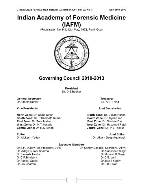
J <strong>Indian</strong> Acad <strong>Forensic</strong> Med. October- December 2011, Vol. 33, No. 4 ISSN 0971-0973<br />
<strong>Indian</strong> <strong>Academy</strong> <strong>of</strong> <strong>Forensic</strong> <strong>Medicine</strong><br />
(<strong>IAFM</strong>)<br />
(Registration No.349, 12th May, 1972, Panji, Goa)<br />
Governing Council 2010-2013<br />
President<br />
Dr. D.S.Badkur<br />
General Secretary Treasurer<br />
Dr.Adarsh Kumar Dr. A.S. Thind<br />
Vice Presidents<br />
North Zone: Dr. Dalbir Singh<br />
South Zone: Dr. P.Sampath Kumar<br />
East Zone: Dr. Tulsi Mahto<br />
West Zone: Dr. H.T. Katade<br />
Central Zone: Dr. R.K. Singh<br />
Editor<br />
Dr. Mukesh Yadav<br />
Dr.B.P. Dubey (Ex. President, <strong>IAFM</strong>)<br />
Dr. Aditya Kumar Sharma<br />
Dr.Sarvesh Tandon<br />
Dr.C.P.Bhaisora<br />
Dr.Pankaj Gupta<br />
Dr.Luv Sharma<br />
Executive Members<br />
i<br />
Joint Secretaries<br />
North Zone: Dr. Dasari Harish<br />
South Zone: Dr. Cyriac Job<br />
East Zone: Dr. Shoban Das<br />
West Zone: Dr. Hasumati Patel<br />
Central Zone: Dr. P.S.Thakur<br />
Joint Editor<br />
Dr. Akash Deep Aggarwal<br />
Dr. Sanjoy Das (Ex. Secretary, <strong>IAFM</strong>)<br />
Dr.Amandeep Singh<br />
Dr.Mukesh K.Goyal<br />
Dr.C.B. Jani<br />
Dr.Jaynti Yadav<br />
Dr.P.K.Tiwari
J <strong>Indian</strong> Acad <strong>Forensic</strong> Med. October- December 2011, Vol. 33, No. 4 ISSN 0971-0973<br />
Journal <strong>of</strong> <strong>Indian</strong> <strong>Academy</strong> <strong>of</strong> <strong>Forensic</strong> <strong>Medicine</strong><br />
(J<strong>IAFM</strong>)<br />
The <strong>of</strong>ficial publication <strong>of</strong> <strong>Indian</strong> <strong>Academy</strong> <strong>of</strong> <strong>Forensic</strong> <strong>Medicine</strong><br />
Editor,<br />
Dr. Mukesh Yadav<br />
Pr<strong>of</strong>essor & H.O.D.,<br />
<strong>Forensic</strong> <strong>Medicine</strong> & Toxicology,<br />
School <strong>of</strong> Medical Sciences and<br />
Research, Sharda University, Greater<br />
Noida-201306, Uttar Pradesh, INDIA<br />
Residence:<br />
G-216, Parsvanath Edens,<br />
Alfa-II, Greater Noida, G.B. Nagar, U.P.INDIA<br />
Ph. No. 0120-2326060, Cell: 09411480753<br />
Email: drmukesh65@yahoo.co.in<br />
Dr. A.K. Srivstava<br />
Pr<strong>of</strong>essor & H.O.D.,<br />
<strong>Forensic</strong> <strong>Medicine</strong><br />
&Toxicology<br />
Subharti Medical College,<br />
Meerut, U.P.<br />
Dr. V.V.Pillay<br />
Pr<strong>of</strong>essor & H.O.D.,<br />
Analytical Toxicology,<br />
Chief <strong>of</strong> Poison Control<br />
Centre<br />
AIMS & R, Cochin-Kerala<br />
Sharma G.K., (New Delhi)<br />
Verma S.K., (New Delhi)<br />
Kaur Balbir, (Srinagar)<br />
Bansal Y., (Chandigarh)<br />
Kumar Shantha B., (Tamilnadu)<br />
Gupta B.D., (Gujrat)<br />
Manju Nath K.H, (Karnatka)<br />
Das Sanjoy, (Uttarakhand)<br />
Bhaisora C.P, (Uttarakhand)<br />
Mahtoo Tulsi, (Jharkhand)<br />
Peer Review Group<br />
Dr. R.K. Gorea<br />
Pr<strong>of</strong>essor & H.O.D.,<br />
<strong>Forensic</strong> <strong>Medicine</strong><br />
&Toxicology<br />
Gian Sagar Medical College,<br />
Banur, Patiala, Punjab<br />
Dr. C.B.Jani<br />
Pr<strong>of</strong>essor & H.O.D.,<br />
<strong>Forensic</strong> <strong>Medicine</strong> and<br />
Toxicology P.S.Medical<br />
College, Karamsad, Distt.<br />
Anand, Gujarat<br />
Advisory Board<br />
Ravindran K, (Pondicherry)<br />
Sabri Imran, (U.P.)<br />
Rastogi Prateek (Karnatka)<br />
Potwary AJ (Assam)<br />
Singh R.K. (Chhatisgarh)<br />
Dongre A.P. (Nagpur)<br />
Rastogi Pooja (U.P.)<br />
Sharma Aditya (H.P.)<br />
Khanagwal V. (Haryana)<br />
Gupta Pankaj (Punjab)<br />
ii<br />
Joint Editor,<br />
Dr. Akash Deep Aggarwal<br />
Assistant Pr<strong>of</strong>essor,<br />
Department <strong>of</strong> <strong>Forensic</strong> <strong>Medicine</strong>,<br />
Government Medical College,<br />
Patiala-147001,<br />
Punjab, INDIA<br />
Residence:<br />
H.No. 14, Desi Mehmandari,<br />
Patiala-147001, Punjab, INDIA<br />
Cell: 9815652621<br />
Email:toakashdeep@yahoo.com<br />
Dr. T.K Bose<br />
Pr<strong>of</strong>essor & H.O.D.,<br />
<strong>Forensic</strong> and State <strong>Medicine</strong><br />
Govt. Medical College<br />
Kolkata, West Bengal<br />
Dr. G. Pradeep Kumar<br />
Pr<strong>of</strong>essor & H.O.D.,<br />
<strong>Forensic</strong> <strong>Medicine</strong><br />
&Toxicology<br />
Kasturba Medical College,<br />
Manipal, Karnatka<br />
Pounder Derrick, (England)<br />
Khaja Shaikh (A.P.)<br />
Basu R (W.B.)<br />
Naik R.S. (Maharastra)<br />
Godhikirakar Madhu (Goa)<br />
Job Cyriac (Kerala)<br />
Vinita K. (U.P.)<br />
Yadav B.N. (Nepal)<br />
Mohite Shailesh (Mumbai)<br />
Singh Abhas Kumar (U.P.)<br />
Printed and published by Dr. Mukesh Yadav, Editor, J<strong>IAFM</strong> and Dr. A. D. Aggarwal, Joint Editor, J<strong>IAFM</strong> on<br />
behalf <strong>of</strong> <strong>Indian</strong> <strong>Academy</strong> <strong>of</strong> <strong>Forensic</strong> <strong>Medicine</strong> at name <strong>of</strong> the press [SHIVANI PRINTERS, NOIDA, U.P.]
J <strong>Indian</strong> Acad <strong>Forensic</strong> Med. October- December 2011, Vol. 33, No. 4 ISSN 0971-0973<br />
Journal <strong>of</strong> <strong>Indian</strong> <strong>Academy</strong> <strong>of</strong> <strong>Forensic</strong> <strong>Medicine</strong><br />
Volume: 33 • Number: 4 • October- December 2011<br />
Contents<br />
Sr. Page<br />
I. From the Editor‘s Desk 285<br />
II. Editorial: Highest compensation for medical negligence A matter <strong>of</strong> great<br />
concern for medical fraternity<br />
Original Research Paper<br />
1. <strong>Forensic</strong> View on Aluminium Phosphide Poisoning.Vijayanath.V, Anitha M.R, Raju.<br />
G.M.,Vijayamahantesh S.N.<br />
2. Age determination from radiological study <strong>of</strong> epiphysial appearance and<br />
union around wrist joint and hand. S.S. Bhise, B. G. Chikhalkar, S. D. Nanandkar, G. S.<br />
Chavan<br />
283<br />
286-288<br />
289-291<br />
292-295<br />
3. The Pattern <strong>of</strong> Poisoning in Khammam. Bharath K. Guntheti, Uday Pal Singh 296-300<br />
4. Radiological study <strong>of</strong> fusion <strong>of</strong> iliac crest by digital method. Gaurang Patel,<br />
Dharmesh Shilajiya, Ganesh Govekar, Chandresh Tailor<br />
5. Effect <strong>of</strong> ageing & environmental condition for detection <strong>of</strong> blood group from<br />
blood stain. Prakash.M.Mohite, Atul Keche, Anil J.Anjankar, Sudhir Ninave.<br />
6. Pattern <strong>of</strong> Burns Cases Brought To Morgue, Sion Hospital, Mumbai: A Two<br />
Year Study. Dhiraj Buchade,Hemant Kukde, Rajesh Dere, Ramesh Savardekar<br />
7. Study <strong>of</strong> pr<strong>of</strong>ile <strong>of</strong> Deaths due to poisoning in Bhavnagar region. Navinkumar M.<br />
Varma, S.D.Kalele<br />
8. Incidence <strong>of</strong> post burn septicemia at a Tertiary Care Hospital. Prabhsharan Singh,<br />
Dasari Harish<br />
9. A Study <strong>of</strong> Rational use <strong>of</strong> Drugs among the Ophthalmic-in-Patients <strong>of</strong> a<br />
Government Teaching Hospital: In View <strong>of</strong> <strong>Forensic</strong> Pharmacology. Kanchan<br />
Kumar Mondal,Supreeti Biswas, Rajat Kanti Biswas, Anjan Adhikari,Biswajit Sukul, Saibendu Kumar<br />
Lahiri,Krishnangshu Ray.<br />
301-305<br />
306-310<br />
311-312<br />
313-318<br />
319-323<br />
324-327<br />
10. Natural deaths in custody: A 10 year mortality study. Rajesh Bardale, Pradeep Dixit 328-331<br />
11. The Study <strong>of</strong> Aluminium Phosphide Poisoning in a Tertiary Care Hospital -<br />
Amritsar. Puneet Khurana, J.S.Dalal, A. S. Multani, H.R. Tejpal<br />
12. Study <strong>of</strong> Medico-legal Case Management in tertiary Care Hospital. A.K. Singh,<br />
Kanchan Singh, Anoop Verma<br />
13 Finger print pattern in different blood groups. Muralidhar Reddy Sangam, A. Ramesh<br />
Babu, K. Krupadanam, K. Anasuya<br />
332-336<br />
337-342<br />
343-345<br />
14 Trends <strong>of</strong> Maxill<strong>of</strong>acial Trauma at Adesh Institute <strong>of</strong> Medical Sciences and 346-348
J <strong>Indian</strong> Acad <strong>Forensic</strong> Med. October- December 2011, Vol. 33, No. 4 ISSN 0971-0973<br />
Research, Bathinda, Punjab. Vishal Garg, Harinder Singh<br />
15 Digital Dermatoglyphis in ABO, Rh Blood Groups. Amit A. Mehta, Anjulika A. Mehta,<br />
Vaibhav Sonar<br />
16 Pattern <strong>of</strong> Neck Findings in Suicidal Hanging: A Study in Manipur. Th. Meera, M.<br />
Bapin Kumar Singh<br />
17 Suicidal Hanging- A Prospective Study. N. Vijayakumari<br />
Review Papers<br />
18 Medicolegal Importance <strong>of</strong> Inca Bone in <strong>Forensic</strong> Identification. D.S. Badkur,<br />
Vandana Sharma, Prabha Badkur<br />
19 Child abuse and its detection in the Dental Office. Rani Somani, Vinita Kushwah, Dilip<br />
Kumar, Jaskirat Khaira<br />
Case Reports<br />
284<br />
349-351<br />
351-354<br />
355-357<br />
358-360<br />
361-365<br />
20 Female Infanticide. S. Praveen 366-369<br />
21 Fatal Hemmorhage Following Extraction <strong>of</strong> First Molar. Dhritiman Nath, Manish<br />
Kumath, Sarvesh Tandon, Mamta Panwar<br />
370-371<br />
22 Importance <strong>of</strong> Crime Scene Visit: A case study. Z.Sasi Kanth, B.RajMohan Lal 372-374<br />
Supplements<br />
News and Views 375<br />
Guidelines to Authors / Contributors 376-377<br />
Membership Form <strong>of</strong> <strong>IAFM</strong> 378<br />
Copy Right © All rights reserved: No part <strong>of</strong> this publication may be reprinted or publish without the<br />
prior permission <strong>of</strong> the Editor, J<strong>IAFM</strong>. Submission <strong>of</strong> all manuscripts to the journal is understood to imply<br />
that it is not being considered for publication elsewhere. Submission <strong>of</strong> multi authored papers implies that<br />
the consent <strong>of</strong> each author has been obtained. In this journal, every effort has been made NOT to publish<br />
inaccurate or misleading information. However, the Editor, Joint Editor, Peer Review Group and Advisory<br />
Board accept NO liability in consequences <strong>of</strong> such statements. The Journal <strong>of</strong> <strong>Indian</strong> <strong>Academy</strong> <strong>of</strong><br />
<strong>Forensic</strong> <strong>Medicine</strong> is indexed in Scopus [Elsevier], Index Copernicus [Poland] & IndMED<br />
[India]. Print ISSN: 0971-0973. Electronic ISSN: 0974-0848. IndMED<br />
www.medind.nic.in/jal/jalm.shtml<br />
Address request for reprint or further information relating to any article may please be made with author and<br />
in case <strong>of</strong> multi authored article, please communicate to Corresponding Author or the First Author
J <strong>Indian</strong> Acad <strong>Forensic</strong> Med. October- December 2011, Vol. 33, No. 4 ISSN 0971-0973<br />
From the Editor’s Desk<br />
J<strong>IAFM</strong><br />
A Quarterly Publication<br />
Volume 33, Number 4, October-December 2011<br />
I feel immense pleasure to present before you the fourth and last issue <strong>of</strong> 2011. I assure you<br />
about the quality <strong>of</strong> research papers and quality <strong>of</strong> printing in future issues. The quality <strong>of</strong> our journal is<br />
increasing day-by-day as evidenced by the demand <strong>of</strong> few papers by US based research agencies,<br />
authors contribution is <strong>of</strong> immense value for me. Your valuable suggestions are always encouraging me<br />
and I heartily welcome for future suggestions. On behalf <strong>of</strong> Executive Committee <strong>of</strong> <strong>IAFM</strong> for the years<br />
2010-2013, I took resolution to further improve the quality and status <strong>of</strong> our Journal. We always learn from<br />
mistakes and try to improve upon these. I am happy to inform you that the journal has been included in<br />
Scopus. SciVerse Scopus is the world‘s largest abstract and citation database <strong>of</strong> peer-reviewed literature<br />
and quality web sources.<br />
Pr<strong>of</strong>essor [Dr.] Mukesh Yadav<br />
Editor, J<strong>IAFM</strong><br />
Subscription Information<br />
Members <strong>of</strong> <strong>IAFM</strong> will receive the free <strong>of</strong> cost.<br />
Non Members and Institutions (Annual Subscription rates)<br />
Personal: In India, Rs. 1000/ (Rest <strong>of</strong> the world: US$ 200/ or equivalent)<br />
Institutions: In India, Rs. 3000/ (Rest <strong>of</strong> the world: US$ 400/ or equivalent)<br />
We Accept: Bank Cheque / Demand Drafts (Add Rs. 50/- for outstation Cheques)<br />
The Scope <strong>of</strong> the Journal covers all aspects <strong>of</strong> <strong>Forensic</strong> <strong>Medicine</strong> and allied fields, research<br />
and applied.<br />
Subscription orders and payments should be made in favour <strong>of</strong><br />
“Editor, J<strong>IAFM</strong>, payable at Greater Noida”<br />
Claims for missing issue:<br />
A copy will be sent free to the member / subscriber provided the claim is made within 2 months <strong>of</strong><br />
publication <strong>of</strong> the issue & self addressed envelop <strong>of</strong> the size 9‖ x 12‖ is sent to the Editor. (Those<br />
who want the journals to be dispatched by Registered Post must affix Rs. 50/ worth postage<br />
stamps).<br />
The journal is indexed with IndMed and made available online by following <strong>website</strong>:<br />
www.medind.nic.in http://indmed.nic.in<br />
www.jiafm.com www.forensicindia.com<br />
www.indianjournals.com<br />
285
J <strong>Indian</strong> Acad <strong>Forensic</strong> Med. October- December 2011, Vol. 33, No. 4 ISSN 0971-0973<br />
Editorial<br />
Highest Compensation for Medical Negligence<br />
A Matter <strong>of</strong> Great Concern for Medical Fraternity<br />
Introduction:<br />
A petitioner claimed a total compensation <strong>of</strong> Rs.770745000/- (Seventy Seven Crores Seven<br />
Lakhs Fourty Five Thousand only). Later he also filed another complaint no. 179 <strong>of</strong> 2000 before National<br />
Consumer Dispute Redressal Commission (NCDRC) against Breach Candy Hospital, Mumbai, its doctors<br />
and functionaries claiming a further compensation <strong>of</strong> Rs.25.30 crore (though the said complaint was later<br />
on withdrawn), thereby made claim <strong>of</strong> compensation exceeding Rs.102 crores, perhaps the highest ever<br />
claimed by any complainant for medical negligence before any consumer fora in India established under<br />
the provisions <strong>of</strong> Consumer Protection Act, 1986. These facts made Dr.Kunal Saha vs.<br />
Dr.Sukumar Mukherjee & Others, O. P. No. 240 <strong>of</strong> 1999, NCDRC, New Delhi, date <strong>of</strong> judgment: 21 st<br />
October 2011, case an extra ordinary in the field <strong>of</strong> medical negligence in <strong>Indian</strong> Context.<br />
Brief facts:<br />
Complainant a doctor by pr<strong>of</strong>ession (husband <strong>of</strong> deceased victim <strong>of</strong> medical negligence<br />
Anuradha) felt that the doctors who treated Anuradha and the hospitals where she was treated were<br />
grossly negligent in her treatment and her death was occasioned due to gross negligence <strong>of</strong> the treating<br />
doctors and hospitals. Complainant, accordingly, got issued a legal notice to as many as 26 persons i.e.<br />
various doctors who treated Anuradha between end <strong>of</strong> April, 1998 to the date <strong>of</strong> her death (May 1998)<br />
alleging negligence and deficiency in service on their part. He filed the complaint on 09.03.1999 before<br />
NCDRC, New Delhi.<br />
This case was filed against few doctors and one hospital at Culcutta, namely, Dr.Sukumar<br />
Mukherjee, Dr.B.Haldar (Baidyanath Halder), Advanced Medicare and Research Institute Limited (AMRI<br />
Hospital) and Dr.Balram Prasad and Dr.Abani Roy Chowdhury (physician) and Dr.Kaushik Nandy (plastic<br />
surgeon), the Directors <strong>of</strong> the AMRI Hospital and others. The complaint was resisted by the doctors and<br />
the hospital on a variety <strong>of</strong> grounds thereby denying any medical negligence or deficiency in service on<br />
their part.<br />
Role <strong>of</strong> Supreme Court:<br />
The Apex Court by order dated 07.08.2009 dismissed the Criminal Appeals filed by Shri Malay<br />
Kumar Ganguly but allowed the Civil Appeal No. 1727 <strong>of</strong> 2007 filed by the complainant and set aside the<br />
order <strong>of</strong> NCDRC dated 01.6.2006 dismissing the complaint and remanded the matter to this Commission<br />
for the limited purpose <strong>of</strong> determining the adequate compensation, which the complainant is entitled to<br />
receive from the subsisting opposite parties by observing as under:<br />
“So far as the judgment <strong>of</strong> the Commission is concerned, it was clearly wrong in opining that<br />
there was no negligence on the part <strong>of</strong> the Hospital or the doctors. We, are, however, <strong>of</strong> the opinion,<br />
keeping in view the fact that Dr.Kaushik Nandy has done whatever was possible to be done and his line<br />
<strong>of</strong> treatment meets with the treatment protocol <strong>of</strong> one <strong>of</strong> the experts viz.. Pr<strong>of</strong>. Jean<br />
Claude Roujeau although there may be otherwise difference <strong>of</strong> opinion, that he cannot be held to be guilty<br />
<strong>of</strong> negligence.<br />
We remit the case back to the Commission only for the purpose <strong>of</strong> determination <strong>of</strong> the quantum<br />
<strong>of</strong> compensation.”<br />
Principle <strong>of</strong> compensation:<br />
“170. Indisputably, grant <strong>of</strong> compensation involving an accident is within the realm <strong>of</strong> law <strong>of</strong> torts. It<br />
is based on the principle <strong>of</strong> restitutio in integrum. The said principle provides that a person entitled to<br />
damages should, as nearly as possible, get that sum <strong>of</strong> money which would put him in the same position<br />
as he would have been if he had not sustained the wrong.” [Livingstone v. Rawyards Coal Co]<br />
Effect <strong>of</strong> contributory negligence:<br />
In this connection, the observations made by the Supreme Court in Para 123, which read as<br />
under:<br />
“To conclude, it will be pertinent to note that even if we agree that there was interference<br />
by Kunal Saha during the treatment, it in no way diminishes the primary responsibility and default in<br />
duty on part <strong>of</strong> the defendants. Inspite <strong>of</strong> a possibility <strong>of</strong> him playing an overanxious role during the<br />
medical proceedings, the breach <strong>of</strong> duty to take basic standard <strong>of</strong> medical care on the part <strong>of</strong><br />
286
J <strong>Indian</strong> Acad <strong>Forensic</strong> Med. October- December 2011, Vol. 33, No. 4 ISSN 0971-0973<br />
defendants is not diluted. To that extent, contributory negligence is not pertinent. It may,<br />
however, have some role to play for the purpose <strong>of</strong> damages.”<br />
On the other hand it was submitted in defense that the Supreme Court though holding that there<br />
was possibility <strong>of</strong> the complainant playing an overanxious role during the medical treatment, the<br />
question <strong>of</strong> contributory negligence was not pertinent but at the same time, it was observed by the<br />
Supreme Court that this contributory negligence on the part <strong>of</strong> the complainant have some role to play for<br />
the purpose. We have therefore no manner <strong>of</strong> doubt that the above noted circumstances <strong>of</strong> interference<br />
which can be said to have been established on record are relevant for the purpose <strong>of</strong> determination <strong>of</strong> the<br />
extent <strong>of</strong> compensation. [Para 8.7]<br />
The next submission put forth on behalf <strong>of</strong> the AMRI Hospital is in regard to the liability <strong>of</strong> the<br />
hospital to pay any compensation much less the compensation claimed by the complainant from it<br />
either on account <strong>of</strong> pecuniary or non-pecuniary damages or special damages. [Para 8.8]<br />
Doctrine <strong>of</strong> Apportionment <strong>of</strong> Liability:<br />
The question before NCDRC was ―whether any amount should be deducted from this amount on<br />
account <strong>of</strong> the contributory negligence <strong>of</strong> the complainant arising out <strong>of</strong> his conduct i.e. interference in the<br />
treatment <strong>of</strong> Anuradha during her hospitalization in AMRI hospital.‖<br />
In Para no. 123, the Supreme Court has held that said interference <strong>of</strong> the complainant may be a relevant<br />
and may have some role to play for determining the amount <strong>of</strong> compensation.<br />
Therefore, having held that complainant has interfered in treatment <strong>of</strong> Anuradha, NCDRC was<br />
inclined to deduct a sum equivalent to 10% from the above payable amount. [Para 13.1]<br />
The most intriguing task before NCDRC was, to apportion the liability to pay the awarded<br />
amount <strong>of</strong> compensation amongst the opposite parties. No straight jacket formula exists or perhaps<br />
can be laid down in a case where so many doctors and hospitals are found negligent in the<br />
treatment <strong>of</strong> patient. The Supreme Court has not indicated the criteria for apportionment <strong>of</strong> the<br />
compensation amongst the opposite parties but going by the findings <strong>of</strong> the Supreme Court in regard to<br />
the nature and extent <strong>of</strong> negligence / deficiency in treatment on the part <strong>of</strong> the opposite parties<br />
and Dr. Abani Roy Chowdhury as enumerated in para 158 to 166 <strong>of</strong> the judgment, we must apply a<br />
formula which appears just to us. Supreme Court has primarily found Dr. Sukumar Mukherjee and AMRI<br />
hospital guilty <strong>of</strong> negligence and deficient in service on several counts. Therefore, going by the said<br />
findings and observations <strong>of</strong> the Supreme Court, ncdrc considered it appropriate to apportion the liability<br />
<strong>of</strong> Dr.Sukumar Mukherjee and AMRI hospital in equal portion i.e., each should pay 25% <strong>of</strong> the awarded<br />
amount. Remaining half <strong>of</strong> the awarded compensation should be divided amongst Dr.B.Haldar and<br />
Dr.Balram Prasad and Dr. Abani Roy Chowdhury (heirs <strong>of</strong> whom have been given up by the complainant<br />
by forgoing his right to claim compensation from them). [50% divided among three parties] [Para 14.1]<br />
Cost <strong>of</strong> Litigation:<br />
In the Annexure submitted along with the synopsis <strong>of</strong> submissions, the complainant has claimed<br />
a sum <strong>of</strong> Rs.11250000/- towards the loss <strong>of</strong> income for missed works, Rs. 7000000/- towards traveling<br />
expenses, a sum <strong>of</strong> Rs. 15000000/- towards advocate fees paid to senior advocates for over 12 years<br />
and a sum <strong>of</strong> Rs. 1500000/- towards other legal expenses. These claims appear to be highly<br />
exaggerated. In any case, the complainant has not furnished any cogent pro<strong>of</strong> <strong>of</strong> having actually<br />
incurred the above expenditure. The Supreme Court had already awarded a cost <strong>of</strong> Rs. 600000/-<br />
while deciding the criminal and civil appeals.<br />
In NCDRC opinion having regard to peculiar facts and circumstances <strong>of</strong> the present case and<br />
as a special case, the complainant is at best entitled to cost <strong>of</strong> Rs. 500000/- (Rupees five lakh only), lest it<br />
becomes too onerous for the opposite parties to pay the same. [Para 15.1]<br />
In view <strong>of</strong> the foregoing discussion, NCDRC concluded as:<br />
The facts <strong>of</strong> this case viz., residence <strong>of</strong> the complainant and Anuradha (deceased) in USA and<br />
they working for gain in that country; Anuradha having been a victim <strong>of</strong> a rare and deadly disease<br />
Toxic Epidermal Necrolysis (TEN) when she was in India during April-May 1998 and could not be<br />
cured <strong>of</strong> the said disease despite her treatment at two superspeciality medical centres <strong>of</strong> Kolkata<br />
and Mumbai and the huge claim <strong>of</strong> compensation exceeding Rs.77 crores made by the<br />
complainant for the medical negligence in the treatment <strong>of</strong> Anuradha makes the present case<br />
somewhat extraordinary. [Para 16.1]<br />
The findings given and observations made by the Supreme Court in its judgment dated<br />
07.08.2009 are absolutely binding on this Commission not only as ratio decidendi but also<br />
as as obiter dicta also. [Para 16.2]<br />
The task entrusted to the Commission may appear to be simple but the facts <strong>of</strong> the present case<br />
and the voluminous evidence led on behalf <strong>of</strong> the complainant has made it somewhat arduous.<br />
287
J <strong>Indian</strong> Acad <strong>Forensic</strong> Med. October- December 2011, Vol. 33, No. 4 ISSN 0971-0973<br />
Still difficult was the task <strong>of</strong> apportionment <strong>of</strong> the liability to pay the awarded amount by the<br />
different opposite parties. [16.3]<br />
Multiplier method provided under the Motor Vehicles Act for calculating the compensation is the<br />
only proper and scientific method for determination <strong>of</strong> compensation even in the cases where<br />
death <strong>of</strong> the patient has been occasioned due to medical negligence / deficiency in service in the<br />
treatment <strong>of</strong> the patient, as there is no difference in legal theory between a patient dying<br />
through medical negligence and the victim dying in industrial or motor accident. The award<br />
<strong>of</strong> lump sum compensation in cases <strong>of</strong> medical negligence has a great element <strong>of</strong> arbitrariness<br />
and subjectivity. [Para 16.4]<br />
The foreign residence <strong>of</strong> the complainant or the patient and the income <strong>of</strong> the deceased patient in<br />
a foreign country are relevant factors but the compensation awarded by <strong>Indian</strong> Fora cannot be<br />
at pars which are ordinarily granted by foreign courts in such cases. Socio economic conditions<br />
prevalent in this country and that <strong>of</strong> the opposite parties / defendants are relevant and must be<br />
taken into consideration so as to modulate the relief. A complainant cannot be allowed to get<br />
undue enrichment by making a fortune out <strong>of</strong> a misfortune. The theoretical opinion /<br />
assessment made by a Foreign Expert as to the future income <strong>of</strong> a person and situation prevalent<br />
in that country cannot form a sound basis for determination <strong>of</strong> future income <strong>of</strong> such person and<br />
the Commission has to work out the income <strong>of</strong> the deceased having regard to her last income<br />
and future prospects in terms <strong>of</strong> the criteria laid down by the Supreme Court. [Para 16.5]<br />
There exists no straight jacket formula for apportionment <strong>of</strong> the awarded compensation amongst<br />
various doctors and hospitals when there are so many actors who are responsible for negligence<br />
and the apportionment has to be made by evolving a criteria / formula which is just going by the<br />
nature and extent <strong>of</strong> medical negligence and deficiency in service established on the part <strong>of</strong><br />
different doctors and hospitals. [Para 16.6]<br />
On a consideration <strong>of</strong> the entirety <strong>of</strong> the facts and circumstances, evidence and material brought<br />
on record, NCDRC held that overall compensation on account <strong>of</strong> pecuniary and non<br />
pecuniary damages works out to Rs.17287500/- in the present case, out <strong>of</strong> which we must deduct<br />
10% amount on account <strong>of</strong> the contributory negligence / interference <strong>of</strong> the complainant in the<br />
treatment <strong>of</strong> Anuradha. That will make the net payable amount <strong>of</strong> compensation to<br />
Rs.15558750/-(rounded <strong>of</strong> toRs.15560000/). From this amount, we must further deduct a sum <strong>of</strong><br />
Rs.2593000/- which was payable by Dr.Abani Roy Chowdhury (deceased) or his Legal<br />
Representative as the complainant has forgone the claim against them. [Para 16.7]<br />
In view <strong>of</strong> the peculiar facts and circumstances <strong>of</strong> the case and as a special case, NCDRC have<br />
awarded a sum <strong>of</strong> Rs. 500000/- as cost <strong>of</strong> litigation in the present proceedings. [Para 16.8]<br />
The above amount shall be paid by opposite parties no. 1 to 4 to the complainant in the following<br />
manner:<br />
i. Dr.Sukumar Mukherjee shall pay a sum <strong>of</strong> Rs.4040000/- (Rupees Forty Lakh Forty Thousand<br />
only) i.e. [Rs.3890000/- towards compensation and Rs.150000/- as cost <strong>of</strong> litigation]<br />
ii. Dr. B.Haldar (Baidyanth Halder) shall pay a sum <strong>of</strong> Rs.2693000/- (Rupees Twenty Six Lakh<br />
Ninety Three Thousand only) i.e. [Rs.2593000/- towards compensation and Rs.100000/-<br />
as cost <strong>of</strong> litigation]<br />
iii. AMRI hospital shall pay a sum <strong>of</strong> Rs.4040000/- (Rupees Forty Lakh Forty Thousand only )<br />
i.e. [Rs.3890000/- towards compensation and Rs.150000/- as cost <strong>of</strong> litigation]<br />
iv. Dr. Balram Prasad shall pay a sum <strong>of</strong> Rs.2693000/- (Rupees Twenty Six Lakh Ninety Three<br />
Thousand only) i.e. [Rs.2593000/- towards compensation and Rs.100000/- as cost <strong>of</strong><br />
litigation]<br />
The opposite parties were directed to pay the aforesaid amounts to the complainant within a<br />
period <strong>of</strong> eight weeks from the date <strong>of</strong> this order (21.10.2011), failing which the amount shall<br />
carry interest @ 12% p.a. w.e.f. the date <strong>of</strong> default. [Para 16.9]<br />
There is an urgent need for medical fraternity to introspect on the issue <strong>of</strong> medical negligence with great<br />
concern in changed global socio-economic scenario and rising <strong>of</strong> medical tourism as potential country for<br />
providing medical care to the world community.<br />
288<br />
Editor<br />
Dr.Mukesh Yadav
J <strong>Indian</strong> Acad <strong>Forensic</strong> Med. October- December 2011, Vol. 33, No. 4 ISSN 0971-0973<br />
Original Research Paper<br />
<strong>Forensic</strong> View on Aluminium Phosphide Poisoning<br />
*Vijayanath, V., **Anitha M.R., ***Raju, G.M., ****Vijayamahantesh, S.N.<br />
Abstract<br />
In India, acute aluminium phosphide poisoning (AAlPP) is a serious health care problem. This<br />
study aimed to determine the characteristics <strong>of</strong> AAlPP and the predictors <strong>of</strong> mortality at the time <strong>of</strong><br />
patients' admission. We studied consecutive admissions <strong>of</strong> patients with AAlPP admitted to the intensive<br />
care unit (ICU). We noted 38 parameters at admission to the hospital and the ICU and compared survivor<br />
and non-survivor groups. A total <strong>of</strong> 54 patients were enrolled comprising 10 females and 44 males and<br />
the mean ingested dose <strong>of</strong> poison was 0.75 ± 0.745 grams. The mortality from AAlPP was 59.3%. We<br />
found the following factors to be associated with an increased risk <strong>of</strong> mortality: a serum creatinine<br />
concentration <strong>of</strong> more than 1.0 mg % (P = 0.01), pH value less than 7.2 (P = 0.014), serum bicarbonate<br />
value less than 15 mmol/L (P = 0.048), need for mechanical ventilation (P = 0.045), need for vasoactive<br />
drugs like dobutamine (P = 0.027) and nor adrenaline (P = 0.048) AAlPP causes high mortality primarily<br />
due to early haemodynamic failure and multi-organ dysfunction.<br />
Key Words: Aluminium Phosphide, Mortality, Poisoning<br />
Introduction:<br />
Acute aluminium phosphide poisoning<br />
(AAlPP) is a large, though under-reported,<br />
problem in the <strong>Indian</strong> subcontinent. Death is<br />
reported to result from pr<strong>of</strong>ound shock,<br />
myocarditis and multiorgan failure. The mortality<br />
rates from AAlPP published in literature vary<br />
from 40-80%. [1]<br />
However, the actual numbers <strong>of</strong> cases<br />
affected are much larger, as less than 5% <strong>of</strong><br />
those with AAlPP eventually reach a tertiary care<br />
centre. [2] Since 1992, when aluminium<br />
phosphide became freely available in the<br />
market, it has reportedly, overtaken all other<br />
forms <strong>of</strong> deliberate poisoning like<br />
organophosphorus and barbiturate poisoning in<br />
India. [3] In one study AAlPP was the major<br />
cause <strong>of</strong> death among all poisonings. [4]<br />
Despite these large numbers, there has<br />
been little progress in our understanding <strong>of</strong> the<br />
characteristics <strong>of</strong> the poison and limited <strong>Indian</strong><br />
data is available on the predictors <strong>of</strong> mortality in<br />
these patients. The purpose <strong>of</strong> this study was to<br />
retrospectively study the pr<strong>of</strong>ile <strong>of</strong> patients<br />
presenting with AAlPP and to identify the factors<br />
at admission that might be useful in predicting<br />
mortality.<br />
Corrosponding Author:<br />
* Asso. Pr<strong>of</strong>. <strong>Forensic</strong> <strong>Medicine</strong> & Toxicology,<br />
VMKV Medical College, Salem, Tamil Nadu<br />
Email; drvijayanath@rediffmail.com<br />
**Asst. Pr<strong>of</strong>. Deptt. <strong>of</strong> Anatomy,<br />
***Asst.Pr<strong>of</strong>. J.J.M.Medical College, Davangere-577004,<br />
****Asso. Pr<strong>of</strong>. SNMC, Navangar, Karnataka<br />
289<br />
Material and Methods:<br />
Study design: All consecutive cases <strong>of</strong> AAlPP<br />
presenting to a tertiary care hospital in northern<br />
karnataka from January 2001 to December 2006<br />
were retrospectively reviewed. The diagnosis <strong>of</strong><br />
AAlPP was based on history <strong>of</strong> consumption <strong>of</strong><br />
the poison (obtained from the patient or the<br />
closest relative) and symptomatology at<br />
admission. Instances where the patients had<br />
presented with an unclear diagnosis <strong>of</strong> poisoning<br />
or where there was consumption <strong>of</strong> more than<br />
one substance were excluded from the study.<br />
All patients were admitted into our ICU<br />
after initial resuscitation and gastric lavage. A<br />
baseline electrocardiogram was recorded and<br />
blood samples for biochemical and<br />
haematological investigations were sent.<br />
Infusion <strong>of</strong> vasoactive agents and mechanical<br />
ventilatory support were instituted where<br />
indicated. We collected 38 variables, like age,<br />
gender, nature <strong>of</strong> poisoning (suicidal/accidental),<br />
the dose poison consumed and the delay in<br />
presentation to hospital. The nature <strong>of</strong> any first<br />
aid instituted before reaching this hospital as<br />
well as laboratory parameters on admission to<br />
the hospital and ICU were also noted.<br />
The severity <strong>of</strong> the poisoning was<br />
assessed from the extent <strong>of</strong> organ dysfunction<br />
(renal, hepatic, neurological, gastrointestinal,<br />
cardiovascular, etc.), the need for mechanical<br />
ventilation and the requirement <strong>of</strong> drugs for<br />
vasoactive support.<br />
Statistical analysis: The SPSS computer<br />
program was used to process the data and<br />
generate the statistics. Univariate analysis was<br />
performed to compare survivors with non
J <strong>Indian</strong> Acad <strong>Forensic</strong> Med. October- December 2011, Vol. 33, No. 4 ISSN 0971-0973<br />
survivors groups (P < 0.05 was considered<br />
significant).<br />
Results:<br />
A total <strong>of</strong> 54 patients with AAlPP were<br />
admitted into our ICU during the study period.<br />
The majority <strong>of</strong> patients were young and in the<br />
age group from 21 to 40 years with males<br />
outnumbering females by more than 4: 1. Most<br />
<strong>of</strong> the cases involved suicidal consumption <strong>of</strong><br />
the poison (92%) and 60% <strong>of</strong> the poison was<br />
consumed in the unexposed form <strong>of</strong> the tablet<br />
and an average <strong>of</strong> 1.53 grams <strong>of</strong> drug was<br />
consumed. There was a mean delay <strong>of</strong> 2.1 ±<br />
1.55 hours before presenting to this hospital.<br />
There was no significant association between<br />
the dose <strong>of</strong> poison consumed or the time delay<br />
in presentation to the hospital with mortality.<br />
At presentation to the hospital, the most<br />
predominant feature experienced by patients<br />
was vomiting and nausea (92.6%). A few<br />
patients had respiratory distress (7%). The mean<br />
Glasgow coma scale at admission was 13.29 ±<br />
2.825 and the mean partial pressure <strong>of</strong> oxygen<br />
in arterial blood (paO2) and partial pressure <strong>of</strong><br />
carbon dioxide in arterial blood (paCO2) in<br />
patients were 72.63 ± 4.06 mm Hg and 26.37 ±<br />
7.46 mm Hg, respectively. A total <strong>of</strong> 14 patients<br />
(25%) had high serum creatinine values at<br />
admission, all <strong>of</strong> whom eventually died. Serum<br />
creatinine levels were found to correlate well<br />
with mortality. Survivors had significantly lower<br />
serum creatinine levels at admission as<br />
compared to non- survivors (0.82 ± 0.1418<br />
milligram per deciliter versus 1.375 ± 0.642 mg<br />
per deciliter respectively, P = 0.011).<br />
The mean pH <strong>of</strong> patients at admission<br />
too, was a good indicator <strong>of</strong> prognosis. Survivors<br />
had a much higher average value (7.284 ±<br />
0.151) than non-survivors (7.148 ± 0.120) and<br />
this difference was statistically significant (P =<br />
0.015). Similarly, serum bicarbonate levels at<br />
admission also correlated well with the eventual<br />
outcome in these patients (P = 0.048).<br />
All patients had normal levels <strong>of</strong> sodium<br />
at admission to the hospital while 48% <strong>of</strong><br />
patients had hypokalemia. These variables were<br />
statistically insignificant. Increased serum levels<br />
<strong>of</strong> bilirubin, aspartate aminotranferase, alanine<br />
aminotransferase and random blood sugar at<br />
admission did not show any association with<br />
mortality. We found that 81% <strong>of</strong> patients had<br />
cardiac symptoms, mainly in the form <strong>of</strong><br />
hypotension and/or arrhythmias on admission to<br />
the hospital. A total <strong>of</strong> 26 patients had<br />
dysrhythmias at admission, <strong>of</strong> which, the<br />
majority (69%) were <strong>of</strong> supraventricular origin.<br />
Though the presence <strong>of</strong> electrocardiographic<br />
abnormalities did not predict mortality, there was<br />
290<br />
a trend towards increasing mortality in patients<br />
with dysrhythmias (P = 0.07).<br />
Immediately on admission into the<br />
intensive care unit, 50% <strong>of</strong> the patients required<br />
mechanical ventilatory support while non<br />
invasive ventilation was used only in one patient.<br />
Eighty nine percent <strong>of</strong> the patients were in shock<br />
at admission despite adequate fluid resuscitation<br />
and needed vasoactive support, predominantly<br />
dobutamine and nor adrenaline. In all patients,<br />
magnesium sulphate was used for treatment<br />
while systemic steroid therapy (injectable<br />
hydrocortisone) was initiated in 70% <strong>of</strong> patients<br />
(mainly depending on the treating physician).<br />
The overall mortality from AAlPP<br />
poisoning in our unit during the study period was<br />
59.3%. The statistically significant factors useful<br />
in predicting mortality in our study were, an<br />
elevated serum creatinine conc. (P = 0.01) at<br />
admission, need for mechanical ventilation (P =<br />
0.045), need for vasoactive drugs like<br />
dobutamine (P = 0.027) and nor adrenaline (P =<br />
0.048), pH value less than 7.2 (P = 0.014), a low<br />
serum bicarbonate value (P = 0.048)<br />
Discussion:<br />
Aluminium Phosphide is an extremely<br />
toxic compound and resulted in a high mortality<br />
rate <strong>of</strong> 59.3% in our study. The toxicity <strong>of</strong><br />
Aluminium Phosphide is attributed to the<br />
liberation <strong>of</strong> phosphine gas which is cytotoxic<br />
and causes free radical mediated injury.<br />
Phosphine a nucleophile, acts as a<br />
strong reducing agent capable <strong>of</strong> inhibiting<br />
cellular enzymes involved in several metabolic<br />
processes. Early studies on phosphine<br />
demonstrated specific inhibitory effects on<br />
mitiochondrial cytochrome c oxidase. [5]<br />
Experimental and observational studies<br />
have subsequently demonstrated that the<br />
inhibition <strong>of</strong> cytochrome c oxidase and other<br />
enzymes leads to the generation superoxide<br />
radicals and cellular peroxides. Cellular injury<br />
subsequently occurs through lipid peroxidation<br />
and other oxidant mechanisms. [6, 7] One study<br />
reported that, serum phosphine levels correlate<br />
positively with the severity <strong>of</strong> poisoning and<br />
levels equal to or less than 1.067 ± 0.16 mg %<br />
appear to be the limit <strong>of</strong> phosphine toxicity. [8]<br />
The major lethal consequence <strong>of</strong><br />
aluminium phosphide ingestion i.e., pr<strong>of</strong>ound<br />
circulatory collapse, is reportedly secondary to<br />
these toxins generated, which lead to direct<br />
effects on cardiac myocytes, fluid loss, and<br />
adrenal gland damage. [9]<br />
In addition, phosphine also has<br />
corrosive effects on tissues. [9] As less than 5%<br />
<strong>of</strong> patients with AAlPP eventually reach a tertiary<br />
care hospital, it is extremely difficult to know the
J <strong>Indian</strong> Acad <strong>Forensic</strong> Med. October- December 2011, Vol. 33, No. 4 ISSN 0971-0973<br />
actual incidence <strong>of</strong> these poisonings in our<br />
country and it is postulated that the spectrum <strong>of</strong><br />
actual cases may be much larger than that<br />
reported. In one study, aluminium phosphide<br />
was found to be the commonest poison<br />
consumed (79.8%) in the Haryana- Rohtak belt<br />
with a mortality <strong>of</strong> 67.6%. [10]<br />
In our study we found that most <strong>of</strong> the<br />
victims were young with males outnumbering<br />
females. As with other poisonings, aluminium<br />
phosphide is a common method <strong>of</strong> attempting<br />
suicide among the younger, productive age<br />
group <strong>of</strong> society. The patients in our study<br />
typically developed gastrointestinal symptoms<br />
early in their presentation and the predominant<br />
early toxic manifestation was cardiac<br />
(hypotension and arrhythmias).<br />
Most patients needed some form <strong>of</strong><br />
vasopressor/ionotrope support and the need to<br />
use these early after admission was clearly<br />
associated with a poorer outcome. One study<br />
reported gastrointestinal symptoms as the<br />
commonest while conduction disturbances and<br />
arrhythmias occurred in 38% <strong>of</strong> patients and an<br />
overall mortality rate <strong>of</strong> 77%. [1]<br />
The management <strong>of</strong> AAlPP remains<br />
purely supportive. Though magnesium sulphate<br />
was used in the treatment <strong>of</strong> all our patients,<br />
there is conflicting data in literature on the role <strong>of</strong><br />
magnesium sulphate in the treatment <strong>of</strong> acute<br />
aluminium phosphide poisoning. [11, 12, 13, 14]<br />
We found five factors that can be<br />
assessed at admission to the hospital to predict<br />
mortality from aluminium phosphide ingestion.<br />
These include an elevated serum creatinine<br />
concentration, a low pH value (less than 7.2), a<br />
low serum bicarbonate value (less than 15), an<br />
early need for mechanical ventilation and for<br />
vasoactive drugs like dobutamine and nor<br />
adrenaline for haemodynamic support. These<br />
findings are similar to that obtained by Louriz et<br />
al., who concluded that the prognostic factors<br />
associated with mortality from AAlPP, included<br />
low Glasgow coma scale score, shock,<br />
electrocardiogram abnormalities, the presence<br />
<strong>of</strong> acute renal failure, low prothrombin rate,<br />
hyperleukocytosis, use <strong>of</strong> vasoactive drugs and<br />
use <strong>of</strong> mechanical ventilation. [15] In a recent<br />
update on AAlPP, the development <strong>of</strong> refractory<br />
shock, ARDS, aspiration pneumonitis, anaemia,<br />
metabolic acidosis, electrolyte imbalance, coma,<br />
severe hypoxia, gastrointestinal bleeding, and<br />
pericarditis were the factors reportedly<br />
associated with poor prognosis.<br />
They also noted that the outcome from<br />
AAlPP correlates best with the number <strong>of</strong><br />
vomiting the patient gets after ingestion and the<br />
severity <strong>of</strong> hypotension that the patient<br />
291<br />
develops. [16] The limitation <strong>of</strong> our study was<br />
that it was retrospectively designed. Hence,<br />
larger prospective studies need to be done in the<br />
future to conclusively support our results.<br />
Conclusion:<br />
We conclude that aluminium phosphide<br />
is an extremely toxic compound with mortality<br />
close to 60%. The variables at admission which<br />
could be used to detect patients at greater risk <strong>of</strong><br />
mortality from AAlPP include, need for<br />
mechanical ventilation, hypotension at<br />
admission requiring vasoactive drugs, low pH<br />
values, low serum bicarbonate levels, low serum<br />
creatinine levels.<br />
References:<br />
1. Chugh SN, Dushayant K, Ram S, Arora B. Incidence and outcome <strong>of</strong><br />
aluminium phosphide poisoning in a hospital study. <strong>Indian</strong> J Med<br />
Res 1991; 94:232-35, 5.<br />
2. Chugh SN. Aluminium phosphide poisoning. J <strong>Indian</strong> Acad Med 1999;<br />
4:83-9.<br />
3. Singh D, Jit I, Tyagi S. Changing Trends in Acute Poisoning in<br />
Chandigarh Zone: A 25- Year Autopsy Experience from a Tertiary<br />
Care Hospital in Northern India. Am J <strong>Forensic</strong> Med Pathol 1999;<br />
20:203-10.<br />
4. Singh D, Dewan I, Pandey AN, Tyagi S. Spectrum <strong>of</strong> unnatural<br />
fatalities in the Chandigarh zone <strong>of</strong> north-west India- a 25-year<br />
autopsy study from a tertiary care hospital. J Clin <strong>Forensic</strong> Med<br />
2003; 10:145-52.<br />
5. Chefurka W, Kashi KP, Bond EJ. The effect <strong>of</strong> phosphine (gas toxic to<br />
insects) on electron transport in mitochondria. Pestic Biochem<br />
Physiol 1976; 6:65-84.<br />
6. Bolter CJ, Chefurka W. Extramitochondrial release <strong>of</strong> hydrogen<br />
peroxide from insect and mouse liver mitochondria using the<br />
respiratory inhibitors phosphine myxothiazol, and antimycin and<br />
spectral analysis <strong>of</strong> inhibited cytochromes. Arch Biochem Biophys<br />
1990; 278:65-72.<br />
7. Chugh SN, Arora V, Sharma A, Chugh K. Free radical scavengers and<br />
lipid peroxidation in acute aluminium phosphide poisoning. <strong>Indian</strong> J<br />
Med Res 1996; 104:190-3.<br />
8. Chugh SN, Pal R, Singh V, Seth S. Serial blood phosphine levels in<br />
acute aluminium phosphide poisoning. J Assoc Physicians India<br />
1996; 44:184-5.<br />
9. Alex T. Proudfoot. Aluminium and zinc phosphide poisoning. Clin<br />
Toxicol 2009; 47:89-100.<br />
10. Siwach SB, Gupta A. The pr<strong>of</strong>ile <strong>of</strong> acute poisonings in Harayana-<br />
Rohtak study. J Assoc Physicians India 1995; 43:756-9.<br />
11. Chugh SN, Kolley T, Kakkar R, Chugh K, Sharma A. A critical<br />
evaluation <strong>of</strong> anti-peroxidant effect <strong>of</strong> intravenous magnesium in<br />
acute aluminium phosphide poisoning. Magnes Res 1997; 10:225-<br />
30.<br />
12. Chugh SN, Jaggal KL, Sharma A, Arora B, Malhotra KC.<br />
Magnesium levels in acute cardiotoxicity due to aluminium<br />
phosphide poisoning. <strong>Indian</strong> J Med Res 1991; 94:437-9.<br />
13. Singh RB, Singh RG, Singh U. Hypermagnesemia following<br />
aluminium phosphide poisoning. Int J Clin Pharmacol Ther Toxicol<br />
1991; 29:82-5.<br />
14. Siwach SB, Singh P, Ahlawat S, Dua A, Sharma D. Serum and<br />
tissue magnesium content in patients <strong>of</strong> aluminium phosphide<br />
poisoning and critical evaluation <strong>of</strong> high dose magnesium sulphate<br />
therapy in reducing mortality. J Assoc Physicians India 1994; 2:<br />
107-10.<br />
15. Louriz M, Dendane T, Abidi K, Madani N, Abouqal R, Zeggwagh<br />
AA. Prognostic factors <strong>of</strong> acute aluminum phosphide poisoning.<br />
<strong>Indian</strong> J Med Sci 2009; 63:227-34.<br />
16. A Wahab, MS Zaheer, S Wahab, RA Khan. Acute aluminium<br />
phosphide poisoning: an update. Hong Kong J Emerg Med 2008;<br />
15:152-5.
J <strong>Indian</strong> Acad <strong>Forensic</strong> Med. October- December 2011, Vol. 33, No. 4 ISSN 0971-0973<br />
Original Research Paper<br />
Age Determination from Radiological Study <strong>of</strong> Epiphysial<br />
Appearance and Union around Wrist Joint and Hand<br />
*S.S. Bhise, **B.G. Chikhalkar, ***S.D. Nanandkar, **G.S. Chavan<br />
Abstract<br />
To establish exact identity <strong>of</strong> an individual age determination is essential not only in cases <strong>of</strong><br />
living but also for the dead too. Age has to be determined not only for identification purpose but also for<br />
various civil and criminal purposes. The determination <strong>of</strong> age presents a task <strong>of</strong> considerable importance<br />
from the view-point <strong>of</strong> the administration <strong>of</strong> justice. A roentgenographic study was carried out with the<br />
objective to assess the general skeletal maturity around wrist and hand, <strong>of</strong> subjects in Mumbai region.<br />
205 males and 94 females between age group <strong>of</strong> 3-25 years attending the outpatient department <strong>of</strong> this<br />
hospital are selected. Age confirmed from history and noting the birth dates. The cases selected after<br />
ruling out the nutritional, developmental, and endocrinal abnormality which affects the skeletal growth.<br />
Data analysis was done in P4 computer using HPSS s<strong>of</strong>tware. At the end conclusions were drawn which<br />
are compared with available results <strong>of</strong> various previous studies<br />
Key words: Age Estimation, Wrist Joint, Radiological<br />
Introduction:<br />
To establish exact identity <strong>of</strong> an<br />
individual age determination is essential not only<br />
in cases <strong>of</strong> living but also for the dead too. Age<br />
has to be determined not only for identification<br />
purpose but also for various civil and criminal<br />
purposes. Determination <strong>of</strong> age presents a task<br />
<strong>of</strong> considerable importance from the view-point<br />
<strong>of</strong> the administration <strong>of</strong> justice. It is not possible<br />
to enunciate a hard and fast rule for age<br />
determination from this union for the whole India<br />
because India is composed <strong>of</strong> areas which differ<br />
in climatic, dietetic and disease factors which<br />
affect skeletal growth. Age Determination <strong>of</strong> an<br />
individual from appearance & fusion <strong>of</strong><br />
ossification centers is a well accepted fact. The<br />
present study was carried out retrospectively to<br />
study the epiphysial appearance and union at<br />
wrist joint with hand in subjects between age<br />
group <strong>of</strong> 3 to 25 years attending outpatient<br />
department <strong>of</strong> this hospital.<br />
Aims and Objectives:<br />
To assess the skeletal maturity at wrist joint<br />
and hand for a known chronological age<br />
Comparative study <strong>of</strong> appearance & fusion <strong>of</strong><br />
ossification centers at wrist jt. and hand.<br />
Corresponding Author:<br />
*Assist. Pr<strong>of</strong>.,<br />
<strong>Forensic</strong> <strong>Medicine</strong> & Toxicology,<br />
Grant Medical College, Mumbai<br />
Email: sadanand_bhise@rediffmail.com<br />
** Associate pr<strong>of</strong>essor,<br />
***Pr<strong>of</strong>essor, HOD<br />
292<br />
To evaluate sex related variation & its<br />
correlation with age.<br />
To know variation if any & exception <strong>of</strong><br />
appearance & fusion <strong>of</strong> centers <strong>of</strong><br />
ossification.<br />
To evaluate the medico-legal aspects <strong>of</strong><br />
different ages.<br />
To suggest any additional radiological<br />
investigation to aid and to reduce range in<br />
determining age.<br />
Material and Methods:<br />
The study was carried out in<br />
Government Hospital in Mumbai which is a<br />
tertiary referral centre attached to Government<br />
Medical College with the objective to assess the<br />
general skeletal maturity <strong>of</strong> wrist joint and hand<br />
<strong>of</strong> subjects in Mumbai region. 205 males and 94<br />
females between age group <strong>of</strong> 3-25 years<br />
attending the outpatient department <strong>of</strong> this<br />
hospital are selected. Age confirmed from<br />
history and noting the birth dates. The cases<br />
selected after ruling out the nutritional,<br />
developmental, and endocrinal abnormality<br />
which affects the skeletal growth. X-ray <strong>of</strong> wrist<br />
with hand is taken at department <strong>of</strong><br />
radiodiagnosis. The epiphysis <strong>of</strong> wrist with hand<br />
were observed appearance (A) and not<br />
appeared (NA) and different phases <strong>of</strong> fusion<br />
were graded according to Dr. William Sangma et<br />
al and Mckern and Stewart 5 stages as fallows<br />
Stage 1 (F1): Non union – when the epiphysial<br />
cartilage did not begin to decrease in thickness<br />
Stage 2(F2): Commence <strong>of</strong> union – when the<br />
thickness <strong>of</strong> epiphysial cartilage was found to be<br />
reduced appreciably (1/4 th united)
J <strong>Indian</strong> Acad <strong>Forensic</strong> Med. October- December 2011, Vol. 33, No. 4 ISSN 0971-0973<br />
Stage 3(F3): Incomplete union – when the<br />
epiphysis has begun to fuse with shaft and<br />
complete union was well underway (1/2 united)<br />
Stage 4(F4): Complete union – when the<br />
epiphysial cartilage was bony in architecture and<br />
its density indistinguishable from the epiphysis<br />
and diaphysis in its neighbourhood but an<br />
epiphysial line called epiphysial scar could still<br />
be distinguished. (3/4 united)<br />
Stage 5(F5): Complete union – with absence <strong>of</strong><br />
epiphysial scar.<br />
Skeletal maturity was evaluated<br />
radiologically studying the various centres <strong>of</strong><br />
ossification and the results were compared with<br />
the previous known standard studies. Only<br />
appearance and last two stage <strong>of</strong> fusion cases<br />
were taken in this paper, remaining cases were<br />
in early stages <strong>of</strong> fusion<br />
Results and observations:<br />
Fusion <strong>of</strong> distal end <strong>of</strong> radius: It is clear from<br />
table-1 that in male subjects in majority <strong>of</strong> cases<br />
in age group 15-16 and 16-17 show near fusion<br />
(F4), where as in age groups17-18 and onwards<br />
majority <strong>of</strong> cases showed fusion (F5)<br />
It is clear from table-2 that in female subjects<br />
in majority <strong>of</strong> cases in age group 15-16 and 16-<br />
17 show near fusion (F4), where as in age<br />
groups17-18 and onwards majority <strong>of</strong> cases<br />
showed fusion (F5)<br />
Appearance <strong>of</strong> distal end <strong>of</strong> ulna: It is clear<br />
from table-3 that in male subject in majority <strong>of</strong><br />
cases in age group 7-8 and 8-9 does not show<br />
appearance <strong>of</strong> distal end <strong>of</strong> ulna. The<br />
appearance <strong>of</strong> ulna is seen in age group 9-10,<br />
10-11, 11-12 in male<br />
It is clear from table-3 that in female<br />
subject in majority <strong>of</strong> cases in age group 6-7 and<br />
7-8 does not show appearance <strong>of</strong> distal end <strong>of</strong><br />
ulna. The appearance <strong>of</strong> distal end <strong>of</strong> ulna is<br />
seen in age group 8-9, 9-10, 10-11 in females<br />
Fusion <strong>of</strong> distal end <strong>of</strong> ulna: It is clear from<br />
table-4 that in male subjects in majority <strong>of</strong> cases<br />
in age group 16-17 and 17-18 show near fusion<br />
(F4), where as in age groups17-18, 18-19 and<br />
onwards majority <strong>of</strong> cases complete showed<br />
fusion (F5).<br />
It is clear from table-5 that in female<br />
subjects in majority <strong>of</strong> cases in age group 15-16<br />
and 16-17 show near fusion (F4), where as in<br />
age groups16-17, 17-18 and onwards majority <strong>of</strong><br />
cases complete showed fusion (F5).<br />
Carpel bones appearance in both males and<br />
females: In male subjects below 8 years <strong>of</strong> age<br />
(13.66%) scaphoid is not appeared where as in<br />
age group 7-8 and above (86.34%) its<br />
appearance is seen. Lunate is not appeared<br />
below 6 years <strong>of</strong> age (6.345) where as in age<br />
group 5-6 and above (93.66%) its appearance is<br />
293<br />
seen. Pisiform is not appeared below 13 years <strong>of</strong><br />
age (34.15%) where as in age group 12-13 and<br />
above (66.85%) its appearance is seen.<br />
Trapezium is not appeared below 6 years <strong>of</strong> age<br />
(10.73%) where as in age group 5-6 and above<br />
(89.27%) its appearance is seen. Trapezoid is<br />
not appeared below 6 years <strong>of</strong> age (2.92%)<br />
where as in age group 5-6 and above (97.07%)<br />
its appearance is seen.<br />
In female subjects below 7 years <strong>of</strong> age<br />
(15.96%) scaphoid is not appeared where as in<br />
age group 6-7 and above (84.04%) its<br />
appearance is seen. Lunate is not appeared<br />
below 5 years <strong>of</strong> age (8.51%) where as in age<br />
group 5-6 and above (91.49%) its appearance is<br />
seen. Pisiform is not appeared below 12 years <strong>of</strong><br />
age (35.11%) where as in age group 10-11 and<br />
above (64.89%) its appearance is seen.<br />
Trapezium is not appeared below 6 years <strong>of</strong> age<br />
(9.57%) where as in age group 5-6 and above<br />
(90.43%) its appearance is seen. Trapezoid is<br />
not appeared below 5 years <strong>of</strong> age (5.32%)<br />
where as in age group 4-5 and above (94.68%)<br />
its appearance is seen.<br />
Fusion <strong>of</strong> base <strong>of</strong> 1 st metacarpal: It is clear<br />
from table-6 that in male subjects in majority <strong>of</strong><br />
cases in age group 15-16, 16-17, and 17-18<br />
show near fusion (F4), where as in age<br />
groups16-17, 17-18 and onwards majority <strong>of</strong><br />
cases showed complete fusion (F5)<br />
It is clear from table-6 that in female<br />
subjects in majority <strong>of</strong> cases in age group 14-15,<br />
15-16, and 16-17 show near fusion (F4), where<br />
as in age groups15-16, 16-17 and onwards<br />
majority <strong>of</strong> cases showed complete fusion (F5)<br />
Fusion <strong>of</strong> phalanges in males: It is clear from<br />
table-7 that in male subjects for proximal row <strong>of</strong><br />
phalanges in majority <strong>of</strong> cases in age group 15-<br />
16, 16-17, and 17-18 show near fusion (F4),<br />
where as in age groups16-17, 17-18 and<br />
onwards majority <strong>of</strong> cases showed complete<br />
fusion (F5). for middle row <strong>of</strong> phalanges in<br />
majority <strong>of</strong> cases in age group 15-16, 16-17, and<br />
17-18 show near fusion (F4), where as in age<br />
groups16-17, 17-18 and onwards majority <strong>of</strong><br />
cases showed complete fusion (F5). for terminal<br />
row <strong>of</strong> phalanges in majority <strong>of</strong> cases in age<br />
group 15-16, 16-17, and 17-18 show near fusion<br />
(F4), where as in age groups16-17, 17-18 and<br />
onwards majority <strong>of</strong> cases showed complete<br />
fusion (F5).<br />
Fusion <strong>of</strong> phalanges in females: It is clear<br />
from table-8 that in female subjects for proximal<br />
row <strong>of</strong> phalanges in majority <strong>of</strong> cases in age<br />
group 14-15, 15-16, and 16-17 show near fusion<br />
(F4), where as in age groups 15-16, 16-17 and<br />
onwards majority <strong>of</strong> cases showed complete<br />
fusion (F5). for middle row <strong>of</strong> phalanges in<br />
majority <strong>of</strong> cases in age group 13-14 and 14-15
J <strong>Indian</strong> Acad <strong>Forensic</strong> Med. October- December 2011, Vol. 33, No. 4 ISSN 0971-0973<br />
show near fusion (F4), where as in age<br />
groups15-16 and onwards majority <strong>of</strong> cases<br />
showed complete fusion (F5). for terminal row <strong>of</strong><br />
phalanges in majority <strong>of</strong> cases in age group 14-<br />
15, and 16-17 show near fusion (F4), where as<br />
in age groups15-16, 16-17 and onwards majority<br />
<strong>of</strong> cases showed complete fusion (F5).<br />
Discussion:<br />
The documented study done previously<br />
in Mumbai region was by Homi S. Mehta is<br />
available for standard comparison in Mumbai<br />
region. Observation <strong>of</strong> present study correlates<br />
with Homi S Mehta for females at the centres <strong>of</strong><br />
distal end <strong>of</strong> radius, ulna. At wrist, the complete<br />
union <strong>of</strong> epiphysis is seen by 18 - 19 years in<br />
males and 16 - 17 years in females. As<br />
compared to Flecker‘s study in Australians and<br />
Davies and Parsons Study in England<br />
ossification center appearance and fusion<br />
occurs one to two years earlier in this study.<br />
The present study signifies that all<br />
centres in females mature 1-2 years earlier than<br />
in Males. These observations correlate with the<br />
previous studies. Comparison <strong>of</strong> observations <strong>of</strong><br />
present study has been made with other workers<br />
with reference to age <strong>of</strong> fusion in both sexes.<br />
(Table 9)<br />
Conclusions:<br />
Apart from consideration <strong>of</strong> centers <strong>of</strong><br />
ossification by Dr. Homi S Mehta for population<br />
<strong>of</strong> Mumbai region additional centers <strong>of</strong><br />
ossification have been studied in this study<br />
which will be helpful to arrive at correct<br />
diagnosis with closer range.<br />
As compared to Bengali Hindu female‘s<br />
ossification center fusion occurs one to two year<br />
later in Mumbai region females. As compared to<br />
Hepworth study in Panjabi region skeletal<br />
maturity is delayed by 6 months to 1 year in<br />
Mumbai region. As this study is done in Mumbai<br />
region the application <strong>of</strong> standards can be<br />
considered ideal for application in Mumbai<br />
region. Due to very narrow borderline range <strong>of</strong><br />
differentiation between various stages <strong>of</strong> fusion<br />
(i.e. Stage 1 to Stage 5), it is difficult to consider<br />
stage <strong>of</strong> fusion as age indicator.<br />
References:<br />
1. Homi S Mehta: Medical Law and Ethics in India 1 st edi. March<br />
1963, p 336 - 339<br />
2. R.N. Karmakar, J.B. MUkharjees Essential <strong>of</strong> forensic <strong>Medicine</strong><br />
and toxicology 3 rd edi. p 126, 146, 147, 154, 155<br />
3. V. B. Gaur, V. B. Sahai, Amarjit Singh, Amit Khout.<br />
Determination <strong>of</strong> age in living by closure <strong>of</strong> cranial sutures a<br />
radiological study et al journal <strong>of</strong> <strong>IAFM</strong> Vol 29 No.1 2007. P<br />
32-34<br />
4. H.Flecker, Roentgenographic observations <strong>of</strong> the times <strong>of</strong><br />
appearance <strong>of</strong> epiphyses and their fusion with the diaphyses,<br />
J. Anat. 67 (1933), pp. 118–164.<br />
5. Krogman WM, Iscan, MY in The human skeleton in <strong>Forensic</strong><br />
<strong>Medicine</strong>, Charles C.Thomas Publisher, Illinois, USA. II<br />
Edition, 1986.<br />
294<br />
6. Hepworth SM. Determination <strong>of</strong> age in <strong>Indian</strong>s from study <strong>of</strong><br />
ossification <strong>of</strong> long bones Ind. Med. Gaz., 64,128,1929<br />
7. Basu SK and Basu S: A contribution to the study <strong>of</strong><br />
diaphysiopiphysial reletion at elbow <strong>of</strong> young Bangalee girls.<br />
<strong>Indian</strong> journal <strong>of</strong> Paediatrics, 5, 202-204, 1938.<br />
Dist. end radius appeared. Dist.<br />
end ulna not appeared (1yr/m)<br />
c) Dist E/O radius in F2 stage. Dist<br />
E/O Ulna appeared (9yr/m)<br />
E) Both Dist. E/O. Radius & ulna<br />
in F3 stage. (14yr/m)<br />
G) Both Dist. E/O. Radius & ulna in<br />
F5 stage. (18yr/m)<br />
b) Dist. end <strong>of</strong> radius in F1<br />
stage. Dist. E/O ulna not<br />
appeared (4yr/m)<br />
D) Dist. E/O radius in F2<br />
stage. Dist E/O ulna in F1<br />
stage (10yr/m).<br />
F) Both Dist. E/O. Radius &<br />
ulna in F4 stage. (17yr/m)
J <strong>Indian</strong> Acad <strong>Forensic</strong> Med. October- December 2011, Vol. 33, No. 4 ISSN 0971-0973<br />
Table-1: Fusion <strong>of</strong> distal end <strong>of</strong> radius in males<br />
Age [Yrs]/ Stage <strong>of</strong> Fusion 15-16 16-17 17-18 18-19 19-20 20-21 21-22 22-25 Total<br />
F4 3 (13%) 8 (34.8%) 12 (52.2%) 0 (0%) 0 (0%) 0 (0%) 0 (0%) 0 (0%) 23 (100%)<br />
F5 0 (0%) 0 (0%) 1 (1.6%) 18 (29.5%) 14 (23%) 10 (16.4%) 7 (11.5%) 11(18%) 61 (100%)<br />
Table-2: Fusion <strong>of</strong> distal end <strong>of</strong> radius in females<br />
Age [Yrs]/Stage <strong>of</strong> 13-14 14-15 15-16 16-17 17-18 18-19 19-20 20-21 21-24 Total<br />
Fusion<br />
F4 1 [8.3%] 1[8.3%] 4 [ 33.4%] 6 [50%] 0 0 0 0 0 12<br />
F5 0 0 0 0 3 [9.1%] 6[18.2%] 6[18.2%] 10[30.3%] 8[24.2%] 61<br />
Table-3: Distal end <strong>of</strong> ulna appearance<br />
Age [Yrs]/ Stage <strong>of</strong> Sex 3-6 6-7 7-8 8-9 9-10 10-11 11-12 12-13 &13-14 Total<br />
Fusion<br />
Not appeared M 23[45.1%] 2[3.9%] 8[15.7%] 9[17.6%] 3[5.9%] 3[5.9%] 3[5.9%] 0 51<br />
F 11[45.8%] 7[29.2%] 5[20.8%] 1[4.2%] 0 0 0 0 24<br />
Appeared M 0 0 0 0 2[14.3%] 7[50%] 5[35.7%] 0 14<br />
F 0 0 0 2[40%] 1[20%] 2[40%] 0 0 5<br />
Table-4: Fusion <strong>of</strong> distal end <strong>of</strong> ulna in males<br />
Age [Yrs]/ Fusion 14-15 15-16 16-17 17-18 18-19 19-20 20-21 21-25 Total<br />
Stage<br />
F4 0 2[9.1%] 7[31.8%] 12[54.5%] 1[4.5%] 0 0 0 22<br />
F5 0 0 0 1[1.7%] 17[18.3%] 14[23.3%] 10[16.7%] 18[30%] 60<br />
Table-5: Fusion <strong>of</strong> distal end <strong>of</strong> ulna in females<br />
Age [Yrs]/ Stage <strong>of</strong> 13-14 14-15 15-16 16-17 17-18 18-19 19-20 20-21 21-24 Total<br />
Fusion<br />
F4 0 1[12.5%] 2[25%] 5[62.5%] 0 0 0 0 0 8<br />
F5 0 0 0 1[2.9%] 3[8.8%] 6[17.6%] 6[17.6%]<br />
Table-6: Fusion <strong>of</strong> base <strong>of</strong> 1<br />
10[29.4%] 8[23.5%] 34<br />
st metacarpal<br />
Age in yrs<br />
sex 12- 13-14 14-15 15-16 16-17 17-18 18-19 19-20 20-25 Total<br />
Stage <strong>of</strong> fusion<br />
13<br />
F4 M 0 0 2[8.7%] 7[30.4%] 9[39.1%] 5[21.7%] 0 0 0 23<br />
F 0 0 3[33.3%] 3[33.3%] 3[33.3%] 0 0 0 0 9<br />
F5 M<br />
F<br />
0<br />
0<br />
0<br />
0<br />
0<br />
0<br />
0<br />
3[7.9%]<br />
3[4.2%]<br />
2[5.3%]<br />
8[11.3%]<br />
3[7.9%]<br />
18[25.4%]<br />
6[15.8%]<br />
14[19.7%]<br />
6[15.8%]<br />
28[39.4%]<br />
18[47.3%]<br />
71<br />
38<br />
Table-7: Fusion <strong>of</strong> phalanges in males<br />
Age [ yrs]<br />
stage <strong>of</strong> fusion<br />
12-13 13-14 14-15 15-16 16-17 17-18 18-19 19-20 20-25 Total<br />
Prox. F4 0 0 0 3[14.3%] 10[47.6%] 7[33.3%] 0 0 0 21<br />
Row F5 0 0 0 0 2[3%] 5[7.5%] 18[26.9%] 14[20.9%] 28[41.8%] 67<br />
Midd. F4 0 1[4.3%] 3[13%] 8[34.8%] 5[21.7%] 6[26.1%] 0 0 0 23<br />
Row F5 0 0 0 0 7[9.5%] 7[9.5%] 18[24.3%] 14[18.9%] 28[37.9%] 74<br />
Term.<br />
Row<br />
F4 0 0 0 7[35%] 9[45%] 3[15%] 0 0 0 20<br />
F5 0 0 0 0 3[4.1%] 10[13.7%] 18[24.7%] 14[19.2%] 28[38.3%] 73<br />
Table-8: Fusion <strong>of</strong> phalanges in females<br />
Age [ yrs] Fusion Stage 12-13 13-14 14-15 15-16 16-17 17-18 18-19 19-20 20-25 Total<br />
Prox. F4 0 2[22.2%] 4[44.4%] 1[11.1%] 2[22.2%] 0 0 0 0 9<br />
Row F5 0 0 0 5[11.9%] 4[9.5%] 3[7.1%] 6[14.3%] 6[14.3%] 18[42.9%] 42<br />
Midd. F4 0 2[28.6%] 4[57.1%] 0 1[14.3%] 0 0 0 0 7<br />
Row F5 0 0 0 6[13.6%] 5[11.4%] 3[6.8%] 6[13.6%] 6[13.6%] 18[40.9%] 44<br />
Term. F4 2[18.2%] 2[18.2%] 4[36.4%] 0 3[27.3%] 0 0 0 0 11<br />
Row F5 0 0 0 6[14.3%] 3[7.1%] 3[7.1%] 6[14.3%] 6[14.3%] 18[42.9%] 42<br />
Table-9: Comparison <strong>of</strong> age <strong>of</strong> fusion by different workers<br />
Author Present study Galstaun study H. S. Mehta Pillai Franklin<br />
Centre <strong>of</strong> ossification Appearance (Yr) Fusion (Yr) Appearance (Yr) Fusion (Yr) Fusion (Yr)<br />
M F M F M F M F M F<br />
Dist. end <strong>of</strong> radius 17-18 16-17 17-18 16.5-17 18-19 16-17 14-18 17-18<br />
Dist. end <strong>of</strong> ulna 9-11 8-10 17-19 16-17 18 17 18-19 16-17 14-18 17-18<br />
scaphoid 7-8 6-7 7-11 6<br />
lunate 5-6 5-6 5 5<br />
pisiform 12-13 10-12 12-17 9-12<br />
trapezium 5-6 5-6 7 5-6<br />
trapezoid 4-6 4-5 4-7 5-6<br />
Base <strong>of</strong> first metacarpel 3-5 ----- 16-18 15-17 4 3 16-18 14-15<br />
Phalanges- prox. row 16-18 15-17 2-4 1.5 17-18 14-15<br />
Midd. row 4-6 16-18 15-17 3 2-3 16-18 14-16<br />
Term. row 4-6 16-18 15-17 3-5 3 17-18 15<br />
295
J <strong>Indian</strong> Acad <strong>Forensic</strong> Med. October- December 2011, Vol. 33, No. 4 ISSN 0971-0973<br />
Original Research Paper<br />
The Pattern <strong>of</strong> Poisoning in Khammam<br />
*Bharath K. Guntheti, **Uday Pal Singh<br />
Abstract<br />
This study was undertaken to study the patterns <strong>of</strong> poisoning cases admitted at Mamata General<br />
Hospital Khammam, Andhra Pradesh from June 2005 to June 2010. The poisoning was common mode <strong>of</strong><br />
suicide and one <strong>of</strong> the common causes <strong>of</strong> death in developing countries, particularly in agricultural<br />
workers. The emphasis was given on age, sex, socioeconomic status, etc. type and mode <strong>of</strong> poisoning.<br />
Organophosphate compounds were the most commonly 74.10% abused substance. The common motive<br />
<strong>of</strong> poisoning was suicidal 93.43% in both male 85.85% and female 14.14% with male to female ratio<br />
6.07: 1%. Peak incidence was observed in the age group 21-30 years 50.59% and majority were from<br />
Hindu community 90.83% as compared to other community.<br />
The overall mortality <strong>of</strong> poisoning is 17.33%. Majority 366 (72.90%) were consumed the<br />
approximate amount <strong>of</strong> poison is 400-500 ml and maximum number <strong>of</strong> deaths 74 985.05%) are<br />
associated with highly toxic amount <strong>of</strong> 400-500ml <strong>of</strong> poison consumed.<br />
The study observed increasing trend <strong>of</strong> suicide by organophosphate compounds and rat poison<br />
,whereas other poisons for committing suicide are less commonly used and here with–reflecting the<br />
pattern <strong>of</strong> poisoning in Khammam.<br />
Key Words: Poisoning Pattern, Suicide, Organophosphate Compounds, Khammam<br />
Introduction:<br />
Poisoning is a common mode <strong>of</strong> suicide<br />
has been known since antiquity. The choice <strong>of</strong><br />
agents abused for poisoning depends on the<br />
availability, cost, and harmful effects <strong>of</strong> poison<br />
and regional consideration. A thorough<br />
knowledge about the nature and depth <strong>of</strong> the<br />
problem in a particular area is essential for early<br />
diagnosis and treatment but also important to<br />
make a new policy by government for prevention<br />
<strong>of</strong> poisoning cases because nature and pr<strong>of</strong>ile <strong>of</strong><br />
different modes <strong>of</strong> poisoning varies significantly<br />
in the different parts <strong>of</strong> India.<br />
The mortality rate due to poisoning in<br />
developed counties is only 1%-2% but in<br />
developing countries like India, it varies in<br />
between 15% -30% and which is one <strong>of</strong> the<br />
common causes <strong>of</strong> death, especially in rural<br />
areas. Due to fast development in the field <strong>of</strong><br />
agriculture and industrial sectors, easy<br />
availability <strong>of</strong> toxic substance in market without<br />
any objection or documentation is becoming<br />
global phenomena but also plays a major role in<br />
accidental and suicidal poisoning in developing<br />
countries like India.<br />
Corresponding Author:<br />
*Assistant Pr<strong>of</strong>essor<br />
Dept. <strong>of</strong> <strong>Forensic</strong> <strong>Medicine</strong> & Toxicology,<br />
Mamata Medical College, Khammam<br />
Andhra Pradesh. India.<br />
Email:bk62743@gmail.com<br />
**Pr<strong>of</strong>essor and Head<br />
296<br />
As per W.H.O., about 3 million cases <strong>of</strong><br />
poisoning occur every year in the world wide, <strong>of</strong><br />
which 99%<strong>of</strong> fatal poisoning cases occur in<br />
developing countries annually.<br />
The aim <strong>of</strong> the present study was to<br />
know the actual magnitude, pattern and pr<strong>of</strong>ile <strong>of</strong><br />
poisoning at a tertiary care hospital in the<br />
Khammam region <strong>of</strong> Andhra Pradesh.<br />
Material and Methods:<br />
This retrospective study was conducted<br />
in Mamata General Hospital Khammam, for a<br />
period <strong>of</strong> 5 years from June 2005 to June 2010,<br />
the General and referral center for Khammam<br />
region <strong>of</strong> Andhra Pradesh. A total 502 cases <strong>of</strong><br />
poisoning were analyzed during this period.<br />
Identification <strong>of</strong> abused substance or etiological,<br />
precipitating factor responsible for poisoning was<br />
made by discussing the matter with patient or<br />
relatives after establishing a proper rapport with<br />
them.<br />
The clinical diagnosis <strong>of</strong> the type <strong>of</strong><br />
poison consumed was based on the actual<br />
history ,presentation <strong>of</strong> the remaining stuff or<br />
container from which the poison had consumed<br />
,gastric aspirates and suggestive clinical<br />
manifestations ,which further confirmed by<br />
postmortem examination and further confirm the<br />
nature <strong>of</strong> the poison as per the reports obtained<br />
by chemical analysis and postmortem findings<br />
are studied to corroborate the diagnosis in cases<br />
where such chemical examination reports<br />
unavailable.
J <strong>Indian</strong> Acad <strong>Forensic</strong> Med. October- December 2011, Vol. 33, No. 4 ISSN 0971-0973<br />
The data was collected from all<br />
poisoning cases admitted in the emergency, in a<br />
Pr<strong>of</strong>orma as per the history given by the patient<br />
or attendant, friends, police, hospital records<br />
with particular emphasis being given on<br />
thedemographic pr<strong>of</strong>ile, pattern <strong>of</strong> trend <strong>of</strong><br />
poisoning, type and motive <strong>of</strong> poisoning. All data<br />
was analyzed, documented and interpretedwith<br />
previous studies and concluded.<br />
Observations:<br />
During the study period 4306<br />
emergency cases were admitted, out <strong>of</strong> 502<br />
cases were <strong>of</strong> poisoning [11.65%]. Remaining<br />
cases were accidents, assaults, burns and other<br />
medical and surgical emergencies. The<br />
incidence <strong>of</strong> poisoning is increasing from year by<br />
year .The incidence <strong>of</strong> poisoning observed in the<br />
present study ranges from 7.87%-9.64% with<br />
average <strong>of</strong> 6.75% and the overall incidence <strong>of</strong><br />
poisoning is 11.65%. It was found that there was<br />
apparent increase in proportion <strong>of</strong> poisoning<br />
cases during last five years. Out <strong>of</strong> total cases <strong>of</strong><br />
poisoning, 431 were male [85.85%] including 10<br />
male children and 71 female [14.14%].<br />
The higher incidence was found in 3 rd<br />
decade <strong>of</strong> life .Incidence <strong>of</strong> poisoning was<br />
observed highest in 3 rd decade, that is from 21-<br />
30 years 248 [50.59%] with more males<br />
followed by 104 [20.71%] 31-40 years age<br />
group and less in the extremes <strong>of</strong> age, 10[<br />
1.99%] cases were observed in age group <strong>of</strong> 0-<br />
10 years Out <strong>of</strong> 431 males were 341 [67.92%]<br />
were married and 90 [17.92%] were unmarried<br />
.out <strong>of</strong> the 71 females 64 [12.74%] were married<br />
and 07 [1.39%] were unmarried.<br />
Married 405 [80.67%] were<br />
outnumbered the unmarried 97 [19.32%]. The<br />
medium socioeconomic class were commonest<br />
398 [79.28%] and followed by low 96 [19.12%]<br />
and the least involved were upper class8<br />
[1.59%]. The maximum were from joint family<br />
318[63.84%] and minimum were from nuclear<br />
family 184 [36.65%]. Area wise distributions <strong>of</strong><br />
poisoning 359 [71.51%] were belong to rural and<br />
143[28.48%] urban area. It was found that in<br />
rural areas numbers <strong>of</strong> poisoning cases were<br />
increased. Reference to religion majority victims<br />
were from Hindu 396 [78.88%] followed by<br />
Muslim 68 [13.15%] and minimum from Christian<br />
38 [06.37] community.<br />
Literacy status reveals that at the time <strong>of</strong><br />
poisoning, most <strong>of</strong> the victims was either literate<br />
361 [71.91%] or educated up to high school level<br />
and 141 [28.08%] illiterate. Out <strong>of</strong> 431 males,<br />
310 [61.75%] were literate and 121 [24.10] were<br />
illiterate .out <strong>of</strong> 71 females 51[10.15%] were<br />
literate and 20 [3.98%] illiterate. Occupation<br />
wise farmers were on the top <strong>of</strong> the list 256<br />
297<br />
[50.99%] that was followed by unemployed 112<br />
[22.31%] and next is house wife 64 [12.74%]<br />
and least 26 [5.17%] students.<br />
Maximum incidence <strong>of</strong> cases were<br />
encountered in summer season 203 [40.35%]<br />
followed by rainy 159 [31.67%] and next is<br />
spring 82 [16.13%] and least during winter 59<br />
[11.75%]. As per place <strong>of</strong> incidence, maximum<br />
victims are consumed poison at domestic front<br />
365 [72.70%] followed by work place 122<br />
[24.30%] and least consumed outside<br />
25[04.98%].Considering the time <strong>of</strong> poison<br />
intake, the majority 389 [77.49%] were<br />
consumed poison during the day time [6 am-6<br />
pm] and 113 [22.50%] night [6pm-6am].<br />
Ingestion was the most common route <strong>of</strong><br />
administration 501 [99.80%] as compared to skin<br />
and inhalation. It to be almost a universal<br />
phenomenon and signifies the need <strong>of</strong> more<br />
emphasis in treatment <strong>of</strong> poisoning by ingestion<br />
as compared to otherrouts.<br />
According to survival period, total 502<br />
cases were hospitalized for various durations,<br />
whereas 166 [33.06%] were hospitalized within<br />
1-4 hours <strong>of</strong> intake <strong>of</strong> poison, spot death was not<br />
recorded and 87 [17.33%] were succumbed<br />
death due to poisoning complications, 12 hours<br />
after poison intake. The deaths could be<br />
related to the doses ingested as well as the<br />
elapsed between ingestion and arrival at the<br />
hospital.<br />
As for level <strong>of</strong> consciousness, maximum<br />
victims were conscious at time <strong>of</strong> admission 417<br />
[88.84%] followed by semi-conscious<br />
56[11.15%] and 29[5.77%] were unconscious.<br />
With reference to the manner <strong>of</strong> poisoning,<br />
suicidal poisoning 469 [93.47%] was the<br />
common motive <strong>of</strong> poisoning followed by<br />
accidental 33[6.57%] and homicidal poisoning<br />
was not reported in this study. As per incidence<br />
<strong>of</strong> poisoning mortality is increasing year by year<br />
that is ranges from 14.63% [2005-06]-18.75%<br />
[2009-10] with overall mortality is 17.33%.It was<br />
found that there was apparent increase in<br />
proportion <strong>of</strong> poisoning cases during last five<br />
years.<br />
According to history, maximum number<br />
<strong>of</strong> victims 366 [72.90%] were consumed 400-<br />
500ml <strong>of</strong> approximate amount <strong>of</strong> poison followed<br />
by 69 [13.74%] were 300-400ml and 21 [4.18%]<br />
were consumed 100-200ml and 19 [3.78%] were<br />
consumed 200-300ml and 13 [2.58%] consumed<br />
50-100ml <strong>of</strong>poison, including one inhaled<br />
poisoning also observed. Maximum deaths<br />
associated with 400-500ml <strong>of</strong> poison 74 [85.05%<br />
followed by 12[13.79%] and 1 [1.14%] with more<br />
than 500ml.<br />
From the study it observed that,<br />
insecticide 390 [77.68%] was commonest poison
J <strong>Indian</strong> Acad <strong>Forensic</strong> Med. October- December 2011, Vol. 33, No. 4 ISSN 0971-0973<br />
followed by zinc phosphide 29 [5.778%] and<br />
benzodiazepines 16[3.18] and medicinal drug<br />
16[3.18%] equally abused and phenolic acid<br />
9[1.79%], glass pieces 1 [0.19%], alcohol<br />
1[0.19%] and kerosene 1[0.19%]. In 16 [3.18%]<br />
cases type <strong>of</strong> substance ingested was not<br />
known. In 23 [4.59%] cases was due to snake<br />
bite 16[3.18%] and scorpion sting 7 [1.38%] also<br />
reported .Organophosphate compounds were<br />
the commonest 372 [74.10%], followed by<br />
Organo Chloro compounds 12 [2.39%] and<br />
Carbamate 6 [1.39%].<br />
The total deaths in this study was<br />
87[17.33%] due to insecticide. Maximum<br />
numbers 72 [82.75%] were <strong>of</strong> poisoning due to<br />
organophosphate compounds followed by zinc<br />
phosphide 12 [13.79%], next Organochlorine 6<br />
[6.89%] and least cases were Carbamate 6<br />
[6.89%].<br />
According to trade name <strong>of</strong> insecticide ,<br />
out <strong>of</strong> 479 cases ,maximum victims were<br />
consumed insecticide poison compound<br />
monochrotophos 264 [70.96%] followed by<br />
malathion 22 [5.91%] next Chlorpyriphos<br />
21[5.64%] and Endosulphon 8 [2.15%],<br />
Quinalphos 6 [1.16%] and Forate 6 [1.16%]<br />
least Dimethoate 2 [0.53%]. In 16 cases<br />
consumed poison was not known and zinc<br />
phosphide 29 [5.77%] also observed.<br />
Discussion:<br />
The comparison <strong>of</strong> scientific<br />
observations <strong>of</strong> present study and these are<br />
indicating some significant aspects. The<br />
incidence <strong>of</strong> poisoning observed in this study<br />
ranges from 7.87% [2005-06] - 9.83% [2009-<br />
2010]. It was found that there was apparent<br />
increase in proportion <strong>of</strong> poisoning cases during<br />
last five years. [23]<br />
Out <strong>of</strong> 502 poisoning cases admitted 87<br />
[17.33%] cases succumbed to death due to<br />
complications <strong>of</strong> poisoning. The overall<br />
mortality <strong>of</strong> poisoning is 17.33% with average<br />
mortality is 6.75% due to acute poisoning. The<br />
incidence was affected with poisoning was more<br />
[1-23] male than female; the ratio is 6.1: 1.<br />
This study reveals that there is an<br />
increasing trend <strong>of</strong> poisoning with age up to 30<br />
years and then declines with a peak incidence in<br />
the age group 21-30 years which represented<br />
248[50.59%cases. The younger and middle<br />
aged persons are more common victims <strong>of</strong><br />
poisoning due to various common<br />
socioeconomic problems. [1-11]<br />
The number <strong>of</strong> cases from rural area [1-<br />
11] was high even the center is in a town city<br />
involve more rural area as catchments <strong>of</strong> the<br />
health center. Whereas rat poisoning incidence<br />
increasing from urban population. [12] Incidence<br />
298<br />
<strong>of</strong> poisoning was more amongst [1-11] married<br />
as compared to unmarried. Married person<br />
were more vulnerable to the poisoning than<br />
unmarried. Maximum cases were from joint<br />
family 4 as compared to nuclear family. [21]<br />
The poisoning is more prevalent among<br />
the medium [19, 21] socioeconomic group.<br />
Incidence <strong>of</strong> poisoning was more amongst<br />
literate than Illiterate which is consistent with<br />
others 4, 8, 21] A season–wise variation was<br />
seen in the poisoning incidence in this study with<br />
summer [2, 18, 19] showing the maximum<br />
numbers <strong>of</strong> victims. The most <strong>of</strong> victims were<br />
consumed poison mostly during day time [2, 19]<br />
as compared to night time .According to religion<br />
maximum victims were from Hindu community<br />
[1, 4]<br />
than other community. Regarding<br />
occupation, majority <strong>of</strong> victims were farmers [21]<br />
as compared to other occupation like<br />
unemployed and house wife.<br />
The maximum numbers <strong>of</strong> victims were<br />
consumed poison at domestic [2] fronts and<br />
work place. Majority victims are conscious [9] at<br />
time <strong>of</strong> admission then partially conscious and<br />
unconscious. Ingestion [23] was the most<br />
common route <strong>of</strong> administration than other<br />
routes. This is to be almost universal<br />
phenomena. Regarding survived period, majority<br />
<strong>of</strong> victims were admitted in hospital within 1-4<br />
hrs [5], no spot death was reported and 87<br />
cases [17.35%] were succumbed death after 12<br />
hours <strong>of</strong> poison intake.<br />
The deaths could be related to the<br />
doses ingested as well as the elapsed between<br />
ingestion and arrival at the hospital .With<br />
reference to manner <strong>of</strong> poisoning, majority cases<br />
are suicidal in nature [1-20] and minimum cases<br />
were accidental and homicidal case was not<br />
found. The higher incidence <strong>of</strong> suicidal manner<br />
can be due to higher use <strong>of</strong> pesticides in an<br />
agricultural state like Andhra Pradesh. The<br />
incidence <strong>of</strong> poisoning is increasing [5] year by<br />
year that is from 7.87%-9.38% with average<br />
6.75%.<br />
The overall mortality was 17.33% in<br />
present study may be due to deference in<br />
pattern <strong>of</strong> poisoning and poison consumed<br />
[i.e.O.P.C], geographical variation and rural area<br />
having more population with agriculture<br />
background as compared to other area. [23] In<br />
16 cases the history <strong>of</strong> a particular type <strong>of</strong><br />
ingested substance was not available.<br />
As regards to current pattern <strong>of</strong><br />
poisoning, the commonest poisoning was<br />
organophosphate compounds 372 [74.10%],<br />
next rat poison [zinc phosphide] 29 [5.77%] and<br />
benzodiazepines 16 [3.18%] and medicinal<br />
drugs 16[3.18%] were equally abused. These<br />
agricultural poisons constituted insecticides 390,
J <strong>Indian</strong> Acad <strong>Forensic</strong> Med. October- December 2011, Vol. 33, No. 4 ISSN 0971-0973<br />
O.P.C-372, Organochlorine -12, and Carbamate<br />
6.these are consistent others. [1-23]<br />
The incidence <strong>of</strong> snake bite is more<br />
common in rural area as compared to other<br />
area. [4, 5, 19, 23] Increasing trend <strong>of</strong> poisoning<br />
by Zinc phosphide [5, 19, 23] and medicinal drug<br />
abuse in urban area .which are basis for<br />
diagnostic and therapeutic facilities improvement<br />
at hospital. According to history, maximum<br />
number <strong>of</strong> victim were consumed 400-500ml <strong>of</strong><br />
approximate amount <strong>of</strong> poison [23], to be<br />
considered as highly toxic and less toxic amount<br />
300-400ml consumed by 69 victims and one<br />
inhalation poison also recorded.<br />
Majority deaths associated with 400-<br />
500ml <strong>of</strong> poison as compared to 300-400ml <strong>of</strong><br />
approximate amount <strong>of</strong> poison consumed and<br />
with more than 500ml. these are consistent with<br />
others. [2, 4, 5, 19, 23] Only 87 [17.33%] cases<br />
succumbed to death with treatment after 12<br />
hours <strong>of</strong> poison intake and no spot death was<br />
observed. [22] Organophosphate compounds<br />
are the most commonly consumed poison<br />
accounting 82.75% <strong>of</strong> total death cases, and<br />
Organo chlorine 9.86% and Carbamate 6.89%.<br />
[19] No characteristic smell was observed from<br />
stomach contents at autopsy in (38) 43.67%<br />
cases. [5, 18, 23]<br />
This is due to majority <strong>of</strong> poisoning<br />
cases are commonest rout <strong>of</strong> administration is<br />
ingestion and followed by gastric lavage is a<br />
choice <strong>of</strong> treatment to empty the stomach<br />
contents leaving no characteristic odour at<br />
autopsy . Mortality is increasing year by year<br />
observed [18, 22, 23] that is from 2.39%-<br />
4.18%with overall mortality 17.33%.<br />
Conclusion:<br />
The present study helps tointerpret<br />
thepattern <strong>of</strong> trends <strong>of</strong> poisons and poisoning is<br />
the commonest method <strong>of</strong> committing suicide<br />
and Organo phosphate compounds are choice<br />
<strong>of</strong> poison in Khammam region <strong>of</strong> Andhra<br />
Pradesh and rural people are more prone to<br />
poisoning due to occupational hazards and day<br />
to day problems and rain dependent pr<strong>of</strong>ession<br />
.It is inevitable that younger generation becomes<br />
victim <strong>of</strong> poisoning with the number increasing<br />
year after year. Absence <strong>of</strong> history <strong>of</strong> a particular<br />
poison is difficult in diagnosis <strong>of</strong> particular<br />
poisoning become more challenging which is<br />
lessen by set up <strong>of</strong> bed side chemical tests and<br />
an analytical toxicological set up.<br />
Higher incidence <strong>of</strong> O.P.C and Zinc<br />
phosphide can be restricted by having a control<br />
on their sale and distribution .The high incidence<br />
<strong>of</strong> suicide by poisoning among young adults can<br />
be checked by frequent psychological<br />
counseling and by tackling their problems<br />
299<br />
sympathetically. Education <strong>of</strong> the community<br />
with regard to proper storage and use will<br />
reduce the incidence <strong>of</strong> poisoning .Laws to strict<br />
the sale <strong>of</strong> commonly abused drugs without<br />
proper prescription <strong>of</strong> a qualified doctor should<br />
be implemented.<br />
References:<br />
1. Aggarwal N K, Aggarwal: B B L: Trends <strong>of</strong> poisoning in Delhi; J I AF<br />
M, 1998, vol 20.No 2; 32-36.<br />
2. B K Guntheti, Sheik Khaja .A study <strong>of</strong> serum cholinesterase levels in<br />
organo phosphorus poisoning cases. , journal <strong>of</strong> <strong>Indian</strong> <strong>Academy</strong> <strong>of</strong><br />
<strong>Forensic</strong> <strong>Medicine</strong>, 2010; 32[4]:332-335.<br />
3. Batra A K, Keoliya A N, Jadhav G U .Poisoning: An unnatural cause <strong>of</strong><br />
morbidity and mortality in Rural India .J A P I, Octo 2003; 51:955-<br />
959.<br />
4. B D Guptha, P C Vagehela .Pr<strong>of</strong>ile <strong>of</strong> fatal poisoning in and around<br />
Jamnagar; J I A F M, 2005; 27 [3]145- 148.<br />
5. Dalal J S, Goria R K, Aggarwal A K, Thind A S and Sandhu S<br />
S.Poisoning trends –a postmortem Study, journal <strong>of</strong> <strong>Indian</strong><br />
<strong>Academy</strong> <strong>of</strong> <strong>Forensic</strong> <strong>Medicine</strong>, 1998; 20[2]:27-31.<br />
6. Dhattarwal S K, Dalal S S. Pr<strong>of</strong>ile <strong>of</strong> deaths due to poisoning in Rothak,<br />
Haryana in the year. Journal <strong>of</strong> <strong>Forensic</strong> <strong>Medicine</strong> and Toxicology<br />
.1995; 14[1]; 51.<br />
7. Dhattarwal S K and Harnam Singh. Pr<strong>of</strong>ile <strong>of</strong> deaths due to poisoning<br />
in Rothak, Haryana. Journal <strong>of</strong> <strong>Forensic</strong> <strong>Medicine</strong> and Toxicology<br />
.2001; 18 [2]: 28-29.<br />
8. EddlestonM., Philips .M R, Self poisoning with pesticides B M J 2004;<br />
328: [3] 42 -44 .<br />
9. Gargi J, Rai H, Chanana A, Raj G, Sharma G, Bagga I J S .Current<br />
trends <strong>of</strong> poisoning .A Hospital Journal <strong>of</strong> Punjab <strong>Academy</strong> <strong>of</strong><br />
<strong>Forensic</strong> <strong>Medicine</strong> and Toxicology .2003; 3:41-45.<br />
10. Kohli A, Benerjee K K .Fatal unintentional poisoning –A five year<br />
review <strong>of</strong> deaths in a teaching hospital in Delhi. Journal <strong>of</strong> <strong>Indian</strong><br />
<strong>Academy</strong> <strong>of</strong> <strong>Forensic</strong> <strong>Medicine</strong>, 2002; 24[3]:86-88.<br />
11. Kapila P, Sekhon H S, Mishra V K .Study <strong>of</strong> poisoning deaths in and<br />
around Shimla, internet <strong>Indian</strong> Journal <strong>of</strong> <strong>Forensic</strong> <strong>Medicine</strong> and<br />
Toxicology .2003; 3:1[2].<br />
12. Mohonty M K ,Siddhartha P ,Arun M, Menezes R G, Palimar V<br />
.Correlation between post mortem diagnosis and survival time in<br />
poisoning deaths .journal <strong>of</strong> <strong>Indian</strong> <strong>Academy</strong> <strong>of</strong> <strong>Forensic</strong> <strong>Medicine</strong><br />
,2005;27[1]:23-27.<br />
13. Murari A, Sharma G K .A comparative study <strong>of</strong> poisoning cases<br />
autopsied in L H M C New Delhi and J IP M E R ,Pondicherry ,<br />
Journal <strong>of</strong> <strong>Forensic</strong> <strong>Medicine</strong> and Toxicology .2000;19[1]:18-20.<br />
14. Nigam M ,Jain AK ,Dubey B P, Sharma V K .Trends <strong>of</strong><br />
organophosphorus poisoning in Bhopal region –An autopsy based<br />
study , ,journal <strong>of</strong> <strong>Indian</strong> <strong>Academy</strong> <strong>of</strong> <strong>Forensic</strong> <strong>Medicine</strong><br />
,2004;26[2]:62-65.<br />
15. Sharma B R,D Harish :poisoning in northern India: changing trends ,<br />
causes and prevention there <strong>of</strong> ;Med .Sci.Law,2002,vol 42,no ;<br />
251-257 .<br />
16. Sharma B R ,DasariG,Sharma V, VijK.The epidemiology <strong>of</strong> poisoning<br />
.An <strong>Indian</strong> view point , Journal <strong>of</strong> <strong>Forensic</strong> <strong>Medicine</strong> and Toxicology<br />
.2000;19[2]:5-11.<br />
17. Singh V P ,Sharma B R ,Dasari H, Kkrishan V .A ten year study <strong>of</strong><br />
poisoning cases in a care hospital ,internet <strong>Indian</strong> Journal <strong>of</strong><br />
<strong>Forensic</strong> <strong>Medicine</strong> and Toxicology .2004;1[2].<br />
18. Singh Karamjit Singh, S. S Oberoi, D S Bhullar: poisoning trends in<br />
the Malwa region <strong>of</strong> Punjab AFMAT, 2003, vol 3; 26-29..<br />
19. Shreemanta Kumar dash: Sociodemographic pr<strong>of</strong>ile <strong>of</strong> poisoning<br />
cases; J I A FM, 2005.27[3]; 133-138.<br />
20. Shetty B Vinay, PawarGurudatta S, Inamadar P I. Pr<strong>of</strong>ile <strong>of</strong><br />
poisoning cases in district and medical college hospitals <strong>of</strong> north<br />
Karnataka. <strong>Indian</strong> Journal <strong>of</strong> <strong>Forensic</strong> <strong>Medicine</strong> and Toxicology<br />
.2008; 2:26-28.<br />
21. Subhash C.Joshi, Arun Joshi, Pranesh Nigam, Godawari Joshi,<br />
Chandra Prakash. Pattern <strong>of</strong> poisoning cases admitted at a tertiary<br />
care centre in the Kumoan region <strong>of</strong> Uttarakhand. <strong>Indian</strong> .Journal <strong>of</strong><br />
<strong>Forensic</strong> <strong>Medicine</strong> and toxicology, janvary-june, 2010; vol 4 [1]:4-5.<br />
22. V Chand Basha, S .SirajMohiyuddin, S Rajesharareddy , L<br />
Anadakumar .A retrospective study <strong>of</strong> organo-phosphorus<br />
compoundpoisoning in tertiary hospital in Hyderabad region. <strong>Indian</strong>
J <strong>Indian</strong> Acad <strong>Forensic</strong> Med. October- December 2011, Vol. 33, No. 4 ISSN 0971-0973<br />
.Journal <strong>of</strong> <strong>Forensic</strong> <strong>Medicine</strong> and toxicology, janvary-june, 2010;<br />
vol 4 [1]:6-8.<br />
23. Rohith C .Z ariawla, S .P Merchant; J I A F M, 31[2], 125-132.<br />
Table 1: Yearwise distribution <strong>of</strong> poisoning<br />
Year No. <strong>of</strong> cases Total admissions %age<br />
2005-6 82 1041 7.87<br />
2006-7 93 1222 7..78<br />
2007-8 106 1099 8.46<br />
2008-9 109 1088 9.64<br />
2009-10 112 1078 9.83<br />
Total 502 4306 11.65<br />
Table 2: Year wise sex <strong>of</strong> victims<br />
Year Male % Female % Total<br />
2005-06 70 13.94 12 2.39 82<br />
2006-07 78 15.53 15 2.98 93<br />
2007-08 90 17.92 16 3.18 106<br />
2008-09 94 18.72 15 2.98 109<br />
2009-10 99 19.72 13 2.58 112<br />
Total 431 71 502<br />
Table 3: Year wise age<br />
Year 50 Total<br />
2005-06 2 13 41 19 7 - 82<br />
2006-07 1 12 44 20 12 4 93<br />
2007-08 2 14 53 21 11 2 106<br />
2008-09 3 17 58 22 8 1 109<br />
2009-10 2 16 61 22 11 - 112<br />
Total (%) 10 (1.99) 72 (14.34) 257 (51.19) 104 920.71) 49 (9.76) 7 (1.39) 502<br />
Table 4: year wise marital status<br />
Year Married male Unmarried male Married female Unmarried female Total<br />
2005-06 58 16 7 1 82<br />
2006-07 69 13 10 1 93<br />
2007-08 70 22 13 1 106<br />
2008-09 71 20 16 2 109<br />
2009-10 73 19 18 2 112<br />
Total (%) 341 (67.92) 90 (17.92) 64 (12.74) 7 (1.39) 502<br />
Table 5: Pattern <strong>of</strong> poisoning<br />
Poison 2005-06 2006-07 2007-08 2008-09 2009-10 No. <strong>of</strong> cases %<br />
Organophosphate and compounds 58 71 78 82 83 372 74<br />
Organochlorine 2 3 2 3 2 12 2.4<br />
Carbamate 1 1 2 1 1 6 1.4<br />
Benzodiazepines 2 3 4 4 3 16 3.2<br />
Rat poison (zinc phosphide) 3 4 5 7 10 29 5.8<br />
Alcohol - - - 1 - 1 0.2<br />
Medicinal Drugs 3 2 3 2 5 15 3<br />
Corrosives 1 - - - - 1 0.2<br />
Phenol 2 1 2 1 2 8 1.6<br />
Datura 1 - - - - 1 0.2<br />
Snake bite 3 3 4 3 3 16 3.2<br />
scorpion sting 2 1 1 2 1 7 1.4<br />
Kerosene 1 - - - - 1 0.2<br />
Glass pieces 1 - - - - 1 0.2<br />
Unknown poison 2 4 5 3 2 16 3.2<br />
Total 82 93 106 109 112 502<br />
Table 6: year-wise mode <strong>of</strong> poisoning<br />
Year Accidental Homicidal Suicidal No Male No female<br />
2005-06 7 - 75 70 12<br />
2006-07 6 - 87 78 15<br />
2007-08 6 - 100 90 16<br />
2008-09 7 - 102 94 15<br />
2009-10 7 - 105 99 13<br />
Total (%) 33 (6.57) - 469 (93.42) 431 (85.85) 71 (14.14)<br />
300
J <strong>Indian</strong> Acad <strong>Forensic</strong> Med. October- December 2011, Vol. 33, No. 4 ISSN 0971-0973<br />
Original Research Paper<br />
Radiological Study <strong>of</strong> Fusion <strong>of</strong> Iliac Crest by Digital Method<br />
*Gaurang Patel, **Dharmesh Shilajiya, ***Ganesh Govekar, ****Chandresh Tailor<br />
Abstract <strong>Forensic</strong> medicine experts have to evaluate age in different medico legal situations. The study<br />
<strong>of</strong> epiphyseal union <strong>of</strong> bones is considered a reasonable scientific and accepted method for estimation <strong>of</strong><br />
age by the courts <strong>of</strong> law all over the world. Most <strong>of</strong> the data generated till now was based on the routine<br />
X-ray methods and not using digital X-Ray method. The present study was carried out on 150 persons <strong>of</strong><br />
age group between 17-21 years by using digital X-ray method in the Department <strong>of</strong> <strong>Forensic</strong> medicine<br />
and Toxicology at Govt. Medical College & New Civil Hospital, Surat.The present study possibly may<br />
generate the data giving shorter age intervals <strong>of</strong> fusion <strong>of</strong> centers <strong>of</strong> iliac crest (as compared to the<br />
standard text books or on the other end, may even positively reconfirm the evidential value <strong>of</strong> current data<br />
by using modern technique <strong>of</strong> digital X-ray.<br />
This study will also study the effect <strong>of</strong> nutritional status, socio-economic condition, type <strong>of</strong><br />
work, height, weight <strong>of</strong> the person on fusion <strong>of</strong> iliac crest.<br />
Key Words: Digital X-ray, Iliac crest, Antero-posteriorly<br />
Introduction:<br />
Estimation <strong>of</strong> age is an important task<br />
for the forensic medicine experts especially in<br />
developing countries, when birth records are<br />
<strong>of</strong>ten not well maintained. Though the general<br />
development including height, weight, and<br />
secondary sexual character are helpful, but<br />
eruption and development <strong>of</strong> teeth are quite<br />
reliable data for estimation <strong>of</strong> age. Changes in<br />
bones especially time related appearance and<br />
fusion <strong>of</strong> different ossification centers in growing<br />
periods are also valuable indices for assessing<br />
the age.<br />
According to Banerjee KK and Aggarwal<br />
BBL [1], the study <strong>of</strong> epiphyseal union <strong>of</strong> bones<br />
is considered a reasonable scientific and<br />
accepted method for estimation <strong>of</strong> age, by the<br />
court <strong>of</strong> law all over the world. There are many<br />
factors which affect the appearance and fusion<br />
<strong>of</strong> various bony centers like environmental,<br />
nutritional etc., so it is necessary to follow the<br />
latest data available for a particular place for<br />
estimation <strong>of</strong> the age <strong>of</strong> the population <strong>of</strong> that<br />
area. The <strong>Indian</strong> population differs widely from<br />
the western population in hereditary, dietary &<br />
socio-economic factors.<br />
Corresponding Author:<br />
*Assistant Pr<strong>of</strong>essor & Head,<br />
Dept. <strong>of</strong> <strong>Forensic</strong> <strong>Medicine</strong> & Toxicology,<br />
G.M.E.R.S. Medical College & Civil Hospital Sola,<br />
Ahmedabad<br />
Email: dr.gaurangp@yahoo.in<br />
** Asso. Pr<strong>of</strong>. BJMC, Ahmedabad<br />
*** Pr<strong>of</strong>. & Head, Govt Medical College, Surat<br />
**** Asst. Pr<strong>of</strong>.<br />
301<br />
Studies done in India are few. Galstaun3<br />
in 1930 and 1937 has done a study in Bengali<br />
population. Bajaj [2] in 1967 has done a study in<br />
Delhi.Other studies done in India are Pillai<br />
4(Madarasis) in 1936, Hepworth [5] (Punjabis) in<br />
1929, Basu and Basu6 (Bengalis) in 1938,<br />
Agarwal and Pathak (Punjabis) in 1957, Das,<br />
Thapar and Grewal (Punjabis) in 1965, Jit<br />
(Punjabis) in 1971, Kalpesh shah 7(in Gujarati‘s)<br />
in 1991, which are all based on the fusion <strong>of</strong><br />
ossification centres. Most <strong>of</strong> the data generated<br />
till now, is based on the routine X-ray method<br />
and not using digital X-Rays.<br />
The Advantages <strong>of</strong> Digital X-Rays are<br />
Increased image quality, Clear View and detect<br />
anomalies in dark or light areas <strong>of</strong> the film,<br />
Highlight certain structures, Enhance cortical<br />
outlines, Change image brightness, Magnify the<br />
image, No lost or damaged images , Reduction<br />
<strong>of</strong> repeat x-rays (reducing the amount <strong>of</strong><br />
radiation to the patient) ,No film or chemicals<br />
used(cheaper) ,No processor maintenance or<br />
darkroom needed ,No need for film filing<br />
envelopes or physical storage space as<br />
compared to the conventional X-rays.<br />
The present study will consider the<br />
nutritional status, socio-economic condition, type<br />
<strong>of</strong> work, height & weight etc. <strong>of</strong> the person. By<br />
comparing the results <strong>of</strong> this study with the<br />
earlier ones, the effect <strong>of</strong> these factors will be<br />
studied.<br />
Materials and Methods:<br />
The present study was conducted in the<br />
Department <strong>of</strong> <strong>Forensic</strong> medicine and<br />
Toxicology, Govt. Medical College & New Civil<br />
Hospital, Surat; from October 2008 to
J <strong>Indian</strong> Acad <strong>Forensic</strong> Med. October- December 2011, Vol. 33, No. 4 ISSN 0971-0973<br />
September 2009. This study was carried out on<br />
150 persons between the age group <strong>of</strong> 17-21 yrs<br />
attending the OPD. Only those persons who<br />
were apparently healthy having age between 17-<br />
21 years were selected. The aim <strong>of</strong> present<br />
study was to finalise the age <strong>of</strong> epiphyseal union<br />
in persons <strong>of</strong> Gujarat state by using digital x-ray<br />
method, so only those persons, who were<br />
residing in this state, since their birth & having<br />
the exact date <strong>of</strong> birth with documentary pro<strong>of</strong><br />
like birth certificate, school leaving certificate,<br />
S.S.C. mark sheet etc were selected.<br />
Persons suffering from any chronic<br />
illnesses like congenital heart disease, any<br />
nutritional deficiency, any endocrinal disorders,<br />
having major skeleton disease or any deformity<br />
were excluded. Written consent for X-ray was<br />
taken from all the subjects. Information for<br />
address, handedness, native <strong>of</strong> the subject,<br />
monthly income, no. <strong>of</strong> family members, diet,<br />
and any habit was taken from the subjects<br />
himself. Their height, weight and sex were<br />
recorded. X-ray <strong>of</strong> the pelvis bone showing iliac<br />
crest, Antero-posterior view was taken by using<br />
digital X-ray method and the cassettes was<br />
submitted. Radiological assessment <strong>of</strong> the<br />
fusion <strong>of</strong> iliac crest was done by reading the Xray<br />
findings <strong>of</strong> s<strong>of</strong>t copies <strong>of</strong> X-rays and was<br />
grouped according to stage <strong>of</strong> fusion as follows.<br />
Stage 0: Fusion not started<br />
Stage I: Union begins near 1/3rd <strong>of</strong> iliac crest<br />
Stage II: Fused in half <strong>of</strong> iliac crest<br />
Stage III: Fused in ¾th part <strong>of</strong> iliac crest<br />
Stage IV: Complete Fusion<br />
Comparison was done with previous studies &<br />
Critical evaluation <strong>of</strong> the results was carried out.<br />
Observations:<br />
We had also included few participants<br />
below 17 years and above 21 Years.<br />
Age and Sex wise distribution: The cases<br />
were divided in the following age groups. The<br />
age group 17-18 yrs was considered as those<br />
who had completed 17 years and 18 years been<br />
not completed, also same for the other three age<br />
groups. We have also included few participants<br />
below 17yrs & above 21 yrs but below 22yrs.<br />
Shows majority <strong>of</strong> participants were males &<br />
belonged to age group b/w 19-20 Years (34 %)<br />
followed by 20-21 Yrs (34 %), 18-19 Years (21.3<br />
%) & 17-18 Years (9.3 %). (Table 1)<br />
Table 1: Age and Sex wise distribution<br />
Age Groups SEX TOTAL<br />
Females Males<br />
J <strong>Indian</strong> Acad <strong>Forensic</strong> Med. October- December 2011, Vol. 33, No. 4 ISSN 0971-0973<br />
students, compounder, fine workers and watch<br />
man. Heavy workers were those who were hard<br />
worker and laborers. In our study majority <strong>of</strong><br />
participants were students 103 (68.7%). (Table<br />
5) In the present study to know the association<br />
between type <strong>of</strong> occupation and age group in<br />
relation to complete fusion <strong>of</strong> iliac crest, age was<br />
divided in two groups i.e. 19<br />
years. The X2 test was applied to know the<br />
association & it was observed that, the observed<br />
difference between age group and occupation in<br />
relation to complete fusion <strong>of</strong> iliac crest was<br />
statistically non significant both in males and<br />
females.<br />
Socio-economic class and fusion <strong>of</strong> iliac<br />
crest: According to modified Prasad<br />
classification8, the subjects were distributed into<br />
five socio economic classes on the basis <strong>of</strong> their<br />
family income and no. <strong>of</strong> persons in that family.<br />
It was observed that the majority <strong>of</strong> participants<br />
belonged to SE Class 1: 55(36.7%), followed by<br />
SE class 4 : 34(22.7%), SE class 2 : 32(21.3%),<br />
SE class 3 : 25(16.17%) and in the last SE class<br />
5 : 4(2.7%)<br />
In this study, to know the effect <strong>of</strong> socio<br />
economic class on complete fusion <strong>of</strong> iliac crest,<br />
the SE class were divided in two groups the<br />
upper class (class 1, 2 and 3) and Lower class<br />
(class 4 and 5) & the Age was divided in three<br />
groups 20. For this<br />
X2 test was applied & found that, the relation<br />
between the age group and SE class was<br />
statistically non significant both in males and<br />
females.<br />
Basal metabolic index and fusion <strong>of</strong> iliac<br />
crest: It was observed that, out <strong>of</strong> total 150<br />
persons 103(68.7%) were averagely built,<br />
38(25.3%) were good and 9(6%) were poorly<br />
built and nourished.<br />
Table 5:<br />
Age Group Under weight Average Over weight Total<br />
21 Yrs. 0 3 1 4<br />
TOTAL 25 75 12 112<br />
Table 6: Here for the study <strong>of</strong> relation <strong>of</strong> Height<br />
and Weight with the fusion <strong>of</strong> iliac crest we had<br />
found the Basal metabolic index9 as follows.<br />
B.M.I. = Weight (kg.) / Height (m2)<br />
Under weight: 25<br />
To know the association <strong>of</strong> fusion <strong>of</strong> iliac crest<br />
with Basal metabolic index, we have clubbed the<br />
above table in three groups 20 Years. It was observed that, the<br />
age group and Basal metabolic index in relation<br />
303<br />
to complete fusion <strong>of</strong> iliac crest was statistically<br />
non significant (x2=4.18, d.f. =4, p value= 0.38).<br />
It was observed that the fusion <strong>of</strong> iliac crest had<br />
no association with Height and Weight, also<br />
calculated in both males and females.<br />
Discussion:<br />
In the present study, the effect <strong>of</strong><br />
various factors like socioeconomic status,<br />
height, weight, basal metabolic rate, nutritional<br />
status, occupation on the fusion <strong>of</strong> iliac crest<br />
was studied.<br />
1. Age and fusion <strong>of</strong> iliac crest:<br />
A) Comparison with studies carried out in<br />
foreign countries: It was found that, the fusion<br />
<strong>of</strong> iliac crest occurs at 19-20 years in males and<br />
at 18-19 years in females, which is a higher age<br />
range than that <strong>of</strong> Flecker10 [Australia] findings,<br />
who observed complete fusion at the age <strong>of</strong> 18<br />
years in males and at the age <strong>of</strong> 15-16 years in<br />
females. Mc-Kern and Stewart12 [New York],<br />
Davies and Parson11 [England] observed the<br />
ages <strong>of</strong> fusion to be still higher, than that <strong>of</strong> our<br />
study, i.e. 23 years without any discrimination<br />
between males and females.<br />
B) Comparision with studies carried out in<br />
India: Our findings are very much in accordance<br />
with that <strong>of</strong> Kalpesh Shah‘s [Gujarat] 7 and R.<br />
K.Gupta13 et al (Kanpur) findings. Galstaun3<br />
[Bengal] found that, the fusion <strong>of</strong> iliac crest was<br />
complete by 20 years in males and by 19 years<br />
in females, except that the fusion in females<br />
starts 1 year in advance i.e. 17-19 years as<br />
compare to Gupta R.K.13 who observed that<br />
the fusion starts at 16 years in males and 15<br />
years in females.<br />
In contrast with the above studies,<br />
William Bilkey Ch. Sangma et al [14] [north east<br />
region <strong>of</strong> India] observed that the fusion is<br />
complete [i.e. 4th degree fusion] by the age <strong>of</strong><br />
22 years in females [no findings about male<br />
subjects were mentioned]. However William<br />
Bilkey Ch. Sangma et al [14] and our study<br />
have shown concurrent findings in that respect,<br />
i.e. as the age increases the degree <strong>of</strong> fusion<br />
also increases.<br />
2. Sex and fusion <strong>of</strong> iliac crest:<br />
As far as the relation <strong>of</strong> sex with fusion<br />
<strong>of</strong> iliac crest is concerned, our findings are very<br />
much in accordance with the findings <strong>of</strong> all other<br />
studies, ( i.e. both foreign and indian studies).<br />
pryor j.w and gerulich, pyle in usa, barret j h in<br />
burma, lall and townsend [15] in up also found<br />
that, the fusion <strong>of</strong> the iliac crest occurs earlier in<br />
females as compared to males and that too by<br />
one to one and half years before.<br />
3. Diet and fusion <strong>of</strong> iliac crest:
J <strong>Indian</strong> Acad <strong>Forensic</strong> Med. October- December 2011, Vol. 33, No. 4 ISSN 0971-0973<br />
Except the present study and kalpesh<br />
shah7, no other researcher has studied the<br />
effect <strong>of</strong> diet on fusion <strong>of</strong> iliac crest. in both the<br />
studies it was observed that there is no effect <strong>of</strong><br />
diet on the fusion <strong>of</strong> iliac crest in both males and<br />
females.<br />
4. Occupation and fusion <strong>of</strong> iliac crest<br />
Kothari D.R. [16] in rajasthan and our<br />
team over here are the only who had tried to<br />
find out the effect <strong>of</strong> nature <strong>of</strong> occupation (that is<br />
hard work\moderate work\sedentary work), on<br />
fusion <strong>of</strong> iliac crest, based on the assumption<br />
that higher metabolic rate in people engaged in<br />
work requiring more physical activity, may lead<br />
to early\late fusion, although we did not find any<br />
such association, Kothari D.R.16 has found<br />
that, fusion occurs earlier in hard workers,<br />
however he has not mentioned how much earlier<br />
it occurs and how he came to this conclusion<br />
[which test <strong>of</strong> significance was applied and what<br />
is the level <strong>of</strong> significance].<br />
5. Socio economic class and fusion<br />
<strong>of</strong> iliac cest:<br />
In the case <strong>of</strong> study <strong>of</strong> relationships <strong>of</strong><br />
socio economic class with fusion <strong>of</strong> iliac crest,<br />
only we and Kalpesh shah et al7 studied under<br />
the asumption that higher socio economic class<br />
will lead to better nutrition and hence may cause<br />
earlier fusion but no such relationship was found<br />
in any <strong>of</strong> the study.<br />
6. Height, weight and basal metabolic<br />
index:<br />
Contrary to the widely believed/proved<br />
facts that dwarfs are because <strong>of</strong> earlier fusion <strong>of</strong><br />
epiphyseal plates, we did not find any<br />
association between height weight and fusion <strong>of</strong><br />
iliac crest, contrary to this Kalpesh shah et al7<br />
found that epiphyseal union occurred earlier in<br />
taller subjects but he has not mentioned how<br />
much earlier it occurs.<br />
Apart from the above factors and<br />
their association with fusion <strong>of</strong> iliac crest the<br />
direction <strong>of</strong> fusion <strong>of</strong> iliac crest and its bilateral<br />
symmetry was also studied in the present study.<br />
A. Direction: In our study we found that iliac<br />
crest fuses postero-anteriorly, that is beginning<br />
from posterior superior iliac spine and<br />
proceeding anteriorly. This is in contrast with<br />
study <strong>of</strong> Mc Kern and Stewart [12] [New York]<br />
who has divided fusion <strong>of</strong> iliac crest into 4<br />
inappropriate confusing stages (stage: 1<br />
internally: Union begins near the anterior<br />
superior iliac spine, stage: 2 internally: Fused in<br />
anterior half; occasionally posterior end.<br />
Externally: Fused at anterior superior<br />
iliac spine, and in middle third, stage:3<br />
Internally: United only at a point just above the<br />
junction <strong>of</strong> the iliac fossa and articular area.<br />
304<br />
Externally: United only at a point <strong>of</strong> greater<br />
anterior thickness, stage: 4 Completed unions)the<br />
projected idea <strong>of</strong> which, as perceived by us,<br />
is that fusion occurs anterior to posteriorly.<br />
But this can be disregarded looking at<br />
the confusing division given above and the fact<br />
that conventional x-ray method was used [study<br />
was carried out in 1954], as compared to our<br />
method that is digital radiography the<br />
advantages <strong>of</strong> which are obvious.<br />
B. Bilateral symmetry: All the studies like Pryor<br />
J .W. USA, Galstaun [19, 20] in Bengal and<br />
Kothari D.R.16 in Rajasthan, along with our<br />
study all are well in accordance with each other ,<br />
i.e. there is no great disparity in ossification <strong>of</strong><br />
iliac crest between the 2 sides <strong>of</strong> the same<br />
individual. [Thus avoiding the need to take<br />
bilateral x-ray <strong>of</strong> any person in future for age<br />
determination]<br />
Conclusions:<br />
1. The complete fusion <strong>of</strong> iliac crest with ilium<br />
occurs at the age <strong>of</strong> 19-20 years in the<br />
males and 18-19 years in the females <strong>of</strong><br />
Gujarat State population.<br />
2. The fusion <strong>of</strong> iliac crest occurs one year<br />
earlier in females than in the males.<br />
3. The fusion <strong>of</strong> iliac crest starts from posterior<br />
superior iliac spine and proceeds anteriorly.<br />
4. The complete fusion <strong>of</strong> iliac crest occurs 3<br />
years earlier in Gujarati population than the<br />
subject <strong>of</strong> England and America.<br />
5. The fusion <strong>of</strong> iliac crest is bilaterally<br />
symmetrical.<br />
6. In the present study there is no association<br />
<strong>of</strong> fusion <strong>of</strong> iliac crest with diet, occupation,<br />
socio economic class, height and the weight<br />
<strong>of</strong> person in both males and females.<br />
References:<br />
1. Banerjee KK and Aggarwal BBL: Estimation <strong>of</strong> Age from Epiphyseal<br />
Union at the wrist and ankle joints in the capital city <strong>of</strong> India, Journal<br />
<strong>of</strong> forensic science international: (1983), 1-39.<br />
2. Bajaj I.D.: Epiphyseal union- Ages <strong>of</strong> epiphyseal union in long bones <strong>of</strong><br />
inferior extremity in U.P. subjects (A study <strong>of</strong> 300 Boys and 25<br />
Girls). Thesis <strong>of</strong> M.S. (Anatomy), King George’s Medical College,<br />
Lucknow (University <strong>of</strong> Lucknow), (November 1954).<br />
3. Galstaun G: A study <strong>of</strong> ossification as observed in <strong>Indian</strong> subjects.<br />
<strong>Indian</strong> J.M. research, (July 1937), 25:267-324.<br />
4. Pillai J.S.: The study <strong>of</strong> epiphyseal union for determining the age <strong>of</strong><br />
South <strong>Indian</strong>s. <strong>Indian</strong> Journal <strong>of</strong> Medical Research, 23(April<br />
1936):1015-1018.<br />
5. Hepworth SM: Determination <strong>of</strong> age in <strong>Indian</strong>s from study <strong>of</strong> the<br />
calcification <strong>of</strong> the long bones. Ind. Med. Gaz. (1929); 64:128.<br />
6. Basu S.K. and Basu S.: The age order <strong>of</strong> epiphyseal union in Bengali<br />
Girls. Journal <strong>of</strong> <strong>Indian</strong> Medical Association, (August 1938). 571.<br />
7. Kalpesh shah: A Study <strong>of</strong> fusion <strong>of</strong> iliac crest in relation to age, sex and<br />
physical development in adolescent boys and Girls (Age group 17-<br />
22 years) in Gujarat. Thesis for M.D. (<strong>Forensic</strong> medicine), Gujarat<br />
University, 1991.<br />
8. Kumar P.: Social Classification need for constant upgrading, <strong>Indian</strong><br />
journal <strong>of</strong> community medicine,18 (1993).: 60-61.<br />
9. K. Park: Preventive and Social <strong>Medicine</strong>. 20 th edition; 2009, 347-348.
J <strong>Indian</strong> Acad <strong>Forensic</strong> Med. October- December 2011, Vol. 33, No. 4 ISSN 0971-0973<br />
10. Flecker H.: Time <strong>of</strong> appearance and Fusion <strong>of</strong> ossification centers as<br />
observed by roentgen graphic methods, American Journal <strong>of</strong><br />
roengenology , 47(January 1942): 97-157<br />
11. Davies, D.A. and Parsons, F.G.: The age order <strong>of</strong> the appearance<br />
and union <strong>of</strong> normal epiphysis as seen by X-rays. Journal <strong>of</strong><br />
Anatomy, 62(October 1972):58-71.<br />
12. Mac kern, T.W. and Stewart, T.D.: Skeletal age changes in young<br />
American males, analyzed from the standpoint <strong>of</strong> identification, New<br />
York, Wenner –Gren found for anthropology Research, 1954.<br />
13. Alok Kumar, Yadav Mukesh, Gupta R.K. et al: Estimation <strong>of</strong> Age<br />
from pelvis – A Radiological study, ijfmt 1(2004):3.<br />
14. William Bilkey Ch. Sangma, Fremingston K. Marak, M. Shyamo<br />
Singh, and Biona Kharrubon: Journal <strong>of</strong> <strong>Indian</strong> <strong>Academy</strong> <strong>of</strong><br />
<strong>Forensic</strong> <strong>Medicine</strong>, 29, vol-4 (2007): 102-104.<br />
15. Lall R and Townsend RS: Ages <strong>of</strong> Epiphyseal Union at the Elbow and<br />
Wrist Joints amongst <strong>Indian</strong> Girls, <strong>Indian</strong> Medical Gazette;<br />
74(1939): 614-616.<br />
16. Kothari D.R.: Age <strong>of</strong> Epiphyseal Union at Elbow and Wrist Joints in<br />
Mar war region <strong>of</strong> Rajasthan, Journal <strong>of</strong> Medical Association;<br />
63(October 1974): 8-16.<br />
Digital x-ray films:<br />
Plate 1: X-ray <strong>of</strong> 19 yr old boy showing fusion<br />
<strong>of</strong> 1/3rd iliac crest (stage 1)<br />
305<br />
Plate 2: X-ray <strong>of</strong> 19 yr 11 months old boy<br />
showing the fusion <strong>of</strong> 1/2 iliac crest (stage 2)<br />
Plate 3: X-ray <strong>of</strong> nineteen-year and eight<br />
months old girl with fusion <strong>of</strong> 3/4th iliac crest<br />
(stage 3)<br />
Plate 4: X-ray <strong>of</strong> 19 yr 6 months old girl with<br />
complete fusion iliac crest (stage 4)<br />
Table 3: Fusion <strong>of</strong> iliac crest in females<br />
Age Gp (yrs) stage 0 1 2 3 4 Tot.al<br />
J <strong>Indian</strong> Acad <strong>Forensic</strong> Med. October- December 2011, Vol. 33, No. 4 ISSN 0971-0973<br />
Original Research Paper<br />
Effect <strong>of</strong> Ageing & Environmental Condition for Detection <strong>of</strong><br />
Blood Group from Blood Stain<br />
*Prakash M. Mohite, **Atul Keche, ***Anil J. Anjankar, ****Sudhir Ninave<br />
Abstract<br />
It is an established fact that laboratory investigations involving biological fluids play a vital role in<br />
crime investigations Blood as a source <strong>of</strong> evidence associated with crime, can provide valuable<br />
information that may solve the case. Proper collection, preservation and dispatch <strong>of</strong> this crucial evidence<br />
to the <strong>Forensic</strong> Science Laboratory is hence very essential. Improper collection and preservation can<br />
weaken or destroy a potential source <strong>of</strong> facts in a case. Many times the suspects may hide valuable blood<br />
stain evidence either on the object or the clothes in different conditions which may adversely affect the<br />
investigation. Hence, proper collection and preservation <strong>of</strong> blood stain is <strong>of</strong> paramount importance, as it<br />
may provide a strong link between an individual and a criminal act.<br />
The present study was undertaken to find out the maximum duration for which blood grouping is<br />
possible when the stains are exposed to varied environmental conditions.<br />
Key Words: Blood Stain, Evidence, Blood grouping, Crime scene.<br />
Introduction<br />
It is an established fact that laboratory<br />
investigations involving biological fluids play a<br />
vital role in crime investigations. It is useful<br />
scientific evidence as it forms an important link<br />
in chain <strong>of</strong> evidence or supports circumstantial<br />
evidence. [1]<br />
Edmond Locard (1877-1966), a lawyer<br />
and a physician, proposed that every criminal<br />
carries some elements with him/ her from the<br />
scene <strong>of</strong> crime by which he / she can be linked<br />
with the crime. Blood and Blood stains, likewise<br />
are a very important entity in medico legal<br />
practices, as factors like source <strong>of</strong> blood and<br />
their stains help in solving the crime <strong>of</strong> Violence,<br />
Sexual <strong>of</strong>fences , Vehicular accident cases,<br />
Disasters etc. [1] It helps to establish the relation<br />
between the <strong>of</strong>fence, <strong>of</strong>fender, the <strong>of</strong>fended and<br />
the <strong>of</strong>fending agent.<br />
A careful preservation <strong>of</strong> Blood samples<br />
and blood stain is therefore important. A careful<br />
laboratory investigation <strong>of</strong> Blood and Blood stain<br />
can yield information useful in the court <strong>of</strong> law<br />
with a varying degree <strong>of</strong> reliability. [2]<br />
Blood stains may be present on any<br />
material. Environmental changes affect its<br />
morphology & its contents.<br />
Corresponding Author:<br />
*Pr<strong>of</strong>essor, Department <strong>of</strong> <strong>Forensic</strong> <strong>Medicine</strong>,<br />
Jawaharlal Nehru Medical College, Sawangi (Meghe),<br />
Wardha (M.S) 442 004.<br />
** Assistant Pr<strong>of</strong>essor<br />
*** Pr<strong>of</strong>essor & Head<br />
**** Associate Pr<strong>of</strong>essor<br />
308<br />
Last but not the least the time lapse<br />
from its occurrence to its examination is also an<br />
important factor. Hence it was thought<br />
appropriate to take up the study to find out the<br />
effect <strong>of</strong> ageing and environmental factors on<br />
blood stains.<br />
Material & Methodology:<br />
This study was carried out in the<br />
Department <strong>of</strong> <strong>Forensic</strong> <strong>Medicine</strong> Jawaharlal<br />
Nehru Medical College, Sawangi (meghe)<br />
Wardha over a period <strong>of</strong> 3 years.<br />
For the study stains were prepared on<br />
different fabrics like Cotton cloth, terelene cloth<br />
and cotton gauze pieces <strong>of</strong> 2" x2" size. Known<br />
blood group samples were taken from the blood<br />
bank, Department <strong>of</strong> Pathology, where grouping<br />
<strong>of</strong> samples is done before transfusion. With the<br />
known blood group, stains were prepared by<br />
putting a drop <strong>of</strong> blood with the help <strong>of</strong> dropper<br />
over the cut pieces <strong>of</strong> fabrics with blood spread<br />
evenly on all parts <strong>of</strong> the fabric. The stains<br />
prepared were labeled (known group and with<br />
date on which it was prepared) and were<br />
subjected to different environmental conditions.<br />
Stains prepared on each fabric were<br />
equally divided and were kept in five different<br />
conditions. The stains, so prepared were stored<br />
in five (5) different conditions as under:<br />
1. Moist: Immediately wrapped in plain paper or<br />
kept in zip locked polythene bag.<br />
2. Semi moist: Stain was allowed to dry for 6-8<br />
hours at room temperature and then<br />
wrapped in plain paper or zip polythene bag.<br />
3. Dry: Stains were completely dried at room<br />
temperature under normal condition and
J <strong>Indian</strong> Acad <strong>Forensic</strong> Med. October- December 2011, Vol. 33, No. 4 ISSN 0971-0973<br />
then wrapped in plain paper or zip polythene<br />
bag.<br />
4. Air dried: Stain was kept directly in front <strong>of</strong><br />
fan to dry and then stored.<br />
5. Heat dried: Stains were dried using indirect<br />
heat e.g. by keeping near the heater and<br />
then wrapped.<br />
Stains belonging to each group were<br />
then stored in different environmental conditions:<br />
one in the refrigerator at 4-6 ° C temperature and<br />
another at room temperature, which varied from<br />
9.4 ° C to 46 ° C during the period <strong>of</strong> study.<br />
These stains were then examined at<br />
fixed and regular intervals <strong>of</strong> time (Weekly) using<br />
absorption elution method, which is the most<br />
common technique used and the observations<br />
were noted.<br />
Observations:<br />
In the present study stain prepared on<br />
different fabric and exposed to different<br />
environmental conditions before wrapping, were<br />
examined after every seven (7) days and result<br />
for clumping <strong>of</strong> cells were noted by absorption<br />
elution method. Clear clumping <strong>of</strong> cells was<br />
taken as positive result while any doubt about<br />
the clumping <strong>of</strong> cells due to any reason was<br />
taken as inconclusive result.<br />
On regular and fixed interval <strong>of</strong> time,<br />
stain examination for blood grouping, it is<br />
observed that stains kept in conditions like moist<br />
condition, air dried or heat dried prepared on<br />
cotton gauze and terelene cloth, gives positive<br />
result upto 6 weeks after that results were<br />
inconclusive. While on cotton cloth stains<br />
exposed to same conditions gives positive result<br />
up to 10 weeks. (Figure 1)<br />
Whereas stains prepared on all these 3<br />
fabrics and wrapped in semi moist condition<br />
the detection <strong>of</strong> blood group on stain<br />
examination is positive for a longer duration i.e.<br />
up to 1 year, irrespective <strong>of</strong> the fabric.<br />
Similarly the stains prepared on these 3<br />
fabrics if dried completely at room temperature<br />
in normal conditions (without using artificial<br />
means for drying) and kept at room temperature<br />
the result vary on different fabrics, the stain on<br />
terelene cloth gives positive result up to 18-20<br />
months i.e. (up to 1 ½ years) , stain on cotton<br />
gauze piece gives positive result up to 24-26<br />
months i.e. ( up to 2 years) and the stains on<br />
cotton cloth it is observed that the result is<br />
positive up to 3 years, the maximum period for<br />
which the study is carried out. (Figure 2)<br />
Overall stains examined, kept under<br />
different environment conditions made on<br />
different fabrics shows positive result up to 6<br />
months in moist condition, up to 1 year in semi<br />
moist condition & up to 3 yrs in stains kept at<br />
309<br />
room temperature in normal condition. (Figure<br />
3)<br />
Discussion:<br />
Detection <strong>of</strong> blood group on blood stain<br />
examination depends on various factors like on<br />
which substrata that stain is present or how the<br />
blood stain was preserved whether the stain was<br />
exposed to extreme temperature, direct sunlight<br />
or was the stain dried using fan or indirect heat<br />
using heater.<br />
And at what interval <strong>of</strong> time the stain is<br />
examined after preserving, whether the stain<br />
was kept at room temperature or was it<br />
refrigerated or frozen if stain is not dispatch to<br />
the concerned forensic science laboratory<br />
immediately. In the present study, all these<br />
factors were taken into consideration and study<br />
was carried out on different parameters as stains<br />
were prepared on different fabrics like cotton<br />
cloth piece, cotton gauze piece and terelene<br />
cloth piece and were exposed to different<br />
environment conditions i.e. stains were either air<br />
dried, kept at 4-6 0 c temperature or in moist<br />
condition wet before packing in poly zip bag or<br />
paper envelop and then examined at different<br />
interval <strong>of</strong> time.<br />
In this study it was found that stains<br />
which were allowed to dry naturally at room<br />
temperature and then wrapped in paper envelop<br />
or zip poly bag gives positive result up to 3 years<br />
on cotton cloth ,which is observed by James et<br />
al. & others. The stains which were either<br />
wrapped in moist conditions or dried using fan or<br />
heat shows positive result upto 6-10 weeks as<br />
heat if applied to the blood stain, causes<br />
physical changes within the blood stain.3In the<br />
present study all the stains dried using fan or<br />
heat on any fibre shows inconclusive results<br />
after 10 weeks as fan blows foreign material<br />
onto the stain leading to contamination & gives<br />
inconclusive result. [4] Grouping on the dried<br />
blood stain is more difficult.<br />
The time lapse, exposure to direct<br />
sunlight, extreme temperature and other natural<br />
conditions yield changes that reduces the<br />
possibility <strong>of</strong> successful grouping <strong>of</strong> the blood<br />
stain. In this study all the stains which were<br />
moist (wet) & semi moist i.e. kept at 4-6 0c<br />
before wrapping in the paper envelop or poly zip<br />
bag shows positive result up to 6 weeks in<br />
cotton gauze & terelene cloth pieces & up to 10<br />
weeks on cotton cloth piece. Dry Stains on<br />
cotton cloth piece gives positive result up to 3<br />
years while on cotton gauze up to 2 year & on<br />
terelene up to 1 ½ year.<br />
Conclusion:<br />
It is concluded that blood stain should<br />
be dried properly. Blood stain should not be
J <strong>Indian</strong> Acad <strong>Forensic</strong> Med. October- December 2011, Vol. 33, No. 4 ISSN 0971-0973<br />
sealed in moist condition, as moisture allows the<br />
growth <strong>of</strong> microorganisms that can destroy or<br />
alter the evidence and causes difficulty in<br />
determining the blood group on blood stain<br />
examination by conventional serological method.<br />
[4] In semi moist stain, there is a possibility <strong>of</strong><br />
bacterial growth thus chances <strong>of</strong> having<br />
contamination. Drying should be avoided using<br />
electric fans or heater. After air drying the stain<br />
should be wrapped in fresh blotting paper or<br />
plain or news paper. No preservative is required.<br />
It can be transported at room temperature. [3]<br />
Proper collection and preservation <strong>of</strong><br />
exhibits is very important for administration <strong>of</strong><br />
justice. [5] Over the years, criminals have tried<br />
many ingenious ways to hide, clean up, and<br />
remove the blood evidence, but it is an area<br />
where criminal justice technology has always<br />
stayed one step ahead <strong>of</strong> them.<br />
Blood has always been considered class<br />
evidence, but the potential exists for<br />
individualized blood typing. Strong possibility<br />
estimates linking a single individual from a blood<br />
stain.6 Blood evidence is usually more<br />
informative in cases where a suspect and victim<br />
are in contact or close proximity. [3] Blood<br />
analysis is a comparison analysis. Blood stains<br />
are not easy to eradicate.<br />
Inferences:<br />
Blood stains:<br />
1) Should be dried properly before storing.<br />
2) Should not be sealed in moist or semi moist<br />
condition. (Moisture allows the growth <strong>of</strong><br />
microorganisms).<br />
3) If in Moist & Semi moist condition, it should<br />
be examined immediately<br />
4) Drying should be avoided using electric fans<br />
or heater.<br />
5) After air drying the stain should be wrapped<br />
in fresh blotting paper or plain or news<br />
paper.<br />
6) No preservative is required.<br />
7) It can be transported at room temperature.<br />
8) Blood can be collected on a ½" X ½" cotton<br />
square cloth piece.<br />
References:<br />
1.James Stuart H, Eckert William C. Interpretation <strong>of</strong> blood stains<br />
evidence at crime scenes, 2 nd edition.<br />
2. Robert. Serological evidence in sexual assault investigations<br />
3. R.J.Grispino.M.A FBI Law enforcement bulletin October, 1990.<br />
4. Anupama Raina et al. Methods <strong>of</strong> collection, preservation and<br />
forwarding <strong>of</strong> biological material for DNA fingerprinting.IIJFMT 1 (2)<br />
2003.<br />
5.D.N.Bhardwaj et al. Preservation <strong>of</strong> exhibits in medico legal cases in<br />
casualty. Jour. <strong>of</strong> Acad. <strong>of</strong> hosp. adm..Vol.15 no.2 (2003-07-2003-<br />
12)<br />
310<br />
6.Borrman, Dodd and Lincoln. Blood group serology, Churchill<br />
Livingstone 1977.<br />
7.George Schiro Collection and preservation <strong>of</strong> Blood evidence from<br />
crime scenes. <strong>Forensic</strong> scientist Louisiana State Police Crime<br />
Laboratory.<br />
8.Modi’s Medical jurisprudence and toxicology 21 st edition 2 nd reprint<br />
2004.<br />
9.Nageshkumar G.Rao., Text book <strong>of</strong> <strong>Forensic</strong> <strong>Medicine</strong> & Toxicology 1 st<br />
edition 2000.<br />
10. Apurba Nandy. Principles <strong>of</strong> <strong>Forensic</strong> <strong>Medicine</strong> 1 st edition 1995.<br />
11. P.V.Guharaj. Text book <strong>of</strong> <strong>Forensic</strong> <strong>Medicine</strong> 2 nd edition 2003.<br />
10<br />
8<br />
6<br />
4<br />
2<br />
0<br />
Figure 1: Blood stain showing positive result<br />
in weeks made on different fabrics kept in<br />
Moist, Air dried & Heat dried condition<br />
3<br />
2<br />
1<br />
0<br />
Fabrics<br />
Cotton<br />
Gauze<br />
Figure 2: Stain showing positive result in<br />
years for blood grouping made on different<br />
fabrics kept in normal condition at room<br />
temperature<br />
3<br />
2<br />
1<br />
0<br />
Fabrics<br />
Conditions<br />
Cotton<br />
gauze<br />
Moist<br />
Figure 3: Stains showing positive result for<br />
grouping kept in different environmental<br />
conditions
J <strong>Indian</strong> Acad <strong>Forensic</strong> Med. October- December 2011, Vol. 33, No. 4 ISSN 0971-0973<br />
Original Research Paper<br />
Pattern <strong>of</strong> Burns Cases Brought to Morgue, Sion Hospital,<br />
Mumbai: A Two Year Study<br />
*Dhiraj Buchade, **Hemant Kukde, **Rajesh Dere, *** Ramesh Savardekar<br />
Abstarct<br />
The present study was carried out in the Department <strong>of</strong> <strong>Forensic</strong> <strong>Medicine</strong> & Toxicology <strong>of</strong><br />
Lokmanya Tilak Municipal Medical College & General Hospital, Sion Mumbai and total 237 cases were<br />
collected for this study purpose. Female victims were most commonly affected as compared to male<br />
victims. The age group <strong>of</strong> 21-30 years 97 (40.93%) was most commonly affected followed by age group<br />
31-40 years 54 (22.79%). The thermal burns 184 (77.63%) was most commonly noted followed by<br />
electrical burns 23 (9.71%). Married females 114 (76.51%) most common victims and most <strong>of</strong> victims<br />
survived for a period <strong>of</strong> 12 to 24 Hours 61 (25.74%). Head, face & neck 206 (86.91%) region <strong>of</strong> body was<br />
most commonly affected followed by Chest 174 (73.41%) region <strong>of</strong> body. The most <strong>of</strong> victims sustained<br />
51 to 75% burns133 (56.12%) and most common manner <strong>of</strong> death was accidental burns 147(62.02%).<br />
Key Words: Burns, Types <strong>of</strong> burns, Percentage <strong>of</strong> burns over the body, Manner <strong>of</strong> death<br />
Introduction:<br />
Burns injuries have been a major cause<br />
<strong>of</strong> concern since prehistoric time to the present<br />
era <strong>of</strong> modern medicine. Burns injuries were<br />
most important as they were commonest cause<br />
<strong>of</strong> unnatural death in India. Dowry deaths by<br />
burns was most common in India & at the same<br />
time accidental burns in females also occur <strong>of</strong>ten<br />
while cooking food in their kitchen. The higher<br />
incidences <strong>of</strong> burns in the Mumbai region and<br />
high mortality rates in these cases motivated us<br />
to undertake this study.<br />
Aims and objectives:<br />
The present study was undertaken with<br />
following aims and objectives:<br />
a. To study the pattern <strong>of</strong> burns injuries in<br />
relation to age and sex.<br />
b. To study the pattern <strong>of</strong> burns injuries in<br />
relation to marital status and types <strong>of</strong> burns.<br />
c. To study the pattern <strong>of</strong> burns injuries in<br />
relation to manner <strong>of</strong> death and period <strong>of</strong><br />
survival.<br />
d. To study the pattern <strong>of</strong> burns injuries in<br />
relation to area <strong>of</strong> body burnt and total<br />
percentage <strong>of</strong> burns sustained.<br />
Corresponding Author:<br />
*Assistant Pr<strong>of</strong>essors,<br />
<strong>Forensic</strong> <strong>Medicine</strong> & Toxicology,<br />
Lokmanya Tilak Municipal Medical College & General<br />
Hospital,<br />
Sion, Mumbai-400022<br />
**Asso. Pr<strong>of</strong>.,<br />
***Pr<strong>of</strong>essor and Head<br />
311<br />
Material and Methods:<br />
The present study was carried out in the<br />
<strong>Forensic</strong> <strong>Medicine</strong> Department <strong>of</strong> Lokmanya<br />
Tilak Municipal Medical College & General<br />
Hospital, Sion Mumbai.<br />
Total 237 cases were collected from 1 st<br />
Jan 2009 to 31 st Dec 2010 from post mortem<br />
reports, statements <strong>of</strong> relatives and police<br />
panchnama. The data was collected in a<br />
predetermined pr<strong>of</strong>orma and it was analysed<br />
and presented for the discussion. The cases<br />
where data was incomplete such cases were<br />
excluded.<br />
Observations & Results:<br />
Present study demonstrated<br />
preponderance <strong>of</strong> female149 (62.86%) victims<br />
over male 88 (37.14%) victims with male/female<br />
ratio <strong>of</strong> 1:1.69. (Table 1) Age gp, 21-30yrs<br />
[40.93%] was most commonly affected followed<br />
by 31-40yrs [22.79%]. (Table 1)<br />
Thermal burns 184 (77.63%) was most<br />
common, followed by electrical burns 23<br />
(9.71%). (Table 2) Married females 114<br />
(76.51%) most common victims <strong>of</strong> present study<br />
followed by married males 63 (71.59%). (Table<br />
3) The most <strong>of</strong> the victims survived for period <strong>of</strong><br />
12 to 24 Hours 61 (25.74%). (Table 4) Head,<br />
face & neck 206 (86.91%) region <strong>of</strong> body was<br />
most commonly affected followed by Chest<br />
174(73.41%) region <strong>of</strong> body. (Table 5) The most<br />
<strong>of</strong> victims sustained 51 to 75% burns133<br />
(56.12%). (Table 6) Most common manner <strong>of</strong><br />
death was accidental burns 147(62.02%). Out <strong>of</strong><br />
total 26 cases <strong>of</strong> spot death <strong>of</strong> burns in 17<br />
(65.38%) cases black coloured soot particles
J <strong>Indian</strong> Acad <strong>Forensic</strong> Med. October- December 2011, Vol. 33, No. 4 ISSN 0971-0973<br />
were found in trachea during post mortem<br />
examination. In the present study total 18<br />
(7.59%) victims found under the influence <strong>of</strong><br />
alcohol and 23 (9.71%) victims were found to be<br />
used kerosene as an inflammable material for<br />
suicidal purpose. (Table 7)<br />
Table 1: Distribution <strong>of</strong> Cases Age-Sex wise<br />
Age gp (Yrs) Male (%) Female (%) Total (%)<br />
1-10 4 (4.54%) 7 (4.69%) 11 (4.64%)<br />
11-20 14 (15.91%) 20 (13.43%) 34 (14.34%)<br />
21-30 34 (38.64%) 63 (42.29%) 97 (40.93%)<br />
31-40 19 (21.59%) 35 (23.48%) 54 (22.79%)<br />
41-50 7 (7.96%) 13 (8.74%) 20 (8.44%)<br />
51-60 6(6.82%) 7 (4.69%) 13 (5.48%)<br />
61-70 4(4.54%) 4 (2.68%) 8 (3.38%)<br />
Total 88 (100%) 149 (100%) 237 (100%)<br />
Table 2: Type <strong>of</strong> burns<br />
Type <strong>of</strong> Burns No <strong>of</strong> cases Percentage (%)<br />
Thermal 184 77.63<br />
Electrical 23 9.71<br />
Chemical 19 8.01<br />
Scalds 11 4.65<br />
Total 237 100<br />
Table 3: Cases according to marital status<br />
Marital status Male (%) Female (%) Total (%)<br />
Married 63 (71.59%) 114 (76.51%) 177 (74.68%)<br />
Unmarried 25 (28.41%) 35(23.49%) 60 (25.32%)<br />
Total 88 (100%) 149 (100%) 237 (100%)<br />
Table 4: Cases according to period <strong>of</strong> Survival<br />
Period <strong>of</strong> survival No <strong>of</strong> cases Percentage (%)<br />
Spot death 26 10.97<br />
Up to 6 Hours 37 15.62<br />
6 to 12 Hours 51 21.52<br />
12 to 24 Hours 61 25.74<br />
24 to 36 Hours 18 7.59<br />
36 to 72 Hours 19 8.02<br />
3 to 7 days 10 4.22<br />
>7 days 15 6.32<br />
Total 237 100<br />
Table 5: Cases according to area <strong>of</strong> body burnt<br />
Area <strong>of</strong> body burnt No <strong>of</strong> cases Percentage (%)<br />
Head, face & neck 206 86.91<br />
Chest 174 73.41<br />
Abdomen 137 57.80<br />
Back 146 61.60<br />
Upper limbs (Right & Left) 98 41.35<br />
Lower limbs (Right & Left) 76 32.06<br />
Genital 28 11.81<br />
Table 6: Cases according to paercentage<br />
area <strong>of</strong> burns<br />
Percentage <strong>of</strong> Male (%) Female Total (%)<br />
body burned (%)<br />
Up to 25 4 (4.54%) 3 (2.02%) 7 (2.95%)<br />
26 to 50 25 (28.41%) 17 11.42%) 42 (17.72%)<br />
51 to 75 41 (46.59%) 92 61.72%) 133 (56.12%)<br />
76 to 100 18 (20.46%) 37 24.84%) 55 (23.21%)<br />
Total 88 (100%) 149 (100%) 237 (100%)<br />
Table 7: Cases according to Manner <strong>of</strong> death<br />
Sr Manner <strong>of</strong> death No <strong>of</strong> cases Percentage (%)<br />
1 Accidental 147 62.02<br />
2 Suicidal 62 26.16<br />
3 Homicidal 28 11.82<br />
Discussion:<br />
The present study demonstrated<br />
preponderance <strong>of</strong> female149 (62.86%) victims<br />
over male 88 (37.14%) victims which also<br />
(%)<br />
312<br />
reported by other researchers. [1, 2, 4, 8, 12, 14]<br />
The age group <strong>of</strong> 21-30 years 97 (40.93%) was<br />
most commonly affected in the present study<br />
which was consistent with the studies <strong>of</strong> other<br />
researchers. [1, 2, 3, 4, 12]<br />
The thermal burns 184 (77.63%) was<br />
most commonly noted which was consistent with<br />
study <strong>of</strong> H.M. Mangal, Akhilesh Pathak 2 . Married<br />
females 114 (76.51%) most common victims <strong>of</strong><br />
present study followed by married males 63<br />
(71.59%) which was consistent with studies <strong>of</strong> 2,<br />
13. The most <strong>of</strong> the victims survived for period <strong>of</strong><br />
12 to 24 Hours 61 (25.74%) which was also<br />
reported in the study <strong>of</strong> H.M. Mangal and it was<br />
not consistent with the study <strong>of</strong> Memchoubi , H.<br />
Nabachandra where period <strong>of</strong> survival <strong>of</strong> less<br />
than 1 Hour 32 (49.23%) was most common.<br />
The most <strong>of</strong> victims sustained 51 to 75%<br />
burns 133 (56.12%) which was consistent with<br />
H.M. Mangal et at but it was not consistent with<br />
the study <strong>of</strong> Usama B.Ghaffar et al where 26 to<br />
50% burns 116 (28.8%) was most commonly<br />
affected the burns victims. Most common<br />
manner <strong>of</strong> death was accidental burns<br />
147(62.02%) which was consistent with other<br />
study. [1, 2, 4, 11, 14]<br />
References:<br />
1. Bangal RS: Thermal Injuries-A study <strong>of</strong> mortality patterns. JFMT; 1995,<br />
XII (1&2), 1-4.<br />
2. H.M. Mangal, Akhilesh Pathak: The Fire is Both “A Blessing & Scourge<br />
to the Mankind” J<strong>IAFM</strong>, 2007 – 29(4) 75-77.<br />
3. Kailash Zine, A. Mugadlimath: Study <strong>of</strong> some socio-etiological aspects<br />
<strong>of</strong> unnatural female deaths at Government Medical College,<br />
Aurangabad. J <strong>Indian</strong> Acad <strong>Forensic</strong> Med, 2009, 31(3) 210-<br />
17.<br />
4. Memchoubi, H. Nabachandra: A Study <strong>of</strong> Burn Deaths in Imphal.<br />
J<strong>IAFM</strong>, 2007 - 29(4) 131-134.<br />
5. Mohanty et al: Self inflicted Burns fatalities in Manipal. <strong>Medicine</strong><br />
Science Law: 2005; 45: 27-30.<br />
6. Murty OP, Paul G: Bride burning and burns-certain differentiating<br />
aspects there<strong>of</strong>. JFMT 1995; XII (3&4): 13-26.<br />
7. Nagesh Kumar G. Rao: Study <strong>of</strong> fatal female burns in Manipur. J<br />
<strong>Forensic</strong> Med Toxicology. 1997; 14(2): 57-59.<br />
8. Rao NG: Study <strong>of</strong> fatal female burns in Manipal. JFMT 1997;14(2): 57-<br />
59.<br />
9. Rooh-ul-Muqim, Mohammad Zareen, Dilbag, Muhammad Hayat:<br />
Epidemiology and outcome <strong>of</strong> Burns at Khyber Teaching Hospital<br />
Peshawar. Pak J Med Sci, 2007; 23(3): 420-24.<br />
10. Satpathy D K: Burning bride-A medicolegal study. Med. Sci. Law<br />
1995; 14: 547-552.<br />
11. Sharma BR, Harish D, Sharma A, Sharma S, Singh H: Accidental<br />
Fatal burns in <strong>Indian</strong> Kitchens: Are they really accidental? J<strong>IAFM</strong>;<br />
2006: 28(1), 14-17.<br />
12. Sharma BR, Sharma V & Harish D: Suicides in Northern India-<br />
Causes, Methods used and Prevention there<strong>of</strong>, Med. Sci. Law,<br />
2003, 43(3); 221-229.<br />
13. Usama B.Ghaffar, Munnawar Husain and Shameen J Rizvi:<br />
Thermal Burn: An Epidemiological Prospective Study. J <strong>Indian</strong><br />
Acad <strong>Forensic</strong> Med, 2008, 30(1) 10-14.<br />
14. Verma VCS and Das P K: Observations on cases <strong>of</strong> burns and their<br />
medicolegal aspects. J <strong>Indian</strong> Acad <strong>Forensic</strong> Med, 1990, 12(1) 19-<br />
24.
J <strong>Indian</strong> Acad <strong>Forensic</strong> Med. October- December 2011, Vol. 33, No. 4 ISSN 0971-0973<br />
Original Research Paper<br />
Study <strong>of</strong> Pr<strong>of</strong>ile <strong>of</strong> Deaths due to Poisoning in Bhavnagar<br />
*Navinkumar M. Varma, **S.D.Kalele<br />
Region<br />
Abstract<br />
The present study was undertaken to evaluate the pattern <strong>of</strong> poisoning deaths in Bhavnagar<br />
region <strong>of</strong> Gujarat. The Paper presents the study <strong>of</strong> 143 cases <strong>of</strong> poisoning during the span <strong>of</strong> one year<br />
from 01/04/2008 to 31/03/2009. Out <strong>of</strong> 878 Post mortem examination done during the study period 143<br />
cases were <strong>of</strong> poisoning. The cases were then analyzed on various parameters in the pr<strong>of</strong>orma prepared<br />
for this purpose. We concluded that majority <strong>of</strong> victims were married, Hindu, males from rural area and<br />
low socioeconomic group. The incidence <strong>of</strong> poisoning was more common during 21-30years <strong>of</strong> life.<br />
Suicidal cases were more common than accidental cases. No case <strong>of</strong> homicidal poisoning was detected<br />
in present study, chemical analysis <strong>of</strong> viscera done in 122 cases. Insecticide still topped the list as killer,<br />
while Aluminium phosphide & snake bite was second common fatal poisoning.<br />
Key Words: Poisoning, Insecticide, Death, Post Mortem, Bhavnagar<br />
Introduction:<br />
Due to rapid development in science<br />
and technology and vast growth in the<br />
Industrial and agricultural sector, the incidence<br />
<strong>of</strong> poisoning is spreading like a wild fire. A<br />
number <strong>of</strong> chemical substances, which are,<br />
developed to save the agriculture products from<br />
rodents and pests, so as to protect the human<br />
beings from starvation, are infact, themselves<br />
proving to be man-eaters. [1, 2]<br />
Death as a reason <strong>of</strong> poisoning/drug<br />
abuse is <strong>of</strong> enormous medical, legal and social<br />
significance.Due to easy availability <strong>of</strong> poisons<br />
and low cost, many people prefer it for the<br />
purpose <strong>of</strong> suicide, as poisons leading peaceful<br />
death. Even though the advanced medical<br />
treatment and awareness, the poisoning cases<br />
are increasing day by day.<br />
Accidental ingestions are most<br />
common in children less than 5 years old.<br />
According to WHO, 3 million acute poisoning<br />
cases with 220000 deaths occur annually. [3, 4]<br />
Of these, 90% <strong>of</strong> fatal poisoning occurs in<br />
developing countries, especially, amongst the<br />
agricultural workers. Developing countries such<br />
as India and Sri Lanka have reported alarming<br />
rates <strong>of</strong> toxicity and deaths due to poisons. [5]<br />
Corresponding Author:<br />
* Asst. Pr<strong>of</strong>. <strong>Forensic</strong> <strong>Medicine</strong> & Toxicology,<br />
GMC, Aurangabad. Add. Plot No. 21, ―Ramdurg‖,<br />
Raghuveer Nagar, Opp: S.F.School, Jalna Road,<br />
Aurangabad- 431001 (Maharashtra)<br />
Email: dr_nmv@yahoo.com<br />
**Pr<strong>of</strong>. & Head, Government Medical College,<br />
Bhavnagar (Gujarat)<br />
313<br />
Everyday around the world almost 700<br />
people die <strong>of</strong> poisoning and several thousands<br />
more are affected by poisoning. It has been<br />
estimated that about 5-6 persons per lakh <strong>of</strong><br />
population die due to poisoning every year. [6]<br />
More recently, Aluminium phosphide,<br />
because it is cheap, easily available, highly<br />
toxic, and has no antidote, has emerged as the<br />
most common suicidal agent. [7, 8]<br />
Since 1985, aluminium phosphide<br />
poisoning has been reported as a commonest<br />
cause <strong>of</strong> intentional poisoning in northern parts<br />
<strong>of</strong> India. Therefore, present study was<br />
undertaken to study epidemiological aspects,<br />
pattern, mortality and other significant features<br />
<strong>of</strong> poisoning and to compare with the<br />
observations <strong>of</strong> various authors.<br />
Material and Methods:<br />
This study has been carried out at<br />
Government Medical College and Sir T.<br />
Hospital, Bhavnagar (Gujarat) during the period<br />
<strong>of</strong> one year from 01/04/2008 to 31/03/2009.<br />
This institute is a referral center for whole <strong>of</strong><br />
Bhavnagar, Junagarh, Amreli region and acts<br />
as an apex referral institution.<br />
During the present study period 878<br />
autopsies were conducted in the mortuary <strong>of</strong><br />
Sir T. Hospital, Bhavnagar, were included. Out<br />
<strong>of</strong> total 878 cases <strong>of</strong> post mortem examination,<br />
poisoning was observed in 143 cases. Relevant<br />
history was obtained from relatives <strong>of</strong> the<br />
deceased or the investigating police <strong>of</strong>ficer.<br />
Post mortem examination was conducted.<br />
Detailed and complete examination <strong>of</strong> bodies<br />
was done. Routine viscera was preserved by<br />
using appropriate preservative according to
J <strong>Indian</strong> Acad <strong>Forensic</strong> Med. October- December 2011, Vol. 33, No. 4 ISSN 0971-0973<br />
nature <strong>of</strong> poison and then sent to regional<br />
<strong>Forensic</strong> Science Laboratory, Junagarh for<br />
chemical analysis examination.<br />
Observations:<br />
Maximum numbers <strong>of</strong> cases were<br />
observed in age group 21 – 30 i.e. 43 cases<br />
(30.06%), followed by 38 cases (26.56%) in 11<br />
– 20 yrs & least incidence was found in 61 – 70<br />
years & 71 -80 years, i.e. 3cases (2.10%) each.<br />
(Table 1)<br />
Maximum numbers <strong>of</strong> poisoning cases<br />
were observed in Sept.2008 21 cases,<br />
(14.68%) followed by July 2008 & August-2008,<br />
17 cases (11.88%) each. Least incidence in the<br />
month <strong>of</strong> March i.e. 4 cases (Table 2)<br />
Out <strong>of</strong> 143 cases incidence was more<br />
in rural area i.e. 83 cases (58.05%) while 58<br />
cases (40.55%) from urban area. (Table 3)<br />
Incidence <strong>of</strong> poisoning cases was<br />
maximally observed in Hindu people 129 cases<br />
(90.20%) (Table 4)<br />
Maximum cases were from Nuclear<br />
family 92 cases (64.33%) as compared to joint<br />
family 48 cases (33.57%). (Table 5)<br />
Out <strong>of</strong> total 143 cases maximum<br />
persons were illiterate 75 cases (52.44%)<br />
followed by Primary 26 cases (18.18%). (Table<br />
6) Incidence was higher in married persons 90<br />
(62.93%) cases compared to unmarried<br />
persons 50 cases (34.97%). Out <strong>of</strong> 93 male<br />
cases, 59 males (41.25%) were married and 31<br />
males (21.68%) were unmarried. Out <strong>of</strong> 50<br />
females, 31 females (21.68%) were married<br />
and 19 females (13.29%) were unmarried. The<br />
male to female ratio <strong>of</strong> married person is 1.9:1<br />
while in unmarried is 1.6: 1(Table 7)<br />
Maximum cases were from lower socioeconomic<br />
class – 94 cases (65.73%) while 42<br />
cases (29.37%) from middle class and 04<br />
cases (02.80%) from upper class. (Table 8)<br />
The maximum incidence <strong>of</strong> poisoning<br />
was found in Agri cultural Labourer – 48 cases<br />
(33.56%), house wife – 25 cases (17.48%).<br />
Followed by dependent 20 cases (13.98%)<br />
followed by Business man 18 cases (12.58%).<br />
own land owner & cultivator, milkman, fruit<br />
man, etc. are included in business category.<br />
(Table 9)<br />
The maximum death were occurred<br />
within 1-6 hours (44.75%) while in 16 cases<br />
(11.18%) within 1- 3 days.In 13 cases (09.10%)<br />
duration <strong>of</strong> survival was within 1 hour.(Table<br />
10)<br />
The incidence <strong>of</strong> suicidal cases was<br />
higher in 103 cases (72.02%) followed by<br />
accidental cases 37 (25.88%). In 03 (02.10%)<br />
cases in was not possible to determine the<br />
manner <strong>of</strong> death. (Table 11)<br />
314<br />
Out <strong>of</strong> 143 cases Viscera was<br />
preserved in 122 cases (85.31%) while in 21<br />
cases (14.69%) viscera was not preserved.<br />
(Table 12) Chemical Analysis reports were<br />
received in 98 (80.32%) cases. Out <strong>of</strong> these 98<br />
cases insecticide poison detected in 59<br />
(48.36%) cases, out <strong>of</strong> which Monocrotophos<br />
detected in 24 (19.68%) cases, Quinalphos in<br />
07 (05.73%) cases, Methyl Parathion in 06<br />
(04.91%) cases, Endosulphan in 06 (04.91%)<br />
cases, while Phoselon, Chlorpyriphos,<br />
Carbamate and Phosphamidon in 01 (00.82%)<br />
case each. Kerosene detected in 02 (01.64%)<br />
cases. Alphos detected in 23 (18.86%) cases.<br />
In 13 (10.66%) cases no poison was detected.<br />
(Table 13) In 143 cases in 39 (27.27%) cause<br />
<strong>of</strong> poisoning was unknown while in 104<br />
(72.73%) cases following were causes <strong>of</strong> fatal<br />
poisoning.<br />
Most common cause <strong>of</strong> the fatal<br />
poisoning is family problem in 40 (27.97%)<br />
cases, folloed by financial problem.In 06<br />
(04.20%) cases illness was the cause <strong>of</strong> fatal<br />
poisoning, in 03 (02.10%) cases poison was<br />
consumed after quarrel, love affair failure was<br />
the cause in 02 cases (01.40%), in 09 (06.30%)<br />
cases poisoning was due to mistake,<br />
unemployment was the cause in 01 (00.70%)<br />
case. Accidental poisoning was in 26 (18.18%)<br />
cases. (Table 14) Common place <strong>of</strong> poisoning<br />
was home in 80 (55.94%) cases, farm was in<br />
32 (22.37%) cases, outsite home in 28<br />
(19.59%) cases and at workplace in 02<br />
(01.40%) cases. (Table 15)<br />
Table 1: Poisoning according to Age &Sex<br />
Age gp Male Female Total<br />
(in yrs) Case % Case % Case %<br />
00 – 10 03 02.10% 02 01.40% 05 03.50%<br />
11 – 20 19 13.28% 19 13.28% 38 26.56%<br />
21 – 30 29 20.27% 14 09.79% 43 30.06%<br />
31 – 40 13 09.10% 06 04.20% 19 13.30%<br />
41 – 50 20 13.98% 03 02.10% 23 16.08%<br />
51 – 60 05 03.50% 04 02.80% 09 06.30%<br />
61 – 70 01 00.70% 02 01.40% 03 02.10%<br />
71 – 80 03 02.10% 00 00.00% 03 02.10%<br />
Total 93 65.03% 50 34.97% 143 100%<br />
Table 2: Month wise distribution<br />
Tot. Hosp. Brought Total<br />
Month<br />
%<br />
PM<br />
Death<br />
dead<br />
Cases<br />
April – 2008 90 06 03 09 06.30%<br />
May – 2008 61 08 03 11 07.69%<br />
June – 2008 75 06 04 10 06.99%<br />
July – 2008 68 10 07 17 11.88%<br />
August – 2008 73 09 08 17 11.88%<br />
Sept. – 2008 85 17 04 21 14.68%<br />
Oct.– 2008 74 05 04 09 06.30%<br />
Nov.– 2008 65 09 06 15 10.48%<br />
Dec. – 2008 75 08 06 14 09.80%<br />
Jan. – 2009 75 06 03 09 06.30%<br />
Feb.– 2009 77 04 03 07 04.90%<br />
March – 2009 60 03 01 04 02.80%<br />
Total 878<br />
91<br />
(10.3)<br />
52<br />
(05.92)<br />
143<br />
(16.28) 100%
J <strong>Indian</strong> Acad <strong>Forensic</strong> Med. October- December 2011, Vol. 33, No. 4 ISSN 0971-0973<br />
Table 3: Poisoning cases according to area<br />
Area Cases %<br />
Urban 58 40.55%<br />
Rural 83 58.05%<br />
Unknown 02 01.40%<br />
Total 143 100%<br />
Table 4: Poisoning cases to religion<br />
Religion Cases %<br />
Hindu 129 90.20%<br />
Muslim 07 04.90%<br />
Christian 01 00.70%<br />
Sindhi 02 01.40%<br />
Other 02 01.40%<br />
Unknown 02 01.40%<br />
Total 143 100%<br />
Table 5: Poisoning cases to type <strong>of</strong> family<br />
Family Cases %<br />
Nuclear 92 64.33%<br />
Joint 48 33.57%<br />
Unknown 03 02.10%<br />
Total 143 100%<br />
Table 6: Poisoning and Educational status<br />
Educational Status Frequency %<br />
Below SSC 20 13.98%<br />
Graduate 03 02.10%<br />
HSC 03 02.10%<br />
Illiterate 75 52.44%<br />
Not Applicable 02 01.40%<br />
Not known 04 02.80%<br />
Post Graduate 01 00.70%<br />
Primary 26 18.18%<br />
SSC 09 06.30%<br />
Total 143 100%<br />
Table 7: Poisoning cases to marital status<br />
Status<br />
Male<br />
Case %<br />
Female<br />
Case %<br />
Total<br />
Case %<br />
Married 59 41.25 31 21.68 90 62.93<br />
Unmarried 31 21.68 19 13.29 50 34.97<br />
Not Known 03 02.10 00 00.00 03 02.10<br />
Total 93 65.03 50 34.97 143 100<br />
Table 8: Poisoning to Socio-economic class<br />
Class Frequency %<br />
Lower 94 65.73%<br />
Middle 42 29.37%<br />
Upper 04 02.80%<br />
Not known 03 02.10%<br />
Table 9: Poisoning to Occupation<br />
Occupation Cases %<br />
Agri Labourer 48 33.56%<br />
Labourer 08 05.60%<br />
Employed 06 04.20%<br />
Business 18 12.58%<br />
Diamond Worker 07 04.90%<br />
House wife 25 17.48%<br />
Unemployed 06 04.20%<br />
Dependent 20 13.98%<br />
Student 02 01.40%<br />
Not known 03 02.10%<br />
Total 143 100%<br />
Table 11: Poisoning cases to manner <strong>of</strong> death<br />
Manner Cases %<br />
Suicide 103 72.02%<br />
Accidental 37 25.88%<br />
Homicidal 00 00.00%<br />
Un determined 03 02.10%<br />
Total 143 100%<br />
315<br />
Table 10: Poisoning to duration <strong>of</strong> survival<br />
Duration Cases %<br />
Spot death 04 02.79%<br />
Within 1 hour 13 09.10%<br />
1 – 6 hours 64 44.75%<br />
6 – 12 hours 15 10.48%<br />
12 – 24 hours 09 06.30%<br />
1 – 3 days 16 11.18%<br />
3 – 7 days 14 09.80%<br />
More than 7 days 08 05.60%<br />
Total 143 100%<br />
Table 12: Poisoning to Viscera preserved<br />
Viscera Preserved Cases %<br />
Yes 122 85.31%<br />
No 21 14.69%<br />
Total 143 100%<br />
Table 13: Poisoning cases to the poisonous<br />
Compound detected after Chemical analysis<br />
Compound Cases %<br />
Aluminium Phosphide 23 18.86%<br />
Monocrotophos 24 19.68%<br />
Quinalphos 07 05.73%<br />
Methyl parathion 06 04.91%<br />
Endosulphan 06 04.91%<br />
Malathion 04 03.28%<br />
Dimethioate 05 04.10%<br />
Dichlorwash 03 02.45%<br />
Kerosene 02 01.64%<br />
Phoselon 01 00.82%<br />
Chlorpyriphos 01 00.82%<br />
Carbamate 01 00.82%<br />
Phosphamidon 01 00.82%<br />
Zinc Phosphide 01 00.82%<br />
No poison detected 13 10.66%<br />
Report not received 24 19.68%<br />
Total 122 100%<br />
Table 14: Poisoning cases to cause<br />
Cause Cases %<br />
Accidental 25 17.48%<br />
Affair 02 01.40%<br />
Catching snake 01 00.70%<br />
Family 40 27.97%<br />
Finance 17 11.88%<br />
Illness 06 04.20%<br />
Mistake 09 06.30%<br />
Quarrel 03 02.10%<br />
Unemployment 01 00.70%<br />
Unknown 39 27.27%<br />
Total 143 100%<br />
Table 15: Poisoning cases to places<br />
Place Frequency %<br />
Farm 32 22.37%<br />
Home 80 55.94%<br />
Not known 01 00.70%<br />
Out Side 28 19.59%<br />
Work Place 02 01.40%<br />
Total 143 100%<br />
Discussion:<br />
Distribution <strong>of</strong> poisoning cases:<br />
Study/Author Total PM cases Poisoning Cases<br />
A. K. Kapoor [7] 1752 205 (11.70%)<br />
Dalal J. S. et al [8] 1059 163 (15.39%)<br />
Dhattarwal S.K. et al [10] 1238 290 (23.42%)<br />
Sanjay Gupta et al [11] 4160 413 (09.92%)<br />
Vaghela P.C. [15] 826 132 (15.98%)<br />
Present study 878 143 (16.28%)
J <strong>Indian</strong> Acad <strong>Forensic</strong> Med. October- December 2011, Vol. 33, No. 4 ISSN 0971-0973<br />
Maximum author found incidence <strong>of</strong><br />
poisoning between 10-20% among all cases <strong>of</strong><br />
PM across the country except Dhattarwal S.K.<br />
et al.<br />
Age wise distribution <strong>of</strong> cases <strong>of</strong> poisoning<br />
Study/Author<br />
Age group showing higher<br />
incidence (Age Group)<br />
Zine K. U et al [3] 16-25<br />
A. K. Kapoor [7] 21-30<br />
Dalal J. S. et al [9] 21-30<br />
Dhattarwal S. K. et al [10] 11-30<br />
Sanjay Gupta et al [11] 21-30<br />
Manish Nigam et al [12] 21-30<br />
Karamjitsingh et al [13] 21-25<br />
Sharma B.R. et al [14] 21-30<br />
Vaghela [15] 21-30<br />
Present study 21-30<br />
Maximum incidence <strong>of</strong> poisoning seen<br />
in 21–30 years age group (30.06%).<br />
Observation <strong>of</strong> all workers in above mentioned<br />
studies show higher incidence <strong>of</strong> poisoning in<br />
the individuals <strong>of</strong> young age group 15-30 years<br />
particularly between 21 – 30 years which can<br />
be explained by the fact that the persons <strong>of</strong> this<br />
young age group are suffering from stress <strong>of</strong><br />
the modern life style, failure or less percentage<br />
in the exams, scolding from parents or<br />
teachers, failure in love, family problems etc.<br />
Sex wise distribution <strong>of</strong> cases <strong>of</strong> poisoning<br />
Study/Author Male % cases Female % cases<br />
Zine K. U et al [3] 68.23 % 31.77%<br />
S. Chaudhry [6] 57.69% 42.30%<br />
Dalal J. S. et a l [9] 63.19 % 36.81 %<br />
Dhattarwal S.K et al [10] 68.62% 31.37%<br />
Sanjay Gupta et al [11] 66.10 % 33.90 %<br />
Karamjitsingh et al [13] 67.43 % 32.57 %<br />
Sharma B.R. et al [14] 69.97 % 30.03 %<br />
Vaghela [15] 62.10% 37.90%<br />
Present study 65.03 % 34.97 %<br />
Male group (65.03%) showing higher<br />
incidence <strong>of</strong> poisoning than the female group<br />
(34.97%).Though all studies were conducted in<br />
different parts <strong>of</strong> India, male predominance was<br />
a common and constant feature in all.<br />
Month wise distribution <strong>of</strong> poisoning cases<br />
Study/Author Months showing higher incidence<br />
S. Chaudhry [6] May, March, June, October<br />
A. K. Kapoor [7] August, July, May, April<br />
Dhattarwal S.K et al [10] June, May, July, October<br />
Sanjay Gupta et al [11] May, June, April, March<br />
Karamjitsingh et al [13] June, July, August, March<br />
Vaghela [15] July, August, September, January<br />
Present study September, July, August, November<br />
As shown in the above mentioned<br />
table, maximum number <strong>of</strong> poisoning deaths<br />
occurred in the month <strong>of</strong> June, July, and<br />
August as compared to other months <strong>of</strong> the<br />
calendar year. This is mostly due to the<br />
increased farming activity like spraying <strong>of</strong><br />
pesticides in this season. We have an<br />
agricultural based country; hence major<br />
population depends upon farming activity.<br />
Majority <strong>of</strong> farming activity is done in monsoon<br />
316<br />
season – months <strong>of</strong> June, July, August and<br />
September.<br />
Area wise distribution <strong>of</strong> cases <strong>of</strong> poisoning<br />
Study/Author Urban Rural<br />
S. Chaudhry [6] 64.90% 35.10%<br />
A. K. Kapoor [7] 31.71% 61.46%<br />
Dalal J. S. et al [9] 23.92 % 74.84 %<br />
Dhattarwal S.K et al [10] 28.97 % 71.03 %<br />
Karamjitsingh et al [13] 36 % 64 %<br />
Sharma B.R. et al [14] 37.24 % 62.76 %<br />
Vaghela [15] 37.12% 62.88%<br />
Present study 40.55 % 58.05 %<br />
As shown in the table, maximum<br />
number <strong>of</strong> poisoning deaths occurred in the<br />
rural areas as compared to the urban areas.<br />
There is more farming activity in rural areas,<br />
due to which the rural population is more<br />
exposed to insecticides and pesticides in their<br />
day-to-day life.<br />
Along with these, lack <strong>of</strong> awareness,<br />
large size <strong>of</strong> family, high percentage <strong>of</strong><br />
ignorance, illiteracy, distance from the hospitals<br />
are the important factors for causing deaths in<br />
rural areas. Poisoning trend also increased in<br />
urban area due to migration and urbanization.<br />
Increasing trend <strong>of</strong> use <strong>of</strong> grain preservative<br />
and other house hold poisons responsible for<br />
poisoning in urban area.<br />
Poisoning cases according to religion:<br />
Higher incidences <strong>of</strong> poisoning were<br />
found in Hindu people. Major population <strong>of</strong><br />
India is Hindu. So probably the incidence <strong>of</strong><br />
poisoning is more in Hindu people<br />
Poisoning cases to educational status<br />
Study/Author Educational status<br />
Dhattarwal S.K et al [10] Below metric, illiterate<br />
Manish Nigam et al [12] Illiterate<br />
Karamjitsingh et al [13] Below metric, illiterate<br />
Vaghela [15] Below metric, illiterate<br />
Present study Illiterate, Primary & below metric<br />
Above mentioned all studies suggest<br />
that incidence <strong>of</strong> poisoning was more in<br />
illiterate and people with less education, this<br />
may be due to lack <strong>of</strong> knowledge and inability<br />
to succeed in life etc. These data also show<br />
that even less educated people have<br />
knowledge <strong>of</strong> poisons and their effects.<br />
Poisoning cases according to marital status<br />
Study/Author Married Unmarried<br />
S. Chaudhry [6] 66.35% 33.65%<br />
Dhattarwal S.K et al [10] 66.55 % 33.45 %<br />
Sanjay Gupta et al [11] 74.81 % 25.19 %<br />
Karamjitsingh et al [13] 68 % 32 %<br />
Sharma B.R. et al [14] 58.86 % 41.14 %<br />
Vaghela [15] 57.60 % 42.40 %<br />
Present study 62.93% 34.97%<br />
From the above table observation is<br />
made that the incidence <strong>of</strong> poisoning was high<br />
in married people compared to that in<br />
unmarried people. Probable causes are the<br />
early marriages in the rural community, familial<br />
responsibilities, social customs, limited sources
J <strong>Indian</strong> Acad <strong>Forensic</strong> Med. October- December 2011, Vol. 33, No. 4 ISSN 0971-0973<br />
<strong>of</strong> income, frustrations, dowry, family quarrels,<br />
mal – adjustments in marriage life, marital<br />
conflicts, low level <strong>of</strong> education, infidelity,<br />
unemployment, etc are the important causes <strong>of</strong><br />
more deaths in married people.<br />
Poisoning cases to socio- economic class<br />
Socio economic Class<br />
Study/Author<br />
showing higher incidence<br />
S. Chaudhry [6] Middle Class, Lower Class<br />
Dhattarwal S.K [10] Middle Class<br />
Vaghela [15] Lower Class<br />
Present study Lower Class<br />
Above mentioned studies show that the<br />
incidence <strong>of</strong> poisoning was higher in middle<br />
and lower socio-economic class compared to<br />
upper class. This can be explained by the fact<br />
that individuals <strong>of</strong> lower and middle socioeconomic<br />
class having less education,<br />
unemployment, more family responsibilities,<br />
financial crisis, comparison <strong>of</strong> their life style to<br />
modern life style, poverty, large family size, etc.<br />
Poisoning cases according to occupation<br />
Occupation showing higher<br />
Study/Author<br />
incidence<br />
Dhattarwal S.K et al [10] Agricultural, labourer<br />
Sanjay Gupta et al [11] Labourer, House wife<br />
Vaghela [15] Farmers<br />
Present study Agricultural Labourer<br />
In the present study, the incidence <strong>of</strong><br />
poisoning was higher in agriculture labourer.<br />
This can be explained by the fact that major<br />
population <strong>of</strong> India mainly depends upon<br />
agricultural activities and seasons. So<br />
sometimes the farmers are able to grow their<br />
crops and sometimes they fail due to seasonal<br />
variations and irregularity <strong>of</strong> monsoon season.<br />
This lead to less income compared to the hard<br />
work done, financial crisis, tensions, etc. for<br />
more deaths among farmers.<br />
Poisoning cases as per the manner <strong>of</strong> death<br />
Study/Author Manner <strong>of</strong> death<br />
S. chaudhry [6] Suicide (92.78%)<br />
Dalal J. S. et al [9] Unclassified (80.98 %)<br />
Karamjitsingh et al [13] Suicide (69.00 %)<br />
Sharma B.R. et al [14] Suicide (89.05 %)<br />
Vaghela [15] Suicide (68.20 %)<br />
Present study Suicide (72.02 %)<br />
Above mentioned all the studies show<br />
that most <strong>of</strong> the cases were suicidal deaths<br />
followed by accidental deaths. This can be<br />
explained by the fact that the inability to cope<br />
up with the demands put by the standards set<br />
by the materialistic modern society is the major<br />
culprit. Easily availability <strong>of</strong> poison, social<br />
problems, financial crises, low level <strong>of</strong><br />
education, immaturity, etc are the other<br />
responsible factors for suicide.<br />
Poisoning cases as per the family<br />
type:<br />
In the present study more deaths due<br />
to poisoning seen in persons who belongs to<br />
317<br />
nuclear family (64.33%) as compared to joint<br />
family (33.57%). Lack <strong>of</strong> guidance <strong>of</strong><br />
experienced and elderly persons leads to take<br />
such extreme step in stressful condition. In joint<br />
family availability <strong>of</strong> other members <strong>of</strong> family to<br />
share sorrow, failure and financial loss and the<br />
guidance <strong>of</strong> senior member <strong>of</strong> family gives<br />
moral support, thus avoids person to takes<br />
such extreme step in stressful condition.<br />
Out <strong>of</strong> total 143 cases <strong>of</strong> poisoning,<br />
viscera were preserved for chemical analysis in<br />
122 cases (85.31%). Out <strong>of</strong> 122 viscera report<br />
received in 98 cases (80.32%). Poison<br />
detected in 85 (69.67%) cases while in 13<br />
cases (10.65%) no poison was detected. Study<br />
<strong>of</strong> Manoj Kumar Mohanthy et al (16) shows<br />
similar finding, in his study, report was received<br />
in 88.40% cases & poison was detected in<br />
70.60% cases while poison was not detected in<br />
17.80% cases. Out <strong>of</strong> total 122 cases,<br />
insecticides were found in 59 cases (48.36%)<br />
followed by aluminium phosphide poisoning –<br />
23 cases (18.85%), snakebite poisoning – 21<br />
cases (17.21%). Among insecticides, the<br />
organophosphorus compounds were found in –<br />
52 cases (88.13%), followed by Organochloro<br />
compounds – 06 cases (10.16%) and<br />
Carbamates – 01 case (01.69%).<br />
Nigam et al (12) have studied that in<br />
organophosphorus poisoning cases, most <strong>of</strong><br />
the victims had consumed cypermethrin and<br />
monocrotophos, and this can be explained by<br />
the fact that these types <strong>of</strong> synthetic<br />
compounds are becoming more potent<br />
insecticides available in the market than rest <strong>of</strong><br />
the other compounds. Similarly in present study<br />
monocrotophos was the most preferred<br />
compound for suicide by victims as it is more<br />
potent, easily available and cheap value. It is<br />
available in the market as various trade name<br />
like Phoskill, Monocil, Microphos, Monocron,<br />
Monokem, Entopos, Corophos etc. and<br />
concentration is 30 – 40 %. It is extremely toxic<br />
and highly toxic compounds and used in almost<br />
all crops as insecticides. Other compounds like<br />
Kinnadon (Phosphamidon), Lacer<br />
(Cypermethrin) etc. are also used as<br />
insecticides in this region but they are<br />
comparatively less toxic and less frequently<br />
used than Phoskill.<br />
Most <strong>of</strong> the authors have studied the<br />
incidence <strong>of</strong> poisoning according to poison on<br />
the history given by police <strong>of</strong>ficers, relatives <strong>of</strong><br />
the victim and by medical cases papers. They<br />
have not mentioned the poisonous compound<br />
in their studies after chemical analysis except<br />
Nigam et al [12] & Vaghela P.C. [15] While in<br />
present study we have scientifically and<br />
accurately calculated the incidence <strong>of</strong>
J <strong>Indian</strong> Acad <strong>Forensic</strong> Med. October- December 2011, Vol. 33, No. 4 ISSN 0971-0973<br />
poisoning according to the poison after the<br />
chemical analysis reports.<br />
Summary:<br />
The present study comprising <strong>of</strong> 143<br />
autopsy cases <strong>of</strong> poisoning were conducted<br />
during the period <strong>of</strong> a year, from 01/04/2008 to<br />
31/03/2009 at the department <strong>of</strong> <strong>Forensic</strong><br />
<strong>Medicine</strong>, Government Medical College & Sir T<br />
Hospital Bhavnagar. Summarily, following<br />
epidemiological and medico legal significant<br />
scientific facts emerged from the study.<br />
During the year 01/04/2008 to<br />
31/03/2009, total 878 autopsies were<br />
conducted and out <strong>of</strong> these 143 autopsies were<br />
cases <strong>of</strong> poisoning.<br />
The incidence <strong>of</strong> poisoning was more<br />
in persons <strong>of</strong> age group 21 – 30 years, while<br />
least incidence was found in the age group <strong>of</strong> 0<br />
– 10 years. Majority <strong>of</strong> victims were male. The<br />
incidence was higher in Hindu people,<br />
Agricultural labourer among males while<br />
housewives among females.<br />
Most <strong>of</strong> the victims were married,<br />
illiterate, were from Nuclear family and low<br />
socio economic class. They were from rural<br />
areas. Majority <strong>of</strong> cases were died on the spot<br />
and within 1-6 hours after the poisoning.<br />
Suicide cases were far more common than<br />
accidental cases, while not even a single case<br />
<strong>of</strong> homicidal was found. Poisoning cases were<br />
more in the months <strong>of</strong> September, July and<br />
August than rest <strong>of</strong> the months <strong>of</strong> calendar<br />
years.<br />
Most common cause <strong>of</strong> poisoning was<br />
found to be family problems. Financial<br />
problems, Illness, Love affairs, are the other<br />
causes. Most common route <strong>of</strong> poisoning was<br />
ingestion. Most common place <strong>of</strong> poisoning<br />
was home.<br />
318<br />
The most preferred poisons were<br />
insecticides for suicidal purpose, while in<br />
accidental cases snakebite poisoning was more<br />
common. After the chemical analysis it is<br />
observed that among insecticides,<br />
Organophosphorus compounds were most<br />
commonly used like Monocrotophos, Malathion,<br />
Methyl parathion etc., followed by<br />
Organochloro compounds, Carbamate. Cases<br />
<strong>of</strong> aluminium phosphide poisoning were also<br />
commoner while acid poisoning was least<br />
common. In 13 cases, no poison could be<br />
detected after chemical analysis.<br />
References:<br />
1. Rathod S. N., (2000) “Study <strong>of</strong> acute poisoning cases admitted at<br />
Govt. Medical College and Hospital, Aurangabad”<br />
2. Sharma BR, Harish D,Singh S, Vij K. Poisoning scenario in<br />
Northern India-Challenges and Sugeestions, JMGIMS 2002; 7 (I):<br />
37-42.<br />
3. Zine K. U. (1998) Pattern <strong>of</strong> acute poisoning at Indira Gandhi Medical<br />
College and Hospital, Nagpur, J<strong>IAFM</strong>, Vol – 20, No. 2, pp; 37 –<br />
39.<br />
4. Reddy K. S. N., “The essentials <strong>of</strong> <strong>Forensic</strong> <strong>Medicine</strong> & Toxicology”.<br />
26 th edition pp; 432 – 436.<br />
5. Senanayke NPH. Mortality due to poisoning in developing<br />
agricultural country over 20 years. Hum Exp Toxicol 1995; 14:<br />
437-45.<br />
6. S. Chaudhry, Prospective study <strong>of</strong> Fatal poisoning cases in Rajkot<br />
Region.<br />
7. A.K. Kapoor, “An epidemiological study <strong>of</strong> Aluminium Phosphide<br />
poisoning at Allahabad.” IIJFMT 4(1) 2006.<br />
8.Jayaram KS. Death pills from pesticides. Nature 1991; 353: 177.<br />
9. Dalal J.S., Goria R.K., Aggrwal A.K., (1998) Poisoning Trends – a<br />
post mortem study, J<strong>IAFM</strong>, Vol-20, No-2, pp.27-31.<br />
10. Dhattarwal S.K. and Harnamsingh (2001) Pr<strong>of</strong>ile <strong>of</strong> death due to<br />
poisoning in Rohtak, Haryana, Journal <strong>of</strong> <strong>Forensic</strong> <strong>Medicine</strong> and<br />
Toxicology, Vol – 14, No. 1, pp. 51<br />
11. Sanjay Gupta, M. I. Sheikh, study and changing trends <strong>of</strong> poisoning<br />
in year 2004-05 at Surat, India. International Journal <strong>of</strong> Medical<br />
Toxicology and legal <strong>Medicine</strong> Vol – 10, No:01, July-Dec 2007,<br />
Pp:16-19.<br />
12. Nigam M., Jain A. K., Dubey B.P. (2004) Trends <strong>of</strong><br />
organophosphorus poisoning in Bhopal region – An autopsy<br />
based study, J<strong>IAFM</strong>, Vol. – 26, No.-2, pp 62 - 65.<br />
13. Shingh K.,Bhullar D.S. (2003) Poisoning Trends in the Malwa<br />
Region <strong>of</strong> Punjab, Journal <strong>of</strong> Punjab <strong>Academy</strong> <strong>of</strong> <strong>Forensic</strong><br />
<strong>Medicine</strong> And Toxicology, Vol-3, No. 26-29.<br />
14. Sharma B.R., Dasari H.,and Vij K. (2000) The epidemiology <strong>of</strong><br />
poisoning – An <strong>Indian</strong> view point, Journal <strong>of</strong> <strong>Forensic</strong> <strong>Medicine</strong><br />
and Toxicology, Vol – 19, No. 2, July-December, pp. 5-11.<br />
15. Vaghela P. C. (2005), “Study <strong>of</strong> prevalence <strong>of</strong> fatal poisoning in<br />
Jamnagar region”<br />
16. Manojkumar Mohanty, Pinnamaneni , Vikaram Palimar, Corelation<br />
between post mortem diagnosis and survival time in<br />
poisoning deaths, J<strong>IAFM</strong>, 2005 Vol-27, No:01, pp:23-27.
J <strong>Indian</strong> Acad <strong>Forensic</strong> Med. October- December 2011, Vol. 33, No. 4 ISSN 0971-0973<br />
Original Research Paper<br />
Incidence <strong>of</strong> Post Burn Septicemia at a Tertiary Care Hospital<br />
*Prabhsharan Singh, **Dasari Harish<br />
Abstract<br />
A severe burn injury is a life threatening incident to the patients in many ways and infection is one<br />
<strong>of</strong> the most common complications <strong>of</strong> burns. This prospective study was conducted to assess the<br />
incidence, magnitude and pr<strong>of</strong>ile <strong>of</strong> post burn septicemia in burn cases coming to Government Medical<br />
College Hospital, Chandigarh. During the one year study period, a total <strong>of</strong> 89 cases <strong>of</strong> burns were<br />
admitted, with an overall male to female ratio <strong>of</strong> 1: 1.14. Septicemia was seen in 60% cases <strong>of</strong> which 28%<br />
were males and 72% were females; incidence <strong>of</strong> septicemia was 40% in males and 73% in females,<br />
respectively. Fifty-two percent <strong>of</strong> the septicemic patients were from rural areas, with 71% incidence <strong>of</strong><br />
septicemia. Flame burns were seen in 96% cases studied, <strong>of</strong> which 66% suffered septicemia. Eighty-eight<br />
percent had suffered accidental burns and they had 58% incidence <strong>of</strong> septicemia. The mean age <strong>of</strong> the<br />
septicemic patients who survived was 26.8 years and it was 30.2 years in the fatal cases. Mean TBSA<br />
was 37% in the septicemic patients who survived and it was 68% in the fatal cases.<br />
Key Words: Burns, Burns Septicemia, Burn Mortality, Chandigarh<br />
Introduction:<br />
Burns and burn injuries are a major<br />
social, economic and public-health problem due<br />
to their mortality, morbidity and long term<br />
disability. They lead to a severe strain on the<br />
financial and pr<strong>of</strong>essional resources <strong>of</strong> any<br />
institution involved in the care <strong>of</strong> the burn victim.<br />
In India, they rank second, after road traffic<br />
accidents, as a leading cause <strong>of</strong> death. [1]<br />
This is compounded by the fact that in<br />
our country, there has been a steady increase in<br />
the incidence <strong>of</strong> female victims <strong>of</strong> burns,<br />
particularly the newly married ones, over the<br />
years, clearly indicating that such deaths cannot<br />
always be attributed to accidents and that<br />
something more sinister is at play-bride burning,<br />
for want <strong>of</strong> dowry. [2]<br />
The vast clinical experience and<br />
understanding regarding burn patho-physiology<br />
revolutionised the treatment <strong>of</strong> burns which<br />
dramatically changed the cause <strong>of</strong> death in such<br />
patients.3-6 Earlier majority deaths were<br />
associated with fluid loss; these are now<br />
preventable by effective fluid resuscitation.<br />
Once the initial acute phase is over, the<br />
morbidity and mortality <strong>of</strong> post burns patients is<br />
mostly linked to burn wound consequences.<br />
Corresponding Author:<br />
*Asst. Pr<strong>of</strong>, <strong>Forensic</strong> <strong>Medicine</strong>, IMC, Indore,<br />
G-304, Shalimar Palms,<br />
Behind Agarwal Public School,<br />
Indore, M.P., Pin: 452016<br />
Email drprabhsharan@gmail.com<br />
**Pr<strong>of</strong> & Head,<br />
Govt. Medical College Hospital, Chandigarh<br />
319<br />
As the patients nowadays survive long<br />
enough for infection to establish and cause<br />
consequent septic complications, it is now<br />
estimated that more than 75% <strong>of</strong> mortality<br />
following burn injuries is related to infections. [2-<br />
9]<br />
Materials and Methodology:<br />
This study was conducted at<br />
Government Medical College and Hospital<br />
(GMCH), Chandigarh. All burn victims admitted<br />
to the Burns Ward <strong>of</strong> the hospital, excluding the<br />
those who were referred out/ discharged/died<br />
within two days <strong>of</strong> the burn event (
J <strong>Indian</strong> Acad <strong>Forensic</strong> Med. October- December 2011, Vol. 33, No. 4 ISSN 0971-0973<br />
Maximum cases were seen in the month<br />
<strong>of</strong> February, (19% (17) <strong>of</strong> burns cases and 21%<br />
(11) septicemic cases). The cases were<br />
distributed evenly throughout the rest <strong>of</strong> the<br />
year. The months from March to August<br />
recorded 20 septicemic cases against a total <strong>of</strong><br />
41 burns with 49% septicemia incidence<br />
whereas the months from September to<br />
February showed 33 septicemic cases against a<br />
total <strong>of</strong> 48 burns with a 69% incidence <strong>of</strong><br />
septicemia. (Figure 1)<br />
The overall incidence <strong>of</strong> septicemia was<br />
72% for the rural population, 70% for slum<br />
dwellers, and 45% for the urban population. In<br />
males, there were equal number <strong>of</strong> cases from<br />
rural and urban areas, 7 each; but the incidence<br />
<strong>of</strong> septicemia was 54% for rural males and 33%<br />
each for their urban and slum counterparts.<br />
Similarly, the number <strong>of</strong> female cases was<br />
almost equal from rural and urban areas but the<br />
incidence <strong>of</strong> septicemia was 81% for rural<br />
females, 57% for urban and 86% for slum<br />
dwellers. (Table 2)<br />
Kitchen was the most common place for<br />
sustaining burn injury, 71% burns and 83% <strong>of</strong><br />
septicemic cases, respectively. The overall<br />
incidence <strong>of</strong> septicemia in these cases was<br />
70%. But a gender split <strong>of</strong> these cases showed<br />
that only 45% <strong>of</strong> males suffering burns in the<br />
kitchen developed septicemia as against 80% <strong>of</strong><br />
females. The percentage <strong>of</strong> septicemia cases<br />
was only 4% in other rooms <strong>of</strong> the house and<br />
9% for burns suffered at workplace, with a<br />
gender-wise septicemia incidence <strong>of</strong> 20% and<br />
36%, respectively (Table 3).<br />
In accidental burns incidence <strong>of</strong><br />
septicemia was 59%, whereas for suicidal burns,<br />
it was 63%. In the accidental burns group,<br />
females recorded a septicemia incidence <strong>of</strong> 75%<br />
as against 36% for males; whereas in suicidal<br />
group, it was 67% for males and 60% for female<br />
burn victims. (Table 4)<br />
Flame burn was the most common type<br />
<strong>of</strong> burns (86% <strong>of</strong> burn cases and 96% <strong>of</strong><br />
septicemic cases). The overall incidence <strong>of</strong><br />
septicemia in flame burn group was 66% (50%<br />
for males and 75% for females. In males one<br />
case each <strong>of</strong> septicemia was recorded in<br />
electrical and chemical burns with a septicemia<br />
incidence <strong>of</strong> 20% and 50% respectively. (Figure<br />
2)<br />
Twenty-two percent <strong>of</strong> the patients who<br />
reported to GMCH within one hour post burn<br />
showed a 40% incidence <strong>of</strong> septicemia. The<br />
patients who recorded a delay between 1-6<br />
hours (40%) had a 66% incidence <strong>of</strong> septicemia;<br />
where as in the 26% patients with a delay <strong>of</strong> 6-<br />
12 hours, septicemia was seen in 100% cases.<br />
For the 26% cases who had reported to GMCH<br />
320<br />
after > 1 day post burn, incidence <strong>of</strong> septicemia<br />
was 57%. The gender wise incidence <strong>of</strong><br />
septicemia was 25% in males and 63% in<br />
females for up-to 1 hour delay group; 46%<br />
versus 77% in 1-6 hours group and 100% each<br />
in the 6-12 hours delay group. (Figure 3)<br />
The TBSA wise incidence <strong>of</strong> septicemia<br />
was recorded at 19% in 70% TBSA range. The gender wise incidence<br />
<strong>of</strong> septicemia was 0% in males and 50% in<br />
females with 70% TBSA in<br />
males and female burns respectively. (Table 5)<br />
The incidence <strong>of</strong> septicemia in the fatal<br />
cases was 86% with a gender-wise incidence <strong>of</strong><br />
87% in males and 86% in females. The same<br />
was 24% in the patients who survived the burn<br />
injury with an incidence <strong>of</strong> only 5% in male<br />
survivors and 47% in female ones. Within the<br />
septicemia group only 17% survived being<br />
constituted by 7% <strong>of</strong> septicemic males and 21%<br />
<strong>of</strong> females. Eighty-three percent <strong>of</strong> post burn<br />
septicemic patients died, with a gender wise<br />
death incidence <strong>of</strong> 93% in septicemic males and<br />
79% in septicemic females. (Table 6, Figure 4)<br />
Discussion:<br />
During the period <strong>of</strong> study, 53 (59.5%)<br />
cases developed septicemia, <strong>of</strong> which 15<br />
(28.3%) were males and 38 (71.7%) were<br />
females. The results <strong>of</strong> the study indicated that<br />
young and productive population (21-35 years<br />
age group) <strong>of</strong> Chandigarh area is not only the<br />
major contributor to burns patient load at GMCH<br />
(65% <strong>of</strong> burns victims) but also has a high risk <strong>of</strong><br />
developing post burn septicemic complications,<br />
with 62% incidence <strong>of</strong> septicemia in this age<br />
group.<br />
The gender analysis <strong>of</strong> septicemic<br />
cases showed a female predominance with a<br />
male to female ratio <strong>of</strong> 1: 2.5, moreover the<br />
incidence <strong>of</strong> septicemia was 74% for females‘ as<br />
compared to 45% for males and 61% in the<br />
overall study. Similar findings have also been<br />
reported by Harish et al [3], Sharma et al [4-6],<br />
Singh et al [7] and Taneja et al. [8]<br />
The hotter months from March to August<br />
recorded 41 burns cases with 49% incidence <strong>of</strong><br />
septicemia, whereas the colder months from<br />
September to February had 48 burns cases that<br />
had 69% incidence <strong>of</strong> septicemia. Ideally it is<br />
expected that infection rates should be lower in<br />
colder conditions so a lower septicemia<br />
incidence is expected, but a higher TBSA<br />
involved in burn injury was seen in these cooler<br />
months so the resultant increased risk <strong>of</strong><br />
septicemia was recorded. Similar findings <strong>of</strong>
J <strong>Indian</strong> Acad <strong>Forensic</strong> Med. October- December 2011, Vol. 33, No. 4 ISSN 0971-0973<br />
burns involving higher TBSA in colder months<br />
has been reported by Harish et al [3], Sharma et<br />
al [4-6] and Alireza et al. [10]<br />
The overall incidence <strong>of</strong> septicemia was<br />
recorded at 72% for rural population, 70% for<br />
slum and 45% for the urban population.<br />
Moreover gender analysis showed that<br />
incidence risk <strong>of</strong> septicemia is more in rural<br />
females (81%) than in rural males (54%). The<br />
low socioeconomic status, unhygienic living<br />
conditions and poor access to burn care health<br />
facilities contribute to expose these post burn<br />
patient to multiple infection sources producing a<br />
higher septicemia incidence in this group. [2-7,<br />
11]<br />
The overall incidence <strong>of</strong> septicemia for<br />
burns suffered in kitchen was 70%, further the<br />
gender split <strong>of</strong> these cases showed that only<br />
45% <strong>of</strong> males burnt in kitchen developed<br />
septicemia as against 80% <strong>of</strong> females‘ burnt in<br />
kitchen. The other rooms <strong>of</strong> the house had a<br />
septicemia incidence <strong>of</strong> 20% and it was 36%<br />
burns suffered at workplace. This higher rate <strong>of</strong><br />
septicemic complications in kitchen burnt cases<br />
specially the female patients was due to the<br />
higher TBSA involved in such burn injuries. [3, 7,<br />
11]<br />
Only 12 (22.6%) <strong>of</strong> the septicemic cases<br />
lived in joint family setup and the incidence <strong>of</strong><br />
septicemia within this group was 63% where as<br />
nuclear families recorded 41 (77.4%) cases with<br />
a 58.5% incidence <strong>of</strong> septicemia within this<br />
group. The difference in risk <strong>of</strong> septicemic<br />
incidence was not much based on family type<br />
but was high with respect to marital status as<br />
81% septicemic patients were married and this<br />
group had 60% incidence <strong>of</strong> septicemia as<br />
compared to 55% in the unmarried group. This<br />
is again explainable by the fact that 84% <strong>of</strong> the<br />
females studied were married and they<br />
constituted 62% <strong>of</strong> the married group and as<br />
females had a higher TBSA involvement by<br />
burns so had higher septicemic risk. [7]<br />
Majority (96.2%) <strong>of</strong> the cases<br />
developing septicemia had suffered flame burns<br />
and this group had 66.2% incidence rate for<br />
septicemia. There was only 1(1.9%) septicemic<br />
case among the electrical burns with 16.7%<br />
incidence rate for septicemia. Similarly, 1 (1.9%)<br />
case developed septicemia in the chemical<br />
burns group with 50% incidence rate for<br />
septicemia within this group.<br />
Accidently suffered burns had a 58.8%<br />
incidence rate for septicemia which was 75% for<br />
suicidal group and the only case <strong>of</strong> homicidal<br />
burns developed septicemia. The higher TBSA<br />
involved by flame burns and by suicidal burns<br />
explains the higher incidence <strong>of</strong> septicemia in<br />
these patients.<br />
321<br />
Patients who reported to GMCH within<br />
one hour post burn showed a 40% incidence <strong>of</strong><br />
septicemia as compared to 66% for the patients<br />
with a delay between 1-6 hours and 100% for<br />
patients with a delay <strong>of</strong> 6-12 hours. But cases<br />
who had reporting to GMCH after > 1 day post<br />
burn the septicemia incidence was 57%. The<br />
delay and gender wise incidence <strong>of</strong> septicemia<br />
was 25% in males and 63% in females for up-to<br />
1 hour delay group; 46% versus 77% in 1-6<br />
hours group and 100% each in the 6-12 hours<br />
delay group.<br />
In the present study it was observed that<br />
the severely burnt injured patients, which had<br />
more <strong>of</strong> females, were directly brought to GMCH<br />
and had higher risk <strong>of</strong> septicemia due to high<br />
TBSA involved despite early treatment. Those<br />
patients who had less severe burns had<br />
reported late or were referred to GMCH after<br />
variable periods for the management <strong>of</strong> other<br />
post burn complications. [12-16]<br />
The incidence <strong>of</strong> septicemia with TBSA<br />
burnt showed an almost directly proportional<br />
relationship with 19% in 70% TBSA range. The female patients<br />
recorded a higher septicemia incidence within<br />
the same TBSA ranges when compared to the<br />
male patients.<br />
The gender wise incidence <strong>of</strong><br />
septicemia was 0% in males and 50% in<br />
females with 70% TBSA in<br />
males and female burns respectively. The mean<br />
TBSA in the patients who developed septicemia<br />
was 61.3% while it was 35.1% in those who did<br />
not develop septicemia. The patients with<br />
severe burn injury are immune-compromised,<br />
with the entire spectrum <strong>of</strong> host defenses being<br />
affected or depressed beyond the mere loss <strong>of</strong><br />
the mechanical barrier <strong>of</strong> skin; this predisposes<br />
the burn victims to burn wound sepsis and to<br />
septicemia. [4-6]<br />
As the study design inherently excluded<br />
majority <strong>of</strong> the deaths caused by burn shock by<br />
excluding deaths which occur within the first 48<br />
hours post burn, so 45 (88%) deaths recorded in<br />
the study were caused by post burn septicemia.<br />
Within the septicemia group 81.8% patients died<br />
and they had a mean TBSAB <strong>of</strong> 67.6%.<br />
The mean age <strong>of</strong> the septicemic<br />
patients who survived was 26.8 years and it was<br />
30.2 years in the fatal septicemic cases.<br />
Similarly the mean TBSA was 37% the<br />
septicemic patients who survived and it was<br />
67.9% in the fatal septicemic cases.<br />
Septicaemia as the major cause <strong>of</strong> death in the<br />
burn victims has also been observed by other
J <strong>Indian</strong> Acad <strong>Forensic</strong> Med. October- December 2011, Vol. 33, No. 4 ISSN 0971-0973<br />
workers also. [1-8, 14, 16] TBSA burnt is the<br />
most important risk factor for development <strong>of</strong><br />
septicemia as well as post burn mortality and<br />
this risk is even higher for females.<br />
References:<br />
1. Subrahmanyam M. Epidemiology <strong>of</strong> burns in a district hospital in<br />
Western India. Burns. 1996;22(6):439-42.<br />
2. Sharma BR, Harish D, Sharma V. kitchen accidents vis-à-vis<br />
dowry deaths. Burns. 2002;28:250-3.<br />
3. Harish D, Kumar A, Sharma BR. Burns septicemia-Leading<br />
cause <strong>of</strong> burn mortality. JPAFMAT, 2008 (2):10-16.<br />
4. Sharma BR, Singh VP, Bangar S, Gupta N. Septicemia: The<br />
Principal Killer <strong>of</strong> Burns Patients. American Journal <strong>of</strong><br />
Infectious Diseases. 2005; 1(3):132-8.<br />
5. Sharma BR. Medicolegal considerations in burns. Physicians<br />
Digest. 2004; 4:35-44.<br />
6. Sharma BR. Delayed death in burns and the allegations <strong>of</strong><br />
medical negligence. Burns. 2006; 32: 269-75.<br />
7. Singh D, Singh A, Sharma AK, Sodhi L. Burn mortality in<br />
Chandigarh Zone: 25 years autopsy experience from a<br />
tertiary care hospital <strong>of</strong> India. Burns. 1998; 24:150-6<br />
8. Taneja N, Emmanuel R, Chari PS, Sharma M. A prospective<br />
study <strong>of</strong> hospital-acquired infections in burn patients at a<br />
tertiary care referral centre in North India. Burns. 2004; 30:<br />
665-9.<br />
9. Bhat VG, Vasaikar SD. Bacteriological pr<strong>of</strong>ile and antibiogram <strong>of</strong><br />
aerobic burn wound isolates in Mthatha, Eastern Cape, South<br />
Africa. South Afr J Epidemiol Infect. 2010;25(4):16-19<br />
10. Alireza E, Ahmad H, Enayat K, Mahmood L, Abbas K.<br />
Epidemiology <strong>of</strong> hospitalized burn patients during 5 years in<br />
Khuzestan province, Iran. Iranian Journal <strong>of</strong> Clinical Infectious<br />
Diseases.2010; 5(1):40-44.<br />
11. Sharma BR, Harish D, Sharma V, Vij K. Kitchen accidents visa-vis<br />
dowry deaths. Burns. 2002; 28:250-3.<br />
12. Serhat O, Yeşim O, Abdullah E, Selçuk A, Ramazan<br />
K,Yasemin H, İlker E, Mesut Ö. The effect <strong>of</strong> delayed<br />
admission in burn centers on wound contamination and<br />
infection rates. Turkish Journal <strong>of</strong> Trauma & Emergency<br />
Surgery 2005: 230-38<br />
13. Zorgani A., Zaidi M., Ranka R., Shahen A.:The pattern and<br />
outcome <strong>of</strong> septicaemia in a burns intensive care<br />
unit.Ann.Burns Diasters, 2002;15: 179-182.<br />
322<br />
14. Bangal RS, Ajankar AJ, Tirpude BH, Naik RS. Burn<br />
Septicemia-Bacteriological Confirmation at Autopsy. Int J Med<br />
Tox Legal <strong>Medicine</strong>. 1998; 1(1): 9-10.<br />
15. Dhar S, Saraf R, Singh K, Raina B. Microbiological Pr<strong>of</strong>ile <strong>of</strong><br />
Chronic Burn Wounds among Patients Admitted in Burn Unit.<br />
JK Science 2007; 9: 182-185<br />
16. Bang RL, Sharma PN, Sanyal S, Najjadah IA. Septicaemia<br />
after burn injury: a comparative study. Burns. 2002; 28: 746-<br />
51.<br />
Figure 2: Burn Wise Incidence <strong>of</strong> Septicemia<br />
Figure 3: Delay Wise Incidence <strong>of</strong> Septicemia<br />
Table 1: Age and Gender Distribution <strong>of</strong> Cases<br />
AGE<br />
IN YRS<br />
Cases<br />
(n=37)<br />
MALE (%)<br />
Septi<br />
(n= 15)<br />
Incidence<br />
septi<br />
Cases<br />
(n=52)<br />
FEMALE (%)<br />
Septi<br />
(n=38)<br />
Incidence<br />
septi<br />
Cases<br />
(n=89)<br />
TOTAL (%)<br />
Septi<br />
Incidence septi<br />
(n=53)<br />
0-15 2 (2.5) 0 (0) 0% 1 (1.2) 1(2.6) 100% 3 (3.4) 1 (1.9) 33.3%<br />
16-20 1 (1.2) 1(6.7) 100% 10 (11.2) 8 (21.1) 80% 11 (12.4) 9 (16.9) 81.8%<br />
21-25 5 (5.6) 2 (13.3) 40% 15 (16.7) 10 (26.3) 66.7% 20 (22.5) 12 (22.6) 60%<br />
26-30 11 (12.4) 5 (33.3) 45.5% 10 (11.2) 8 (21.1) 80% 21 (23.6) 13 (24.5) 61.9%<br />
31-35 8 (8.9) 2 (13.3) 25% 9 (10.1) 6 (15.8) 66.7% 17 (19.1) 8 (15.4) 47.1%<br />
36-40 1 (1.2) 1 (6.7) 100% 2 (2.5) 1 (2.6) 50% 3 (3.4) 2 (3.8) 66.7%<br />
41-50 6 (6.7) 2 (13.3) 33.3% 4 (4.5) 3 (7.9) 75% 10 (11.2) 5 (9.4) 50%<br />
51-60 3 (3.4) 2 (13.3) 66.7% 1 (1.2) 1 (2.6) 100% 4 (4.5) 3 (5.7) 75%<br />
>60 0 (0) 0 (0) 0% 0 (0) 0 (0) 0% 0 (0) 0 (0) 0%<br />
TOTAL 37 (100) 15 (100) 40.5% 52 (100) 38 (100) 73.1% 89 (100) 53 (100) 59.6%<br />
Table 2: Place <strong>of</strong> Residence- Wise distribution <strong>of</strong> Cases<br />
MALES FEMALES TOTAL<br />
ADDRESS<br />
CASES<br />
(N=37)<br />
SEPTI<br />
(n=15)<br />
Incidence<br />
Septi<br />
CASES<br />
(N=52)<br />
SEPTI<br />
(n=38)<br />
Incidence<br />
Septi<br />
CASES<br />
(N=89)<br />
SEPTI<br />
(n=53)<br />
Incidence<br />
Septi<br />
RURAL 13 (35.1%) 7 (46.7%) 53.8% 26 (50%) 21 (55.3%) 80.8% 39 (43.8%) 28 (52.8%) 71.8%<br />
URBAN 21 (56.8%) 7 (46.7%) 33.3% 19 (36.5%) 11 (29%) 57.9% 40 (44.9%) 18 (34%) 45%<br />
SLUM 3 (8.1%) 1 (6.7%) 33.3% 7 (13.5%) 6 (15.8%) 85.7% 10 (11.2%) 7 (13.2%) 70%<br />
TOTAL 37 (100%) 15 (100%) 40.6% 52 (100%) 38 (100%) 73.1% 89 (100%) 53 (100%) 59.6%<br />
Incidence <strong>of</strong> septicaemia<br />
100%<br />
80%<br />
60%<br />
40%<br />
20%<br />
0%<br />
100%<br />
90%<br />
80%<br />
70%<br />
60%<br />
50%<br />
40%<br />
30%<br />
20%<br />
10%<br />
0%<br />
NON SEPTICEMIC SEPTICEMIC<br />
12<br />
8<br />
26<br />
51<br />
23<br />
7<br />
2<br />
0<br />
4<br />
0<br />
Septicaemic Non-septicaemic<br />
11<br />
1<br />
7 days<br />
0<br />
1
J <strong>Indian</strong> Acad <strong>Forensic</strong> Med. October- December 2011, Vol. 33, No. 4 ISSN 0971-0973<br />
Figure 1: Month Wise Incidence <strong>of</strong> Septicemia<br />
Table 3: Place <strong>of</strong> Burn- Wise distribution <strong>of</strong> Cases<br />
PLACE OF<br />
BURNS<br />
CASES<br />
(N=37)<br />
MALES<br />
SEPTI<br />
(n=15)<br />
Incidence<br />
Septi<br />
CASES<br />
(N=52)<br />
FEMALES<br />
SEPTI<br />
(n=38)<br />
Incidence<br />
Septi<br />
CASES<br />
(N=89)<br />
TOTAL<br />
SEPTI<br />
(n=53)<br />
Incidence<br />
Septi<br />
Kitchen 18 (48.7%) 8 (53.3%) 44.4% 45 (86.5%) 36 (94.7%) 80% 63 (70.8%) 44 (83%) 69.8%<br />
Other<br />
room<br />
5 (13.5%) 2 (13.3%) 40% 5 (9.6%) 2 (5.3%) 40% 10 (11.2%) 2 (3.8%) 20%<br />
Workplace 14 (37.8%) 5 (33.3%) 35.7% 0 (0%) 0 (0%) 0% 14 (15.7%) 5 (9.4%) 35.7%<br />
Table 4: Manner <strong>of</strong> Burn- Wise Distribution <strong>of</strong> Cases<br />
MALES FEMALES TOTAL<br />
MANNER OF CASES SEPTI Incidence CASES SEPTI Incidence CASES SEPTI Incidence<br />
BURN<br />
(N=37) (n=15) Septi (N=52) (n=38) Septi (N=89) (n=53) Septi<br />
ACCIDENTAL 33 (89.2%) 12 (80%) 36.4% 47 (90.4%) 35 (92.1%) 74.5% 80 (89.9%) 47 (88.7%) 58.8%<br />
SUICIDAL 3 (8.1%) 2 (13.3%) 66.7% 5 (9.6%) 3 (7.9%) 60% 8 (9%) 5 (9.4%) 62.5%<br />
HOMICIDAL 1 (2.7%) 1 (6.7%) 100% 0 (0%) 0 (0%) 0% 1 (1.1%) 1 (1.9%) 100%<br />
37 (100%) 15 (100%) 52 (100%) 38 (100%) 89 (100%) 53 (100%)<br />
Table 5: Distribution <strong>of</strong> Cases by Total Body Surface Burnt<br />
Male Female Total<br />
TBSA%<br />
Cases<br />
(N=37)<br />
Septi<br />
(N=15)<br />
Incidence<br />
Septi<br />
Cases (N=52) Septi (N=38)<br />
Incidence<br />
Septi<br />
Cases<br />
(N=89)<br />
Septi<br />
(N=53)<br />
Incidence<br />
Septi<br />
80 % 3 (8.1%) 2 (13.3%) 66.7% 14 (27%) 10 (26.3%) 71.4% 17 (19.1%) 14 (26.4%) 82.4%<br />
Table 6: Gender Distribution <strong>of</strong> Fatal Septicemic Cases<br />
MALES FEMALES TOTAL<br />
Outcome <strong>of</strong> Cases Septi Incidence Cases Septi Incidence Cases<br />
Septi Incidence<br />
burn<br />
(N=37) (N=15) Septi (N=52) (N=38)<br />
septi<br />
(N=89) (N=53) Septi<br />
Died 16 (43.2%) 14 (93.3%) 87.5% 35 (67.3%) 30 (79%) 85.7% 51 (57.3%) 44 (83%) 86.3%<br />
Survived 21 (56.8%) 1 (6.7%) 4.8% 17 (32.7%) 8 (21%) 47.1% 38 (42.7%) 9 (17%) 23.7%<br />
Figure 4: Incidence <strong>of</strong> Death in Septicemic and Non-Septicemic Cases<br />
323
J <strong>Indian</strong> Acad <strong>Forensic</strong> Med. October- December 2011, Vol. 33, No. 4 ISSN 0971-0973<br />
Original Research Paper<br />
A Study <strong>of</strong> rational use <strong>of</strong> Drugs among the Ophthalmic-in-<br />
Patients <strong>of</strong> a Government Teaching Hospital<br />
In View <strong>of</strong> <strong>Forensic</strong> Pharmacology<br />
*Kanchan Kumar Mondal, **Supreeti Biswas, ***Rajat Kanti Biswas, ****Anjan Adhikari,<br />
*****Biswajit Sukul, ******Saibendu Kumar Lahiri, *******Krishnangshu Ray<br />
Abstract:<br />
Inappropriate, irrational and cost-ineffective practices <strong>of</strong> pharmaceuticals are worldwide<br />
phenomena. A retrospective study was conducted among the Ophthalmic-in-patients to investigate the<br />
nature <strong>of</strong> utilization <strong>of</strong> drugs in respect <strong>of</strong> rationality correlating the clinical and forensic pharmacology.<br />
Prescriptions in the Bed Head Tickets were the study samples which were analyzed according to the<br />
WHO/INRUD Indicators. Incurred cost per day per prescription was calculated. Commonly prescribed<br />
drugs were also studied. Result revealed that proportion <strong>of</strong> drugs from Essential <strong>Medicine</strong>s List (EML)<br />
was 51.62%, while 54.05% was prescribed by generic names. Most commonly prescribed drugs were<br />
antibiotics (100%), analgesic-anti-inflammatory agents (100%) and mydriatic-cycloplegic agents<br />
(91.98%).<br />
Average number <strong>of</strong> drugs per prescription was 4.03±1.5 and average cost per day per<br />
prescription was 99.22 INR (<strong>Indian</strong> Rupees). Injectables were prescribed in 97.36% cases, and 10.81% <strong>of</strong><br />
total drugs (37) prescribed. Prescribing practices were not always in accordance with the WHO criteria for<br />
rational use <strong>of</strong> drugs. It is suggested that there is a felt need to provide more inputs to the<br />
Ophthalmologists to promote rational use <strong>of</strong> drugs.<br />
Key Words: Essential medicine, Fluoroquinolones, INRUD indicators, Mydriatics, Polypharmacy<br />
Introduction:<br />
A number <strong>of</strong> interfaces exist between<br />
drugs, medical practice, and the law. It is<br />
essential in modern society that every individual<br />
involved in the health care system have at least<br />
a cursory exposure to those aspects <strong>of</strong> the law<br />
that are intimately involved with the practice <strong>of</strong><br />
medicaltherapeutics.‖[1]<br />
<strong>Forensic</strong> Pharmacology <strong>of</strong>fers a<br />
unique and comprehensible account <strong>of</strong><br />
pharmacological methods and knowledge<br />
dealing with illicit drug use, poisoning and<br />
toxicology, and the relevant aspects <strong>of</strong><br />
prescription writing. [1, 2]<br />
Corresponding Author:<br />
* Asso. Pr<strong>of</strong>. Ophthalmology, RGKMC, Kolkata.<br />
Email: kanchan.kr.mondal@gmail.com<br />
**Pr<strong>of</strong>. & HOD, Pharmacology, BMC, Burdwan<br />
***Demonstrator, Pharmacology, RGKMC, Kolkata.<br />
****Asst. Pr<strong>of</strong>. Pharmacology, RGKMC, Kolkata<br />
*****Asso. Pr<strong>of</strong>., FMT, NRSMC, Kolkata<br />
******Pr<strong>of</strong>. & Head, PSM, RGKMC, Kolkata<br />
*******Pr<strong>of</strong>., Deptt <strong>of</strong> Clinical & Experimental<br />
Pharmacology, & Director, Calcutta School <strong>of</strong> Tropical<br />
<strong>Medicine</strong>, Kolkata<br />
324<br />
It focuses on the detection, analysis,<br />
and consultation pertaining to the negligence in<br />
prescribing and administering drugs, and<br />
adverse drug effects that result in harm, criminal<br />
activity, or death. [1, 3] Drug utilization research,<br />
part <strong>of</strong> clinical pharmacology, explores the links<br />
between pharmacotherapeutics and forensic<br />
science (forgery <strong>of</strong> prescriptions).<br />
Inappropriate and cost-ineffective uses<br />
<strong>of</strong> pharmaceuticals are worldwide phenomena<br />
especially in the developing countries. [4] Drug<br />
utilization research may provide insights into<br />
different aspects <strong>of</strong> drug prescribing including<br />
pattern, determinants, outcomes <strong>of</strong> drug use and<br />
quality control cycle (signals <strong>of</strong> irrational use and<br />
interventions to improve drug use). [4]<br />
Rational use <strong>of</strong> drugs should be one<br />
which meets up patients‘ clinical needs in doses<br />
that meet their own individual requirements for<br />
an adequate period <strong>of</strong> time at the lowest cost to<br />
them and their community. [5] In spite <strong>of</strong><br />
extensive programs on rational use <strong>of</strong> drugs and<br />
the Essential <strong>Medicine</strong> List (EML) <strong>of</strong> WHO, [6]<br />
prescribing behavior has not been changed<br />
significantly. In the legal and ethical context<br />
special care needs to be taken, when treating<br />
institutionalized populations. [7] Several studies<br />
<strong>of</strong> drug utilization pattern for varieties <strong>of</strong> morbid
J <strong>Indian</strong> Acad <strong>Forensic</strong> Med. October- December 2011, Vol. 33, No. 4 ISSN 0971-0973<br />
conditions were carried out and published. But a<br />
few ocular drug utilization data has been<br />
generated, that also upon Out Patient<br />
Department, [8-11] and there exists a paucity <strong>of</strong><br />
reports upon indoor ocular patients. Hence, the<br />
present study has been contemplated to<br />
ascertain the situation in this sector aiming to<br />
focus on drug utilization pattern and to identify<br />
the basic defects in rational use <strong>of</strong> drugs.<br />
Materials and Methods:<br />
The study was record based,<br />
retrospective and cross sectional. Preparatory<br />
work was carried out in the Department <strong>of</strong><br />
Pharmacology. Necessary approval was<br />
obtained to handle the records from the<br />
Department <strong>of</strong> Ophthalmology and Medical<br />
Record Section (MRS) <strong>of</strong> the Institution. The<br />
study protocol was approved by the Institutional<br />
Ethics Committee. Prescriptions from Bed Head<br />
Tickets (BHTs) <strong>of</strong> all discharged patients <strong>of</strong><br />
Ophthalmology Department between April 2007<br />
and March 2008 were the study material and<br />
collected from the MRS.<br />
Census method <strong>of</strong> sampling <strong>of</strong> data was<br />
followed to collect all those BHTs. Data from<br />
those BHTs were copied in a structurally<br />
designed Case Report Form (Annexure – I) and<br />
the process was exercised for a period <strong>of</strong> 6<br />
months.<br />
All collected data were compiled,<br />
audited and analyzed according to the<br />
WHO/INRUD (International Network for Rational<br />
Use <strong>of</strong> Drugs) indicators [12] for number <strong>of</strong><br />
drugs per prescription, number <strong>of</strong> antibiotics per<br />
prescription, number <strong>of</strong> drugs prescribed by<br />
generic name, number <strong>of</strong> drugs prescribed from<br />
the WHO Model List <strong>of</strong> Essential <strong>Medicine</strong>s<br />
(EML), [6] and number <strong>of</strong> injectables per<br />
prescription. The cost per day per prescription<br />
was calculated in <strong>Indian</strong> Rupees (INR).<br />
Moreover, the commonly prescribed drugs were<br />
also looked into. All the data were expressed in<br />
standard statistical format.<br />
Results:<br />
Screening the records <strong>of</strong> one year from<br />
MRS, 948 BHTs were obtained as prescriptions<br />
for ophthalmic-in-patients (Table 1). Majority<br />
(97.36%) <strong>of</strong> them was surgical cases (923)<br />
including cataract surgery,<br />
dacryocystorhinostomy, dacryocystectomy,<br />
pterygium excision, repair <strong>of</strong> corneal rupture,<br />
evisceration, enucleation, trabeculectomy, lid<br />
mass removal, lid repair, correction <strong>of</strong> ptosis,<br />
squint and entropeon. Medical cases were<br />
corneal ulcer, uveitis, hyphaema,<br />
endophthalmitis, vitreous hemorrhage,<br />
retrobulbar neuritis, acute dacryocystitis and<br />
glaucoma. Table 1 shows the pattern <strong>of</strong> drug<br />
325<br />
utilization for ophthalmic-indoor prescriptions (N<br />
= 948), whereas Table 2 describes the<br />
prescribing behavior for total drugs (n = 37)<br />
prescribed over 948 prescriptions. Among the<br />
studied prescriptions, the most common groups<br />
<strong>of</strong> drugs prescribed were antibiotics (100%),<br />
analgesic-anti-inflammatory agents (100%) and<br />
mydriatic-cycloplegic agents (91.98%).<br />
Average number <strong>of</strong> drugs per day per<br />
prescription was 4.03±1.5. Number <strong>of</strong> antibiotics<br />
per prescription was at least one with<br />
preponderance <strong>of</strong> fluoroquinolones. Different<br />
members <strong>of</strong> fluoroquinolone family with<br />
preponderance <strong>of</strong> norfloxacin seconded by<br />
<strong>of</strong>loxacin were prescribed. In 2.71% cases 2<br />
antibiotics (one topical with one oral) were<br />
prescribed. For 948 prescriptions 96.42% was<br />
topical fluoroquinolones whereas 3.58% was<br />
oral antimicrobial agents out <strong>of</strong> which mostly<br />
fluoroquinolones.<br />
Analgesic-anti-inflammatory agents<br />
were NSAIDs in majority (95.63%) cases and<br />
corticosteroids in few (13.56%) cases. NSAIDs<br />
were prescribed in the form <strong>of</strong> either topical<br />
flurbipr<strong>of</strong>en or oral ibupr<strong>of</strong>en. Antacid (96.22%)<br />
and or H2 blockers (78.18%) were prescribed too<br />
<strong>of</strong>ten along with ibupr<strong>of</strong>en. Surgical cases<br />
(97.36%) were prescribed with injectables which<br />
was 10.81% out <strong>of</strong> total drugs (37) prescribed<br />
over 948 prescriptions. Number <strong>of</strong> injectables<br />
per prescription only for surgical cases was 2±1.<br />
Topical and oral NSAIDs were prescribed during<br />
pre- and post-operative period respectively.<br />
Since there is no Hospital Formulary,<br />
the prescription <strong>of</strong> drugs was studied from the<br />
EML. Results showed that proportion <strong>of</strong> drugs<br />
prescribed from EML was 51.62%, and 54.05%<br />
was prescribed by generic names. Symptomatic<br />
drugs were 56.76 % <strong>of</strong> total drugs (37)<br />
prescribed.<br />
Average cost per day per prescription<br />
was INR 99.22 ranging between INR 57.95 and<br />
INR 140.50. Most commonly prescribed<br />
mydriatic-cycloplegic agent was tropicamide<br />
(90.56%); in 73.10% cases cyclopentolate and<br />
in few cases (1.26%) atropine were prescribed.<br />
Prescriptions containing other medications like<br />
timolol, acetazolamide, diazepam, alprazolam<br />
and tear substitutes were observed in some<br />
encounters. Among the surgical cases children<br />
(1.3%) received general anaesthesia for the<br />
surgery. Local anaesthetic agent (LA) was<br />
utilized for the adult or older patients. Lignocaine<br />
was the routinely used LA in combination with<br />
adrenaline, bupivacaine and hyaluronidase.<br />
Patients with raised blood pressure received LA<br />
without adrenaline.
J <strong>Indian</strong> Acad <strong>Forensic</strong> Med. October- December 2011, Vol. 33, No. 4 ISSN 0971-0973<br />
Discussion:<br />
<strong>Forensic</strong> Pharmacology may be<br />
grouped into four categories: Drugs and public<br />
safety, Federal food, drug and cosmetic laws,<br />
Federal drug abuse control laws, Drugs and<br />
medical malpractice. [1] The present study was<br />
in relation to the fourth category (drugs and<br />
medical malpractice). We studied the relevant<br />
and judicial (proper and improper) use <strong>of</strong><br />
therapeutic agents administered to hospitalized<br />
ocular patients. Inadequacy <strong>of</strong> data regarding<br />
drug utilization pattern for in-patients <strong>of</strong><br />
Ophthalmology prompted us to study the<br />
utilization pattern <strong>of</strong> drugs prescribed for<br />
ophthalmic in-patients. The present study<br />
exclusively attempted to estimate the prevalence<br />
<strong>of</strong> ocular medications and the rationality <strong>of</strong> their<br />
use.<br />
Pattern <strong>of</strong> drug prescribing was not<br />
always based on WHO criteria for rational use <strong>of</strong><br />
drugs. Drugs prescribed from EML were<br />
average with prevalence <strong>of</strong> antibiotics,<br />
analgesic-anti-inflammatory agents and<br />
mydriatic-cycloplegic agents. Prevalence <strong>of</strong><br />
generic prescription was average (54.05%)<br />
which needs improvement. The notable<br />
observation pertaining to the choice <strong>of</strong> antibiotic<br />
prophylaxis in surgical cases was the use <strong>of</strong><br />
fluoroquinolones. Different types <strong>of</strong><br />
fluoroquinolones <strong>of</strong> different price were<br />
prescribed.<br />
Most commonly prescribed analgesicanti-inflammatory<br />
agent was NSAID which was<br />
effective for both medical and surgical cases.<br />
Sometimes corticosteroids were also prescribed<br />
alone or simultaneously with NSAID for relief <strong>of</strong><br />
pain and inflammation. It was used minimally<br />
and indicated in few severe cases in the present<br />
study. Corticosteroid, advocated earlier as<br />
mainstay therapy in ocular inflammation, has<br />
been restricted because <strong>of</strong> advantages <strong>of</strong><br />
NSAIDs in producing analgesia and sustained<br />
pupillary dilatation, preventing postoperative<br />
cystoid macular oedema, and reducing different<br />
intra- and extra-ocular inflammation. [13]<br />
Prescriptions <strong>of</strong> tropicamide eye drop<br />
with or without phenylephrine for older patients<br />
and atropine ointment for children as mydriaticcycloplegic<br />
agents were rational. Cyclopentolate<br />
was usually prescribed post-operatively instead<br />
<strong>of</strong> tropicamide. Use <strong>of</strong> LA without adrenaline for<br />
patients with raised blood pressure was also<br />
rational. Bupivacaine was used for prolonged<br />
action <strong>of</strong> LA. Hyaluronidase helps in diffusion <strong>of</strong><br />
LA. Symptomatic drugs were prescribed more<br />
than essential medicines indicating<br />
polypharmacy. Adverse events and deaths have<br />
been increased due to legal and illegal<br />
326<br />
(inappropriate) medication use in combination<br />
and overdose, polypharmacy and complex<br />
medication regimens due to increased<br />
availability <strong>of</strong> medications. [1, 3, 7]<br />
In the present study no adverse reaction<br />
was noted. Polypharmacy leads to economic<br />
burden on patients and society, and makes<br />
healthcare unaffordable to the common people.<br />
The varied price <strong>of</strong> the individual member and<br />
formulation <strong>of</strong> fluoroquinolones led to varied<br />
ranges <strong>of</strong> cost per day per prescription in our<br />
study. In spite <strong>of</strong> today's cost-containment<br />
pressures health care providers should create a<br />
cost-effective treatment which meets the<br />
standards <strong>of</strong> care. [7]<br />
Drug utilization study should be followed<br />
by feedbacks to the prescribers, since the<br />
feedback was shown to have a favorable impact<br />
on compliance with hospital guidelines,<br />
especially on antimicrobial prescribing. [14, 15]<br />
Considering cost-benefit ratio it is necessary to<br />
prescribe medicines in generic names from<br />
EML, and prescribers‘ education is essential in<br />
this respect. Lack <strong>of</strong> standardized training and<br />
certification regarding complexity <strong>of</strong> forensic<br />
pharmacology have left a void. [3]<br />
There is a clear need for the<br />
development <strong>of</strong> prescribing guidelines and<br />
educational initiatives to encourage the rational<br />
and appropriate use <strong>of</strong> drugs. [16]<br />
Improvements could be achieved through<br />
continuing medical education on rational<br />
prescribing practices. The curriculum for such<br />
education should emphasize upon many agenda<br />
like Good Clinical Practices, Standard Treatment<br />
Guidelines, Essential <strong>Medicine</strong>s List and<br />
Unbiased Sources <strong>of</strong> Drug Information. [4, 6, 12,<br />
16]<br />
A carefully considered formulated<br />
treatment plan is crucial for avoiding both tragic<br />
outcomes and malpractice litigation. [7] Every<br />
institution must have a Drugs and Therapeutics<br />
Committee under supervision <strong>of</strong> which several<br />
activities like publishing Hospital Formulary,<br />
prescription auditing may perform.<br />
Conclusion:<br />
Results indicate that there is scope for<br />
improvement <strong>of</strong> prescribing habits through<br />
minimizing the cost by rational use <strong>of</strong><br />
antimicrobial agents, generic prescription and<br />
reducing polypharmacy. Towards achieving<br />
these successes the necessity <strong>of</strong> continuous<br />
supervision and imparting education to the<br />
ophthalmologists at the tune <strong>of</strong> WHO Program<br />
on Rational Use <strong>of</strong> Drugs [4, 5] could not be<br />
ruled out.
J <strong>Indian</strong> Acad <strong>Forensic</strong> Med. October- December 2011, Vol. 33, No. 4 ISSN 0971-0973<br />
Table 1: Drug utilization in ophthalmicindoor<br />
prescriptions (N = 948)<br />
S Indicators <strong>of</strong> Drug Utilization Number / %age<br />
1 Average number <strong>of</strong> drugs per prescription 4.03±1.5<br />
2 Number <strong>of</strong> antibiotics per prescription 1<br />
3 Antibiotics (topical 96.42%, oral 3.58%) 100%<br />
4 Analgesic-anti-inflammatory agents 100%<br />
5 Mydriatic-cycloplegic agents 91.98%<br />
6 Number <strong>of</strong> injections per prescription 2±1<br />
(only for surgical cases : 97.36%)<br />
7 No. <strong>of</strong> total drugs prescribed over N = 948 37<br />
8 Average cost per day per prescription INR 99.22<br />
Table 2: Prescribing behavior for total drugs<br />
(n = 37)<br />
S. Indicators <strong>of</strong> prescription audit %age<br />
1 Essential drugs used from EML<br />
51.62 %<br />
2 Symptomatic drugs 56.76 %<br />
3 Generic prescription 54.05 %<br />
4 Dosage form<br />
topical (eye drops & ointment) 35.14 %<br />
oral (tablet, capsule & syrup) 54.05 %<br />
injections (i.m / i.v) 10.81 %<br />
5 Strength <strong>of</strong> the preparations 67.57 %<br />
References:<br />
1. Maickel RP. <strong>Forensic</strong> Pharmacology. In: Pradhan SN, Maickel<br />
RP, Dutta SN (eds). Pharmacology in medicine: Principles and<br />
practice. Maryland, USA: SP Press International Inc. 1986. p 68 –<br />
73.<br />
2. Ferner RE (Author), Norman E (Contributor). <strong>Forensic</strong><br />
Pharmacology: <strong>Medicine</strong>s, Mayhem, and Malpractice (Oxford<br />
Medical Publications). Rawlins MD (Foreword). 1995. Available from<br />
: ukcatalogue.oup.com/product/9780198548263.do - United<br />
Kingdom, & http://www.amazon.com/ > Books >Health, Mind &<br />
Body.<br />
3. AIPS.net Center Directory – Center for <strong>Forensic</strong> Pharmacology.<br />
Available from :<br />
www.aips.net/centers/detail/center_for_forensic_pharmacology<br />
327<br />
4. World Health Organization (WHO). Introduction to Drug Utilization<br />
Research. WHO Library Cataloguing-in-Publication Data. Oslo,<br />
Norway, 2003. p 6 – 48. Available from:<br />
www.whocc.no/filearchive/.../drug_utilization_research.pdf<br />
5. WHO. The Rational Use <strong>of</strong> Drugs – Reports <strong>of</strong> the Conference <strong>of</strong><br />
Experts, convened by the WHO, Nairobi, 25-29 November, 1985.<br />
6. WHO model list <strong>of</strong> essential medicines. 15 th edition, March, 2007. p<br />
1 – 27. Available on<br />
www.who.int/medicines/.../08_ENGLISH_indexFINAL_EML15.pdf<br />
7. Bursztajn HJ, Brodsky A. Ethical and Legal Dimensions <strong>of</strong><br />
Benzodiazepine Prescription February 16, 1997. Available from :<br />
www.forensic-psych.com/articles/artBenzo.php<br />
8. Biswas NR, Jindal S, Siddiquei MM, Maini R. Patterns <strong>of</strong><br />
prescription and drug use in Ophthalmology in a tertiary hospital in<br />
Delhi. Br J Clin Pharmacol 2001;51(3):267 - 9.<br />
9. Mohanty M, Mohapatra S. Drug utilization pattern <strong>of</strong> topical ocular<br />
antimicrobials in a tertiary care hospital. <strong>Indian</strong> J Pharmacol<br />
2003;35(6): 399.<br />
10. Nehru M, Kohli K, Kapoor B, Sadhotra P, Chopra V, Sharma R.<br />
Drug utilization study in outpatient Ophthalmology Department <strong>of</strong><br />
Government Medical College Jammu.JK Science 2005;7(3): 149 -<br />
51.<br />
11. Stein JD, Sloan FA, Lee PP. Rates <strong>of</strong> glaucoma medication<br />
utilization among older adults with suspected glaucoma 1992 –<br />
2002. Amer J Ophthalmol 2007; 143(5): 870 - 2.<br />
12. Holloway K (Editor), Green T. Drugs and Therapeutics<br />
Committees– A Practical Guide. World Health Organization, 2003.<br />
Geneva, Switzerland. WHO/EDM/PAR/2004.1<br />
13. Reddy MS, Suneetha N, Thomas RK, Battu RR. Topical<br />
dicl<strong>of</strong>enac sodium for treatment <strong>of</strong> post-operative inflammation in<br />
cataract surgery. <strong>Indian</strong> J Ophthalmol 2000;48:223. Available from :<br />
http://www.ijo.in/text.asp?2000/48/3/223/14870<br />
14. Arnold FW, McDonald LC, Smith RS, Newman D, Ramirez JA.<br />
Improving Antimicrobial use in the hospital setting by providing<br />
usage feedback to prescribing physicians. Infect Control Hosp<br />
Epidemiol 2006;27(4):378-82.<br />
15. Willems L, Simoens S, Laekeman G. Follow-up <strong>of</strong> antibiotic<br />
prophylaxis: impact on compliance with guidelines and financial<br />
outcomes. J Hosp Infect 2005;60(4):333-9.<br />
16. de Vries TPGM, Henning RH, Hogerzeil HV, Fhresle DA. Guide<br />
to Good Prescribing (A practical manual). World Health<br />
Organization Action Programme on Essential Drugs.1994,<br />
Geneva.WHO/DAP/94.11<br />
Annexure – I<br />
CASE REPORT FORM<br />
No. Date <strong>of</strong> collection:<br />
Patient’s personal data<br />
Name: Age: Sex: Religion:<br />
Address with P.S.:<br />
Date & Day <strong>of</strong> Admission: Registration No.<br />
Diagnosis: Date <strong>of</strong> Discharge:<br />
Investigations; BP: Tn.: PPBS: Microbiology: Others<br />
Surgical Cases: Date <strong>of</strong> Surgery:<br />
Type <strong>of</strong> Surgery done: ECCE ± IOL (PC/AC ) / DCR / DCT / Trab. / Others<br />
Anaesthesia: GA / LA<br />
Drugs prescribed: Medical Cases./ Surgical Cases: 1. Pre-operative, 2. Per-operative, 3. Postoperative<br />
Drugs‘ Name Dose / Dosage form Route <strong>of</strong> administration F Frequency Duration<br />
Outcome <strong>of</strong> Treatment in relation to efficacy, safety and cost <strong>of</strong> drugs<br />
Sign.<strong>of</strong> Data Collector Sign. <strong>of</strong> Chief Investigator
J <strong>Indian</strong> Acad <strong>Forensic</strong> Med. October- December 2011, Vol. 33, No. 4 ISSN 0971-0973<br />
Original Research Paper<br />
Natural Deaths in Custody: A 10 Year Mortality Study<br />
*Rajesh Bardale, **Pradeep Dixit<br />
Abstract<br />
Death in custody is usually associated with apprehension and community concern, and raises the<br />
suspicion <strong>of</strong> violation <strong>of</strong> human rights. Studies conducted in few countries have shown that people in<br />
custody have increased rate <strong>of</strong> morbidity and mortality than those not in custody. Many <strong>of</strong> these deaths<br />
are premature deaths and can be prevented with proper care and treatment. The present postmortem<br />
examination based retrospective study was undertaken to examine natural deaths in custody and provide<br />
mortality pattern in this population. A total 165 autopsies were performed during the 10 year period out <strong>of</strong><br />
which 142 (86.06%) cases were <strong>of</strong> natural deaths. The study population consists <strong>of</strong> 138 (97.18%) males<br />
and 4 (2.81%) females and their age ranged from 21 to 78 years (mean age 46.80 years). It was noted<br />
that most common cause <strong>of</strong> death was diseases <strong>of</strong> the respiratory system (40.8%) followed by<br />
cardiovascular system (19.01%).<br />
Key Words: Death, <strong>Forensic</strong>, Autopsy, Custody, Police Cell, Prison<br />
Introduction:<br />
Death in custody whether during arrest,<br />
in police lock-up, or in jail, is usually associated<br />
with apprehension and community concern, and<br />
raises the suspicion <strong>of</strong> violation <strong>of</strong> human rights.<br />
If such death is due to natural cause then<br />
various issues such as negligence in providing<br />
medical aid or improper health care facilities etc<br />
are raised. Correspondingly studies conducted<br />
in few countries have shown that people in<br />
custody have increased rate <strong>of</strong> morbidity and<br />
mortality than those not in custody. [1, 2]<br />
Many <strong>of</strong> these deaths are premature<br />
deaths and can be prevented with proper care<br />
and treatment. In addition, having knowledge<br />
and data regarding such deaths is important to<br />
focus attention on prison medical services and<br />
can facilitate the implementation <strong>of</strong> preventive<br />
programs. Such studies would also guide the<br />
prison authorities in setting priorities for the<br />
allocation <strong>of</strong> their healthcare services and<br />
budget. [3]<br />
But for this purpose updated studies <strong>of</strong><br />
morbidity and mortality are required and such<br />
data is lacking in India. Therefore, we undertook<br />
this study to examine natural deaths in custody<br />
and provide mortality pattern in this population.<br />
Corresponding Author:<br />
* Lecturer, Dept. <strong>of</strong> <strong>Forensic</strong> <strong>Medicine</strong>,<br />
Govt. Medical College & Hospital, Nagpur-440 003<br />
Email: bardalerv@yahoo.co.in<br />
**Pr<strong>of</strong>essor & Head, FMT Deptt,<br />
Govt. Medical College & Hospital, Nagpur<br />
328<br />
Material and Methods:<br />
This is a postmortem examination based<br />
retrospective study conducted in Department <strong>of</strong><br />
<strong>Forensic</strong> <strong>Medicine</strong>, Government Medical College<br />
and Hospital, Nagpur.<br />
We examined all available files <strong>of</strong> inquest<br />
papers, autopsy reports, toxicological analysis<br />
reports, histopathology reports and case papers<br />
into the death <strong>of</strong> people in custody through 2000<br />
to 2009. A standard Pr<strong>of</strong>orma was designed to<br />
collect the information to ensure consistency for<br />
the whole sample. Information collected includes<br />
age, sex, and type <strong>of</strong> custody (jail or police cell),<br />
place <strong>of</strong> death, medical attention received, and<br />
presence <strong>of</strong> any associated disease, history <strong>of</strong><br />
any psychiatric illness, substance abuse and<br />
cause <strong>of</strong> death. Only deaths due to natural<br />
causes were included in the study.<br />
Results:<br />
During the 10 year period, total 165<br />
autopsies were performed out <strong>of</strong> which 142<br />
(86.06%) cases were <strong>of</strong> natural deaths. The<br />
year-wise distribution <strong>of</strong> the cases is presented<br />
in fig 1.<br />
Age and Sex:<br />
The study population consists <strong>of</strong> 138<br />
(97.18%) males and 4 (2.81%) females and their<br />
age ranged from 21 to 78 years (mean age<br />
46.80 years) (table 1). The age-wise distribution<br />
<strong>of</strong> case is provided in table 2 and maximum<br />
number <strong>of</strong> deaths were recorded in the age<br />
group <strong>of</strong> 61 years and above (19.01%) followed<br />
by 31-35 years age group (13.38%). About<br />
28.87% <strong>of</strong> deaths were noted in age group 21 to<br />
35 years.
J <strong>Indian</strong> Acad <strong>Forensic</strong> Med. October- December 2011, Vol. 33, No. 4 ISSN 0971-0973<br />
Type <strong>of</strong> Custody:<br />
Table 3 provides the type <strong>of</strong> custody<br />
and it was observed that 95.07% death occurred<br />
in jail while 4.92% death occurred in police lockups.<br />
Cause <strong>of</strong> Death:<br />
Table 4 provides system-wise affection<br />
<strong>of</strong> the diseases and it was noted that most<br />
common cause <strong>of</strong> death was diseases <strong>of</strong> the<br />
respiratory system (40.8%) followed by<br />
cardiovascular system (19.01%). Table 5<br />
provides the cause <strong>of</strong> death and it was observed<br />
that maximum number <strong>of</strong> deaths were caused<br />
due to tuberculosis (n = 36, 25.35%) followed by<br />
ischemic heart disease/myocardial infarction (n<br />
=24, 16.90%) and consolidation (n=20, 14.08%).<br />
The division <strong>of</strong> tuberculosis related<br />
death is given in table 6. While analyzing age<br />
and cause <strong>of</strong> death, it was found that the<br />
majority <strong>of</strong> deaths from respiratory system<br />
causes were due to pulmonary tuberculosis and<br />
consolidation and the mean age at death was<br />
45.70 years while majority <strong>of</strong> deaths from<br />
cardiovascular causes were due to ischemic<br />
heart diseases and the mean age at death was<br />
51.29 years.<br />
Associated Conditions:<br />
Table 7 shows the presence <strong>of</strong><br />
associated illness/ condition present in the<br />
deceased individuals. It was noted that systemic<br />
hypertension was common disorder (n=24,<br />
16.09%) followed by presence <strong>of</strong> HIV (n = 20,<br />
14.08%), cerebrovasculr episode/paresis (n =<br />
15, 10.56%) and anemia (n=15, 10.56%).<br />
Similarly 10 (7.04%) individuals had chronic<br />
renal disease, 10 (7.04%) had alcoholic liver<br />
disease while 8 (5.63%) had seizure disorder.<br />
Discussion:<br />
In legal parlance custody is defined as<br />
any point in time when a person‘s freedom <strong>of</strong><br />
movement has been denied by law enforcement<br />
agencies such as during transport prior to<br />
booking or during arrest, prosecution,<br />
sentencing and correctional confinement. [4]<br />
However, the persons held in custody retain<br />
their basic constitutional right except for their<br />
right to liberty and a qualified right to privacy. [5]<br />
The custodians are bound by the law to provide<br />
adequate necessary amenities to ensure the<br />
health and safety <strong>of</strong> persons in their custody,<br />
including medical assistance and treating the<br />
inmates in a humane manner. [4]<br />
The person who is held in custody is<br />
totally dependent on his or her custodian for<br />
proper care and enough medical attention. [6]<br />
Therefore whenever death occurs in custody, a<br />
thorough investigation is needed.<br />
329<br />
This investigation will not only ascertain<br />
the cause and circumstances leading to death<br />
but many times will alleviate the anxiety and<br />
speculations <strong>of</strong> the relatives as to what might<br />
have happened inside a police or prison cell.<br />
Studies <strong>of</strong> deaths while in custody show<br />
that natural death is much more common than<br />
both suicides and homicides combined. [4-6, 7]<br />
On global scenario, many <strong>of</strong> the custodial<br />
deaths are related to natural causes. A study in<br />
Ontario showed 41% natural death, England and<br />
Wales had 44.3% natural deaths while in<br />
California it accounts for 62% <strong>of</strong> custodial<br />
deaths. [1, 4] In a study conducted by Wobeser<br />
et al (2002) in Ontario, Canada, the authors had<br />
noted that the rate <strong>of</strong> death by both violent and<br />
natural causes in custody exceeded by far the<br />
rates in the general population. [2]<br />
It was noted that rate <strong>of</strong> death from<br />
cardiovascular diseases were higher than the<br />
rate in the Canadian male population and about<br />
24% deaths occurred in inmates having age less<br />
than 30 years. In one <strong>of</strong> the larger retrospective<br />
study conducted by the Police Research Group<br />
(PRG) <strong>of</strong> the Home Office in England and Wales<br />
over the 6 year period between 1990 to 1996,<br />
the common five cause <strong>of</strong> death were heart<br />
problems (39%), head injury (33%), lung<br />
problems (10%), epilepsy (5%) and liver<br />
problems (4%). [8]<br />
Considering India, in recent years few<br />
studies have looked at the cause <strong>of</strong> death<br />
among people in custody but most <strong>of</strong> these<br />
studies dealt with all types <strong>of</strong> death and very<br />
little information is available regarding the<br />
natural deaths in custody. [4-6]<br />
About 97.18% deaths were noted in<br />
men and the finding is consistent with other<br />
studies conducted worldwide. [7, 9-12] While<br />
analyzing the death by age, it was noted that<br />
about 28.87% deaths were in the age group <strong>of</strong><br />
21 to 35 years while 19.71% deaths were noted<br />
in above 61 years age group. Death occurring in<br />
early age is <strong>of</strong> great concern and highlights the<br />
importance <strong>of</strong> effective implementation <strong>of</strong><br />
screening and diagnostic program.<br />
Routine enquiry about presence <strong>of</strong> any<br />
disease or taking treatment for any aliment will<br />
solve the problems in many cases. Similarly the<br />
elder prisoners should undergo routine<br />
evaluation and necessary treatment should be<br />
provided.<br />
The majority <strong>of</strong> deaths from respiratory<br />
system causes were due to pulmonary<br />
tuberculosis and consolidation and the mean<br />
age at death was 45.70 years while majority <strong>of</strong><br />
deaths from cardiovascular causes were due to<br />
ischemic heart diseases and the mean age at<br />
death was 51.29 years.
J <strong>Indian</strong> Acad <strong>Forensic</strong> Med. October- December 2011, Vol. 33, No. 4 ISSN 0971-0973<br />
The findings are in agreement with<br />
Fazel et al however, Wobeser et al and<br />
Frueshwald et al had noted majority <strong>of</strong> deaths by<br />
cardiovascular diseases in young people. [2, 3]<br />
As far as tuberculosis is considered,<br />
about 25.35% inmates were suffering from the<br />
infection and amongst them 11.97% had<br />
disseminated tuberculosis while 14.08% were<br />
associated with HIV infection. About 4.22%<br />
inmates were defaulter. Previous reports also<br />
highlight the high prevalence <strong>of</strong> tuberculosis in<br />
prisoners. [4-6]<br />
Prisoners constitute a high risk group for<br />
acquisition <strong>of</strong> tubercular infection as compared<br />
to general population owing to overcrowding,<br />
closed living conditions, insufficient ventilation,<br />
poor living conditions and poor nutrition. [13]<br />
There is urgent need to identify such individuals<br />
and initiate treatment. Nevertheless, the public<br />
health authorities should initiate a special action<br />
plan for these inmates. The associated<br />
conditions affecting the prisoners and it were<br />
noted that 16.90% had systemic hypertension,<br />
10.56% had cerebrovascular episode, 10.56%<br />
had anemia, and 7.04% had chronic renal failure<br />
and so on. (Table 7) Therefore identifying such<br />
individuals and providing appropriate treatment<br />
would prolong the life <strong>of</strong> inmate.<br />
Conclusion and Suggestions:<br />
Custodial deaths are among the most<br />
difficult and contentious deaths for investigation.<br />
The inmates in custody are marginalized<br />
populations that have poor access to healthcare<br />
in the community. The Magistrate inquest is<br />
conducted for all deaths in custody and is the<br />
only means <strong>of</strong> inquiry available to obtain<br />
information. Unfortunately, the inquiry reels<br />
around the cause <strong>of</strong> death and nothing<br />
substantial surfaces out regarding preventive<br />
aspect. The Article 21 <strong>of</strong> the Constitution <strong>of</strong> India<br />
enshrines the fact that no person shall be<br />
deprived <strong>of</strong> his life and personal liberty except<br />
according to the procedure established by the<br />
law. Though it is true that not every case <strong>of</strong><br />
death in custody will be avoidable but it is<br />
possible to reduce the number <strong>of</strong> such deaths by<br />
preventive measures. [14] An attempt had been<br />
made in this study to identify the mortality<br />
pattern in custodial setting and some measures<br />
are suggested as below:<br />
Reception Screening:<br />
The prison authorities should have<br />
detailed history <strong>of</strong> the inmate along with<br />
treatment record, follow-up record,<br />
investigations, any operative measures taken<br />
etc. History regarding alcohol dependence/drug<br />
abuse should be recorded. Any history <strong>of</strong><br />
psychiatric illness needs attention. A specific<br />
330<br />
form may be designed for this purpose and<br />
should be filled at the time <strong>of</strong> reception. If<br />
needed, initial medical evaluation should be<br />
undertaken.<br />
Medical Facilities:<br />
Improper medical facilities and inability<br />
to provide timely medical aid are the major areas<br />
and the prison administration needs to work on<br />
this aspect. A structured delivery <strong>of</strong> health care<br />
services can be provided by setting ―Prison<br />
Health Services‖. The coordination with<br />
academic institutes should be encouraged and<br />
for this purpose the help <strong>of</strong> telemedicine could<br />
be taken to access to subspecialties and superspecialties.<br />
The management <strong>of</strong> illness could be<br />
improved by using standard disease<br />
management guidelines. The medical record<br />
should be computerized. The Doctors working in<br />
these institutes should be encouraged to<br />
undergo periodic continued medical education<br />
program. This will lead to improved medical care<br />
for prisoners and this not only would be a<br />
humane course <strong>of</strong> action, but also would serve<br />
the best interests <strong>of</strong> society as a whole. [15]<br />
Improving the Prison Condition:<br />
The prison conditions need<br />
improvement relating to the design and<br />
structure. Decongestion <strong>of</strong> the cells is needed.<br />
The dietary plan for the inmate as per illness (for<br />
e.g. chronic renal failure, diabetes mellitus)<br />
should be considered. If needed, the help <strong>of</strong><br />
dietician should be taken in consultation with the<br />
treating physician.<br />
Research:<br />
Commissioning research and<br />
interdisciplinary help is the need <strong>of</strong> the hour.<br />
Feedback from the referral institutes and<br />
monitoring <strong>of</strong> the existing health care facilities<br />
should be undertaken.<br />
Training <strong>of</strong> Custodian People:<br />
The people related with custodial<br />
responsibilities should be properly trained for<br />
providing primary aid and to recognize when<br />
medical intervention is required. Similarly these<br />
people should be sensitized to treat the inmates<br />
with compassion. Awareness <strong>of</strong> prisoners: this is<br />
important aspect because without the initiative<br />
and willingness <strong>of</strong> these people any action taken<br />
by the administration would be ineffective.<br />
References:<br />
1. Fazel S, Benning R. Natural deaths in male prisoners: A<br />
20 year mortality study. Eur J Public Health 2006;16:<br />
441-4.<br />
2. Wobeser WL, Datema J, Bechard B, Ford P. Causes <strong>of</strong><br />
death among people in custody in Ontario, 1990-1999.<br />
CMAJ 2002; 167: 1109-13.<br />
3. Fruehwald S, Frottier P. Death behind bars. CMAJ<br />
2002;167:1127-8
J <strong>Indian</strong> Acad <strong>Forensic</strong> Med. October- December 2011, Vol. 33, No. 4 ISSN 0971-0973<br />
4. Bansal YS, Murali G, Singh D. Custodial deaths – an<br />
overview <strong>of</strong> the prevailing healthcare scenario. J <strong>Indian</strong><br />
Acad <strong>Forensic</strong> Med 2010; 32: 315-7<br />
5. Bardale R, Shrigiriwar M, Vyawahare MS, Dixit PG,<br />
Tayade SN. Death behind bars: A five-year study <strong>of</strong><br />
custodial deaths. Medicolegal Update 2005;5:10-12<br />
6. Sonar V. A retrospective study <strong>of</strong> prison deaths in western<br />
Maharashtra (2001-2008). Medicolegal Update<br />
2010;10:112-4<br />
7. Huddleston DJ, Kocoshis TA. Death in custody due to a<br />
colopericardial fistula. Am J <strong>Forensic</strong> Med Pathol<br />
1997;18:194-8<br />
8. Young JG, Chiasson DA, Cairns JT, Payne-James J,<br />
Carter EJ. Custody and restraint deaths. In: Payne-<br />
James J, Busuttil A, Smock W (eds) <strong>Forensic</strong> <strong>Medicine</strong><br />
– clinical and pathological aspects, 1 st ed 2003.<br />
Greenwich Medical Media Ltd, London. 115-32<br />
9. Copeland AR. Death in custody revisited. Am J <strong>Forensic</strong><br />
Med Pathol 1984;5:121-4<br />
10.Bhana BD. Custody related deaths in Durban, South<br />
Africa, 1998-2000. Am J <strong>Forensic</strong> Med Pathol<br />
2003;24:202-7<br />
11.Southall P, Grant J, Fowler D, Scott S. Police custody<br />
deaths in Maryland, USA: An examination <strong>of</strong> 45 cases.<br />
J <strong>Forensic</strong> Leg Med 2008;15:227-30<br />
12.Smialek JE, Spitz WU. Death behind bars. JAMA<br />
1978;240:2563-4<br />
13.Stead WW. Undetected tuberculosis in prison. JAMA<br />
1978;240:2544-7<br />
14.Heide S, Kleiber M, Hanke S, Stiller D. Deaths in<br />
German police custody. Eur J Public Health<br />
2009;19:597-601<br />
15.Barclay WR. Medical care in prisons. JAMA 1978; 240:<br />
2570.<br />
Table 1: Sex-wise distribution <strong>of</strong> cases<br />
Sex No. <strong>of</strong> cases (%) Range (in years) Mean age<br />
Male 138 (97.18) 21-78 46.63<br />
Female 4 (2.81%) 40-70 52.5<br />
Combined 142 21-78 46.80<br />
Table 2: Age-wise distribution <strong>of</strong> cases<br />
Age group (in yrs) Male Female Total cases %<br />
21-25 9 -- 9 6.33<br />
26-30 9 -- 9 6.33<br />
31-35 23 -- 23 16.19<br />
36-40 17 2 19 13.38<br />
41-45 15 -- 15 10.56<br />
46-50 12 -- 12 8.45<br />
51-55 16 -- 16 11.26<br />
56-60 10 1 11 7.74<br />
> 61 27 1 28 19.71<br />
Table 3 showing type <strong>of</strong> custody<br />
Type <strong>of</strong> custody Number <strong>of</strong> cases %<br />
Jail 135 95.07<br />
Police 7 4.92<br />
Table 4: System-wise cause <strong>of</strong> death<br />
System affected No. <strong>of</strong> cases %<br />
Cardiovascular system 27 19.01<br />
Central nervous system 17 11.97<br />
Gastrointestinal system 12 8.45<br />
Genitourinary system 10 7.04<br />
Infectious diseases 08 5.63<br />
Neoplasm 9 6.33<br />
Respiratory system 58 40.84<br />
Others 1 0.70<br />
Table 6: Tuberculosis related deaths<br />
Tuberculosis Number <strong>of</strong> cases %<br />
Pulmonary tuberculosis 19 13.38<br />
Disseminated tuberculosis 17 11.97<br />
Associated with HIV 20 14.08<br />
331<br />
Table 5: Cause <strong>of</strong> death<br />
Cause <strong>of</strong> death No. <strong>of</strong> cases %<br />
Adenocarcinoma ovary 2 1.40<br />
Adenocarcinoma intestine 1 0.70<br />
Bronchial asthma 2 1.40<br />
Carcinoma larynx 2 1.40<br />
Cerebral infarction 2 1.40<br />
Carcinoma gall bladder 1 0.70<br />
Carcinoma lung 1 0.70<br />
Carcinoma oesophagus 1 0.70<br />
Cardiac tamponade 1 0.70<br />
Cerebral abscess 1 0.70<br />
Cerebral oedema 3 2.11<br />
Consolidation 20 14.08<br />
Encephalitis 2 1.40<br />
Epilepsy 1 0.70<br />
Gastric carcinoma 1 0.70<br />
Gastroenteritis 2 1.40<br />
Gastrointestinal hemorrhage 1 0.70<br />
Hemolytic anemia 1 0.70<br />
Hepatitis 2 1.40<br />
Hepatic failure 3 2.11<br />
Intestinal obstruction 1 0.70<br />
Intracranial hemorrhage 7 4.92<br />
Ischemic heart disease 24 16.90<br />
Meningitis 1 0.70<br />
Myocarditis 2 1.40<br />
Pancreatitis 3 2.11<br />
Renal failure 10 7.04<br />
Septicemia 8 5.63<br />
Tuberculosis 36 25.35<br />
Table 7: Presence <strong>of</strong> associated illness<br />
Associated conditions No.<strong>of</strong> cases %<br />
Anemia 15 10.56<br />
Alcohol dependence 7 4.92<br />
Alcohol liver disease 10 7.04<br />
Bronchial asthma 2 1.40<br />
Cerebrovascular episode/paraplegia 15 10.56<br />
Chronic obstructive pulmonary disease 6 4.22<br />
Chronic renal disease 10 7.04<br />
Chronic Suppurative Otitis Media 2 1.40<br />
Cocaine abuse 1 0.70<br />
Dementia 3 2.11<br />
Diabetes mellitus 3 2.11<br />
Generalized cerebral atrophy 2 1.40<br />
Hepatitis 3 2.11<br />
Human immunodeficiency virus disease 20 14.08<br />
Hypoprotenemia 7 4.92<br />
Ischemic heart disease 8 5.63<br />
Lepromatous leprosy 1 0.70<br />
Parkinsonism 1 0.70<br />
Psychiatric illness 6 4.22<br />
Pyelonephritis 2 1.40<br />
Rheumatoid arthritis 1 0.70<br />
Seizure disorder 8 5.63<br />
Sickle cell disease 2 1.40<br />
Systemic hypertension 24 16.90<br />
Tuberculosis (defaulter) 6 4.22<br />
Fig1: year-wise distribution <strong>of</strong> the cases<br />
30<br />
20<br />
10<br />
0
J <strong>Indian</strong> Acad <strong>Forensic</strong> Med. October- December 2011, Vol. 33, No. 4 ISSN 0971-0973<br />
Original Research Paper<br />
The Study <strong>of</strong> Aluminium Phosphide Poisoning in a<br />
Tertiary Care Hospital, Amritsar<br />
*Puneet Khurana, **J.S.Dalal, ***A. S. Multani, ****H.R. Tejpal<br />
Abstract:<br />
Trend <strong>of</strong> poisoning had been constantly changing throughout the world, as the advent <strong>of</strong> new<br />
agents, has always replaced the older one due to their innumerable merits and demerits. Similarly in<br />
India, the pattern <strong>of</strong> ingested poisons has also changed. A number <strong>of</strong> chemical substances which are<br />
developed to save the agricultural product from rodents and pests are in fact themselves proving to be<br />
disastrous for mankind. Recently in the last two decades aluminium phosphide [Alphos] used as<br />
pesticide, rodenticide, fumigant for storage <strong>of</strong> food grains has created havoc with the human lives<br />
differently i.e. suicidal, accidental and very rarely homicidal poisoning agent. Alphos, a solid fumigant<br />
used for storage <strong>of</strong> food grains, freely accessible in the market is commonly used suicidal agent with high<br />
mortality rate as no antidote or specific treatment is available.<br />
The present study <strong>of</strong> 50 cases <strong>of</strong> aluminium phosphide poisoning showed male dominance,<br />
common in sikhs and educated persons. Heart is the first organ to be affected. Shock was present in<br />
majority <strong>of</strong> cases. Sufficient time was available for recording dying declaration.<br />
Key Words: Aluminium Phosphide, Poisoning, Suicide, Mortality Rate<br />
Introduction:<br />
Trend <strong>of</strong> poisoning had been constantly<br />
changing throughout the world with advent <strong>of</strong><br />
new agents. In India, the pattern <strong>of</strong> ingested<br />
poisons has also changed.<br />
A number <strong>of</strong> chemical substances which<br />
are developed to save the agricultural product<br />
from rodents and pests are in fact themselves<br />
proving to be disastrous for mankind. Recently<br />
in the last two decades Alphos used as<br />
pesticide, rodenticide, fumigant for storage <strong>of</strong><br />
food grains has created havoc with the human<br />
lives differently i.e. suicidal, accidental and very<br />
rarely homicidal poisoning agent.<br />
Alphos is an inorganic compound<br />
Alphos is a solid fumigant in use since 1940s. [1]<br />
It has emerged as an ideal fumigant because <strong>of</strong><br />
its low cost, long lasting effects, easy to handle,<br />
its action being in the gaseous form so easy<br />
penetration to areas otherwise inaccessible for<br />
application (grain storage areas, rodent run<br />
away).<br />
Corresponding Author:<br />
*Asst. Pr<strong>of</strong>, <strong>Forensic</strong> <strong>Medicine</strong> & Toxicology,<br />
Christian Medical College [CMC], Ludhiana, Punjan<br />
** Pr<strong>of</strong>. & Head,<br />
***Ex. Pr<strong>of</strong>. & Head, <strong>Medicine</strong> Department,<br />
Government Medical College, Amritsar<br />
**** Ex. Additional Pr<strong>of</strong>essor, FMT, GMC, Amritsar<br />
332<br />
It is available as white coloured or dark<br />
brown tablets <strong>of</strong> 3 gm each with 20 mm diameter<br />
and 5 mm thickness, packed in a pen shaped air<br />
tight plastic/tin container and remains fresh as<br />
long as the container is not opened.<br />
Each tablet <strong>of</strong> Alphos consists <strong>of</strong> 56%<br />
w/w Alphos as active ingredient and 44% w/w<br />
aluminium carbonate or carbamate as the inert<br />
ingredient. It librates phosphine gas (active<br />
ingredient) which in its pure form is colourless<br />
and odourless but smell like garlic or stale fish<br />
due to added impurities.<br />
History and Review <strong>of</strong> Literature:<br />
Since 1960, the mean death rate from<br />
suicide made by WHO has remained at about 10<br />
per 100,000 (WHO 1974). Currently the rate<br />
varies from 5 to 40 per 100000 with high<br />
Scandinavian countries. The rate <strong>of</strong> attempted<br />
suicide is 8-10 times more than the completed<br />
suicide. [2] There were both accidental and<br />
suicidal poisoning with the aluminium<br />
phosphide. The number <strong>of</strong> self poisoning is<br />
much more than accidental cases. Majority <strong>of</strong><br />
patients are from 15-35 years age groups with<br />
rural background<br />
Fatal Dose: Less than 500 mg <strong>of</strong> unexposed<br />
pellet <strong>of</strong> aluminium phosphide is lethal for an<br />
adult (usual being 150 - 500 mg for a 70 kg<br />
individual). The inhalation <strong>of</strong> phosphine at a<br />
concentration <strong>of</strong> 290- 300 ppm is dangerous to<br />
life, 400-600 ppm, it is lethal within half an hour;<br />
1000 ppm, it is rapidly fatal. [3]
J <strong>Indian</strong> Acad <strong>Forensic</strong> Med. October- December 2011, Vol. 33, No. 4 ISSN 0971-0973<br />
Fatal Period is One hour to four days.<br />
Majority die within twenty-four hours. [4] Singh et<br />
al 1989 studied cardiovascular manifestations <strong>of</strong><br />
aluminium phosphide intoxication and reported<br />
massive focal myocardial damage resulting in<br />
raised cardiac enzymes. Out <strong>of</strong> 32 cases studied<br />
cardiac arrhythmias were present in 28 cases<br />
and hyper magnesia in 13 patients. Twenty two<br />
patients died <strong>of</strong> which 18 cases died with in 24<br />
hours <strong>of</strong> ingestion <strong>of</strong> aluminium phosphide.<br />
Histopathological examination after autopsy or<br />
biopsy <strong>of</strong> organs showed marked congestion,<br />
patchy necrosis and mononuclear cell infiltration,<br />
severe peripheral capillary leakage in lungs,<br />
heart, stomach, kidney and brain. There is either<br />
patchy or global myocardial injury. There is<br />
alveolar capillary membrane damage which<br />
could cause ARDS. Thickening <strong>of</strong> alveoli,<br />
cellular aggregation around bronchioles, patchy<br />
areas <strong>of</strong> gray hepatisation consistent with ARDS<br />
could also occur. [5]<br />
Gupta & Rao 1995 studied 30 cases <strong>of</strong><br />
aluminium phosphide poisoning admitted to<br />
emergency medical wards <strong>of</strong> Rajendra Hospital,<br />
Patiala to find out its clinical pr<strong>of</strong>ile. The poison<br />
was taken orally for suicidal purposes in all the<br />
30 cases. M: F ratio was 1.8: 1 and commonest<br />
age group was 21-30 Years (53.33%). Cardio<br />
respiratory collapse, shock, vomiting with<br />
relatively preserved sensorium was hallmark<br />
<strong>of</strong> clinical presentation. Cases which survived for<br />
more than 12 hours had end organ damage<br />
such as myocarditis [7], ARF [4] and<br />
hepatocellular damage. Overall mortality rate<br />
was 80% and they concluded that aluminium<br />
phosphide although a potent insecticide is a<br />
human "killer with high mortality rate” and<br />
hence need to check its easy availability. [6]<br />
Aims and Objectives:<br />
Our study consists <strong>of</strong> 50 admitted cases<br />
to <strong>Medicine</strong> Department <strong>of</strong> Guru Nanak Dev<br />
Hospital attached to Government Medical<br />
College, Amritsar and was done to determine:<br />
1. Various epidemiological features such as<br />
age, sex, religion, educational status <strong>of</strong> the<br />
patients.<br />
2. The sequence <strong>of</strong> signs and symptoms in<br />
relation to cardiovascular system.<br />
3. The ECG changes.<br />
4. Time elapsed between the ingestion and<br />
admission to the hospital and time for dying<br />
declaration.<br />
5. Mortality Rate<br />
Material & Methods:<br />
Our study was on 50 cases <strong>of</strong><br />
aluminium phosphide poisoning admitted to<br />
<strong>Medicine</strong> Department <strong>of</strong> Guru Nanak Dev<br />
hospital, attached to Government Medical<br />
333<br />
College, Amritsar during the period from 01-10-<br />
2004 to 15-04-2006. The diagnosis was done by<br />
history <strong>of</strong> ingestion, garlic/decaying fish like odor<br />
and confirmation was done by silver nitrate filter<br />
paper test with gastric aspirate having high<br />
sensitivity even with low concentration <strong>of</strong><br />
phosphine which darkens the paper and to<br />
prevent false positive cases another filter paper<br />
impregnated with silver nitrate has to be kept<br />
outside as control. The data and information<br />
pertaining to the cases were collected by<br />
interviewing the patient, relatives or friends <strong>of</strong><br />
the patient and recorded on the pr<strong>of</strong>orma.<br />
Procedure: With Gastric aspirate:<br />
5 ml <strong>of</strong> gastric aspirate is mixed with 15<br />
ml <strong>of</strong> water, a round strip <strong>of</strong> filter paper impregnated<br />
with AgNO3 (0.1 N) was placed on mouth<br />
<strong>of</strong> flask & heated at 500C for 15-20 min.<br />
Blackening <strong>of</strong> filter paper occurred as follows:<br />
[7, 8]<br />
2AlP + 6 H2O → 2Al (OH)3 + 2PH3(↑)<br />
4H2O+8AgNO3+PH3 → 8Ag+H3PO4+8HNO3 (Black)<br />
Observations:<br />
Table 1: Age & Sex Wise Distribution<br />
Male Female Total<br />
Age in years<br />
No. %age No. %age No. %age<br />
0-10 ----- ----- ----- ----- ----- -----<br />
11-20 4 8% 5 10% 9 18%<br />
21-30 12 24% 5 10% 17 34%<br />
31-40 10 20% 5 10% 15 30%<br />
41-50 5 10% 2 4% 7 14%<br />
Above 50 2 4% ----- ----- 2 4%<br />
Total 33 66% 17 34% 50 100%<br />
In 50 cases, 33(66%) were males and<br />
17(34%) were females with male dominating the<br />
study as shown in table 1. Male: female ratio<br />
being 1.94:1. Majority <strong>of</strong> the patients were i.e.<br />
34% (24% males & 10% females) in the 21-30<br />
years age group. There was no case in the age<br />
group <strong>of</strong> 0-10 years in both males and females.<br />
The minimum age was 16 yrs both in males &<br />
females. The maximum age was 63 years in<br />
males and 42 yrs in females in our study.<br />
Table 2: Incidence & Distribution <strong>of</strong> Religion<br />
Male Female Total<br />
Religion<br />
No. %age No. %age No. %age<br />
Hindu 14 28% 04 08% 18 36%<br />
Sikh 15 30% 13 26% 28 56%<br />
Christian 04 08% ----- ------ 04 08%<br />
Total 33 66% 17 34% 50 100%<br />
A large chunk (56% patients) belonged<br />
to Sikh religion followed by Hindus (36%) <strong>of</strong> all<br />
poisoning cases. Out <strong>of</strong> these 30% were males<br />
and 26% were females in Sikh community and<br />
28% males, 08% females in Hindu community.<br />
There were 08% males & no female from<br />
Christian community. (Table 2)<br />
Table 6: Mortality Rate in Alphos Poisoning<br />
Expired Survived<br />
No. <strong>of</strong> patients<br />
No. %age No. %age<br />
50 38 76% 12 24%
J <strong>Indian</strong> Acad <strong>Forensic</strong> Med. October- December 2011, Vol. 33, No. 4 ISSN 0971-0973<br />
Table 3: Educational Status <strong>of</strong> Individuals<br />
Educational Male Female Total<br />
status<br />
No. %age No. %age No. %age<br />
Educated 26 52% 10 20% 36 72%<br />
Uneducated 07 14% 07 14% 14 28%<br />
Total 33 66% 17 34% 50 100%<br />
The educational status <strong>of</strong> 50 patients is<br />
shown in Table 3. The incidence <strong>of</strong> poisoning<br />
was seen more in educated persons (72%). Out<br />
<strong>of</strong> which 52% were males & 20% were females.<br />
Table 4: CVS Manifestations in Alphos<br />
Symptoms & Signs No. <strong>of</strong> Cases<br />
No. %age<br />
Palpitation 46 92<br />
Giddiness 47 94<br />
Restlessness 47 94<br />
Pulse rate More than 16 08 32<br />
90/min<br />
16 02<br />
60-90/min less than<br />
60/min Unrecordable<br />
01 25 50<br />
Blood Pressure Unrecordable 25 22 50 44<br />
SystolicB.P≤90mmHg<br />
SystolicB.P>90mmHg<br />
03 06<br />
Shock 47 94<br />
The patients were presented with<br />
restlessness (94%), giddiness (94%) and<br />
palpitation (92%). The major physical sign shock<br />
was present in 94%. Pulse rate was below 60<br />
/min in 2% cases and unrecordable in 50%<br />
cases. B.P was unrecordable in 50% cases &<br />
systolic blood pressure was below or equal to 90<br />
mm <strong>of</strong> Hg in 44% cases.<br />
Table 5: ECG Changes in Alphos Poisoning<br />
E.C.G Changes No. <strong>of</strong> Cases<br />
No. %age<br />
Supraventicular tachycardia 08 20.51%<br />
Atrial fibrillation 07 17.95%<br />
Brady tachyarrhythmia 02 5.13%<br />
ST-T segment elevation 02 5.13%<br />
ST-T segment depression 22 56.41%<br />
Ventricular tachycardia 01 2.56%<br />
Out <strong>of</strong> 50 patients, 39 patients <strong>of</strong> Alphos<br />
poisoning where ECG was done at admission or<br />
subsequently, formed the subject matter <strong>of</strong> this<br />
study. In 11 patients ECG was not possible due<br />
to early death. ECG changes were analyzed for<br />
type & pattern <strong>of</strong> ST-T changes, arrhythmias.<br />
Most common ECG abnormality observed was<br />
ST-T depression (56.41%). ST-T segment<br />
elevation was seen in 5.13%cases.<br />
Supraventicular tachycardia (20.51%), atrial<br />
fibrillation (17.95%) bradytachyarrhythmia<br />
(5.13%) & ventricular tachycardia (2.56%).<br />
Table 7: Time <strong>of</strong> Intake <strong>of</strong> Poison Mortality<br />
Time elapsed between No. <strong>of</strong> patients No. <strong>of</strong><br />
intake <strong>of</strong> poison and died<br />
patients<br />
admission to the<br />
survived<br />
hospital<br />
No. %age No. %age<br />
0-2 hrs 7 14% 06 12%<br />
+2-4hrs 25 50% 06 12%<br />
+4-6hrs 04 08% ----- -----<br />
+6-8hrs 01 02% ----- ------<br />
More than 8hrs 01 02% ----- -----<br />
Total 38 76% 12 24%<br />
334<br />
Time elapsed between intake <strong>of</strong> poison<br />
and admission to the hospital. 24% survived<br />
cases reached hospital within 4hrs after the<br />
ingestion <strong>of</strong> the poison. Not a single patient was<br />
reported to survive when the time elapsed<br />
exceeded 4 hours. (Table 7)<br />
Discussion:<br />
Age & Sex wise Distribution: Majority <strong>of</strong> the<br />
patients (24% males & 10% females) in our<br />
study were young in the age group <strong>of</strong> 21-30<br />
years. There was decline in incidence with<br />
increasing age. Frustration due to imbalance<br />
between ambitions and available avenues, poor<br />
performance in studies, strict discipline by<br />
parents and unsuccessful love affairs were the<br />
major causes <strong>of</strong> poisoning in young generation.<br />
In our study males were more prone to<br />
poisoning as compared to females (Table 1).<br />
This ratio coincides with ratio observed by<br />
Chugh et al 1991 . [9] (2: 1 & mean age 26.5<br />
years). The high incidences in males may be<br />
because males were more exposed to stress &<br />
strain, economic instability and also to<br />
occupational hazards.<br />
Incidence & Distribution <strong>of</strong> Religion: As far as<br />
incidence and distribution <strong>of</strong> religion <strong>of</strong><br />
aluminium phosphide poisoning cases is<br />
concerned a large chunk belonged to Sikh<br />
religion comprising <strong>of</strong> 56% <strong>of</strong> all poisoning cases<br />
(Table 2). In contrast Sinha et al 1999 [10]<br />
observed that incidences were more in Hindus<br />
(approx. 85%) as compared to other religions.<br />
The high rate <strong>of</strong> incidences in Sikhs could be<br />
due to the reason that they form the main chunk<br />
<strong>of</strong> population <strong>of</strong> this region.<br />
Educational status: 72% cases were educated<br />
and 28% were uneducated (Table 3). The same<br />
was observed by Siwach and Gupta 1995 11 in<br />
their study <strong>of</strong> acute poisoning in Haryana –<br />
Rohtak study where 65.30% cases were<br />
educated having education above primary and<br />
34.70% cases were either illiterate or having<br />
education below primary. Puranim et al 1989<br />
[12] studied 33 cases <strong>of</strong> acute aluminium<br />
phosphide poisoning where education level <strong>of</strong> all<br />
cases were below H.S class. The poisoning<br />
among educated persons are more because <strong>of</strong><br />
many reasons like failure on education front,<br />
lack <strong>of</strong> employment opportunities, a more<br />
stressful life, urbanization, breakup in family<br />
support system, economic instability etc.<br />
Patients came to emergency with<br />
symptoms <strong>of</strong> restlessness (94%), giddiness<br />
(94%) and palpitation (92%) as shown in Tables<br />
4 & 8. Majority <strong>of</strong> the patients were restless in<br />
our study as well as in study by Singh, Rastogi &<br />
Singh 1989. [5] Cardiovascular manifestations<br />
may be due to direct toxicity or systemic
J <strong>Indian</strong> Acad <strong>Forensic</strong> Med. October- December 2011, Vol. 33, No. 4 ISSN 0971-0973<br />
absorption <strong>of</strong> phosphine. Shock (94%) was the<br />
major physical signs observed in our study.<br />
Same was observed by Gupta, Malik & Sharma<br />
1995. [14] Shock was invariably present in<br />
majority <strong>of</strong> cases signifying that the<br />
cardiovascular system bears the burnt earliest<br />
and is most severely affected.<br />
Pulse rate was below 60/min in 2%<br />
cases and unrecordable in 50% cases. B.P was<br />
unrecordable in 50% cases and systolic blood<br />
pressure was below or equal to 90 mm <strong>of</strong> Hg in<br />
44% cases our results are comparable to the<br />
findings by other workers. Acute cardiovascular<br />
collapse with low to unrecordable B.P, false<br />
steady pulse is the commonest mode <strong>of</strong><br />
presentation. [15]<br />
Out <strong>of</strong> 50 patients, only 39 patients <strong>of</strong><br />
aluminium phosphide poisoning where ECG was<br />
done at admission or subsequently, formed the<br />
subject matter <strong>of</strong> this study. In 11 patients ECG<br />
was not possible because the patients died<br />
within few minutes after admission. The various<br />
ECG changes present were shown in Table 5.<br />
ECG changes in aluminium phosphide poisoning<br />
are diverse as mentioned in so many previous<br />
studies by various authors (Table 9) and also<br />
from our study. ECG changes have been a<br />
cardinal feature <strong>of</strong> aluminium phosphide toxicity<br />
to the myocardium. Chugh et al 1991<br />
[9]<br />
proposed that the probable mechanisms <strong>of</strong> ECG<br />
changes:<br />
1. Disturbances in permeability to ions causing<br />
transmembrane action potential alterations<br />
and subsequent myocardial necrosis.<br />
2. General protoplasmic poisons interfering<br />
with enzyme process (cytochrome oxidase).<br />
3. Direct toxic effect on myocardium producing<br />
ischaemia or necrosis <strong>of</strong> muscle tissue.<br />
4. Hypomagnesemia.<br />
Overall, ST-T segment changes<br />
dominated the ECG changes in present study<br />
with total <strong>of</strong> 61.54% the cases showing the<br />
same. The preponderance <strong>of</strong> ST-T changes<br />
denotes that there is ischaemia, toxic<br />
myocarditis or myocardial necrosis. This is<br />
evident from postmortem examination <strong>of</strong><br />
patients showing myocardial damage. The ECG<br />
changes vary in different studies because <strong>of</strong><br />
difference in number <strong>of</strong> patients studied, the<br />
amount <strong>of</strong> poison consumed and already<br />
existing status <strong>of</strong> the heart. The mechanism <strong>of</strong><br />
death from aluminium phosphide poisoning<br />
appears to be cardiogenic as evidenced by<br />
cardiogenic shock and ECG abnormalities. The<br />
ECG changes were not secondary to shock and<br />
some ECG abnormalities appeared before a<br />
substantial fall in blood pressure.[17]<br />
Mortality rate in alphos poisoning cases:<br />
335<br />
Our study showed mortality rate 76% as<br />
in Table 6. The mortality rate observed by the<br />
different workers were 37.5% (Chopra et al<br />
1986) [19], 85% (Agarwal, Agarwal & Jain) [20],<br />
52% (Kalra et al 1991) [21] and 80% (Chugh et<br />
al 1996 22 ). This mortality rate difference could be<br />
because <strong>of</strong> variation in the number <strong>of</strong> patients<br />
studied, facilities available to treat the patient<br />
and first aid / treatment received at the<br />
periphery. The dose and freshness <strong>of</strong> the tablets<br />
also played a significant role in deciding<br />
mortality. The high mortality is due to rapid<br />
absorption <strong>of</strong> phosphine throughout the body<br />
producing organ damage and lack <strong>of</strong> any<br />
specific antidote available.<br />
Table 10: Time Elapsed from intake <strong>of</strong> Poison<br />
with Mortality<br />
Study Mean pre-hospital time in hours<br />
Singh et al 1985 [23] 5.3<br />
Sepaha et al 1985 [24] 3.20<br />
Chopra et al 1986 [19] 4.3<br />
Kalra et al 1991 [2]] 3.5<br />
Present study 2006 3.02<br />
In our study the minimum time elapsed between<br />
intake <strong>of</strong> poison & admission to the hospital is<br />
50 minutes and maximum time was 9.10 hours.<br />
Only those cases who reached hospital within 4<br />
hours survived (24%). The mean pre-hospital<br />
time is 3.02hours. Our results are comparable<br />
with Sepaha et al 1985 [24] (Table10). Gupta &<br />
Rao 1995 [6] studied 30 cases and majority <strong>of</strong><br />
cases brought to the hospital within 2 hrs and<br />
mortality rate is 80%. Siwach et al 1998 [18]<br />
observed that 80% <strong>of</strong> the cases reported within<br />
2 hrs <strong>of</strong> intake <strong>of</strong> poison & none <strong>of</strong> the patient<br />
had received any primary treatment at home or<br />
at the nearest PHC/Civil hospital and the<br />
mortality rate was 43.3%. Easy availability <strong>of</strong><br />
transportation facility, easy understanding <strong>of</strong><br />
symptoms, nearby availability <strong>of</strong> hospital and<br />
prompt treatment {immediate induction <strong>of</strong><br />
vomiting, gastric lavage, immediate treatment<br />
with cardioprotective drugs, intensive supportive<br />
treatment and oxygen therapy} are some <strong>of</strong> the<br />
factors which could reduce mortality rate.<br />
Summary:<br />
1. The male to female ratio was 1.94: 1.<br />
2. Poisoning was predominant in age group<br />
21-30 years (34%).<br />
3. Majority <strong>of</strong> victims (56%) belonged to Sikh.<br />
4. Educated victims (72%) outnumbered the<br />
uneducated ones.<br />
5. Shock (94%), unrecordable pulse rate<br />
(50%) were major signs suggesting<br />
myocardial damage.<br />
6. The main ECG changes were ST-T segment<br />
depression (56.41%), supraventricular<br />
tachycardia (20.51%), atrial fibrillation<br />
(17.95%).
J <strong>Indian</strong> Acad <strong>Forensic</strong> Med. October- December 2011, Vol. 33, No. 4 ISSN 0971-0973<br />
7. Mortality rate was 76%.<br />
8. The mean time elapsed between intake <strong>of</strong><br />
poison and admission to the hospital was 3<br />
hours 2 minutes.<br />
Conclusions:<br />
Alphos is a systemic lethal protoplasmic<br />
poison. The clinical hallmark <strong>of</strong> acute poisoning<br />
includes arrhythmias and shock. Heart is the first<br />
organ to be affected. Sufficient time is available<br />
to record the dying declaration. There is no<br />
antidote available hence treatment is entirely<br />
symptomatic and is directed mainly to remove<br />
the poison from the stomach or to prevent its<br />
absorption. Public awareness and immediate<br />
first aid can be life saving. The forensic<br />
medicine departments must have well<br />
established toxicology units where not only the<br />
patients can be managed but also research<br />
oriented programmes can be carried out. The<br />
present study showed increased in alphos<br />
poisoning cases in this part <strong>of</strong> India.<br />
References:<br />
1. Vij K. <strong>Forensic</strong> Toxicology. Textbook <strong>of</strong> <strong>Forensic</strong> <strong>Medicine</strong> and<br />
Toxicology. III rd ed. Elsevier- A division <strong>of</strong> Reed Elsevier India<br />
Private Limited; New Delhi 2005: 603, 750-5.<br />
2. Narang RL, Mishra BP, Mohan N. Attempted suicide in Ludhiana.<br />
<strong>Indian</strong> Journal <strong>of</strong> Psychiatry 2000; 42(1): 83-7.<br />
3. Chugh SN. Aluminium phosphide poisoning. Journal <strong>of</strong> Association <strong>of</strong><br />
Physician <strong>of</strong> India 1992; 40 (6): 401-5.<br />
4. Reddy KSN. Toxicology. The Essentials <strong>of</strong> <strong>Forensic</strong> <strong>Medicine</strong> and<br />
Toxicology. 25 th ed. K. Suguna Devi, Malakpet; Hyderabad 2005:<br />
418-422, 445-7.<br />
5. Singh RB, Rastogi SS, Singh DS. Cardiovascular manifestations <strong>of</strong><br />
aluminium phosphide poisoning intoxication. Journal <strong>of</strong> Association<br />
<strong>of</strong> Physician <strong>of</strong> India 1989; 37(9):590-1.<br />
6. Gupta RS, Rao HK. Clinical pr<strong>of</strong>ile <strong>of</strong> aluminium phosphide poisoning<br />
as seen at M.C Patiala. Journal <strong>of</strong> Association <strong>of</strong> Physician <strong>of</strong> India<br />
1995; 43 (12): 907.<br />
7. Chugh SN, Santram, Chugh K, Malhotra KC. Spot diagnosis <strong>of</strong><br />
aluminium phosphide ingestion: An application <strong>of</strong> a simple test.<br />
Journal <strong>of</strong> Association <strong>of</strong> Physician <strong>of</strong> India 1989; 37 (3): 219 – 20.<br />
8. Mital HS, Mehrotra TN, Dwivedi KK, Gera M. A study <strong>of</strong> aluminium<br />
phosphide poisoning with special reference to its spot diagnosis by<br />
silver nitrate test. Jour. <strong>of</strong> Association <strong>of</strong> Physician <strong>of</strong> India 1992;<br />
40(7): 473- 4.<br />
9. Chugh SN, Chugh K, Santram, Malhotra KC Electrocardiographic<br />
abnormalities in aluminium phosphide poisoning with special<br />
336<br />
reference to its incidence, pathogenesis, mortality and<br />
histopathology. Journal <strong>Indian</strong> Medical Association 1991; 88(2): 32-<br />
5.<br />
10. Sinha US, Kapoor AK, Agnihotri AK, Srivastava PC. A pr<strong>of</strong>ile <strong>of</strong><br />
poisoning cases admitted in SRN hospital, Allahabad with special<br />
reference to aluminium phosphide poisoning. Journal <strong>of</strong> <strong>Forensic</strong><br />
<strong>Medicine</strong> and Toxicology 1999; 16(1): 40- 3.<br />
11. Siwach SB, Gupta A. The pr<strong>of</strong>ile <strong>of</strong> acute poisoning in Haryana–<br />
Rohtak Study. Journal <strong>of</strong> Association <strong>of</strong> Physician <strong>of</strong> India 1995; 43<br />
(11): 756- 9.<br />
12. Puranim CM, Raman PG, Sarmar P, Kulkarni CV, Verma M.<br />
Aluminium phosphide poisoning–A clinical, biochemical and<br />
histopathological study. Journal <strong>of</strong> Association <strong>of</strong> Physician <strong>of</strong> India<br />
1989; 37(1): 29.<br />
13. Khosla SN, Handa R, Khosla P. Aluminium phosphide poisoning.<br />
Tropical Doctor 1992; oct.: 155-7.<br />
14. Gupta MS, Malik A, Sharma VK. Cardiovascular manifestations in<br />
aluminium phosphide poisoning with special reference to<br />
echocardiographic changes. Journal <strong>of</strong> Association <strong>of</strong> Physician <strong>of</strong><br />
India 1995; 43 (11): 773- 80.<br />
15. Sainani GS, Anand MP, Chugh MP et al. Toxicology. API Textbook<br />
<strong>of</strong> medicine 5 th ed. Association <strong>of</strong> Physician <strong>of</strong> India; Bombay1992:<br />
1394-5.<br />
16. Katira R, Elhence GP, Srivastava SSL, Agarwala RK (Meerat). ECG<br />
changes in aluminium phosphide poisoning. Journal <strong>of</strong> Association<br />
<strong>of</strong> Physician <strong>of</strong> India 1989; 37(1): 66.<br />
17. Katira R, Elhence GP, Mehrotra ML, Srivastava SSL, Mitra A,<br />
Agarwala R, Ram A. A study <strong>of</strong> aluminium phosphide poisoning<br />
with special reference to electrocardiographic changes. Journal <strong>of</strong><br />
Association <strong>of</strong> Physician <strong>of</strong> India 1990; 38(7): 471- 3.<br />
18. Siwach SB, Singh H, Jagdish, Katyal VK, Bhardwaj G. Cardiac<br />
arrhythmias in aluminium phosphide poisoning studied by on<br />
continuous holter and cardioscopic monitoring. Journal <strong>of</strong><br />
Association <strong>of</strong> Physician <strong>of</strong> India 1998; 46(7): 598-601.<br />
19. Chopra JS, Kalra OP, Malik VS, Sharma R, Chandna A. Aluminium<br />
phosphide poisoning: A prospective study <strong>of</strong> 16 cases in one year.<br />
Postgraduate Medical Journal 1986; 62: 1113- 5.<br />
20. Agarwal HK, Agarwal MP, Jain S. Aluminium phosphide poisoning –<br />
Study <strong>of</strong> forty cases. Journal <strong>of</strong> Association <strong>of</strong> Physician <strong>of</strong> India<br />
1989; 37(1): 66.<br />
21. Kalra GS, Anand IS, Jit I, Bushnurmath B, Wahi PL. Aluminium<br />
phosphide poisoning: Haemodynamic observations. <strong>Indian</strong> Heart<br />
Journal 1991; 43(3); 175-8.<br />
22. Chugh SN, Pal R, Singh V, Seth S. Serial blood phosphine levels in<br />
acute aluminium phosphide poisoning. Journal <strong>of</strong> Association <strong>of</strong><br />
Physician <strong>of</strong> India 1996; 44 (3): 184- 5.<br />
23. Singh S, Dilawari JB, Vashisht R, Malhotra HS, Sharma BK.<br />
Aluminium phosphide ingestion. British Medical Journal1985; 290:<br />
1110-1.<br />
24. Sepaha GC, Bharani AK, Jain SM, Raman PG. Acute aluminium<br />
phosphide poisoning. JIMA 1985; 83(11): 378- 9.<br />
Table 8: CVS Manifestations in Aluminium Phosphide Poisoning Cases<br />
Symptoms & Signs Singh RB et al Khosla et al Gupta MS et al Present study 2006<br />
Palpitation 78% - 12% 92%<br />
Giddiness 78% - - 94%<br />
Restlessness Majority - - 94%<br />
Pulse rate More than 90/min<br />
35% 35% 24% ___ 32% 16%<br />
60-90/min less than 60/min Unrecordable -<br />
}15%<br />
52% 12%<br />
02% 50%<br />
Systolic B.P≤90mmHg Systolic B.P>90mmHg - }61% ___ 50% 44% 06%<br />
Shock - 61% 100% 94%<br />
Table 9: ECG Changes in Aluminium Phosphide Poisoning Cases<br />
Study S V T Atrial Fibrillation Bradytachy Arrhythmias ST-T Segment<br />
Depression<br />
ST-T Segment Elevation V. T.<br />
Singh, Rastogi & Singh (1989) [5] 6.25% 9.37% 3.12% 18.75% 6.25% ---<br />
Katira et al 1989 [16] 75% 10% --- --- 33% ---<br />
Katira et al 1990 [17] 17.5% 5.2% --- 63.1% 21% 17.5%<br />
Chugh et al 1991 [9] 17.9% 7.4% 2.1% 6.3% 22.1% 2.1%<br />
Gupta, Malik & Sharma (1995) [14] 20% 4% 25% 36% 4% 4%<br />
Siwach et al 199818 46.7% 20% --- 90% 10% 40%<br />
Present study 2006 20.51% 17.95% 5.13% 56.41% 5.13% 2.56%
J <strong>Indian</strong> Acad <strong>Forensic</strong> Med. October- December 2011, Vol. 33, No. 4 ISSN 0971-0973<br />
Original Research Paper<br />
Study <strong>of</strong> Medico-legal Case Management in Tertiary Care<br />
*A.K. Singh, **Kanchan Singh, ***Anoop Verma<br />
Hospital<br />
Abstract<br />
Article 21 <strong>of</strong> the constitution guarantees protection <strong>of</strong> life and liberty to every citizen as well as<br />
non-citizens. Right to health care and medical assistance is integral to the right to life and the state has a<br />
constitutional obligation to provide health facilities. Failure <strong>of</strong> a government and other hospital to provide a<br />
patient timely medical treatment results in violation <strong>of</strong> the patient‘s right to life.<br />
The study comprising about medicolegal case (MLC) management, awareness and prevalent<br />
practices among the health care pr<strong>of</strong>essionals especially among those who directly or indirectly involve<br />
with treatment <strong>of</strong> patients. As for awareness is concern regarding especially regarding procedural<br />
awareness, the documentation and various other medico-legal issues were studied in detail. Contrary to<br />
the popular belief that doctors who deals with the extreme situation <strong>of</strong> life but suffer some kind <strong>of</strong><br />
medicolegal phobia in his work except few especially those who are in feeling <strong>of</strong> government immunity<br />
have genuine or in-genuine boldness in dealing with medicolegal cases.<br />
Key Words: Right for protection <strong>of</strong> Life, Accidents, Right to Privacy, Human Dignity, Medicolegal Case<br />
Introduction:<br />
No greater opportunity, no greater<br />
responsibility, no greater obligation can fall to<br />
any other human being than to become a<br />
medical pr<strong>of</strong>essional. In the care <strong>of</strong> suffering the<br />
medical pr<strong>of</strong>essionals need scientific knowledge,<br />
technical skill, moral understanding <strong>of</strong> pr<strong>of</strong>ession<br />
and awareness about the relevant laws <strong>of</strong> the<br />
land. Primarily doctors but also on other ancillary<br />
and administrative staff <strong>of</strong> the hospital have<br />
several ethical and legal obligations in the<br />
performance <strong>of</strong> their duties.<br />
It is important therefore, that every<br />
concerned person <strong>of</strong> the hospital, related with<br />
treatment and care <strong>of</strong> the patients must<br />
understand the nature <strong>of</strong> obligations and thus<br />
fulfills these obligations to the best <strong>of</strong> their<br />
ability, to maintain nobility <strong>of</strong> medical pr<strong>of</strong>ession.<br />
Most <strong>of</strong> doctor, irrespective <strong>of</strong> his/her<br />
specialty, would have been faced certain cases,<br />
which at the time or subsequently, would be<br />
labeled as ―medico-legal‖.<br />
Corresponding Author<br />
* Associate Pr<strong>of</strong>essor,<br />
<strong>Forensic</strong> <strong>Medicine</strong> and Toxicology,<br />
SRMS IMS, Bareilly, U.P.<br />
** SR in OBS & Gynae Deptt. ELMC&H, (PGDHHM)<br />
***Associate Pr<strong>of</strong>essor<br />
CSMMU Lucknow, U.P.<br />
337<br />
Not only medical pr<strong>of</strong>essionals but also<br />
various private medical institutions are usually<br />
apprehensive in dealing with these, for,<br />
according to them, an MLC (Medico-legal Case)<br />
implies – lot <strong>of</strong> disputes, unwanted burden,<br />
‗rough speaking‘ police <strong>of</strong>ficials, ‗inordinate<br />
hours‘ in the court, ‗unrelenting‘ defense<br />
counsels, etc. Because <strong>of</strong> this ―fear-factor‖, they<br />
either try to avoid the cases or try to ‗get rid <strong>of</strong>‘<br />
them as soon as possible. With these improper<br />
understanding about implications <strong>of</strong> the cases,<br />
they invite mistakes, deliberation, which may<br />
land them in trouble.<br />
Members <strong>of</strong> the medical pr<strong>of</strong>ession are<br />
liable to be called upon to give medico-legal<br />
assistance in varied circumstances and<br />
situations by police and law. Like any other<br />
witness, the medical practitioner is also bound to<br />
answer truly all questions posed to him in the<br />
court <strong>of</strong> law. The law usually requires reliable<br />
evidence <strong>of</strong> facts and authoritative opinions.<br />
Thus, a doctor has not only to play a vital<br />
role struggling to save the life <strong>of</strong> the patients or<br />
relieving their sufferings but also has to fulfill the<br />
required minimum formalities on medico-legal<br />
aspects in each case. This emphasizes the most<br />
frequent dilemma faced by the doctors: which<br />
case is to be booked as medico-legal?<br />
Aims and Objectives:<br />
This study was designed to know the<br />
following main objectives.<br />
a. To know the status <strong>of</strong> awareness regarding<br />
medico-legal case management, among the
J <strong>Indian</strong> Acad <strong>Forensic</strong> Med. October- December 2011, Vol. 33, No. 4 ISSN 0971-0973<br />
medical and paramedical staff <strong>of</strong> the tertiary<br />
care hospital.<br />
b. To know the various medico legal<br />
procedures being followed in institution.<br />
c. To know about status <strong>of</strong> various<br />
documentation concerning medico-legal<br />
case management.<br />
d. To find out commonly encountered problems<br />
or specific problem by the doctors during<br />
medico legal case management.<br />
e. Try to find out solution <strong>of</strong> the commonly<br />
encountered problems/specific problem by<br />
the doctors during medico-legal case<br />
management.<br />
Material & Methods:<br />
The material for the present study was<br />
comprised <strong>of</strong> information gathered by the<br />
medical pr<strong>of</strong>essionals (especially Doctors)<br />
working in casualty and different clinical and<br />
para-clinical departments, who were directly or<br />
indirectly engaged in treatment and care <strong>of</strong><br />
various kind <strong>of</strong> medico-legal case management.<br />
And it was by means <strong>of</strong> written questionnaire<br />
with suggestive answers in the form <strong>of</strong> MCQs as<br />
well as short conclusive answers, and<br />
information reveal by them in interview. This<br />
study was carried out at tertiary care hospitals <strong>of</strong><br />
Lucknow (Era‘s Lucknow Medical college and<br />
others), after due informed consent <strong>of</strong><br />
participants and with due assurance <strong>of</strong><br />
confidentiality, ethical regulations and repute <strong>of</strong><br />
the institution and individual.<br />
The questionnaire was designed to<br />
know the standard <strong>of</strong> knowledge and skill,<br />
practical application, customary practices, in<br />
their hospital as it may or may not be within the<br />
control <strong>of</strong> the doctors and ancillary staff.<br />
Appropriateness in documentation in case-files,<br />
record keeping, care <strong>of</strong> preserving material <strong>of</strong><br />
evidence <strong>of</strong> medicolegal cases for purpose <strong>of</strong><br />
law as per guidelines <strong>of</strong> regulatory authorities.<br />
This study was conducted with 119<br />
doctors out <strong>of</strong> approximately two hundred. The<br />
questionnaire was mainly designed to extract<br />
knowledge in regard to promptness, procedural<br />
awareness, awareness <strong>of</strong> law, insight about<br />
causes <strong>of</strong> negligence and their experience with<br />
agencies if crime investigation and<br />
administration <strong>of</strong> justice. Finally various grouped<br />
responses were correlated with the nature <strong>of</strong><br />
customary practice in their subjects and medicolegal<br />
requirements and expectations <strong>of</strong> law.<br />
Observation:<br />
During this study the group response<br />
emphasizing awareness status regarding<br />
medico-legal case management was analyzed<br />
precisely and they are grouped under four<br />
categories like Aware, Partly Aware, Not Aware<br />
338<br />
and No response. Awareness responses were<br />
grouped according to accuracy <strong>of</strong> knowledge,<br />
tested with their written responses and their<br />
clarifications during personal interviews. Aware<br />
are those who have more than 60% correct<br />
response, partly aware are between 30% to 60%<br />
correct responses and not aware are those<br />
having below 30% correct responses. Those<br />
responses which were not attempted in the<br />
provided questionnaire and no comment were<br />
given in personal interview.<br />
Awareness status regarding different<br />
medico-legal case management like medicolegal<br />
cases, Promptness in examination <strong>of</strong> victim<br />
<strong>of</strong> sexual assaults, Death certification in MLC,<br />
Evidence Preservation in MLC, Medico-legal<br />
severity <strong>of</strong> injury, Importance <strong>of</strong> Identity in MLC,<br />
Criminal Abortion Cases, Awareness <strong>of</strong> Law<br />
related to medical Practice, Real causes <strong>of</strong><br />
Negligence complaints, Awareness regarding<br />
the provisions <strong>of</strong> PCPNDT Act, Importance <strong>of</strong><br />
good medical record and Rights <strong>of</strong> the patients<br />
etc. 33.69% were aware, 31.02% were partly<br />
aware and 24.93% were not aware and 10.36%<br />
has given no response in related subject matter<br />
or issues. (Table 1)<br />
Discussion:<br />
“The health <strong>of</strong> my patient will be my<br />
first consideration.‖ These words <strong>of</strong><br />
Declaration <strong>of</strong> Geneva <strong>of</strong> the World Medical<br />
Association bind the physicians towards their<br />
duty to promote and safeguard the health <strong>of</strong> the<br />
people.<br />
A medico-legal case is a case <strong>of</strong> injury/<br />
illness where the attending doctor, after eliciting<br />
history and examining the patient, thinks that<br />
some investigation by law enforcement agencies<br />
is essential to establish and fix responsibility for<br />
the case in accordance with the law <strong>of</strong> the land.<br />
[1] It can also be defined as a case <strong>of</strong> injury or<br />
ailment, etc., in which investigations by the lawenforcing<br />
agencies are essential to fix the<br />
responsibility regarding the causation <strong>of</strong> the said<br />
injury or ailment. [2]<br />
In the casualty, while attending to an<br />
emergency, the doctor should understand that<br />
his first priority is to save the life <strong>of</strong> the patient.<br />
He should do everything possible to resuscitate<br />
the patient and ensure that he is out <strong>of</strong> danger.<br />
All legal formalities stand suspended till this<br />
is achieved. This has been clearly exemplified<br />
by the Hon‘ble Supreme Court <strong>of</strong> India in<br />
Parmananda Katara Vs Union <strong>of</strong> India: [3]<br />
―Every doctor is bound to provide<br />
medical aid to the victims irrespective <strong>of</strong> the<br />
cause <strong>of</strong> injury; he cannot take any excuse <strong>of</strong><br />
allowing law to take its course‖. In the same<br />
case, the MCI (Medical Council <strong>of</strong> India) filed an
J <strong>Indian</strong> Acad <strong>Forensic</strong> Med. October- December 2011, Vol. 33, No. 4 ISSN 0971-0973<br />
affidavit stating that ‖the MCI expects that all<br />
registered medical practitioners must attend to<br />
the sick and the injured immediately and it is the<br />
duty <strong>of</strong> the medical practitioner to make<br />
immediate and timely medical care available to<br />
every injured person, whether he is injured in an<br />
accident or otherwise…..Life <strong>of</strong> a person is far<br />
more important than the legal formalities‖ Again,<br />
in Pattipati Venkaiah Vs State <strong>of</strong> APPROACHES<br />
[4], the Hon‘ble High Court <strong>of</strong> Andhra Pradesh<br />
decreed that ―doctor‘s duty is to attend to the<br />
injuries <strong>of</strong> the person produced before him. His<br />
primary effort should be to save the life <strong>of</strong> the<br />
patient and then inform the police/ document<br />
clearly all the injuries observed by him in<br />
medicolegal cases‖. This means that the duty <strong>of</strong><br />
the doctor to provide medical aid, even in MLCs,<br />
has been extended to the private doctors also.<br />
The next important duty is to identify,<br />
after carefully analyzing the injuries on the<br />
person <strong>of</strong> the patient, the history given, and the<br />
other circumstances <strong>of</strong> the case; whether the<br />
said case falls under the category <strong>of</strong> an MLC or<br />
not. If it does fall in this category, then he must<br />
register the case as an MLC and/ or intimate the<br />
same to the nearest police station, either by<br />
telephone or in writing. An acknowledgement <strong>of</strong><br />
receipt <strong>of</strong> such a message should be taken for<br />
future reference. If the intimation is given orally<br />
or on phone, the diary number (DD or the Daily<br />
Docket number) should be taken down as pro<strong>of</strong><br />
<strong>of</strong> intimation and should be properly<br />
documented in the patient‘s records. According<br />
to the Hon‘ble Supreme Court, ―whenever any<br />
medico-legal case comes to the hospital, the<br />
medical <strong>of</strong>ficer on duty should inform the Duty<br />
Constable, giving the name, age, sex <strong>of</strong> the<br />
patient and the place <strong>of</strong> occurrence <strong>of</strong> the<br />
incident and should start the treatment <strong>of</strong> the<br />
patient. It will be the duty <strong>of</strong> the said Constable<br />
to inform the nearest concerned police station or<br />
higher police functionaries for further action‖[3].<br />
Every big hospital usually has either a police<br />
post at the casualty or has a police <strong>of</strong>ficial<br />
posted there for this purpose. Police should also<br />
be informed regarding the discharge/ death <strong>of</strong><br />
the said patient in the Casualty/ any other<br />
department <strong>of</strong> the hospital.<br />
A medico-legal register should be<br />
maintained in the casualty <strong>of</strong> every hospital and<br />
details <strong>of</strong> all medico-legal cases should be<br />
entered in this register, including the time and<br />
date <strong>of</strong> examination and the name <strong>of</strong> the doctor<br />
who is dealing with the case. This would be <strong>of</strong><br />
immense help for future reference, when the<br />
patient through the court/ the police, requests for<br />
a copy <strong>of</strong> the medicolegal report. A case may be<br />
registered as an MLC even if it is brought<br />
several days after the incident.<br />
339<br />
In current medical practice most therapeutic<br />
as well as diagnostic procedures involve<br />
inherent risks. This is due to high technology<br />
computerized centre for diagnostic & therapeutic<br />
medical care, the intensified competition, more<br />
and more medico-legal awareness, economic<br />
stress, and inclusion <strong>of</strong> medical care delivery in<br />
consumer protection act 1986 leaded to the<br />
breakdown <strong>of</strong> the trustworthy traditional<br />
relationship between patient and licensed<br />
medical care provider.. It is the duty <strong>of</strong> the<br />
physician to promote and protect the health with<br />
respect to rights and autonomy <strong>of</strong> the patient.<br />
Except emergency cases, in elective care where<br />
there is time for consultation, clinicians should<br />
discuss the case in detail with the patient or with<br />
his nominated representatives. In India the real<br />
enemy <strong>of</strong> informed consent is insufficient<br />
resources and inadequate manpower to allow<br />
enough available time for the detailed<br />
communication to occur. However, it should be<br />
remembered that an informed consent is a<br />
patient’s right and a physician’s duty.<br />
The medical science is advancing and<br />
law is encroaching in almost every area <strong>of</strong><br />
medical practice. It also cannot be ignored that<br />
society deserves the right to nourish great<br />
expectation from the advances and modern<br />
medicine and legal provisions aimed at their<br />
welfare. [5]<br />
The average percentage <strong>of</strong> awareness<br />
is as 33.89% were aware, 31.09% were partly<br />
aware and 24.79% were not aware and 10.36%<br />
has given no response in related subject matter<br />
or issues. (Table 2)<br />
The average percentage <strong>of</strong> awareness<br />
is as 27.90% were aware, 30.08% were partly<br />
aware and 30.42% were not aware and 11.60%<br />
has given no response in related subject matter<br />
or issues. (Table 3)<br />
Study reveals that few commonly<br />
encountered problems or specific<br />
problems/query by the doctors who are dealing<br />
with medico-legal cases. (Table 4) In the<br />
present study 33.69% medical pr<strong>of</strong>essional are<br />
overall aware about various implications <strong>of</strong><br />
medico legal case management, 31.02% are<br />
partly aware and 24.93% are not aware. Most <strong>of</strong><br />
the people are either well aware (55.46%) or<br />
partly aware (41.18%) about the cases which<br />
qualify as medico-legal cases. Overall<br />
awareness response regarding medico legal<br />
case management, 33.69% doctors <strong>of</strong> the study<br />
group know that they have to attend the MLC‘s<br />
as per merit <strong>of</strong> the case, 31.02% partly aware<br />
and 24.93% did not know it.<br />
This study reveals very important finding<br />
that very few person were really aware about the<br />
importance <strong>of</strong> evidence preservation in MLC
J <strong>Indian</strong> Acad <strong>Forensic</strong> Med. October- December 2011, Vol. 33, No. 4 ISSN 0971-0973<br />
cases (32.77%), Importance <strong>of</strong> Identity in MLC<br />
(27.73%), criminal abortion cases (22.69%),<br />
importance <strong>of</strong> good medical record (26.89%)<br />
and rights <strong>of</strong> the patients (21.08%).<br />
Almost every medical specialty might<br />
have exposed to medicolegal issues but<br />
obstetrics and gynecology and surgery are few<br />
<strong>of</strong> the risk prone specialties. The varied and<br />
unpredictable nature <strong>of</strong> obstetrics‘ practice and<br />
increasing complexities <strong>of</strong> various gynecological<br />
procedures put these specialist in an inherent<br />
problematic zone <strong>of</strong> complains and litigations<br />
similar situation arises when the dilemma is as<br />
to whether death in a MLC is to be certified or<br />
not. It was found in this study that 26.05% were<br />
confused about procedure <strong>of</strong> death certification<br />
and 31.09% were not aware <strong>of</strong> it at all, which is<br />
more or less similar to the findings <strong>of</strong><br />
S.D.Nanandkar and G.S.Chavan in their study <strong>of</strong><br />
assessment <strong>of</strong> medico legal awareness <strong>of</strong><br />
practicing Obstetrician and gynecologist. They<br />
observe that 18.98% were confused and 35.45%<br />
were not aware <strong>of</strong> it at all. [6]<br />
The act <strong>of</strong> omission ranges from failure<br />
to obtain consent to failure to use aseptic<br />
technique. Same is the story for act <strong>of</strong><br />
commission. Here they range from operation<br />
resulting in sterility to problems <strong>of</strong> pregnancy<br />
following tubectomy. [7] There is tremendous<br />
rise in the cases <strong>of</strong> violence against women in<br />
last few decades. In this category particularly<br />
important are the cases <strong>of</strong> sexual assaults,<br />
marital torture and child abuse. In these cases in<br />
addition to the clinical competence law requires<br />
these pr<strong>of</strong>essionals to be wise and well versed<br />
in understanding and implementing legal<br />
provisions. The study observations indicate that<br />
27.73% doctors were ignorant <strong>of</strong> promptness<br />
and procedures <strong>of</strong> examining victim <strong>of</strong> sexual<br />
assault.<br />
Quality treatment, information, safety<br />
and redressal are the important rights <strong>of</strong> patient<br />
recognized by CPA. It was clear in our study that<br />
38.66% <strong>of</strong> doctors had the insight <strong>of</strong> real causes<br />
<strong>of</strong> negligence, litigations while 32.77% were<br />
confused and 19.33% were not aware <strong>of</strong> the<br />
same. In recent years the CPA 1986 has widen<br />
its scope mainly in relation to issues like<br />
vicarious liability and corporate negligence. [8]<br />
The cases <strong>of</strong> prenatal, abortion (MTP<br />
Act, 1971 and Amended in 2002) [9] and<br />
assisted reproduction exposed the obstetricians<br />
and gynecologist to grate pr<strong>of</strong>essional stress<br />
and controversies. These specialists too<br />
understand the gravity <strong>of</strong> change in appreciation<br />
<strong>of</strong> the situation by law. Because previously the<br />
sex determination was pr<strong>of</strong>essional misconduct<br />
but now same treated as proscribed cases. [10]<br />
340<br />
It was noticed that 20.17% Doctors were<br />
not aware <strong>of</strong> laws governing there practice<br />
however 37.82% <strong>of</strong> them knew about details <strong>of</strong><br />
PC PNDT Act which is somewhat better<br />
situation in comparison <strong>of</strong> private practitioners<br />
32.91 were not aware <strong>of</strong> law as revealed in the<br />
study <strong>of</strong> S.D.Nanandkar and G.S.Chavan in their<br />
study <strong>of</strong> assessment <strong>of</strong> medico legal awareness<br />
<strong>of</strong> practicing Obstetrician and gynecologist.<br />
Awareness regarding various medicolegal<br />
procedures <strong>of</strong> medico-legal Case<br />
Management is 33.89% were aware, 31.09%<br />
were confused and 24.79% did not know at all.<br />
The average percentage <strong>of</strong> awareness<br />
is as 27.90% were aware, 30.08% were not well<br />
acquainted and 30.42% were not aware at all.<br />
Despite <strong>of</strong> all measures large proportion<br />
<strong>of</strong> doctors were guarded and tight liped about<br />
but expressed their worries about inadequate<br />
and incompetent supporting staff and their<br />
negligent acts which is prima-facie under the<br />
control <strong>of</strong> doctor but not in actual. They also feel<br />
lack <strong>of</strong> pr<strong>of</strong>essional independence <strong>of</strong> some<br />
degree but he has to survive also. And it is very<br />
hard to prove corporate negligence due to<br />
master-servants relation rather than employeremployees<br />
relationship especially in private and<br />
corporate sectors.<br />
The <strong>Indian</strong> Medical Council<br />
(Pr<strong>of</strong>essional Conduct, Etiquette and Ethics)<br />
Regulation, 2002 [11] has made few things quite<br />
clear as regards to legal awareness <strong>of</strong> the<br />
doctors. In these regulations it has been<br />
specifically mentioned that the physicians shall<br />
observe the law <strong>of</strong> the country in regulating the<br />
practice <strong>of</strong> medicine and shall also not assist<br />
others to evade such laws. He should be<br />
cooperative in observance and enforcement <strong>of</strong><br />
sanitary law and regulation in the interest <strong>of</strong><br />
public health.<br />
A physician should the provision <strong>of</strong> state<br />
Acts like Pharmacy Act, Drug and Cosmetics<br />
Act, Narcotics Drugs and Psychotropic<br />
Substance Act, Medical Termination Of<br />
Pregnancy Act, Mental Health Act,<br />
Transplantation <strong>of</strong> Human Organ Act,<br />
Environmental Protection Act, PC PNDT Acts,<br />
Biomedical Waste Rules, Persons with Disability<br />
Act (MCI Act, 1956 Amended in 1997- 2000 and<br />
2002) and Right to the Information Act, 2005.<br />
One interesting finding in the study was<br />
regarding overall impression <strong>of</strong> these doctors<br />
about agencies concerned with the crime<br />
investigation and administration <strong>of</strong> justice.<br />
It was observed that a significant<br />
number <strong>of</strong> them had good opinion but<br />
proportionate quantum <strong>of</strong> average and bad<br />
opinion can be expected to give appropriate
J <strong>Indian</strong> Acad <strong>Forensic</strong> Med. October- December 2011, Vol. 33, No. 4 ISSN 0971-0973<br />
massage to the concern hence cannot be<br />
ignored.<br />
In the changing social and legal<br />
scenario the benefits <strong>of</strong> being doctors are<br />
certainly (may be slowly) reducing as compared<br />
to the liabilities. When it comes to the surgeon,<br />
obstetricians and gynecologists, radiologist and<br />
pathologist same facts apply (may be slightly to<br />
greater extent). In this connection the stories not<br />
yet complete for obstetricians and gynecologists.<br />
This specialty is probably going to witness more<br />
stressful days in future. This is expected to be<br />
probably so in relation to the matters <strong>of</strong> assisted<br />
reproduction and cloning.<br />
Suggestion and Recommendation:<br />
1. The medical ethics, acts related to medical<br />
practice should be emphasized in post<br />
graduate curriculum and examinations.<br />
2. The periodical CME programmes specially<br />
by involving forensic <strong>Medicine</strong> specialty and<br />
Integrated reorientation programmes should<br />
be made compulsory for all doctors in<br />
particular surgeon, obstetricians and<br />
gynecologists, radiologist and pathologists.<br />
3. Doctor should know his Duties regarding<br />
general aspects <strong>of</strong> medical practice, Duty to<br />
Up-to-date his knowledge and skill, Duty to<br />
attend the patient, Duty to special attention<br />
to sick and patient <strong>of</strong> tender ages, Duty to<br />
proper investigations, Duty to proper<br />
prescription and instructions, Duty to issue<br />
the needful certificates, Duty to inform the<br />
concerned responsible authorities and Duty<br />
to act in accordance with the law.<br />
4. Doctor should also know about need <strong>of</strong><br />
proper information to the patient and proper<br />
inform consent and to maintain pr<strong>of</strong>essional<br />
secrecy.<br />
5. Doctor should well acquainted about Duties<br />
in relation <strong>of</strong> operative case that Not<br />
delegate duty to operate another doctor, Not<br />
to experiment without valid reason and<br />
without consent, Inform the patient about<br />
nature <strong>of</strong> operation and risk involved,<br />
Particular about site and extent <strong>of</strong> operation,<br />
Assistance by qualified and experienced<br />
anesthetist only, Use only properly sterilized<br />
instruments, Care not to leave any<br />
instrument or swab in body cavity, Proper<br />
post operative care, D.O.T. should be<br />
followed by PM examination.<br />
6. Doctor should well acquainted about Duties<br />
while dealing with poisoning cases that<br />
utmost priority to save the life, give<br />
emergency treatment, if necessary refer to<br />
higher center if condition demands and<br />
permit the shift, Doctor should himself<br />
record full history, Collect and preserve the<br />
341<br />
vomits, stool, urine, blood, clothes, doubtful<br />
container, Arrange reliable attendant for<br />
patient, Police information, Dying declaration<br />
if death is apprehend and Recommendation<br />
for PM Examination if death.<br />
7. Doctor should well acquainted about when<br />
he has slightest doubt <strong>of</strong> Medico-legal cases<br />
then he should send police information (s.39<br />
<strong>of</strong> Cr.P.C.), he should carefully examine and<br />
treat the patient, he should record date,<br />
time, place, brought by whom and<br />
examination finding, he should record dying<br />
deposition/declaration if there is eminent<br />
danger <strong>of</strong> death. He should not issue death<br />
certificates in death occurred in suspicious<br />
circumstances and recommend Postmortem<br />
examination for cause <strong>of</strong> death and police<br />
should be informed.<br />
8. Doctor should well aware about Laws in<br />
relation to medical practices (Legal<br />
obligation). Those are as followings IPCs.<br />
Sec 118, Sec 176, Sec 177, Sec 182, Sec<br />
191, Sec 192, Sec 193, Sec 194, Sec<br />
195, Sec 197, Sec 201, Sec 203 and Sec<br />
204. IPC section 312 to 316 are especially<br />
important for doctors who are in the practice<br />
<strong>of</strong> obstetrics and gynecology besides the<br />
above. Sec 39 Cr.P.C. public to give<br />
information <strong>of</strong> certain <strong>of</strong>fence.<br />
Doctor should understand the need <strong>of</strong><br />
dedication and noble tradition <strong>of</strong> pr<strong>of</strong>ession.<br />
Health <strong>of</strong> patient should be first and foremost<br />
consideration for him. The physician must know<br />
his legal and ethical duties to the patient. Proper<br />
informed consent and only the privileged<br />
communication <strong>of</strong> secrecy are the benchmark <strong>of</strong><br />
his safety. No female patient should be<br />
examined, except in and actual emergency,<br />
unless a third person is present.<br />
Consultation with colleagues should be<br />
encouraged. There need not to emphasize the<br />
importance <strong>of</strong> proper communication skill and<br />
vigilantly dealing <strong>of</strong> the medico legal cases.<br />
"Ideal" medical records should be kept in every<br />
case. Doctor should use the reasonable degree<br />
<strong>of</strong> skill, care, knowledge prudence in treatment<br />
<strong>of</strong> his patient.<br />
Lastly he should never forget<br />
“IGNORANCE OF LAW IS NOT AN EXCUSE”<br />
References:<br />
1. Dogra TD, Rudra A. Lyon’s Medical Jurisprudence & Toxicology. 11 th<br />
Ed. Delhi Law House. 2005:367.<br />
2. Jayapalan VK. Practical Medico-Legal Manual.1st Ed. <strong>Indian</strong> <strong>Academy</strong><br />
<strong>of</strong> <strong>Forensic</strong> <strong>Medicine</strong> (Publisher). 1988: 26.<br />
3. Parmananda Katara Vs Union <strong>of</strong> India. AIR 1986 SC 2039<br />
4. Pattipati Venkaiah Vs State <strong>of</strong> Andhra Pradesh. 1985(2) Crimes 746<br />
at pp 749.<br />
5. Legal <strong>Medicine</strong> Annual 1978, pp 245-255.<br />
6. S.D. Nanandkar, G.S.Chavan; Assessment <strong>of</strong> medico legal awareness<br />
<strong>of</strong> practicing Obstetrician and gynecologist; J <strong>Indian</strong> Acad <strong>Forensic</strong>
J <strong>Indian</strong> Acad <strong>Forensic</strong> Med. October- December 2011, Vol. 33, No. 4 ISSN 0971-0973<br />
Med, 30(3); p136-140. The Medical Pr<strong>of</strong>ession and Law 1992 pp<br />
14.<br />
7. The Consumer Protection Act, 1998.<br />
342<br />
8. The Medical Termination <strong>of</strong> Pregnancy act 1971.<br />
9. The PC & PNDT act 1994 amd 2003.<br />
10. The MCI Act 1956; Amended in 1997-2000 & 2002.<br />
Table- 1: Awareness status regarding medico-legal Case Management<br />
Sr. No. Particular <strong>of</strong> subject matter/ issues Awareness Response Total number <strong>of</strong> cases 119<br />
Aware Partly Aware Not Aware No response<br />
1. Medico-legal Cases 66 (55.46%) 49 (41.18%) 01 (0.84%) 03 (2.52%)<br />
2. Promptness in examination <strong>of</strong> victim <strong>of</strong> sexual<br />
assaults<br />
45 (37.82%) 31 (26.05%) 33 (27.73%) 10 (8.40%)<br />
3. Death certification in MLC 43 (36.13%) 31 (26.05%) 37 (31.09%) 08 (6.72%)<br />
4. Evidence Preservation in MLC 39 (32.77%) 37 (31.09%) 24 (20.17%) 19 (15.97%)<br />
5. Medico-legal severity <strong>of</strong> injury 36 (30.25%) 43 (36.13%) 28 (23.53%) 12 (10.08%)<br />
6. Importance <strong>of</strong> Identity in MLC 33 (27.73%) 39 (32.77%) 31 (26.05%) 16 (13.45%)<br />
7. Criminal Abortion Cases 27 (22.69%) 31 (26.05%) 49 (41.18%) 12 (10.08%)<br />
8. Awareness <strong>of</strong> Law related to medical Practice 44 (36.97%) 23 (19.33%) 38 (31.93%) 14 (11.77%)<br />
9. Real causes <strong>of</strong> Negligence complaints 46 (38.66%) 39 (32.77%) 23 (19.33%) 11 (9.24%)<br />
10. Awareness regarding the provisions <strong>of</strong> PCPNDT Act 45 (37.82%) 37 (31.09%) 24 (20.17%) 13 (10.92%)<br />
11. Importance <strong>of</strong> good medical record 32 (26.89%) 42 (35.29%) 29 (24.37%) 16 (13.45%)<br />
12. Rights <strong>of</strong> the patients 25 (21.08%) 41 (34.45%) 39 (32.77%) 14 (11.77%)<br />
Average percentage 33.69% 31.02% 24.93% 10.36%<br />
Table-2: Status <strong>of</strong> various medico-legal procedures <strong>of</strong> medico-legal Case Management<br />
Sr. No. Particular <strong>of</strong> subject matter/ issue Awareness Response /Total number <strong>of</strong> cases 119<br />
Aware Partly Aware Not Aware No response<br />
1. Injury report/ Register/MLC Register 67 (56.30%) 36 (30.25%) 11 (9.24%) 05 (4.20%)<br />
2. Ideal Medico-legal record Keeping 25 (21.01%) 41 (34.45%) 34 (28.57%) 19 (15.97%)<br />
3. Recording <strong>of</strong> Dying Declaration/ Deposition. 34 (28.57%) 39 (32.77%) 35 (29.41%) 11 (9.24%)<br />
4. MLC Investigation 38 (31.93%) 44 (36.98%) 29 (24.37%) 08 (6.72%)<br />
5. Poisoning cases 37 (31.09%) 33 (27.73%) 36 (30.25%) 13 (10.92%)<br />
6. Negligence in MLC Cases 41 (34.45%) 29 (24.37%) 32 (26.89%) 17 (14.29%)<br />
Average percentage 33.89% 31.09% 24.79% 10.22%<br />
Table-3: Awareness Status about various documentation proceude in MLC Management<br />
Sr. No. Particular <strong>of</strong> subject matter/ issue Awareness Response Total number <strong>of</strong> cases 119<br />
Aware Partly Aware Not Aware No Response<br />
1. MLC Cases File? 36 (30.25%) 39 (32.77%) 29 (24.37%) 15 (12.61%)<br />
2. MLC Record Keeping? 25 (21.01%) 41 (34.45%) 34 (28.57%) 19 (15.97%)<br />
3. MLC Investigations? 38 (31.93%) 44 (36.98%) 29 (24.37%) 08 (6.72%)<br />
4. Asses <strong>of</strong> Medical Records? 34 (28.57%) 26 (21.85%) 46 (38.66%) 13 (10.92%)<br />
5. Safety <strong>of</strong> Medical records? 33 (27.73%) 29 (24.37%) 43 (36.13%) 14 (11.77%)<br />
Average percentage 27.90% 30.08% 30.42% 11.60%<br />
Table-4: Status <strong>of</strong> specific problem medico-legal Case Management<br />
Sr. No. Particular <strong>of</strong> subject matter/ issue Problemin (%)<br />
1. No significant History <strong>of</strong> MLC? 76%<br />
2. Household Accidents? 67%<br />
3. Illegal pregnancy/criminal abortion? 63%<br />
4. Patient <strong>of</strong> MLC case absconds? 59%<br />
5. Attendant absconds? 57%<br />
6. Cases in which Dying Declaration/Deposition required? 51%<br />
7. Financial Crisis <strong>of</strong> patient? 50%<br />
8. Non-cooperation <strong>of</strong> IO? 43%<br />
9. Problem related to preservation evidence material? 43%<br />
10. Attitude and behavior <strong>of</strong> IO/Agency? 41%<br />
11. Patient insisting/refusing for MLC Cases? 39%<br />
12. <strong>Forensic</strong> medicine/Autopsy? 29%<br />
13. Court/Witness? 27%<br />
14. MLC Patient died at higher center? 23%
J <strong>Indian</strong> Acad <strong>Forensic</strong> Med. October- December 2011, Vol. 33, No. 4 ISSN 0971-0973<br />
Original Research Paper<br />
Finger Print Pattern in Different Blood Groups<br />
*Muralidhar Reddy Sangam, **A. Ramesh Babu, ***K. Krupadanam, ***K. Anasuya<br />
Abstract:<br />
Dermatoglyphics is study <strong>of</strong> pattern <strong>of</strong> fine ridges on fingers, palms and soles. The term<br />
dermatoglyphics was coined by Cummins. The type <strong>of</strong> finger print is unique and is based on genetic<br />
characters <strong>of</strong> each individual. They develop between 2 nd and 3 rd months <strong>of</strong> intra uterine life and remain<br />
unchanged in an individual through out life. Finger prints are regarded as the most reliable tool for<br />
personal identification. Due to their immence potential particularly in forensic medicine, the study <strong>of</strong> finger<br />
print pattern was carried out in relation to various ABO blood groups.<br />
The study was carried out in the Department <strong>of</strong> Anatomy, NRI medical college, Chinakakani,<br />
Guntur. 506 students <strong>of</strong> known blood group were selected for the study. The finger prints were collected,<br />
studied and analyzed statistically. Thumbs presented high frequency <strong>of</strong> whorls in A+ves. Index and ring<br />
fingers were associated with high frequency <strong>of</strong> whorls in A-ves and AB+ves.<br />
Key Words: Finger Print, Blood Group, Commins, Intra Uterine Life<br />
Introduction:<br />
Dermatoglyphics is study <strong>of</strong> pattern <strong>of</strong><br />
fine ridges on fingers, palms and soles. The<br />
term dermatoglyphics was coined by Cummins.<br />
[1] The type <strong>of</strong> finger print is unique and is<br />
based on genetic characters <strong>of</strong> each individual.<br />
They develop between 2 nd and 3 rd months <strong>of</strong><br />
intra uterine life and remain unchanged in an<br />
individual through out life. They form the most<br />
reliable criteria for personal identification.<br />
Forest reported that dermatoglyphics<br />
from a part <strong>of</strong> structural constitution [2]. Blotegrel<br />
and Blotegrel expressed the correlation between<br />
the physical characters and blood groups [2]. An<br />
attempt is made to study the distribution <strong>of</strong> finger<br />
print pattern in different ABO blood groups and<br />
to find the correlation between them.<br />
Material and Methods:<br />
The study was carried out in the<br />
department <strong>of</strong> Anatomy, NRI Medical College,<br />
Chinakakani, Guntur during the period <strong>of</strong> 2009-<br />
2011. 506 students <strong>of</strong> known blood group were<br />
randomly selected for the study. For taking the<br />
finger prints, Cummins ink method was used.<br />
Printer‘s ink was uniformly spread on a glass<br />
slab with the help <strong>of</strong> a roller. Finger prints were<br />
taken after washing the hands with soap water<br />
and after complete drying. The pattern <strong>of</strong> finger<br />
print, total and absolute ridge counts were<br />
observed with the help <strong>of</strong> hand lens.<br />
Corresponding Author:<br />
*Asst. Pr<strong>of</strong>. Anatomy,<br />
NRI Medical College Chinakakani, Guntur<br />
**Pr<strong>of</strong>essor <strong>of</strong> <strong>Forensic</strong> <strong>Medicine</strong>, NRIMC<br />
***Pr<strong>of</strong>essor <strong>of</strong> Anatomy, NRIMC<br />
343<br />
The finger print patterns were classified<br />
into loops (ulnar and radial), whorls and arches<br />
according to Henry‘s system <strong>of</strong> classification.<br />
According to Holt [3], the ridge count<br />
consists <strong>of</strong> number <strong>of</strong> ridges which touch the<br />
straight line running from the triradius to the<br />
centre <strong>of</strong> the pattern. Total and absolute finger<br />
ridge counts were studied. The distribution <strong>of</strong><br />
finger print pattern in both hands <strong>of</strong> individuals<br />
with different ABO blood groups was evaluated<br />
and analyzed statistically.<br />
Observations:<br />
We studied 506 individuals with known<br />
blood groups. Male: Female ratio is 1.12: 1.<br />
Majority <strong>of</strong> individuals belongs to O group,<br />
followed by B, A and AB respectively.<br />
Pattern: Loops are most common pattern<br />
(56.2%), followed by whorls (39.4%) and arches<br />
(4.4%) respectively. Frequency <strong>of</strong> loops and<br />
arches were more in females compared to<br />
males. Frequency <strong>of</strong> whorls is more in males.<br />
Chi-Square is 15.3145 and P-value<br />
J <strong>Indian</strong> Acad <strong>Forensic</strong> Med. October- December 2011, Vol. 33, No. 4 ISSN 0971-0973<br />
on ring fingers, the frequency <strong>of</strong> whorls is<br />
highest. But in A+ves, thumbs presented high<br />
frequency <strong>of</strong> whorls. In A-ves and AB+ves the<br />
index finger presented high frequency <strong>of</strong> whorls.<br />
Total finger ridge count and absolute<br />
ridge count in different blood groups were<br />
tabulated as follows and analyzed statistically.<br />
Loops are similar in Rh+ & Rh- gp.<br />
Arches were slightly more in Rh+ & whorls<br />
were more in Rh-<br />
Table 1: Distribution <strong>of</strong> sex & blood groups<br />
Blood group Males Females Total<br />
O group 101 (19.69%) 116 (22.9%) 217 (42.9%)<br />
A group 47 (9.3%) 45 (8.9%) 92 (18.2%)<br />
B group 98 (19.4%) 65 (12.8%) 163 (32.2%)<br />
AB group 22 (4.3%) 12 (2.4%) 34 (6.7%)<br />
Total 268 (53%) 238 (47%) 506 (100%)<br />
Table 7: Pattern on all fingers <strong>of</strong> O group<br />
Loops Whorl Arches<br />
O+ve O-ve O+ve O-ve O+ve O-ve<br />
RT 110 7 91 5 4 -<br />
(53.6%) (58.3%) (44.4%) (41.6%) (1.9%) -<br />
RI 101 6 86 6 18 -<br />
(49.2%) (50%) (41.9%) (50%) (8.7%) -<br />
RM 156 12 43 - 6 -<br />
(76%) (100%) (21%) (2.9%) -<br />
RR 80 7 121 5 4 -<br />
(39%) (58.3%) (59%) (41.6%) (1.9%) -<br />
RL 154 9 49 3 2 -<br />
(75.1%) (75%) (23.9%) (25%) (0.9%) -<br />
RT 111 7 88 5 6 -<br />
(54.1%) (58.3%) (42.9%) (41.6%) (2.9%) -<br />
RL 116 4 67 8 22 --<br />
(56.6%) (33.3%) (32.7%) (66.6%) (10.7%) -<br />
RM 137 8 53 4 15 --<br />
(66.8%) (66.6%) (25.8%) (33.3%) (7.3%) -<br />
RR 81 6 120 6 4 -<br />
(39.5%) (50%) (58.5%) (50%) (1.9%) -<br />
RL 158 5 47 7 - -<br />
(77%) (41.6%) (22.9%) (58.3%) -<br />
Table 4: Fingerprint pattern<br />
Loops Whorls Arches<br />
Ulnar Radial Total<br />
O 1242 33 1275 814 81<br />
O (57.3%) (1.5%) (58.8%) (37.5%) (3.7%)<br />
A 490 18 508 389 23<br />
A (53.3%) (1.9%) (55.2%) (42.3%) (2.5%)<br />
B 851 38 889 626 115<br />
B (52.3%) (2.3%) (54.5%) (38.4%) (7.1%)<br />
AB 156 14 170 168 02<br />
AB (45.9%) (4.1%) (50%) (49.4%) (0.6%)<br />
Table 11: Total finger ridge count<br />
Blood groups TFRC Mean ±S.D t – value P -value<br />
O+ve 138.64 ± 48.2 41.26
J <strong>Indian</strong> Acad <strong>Forensic</strong> Med. October- December 2011, Vol. 33, No. 4 ISSN 0971-0973<br />
Table 3: Finger print pattern sex-wise<br />
Males Females Total<br />
(2680) (2380) (5060)<br />
Loops<br />
1400<br />
(52.3%)<br />
1442<br />
(60.5%)<br />
2842<br />
(56.2%)<br />
Whorls<br />
1180 817 1997<br />
(44%) (34.3%) (39.4%)<br />
Arches<br />
100<br />
(3.7%)<br />
121<br />
(5.1%)<br />
221<br />
(4.4%)<br />
Chi-Square = 50.989 and P-value
J <strong>Indian</strong> Acad <strong>Forensic</strong> Med. October- December 2011, Vol. 33, No. 4 ISSN 0971-0973<br />
Original Research Paper<br />
Trends <strong>of</strong> Maxill<strong>of</strong>acial Trauma at Adesh Institute <strong>of</strong> Medical<br />
*Vishal Garg, **Harinder Singh<br />
Abstract<br />
Sciences and Research, Bathinda, Punjab<br />
A four years retrospective study from April 1, 2007 to March 31, 2011, was conducted to analyze<br />
the pattern and magnitude <strong>of</strong> maxill<strong>of</strong>acial trauma in medico-legal cases coming to the casualty <strong>of</strong> Adesh<br />
Institute <strong>of</strong> Medical Sciences and Research, Bathinda, a rural area <strong>of</strong> Punjab. The study revealed that out<br />
<strong>of</strong> 1237 medico-legal cases 130 (10.5%) suffered maxill<strong>of</strong>acial trauma. The commonest age group prone<br />
to maxill<strong>of</strong>acial injury was between 16-30 years. Male preponderance was quiet evident (6: 1). The<br />
commonest cause <strong>of</strong> such injuries was road traffic accident including 83.1% <strong>of</strong> the total cases. S<strong>of</strong>t tissue<br />
was the most common type <strong>of</strong> maxill<strong>of</strong>acial trauma (52.3%). Most common bones involved were nasal<br />
bone and mandible (18.5% each) and the commonest associated injury was involvement <strong>of</strong> limbs<br />
(30.0%). Most common weapon involved was blunt (90.8%). Drawing public attention and awareness<br />
towards the traffic rules especially use <strong>of</strong> helmets by the motorcyclists and separation <strong>of</strong> pedestrians from<br />
motor vehicles could possibly reduce the number <strong>of</strong> maxill<strong>of</strong>acial trauma cases.<br />
Key Words: Medico-Legal Case, Maxill<strong>of</strong>acial Trauma, Road Traffic Accidents<br />
Introduction:<br />
Maxill<strong>of</strong>acial injuries occur in a<br />
significant proportion <strong>of</strong> medico-legal cases &<br />
pose a therapeutic challenges to trauma,<br />
maxill<strong>of</strong>acial and plastic surgeons practicing in<br />
developing countries. [1] Being most exposed<br />
part <strong>of</strong> the body face is particularly vulnerable to<br />
traumatic injuries. In rural area road traffic<br />
accidents are still the major cause <strong>of</strong><br />
maxill<strong>of</strong>acial trauma. This may be due to the<br />
lack <strong>of</strong> enforcement <strong>of</strong> traffic laws by police and<br />
insufficient compliance <strong>of</strong> the population in<br />
obeying traffic rules. [2, 3]<br />
The present study is aimed at<br />
determining trends <strong>of</strong> maxill<strong>of</strong>acial trauma to<br />
create public awareness & help guide the<br />
development <strong>of</strong> its preventive measures.<br />
Material and Methods:<br />
The study was retrospective analysis<br />
from Apr 1st, 2007 to Mar 31st, 2011 <strong>of</strong><br />
maxill<strong>of</strong>acial trauma in all medico-legal cases<br />
admitted in the emergency department <strong>of</strong><br />
AIMSR, Bathinda. The institute is situated in the<br />
rural area <strong>of</strong> South-West Punjab along the side<br />
<strong>of</strong> National Highway (NH-64).<br />
Corresponding Author:<br />
*Asso. Pr<strong>of</strong>, <strong>Forensic</strong> <strong>Medicine</strong> & Toxicology,<br />
Adesh Institute <strong>of</strong> Medical Sciences and Research<br />
(AIMSR),<br />
Barnala Road, Bathinda,<br />
Punjab, INDIA. (PIN-151109)<br />
Email: drvishalg@yahoo.co.in<br />
346<br />
Information regarding gender, age,<br />
demography, mode <strong>of</strong> occurrence, type <strong>of</strong> injury<br />
was confirmed from the hospital records, victim‘s<br />
attendants and police.<br />
The collected data were analyzed, observations<br />
discussed and compared with other studies.<br />
Aims and Objectives:<br />
1. To analyse causative agents & pattern <strong>of</strong><br />
cases.<br />
2. To draw public attention and awareness<br />
towards facial trauma.<br />
3. To suggest preventive measures, this<br />
possibly reduce incidence <strong>of</strong> these cases.<br />
Observations and Results:<br />
Demographic pr<strong>of</strong>ile: Maxill<strong>of</strong>acial trauma<br />
contributed for 10.5% (130/1237) <strong>of</strong> all MLC<br />
attending the emergency department <strong>of</strong> the<br />
institution during the study period <strong>of</strong> four years.<br />
The percentage <strong>of</strong> male victims (85.4%) was<br />
more than the females (14.6%), in the ratio <strong>of</strong><br />
6:1 and the commonest age group involved was<br />
between 16-30 years (54 cases; 44.6%)<br />
followed by 31-45 years (41 cases; 37.7%)<br />
(Table 1) Rural victims (57.7%) were more than<br />
the urban which comprised 42.3% <strong>of</strong> the total<br />
cases. (Table-2)<br />
Cause <strong>of</strong> injury: The most common cause <strong>of</strong><br />
facial trauma was road traffic accident (108<br />
cases; 83.1%) followed by assault (10 cases;<br />
7.7%) (Table 3) Motorcyclists formed the<br />
majority amongst road traffic accidents.<br />
Type <strong>of</strong> Injury: Out <strong>of</strong> 130 patients, 68 cases<br />
(52.3%) had s<strong>of</strong>t tissue injuries and 58 cases<br />
(44.6%) suffered from fracture <strong>of</strong> different facial
J <strong>Indian</strong> Acad <strong>Forensic</strong> Med. October- December 2011, Vol. 33, No. 4 ISSN 0971-0973<br />
bones. There were 3 cases that had fracture or<br />
extraction <strong>of</strong> teeth and one case had dislocation<br />
<strong>of</strong> Temporo-Mandibular Joint (Table 4).<br />
Mandible and nasal bone fractures were the<br />
most common types <strong>of</strong> bony injuries comprising<br />
24 cases each (18.5%). (Table 5)<br />
Associated Injuries: The most common<br />
associated injury was involvement <strong>of</strong> limbs (39<br />
cases; 30.0%) followed by head (28 cases;<br />
21.5%) (Table 5)<br />
Table 1: Age & Gender Wise Distribution<br />
Male Female Total Percentage<br />
0-15 4 5 9 6.9<br />
16-30 54 4 58 44.6<br />
31-45 41 8 49 37.7<br />
46-60 12 2 14 10.8<br />
>60 0 0 0 0.0<br />
Total 111 19 130 100.0<br />
Table 2: Gender & Area Wise Distribution<br />
Area Male Female Total %age<br />
Rural 67 8 75 57.7<br />
Urban 44 11 55 42.3<br />
Total 111 19 130 100.0<br />
Table 3 – Cause <strong>of</strong> Injury Wise Distribution<br />
Cause <strong>of</strong> Injury Number %age<br />
RTA 108 83.1<br />
Assault 10 7.7<br />
FFH 5 3.8<br />
Electric Burns 4 3.1<br />
Burns 2 1.5<br />
Railway Accident 1 0.8<br />
Total 130 100.0<br />
Table 4: Type <strong>of</strong> Injury<br />
Type <strong>of</strong> Injury Number %age<br />
S<strong>of</strong>t Tissue 68 52.3<br />
Facial Bones 58 44.6<br />
Teeth 3 2.3<br />
Joint 1 0.8<br />
Total 130 100.0<br />
Table 5: Type <strong>of</strong> Bone Involved<br />
Type <strong>of</strong> Bone Number %age<br />
Nasal 24 18.5<br />
Mandible 24 18.5<br />
Maxilla 11 8.5<br />
Zygomatic 4 3.1<br />
Orbit 3 2.3<br />
Total 66 50.8<br />
Table 5: Associated Site <strong>of</strong> Injury<br />
Associated Site Number %age<br />
Limbs 39 30.0<br />
Head 28 21.5<br />
Chest 14 10.8<br />
Neck 5 3.8<br />
Abdomen 1 0.8<br />
Total 87 66.9<br />
Table 6: Type <strong>of</strong> Weapon Involved<br />
Weapon Number %ge<br />
Blunt 118 90.8<br />
Sharp 5 3.8<br />
Electric Burns 4 3.1<br />
Flame Burns 2 1.5<br />
Firearm 1 0.8<br />
Total 130 100.0<br />
Weapon Used: Most common weapon involved<br />
was blunt (118 cases; 90.8%). (Table 6)<br />
347<br />
Discussion:<br />
During the period <strong>of</strong> four years (April 1 st ,<br />
2007 to March 31 st , 2011) the present study<br />
revealed that out <strong>of</strong> total 1237 medico-legal<br />
cases admitted in the emergency department<br />
130 (10.5%) cases presented with maxill<strong>of</strong>acial<br />
trauma. The higher prevalence <strong>of</strong> males in<br />
maxill<strong>of</strong>acial trauma (6: 1) is well documented<br />
[1, 2, 4-7]. It is due to greater male exposure on<br />
roads, active social life and drug use. Majority <strong>of</strong><br />
the victims were between the age group <strong>of</strong> 16-<br />
30 year 54 (44.6%). This is possibly due to the<br />
fact that during this phase <strong>of</strong> life there is great<br />
personal independence, social excitement,<br />
intense mobility, careless driving and<br />
involvement in violence. [2, 8]<br />
Rural victims also outnumbered the<br />
urban 1.4:1, which probably is due to less<br />
awareness towards the traffic rules. The most<br />
common cause <strong>of</strong> maxill<strong>of</strong>acial trauma was road<br />
traffic accident 108 (83.1%), consistent with<br />
other studies [1, 2, 7, 8] and amongst them<br />
motorcyclists formed the majority which again<br />
signifies the importance <strong>of</strong> traffic rules and use<br />
<strong>of</strong> protection measures. Most <strong>of</strong> the cases<br />
suffered from s<strong>of</strong>t tissue injury 68 (52.3%) and<br />
fracture <strong>of</strong> facial bones 58 (44.6%) and the most<br />
common bones fractured were mandible and<br />
nasal bone 24 (18.5%) each. This is consistent<br />
with other studies. [1, 2, 7, 8] Maximum<br />
associated injuries were involvement <strong>of</strong> limbs<br />
and head 39 (30.0%) and 28 (21.5%)<br />
respectively. Most common weapon involved in<br />
maxill<strong>of</strong>acial trauma was blunt 118 (90.8%) as<br />
face is most exposed part and prone to injuries<br />
especially in road traffic accidents.<br />
Conclusion:<br />
Management <strong>of</strong> injured patient should<br />
also be aimed at reducing the incidences <strong>of</strong><br />
maxill<strong>of</strong>acial injuries by using preventive and<br />
interventional programs. There is need to ensure<br />
strict compliance <strong>of</strong> traffic rules and regulations,<br />
implement improved safety devices in<br />
automobiles, use <strong>of</strong> helmets by motorcyclists,<br />
separation <strong>of</strong> pedestrians from motor vehicles<br />
and educating people to obey traffic rules<br />
especially at school level and in rural areas<br />
could reduce the number <strong>of</strong> maxill<strong>of</strong>acial trauma.<br />
References:<br />
1. Chalya PL, McHembe M, Mabula JB, Kanumba ES and Gilyoma JM.<br />
Etiological Spectrum, Injury Characteristics and Treatment Outcome<br />
<strong>of</strong> Maxill<strong>of</strong>acial Injuries in a Tanzanian Teaching Hospital. J Trauma<br />
Manag Outcome. 2011; 5(1): 7.<br />
2. Lima Júnior SM, Santos SE, Kluppel LE, Asprino L, Moreira RW<br />
and de Moraes M. A Comparison <strong>of</strong> Motorcycle and Bicycle<br />
Accidents in Oral and Maxill<strong>of</strong>acial Trauma. J Oral Maxill<strong>of</strong>ac Surg.<br />
2011; Jun; 10. [Epub ahead <strong>of</strong> print]<br />
3. Chrcanovic BR. Factors Influencing the Incidence <strong>of</strong> Maxill<strong>of</strong>acial<br />
Fractures. Oral Maxill<strong>of</strong>acial Surg. Jun 2011; 9. [Epub ahead <strong>of</strong><br />
print]
J <strong>Indian</strong> Acad <strong>Forensic</strong> Med. October- December 2011, Vol. 33, No. 4 ISSN 0971-0973<br />
4. Gandhi S, Ranganathan LK, Solanki M, Mathew GC, Singh I and<br />
Bither S. Pattern <strong>of</strong> Maxill<strong>of</strong>acial Fractures at a Tertiary Hospital in<br />
Northern India: A 4-year Retrospective Study <strong>of</strong> 718 Patients. Dent<br />
Traumatol. Jun 2011; 3. doi: 10.1111/j.1600-9657.2011.00996.x.<br />
[Epub ahead <strong>of</strong> print]<br />
5. Mesgarzadeh AH, Shahamfar M, Azar SF and Shahamfar J. Analysis<br />
<strong>of</strong> the Pattern <strong>of</strong> Maxill<strong>of</strong>acial Fractures in North Western <strong>of</strong> Iran: A<br />
Retrospective Study. J Emerg Trauma Shock. Jan 2011; 4(1): 48-<br />
52.<br />
6. Zix JA, Schaller B, Lieger O, Saulacic N, Thoren HA and Iizuka T.<br />
Incidence, Aetiology and Pattern <strong>of</strong> Mandibular Fractures in Central<br />
<strong>Forensic</strong> Person Leaves us to settle in Heaven<br />
Pr<strong>of</strong>essor [Dr.] Baljeet Singh<br />
[1956-2011]<br />
348<br />
Switzerland. Swiss Med Wkly. 2011 May 27; 141:w13207. doi:<br />
10.4414/smw.2011.13207.<br />
9. Agnihotri AK, Joshi HS and Tsmilshina N. Study <strong>of</strong> Crani<strong>of</strong>acial<br />
Trauma in a Tertiary Care Hospital, West Nepal. Medico-Legal<br />
Update. 2005; 5(1): 1-3.<br />
10. Hussaini HM, Rahman NA, Rahman RA, Nor GM, AI Idrus SM and<br />
Ramli R. Maxill<strong>of</strong>acial Trauma with Emphasis on S<strong>of</strong>t-tissue<br />
Injuries in Malaysia. International Journal <strong>of</strong> Oral and Maxill<strong>of</strong>acial<br />
Surgery. Sep 2007; 36(9): 797-801.<br />
MBBS : 1974 Batch from KGMC, Lucknow, U.P.<br />
MD : Completed in 1983<br />
Joined Department <strong>of</strong> <strong>Forensic</strong> <strong>Medicine</strong> & Toxicology, KGMC, Lucknow as Lecturer in 1984<br />
promoted to Pr<strong>of</strong>essor in 1999<br />
He was Pr<strong>of</strong>essor & Head on 9th September 2011<br />
OBITUARY<br />
A simple noble soul whose life was full <strong>of</strong> love, sincerety, and ever smiling<br />
We owe our today and tommorw to noble thoughts and invaluable guidance
J <strong>Indian</strong> Acad <strong>Forensic</strong> Med. October- December 2011, Vol. 33, No. 4 ISSN 0971-0973<br />
Original Research Paper<br />
Digital Dermatoglyphis in ABO, Rh Blood Groups<br />
*Amit A. Mehta, **Anjulika A. Mehta, ***Vaibhav Sonar<br />
Abstract<br />
Dermatoglyphics, the study <strong>of</strong> fingerprints are constant and individualistic. The ridge pattern<br />
depends upon cornified layer <strong>of</strong> epidermis as well as dermal papillae. This study was conducted to<br />
correlate between digital dermatoglyphics patterns in ABO, Rh blood groups and evaluates their<br />
significance. A total <strong>of</strong> 200 first year MBBS students with known blood groups from 2004 and 2005 batch<br />
<strong>of</strong> IGGMC, Nagpur were included in the study. Fingerprints were obtained by printing method. Parameters<br />
studied were arches, whorls, loops.<br />
It was concluded that, whorls were highest in B blood group and the difference was significant<br />
with O blood group. Loops were highest in O blood group and were significant with A, B, AB blood<br />
groups. Arches were highest in AB blood group and were statistically significant with B and O blood<br />
groups. Arches were higher in Rh negative blood group differing statistically with Rh positive blood group.<br />
Key Words: Dermatoglyphics, Whorl, Loop, Arch, Head and Neck Malignancy<br />
Introduction:<br />
Dermatoglyphics is defined as scientific<br />
study <strong>of</strong> epidermal ridges and their configuration<br />
on the volar aspect <strong>of</strong> the palmar and plantar<br />
regions. [1] The ridge pattern depends upon<br />
cornified layer <strong>of</strong> epidermis as well as dermal<br />
papillae. The characteristics patterns <strong>of</strong><br />
epidermal ridges are differentiated in their<br />
primitive forms during third and fourth month <strong>of</strong><br />
fetal life. [2] Dermatoglyphics, the study <strong>of</strong><br />
fingerprints are constant and individualistic. [3]<br />
Herschel used fingerprints for personal<br />
identification in India. [4]<br />
Fingerprints are classified into three patterns. [5]<br />
1. Loops (60-65%)<br />
2. Whorls (30-35%)<br />
3. Arches (5%)<br />
Blood group system was discovered in<br />
1901 by Karl Landsteiner. [5] So far 19 major<br />
groups have been identified <strong>of</strong> which ―ABO‖ and<br />
―Rhesus‖ groups are <strong>of</strong> major importance. The<br />
genetics <strong>of</strong> blood groups is proved by the fact<br />
that specific diseases are common in particular<br />
blood group; for example, duodenal ulcers in ―O‖<br />
blood group; gastric cancer in ―A‖ blood group.<br />
[7, 8]<br />
Corresponding Author:<br />
*Asst Pr<strong>of</strong>. Department <strong>of</strong> Anatomy,<br />
Kamineni Institute <strong>of</strong> Medical Sciences, Nalgonada<br />
District, Narketpally-80254<br />
Email: dramit_mehta@yahoo.com<br />
**Tutor, Department <strong>of</strong> Physiology<br />
***Assistant Pr<strong>of</strong>essor<br />
Department <strong>of</strong> <strong>Forensic</strong> <strong>Medicine</strong>, GMC, Miraj<br />
349<br />
Though extensive research work has<br />
been carried out regarding dermatoglyphics and<br />
blood group system independently; combined<br />
study correlating the two entities are few. So, to<br />
bring forth correlation between dermatoglyphics<br />
and blood groups and evaluate their<br />
significance, present study has been carried out.<br />
Aims and Objectives<br />
1. To evaluate the percentage distribution <strong>of</strong><br />
ABO, Rh blood groups.<br />
2. To study the correlation between ABO, Rh<br />
blood groups and dermatoglyphics patterns.<br />
3. If any such correlation is observed, to<br />
evaluate the significance <strong>of</strong> such correlation<br />
by statistical methods.<br />
4. To compare the results <strong>of</strong> the present study<br />
with that <strong>of</strong> previous workers.<br />
Materials and Methods:<br />
In the present study 200 students were<br />
taken, out <strong>of</strong> which 127 were males and 73 were<br />
females. The subjects taken for the study were<br />
first year MBBS students from 2004 and 2005<br />
batch <strong>of</strong> Indira Gandhi Government Medical<br />
College, Nagpur. All the subjects were healthy<br />
with known blood groups and their age ranges<br />
from 17 to 22 years. Written informed consent<br />
was taken from the study subjects.<br />
Two types <strong>of</strong> blood group system were used:<br />
1. ABO blood group system – Subjects were<br />
grouped into four blood groups → A, B,<br />
AB and O.<br />
2. Rh blood group system – Subjects were<br />
grouped into two blood groups → Rh<br />
positive and Rh negative.<br />
Dermatoglyphics prints were taken by<br />
using Ink Method by ―Cummins and Midlo.‖ [1]<br />
The materials used were printers, duplicating ink<br />
from Kores, cardboard, roller, gauze pads and
J <strong>Indian</strong> Acad <strong>Forensic</strong> Med. October- December 2011, Vol. 33, No. 4 ISSN 0971-0973<br />
sheets <strong>of</strong> paper. Subjects were asked to wash<br />
and dry their hands. A small quantity <strong>of</strong> ink was<br />
applied over the palm with a gauze piece and<br />
smeared thoroughly and uniformly. A sheet <strong>of</strong><br />
paper was kept at the edge <strong>of</strong> the table. The<br />
palm was placed on the sheet from proximal to<br />
distal end. Then it was lifted from the paper in<br />
the reverse order. The fingers were also printed<br />
by rolling them from radial to ulnar side to<br />
include all the patterns. The printed sheets were<br />
coded with name, age, sex, blood group. Prints<br />
were analysed with the help <strong>of</strong> magnifying hand<br />
glass. Parameters observed were loops, whorls,<br />
arches. The data obtained was tabulated and<br />
statistically analysed by using Chi-square test<br />
and ‗Z test‘ for comparing digital<br />
dermatoglyphics between two blood groups<br />
separately.<br />
Observations:<br />
In the present study 200 students were<br />
taken, out <strong>of</strong> which 127 were males and 73 were<br />
females.<br />
Table 1 shows that maximum (41.5%) <strong>of</strong><br />
the study subjects belong to B blood group,<br />
whereas AB blood group contributes minimum<br />
(5.5%) <strong>of</strong> the study subjects.<br />
Table 2 shows that maximum (95%) <strong>of</strong><br />
the study subjects belong to Rh positive group,<br />
as compared to Rh negative group which<br />
contributes only 5% <strong>of</strong> the study subjects.<br />
Table 3 shows frequency and<br />
percentagewise distribution <strong>of</strong> various fingertip<br />
patterns in A, B, AB and O blood groups. It was<br />
observed that percentage <strong>of</strong> whorls was highest<br />
in B blood group (43.25%) and lowest in O blood<br />
group (29.02%). Also, percentage <strong>of</strong> arches in<br />
AB blood group was highest (15.46%) as<br />
compared to lowest in B blood group (6.15%).<br />
Similarly, percentage <strong>of</strong> loops was highest in O<br />
blood group (61.80%) and lowest in AB blood<br />
group (47.27%).<br />
Table 4 shows frequency and<br />
percentagewise distribution <strong>of</strong> various fingertip<br />
patterns in Rh positive and Rh negative blood<br />
groups. It was observed that in Rh positive blood<br />
group 38.37% were total whorls, 8% were total<br />
arches, 53.63% were total loops. Also, in Rh<br />
negative blood group it was observed that 35%<br />
were total whorls, 14% were total arches, 51%<br />
were total loops.<br />
Discussion:<br />
The present study reveals that there<br />
was an association between distribution <strong>of</strong><br />
fingerprint (dermatoglyphic) pattern and blood<br />
groups. The general distribution pattern <strong>of</strong> the<br />
primary finger print was <strong>of</strong> the same order in<br />
individuals with A, B, AB and O blood group i.e.<br />
high frequency <strong>of</strong> loops, moderate <strong>of</strong> whorls and<br />
350<br />
low <strong>of</strong> arches. The same findings were seen in<br />
Rh-positive and Rh-negative individuals <strong>of</strong> ABO<br />
blood group. [9, 10, 11]<br />
In our study, percentage <strong>of</strong> whorls were<br />
highest in B blood group (43.25%) and lowest in<br />
O blood group (29.02%) which was contrary to<br />
the findings <strong>of</strong> Mahajan et al (1986) and<br />
Kshirsagar et al (2001) who observed higher<br />
percentage <strong>of</strong> whorls in O blood group and lower<br />
percentage in AB blood group. Similarly,<br />
Bharadwaja et al (2004) observed higher<br />
percentage <strong>of</strong> whorls in AB blood group and<br />
lower percentage in A blood group.<br />
Percentage <strong>of</strong> arches in AB blood group<br />
was highest (15.46%) in our study as compared<br />
to lowest in B blood group (6.15%) which<br />
correlates with the finding <strong>of</strong> Mahajan et al<br />
(1986) and Kshirsagar et al (2001) <strong>of</strong> lowest<br />
percentage <strong>of</strong> arches in B blood group. Contrary<br />
to our findings, Bharadwaja et al (2004)<br />
observed higher percentage <strong>of</strong> arches in B blood<br />
group and lower percentage in AB blood group.<br />
In our study, percentage <strong>of</strong> loops were<br />
highest in O blood group (61.80%) and lowest in<br />
AB blood group (47.27%) which correlates with<br />
the finding <strong>of</strong> Bharadwaja et al (2004) <strong>of</strong> having<br />
lowest percentage in AB blood group. However,<br />
Mahajan et al (1986) and Kshirsagar et al (2001)<br />
observed higher percentage <strong>of</strong> loops in B and<br />
AB blood groups respectively; while lower<br />
percentage in O blood group.<br />
Also in our study, percentage <strong>of</strong> whorls<br />
were highest in Rh +ve blood group (38.37%)<br />
and lowest in Rh -ve blood group (35%) which<br />
was contrary to the findings <strong>of</strong> Kshirsagar et al<br />
(2001) and Bharadwaja et al (2004).<br />
Percentage <strong>of</strong> arches in Rh –ve blood<br />
group was highest (14%) in our present study as<br />
compared to lowest in Rh +ve blood group (8%)<br />
which correlates with the findings <strong>of</strong> Kshirsagar<br />
et al (2001) and Bharadwaja et al (2004).<br />
Also in our study, percentage <strong>of</strong> loops<br />
were highest in Rh +ve blood group (53.63%)<br />
and lowest in Rh -ve blood group (51%) which<br />
correlates with the findings <strong>of</strong> Kshirsagar et al<br />
(2001) and Bharadwaja et al (2004).<br />
Conclusion:<br />
• Whorls were highest in B blood group and<br />
the difference was statistically significant<br />
with O blood group.<br />
• Arches were highest in AB blood group and<br />
the difference was statistically significant<br />
with B and O blood group.<br />
• Loops were highest in O blood group and<br />
the difference was statistically significant<br />
with A, B, AB blood group.
J <strong>Indian</strong> Acad <strong>Forensic</strong> Med. October- December 2011, Vol. 33, No. 4 ISSN 0971-0973<br />
• Arches were highest in Rh -ve blood group<br />
as compared to in Rh +ve blood group and<br />
the difference was statistically significant.<br />
References:<br />
1. Cummins H. Palmar and Plantar Epidermal Ridge Configuration<br />
(Dermatoglyphics) in Europeans and Americans. Am. J. Phy.<br />
Anthrop. 1926; 179: 741-802.<br />
2. Purkinje JE. Physiological Examination <strong>of</strong> Visual Organ and <strong>of</strong> The<br />
Cutaneous System. Brirlaree / Vratisavial Typis Universities, 1823<br />
(translated to English by Cummins H and Kennedy RW: Am. J. Crim<br />
Law. Criminal 1940; 31: 343-356.<br />
3. Faulds H. The Skin Furrows <strong>of</strong> the Hand. Nahest 1880; 22: 605.<br />
4. Herschel WJ. Skin Furrows <strong>of</strong> the Hand. Wahul 1880; 23: 76.<br />
5. Galton F. Finger Prints. London: Macmillan and Co. 1892.<br />
6. Bijlani RL. Textbook <strong>of</strong> Physiology, 2 nd ed. Blood Groups: 93 – 94.<br />
7. Ian Aird et al. A relation between cancer <strong>of</strong> stomach and the ABO<br />
blood groups. British Medical Journal 1953; 799 – 801.<br />
8. Ian Aird et al. The blood groups in relation to peptic ulceration and<br />
carcinoma <strong>of</strong> colon, rectum, breast and bronchus. British Medical<br />
Journal 1969; II: 315 – 321.<br />
9. Kshirsagar SV, et al. Study <strong>of</strong> fingerprint patterns in ABO blood<br />
group. J Anat Soc India 2003; 52 (1) : 82-115.<br />
10. Mahajan AA. Dermatoglyphics and ABO blood group. Thesis<br />
submitted for MS Anatomy, Aurangabad, 1986.<br />
11. Bharadwaja A, et al. Pattern <strong>of</strong> fingerprints in different ABO blood<br />
groups. J<strong>IAFM</strong>, 2004; 26(1): 6 – 9.<br />
Table 1: Distribution <strong>of</strong> A, B, AB and O blood groups<br />
Blood group Male Female Total<br />
No. % No. % No. %<br />
A 25 19.69 20 27.40 45 22.50<br />
B 54 42.51 29 39.63 83 41.50<br />
AB 08 6.30 03 4.10 11 5.50<br />
O 40 31.50 21 28.77 61 30.50<br />
Total 127 100 73 100 200 100<br />
Table 2: Distribution <strong>of</strong> Rh positive and Rh negative blood groups<br />
Bld gp. Male Female Total<br />
No. % No. % No. %<br />
Rh +ve 121 95.28 69 94.52 190 95<br />
Rh -ve 06 4.72 04 5.48 10 5<br />
Total 127 100 73 100 200 100<br />
Table 3: Distribution <strong>of</strong> various fingertip patterns in A, B, AB and O blood groups<br />
Blood group whorls (%) Arches (%) Lloops (%) Total (%)<br />
A 187(41.56) 42(9.33) 221(49.11) 450(100)<br />
B 359(43.25) 51(6.15) 420(50.60) 830(100)<br />
AB 41(37.27) 17(15.46) 52(47.27) 110(100)<br />
O 177(29.02) 56(9.18) 377(61.80) 610(100)<br />
Table 4: Various fingertip patterns in Rh positive and Rh negative blood groups<br />
Blood Group Whorls (%) Arches (%) Loops (%) Total (%)<br />
Rh +ve 729 (38.37) 152 (8.0) 1019(53.63) 1900 (100.0)<br />
Rh -ve 35 (35.0) 14 (14.0) 51 (51.0) 1900 (100.0)<br />
351
J <strong>Indian</strong> Acad <strong>Forensic</strong> Med. October- December 2011, Vol. 33, No. 4 ISSN 0971-0973<br />
Original Research Paper<br />
Pattern <strong>of</strong> Neck Findings in Suicidal Hanging<br />
*Th.Meera, **M. Bapin Kumar Singh<br />
A Study in Manipur<br />
Abstract<br />
Deaths due to hanging are common among suicides. In a study on 84 cases <strong>of</strong> suicidal hanging<br />
brought for autopsy to the mortuary <strong>of</strong> the Regional Institute <strong>of</strong> Medical Sciences, Imphal during 2004 to<br />
2008, it was observed that 77.38% <strong>of</strong> the cases were males and 22.62% were females. The highest<br />
number <strong>of</strong> victims was in the age range <strong>of</strong> 21-40 years. 73.81% <strong>of</strong> the cases committed suicide indoors<br />
and 57.14% <strong>of</strong> them used ropes as ligature material. 85.75% <strong>of</strong> the victims had fixed knots with a single<br />
turn and 10.71% had slip knots. Complete atypical hanging constituted 88.10% <strong>of</strong> the cases. 23.81% <strong>of</strong><br />
the cases had tear <strong>of</strong> the carotid artery and 3.57% had hyoid fractures. None <strong>of</strong> the cases had cricoid or<br />
trachea fractures. The neck findings vary depending upon the composition, multiplicity and tightness <strong>of</strong><br />
the ligature material used, the suspension time, type <strong>of</strong> hanging, etc. Hence, the possible findings in a<br />
suspected case <strong>of</strong> hanging must be always anticipated so as to avoid any erroneous opinion.<br />
Key Words: Suicidal Hanging, Ligature Mark, Hyoid Fracture, Carotid Artery Tear<br />
Introduction:<br />
Hanging is that form <strong>of</strong> asphyxia which<br />
is caused by suspension <strong>of</strong> the body by a<br />
ligature which encircles the neck, the<br />
constricting force being the weight <strong>of</strong> the body .<br />
[1] It is a common method <strong>of</strong> committing suicide.<br />
In India, hanging is among the top five methods<br />
<strong>of</strong> choice for committing suicide, the other<br />
preferred methods being poisoning, drowning,<br />
burning and jumping from a tall structure, etc. [2]<br />
According to the NCRB (National crime reports<br />
bureau) report 2009, the incidence <strong>of</strong> suicide by<br />
hanging in India is 31.7% in 2007, 32.2% in<br />
2008 and 31.5% in 2009. In medicolegal<br />
practice, a meticulous postmortem examination<br />
supplemented with a thorough crime scene<br />
examination <strong>of</strong>ten helps in coming to a concrete<br />
opinion in hanging cases.<br />
Methods:<br />
84 cases <strong>of</strong> suicidal hanging brought for<br />
autopsy to the mortuary <strong>of</strong> the Regional Institute<br />
<strong>of</strong> Medical Sciences, Imphal during 2004 to 2008<br />
have been considered for this study. A detailed<br />
history along with the crime scene investigation<br />
findings and photographs were thoroughly<br />
studied before conducting the postmortem.<br />
Corresponding Author:<br />
* Associate Pr<strong>of</strong>essor, <strong>Forensic</strong> <strong>Medicine</strong> Department,<br />
Regional Institute <strong>of</strong> Medical Sciences, Imphal,<br />
Manipur – 795004<br />
Email: meera_th@yahoo.com<br />
** Demonstrator,<br />
JLN Institute <strong>of</strong> Medical Sciences, Imphal, Manipur<br />
352<br />
A standard autopsy technique was<br />
adopted and a meticulous examination <strong>of</strong> the<br />
neck findings was carried out. The findings were<br />
recorded and analysis was done to find out the<br />
age and sex incidence <strong>of</strong> suicidal hanging, type<br />
<strong>of</strong> hanging, type <strong>of</strong> the ligature material used,<br />
number <strong>of</strong> turns <strong>of</strong> the ligature and type <strong>of</strong> the<br />
knot, contusion <strong>of</strong> neck tissues, fracture <strong>of</strong> hyoid<br />
and thyroid, tear <strong>of</strong> carotid artery, etc.<br />
Results:<br />
It was observed that that maximum<br />
number <strong>of</strong> cases was seen in the age group 21-<br />
30 years (25%) followed by 31 - 40 years<br />
(22.62%). Males accounted for 77.38% <strong>of</strong> the<br />
cases as compared to 22.62% in females in the<br />
ratio <strong>of</strong> 3: 1. (Table 1)<br />
73.81% <strong>of</strong> the cases occurred indoors<br />
viz. bedroom, bathroom, toilet, etc. and the<br />
remaining occurred outdoors (26.19%) as shown<br />
in Table 2. Complete and atypical hanging was<br />
seen in 88.10% <strong>of</strong> the cases and partial hanging<br />
in only 8.33%. Typical hanging was seen in only<br />
3 (3.57%) cases. (Table 3) 36 cases (57.14%)<br />
used cloth as a ligature material which were<br />
either scarf, towel, Khudei (Lungi worn by men in<br />
the local population), etc. and 42.86% used<br />
ropes - nylon or jute. (Table 4)<br />
It is observed from Table 5 that single<br />
turn <strong>of</strong> the ligature with a fixed knot was seen in<br />
72 cases (85.71%) and slip knot in 10.71% <strong>of</strong><br />
the cases. 97.62% <strong>of</strong> cases showed pale, white,<br />
glistening tissue underneath the ligature mark<br />
and only 2.38% <strong>of</strong> the cases showed mild<br />
contusion <strong>of</strong> neck tissue. (Table 6) In 3 cases
J <strong>Indian</strong> Acad <strong>Forensic</strong> Med. October- December 2011, Vol. 33, No. 4 ISSN 0971-0973<br />
(3.57%), there was fracture <strong>of</strong> the greater cornua<br />
<strong>of</strong> the hyoid bone and fracture <strong>of</strong> the thyroid<br />
cartilage in 2.38% <strong>of</strong> the cases (Table 7), and<br />
these cases were above 40 years <strong>of</strong> age. None<br />
<strong>of</strong> the cases had fracture <strong>of</strong> the cricoid or<br />
trachea. As shown in Table 8, transverse tear <strong>of</strong><br />
the carotid artery was seen in 22 cases<br />
(26.81%) <strong>of</strong> the cases and 3 out <strong>of</strong> these 22<br />
cases had bilateral carotid tear.<br />
Discussion:<br />
In the present study, the maximum<br />
number <strong>of</strong> cases was seen in the age group 21-<br />
40 years. These findings may be compared with<br />
the findings <strong>of</strong> Sharma BR et al [3] and Nikolic S<br />
et al. [4] Some <strong>of</strong> the causative factors in these<br />
age groups in the present study include drug<br />
abuse, mental illness, unemployment, poverty<br />
and family problems in males; and poverty and<br />
marital disharmony mostly in females.<br />
26.19% <strong>of</strong> the cases committed suicide<br />
outdoors while the remaining 73.81% were<br />
found indoors. This may be compared with the<br />
observation <strong>of</strong> Ahmad and Hossain [5] in a study<br />
in Bangladesh who observed that 97.93% <strong>of</strong> the<br />
cases were recovered from inside the living<br />
rooms and 2.06% from outside. Charoonnate et<br />
al [6] observed higher number <strong>of</strong> incomplete<br />
hanging cases (55%) compared to complete<br />
hanging cases (45%) in a study at Thailand. This<br />
is in sharp contrast to the present study where<br />
complete hanging was seen in 91.67% <strong>of</strong> the<br />
cases.<br />
This may be explained by the fact that<br />
most <strong>of</strong> the victims in the present study<br />
committed suicide by hanging indoors and when<br />
a victim attempts to commit suicide in a room,<br />
they usually use a chair or a stool for standing to<br />
reach the point <strong>of</strong> suspension which may be<br />
ceiling fans, beams, etc. and later on push them<br />
away by feet and hence most had complete<br />
suspension. This opinion is in concurrence with<br />
the explanation <strong>of</strong> Ahmad and Hossain. [5]<br />
Atypical ligature mark was observed in majority<br />
<strong>of</strong> cases in studies conducted by Simonson J [7]<br />
and Feigin G [8], which may be compared with<br />
the present study.<br />
57.14% <strong>of</strong> the cases used cloth as a<br />
ligature material; this could be because <strong>of</strong> the<br />
fact that clothes are usually the most easily<br />
available ligature material in a household at the<br />
material moment. Similar findings were observed<br />
by Ahmad and Hossain. [5]<br />
85.71% <strong>of</strong> the cases had single turn <strong>of</strong><br />
the ligature with a fixed knot and 10.71% had<br />
single turn with a slip knot, and 3 cases (3.57%)<br />
had double ligature marks. Similar findings were<br />
observed in the studies conducted by Sarangi<br />
MP [9] and Sharma ASK. [10] The possible<br />
353<br />
explanation is that it could be because <strong>of</strong> the fact<br />
that in most <strong>of</strong> the cases the ligature materials<br />
were long, and strong enough to suspend the<br />
victim with a single turn.<br />
In a study by Sharma BR et al, [3]<br />
contusions and lacerations <strong>of</strong> musculature were<br />
found in 34% <strong>of</strong> the cases. This is in sharp<br />
contrast to the findings <strong>of</strong> the present where only<br />
2.38% <strong>of</strong> the cases showed contusion <strong>of</strong> the<br />
neck tissue, and this was observed in those<br />
victims with slip knots <strong>of</strong> the ligatures.<br />
Green et al [11] did a prospective study<br />
<strong>of</strong> 40 cases <strong>of</strong> suicidal hanging in Australia and<br />
detected hyoid bone and/or thyroid cartilage<br />
fractures in 47.5%(17). Paparo and Siegel [12]<br />
found fractures <strong>of</strong> the hyoid bone and thyroid<br />
cartilage in 20% and 46% in a prospective study.<br />
In the present study, fractures <strong>of</strong> the hyoid bone<br />
and thyroid cartilage were detected in only<br />
3.57% and 2.38% <strong>of</strong> the cases. Moreover,<br />
defining fractures only as ante-mortem fractures<br />
can cause a lower frequency <strong>of</strong> fractures than<br />
studies that do not rule out postmortem<br />
fractures. [11] In this study, hyoid bone and<br />
thyroid cartilage fractures were seen in those<br />
victims who were in the older age ranges i.e.<br />
above 40 years, the reason being ossification<br />
increasing with the age after 30 years. Similar<br />
findings were observed in the studies done by<br />
Sarangi MP [9], Betz P and Eisenmenger W. [13]<br />
Further, fractures <strong>of</strong> the hyoid bone and<br />
thyroid cartilage were observed in the<br />
incomplete hanging group by Charoonnate et al<br />
[6], while the present study found no fracture in<br />
the partial hanging group. However, the present<br />
study may be compared with Sharma et al [14]<br />
who observed that fractures were more frequent<br />
in complete hanging cases. None <strong>of</strong> the cases<br />
had fracture <strong>of</strong> the trachea or cricoid cartilage.<br />
This is in agreement with the findings <strong>of</strong> Green H<br />
et al. [11]<br />
In a study by Suárez-Peñaranda JM<br />
[15], vascular lesions are clearly more<br />
infrequent: intimal injuries were found in the<br />
carotid artery (9.1%) and ruptures <strong>of</strong> the carotid<br />
adventitial layer (21.7%). This could be partially<br />
associated with the use <strong>of</strong> a hard fixed noose<br />
and body weight. In the present study, 26.19%<br />
<strong>of</strong> the cases showed tear <strong>of</strong> the carotid artery<br />
and most <strong>of</strong> the victims used ropes with a fixed<br />
noose and there was involvement <strong>of</strong> drop from a<br />
relatively higher point <strong>of</strong> suspension.<br />
Conclusion:<br />
Neck findings in suicidal hanging cases<br />
vary depending upon the composition,<br />
multiplicity and tightness <strong>of</strong> the ligature material<br />
used, the suspension time, type <strong>of</strong> hanging, etc.<br />
Hence, the possible findings in a suspected case
J <strong>Indian</strong> Acad <strong>Forensic</strong> Med. October- December 2011, Vol. 33, No. 4 ISSN 0971-0973<br />
<strong>of</strong> hanging must be always anticipated so as to<br />
avoid any erroneous opinion.<br />
Table 1: Age and sex distribution<br />
Age (in years) Male Female<br />
No. P.C (%) No. P.C<br />
0 – 10 0 0 0 0<br />
11 – 20 4 4.76 1 1.19<br />
21 – 30 21 25.00 9 10.71<br />
31 – 40 19 22.62 5 5.95<br />
41 – 50 13 15.48 4 4.76<br />
51 & above 8 9.52 0 0<br />
Total 65 77.38 19 22.62<br />
Table 2: Place <strong>of</strong> occurrence<br />
Place No. P.C (%)<br />
Indoors 62 73.81<br />
Outdoors 22 26.19<br />
Total 84 100<br />
Table 3: The type <strong>of</strong> hanging<br />
Type Typical <strong>of</strong><br />
Atypical<br />
hanging No. P.C No. P.C (%)<br />
Complete 3 3.57 74 88.10<br />
Partial 0 0 7 8.33<br />
Total 3 3.57 81 96.43<br />
Table 4: Type <strong>of</strong> ligature material used<br />
Ligature material No. P.C (%)<br />
Cloth 48 57.14<br />
Rope 36 42.86<br />
Total 84 100<br />
Table 5: Number <strong>of</strong> turns <strong>of</strong> ligature and type<br />
<strong>of</strong> knot<br />
No. <strong>of</strong> turns Fixed knot Slip knot<br />
No. P.C No. P.C<br />
One 72 85.71 9 10.71<br />
Two 3 3.57 0 0<br />
More than two 0 0 0 0<br />
Total 75 89.28 9 10.71<br />
Table 6: Injury to the neck tissue<br />
Type No. P.C (%)<br />
Without contusion 82 97.62<br />
With contusion 2 2.38<br />
Total 84 100<br />
354<br />
Table 7: Incidence <strong>of</strong> hyoid and thyroid<br />
fractures (n = 5)<br />
Fracture No. P.C<br />
Hyoid 3 3.57<br />
Thyroid 2 2.38<br />
Cricoid 0 0<br />
Trachea 0 0<br />
** All the cases were above 40 years<br />
Table 8: Incidence <strong>of</strong> carotid artery tear<br />
(n=22; 26.19%)<br />
Carotid tear No. P.C (%)<br />
Without any other neck structure injury 20 23.81<br />
With hyoid fracture 1 1.19<br />
With thyroid fracture 1 1.19<br />
Bilateral carotid tear: 3 cases<br />
References:<br />
1. Reddy KSN. Mechanical asphyxia. The essentials <strong>of</strong> <strong>Forensic</strong> <strong>Medicine</strong><br />
and toxicology, 20 th Edn. K. Suguna Devi, Hyderabad.<br />
2001:286:293.<br />
2. Pillay VV. Mechanical asphyxia, Text <strong>of</strong> <strong>Forensic</strong> <strong>Medicine</strong> and<br />
toxicology 15 th Edn. Paras Medical Publishers, Hyderabad, New<br />
Delhi; 2010: 279 -286<br />
3. Sharma BR, Singh VP, Harish D. Neck structure injuries in Hangingcomparing<br />
retrospective and prospective studies. Med Sci Law.<br />
2005 Oct;45(4):321-30.<br />
4. Nikolic S, Micic J, Atanasijevic T, Djokic V, Djonic D. “Analysis <strong>of</strong><br />
Neck Injuries in Hanging” The American Journal <strong>of</strong> <strong>Forensic</strong><br />
<strong>Medicine</strong> and Pathology 2003; 24(2): 179-182.<br />
5. Ahmad M, Hossain MZ. Hanging as a method <strong>of</strong> suicide retrospective<br />
analysis <strong>of</strong> postmortem cases. JAFMC Bangladesh. 2010 Dec; Vol<br />
6 No 2: 37-39<br />
6. Charoonnate N. Narongchai P, Vongvaivet S. Fractures <strong>of</strong> the Hyoid<br />
Bone and Thyroid Cartilage in Suicidal Hanging. J Med Assoc Thai<br />
2010; Vol. 93 No. 10:1211-1216<br />
7. Simonsen J. “Patho-anatomic findings in neck structures in<br />
asphyxiation due to hanging: a survey <strong>of</strong> 80 cases”. <strong>Forensic</strong><br />
Science International 1988; 38:83-91.<br />
8. Feigin G. “Frequency <strong>of</strong> neck organ fractures in hanging”. American<br />
Journal <strong>of</strong> <strong>Forensic</strong> <strong>Medicine</strong> & Pathology 1999; 20(2): 128-130.<br />
9. Sarangi M.P. “Ligature Mark/s- In <strong>Forensic</strong> Pathologist’s Perspective.”<br />
Journal <strong>of</strong> <strong>Forensic</strong> <strong>Medicine</strong> and Toxicology 1998; 15(1): 99-102.<br />
10. Sharma ASK, Murthy OP, Dogra TD. “Study <strong>of</strong> ligature marks in<br />
asphyxial deaths <strong>of</strong> hanging and strangulation”. International<br />
Journal <strong>of</strong> Medical Toxicology and Legal <strong>Medicine</strong> 2002; 4(2):21-24.<br />
11. Green H., James R. A., Gilbert J. D. and Byard R. W. “Fractures <strong>of</strong><br />
the hyoid bone and laryngeal cartilages in suicidal hanging”. Journal<br />
<strong>of</strong> Clinical <strong>Forensic</strong> <strong>Medicine</strong> 2000; 7(3): 123-126.<br />
12. Paparo GP, Siegel H. Neck markings and fractures in suicidal<br />
hangings. <strong>Forensic</strong> Sci Int. 1984 Jan;24(1):27-35.<br />
13. Betz P and Eisenmenger W. “Frequency <strong>of</strong> throat-skeleton fractures<br />
in hanging”. The American Journal <strong>of</strong> <strong>Forensic</strong> <strong>Medicine</strong> &<br />
Pathology 1996; 17(3): 191-193.<br />
14. Sharma BR, Harish D, Sharma A, Sharma S, Singh H. Injuries to<br />
neck structures in deaths due to constriction <strong>of</strong> neck, with a special<br />
reference to hanging. J <strong>Forensic</strong> Leg Med 2008; 15: 298-305.<br />
15. Suárez-Peñaranda JM, Alvarez T, Miguéns X, Rodríguez-Calvo<br />
MS, de Abajo BL, Cortesão M, Cordeiro C, Vieira DN, Muñoz JI.<br />
Characterization <strong>of</strong> lesions in hanging deaths. J <strong>Forensic</strong> Sci. 2008<br />
May; 53(3): 720-3.
J <strong>Indian</strong> Acad <strong>Forensic</strong> Med. October- December 2011, Vol. 33, No. 4 ISSN 0971-0973<br />
* N. Vijayakumari<br />
Original Research Paper<br />
Suicidal Hanging: A Prospective Study<br />
Abstract<br />
The objective <strong>of</strong> this study was to focus on various factors associated with suicide by hanging at<br />
Chennai, India; with a view to identify the areas <strong>of</strong> intervention. A prospective study was carried out on 65<br />
cases <strong>of</strong> death due to suicide by hanging which was received by the Institute <strong>of</strong> <strong>Forensic</strong> <strong>Medicine</strong>,<br />
Madras Medical College, Chennai, India, during the period <strong>of</strong> August 2008- July 2009.<br />
In the present study, 84.7%% <strong>of</strong> the cases were below the age <strong>of</strong> 40years, time <strong>of</strong> hanging in<br />
50.8% <strong>of</strong> the cases was 3am-12noon, place <strong>of</strong> hanging in 95.5% <strong>of</strong> the cases was their residence, 92.3%<br />
were living with their family and 69.2% were married. Most frequent precipitating factors were marital<br />
unhappiness (33.8%), problems associated with organic disease (18.5%) and dowry harassment (16.8%).<br />
To reduce the incidence <strong>of</strong> suicides by hanging, there is urgent need to focus on these factors.<br />
Key Words: Chennai; Suicide, Hanging, Precipitating Factor<br />
Introduction:<br />
Suicide is a major socioeconomic and<br />
public health issue worldwide. Hanging is one <strong>of</strong><br />
the 10 leading causes <strong>of</strong> death in the world<br />
accounting more than a million deaths annually<br />
[1]. In India, hanging is second common method<br />
<strong>of</strong> committing suicide after poisoning. Over the<br />
past 30 years the incidence <strong>of</strong> suicide by<br />
hanging is on increase, especially among young<br />
adults [2]. The fact that 71% <strong>of</strong> suicides in India<br />
are by persons below the age <strong>of</strong> 44 years<br />
imposes a huge social, emotional and economic<br />
burden on our society [3]. Its prevention is still a<br />
challenging job for public health authorities. A<br />
detailed knowledge <strong>of</strong> various factors associated<br />
with suicidal hanging in that particular<br />
geographical area is very much necessary to<br />
prevent such suicides. Keeping this in mind we<br />
conducted a prospective study at Chennai, to<br />
focus on the various factors associated with<br />
suicidal hanging; with a view to identify the areas<br />
<strong>of</strong> intervention.<br />
Material and Methods:<br />
This prospective study was conducted<br />
during August 2008 to July 2009 at the Institute<br />
<strong>of</strong> <strong>Forensic</strong> <strong>Medicine</strong>, Madras Medical College,<br />
Chennai, India. During this period we received<br />
65 cases <strong>of</strong> deaths due to suicide by hanging for<br />
autopsy.<br />
Corresponding Author:<br />
*Assistant Pr<strong>of</strong>essor,<br />
Annapoorana Medical College and Hospitals,<br />
Salem, Tamilnadu, India<br />
E-mail: vijayakumari1969@ymail.com<br />
355<br />
We included cases in which the history<br />
and scene <strong>of</strong> crime examination report given by<br />
the investigating police <strong>of</strong>ficer and history given<br />
by the close relatives and friends who attended<br />
the inquest were suggestive <strong>of</strong> suicide by<br />
hanging.<br />
Information regarding age, sex, marital<br />
status, living circumstances, place and time <strong>of</strong><br />
hanging, ligature material and ligature points<br />
used, any history <strong>of</strong> alcoholism and drug<br />
addiction (details <strong>of</strong> toxicology report) were<br />
collected. We took a detailed history regarding<br />
the possible precipitating factors for committing<br />
suicide. We also collected information regarding<br />
previous illnesses and the treatment pattern.<br />
Results:<br />
In the present study, 65 cases <strong>of</strong> deaths<br />
due to suicide by hanging were studied. In that<br />
52.3% were men and 47.7% were women. The<br />
commonest age involved was 21-30yrs, 38.5%<br />
<strong>of</strong> cases. We observed maximum number <strong>of</strong><br />
suicidal hanging (84.7%) by the persons below<br />
the age <strong>of</strong> 40years. Majority <strong>of</strong> suicidal hanging<br />
by males was in the age group <strong>of</strong> 31-40 years<br />
(21.6%) but in females it was 21-30 years<br />
(24.6%) (Table1). At the time <strong>of</strong> committing<br />
suicide, 92.3% <strong>of</strong> cases were living with their<br />
family and 69.2% were married (Table 2). In<br />
95.5% <strong>of</strong> cases, the place <strong>of</strong> hanging was at<br />
their own residence. Most <strong>of</strong> the cases (50.8%)<br />
hanged themselves during the early hours <strong>of</strong> the<br />
day around 3am-12noon. The most commonly<br />
used ligature materials were nylon materials<br />
(saree, dupatta, rope) and ligature points were<br />
ceiling fans, beams and window grills. The<br />
commonest reason for hanging was marital<br />
unhappiness (33.8%) followed by organic
J <strong>Indian</strong> Acad <strong>Forensic</strong> Med. October- December 2011, Vol. 33, No. 4 ISSN 0971-0973<br />
disease (18.5%), dowry harassment (16.8%),<br />
financial problems (7.7%), failure in exams<br />
(6.2%) and psychiatric illness (6.2%). Other rare<br />
causes were dispute with parents/children/lover<br />
(4.6%), infertility (3.1%) and unemployment<br />
(3.1%) (Table3). Suicide note was left behind in<br />
only five cases. Post mortem blood investigation<br />
does not reveal alcohol intoxication or drug<br />
addiction in any <strong>of</strong> the cases.<br />
Discussion:<br />
In the present study <strong>of</strong> 65 cases <strong>of</strong><br />
suicidal hanging, the incidence among males<br />
and females was almost equal. In Chennai,<br />
significant number <strong>of</strong> women too commits<br />
suicide by hanging. Majority (84.7%) <strong>of</strong> the<br />
victims were below the age <strong>of</strong> 40years. The<br />
incidence among females below the age <strong>of</strong><br />
30yrs was on higher side (38.5%) when<br />
compared to males (18.4%). High suicide rate<br />
among young adults, especially females impose<br />
a huge social, emotional and economic burden<br />
on our society. Both in developed and<br />
developing countries suicides among young<br />
seem to be on increase. [4, 5, 6, 7] Stresses<br />
associated with marriage, dependency, dowry<br />
related problems, interpersonal differences with<br />
spouse and his relatives were the major factors<br />
in <strong>Indian</strong> women. [8]<br />
Time <strong>of</strong> hanging in 50.8% <strong>of</strong> cases was<br />
during the early hours <strong>of</strong> the day around 3am-<br />
12noon and 21.5% in the afternoon i.e., when<br />
most <strong>of</strong> them were either asleep or gone out to<br />
work. Very few people hanged themselves in the<br />
evening and night hours. This is in contrast to<br />
other studies where night time was commonly<br />
preferred [5].<br />
The most commonly used ligature<br />
materials were nylon materials (saree, dupatta<br />
and rope) and ligature points were ceiling fans,<br />
beams and window grills. In another study done<br />
by B.R. Sharma et al. (2008) dupatta was the<br />
commonly used ligature material by females,<br />
whereas saree was used by males [5, 9]. Easy<br />
availability <strong>of</strong> these materials widely makes<br />
restriction very difficult. Hence to prevent<br />
suicides by hanging we should identify the<br />
various precipitating factors and focus on its<br />
prevention.<br />
At the time <strong>of</strong> committing suicide,<br />
majority (92.3%) <strong>of</strong> cases was living with their<br />
family and the place <strong>of</strong> hanging was their<br />
residence. We observed that 69% <strong>of</strong> them were<br />
married and living to-gather, 6% were either<br />
divorced or separated. The main precipitating<br />
factor in majority <strong>of</strong> the cases was marital<br />
unhappiness (34%). This correlates with the<br />
above fact. Similar observation has been<br />
reported in other studies as well [3, 6, 10, 11].<br />
356<br />
Competitive life, increased aspiration, financial<br />
instability, lack <strong>of</strong> meaningful relationships,<br />
inability to solve interpersonal problems was the<br />
main contributing factors. In India, breakdown <strong>of</strong><br />
joint family system which provided emotional<br />
and social support system is also an important<br />
factor for family dysfunctions. [13] Conflicts<br />
relating to marriage like dowry harassment<br />
(16.8%) which is one <strong>of</strong> the major social evil in<br />
our country also had a crucial role. [6, 11]<br />
Organic illness was also an important<br />
precipitating factor (18.5%) next to marital<br />
unhappiness. In our study, chronic abdominal<br />
pain, cardiac disease, epilepsy and cancer were<br />
frequently associated with suicidal hanging. This<br />
observation is consistent with other studies. [10,<br />
11] Association with psychiatric illnesses was<br />
just 6.2%. One was a case <strong>of</strong> schizophrenia and<br />
other was a case <strong>of</strong> chronic depression. But in<br />
developed countries, psychiatric illnesses were<br />
the common precipitating factor. [6, 13, 14]<br />
Inability to take proper treatment due to financial<br />
problems, poor prognosis, concerns <strong>of</strong> debility,<br />
fear <strong>of</strong> posing a burden to loved ones were the<br />
reasons for committing suicide. Here the treating<br />
doctors, nurses, other health care pr<strong>of</strong>essionals<br />
and family members have to play a crucial role.<br />
Identifying the depressed patients and treating<br />
them will prevent many suicides.<br />
Among the 7 cases <strong>of</strong> suicidal hanging<br />
in the age group <strong>of</strong> 11-20yrs, in 4cases (6.2%)<br />
the precipitating factor was failure in school<br />
examinations. It is a tragic and preventable<br />
public health problem all over the world [5, 11].<br />
High competition among school children, high<br />
expectation from parents and teachers, inability<br />
to attain their goals are the main reasons for<br />
such suicides. In contrast to other studies, the<br />
incidence <strong>of</strong> suicidal hanging due to financial<br />
problems (7.7%), unemployment (3.1%) and<br />
infertility (3.1%) were very less.<br />
Conclusion:<br />
The high incidence <strong>of</strong> suicidal hangings<br />
among young adults, especially females<br />
imposes a huge socioeconomic burden on our<br />
society. Private nature <strong>of</strong> hanging and easy<br />
availability <strong>of</strong> ligature materials and ligature<br />
points makes prevention <strong>of</strong> suicide by hanging a<br />
difficult task. Marital unhappiness, problems<br />
associated with organic illnesses and dowry<br />
harassment are the main causative factors for<br />
suicidal hangings at Chennai. Family members,<br />
friends, teachers, healthcare pr<strong>of</strong>essionals<br />
especially psychiatrists have to play a major role<br />
in primary and secondary prevention <strong>of</strong> suicidal<br />
hangings. Also a change regarding social<br />
practices and perceptions in India will prevent<br />
most <strong>of</strong> the suicides.
J <strong>Indian</strong> Acad <strong>Forensic</strong> Med. October- December 2011, Vol. 33, No. 4 ISSN 0971-0973<br />
References:<br />
1. Mohanty S, Sagu H, Mohanty MK, Patnaik M. Suicide in India: A four<br />
year retrospective study. J <strong>Forensic</strong> Leg Med 2007; 14(2):185-189.<br />
2. David Gunnell, Olive Bennewith, Keith Hawton, Sue Simkin and Nav<br />
Kapur. The epidemiology and prevention <strong>of</strong> suicide by hanging: a<br />
systematic review. International Journal <strong>of</strong> Epidemiology 2005;<br />
34(2):433-442.<br />
3. Vijaykumar L. Suicide and its prevention: The urgent need in India.<br />
<strong>Indian</strong> J Psychiatry 2007; 49:81-4.<br />
4. Bennewith Olive, Gunnell David, Kapur Navneet, Turnbull Pauline,<br />
Simkin Sue, Sutton Lesley, et al. British Journal <strong>of</strong> Psychiatry<br />
2005 ;186(3):260-261.<br />
5. Ahmad M, Hossain MZ. Hanging as a method <strong>of</strong> suicide-retrospective<br />
analysis <strong>of</strong> postmortem cases. JAFMC Bangladesh 2010; 6(2):37-<br />
39.<br />
6. Eddlesto M, Rezvi SMH, Hawton K. Deliberate Self Harm in Srilanka;<br />
an overlook tragedy in the developing world. BMJ 1998; 7151:133-<br />
135.<br />
7. Wassermann D, Cheng Q, Jiang GX. Global suicide rates among<br />
young people aged 15–19. World Psychiatry 2005; 4:114–20.<br />
8. Lalwani S, GASK Sharma, Rautji R, T Millo. Study <strong>of</strong> suicide among<br />
young and middle age adults in South Delhi. <strong>Indian</strong> J. Prev. Soc.<br />
Med. 2004; 35:173-178.<br />
9. Sharma BR, Harish D, Anup Sharma, Swati Sharma, Harshabad<br />
Singh. Injuries to neck structures in deaths due to constriction <strong>of</strong><br />
neck, with a special reference to hanging. Journal <strong>of</strong> <strong>Forensic</strong> and<br />
Legal <strong>Medicine</strong> 2008; 15:298-305.<br />
10. Ravindra Fernando, Medhani Hewagama, Priyangika, Sonali<br />
Range, and Shashi Karunaratne. Study <strong>of</strong> suicides reported to the<br />
Coroner in Colombo, Sri Lanka. <strong>Medicine</strong> science and the law 2010;<br />
50(1):25-28.<br />
11. Binaya K. Bastia, Nilamadhab Kar. A Psychological Autopsy Study <strong>of</strong><br />
Suicidal Hanging from Cuttack, India: Focus on Stressful Life<br />
Situations. Archives <strong>of</strong> Suicide Research 2009; 13(1):100-104.<br />
12. De Leo D. The interface <strong>of</strong> schizophrenia, culture and suicide, Suicide<br />
Prevention-Meeting the challenge together. Vijayakumar L, editor.<br />
Orient Longman: 2003. P.11-41.<br />
13. David N. Juurlink, Nathan Herrmann, John P. Szalai, Alexander<br />
Kopp and Donald A. Redelmeier. Medical Illness and the Risk <strong>of</strong><br />
Suicide in the Elderly. Arch Intern Med. 2004; 164:1179-1184.<br />
357<br />
14. Angst J, Angst F, Stassen HH. Suicide risk in patients with major<br />
depressive disorder. J Clin Psychiatry 1999 ;( 60 suppl) 2:57-62.<br />
Table 1: Distribution <strong>of</strong> Suicidal Hanging<br />
Cases by Gender and Age Groups<br />
Age<br />
groups<br />
(yrs)<br />
Gender Total<br />
Male Female<br />
No. % No. % No. %<br />
70 1 1.5 - - 1 1.5<br />
Total 34 52.3 21 47.7 65 100<br />
Table 2: Marital Status <strong>of</strong> Victims <strong>of</strong> Suicidal<br />
Hanging<br />
Marital status No. %<br />
Married 45 69.2<br />
Single 13 20.0<br />
Widowed 03 4.6<br />
Separated 02 3.1<br />
Divorced 02 3.1<br />
Total 65 100<br />
Table 3: Reason Given at the Inquest for<br />
Committing Suicide<br />
Reason for suicide No. %<br />
Marital unhappiness 22 33.8<br />
Organic disease 12 18.5<br />
Dowry harassment 11 16.8<br />
Financial problems 05 7.7<br />
Psychiatric illness 04 6.2<br />
Failure in school exams 04 6.2<br />
Dispute wit parents/children/lover 03 4.6<br />
Infertility 02 3.1<br />
Unemployment 02 3.1<br />
Total 65 100
J <strong>Indian</strong> Acad <strong>Forensic</strong> Med. October- December 2011, Vol. 33, No. 4 ISSN 0971-0973<br />
Review Paper<br />
Medicolegal Importance <strong>of</strong> Inca Bone in <strong>Forensic</strong><br />
*D.S. Badkur, **Vandana Sharma, ***Prabha Badkur<br />
Identification<br />
Abstract<br />
Skull is usually available human bone for establishing personal identity. Presence <strong>of</strong> Inca bone in<br />
human skull is one such feature that may be identified on radiological examination and if earlier recorded<br />
help in establishing the identity <strong>of</strong> deceased. Complete division <strong>of</strong> membranous and cartilaginous part <strong>of</strong><br />
occipital bone by a transverse suture extending between the two lambdoid sutures at the level <strong>of</strong> highest<br />
nuchal line above the external occipital protuberance, results in rare cranial variation presenting as Inca<br />
bone.During medicolegal examination <strong>of</strong> one male skull at Madhya Pradesh Medicolegal Institute Bhopal,<br />
presence <strong>of</strong> Inca bone was found.<br />
The interparietal bone is formed by a part <strong>of</strong> the squamous occipital bone bounded by two<br />
lambdoid sutures on two sides and additional longitudinal suture on both sides between lambdoid suture<br />
and external occipital protuberance forming diamond shaped Os Incae Centrale that can also be<br />
considered as intra-occipital bone. Such rare findings have significant anthropological and embryological<br />
basis. Specific anatomical feature if earlier documented in the clinico-radio-pathological reports <strong>of</strong> the<br />
deceased provides opportunity for establishing the identity.<br />
Key Words: Inca Bone, Transverse Suture, <strong>Forensic</strong> Identification, Highest Nuchal Line, Lambdoid<br />
Suture<br />
Introduction:<br />
The identification <strong>of</strong> skeletal remains is<br />
usual problem faced by the forensic experts.<br />
Identification <strong>of</strong> specific oesteological features in<br />
form <strong>of</strong> congenital or acquired malformations<br />
and its comparison with the antemortem clinicoradiological<br />
hospital records <strong>of</strong> the deceased<br />
help in establishment <strong>of</strong> the identity. <strong>Forensic</strong><br />
experts may come across such rare congenital<br />
anamolies during the examination <strong>of</strong> skeletal<br />
remains. [1]<br />
The interparietal part <strong>of</strong> occipital bone<br />
may be divided by a transverse suture at the<br />
level <strong>of</strong> highest nuchal line and squamous part<br />
<strong>of</strong> occipital bone above this suture is the Inca<br />
bone. It was first described by M.E. Rivero and<br />
S.J. Tschudy (1851) in cranium <strong>of</strong> Peruvian<br />
origin. The same variation was also described as<br />
Os interparietale.<br />
Transverse suture extends between the<br />
two lambdoid sutures at the highest nuchal line<br />
above external occipital protuberance dividing<br />
the membranous and cartilaginous part <strong>of</strong><br />
occipital bone.<br />
Corresponding Author:<br />
**Assistant Pr<strong>of</strong>essor, Department <strong>of</strong> Anatomy,<br />
G.M.C. Bhopal. (M.P.) 462003<br />
Email: drvandeep@gmail.com,<br />
*Director, MP Medicolegal Institute, Bhopal<br />
*** Medical Officer, MP Medicolegal Institute, Bhopal<br />
358<br />
The Inca bone may be subdivided by the<br />
presence <strong>of</strong> additional longitudinal and<br />
transverse suture leading to bipartite, tripartite<br />
and multipartite Inca bones. Os Incae centrale is<br />
diamond shaped and formed by additional<br />
longitudinal suture extending between external<br />
occipital protuberance and lambdoid suture <strong>of</strong><br />
both sides. [2, 3, 4]<br />
The frequency distribution <strong>of</strong> the Inca<br />
bone in modern populations has been studied by<br />
various observers in different populations. The<br />
incidence <strong>of</strong> Inca bone is very low in the western<br />
hemisphere <strong>of</strong> the old world except for sub-<br />
Saharan Africa. Among the modern population<br />
originally derived from eastern Asian population<br />
stock, the frequencies are highest in some <strong>of</strong> the<br />
marginal isolated groups. In <strong>Indian</strong> subcontinental<br />
population relatively low incidence <strong>of</strong><br />
Inca bone was reported by Srivastava (1977)<br />
and Pal et al (1984) who have studied<br />
interparietal and pre interparietal bones. [5, 6, 7,<br />
8, 9, 10, 11]<br />
Purkait R. and Chandra H. (1989) had<br />
suggested nomenclature <strong>of</strong> Inca variants on the<br />
basis <strong>of</strong> ossification centres and described its<br />
medicolegal importance as corroborative<br />
evidence for identity <strong>of</strong> deceased. [12]<br />
On the basis <strong>of</strong> application <strong>of</strong> this<br />
knowledge in forensic identification, a case <strong>of</strong><br />
inca bone is reported which was detected during<br />
the medicolegal examination <strong>of</strong> skull and other
J <strong>Indian</strong> Acad <strong>Forensic</strong> Med. October- December 2011, Vol. 33, No. 4 ISSN 0971-0973<br />
skeletal remains <strong>of</strong> an unknown burnt body that<br />
were recovered from Vidisha district in Madhya<br />
Pradesh. It is a rare anamoly reported in central<br />
India region. The clinico-radiological importance<br />
and forensic implications are discussed.<br />
Case Report: An unknown burnt body<br />
<strong>of</strong> male, aged 50-55 years was recovered from<br />
farm at Jivajipur in Vidisha district <strong>of</strong> Madhya<br />
Pradesh. The burnt face <strong>of</strong> the body was<br />
wrapped in 2 plastic bags <strong>of</strong> fertilizers. All the<br />
body organs stomach, intestine, liver, spleen,<br />
urinary bladder and genital organs were burnt.<br />
Preliminary investigation had not established the<br />
identity <strong>of</strong> the deceased.<br />
The dead burnt body was examined at<br />
Madhya Pradesh Medicolegal Institute at<br />
Bhopal, Madhya Pradesh. Examination revealed<br />
a burnt male body <strong>of</strong> average built aged 50-55<br />
years having burns <strong>of</strong> 5 to 6 degree.<br />
Antemortem injury over occipital region was<br />
caused by hard and blunt heavy object along<br />
with fracture <strong>of</strong> 5th cervical vertebra. Periosteal<br />
echmyosis was present over the occipital region.<br />
Face and anterior aspect <strong>of</strong> neck were deeply<br />
burnt and charred. Post mortem burns were over<br />
face, neck, upper limbs, chest and abdomen and<br />
lower limbs. Death was homicide in nature and<br />
injury in the neck was sufficient to cause death.<br />
Duration <strong>of</strong> death was calculated to be in<br />
between 24-36 ±12 hours.<br />
During skull examination Inca bone was<br />
found. The Os Incae Centrale is bounded by<br />
right and left superior-lateral border formed by<br />
part <strong>of</strong> lambdoid suture extending from lambda<br />
upto 3 cms on both sides and inferio-medial<br />
border formed by two right and left oblique<br />
suture <strong>of</strong> 4.5 cms extending from approximately<br />
mid <strong>of</strong> the lambdoid suture to external occipital<br />
protuberance encompassing the diamond<br />
shaped area. The investigating agency was<br />
provided with the information along with the<br />
postmortem report that can be used for the<br />
confirmation <strong>of</strong> the identity <strong>of</strong> the deceased if<br />
any comparable antemortem radiographic record<br />
<strong>of</strong> lost people in that region was made available<br />
during investigation.<br />
Discussion:<br />
Occipital bone forming the back and<br />
base <strong>of</strong> the cranium, is trapezoid and internally<br />
concave. It encloses the foramen magnum,<br />
expanded plate posterio-superior to this is<br />
squama, and the massive quadrilateral part<br />
anterior to foramen magnum is basilar part. On<br />
each side <strong>of</strong> the foramen are lateral parts,<br />
Squama is convex externally with external<br />
occipital protuberance along with two curved<br />
lines extending laterally called as superior<br />
nuchal lines and above it is faintly perceptible<br />
359<br />
highest nuchal line. The superior angle at the<br />
squama summit meets the occipital angle <strong>of</strong><br />
parietal bones. The lateral angles <strong>of</strong> the squama<br />
project between the parietal and temporal<br />
bones. The lambdoid borders extend from<br />
superior to lateral angles. Varity <strong>of</strong> ossicles may<br />
occur at or near the lambda known as interparietal.<br />
[1, 2, 11]<br />
The formation <strong>of</strong> Inca bone has<br />
embryological and oesteological basis. The<br />
portion <strong>of</strong> the occipital bone inferior to superior<br />
nuchal line ossified in cartilage whereas superior<br />
portion ossified in membrane. A common<br />
description <strong>of</strong> occipital ossification states that the<br />
highest nuchal lines the squama is developed in<br />
a fibrous membrane and ossified from two<br />
centers, one on each side from above the<br />
second fetal month; this part may remain<br />
separate as interparietal bone.<br />
The rest is preformed in cartilage below<br />
the highest nuchal lines, the squama ossifies<br />
from two centers appearing in about the seventh<br />
week and soon uniting, these two regions <strong>of</strong><br />
squama unite in the third post natal month but<br />
the line <strong>of</strong> union is recognizable at birth. To the<br />
cartilaginous supra occipital part, five<br />
endochondral centers, a pair each for right and<br />
left lateral segments and one for the central<br />
segment (Kerckring‘s Centre) appears.<br />
The formation <strong>of</strong> Inca bone is caused<br />
because <strong>of</strong> failure <strong>of</strong> fusion <strong>of</strong> interparietal bones<br />
formed by primary ossification centres. Purkait<br />
R. and Chandra H. (1989) had described these<br />
primary ossification centres as elements and<br />
named them as 1, 2, 3 and 4. Various variant<br />
presentations <strong>of</strong> Inca bone were described on<br />
the basis <strong>of</strong> possible combinations <strong>of</strong> fusion and<br />
non fusion <strong>of</strong> these ossification centres or<br />
elements. [5, 9, 10, 12, 13] Inca bone being a<br />
part <strong>of</strong> occipital bone can also be considered as<br />
intraoccipital bone thus giving a novel dimension<br />
to existing understanding.<br />
Srivastava (1977) after study <strong>of</strong> 620<br />
human skulls for anamolies <strong>of</strong> occipital squama<br />
has proposed that the membranous or dermal<br />
part above the nuchal lines as compounded <strong>of</strong><br />
interparietal and pre-interparietal parts, the<br />
interparietal consisting <strong>of</strong> two lateral plates and a<br />
central piece. The intra membranous centre‘s<br />
proposed for these are a pair for each lateral<br />
plate and two for the central piece, additionally a<br />
pair <strong>of</strong> centres for pre interparietal bone.<br />
[9,10]Pal et al (1984) have stated that the pre<br />
interparietal elements are sutural but it has to be<br />
considered that much uncertainty persists<br />
regarding the status <strong>of</strong> these accessory ossicles.<br />
Fusion may fail, partly or completely, between<br />
any <strong>of</strong> these elements. [6, 7]
J <strong>Indian</strong> Acad <strong>Forensic</strong> Med. October- December 2011, Vol. 33, No. 4 ISSN 0971-0973<br />
Anthropologically, the occurrence <strong>of</strong><br />
Inca bone in fossils <strong>of</strong> hominids such as<br />
Australopithecus, Homo erectus and early<br />
homosapiens was described by various authors.<br />
[11, 14, 15] Wu and Wu (1985) reported that<br />
presence <strong>of</strong> Inca bone was important primitive<br />
feature found in Chinese Homo erectus and<br />
early Homo sapiens. [15] Thus the Inca bone<br />
regarded as one <strong>of</strong> the character that forms the<br />
morphological basis <strong>of</strong> multiregional model <strong>of</strong><br />
origin <strong>of</strong> modern human population in East Asian<br />
region. [16] Pedigree studies showed that the<br />
Inca bone is inherited as dominant trait with 50%<br />
penetrance and suggested genetic background<br />
for occurrence <strong>of</strong> Inca bone. [17] The earlier<br />
studies in central and south Asian region<br />
estimated the frequency <strong>of</strong> all types <strong>of</strong> Inca bone<br />
to be 0.0732, 0.0635, 0.0403, 0.0056, 0.0514,<br />
0.0 in Tibetan and Nepalese, Assam and<br />
Sikimise, East <strong>Indian</strong>, South <strong>Indian</strong>, Northwest<br />
<strong>Indian</strong> and Kazakhs population respectively. [11]<br />
Skull is an important bone in<br />
identification <strong>of</strong> an individual. The skull is<br />
implicated in the determination <strong>of</strong> gender, age<br />
and sex. Ossenberg (1969) discussed that<br />
wormian bones are more common in skulls with<br />
Inca bones than those without it. [3] She had<br />
pointed that among modern population the<br />
frequency <strong>of</strong> Inca bone are highest in marginal<br />
isolates those have retained traits <strong>of</strong> early<br />
ancestral population. Inca bone is considered to<br />
be regional character <strong>of</strong> East-Asians providing<br />
evidence <strong>of</strong> regional continuity. Such occurrence<br />
<strong>of</strong> Inca bone has not only paleoanthropological,<br />
morphological, and evolutionary importance but<br />
plays important role in medicolegal cases for<br />
establishing identity.<br />
Conclusion:<br />
During medicolegal investigations,<br />
detection <strong>of</strong> congenital and oesteological<br />
anamolies play vital role in identification <strong>of</strong> the<br />
deceased, especially where it can be compared<br />
with the available antemortem records. A person<br />
with Inca bone may have clinico- radiological<br />
records and there are chances <strong>of</strong> misdiagnosed<br />
fracture line in the skull. Thus there are chances<br />
<strong>of</strong> antemortem radiological reports that can be<br />
correlated with postmortem findings. So, from<br />
medicolegal perspective, it has implications for<br />
establishing identity <strong>of</strong> the deceased. Thus when<br />
skull as skeletal remains is bought in for<br />
medicolegal examination, identifying Inca bone<br />
can be helpful in conforming identity.<br />
360<br />
References:<br />
1. Krogman WM. The human skeleton in <strong>Forensic</strong> <strong>Medicine</strong>: Chap-1<br />
Introduction: General statement and scope <strong>of</strong> problems.<br />
Springfield: Charles C Thomas,3rd Print,1978,3-17<br />
2. Roger W Soames in Gray’s Anatomy-Chapter 6 Skeletal system;<br />
Churchill Livingstone: 38th Ed., 2000, 582-585.<br />
3. Ossenberg N. The influence <strong>of</strong> artificial cranial deformation on<br />
discontinuous morphological traits. Am J <strong>of</strong> physical anthropology<br />
1970:33,375-372.<br />
4. Oetteking B . The jesup north pacific expedition XI, craniology <strong>of</strong> the<br />
north pacific coast. New York:GE stechert 1930,11:1-391<br />
5. Bennett KA. The etiology and genetics <strong>of</strong> wormian bones. Am J<br />
phys Anthropol, 1965:23:255-260.<br />
6. Pal GP, Tamankar BP, Routal RV, Bhagwat SS. The ossification <strong>of</strong><br />
the membranous part <strong>of</strong> the squamous occipital bone in man.<br />
Journal <strong>of</strong> Anatomy 1984:138,259-266.<br />
7. Pal GP: Variations <strong>of</strong> the interparietal bone in man. Jour. <strong>of</strong> Anatomy<br />
1987:152,205-208.<br />
8. Saxena SK, Chowdhary DS, Jain SP. Interpretable bones in<br />
Nigerian skulls. Journal <strong>of</strong> Anatomy 1986:144, 235-237.<br />
9. Srivastava HC. Development <strong>of</strong> ossification centers in the squamous<br />
portion <strong>of</strong> the occipital bone in man. Jour. <strong>of</strong> Anatomy<br />
1977:124,643-649.<br />
10. Srivastava HC: Ossification <strong>of</strong> the membranous portion <strong>of</strong> the<br />
squamous part <strong>of</strong> the occipital bone in man. Jour. <strong>of</strong> Anatomy<br />
1992:180, 219-224.<br />
11. Hanihara T, Ishida H. Os Incae: Variation in frequency in major<br />
human population groups. Jour. Of Anatomy, 2001, 198,137-152.<br />
12. Purkait R, Chandra H. The identity <strong>of</strong> INCA-Medicolegal<br />
importance and suggested nomenclature <strong>of</strong> variants. J Anat. Soc.<br />
Of India, 1989, Vol38, No.3, 162-171.<br />
13. Gopinathan K. A rare anamoly <strong>of</strong> 5 ossicles in the preinterparitel<br />
part <strong>of</strong> the squamous occipital bone in north <strong>Indian</strong>s. Jour. <strong>of</strong><br />
Anatomy.1992: 180, 201-202.<br />
14. Dart RA. The makapansgat proto human Australopithecus<br />
Prometheus. Am J <strong>of</strong> Phy. Anthropology 6,259-284.<br />
15. Wu X, Wu M. Early Homo sapiens in China. In Palaeoanthropology<br />
and Palaeolithic Archaeology in the peoples <strong>of</strong> Republic <strong>of</strong> China.<br />
Ed. Wu R, Olsen JW New York: Academic Press.1985: 90-106.<br />
16. Wolp<strong>of</strong>f MH : Multiregional evolution- The fossil alternative to eden,<br />
Behavioural and biological perspectives on the origins <strong>of</strong> modern<br />
humans: Ed Mellars P, Stringer CB, Edinburg Uni. Press<br />
1989:62-108.<br />
17. Torgersen JH . Hereditary factors in the sutural pattern <strong>of</strong> the skull.<br />
Acta Radiologica 1951:36,374-382.
J <strong>Indian</strong> Acad <strong>Forensic</strong> Med. October- December 2011, Vol. 33, No. 4 ISSN 0971-0973<br />
Review Paper<br />
Child abuse and its detection in the Dental Office<br />
*Rani Somani, **Vinita Kushwaha, ***Dilip Kumar, ****Jaskirat Khaira<br />
Abstract<br />
Child abuse is a major public health problem all over the world. There are four major types <strong>of</strong><br />
abuse: physical abuse, sexual abuse, emotional abuse and neglect. Although the injuries <strong>of</strong> child abuse<br />
are many and varied, several types <strong>of</strong> injuries are common to abuse. Many <strong>of</strong> these injuries are within the<br />
scope <strong>of</strong> dentistry or easily observed by the dental pr<strong>of</strong>essional in the course <strong>of</strong> routine dental treatment.<br />
It is important to realize that all members <strong>of</strong> the dental team have a unique opportunity and a legal<br />
obligation to assist in the struggle against child abuse. This requires clinical significance because a high<br />
proportion <strong>of</strong> abused children suffer injuries to the face and head, including the oral and perioral regions.<br />
These injuries may be observed during the course <strong>of</strong> dental treatment and in some cases even before the<br />
child is seated in the dental chair.<br />
Key Words: Child Abuse, Causes, Public Health, Pr<strong>of</strong>essional, Dental, Legal<br />
Introduction:<br />
In recent years, the community has<br />
become increasingly aware <strong>of</strong> the problem <strong>of</strong><br />
child abuse in society. Child abuse is prevalent<br />
in every segment <strong>of</strong> society and is witnessed in<br />
all social, ethnic, religious and pr<strong>of</strong>essional<br />
strata. [1] Child abuse is the physical, sexual,<br />
emotional mistreatment, or neglect <strong>of</strong> children.In<br />
the United States, the Centers for Disease<br />
Control and Prevention (CDC) define child<br />
maltreatment as any act or series <strong>of</strong> acts <strong>of</strong><br />
commission or omission by a parent or other<br />
caregiver that results in harm, potential for harm,<br />
or threat <strong>of</strong> harm to a child. [2]<br />
Most child abuse occurs in a child's<br />
home, with a smaller amount occurring in the<br />
organizations, schools or communities the child<br />
interacts with. Different jurisdictions have<br />
developed their own definitions <strong>of</strong> child abuse<br />
for the purposes <strong>of</strong> removing a child from his/her<br />
family and/or prosecuting a criminal charge.<br />
According to Journal <strong>of</strong> Child Abuse and<br />
Neglect, child abuse is "any recent act or failure<br />
to act on the part <strong>of</strong> a parent or caretaker which<br />
results in death, serious physical or emotional<br />
harm, sexual abuse or exploitation, an act or<br />
failure to act which presents an imminent risk <strong>of</strong><br />
serious harm". [3]<br />
Corresponding Author:<br />
*Pr<strong>of</strong>essor and Head, Department <strong>of</strong> Paedodontics<br />
and Preventive Dentistry,<br />
D.J. College <strong>of</strong> Dental Sciences and Research,<br />
Modinagar -201204, U.P., India<br />
**Associate Pr<strong>of</strong>., Deptt. <strong>of</strong> <strong>Forensic</strong> <strong>Medicine</strong> &<br />
Toxicology, MNM College, Muzaffernagar, U.P.<br />
***Senior Consultant, Max Super-Speciality Hospital,<br />
New Delhi<br />
****PG Student<br />
361<br />
The indicators that may be noticeable to<br />
the dental pr<strong>of</strong>essional include trauma to the<br />
teeth and injuries to the mouth, lips, tongue or<br />
cheeks that are not consistent with an<br />
accident. [4] Other common signs <strong>of</strong> child abuse<br />
include fractures <strong>of</strong> the maxilla and mandible<br />
and oral burns. Injuries to the upper lip and<br />
maxillary labial frenum may be a characteristic in<br />
severely abused young children. [5]<br />
When an individual is attacked for<br />
whatever reason, the head, neck and facial<br />
areas are <strong>of</strong>ten involved. Dental pr<strong>of</strong>essionals<br />
are in a unique position to identify possible<br />
cases <strong>of</strong> child abuse and neglect. Dental care<br />
providers are more likely to see evidence <strong>of</strong><br />
physical abuse than are the other health care<br />
workers, as it has been reported that or<strong>of</strong>acial<br />
trauma is present in approximately 50-75% <strong>of</strong> all<br />
reported cases <strong>of</strong> physical child abuse.<br />
Prevalence:<br />
According to the American National<br />
Committee to Prevent Child Abuse, in 1997<br />
neglect represented 54% <strong>of</strong> confirmed cases <strong>of</strong><br />
child abuse out <strong>of</strong> which physical abuse 22%,<br />
sexual abuse 8%, emotional maltreatment 4%,<br />
and other forms <strong>of</strong> maltreatment 12%. [6]<br />
A UNICEF report on child wellbeing<br />
[7] stated that the United States and the United<br />
Kingdom ranked lowest among industrial nations<br />
with respect to the wellbeing <strong>of</strong> children. It also<br />
found that child neglect and child abuse were far<br />
more common in single-parent families than in<br />
families where both parents are present.<br />
In the USA, neglect is defined as the<br />
failure to meet the basic needs <strong>of</strong> children<br />
including housing, clothing, food and access to<br />
medical care. Researchers found over 91,000<br />
cases <strong>of</strong> neglect in one year (from October 2005<br />
to 30 September 2006) using information from a
J <strong>Indian</strong> Acad <strong>Forensic</strong> Med. October- December 2011, Vol. 33, No. 4 ISSN 0971-0973<br />
database <strong>of</strong> cases verified by protective services<br />
agencies. [8]<br />
Neglect could also take the form<br />
<strong>of</strong> financial abuse by not buying the child<br />
adequate materials for survival. [9] The U.S.<br />
Department <strong>of</strong> Health and Human<br />
Services reports that for each year between<br />
2000 and 2005, "female parents acting alone"<br />
were most likely to be perpetrators <strong>of</strong> child<br />
abuse. [10]<br />
Risk Factors:<br />
Child abuse seldom results from one<br />
cause; rather, many risk factors usually<br />
interact. [11, 12] Factors such as child's<br />
disability or a parent with depression predispose<br />
children to maltreatment. [12, 13] Within a<br />
family, intimate partner violence increases<br />
children's risk <strong>of</strong> abuse. In communities, factors<br />
such as dangerous neighborhoods or poor<br />
recreational facilities increase risk. [14] Societal<br />
factors, such as poverty and associated burdens<br />
contribute substantially to risk <strong>of</strong> maltreatment.<br />
However, children in all social classes can be<br />
maltreated, and physicians need to guard<br />
against biases toward low-income families. [15]<br />
Known risk factors for physical abuse<br />
are teenage pregnancy, unwanted pregnancy,<br />
prematurity, developmental disorders and/or<br />
chronic illness, twin pregnancy, substance<br />
abuse, poverty, lack <strong>of</strong> knowledge <strong>of</strong> parenting,<br />
child health and development. [16] Child<br />
characteristics are also important in child abuse<br />
including age, a previous history <strong>of</strong> abuse and<br />
comorbid conditions belonging to the child.<br />
About 71% <strong>of</strong> children are abused between the<br />
ages <strong>of</strong> 1 and 12. Children under the age <strong>of</strong> 4<br />
are at the greatest risk <strong>of</strong> severe injury, and<br />
account for 79% <strong>of</strong> child maltreatment fatalities,<br />
with infants under 1 year accounting for 44% <strong>of</strong><br />
deaths. Children with learning disabilities,<br />
conduct disorders, chronic illnesses, mental<br />
retardation, prematurity, or other handicaps are<br />
at increased risk <strong>of</strong> incurring abuse. [17]<br />
Forms <strong>of</strong> Child Maltreatment:<br />
Child maltreatment may occur either<br />
within or outside the family. The proportion <strong>of</strong><br />
interfamilial to extra familial cases varies with<br />
the type <strong>of</strong> abuse as well as the gender and age<br />
<strong>of</strong> child. Each <strong>of</strong> the following conditions may<br />
exist as separate or concurrent diagnoses.<br />
Physical Abuse:<br />
Physical abuse <strong>of</strong> children is most <strong>of</strong>ten<br />
inflicted by a caregiver or family member but<br />
occasionally by a stranger. The most common<br />
manifestations include bruises, burns, fractures,<br />
head trauma, and abdominal injuries. A small<br />
but significant number <strong>of</strong> unexpected pediatric<br />
deaths, particularly in infants and very young<br />
362<br />
children (eg, sudden infant death syndrome), are<br />
related to physical abuse. [18]<br />
Sexual Abuse:<br />
Sexual abuse is defined as the<br />
engaging <strong>of</strong> dependent, developmentally<br />
immature children in sexual activities that they<br />
do not fully comprehend and to which they<br />
cannot give consent or activities that violate the<br />
laws and taboos <strong>of</strong> a society. It includes all<br />
forms <strong>of</strong> incest, sexual assault or rape, and<br />
pedophilia. This includes fondling, oral–genital–<br />
anal contact, all forms <strong>of</strong> intercourse or<br />
penetration, exhibitionism, voyeurism,<br />
exploitation or prostitution, and the involvement<br />
<strong>of</strong> children in the production <strong>of</strong> pornography. [18,<br />
21]<br />
Emotional Abuse:<br />
Emotional or psychological abuse has<br />
been defined as the rejection, ignoring,<br />
criticizing, isolation, or terrorizing <strong>of</strong> children, all<br />
<strong>of</strong> which have the effect <strong>of</strong> eroding their selfesteem.<br />
The most common form is verbal abuse<br />
or denigration. Children who witness domestic<br />
violence should be considered emotionally<br />
abused. [19]<br />
Physical Neglect:<br />
Physical neglect is the failure to provide<br />
the necessary food, clothing, and shelter and a<br />
safe environment in which children can grow<br />
and develop. Although <strong>of</strong>ten associated with<br />
poverty or ignorance, physical neglect involves a<br />
more serious problem than just lack <strong>of</strong><br />
resources. There is <strong>of</strong>ten a component <strong>of</strong><br />
emotional neglect and either a failure or an<br />
inability, intentionally or otherwise, to recognize<br />
and respond to the needs <strong>of</strong> the child. [21]<br />
Emotional Neglect:<br />
The most common feature <strong>of</strong> emotional<br />
neglect is the absence <strong>of</strong> normal parent–child<br />
attachment and a subsequent inability to<br />
recognize and respond to an infant‘s or child‘s<br />
needs. A common manifestation <strong>of</strong> emotional<br />
neglect in infancy is nutritional (nonorganic)<br />
failure to thrive.<br />
Medical Care Neglect<br />
Medical care neglect is failure to provide<br />
the needed treatment to infants or children with<br />
life-threatening illness or other serious or chronic<br />
medical conditions. [19]<br />
Munchausen Syndrome by Proxy:<br />
Munchausen syndrome by proxy is a<br />
relatively unusual disorder in which a caregiver,<br />
usually the mother, either simulates or creates<br />
the symptoms or signs <strong>of</strong> illness in a child. The<br />
child can present with a long list <strong>of</strong> medical<br />
problems or <strong>of</strong>ten bizarre recurrent complaints.<br />
Fatal cases have been reported. [20]
J <strong>Indian</strong> Acad <strong>Forensic</strong> Med. October- December 2011, Vol. 33, No. 4 ISSN 0971-0973<br />
Detecting Child abuse in the Dental Office:<br />
When a child presents for examination,<br />
particularly if there is an injury involved, the<br />
history may alert the dental team to the<br />
possibility <strong>of</strong> child abuse. Indeed, the history<br />
may be the single most important source <strong>of</strong><br />
information. [22] Because legal proceedings<br />
could follow, the history should be recorded in<br />
detail. While one should always realize that<br />
there are other possible explanations, the<br />
possibility <strong>of</strong> child abuse or neglect should be<br />
considered.<br />
General Physical Findings:<br />
Before examining the mouth, alert<br />
members <strong>of</strong> the dental team may note general<br />
physical findings that are consistent with child<br />
abuse or neglect:<br />
1. The child‘s nutritional state is poor and<br />
growth is subnormal.<br />
2. Extraoral injuries are noted. They may be in<br />
various stages <strong>of</strong> healing, indicating the<br />
possibility <strong>of</strong> repeated trauma<br />
3. There may be bruises or abrasions that<br />
reflect the shape <strong>of</strong> the <strong>of</strong>fending object,<br />
e.g., belt buckle, strap, hand.<br />
4. Cigarette burns or friction burns may be<br />
noted, e.g., from ligatures on wrists, gag on<br />
mouth.<br />
5. There may be bite marks, bald patches<br />
(where hair has been pulled out), injuries on<br />
extremities or on the face, eyes, ears, or<br />
around the mouth.<br />
As always, the examiner must<br />
remember that there may be explanations other<br />
than child abuse for some <strong>of</strong> these findings.<br />
Findings on Dental Examination:<br />
Examination <strong>of</strong> dental injuries includes<br />
thorough visual observation, radiographic<br />
studies, manipulation <strong>of</strong> the jaws, pulp vitality<br />
tests, and percussion. Transillumination may<br />
also be helpful.<br />
Typical Oral Lesions:<br />
Both oral and facial injuries <strong>of</strong> child<br />
abuse may occur alone or in conjunction with<br />
injuries to other parts <strong>of</strong> the body. The oral<br />
lesions associated with child abuse are usually<br />
bruises, lacerations, abrasions, or fractures.<br />
Suspicion <strong>of</strong> child abuse should be particularly<br />
strong when new injuries are present along with<br />
older injuries.<br />
Thus scars, particularly on the lips, are<br />
evidence <strong>of</strong> previous trauma and should alert<br />
the investigator to the possibility <strong>of</strong> child abuse.<br />
As noted earlier, further investigation is required<br />
when the explanation for the injuries does not<br />
justify the clinical findings.<br />
Tearing <strong>of</strong> the labial or lingual frenum:<br />
Tears <strong>of</strong> the frenula, particularly the<br />
labial frenum, are frequently seen in child abuse<br />
363<br />
cases. These injuries may result from blunt force<br />
trauma. For example, the labial frenum may be<br />
torn when a hand or other blunt object is forcibly<br />
applied to the upper lip to silence the child .<br />
Injuries <strong>of</strong> this type may also occur in forced<br />
feeding, as a result <strong>of</strong> the bottle being forced<br />
into the mouth. [23]<br />
Oral mucosa torn from gingival:<br />
Blunt force trauma to the lower face may<br />
also cause the mucosal lining <strong>of</strong> the inner<br />
surface <strong>of</strong> the lip to be torn away from the<br />
gingiva. A forceful slap, for example, may have<br />
this effect. The location and extent <strong>of</strong> the injury<br />
will depend on the magnitude <strong>of</strong> force and the<br />
location and direction <strong>of</strong> the blow.<br />
Loosened, fractured, or avulsed teeth:<br />
Severe trauma to the lower face may<br />
loosen teeth, completely displace them from<br />
their alveolar sockets, and/or cause dental<br />
fractures. It is not uncommon for root fractures<br />
to occur, but this finding may be missed unless<br />
the radiographs are examined carefully. These<br />
injuries, as well as most other traumatic injuries,<br />
may be accidental rather than abusive.<br />
Therefore, one must always determine<br />
whether the injury is compatible with the<br />
explanation given. If the dental injuries resulted<br />
from a fall, for example, one would expect to<br />
also find bruised or abraded knees, hands, or<br />
elbows. When these additional injuries are not<br />
present, further inquiry is required. [24]<br />
Previously missing teeth:<br />
In examining a child who has<br />
experienced recent trauma, it may be noted that<br />
one or more teeth has been lost prior to the<br />
present incident. The etiology <strong>of</strong> this earlier tooth<br />
loss should be investigated. If it was due to ―an<br />
accident‖, a pattern <strong>of</strong> repeated trauma has<br />
been established. This pattern needs to be<br />
evaluated, and child abuse is one <strong>of</strong> the<br />
possibilities to be considered.<br />
Trauma to the lip:<br />
It is not uncommon to find contusions,<br />
lacerations, burns, or scars on the lips <strong>of</strong> abused<br />
children. Bruises to the lip may result from<br />
forced feeding. Burns on the lip, as well as burns<br />
on the face or tongue, may be signs <strong>of</strong> physical<br />
punishment. [23] Bruises at the angles <strong>of</strong> the<br />
mouth may result from efforts to gag or silence a<br />
child.<br />
Trauma to the tongue:<br />
The tongue <strong>of</strong> an abused child may<br />
exhibit abnormal anatomy or function due to<br />
scarring. [27] This may result from a burn or<br />
other trauma.<br />
Other s<strong>of</strong>t tissue injuries:<br />
Trauma to the mouth may also cause<br />
ulceration <strong>of</strong> the palate or uvula. Additionally,<br />
lacerations are sometimes found in the floor <strong>of</strong>
J <strong>Indian</strong> Acad <strong>Forensic</strong> Med. October- December 2011, Vol. 33, No. 4 ISSN 0971-0973<br />
the mouth, which may be caused by forced<br />
bottle feeding.<br />
Fractures <strong>of</strong> jaws and associated structures:<br />
Fractures <strong>of</strong> the maxilla, mandible, and<br />
other cranial bones may be found in cases <strong>of</strong><br />
child abuse. If the radiologic study shows signs<br />
<strong>of</strong> old as well as new fractures, a pattern <strong>of</strong><br />
repeated trauma has been found, and needs to<br />
be investigated with reference to possible child<br />
abuse. The examination for maxill<strong>of</strong>acial<br />
fractures is performed within the concept <strong>of</strong><br />
overall patient care, including airway<br />
maintenance, control <strong>of</strong> hemorrhage, and<br />
neurologic examination.<br />
In a significant number <strong>of</strong> jaw fractures<br />
there is also damage to associated structures,<br />
including the cribriform plate, nasal, and<br />
zygomatic bones. Intracranial lesions and skull<br />
fractures may also be present. [28] The clinical<br />
examination includes both extraoral and intraoral<br />
palpation. Bilateral palpation is helpful to detect<br />
asymmetry. Swelling or ecchymosis in the lower<br />
face is suggestive <strong>of</strong> fractures <strong>of</strong> the mandible.<br />
Fractures should also be suspected if<br />
there is an abrupt change in the occlusal level <strong>of</strong><br />
the teeth. This may be associated with open<br />
bite, difficulty in opening the mouth, and facial<br />
asymmetry. Other signs and symptoms include<br />
abnormal mobility <strong>of</strong> bony structures, or the<br />
ability to move the mandible beyond its normal<br />
excursion in any direction.<br />
Dingman and Natvig suggested<br />
supporting the angle <strong>of</strong> the mandible and<br />
pressing the anterior mandibular region up and<br />
down to detect fractures <strong>of</strong> the body <strong>of</strong> the<br />
mandible. [28] Crepitation and deviation <strong>of</strong> the<br />
midline on closing may be diagnostic signs, as<br />
well. Pain in the area <strong>of</strong> the temporomandibular<br />
joints may suggest fractures in this region.<br />
General neglect <strong>of</strong> the mouth:<br />
A child with rampant, untreated dental<br />
decay and poor oral hygiene is suffering from<br />
significant neglect. The consequences may be<br />
pain, infection, and a threat to the child‘s general<br />
health and well-being. The medical or dental<br />
practitioner who observes this condition,<br />
particularly if it continues after having been<br />
brought to the attention <strong>of</strong> the parents, should<br />
realize that the situation is no different than<br />
having parents neglect any other important<br />
medical condition. Moreover, this may be a sign<br />
<strong>of</strong> a more generalized problem in caring for the<br />
child. Blain reports that a preliminary study<br />
supports the high correlation between dental<br />
neglect and CAN (child abuse and neglect). [29]<br />
As reported by Blain, the following<br />
conditions should be considered reasons for<br />
reporting if the caretaker consciously fails to<br />
364<br />
follow treatment recommendations in potentially<br />
life-threatening situations:<br />
1. Failure to provide prescribed antibiotics.<br />
2. Failure to seek treatment for cellulitis and its<br />
associated infections.<br />
3. Failure to seek treatment for any acute or<br />
chronic infection, including dental caries,<br />
when underlying life-threatening system<br />
conditions are present such as subacute<br />
bacterial endocarditis, glomerulonephritis, or<br />
juvenile-onset diabetes.<br />
Consideration should be given to<br />
reporting the following conditions. If the dental<br />
situation is deteriorating to the point where<br />
irreversible harm will be done, leading to pain,<br />
discomfort, or a decrease in health or welfare:<br />
1. Diagnosed caries or periodontal diseases<br />
which have been referred for treatment and<br />
caretakers have failed to keep<br />
appointments.<br />
2. Presence <strong>of</strong> untreated traumatic injuries as<br />
indicated by nonvital teeth, avulsed<br />
permanent teeth, and injuries to s<strong>of</strong>t tissues,<br />
including signs <strong>of</strong> scarring.<br />
3. Failure to seek recommended treatment for<br />
diagnosed severe malrelationships <strong>of</strong> the<br />
maxilla and mandible, including crani<strong>of</strong>acial<br />
anomalies, which may result in deficient<br />
speech, esthetic deformities, and<br />
psychological disturbances.<br />
Associated Facial Lesions:<br />
Becker et al. found that in their series <strong>of</strong><br />
facial injuries in abused children, 66% <strong>of</strong> the<br />
injuries were contusions and ecchymoses, 28%<br />
were abrasions and lacerations, 3% were burns,<br />
2% were fractures, and 1% were bites. [30]<br />
Knowledge <strong>of</strong> the color changes associated with<br />
bruising may be important in determining when<br />
the injury occurred, and in determining whether<br />
other injuries occurred during the same event or<br />
at different times.<br />
Kessler and Hyden point out that after<br />
the injury occurs, the area is usually tender and<br />
swollen, but the bruise may not be visible as a<br />
contusion or ecchymosis for 24 to 72 hours. A<br />
reddish-blue or purple color may be visible<br />
immediately or within the first 5 days. This initial<br />
color may change to green in 5 to 7 days, then<br />
to yellow in 7 to 10 days, then to brown in 10 to<br />
14+ days, before clearing in 2 to 4 weeks. [22]<br />
Injuries to the face may include trauma<br />
to the eyes, ears, and nose, as well as to the<br />
oral cavity. Blunt force trauma to the eye may<br />
cause periorbital bruises (black eyes), acute<br />
hyphema (blood in the anterior chamber <strong>of</strong> the<br />
eye), retinal and subconjunctival hemorrhage,<br />
ruptured globe, dislocated lens, optic atrophy,<br />
traumatic cataract, and detached retina. [22]
J <strong>Indian</strong> Acad <strong>Forensic</strong> Med. October- December 2011, Vol. 33, No. 4 ISSN 0971-0973<br />
Direct trauma to the nose may cause<br />
deviated septum due to cartilage injury or<br />
hematoma formation. Such trauma may also<br />
cause nasal fractures, with accompanying<br />
bilateral periorbital ecchymosis. Injuries to the<br />
ear may be associated with twisting and<br />
bruising, while repeated blows may eventually<br />
result in a ―cauliflower ear‖.<br />
Blows to the ear can also rupture the<br />
tympanic membrane or cause hemorrhage and<br />
hematoma formation. [23] Bruises from hand<br />
slapping are not uncommon. In such cases the<br />
bruise may reproduce the outline <strong>of</strong> the hand in<br />
startling detail. Other cutaneous injuries may<br />
also take the shape <strong>of</strong> the object used to inflict<br />
the injury, such as a belt buckle or looped<br />
electric cord. [23] It has been suggested that<br />
whenever bruises occur on both sides <strong>of</strong> the<br />
mouth or face at once, or if there is scarring <strong>of</strong><br />
the lips, abuse should be suspected. Also, the<br />
presence <strong>of</strong> injuries on multiple body surfaces<br />
suggests abuse. McNeese and Hebeler point<br />
out that such multiplanar injuries would occur<br />
accidentally only as a result <strong>of</strong> tumbling falls<br />
(e.g., falling down stairs) or trauma incurred<br />
during automobile accidents. [23]<br />
Lips and corners <strong>of</strong> the mouth may<br />
show contusions, lacerations, burns, or scars<br />
due to the frequency <strong>of</strong> attack to the mouth in<br />
abused children. Bite marks on the face <strong>of</strong><br />
children are most commonly found on or around<br />
the cheeks. However, they may occur on the<br />
ear, nose, chin, or elsewhere.<br />
Conclusion:<br />
As most <strong>of</strong> the abuse injuries occur in<br />
the head and neck, dentists can easily diagnose<br />
them and as a oral care pr<strong>of</strong>essional it is our<br />
duty to detect such abuses at an early stage to<br />
prevent further harm to the child and counseling<br />
<strong>of</strong> abusive caretaker Reported cases <strong>of</strong> child<br />
abuse and corporal punishment, both new and<br />
under management and treatment, require<br />
continual monitoring. It is becoming increasingly<br />
important for dentists to recognize some <strong>of</strong> the<br />
more obvious manifestations <strong>of</strong> physical abuse.<br />
The involvement <strong>of</strong> dentists in child<br />
protection teams would be beneficial in two<br />
ways: dentists would become aware <strong>of</strong> their role<br />
and would assist in the training <strong>of</strong> physicians<br />
and other pr<strong>of</strong>essionals. In turn, non-dental<br />
practitioners would benefit from consultations<br />
with dentists in the evaluation <strong>of</strong> physical and<br />
sexual abuse or neglect, especially those<br />
dentists who have experience or expertise with<br />
children.<br />
365<br />
References:<br />
1. Naidoo S. A pr<strong>of</strong>ile <strong>of</strong> the oro-facial injuries in child physical abuse at a<br />
children's hospital. Child Abuse Negl 2000;24:521-34<br />
2. Leeb, R.T.; Paulozzi, L.J.; Melanson, C.; Simon, T.R.; Arias, I. (1<br />
January 2008). "Child Maltreatment Surveillance: Uniform<br />
Definitions for Public Health and Recommended Data<br />
Elements". Centers for Disease Control and Prevention. Retrieved<br />
20 October 2008.<br />
3. Herrenkohl, R.C. (2005). "The definition <strong>of</strong> child maltreatment: from<br />
case study to construct". Child Abuse and Neglect 29 (5): 413.<br />
4. Carpenter RF. The prevalence and distribution <strong>of</strong> bruising in babies.<br />
Arch Dis Child 1999; 80: 363-6.<br />
5. Cavalcanti AL. Child abuse: Oral manifestations and their recognition<br />
by dentists. Rev Odontol UNICID 2003; 1: 123-8.<br />
6. "Child Abuse and Neglect Statistics". National Committee to Prevent<br />
Child Abuse. 1998. Archived from the original on 1998-05-15.<br />
7. Child Poverty in Respective: An Overview <strong>of</strong> Child Wellbeing in Rich<br />
Countries. UNICEF: Innocenti Research Center, Report Card 7.<br />
8. "Sometimes They Can't Afford to Leave their Abusers", Santa Ynez<br />
Valley Journal, California, 22 October 2009.<br />
9. Stats for 2000; Stats for 2001; Stats for 2002; Stats for 2003; Stats for<br />
2004; Stats for 2005.<br />
10. Svedin CG, Wadsby M, Sydsjφ G. Mental health, behaviour<br />
problems and incidence <strong>of</strong> child abuse at the age <strong>of</strong> 16 years. A<br />
prospective longitudinal study <strong>of</strong> children born at psychosocial risk.<br />
Eur Child Adolesc Psychiatry 2005; 14: 386-96.<br />
11. Wu SS, Ma CX, Carter RL, Ariet M, Feaver EA, Resnick MB, et al.<br />
Risk factors for infant maltreatment: a population-based study. Child<br />
Abuse Negl 2004; 28: 1253-64.<br />
12. Kendall-Tackett K, Lyon T, Taliaferro G, Little L. Why child<br />
maltreatment researchers should include children′s disability status<br />
in their maltreatment studies. Child Abuse Negl 2005; 29: 147-51.<br />
13. Wilson SL, Kuebli JE, Hughes HM. Patterns <strong>of</strong> maternal behavior<br />
among neglectful families: implications for research and<br />
intervention. Child Abuse Negl 2005; 29: 985-1001.<br />
14. Korbin JE. Neighborhood and community connectedness in child<br />
maltreatment research. Child Abuse Negl 2003; 27: 137-40.<br />
15. Lane WG, Rubin DM, Monteith R, Christian CW. Racial differences<br />
in the evaluation <strong>of</strong> pediatric fractures for physical abuse. JAMA<br />
2002; 288: 1603-9.<br />
16. Rivara FP, DiGuiseppi C, Thompson RS, Calonge N. Risk <strong>of</strong> injury<br />
to children less than 5 years <strong>of</strong> age in day care versus home care<br />
settings. Pediatrics 1989; 84: 1011-6.<br />
17. Swerdlin A, Berkowitz C, Craft N. Cutaneous signs <strong>of</strong> child abuse. J<br />
Am Acad Dermatol 2007; 57: 371-92.<br />
18. American <strong>Academy</strong> <strong>of</strong> Pediatrics: Distinguishing sudden infant death<br />
syndrome from child abuse fatalities. Pediatrics 2001; 107(2): 437.<br />
19. American <strong>Academy</strong> <strong>of</strong> Pediatrics: Visual Diagnosis <strong>of</strong> Child Abuse.<br />
[CD ROM], 2nd ed, 2002.<br />
20. American <strong>Academy</strong> <strong>of</strong> Pediatrics: Visual Diagnosis <strong>of</strong> Child Sexual<br />
Abuse. [Slide Set Atlas] American <strong>Academy</strong> Pediatrics, 2002.<br />
21. American <strong>Academy</strong> <strong>of</strong> Pediatrics: Diagnostic imaging <strong>of</strong> childrenabuse.<br />
Pediatrics 2000; 105(6): 1345.<br />
22. Kessler, D. B. and Hyden, P. Physical, sexual, and emotional abuse<br />
<strong>of</strong> children, Clin. Symp. 43(1), 4, 1991.<br />
23. McNeese, M. C. and Hebeler, J. R., The abused child: a clinical<br />
approach to identification and management Clin. Symp., 29(5), 1,<br />
1977.<br />
24. Andreasen, J. O. Traumatic Injuries <strong>of</strong> the Teeth, W. B. Saunders,<br />
Philadelphia, 1981, chap. 1.<br />
25. Finn, S. B., Clinical Pedodontics, W. B. Saunders, Philadelphia, 1973,<br />
chap. 11.<br />
26. Hamilton, J. Child abuse: the dentist’s responsibility, Chicago Dent.<br />
Soc. Rev., 83 (9), 19, 1990.<br />
27. Blain, S. M. Child abuse, Pediatric Dentistry; Scientific Foundations<br />
and ClinicalPractice, Stewart, R. E. et al., C. V. Mosby, St. Louis,<br />
1981, chap. 64.<br />
28. Dingman, R.O. and Natvig, P., Surgery <strong>of</strong> Facial Fractures, W. B.<br />
Saunders,<br />
Philadelphia, 1964, chap 3.<br />
29. Blain, S. M., Abuse and neglect as a component <strong>of</strong> pediatric treatment<br />
planning J. Calif. Dent. Assoc., 19(9), 1991.<br />
30. Becker, D. B. et al., Child abuse and dentistry: or<strong>of</strong>acial trauma and<br />
its recognition by dentists, J. Am. Dent. Assoc., 97(7) 24, 1978.
J <strong>Indian</strong> Acad <strong>Forensic</strong> Med. October- December 2011, Vol. 33, No. 4 ISSN 0971-0973<br />
*S. Praveen<br />
Case Report<br />
Female Infanticide<br />
Abstract<br />
Killing infant girls is something most <strong>of</strong> us cannot imagine. As shocking and disturbing as this<br />
behavior is, however, we must look at in within its cultural context. In some <strong>Indian</strong> communities there is a<br />
preference for male children. This attitude is rooted in a complex set <strong>of</strong> social, cultural and economic<br />
factors. Daughters will require a sizable financial dowry in order to marry. This dowry system, lack <strong>of</strong><br />
economic independence, social customs and traditions has relegated the female to a secondary status.<br />
Because daughters leave their families <strong>of</strong> origin, they are <strong>of</strong>ten regarded as temporary members <strong>of</strong> their<br />
families and a drain on its wealth.<br />
There is an expression in India that ―bringing up a daughter is like watering a neighbor‘s plant‖. In<br />
most <strong>of</strong> such cases <strong>of</strong> female infanticide the perpetrator is the mother <strong>of</strong> the infant. Here we are<br />
highlighting a case where a new born female was suspected to have been brutally killed by her parent.<br />
Key Words: Female, Infanticide, Autopsy, Manual Strangulation, Head Injury<br />
Introduction:<br />
In our country on one side woman is<br />
regarded as the epitome <strong>of</strong> life, she is revered<br />
and worshipped as exemplified by the river<br />
names which are named after female<br />
goddesses, and in the same breath she is<br />
ostracized and burnt in the name <strong>of</strong> dowry and<br />
the situation is far more pathetic in certain male<br />
dominated communities <strong>of</strong> northern India, where<br />
the birth <strong>of</strong> a female child is regarded as a curse<br />
and that <strong>of</strong> male child as a boon.<br />
This is an age old scrouge <strong>of</strong> our<br />
society- which led to the widening <strong>of</strong> male<br />
female ratio and further to the introduction <strong>of</strong><br />
PNDT act so also Infanticide is defined as the<br />
deliberate, unlawful, destruction <strong>of</strong> the child<br />
under the age <strong>of</strong> one year, by act <strong>of</strong> omission or<br />
by act <strong>of</strong> commission. [1]<br />
The term is applied technically to those<br />
cases in which the mother kills her child at or<br />
soon after its birth. It‘s reported that female<br />
infanticide existed in India since 1789 in several<br />
districts <strong>of</strong> Rajasthan and Gujarat. The female<br />
infanticide was so widespread in jadeja (Rajput)<br />
families <strong>of</strong> Kutch and saurashtra, that only five <strong>of</strong><br />
such families were found who had not killed their<br />
newborn daughters. [2]<br />
There were alarming reports <strong>of</strong> the baby<br />
girls being murdered even in the areas where<br />
the practice did not exist earlier.<br />
Corresponding Author<br />
*Associate Pr<strong>of</strong>essor,<br />
Dept <strong>of</strong> <strong>Forensic</strong> <strong>Medicine</strong> and Toxicology,<br />
M.S. Ramaiah Medical College<br />
366<br />
Poverty, ignorance <strong>of</strong> family planning,<br />
cost <strong>of</strong> dowry etc…. [3] have been reported as<br />
the possible causes for this crime. Son<br />
preference has become daughter hatred in<br />
recent decades due to widespread legitimization<br />
<strong>of</strong> this form <strong>of</strong> violence against newborn female<br />
infants.<br />
The census fig. for overall sex ratio and<br />
child population in the 0-6 age group revealed<br />
girls were subjected to double whammy.<br />
Females were not only aborted before birth but<br />
also victims <strong>of</strong> prejudice after birth. Girls are<br />
discriminated against with regard to nutrition,<br />
medical attention and general care as compared<br />
to male children.4 we are discussing death <strong>of</strong> an<br />
alive full term female baby delivered in<br />
government hospital which was found dead after<br />
4 hours under mysterious circumstances.<br />
History furnished by police:<br />
On 6/5/2011 Mrs.Seetha was admitted<br />
in government maternity hospital where she<br />
gave birth to a female child at 5.43 pm. On the<br />
same day between 9.30 pm and 10 pm the baby<br />
was found dead. The treating Obstetrician, Dr<br />
Shobha Nagesh complained to the Peenya<br />
police about the suspicious death <strong>of</strong> the baby. A<br />
case was booked U/S 174 ‗C‘ Crpc and the<br />
requisition for autopsy was given by police.<br />
Autopsy Findings:<br />
External examination:<br />
The dead body <strong>of</strong> female baby<br />
measuring 51 cms in length and weighing 3 Kgs,<br />
fair in complexion. Postmortem staining faintly<br />
present over the back <strong>of</strong> the body and fixed.<br />
Rigor mortis appreciated all over the body. Dried<br />
me conium stains present over the anal region<br />
and at places over both lower limbs. Scalp hairs<br />
are black in colour measuring 2-3 cms in length,
J <strong>Indian</strong> Acad <strong>Forensic</strong> Med. October- December 2011, Vol. 33, No. 4 ISSN 0971-0973<br />
anterior and posterior fontanelle present.<br />
Eyelashes present measuring 0.2-0.3 cms. Eyes<br />
closed, pupils dilated and fixed. Subconjunctival<br />
hemorrhage present over both eyes. Lanugo<br />
hair present over the top <strong>of</strong> both shoulders.<br />
Palmar and sole creases are present. Fingernail<br />
projects beyond the tip <strong>of</strong> fingers.<br />
Measurements:<br />
Length <strong>of</strong> the baby : 51 cms.<br />
Head circumference : 33 cms.<br />
Chest circumference : 29 cms.<br />
Abdominal circumference: 28 cms.<br />
Umbilical cord stump: 2cm in length, clamped<br />
with a blue clip.<br />
Ossification centers:<br />
Center for manubrium sterni, upper 3 segments<br />
<strong>of</strong> body <strong>of</strong> sternum, calcaneum, talus and lower<br />
end <strong>of</strong> femur have appeared.<br />
Injuries:<br />
1. Multiple contused abrasions sizes varying<br />
from 3 x 2 cm and 0.5 x 0.5 cm present all<br />
over the face.<br />
2. Contusion 3 x 3 cms present over right side<br />
<strong>of</strong> forehead.<br />
3. Multiple contusions size varying from5 x 5<br />
cm and 3 x 3 cms present over front <strong>of</strong> neck,<br />
chest and abdomen. On dissection <strong>of</strong> the<br />
neck, blood extravagated into the layers <strong>of</strong><br />
the skin, strap muscles and into thyroid<br />
gland.<br />
Internal examination:<br />
Scalp: On reflection shows extravasations<br />
all over.<br />
Skull: shows sutural separation <strong>of</strong> the<br />
posterior and lamdoid fontalelle‘s.<br />
Brain and Meninges: Meningeslacerated<br />
irregularly and brain covered with blood and<br />
blood clots.<br />
Larynx and trachea: Shows contusion.<br />
Diaphragm: Situated at the level <strong>of</strong> 6th and<br />
7th rib.<br />
Lungs: Both lungs are congested and<br />
edematous.<br />
Heart: Intact and pale.<br />
Peritoneal cavity: contains about 400 ml <strong>of</strong><br />
blood and blood clots.<br />
Liver: lacerated irregularly.<br />
Kidneys: Both kidneys contused, blood<br />
extravasations seen in perinephric tissues.<br />
Histopathological report:<br />
Brain: Subarachnoid haemorrhage<br />
Thymus: Congestion<br />
Lung: Pulmonary haemorrhage with<br />
congestion<br />
Myocardium: Haemorrhage<br />
Trachea, thyroid: Haemorrhage with<br />
congestion<br />
367<br />
Liver: congestion<br />
Bilateral kidneys, suprarenal’s:<br />
Congestion and adrenal haemorrhage<br />
Spleen: Congestion<br />
Pancreas: Congestion<br />
Photograph 1: The multiple injuries on face,<br />
neck and chest<br />
Photograph 2: The diffuse extravasations <strong>of</strong> the<br />
neck muscles<br />
Photograph 3: The irregularly lacerated right<br />
lobe <strong>of</strong> the liver
J <strong>Indian</strong> Acad <strong>Forensic</strong> Med. October- December 2011, Vol. 33, No. 4 ISSN 0971-0973<br />
Photograph 4: The diffuse extravasation in the<br />
scalp and also sutural separation <strong>of</strong> the posterior<br />
and lambdoid fontenelles<br />
Hospital case records:<br />
Government Maternity Hospital, Mallasandra<br />
Admission no: 4/42<br />
Full term normal delivered female baby born on<br />
06/05/2011 at 5:43 PM. Normal anthropometry<br />
and APGAR score. Baby was found dead under<br />
mysterious condition and police informed.<br />
Opinion (Cause <strong>of</strong> Death):<br />
On perusal <strong>of</strong> history, hospital case<br />
records, autopsy findings and histopathological<br />
report, cause <strong>of</strong> death was opined as ―Death is<br />
due to combined effect <strong>of</strong> manual<br />
strangulation and blunt injuries to the head,<br />
chest and abdomen sustained (Homicidal in<br />
manner)”.<br />
Discussion:<br />
Infanticide, or the murder <strong>of</strong> a new-born<br />
child, is not treated as a specific crime, but is<br />
tried by the same rules as in cases <strong>of</strong> felonious<br />
homicide. The term is applied technically to<br />
those cases in which the mother kills her child<br />
at, or soon after, its birth. She is <strong>of</strong>ten in such a<br />
condition <strong>of</strong> mental anxiety as not to be<br />
responsible for her actions. The child must have<br />
had a separate existence. To constitute 'live<br />
birth,' the child must have been alive after its<br />
body was entirely born--that is, entirely outside<br />
the maternal passages--and it must have had an<br />
independent circulation, though this does not<br />
imply the severance <strong>of</strong> the umbilical cord. Every<br />
child is held in law to be born dead until it has<br />
been shown to have been born alive. Killing a<br />
child in the act <strong>of</strong> birth and before it is fully born<br />
is not infanticide, but if before birth injuries are<br />
inflicted which result in death after birth, it is<br />
murder. Medical evidence will be called to show<br />
that the child was bornalive. [5] The methods <strong>of</strong><br />
death usually employed are:<br />
1. Suffocation by the hand or a cloth.<br />
2. Strangulation with the hands, by a tape or<br />
ribbon, or by the umbilical cord itself.<br />
368<br />
3. Blows on the head, or dashing the child<br />
against the wall.<br />
4. Drowning by putting it in the privy or in a<br />
bucket <strong>of</strong> water.<br />
5. Omission: by neglecting to do what is<br />
absolutely necessary for the newly-born<br />
child--e.g., not separating the cord; allowing<br />
it to lie under the bed-clothes and be<br />
suffocated.6 In our case, the manner was<br />
found to be manual strangulation, blows on<br />
the head and dashing <strong>of</strong> child against wall<br />
which was due to infanticide due to act <strong>of</strong><br />
commission.<br />
It is unfortunate that the parents also<br />
view her as a liability. This attitude is rooted in a<br />
complex set <strong>of</strong> social, cultural, and economic<br />
factors. It is the dowry system, lack <strong>of</strong> economic<br />
independence, social customs and traditions<br />
that have relegated the female to a secondary<br />
status. The degree may vary but the neglect <strong>of</strong><br />
the girl child and discrimination goes hand-in-<br />
hand. Poverty, ignorance <strong>of</strong> family planning, cost<br />
<strong>of</strong> dowry, etc. have been reported as the<br />
possible causes for this crime (Tandon, 1999:<br />
46-57). [3]<br />
A study conducted by Premi and<br />
Raju (1996) in Behind district in Madhya<br />
Pradesh and Barmer and Jaisalmer districts in<br />
Rajasthan found that female infanticide was<br />
community specific. [7]<br />
Villages that were inhabited entirely or<br />
predominantly by Gujars, Yadavs (Ahir) and the<br />
Rajputs had the lowest sex ratio. Incidentally,<br />
the parents in our case belonged to Gujars<br />
community. In multi-caste villages the chances<br />
<strong>of</strong> female infanticide reduced very substantially.<br />
The twin process <strong>of</strong> ‗elimination <strong>of</strong> unborn<br />
daughters‘ and the ‗slow killing‘ through neglect<br />
and discrimination <strong>of</strong> those that are born has<br />
become a matter <strong>of</strong> concern. Most <strong>of</strong> the<br />
women reported that they killed their babies<br />
under pressure from their husbands which was<br />
evident in our case.<br />
Many a time the husbands would<br />
beat up their wives and force them to kill the<br />
female child because she is an economic<br />
burden. The girls are devalued not only because<br />
<strong>of</strong> the economic considerations but also<br />
because <strong>of</strong> socio-cultural factors, such as,<br />
the belief that son extends the lineage,<br />
enlarges the family tree, provides protection<br />
safety and security to the family and is<br />
necessary for salvation as he alone can light<br />
the funeral pyre and perform other death<br />
related rites and rituals. Such situations occur<br />
more so among poverty stricken and orthodox<br />
families. [8]<br />
Legally infanticide amounts to homicide<br />
and all legal provisions applicable to the <strong>of</strong>fence
J <strong>Indian</strong> Acad <strong>Forensic</strong> Med. October- December 2011, Vol. 33, No. 4 ISSN 0971-0973<br />
<strong>of</strong> homicide are applicable to infanticide (Section<br />
318 concealment <strong>of</strong> birth by secret disposal <strong>of</strong><br />
the dead body amounts to culpable homicide).<br />
Public opinion about neonaticide (the killing <strong>of</strong> a<br />
newborn within the first 24 hours <strong>of</strong> life) has<br />
varied across time and cultures. Some nations<br />
have passed legislation on behalf <strong>of</strong> maternal<br />
<strong>of</strong>fenders with the assumption that childbirth, a<br />
time <strong>of</strong> unique biological change, may lead to<br />
mental disturbance.<br />
1. The perception that an <strong>of</strong>fender's<br />
emotional and physical turmoil during<br />
the birth and homicide reduces her<br />
culpability.<br />
2. The sentiment that neonaticide<br />
<strong>of</strong>fenders are more "redeemable" than<br />
other <strong>of</strong>fenders.<br />
3. The uncertainty about the personhood<br />
<strong>of</strong> a fetus or newborn. [9]<br />
Neonaticides were most <strong>of</strong>ten<br />
committed by poor, relatively young, single<br />
women who lacked prenatal care. Efforts to<br />
better prevent these tragedies should include<br />
improved sex education and contraceptive<br />
access. Two legal responses to the problem <strong>of</strong><br />
neonaticide: [10]<br />
The prevailing public view on women<br />
who kill their babies is that they are<br />
either monsters or psychotic, or both.<br />
[11]<br />
The psychiatric and legal communities<br />
recognize that the issue is not as simply<br />
dichotomous as this. Evidence suggests<br />
that there are important distinctions to<br />
be drawn between different types <strong>of</strong><br />
baby deaths and that this may have<br />
implications for identification,<br />
punishment, and/or treatment <strong>of</strong><br />
potential and actual perpetrators.<br />
According to a recent report by the<br />
United Nations Children's Fund<br />
(UNICEF) up to 50 million girls and<br />
women are missing from India‘s<br />
population as a result <strong>of</strong> systematic<br />
gender discrimination in India.<br />
In most countries in the world, there are<br />
approximately 105 female births for<br />
every 100 males.<br />
369<br />
In India, there are less than 93 women<br />
for every 100 men in the population.<br />
Increasing female feticide in India could<br />
spark a demographic crisis where fewer<br />
women in society will result in a rise in<br />
sexual violence and child abuse as well<br />
as wife-sharing, the United Nations<br />
warned. This in turn will deteriorate the<br />
social value system and can cause<br />
crisis situation.<br />
The government has initiated many<br />
steps and to bring an end to this social evil, and<br />
to bring a change in the attitudes <strong>of</strong> the people<br />
in society. It is in this direction that many laws,<br />
Acts and schemes have been initiated, like:<br />
The Laws favoring Girl Education.<br />
The Laws favoring Women's right.<br />
The Laws favoring Equal Property Share for<br />
a daughter.<br />
Other schemes for girl child.<br />
References:<br />
1. Nagesh Kumar Rao. G. “Text book <strong>of</strong> <strong>Forensic</strong> <strong>Medicine</strong> and<br />
Toxicology”. 2nd edn. New Delhi: Jaypee brothers, Medical<br />
publishers (p) Ltd; 2010. p. 382.<br />
2. Desai, N. (1988). Born to die. The <strong>Indian</strong> Post, 7th October,<br />
1988, Bombay.<br />
3. Tandon S. L. “Penal Sanctions on Violence against Women: An<br />
Appraisal, In: Centre for Social Research, Violence against<br />
Women in Delhi: Determinants and Remedies New<br />
Delhi”.1999; Centre for Social Research p. 46-57.<br />
4. Sneh Lata Tandon and Renu Sharma. “Female Foeticide and<br />
infanticide in India: An analysis <strong>of</strong> crimes against girl<br />
children”. 2006 vol 1(1). International Journal <strong>of</strong> Criminal<br />
Justice Sciences.<br />
5. WWW.forensicmedicine.Ca/forensics/infanticide. cited on 25/5/11<br />
6. Narayan Reddy.K.S. “Essentials <strong>of</strong> <strong>Forensic</strong> <strong>Medicine</strong> and<br />
Toxicology”. 29th edn. Hyderabad: Om Sai graphics; 2010.<br />
P.396-397<br />
7. Premi M. K. and S. Raju. “Imbalance <strong>of</strong> Child Sex Ratio in<br />
Madhya Pradesh and Rajasthan. Unpublished report <strong>of</strong> the<br />
study sponsored by Department <strong>of</strong> Women and Child<br />
Development, Ministry <strong>of</strong> Human Resource Development,<br />
Govt <strong>of</strong> India. New Delhi”.1996. Centre for the Study <strong>of</strong><br />
Regional Development, Jawaharlal Nehru University.<br />
8. Srivastava, S.P. “The Perils <strong>of</strong> Pre-Birth Murder – A Sociological<br />
Analysis <strong>of</strong> Female Foeticide”. 2001 Vol.47 (10). Journal <strong>of</strong><br />
Social Welfare.p.7-12.<br />
9. Shelton JL, Muirhead Y, Canning KE. “Ambivalence toward<br />
mothers who kill: An examination <strong>of</strong> 45 U.S. cases <strong>of</strong><br />
maternal neonaticide”. 2010, vol 28 (6). Behav Sci Law<br />
.p.812-31<br />
10. Friedman SH, Resnick PJ. “Neonaticide Phenomenology and<br />
considerations for Prevention”. 2008 .Int J Law Psychiatry.<br />
11. Porter T, Gavin H. “Infanticide and neonaticide: a review <strong>of</strong> 40<br />
years <strong>of</strong> research literature on incidence and causes”. 2010,<br />
vol 11(3).Trauma Violence Abuse. P. 99-112
J <strong>Indian</strong> Acad <strong>Forensic</strong> Med. October- December 2011, Vol. 33, No. 4 ISSN 0971-0973<br />
Case Report<br />
Fatal Hemmorhage Following Extraction <strong>of</strong> First Molar<br />
*Dhritiman Nath, **Manish Kumath, *** Sarvesh Tandon, ****Mamta Panwar<br />
Abstract<br />
A case involving extraction <strong>of</strong> right upper 1st molar leading to massive hemorrhage and death in<br />
an undiagnosed case <strong>of</strong> Arteriovenous Venous Malformation <strong>of</strong> maxilla is presented along with the<br />
relevant investigations, standard treatment to be given and the related medicolegal issues. A 16 year old<br />
male presented to a dentist with history <strong>of</strong> Grade III mobility <strong>of</strong> right upper 1st molar without any known<br />
etiology. The dentist extracted the tooth without properly assessing the patient clinically and advising any<br />
relevant investigations thereby leading to massive hemorrhage and death <strong>of</strong> the patient. Proper clinical<br />
assessment, relevant investigations adequate treatment is to be given or the surgeon faces the risk <strong>of</strong><br />
legal action for medical negligence under the provisions <strong>of</strong> Sec.304A <strong>of</strong> IPC. History taking, proper clinical<br />
examination, relevant investigations and adequate treatment are imperative in treating any case and in<br />
case <strong>of</strong> complications, the patient must be shifted to the ED <strong>of</strong> the nearest hospital without any delay.<br />
Key Words: Tooth Extraction, Negligence, Precautions<br />
Introduction:<br />
List <strong>of</strong> complications related to tooth<br />
extraction is too long & varies from simple to<br />
severe. The life threatening intraoperative or<br />
postoperative hemorrhage is one <strong>of</strong> the few<br />
complications for which a dentist has to actively<br />
initiate a life saving treatment. A case <strong>of</strong> life<br />
threatening hemorrhage leading to death after<br />
extraction <strong>of</strong> first molar, the treatment initiated<br />
and the subsequent autopsy findings are<br />
presented along with the investigations to be<br />
done and the standard treatment to be given<br />
along with the medicolegal issues related to it.<br />
Case report:<br />
A 16 year old male presented to a<br />
dentist with history <strong>of</strong> pain and swelling on right<br />
side <strong>of</strong> face. On examination it was found that<br />
there was Grade III mobility <strong>of</strong> the right upper<br />
1st molar (tooth number 16). There was no<br />
history <strong>of</strong> any past or present significant illness<br />
like coagulation disorders, liver disorders,<br />
prolonged hospitalization or medications. He<br />
was put on antibiotics and pain killers to reduce<br />
inflammation and swelling with instructions to<br />
report back after 5 days for tooth extraction.<br />
Patient reported after 3 days with no<br />
decrease in the size <strong>of</strong> the swelling and was<br />
sent back with advice to continue medications<br />
for 2 more days.<br />
Corresponding Author:<br />
**Asso. Pr<strong>of</strong>., <strong>Forensic</strong> <strong>Medicine</strong> & Toxicology, 208,<br />
Vardhman Mahavir Medical College & Safdarjung<br />
Hospital [VMMC&SH], New Delhi<br />
New Delhi Email: drkumath@gmail.com<br />
*Lecturer, FMT, Seth GSMC & KEM Hospital<br />
***Senior Specialist<br />
****Deptt. <strong>of</strong> Anesthesiology<br />
370<br />
Patient reported back after 2 days for<br />
extraction <strong>of</strong> the right 1st upper molar. Routine<br />
preoperative investigations were not done and<br />
the dentist planned to extract the tooth under<br />
LA. Local anesthesia with Inj. Adrenaline<br />
(1:80,000) was administered. A jet <strong>of</strong> blood<br />
started gushing out as soon as the tooth was out<br />
<strong>of</strong> the socket. The concerned dental surgeon<br />
tried to achieve hemostasis by compression<br />
followed by ice pack along with Inj. Ethamsylate<br />
0.5 mg IM with no desired effect. The case was<br />
referred to the ED <strong>of</strong> a large tertiary level<br />
hospital and diagnosed as Arterio Venous<br />
Malformation. Bone wax and barrel bandaging<br />
along with blood transfusion was tried. However<br />
hemostasis could not be achieved and the<br />
patient was declared dead due to shock<br />
consequent upon blood loss.<br />
The case was brought to mortuary. On<br />
general examination, it was seen that there were<br />
dried blood stains all over the body. The body<br />
looked pale. Gauze and wax packing along with<br />
antemortem blood clots were recovered from<br />
number 16 alveolar space. Size <strong>of</strong> the alveolar<br />
cavity was 1.5 cms in length, 1 cm broad and<br />
3.5 cms in depth. Visceral organs were pale and<br />
no other abnormality was observed.<br />
Discussion:<br />
Though patients with tooth extraction<br />
may present with various post – extraction<br />
complications but death following extraction is<br />
rare. It is imperative on the part <strong>of</strong> the doctor to<br />
take maximum precautions before, after and<br />
during any dental procedure.<br />
Most cases <strong>of</strong> dental diseases are<br />
diagnosed by a proper clinical history and<br />
physical examination. [1] Radiological<br />
investigations are relevant in most dental
J <strong>Indian</strong> Acad <strong>Forensic</strong> Med. October- December 2011, Vol. 33, No. 4 ISSN 0971-0973<br />
diseases and the dentist is expected to advise<br />
as needed. However in an <strong>Indian</strong> scenario due<br />
to lack <strong>of</strong> infrastructure, there is limited access<br />
to radiological equipments like CT scan, MRI<br />
etc. Under such conditions, the importance <strong>of</strong><br />
having a proper clinical history and thorough<br />
medical examination can never be undermined.<br />
In the above mentioned case, the concerned<br />
dentist neither took a proper clinical history or<br />
did a thorough clinical examination nor advised<br />
any routine investigation even though the patient<br />
presented with Grade III mobility <strong>of</strong> the tooth<br />
without any known etiology. Dental x-rays along<br />
with blood investigations like Hb%, BT, CT are<br />
important screening tests to detect<br />
hematological disorders and should have been<br />
advised before starting the dental extraction.<br />
In view <strong>of</strong> history, examination &<br />
investigations for proper diagnosis & treatment<br />
<strong>of</strong> patient, several authors have devised various<br />
protocols that can be followed before starting<br />
any dental procedure. The advantage <strong>of</strong> having<br />
a definite standard protocol is that the whole<br />
process <strong>of</strong> diagnosis and treatment becomes<br />
systematized resulting in better patient care and<br />
less errors on the part <strong>of</strong> the doctor. Peterson et<br />
al had devised a protocol that may be followed<br />
before tooth extraction: [2]<br />
1. Biographic Data: Name, Address, Age, Sex,<br />
Occupation & marital status etc are<br />
obtained.<br />
2. Chief complaint and history:<br />
3. Medical History: Any history <strong>of</strong> relevant<br />
diseases [Rheumatic heart disease, heart<br />
abnormalities, diabetes, liver diseases,<br />
tuberculosis, kidney trouble] are elicited.<br />
4. Social and family medical histories:<br />
Prevalence <strong>of</strong> relevant diseases in the<br />
family, their socio economic status, any<br />
health related habits or addictions etc are to<br />
be investigated.<br />
5. Review <strong>of</strong> systems: Different systems <strong>of</strong><br />
body are to be examined to reveal<br />
undiagnosed medical conditions unknown to<br />
patient.<br />
6. Physical examination <strong>of</strong> oral cavity & entire<br />
maxill<strong>of</strong>acial region is to be done.<br />
7. Laboratory and radiographic examinations.<br />
In this case, radiological studyhave<br />
shown little or no change, but CT scan may<br />
show the shape, extent & boundaries <strong>of</strong> lytic<br />
expansion <strong>of</strong> intraosseous AVM. [1, 3] MR<br />
study would have been best employed for<br />
characteristics <strong>of</strong> the lesion. [4] Angiography is<br />
currently the gold standard for determination <strong>of</strong><br />
location and flow characteristics <strong>of</strong> vascular<br />
lesions. [5]<br />
For management <strong>of</strong> small AVMs,<br />
surgeons advocate embolization <strong>of</strong> feeder<br />
371<br />
vessels in combination with intraosseous inj. <strong>of</strong><br />
embolizing agents to permanently obliterate the<br />
lesion[6]. For large AVM, maxillectomy or<br />
mandibulectomy is the treatment <strong>of</strong> choice even<br />
though it is associated with significant<br />
disfigurement <strong>of</strong> the face. [7] If the patient<br />
presents with massive bleeding during extraction<br />
or post – extraction, local measures like digital<br />
pressure with gauze, bone wax etc are applied.<br />
If bleeding fails to stop, surgical ligation <strong>of</strong> the<br />
main feeder vessel (lingual, facial, internal<br />
maxillary or external carotid artery) is done.<br />
Legal issues pertaining to such kind <strong>of</strong><br />
cases:<br />
In this case, doctor could be charged for<br />
negligence under Sec 304A IPC, in case <strong>of</strong><br />
complaint. Sec 304A IPC states ―Whoever<br />
causes the death <strong>of</strong> any person by doing any<br />
rash or negligent act not amounting to culpable<br />
homicide, shall be punished with imprisonment<br />
<strong>of</strong> either description for a term which may extend<br />
to two years, or with fine, or with both‖[8].<br />
Conclusion:<br />
Though dental extraction is considered<br />
to be a minor surgical procedure, a few cases<br />
may present with life threatening complications<br />
including hemorrhage. Careful & relevant history<br />
taking, physical and dental examinations prior to<br />
dental procedures are a must to avoid<br />
intraoperative and post operative complications.<br />
Oral & maxill<strong>of</strong>acial surgeon plays a<br />
crucial role in diagnosing & managing these<br />
emergencies in ED. Active bleeding which<br />
cannot be controlled by local measures in the<br />
dental <strong>of</strong>fice should be referred to the nearest<br />
hospital ED so that the airway can be secured<br />
and the hemorrhage managed appropriately.<br />
References:<br />
1.Niimi Y, Song JK, Berenstein A. Current Endovascular Management<br />
<strong>of</strong> Maxill<strong>of</strong>acial Vascular Malformations. Neuroimag Clin N Am<br />
2007; 17: 223 – 237.<br />
2.Peterson LJ, Ellis Edward, Hupp JR et al. Preoperative Health Status<br />
Evaluation. Contemporary Oral and Maxill<strong>of</strong>acial Surgery. 4th Ed,<br />
pp 2 – 10. Elsevier India, 2003.<br />
3.Sakkas N, Schramm A, Metzger MC et al. Arteriovenous malformation<br />
<strong>of</strong> the mandible: A life – threatening situation. Ann Hematol 2007;<br />
86: 409-13.<br />
4.Remonda L, Schroth G, Ozdoba C et al. Facial Intraosseous<br />
Arteriovenous Malformations: CT and MR features. J. Comput.<br />
Assist. Tomogr. 1995; 19(2): 277 – 81.<br />
5.Kademani D, Costello BJ, Ditty D et al. An alternative approach to<br />
Maxill<strong>of</strong>acial Arteriovenous Malformations with transosseous Direct<br />
Puncture Embolization. Oral Surg Oral Med Oral Pathol Oral Radiol<br />
Endod 2004; 97: 701 – 6.<br />
6.Siu WW, Weill A, Gariepy JL et al. Arteriovenous Malformation <strong>of</strong> the<br />
Mandible: Embolization and Direct Injection Therapy. J Vasc Interv<br />
Radiol 2006; 12(9): 1095 – 98.<br />
7.Kacker A, Heier L, Jones J. Large Intraosseous Arteriovenous<br />
Malformation <strong>of</strong> the Maxilla – A case report with review <strong>of</strong> literature.<br />
Int J Pedia Otorhinolaryngol 2000; 52: 89 – 92.<br />
8. Mallick M. Criminal Manual. pp 137. New Delhi: Pr<strong>of</strong>essional Book<br />
Publications, 2004.
J <strong>Indian</strong> Acad <strong>Forensic</strong> Med. October- December 2011, Vol. 33, No. 4 ISSN 0971-0973<br />
Case Report<br />
Importance <strong>of</strong> Crime Scene Visit: A Case Study<br />
*Z.Sasi Kanth, **B.RajMohan Lal<br />
Abstract<br />
Crime Scene visit, is so necessary in coming to a conclusion in some <strong>of</strong> mysterious cases is a must,<br />
in Identifying the dead, time since death & cause <strong>of</strong> Death. Visit <strong>of</strong> crime scene, considering that the<br />
examination <strong>of</strong> the Locus <strong>of</strong> a crime is the duty <strong>of</strong> I.O. and also the <strong>Forensic</strong> Expert, when he views the<br />
body in situ. Thus, if a dead body is seen directly by <strong>Forensic</strong> expert without explanation by I.O in the<br />
P.M. Room, he may come to quite erroneous opinions about the origin <strong>of</strong> various injuries.<br />
But when he sees the body in situ, with various surrounding objects, goes a long way towards<br />
avoiding such mistakes. However there are some occasions when preservation <strong>of</strong> the scene for the<br />
expert is waste <strong>of</strong> time in situations like, Deaths due to Accident, Assault, Stabbing, and Railway Track<br />
Deaths. These can be well recorded by photographs itself is enough in such conditions.<br />
Key Words: Crime Scene, Visit, <strong>Forensic</strong> Expert, Accident, Assault<br />
Introduction:<br />
Crime Scene visiting is so necessary for<br />
coming to a conclusion in some <strong>of</strong> the<br />
mysterious cases is a must, in Identifying a dead<br />
person, time since death & cause <strong>of</strong> Death.<br />
During the course <strong>of</strong> the practice <strong>of</strong> <strong>Forensic</strong><br />
<strong>Medicine</strong> a <strong>Forensic</strong> expert will visit crime scene<br />
considering that the examination <strong>of</strong> the Locus <strong>of</strong><br />
a crime is the duty <strong>of</strong> I.O and also the <strong>Forensic</strong><br />
expert, when he views the body in situ. Thus, if a<br />
dead body is seen directly by <strong>Forensic</strong> expert<br />
without explanation by I.O in the P.M. Room, he<br />
may come to quite erroneous opinions about the<br />
origin <strong>of</strong> various injuries.<br />
But when he sees the body in situ, with<br />
various surrounding objects, goes a long way<br />
towards avoiding such mistakes. Even some<br />
times viewing the scene <strong>of</strong> crime, after<br />
completing the autopsy may be <strong>of</strong> considerable<br />
value.<br />
Personal duties in scene crime:<br />
The function <strong>of</strong> an experienced forensic<br />
expert at the scene <strong>of</strong> death is generally to<br />
assess the environment, the local circumstances<br />
and the position <strong>of</strong> the body in relation to the<br />
near by objects, the condition <strong>of</strong> the body. In<br />
many instances crime can rapidly be excluded in<br />
favor <strong>of</strong> accidents, suicides or even natural<br />
causes. This is most useful and cost effective<br />
function as a spurious murder investigation<br />
involving expensive public facilities like police<br />
force, vehicles in investigation process.<br />
Corresponding Author:<br />
*Tutor/Asst.Prosfessor, Dept.<strong>Forensic</strong> <strong>Medicine</strong>,<br />
Kakatiya Medical College, Warangal<br />
Email: sasidiab@yahoo.co.in<br />
**Pr<strong>of</strong>. & HOD<br />
372<br />
The <strong>Forensic</strong> Expert should always<br />
have appropriate equipments like camera, hand<br />
lens, papers, pencils, pens.<br />
A sketch or photo graph is some times<br />
useful for instant recording <strong>of</strong> scene <strong>of</strong> crime.<br />
1. Observe scene do not be hasty to<br />
handle it.<br />
2. Photographer, Sketch preparer,<br />
Evidence recovery personal should be<br />
there.<br />
3. Draw a simple sketch or Diagram <strong>of</strong> the<br />
Position <strong>of</strong> the body with location <strong>of</strong><br />
blood stains, which gives much<br />
information. Measure the distances<br />
between body and the surrounding<br />
article or objects. Forms part <strong>of</strong> the<br />
experts original notes, when he attends<br />
the court, he can refresh his memory<br />
after seeing original records.<br />
4. Protect the scene.<br />
5. Photographs in all angles, close, and<br />
near pictures should be taken by a<br />
pr<strong>of</strong>essional photographer or by forensic<br />
expert itself.<br />
6. Conduct final survey.<br />
7. Release crime scene.<br />
P.M. Report:<br />
A case where we attended to see after<br />
information by I.O, to be strange, a<br />
person hanging over the middle <strong>of</strong> a<br />
window.<br />
External Findings:<br />
Aged about 30 yrs.Height <strong>of</strong> the person<br />
is 159 cms, scalp Hair – Black, Easily<br />
Detachable wearing blue coloured Half Shorts<br />
with some stains & Discharge. Body was in a<br />
stage Advanced putrefactive changes. Face<br />
bloated, eyes protruded & collapsed.Scrotum
J <strong>Indian</strong> Acad <strong>Forensic</strong> Med. October- December 2011, Vol. 33, No. 4 ISSN 0971-0973<br />
Distended, Skin is peeling over many places.<br />
Maggots <strong>of</strong> 1cm Size crawling over the<br />
body.P.M. Lividity present in Lower Limbs.<br />
Injuries:<br />
1. Transverse pressure abrasion (Horizontal)<br />
present over the Front <strong>of</strong> the Neck above<br />
the level <strong>of</strong> thyroid cartilage measuring 6 X<br />
2.5 cms, and 4 X 2.5 cms with a gap <strong>of</strong> 4<br />
cms on Right side 6 cms below the Right ear<br />
and over left side it is 4 cms lower end <strong>of</strong><br />
mandible.<br />
2. Two Transverse (Horizontal) pressure<br />
abrasions present over the back <strong>of</strong> the chest<br />
with gap <strong>of</strong> the 9 cms to the level <strong>of</strong> Lower<br />
part <strong>of</strong> shoulder blades measuring Left side<br />
<strong>of</strong> 10 X2.5 cms, Right side is 9X2.5 cms.<br />
3. Pressure Abrasion over the inner side <strong>of</strong> the<br />
left wrist <strong>of</strong> 2X2.5 cms.<br />
Internal Findings:<br />
Skull intact & Hyoid, Thyroid cartilages<br />
are intact, no fracture<br />
Ribs & Chest cage - No Fracture<br />
Lungs - S<strong>of</strong>tened, became Black mass<br />
Heart – Flabby<br />
Intestines - Distended due to putrefactive gases<br />
Liver - S<strong>of</strong>tened, became black mass.<br />
Pancreas, Genital Organs, spleen, Adrenals -<br />
N.P.<br />
Kidneys – S<strong>of</strong>tened<br />
Observation Findings in Crime:<br />
1. As the crime scene helped the forensic<br />
personal for coming to a conclusion in the<br />
cause <strong>of</strong> death. In this case media wrote<br />
that the building owners killed a person and<br />
the placed him in a window.<br />
2. He is a scrap lifter on the roads.<br />
3. He tried to lift the scrap present inside the<br />
room where the waste material is placed in a<br />
room door locked from outside.<br />
4. He tried for safe landing through window,<br />
his body, lower limbs easily passed into the<br />
room and feet failed to touch the hard<br />
ground, where the floor inside is lower than<br />
outside.<br />
5. Injuries correspond with the patterned<br />
pressure abrasions <strong>of</strong> the window rods.<br />
6. Clothes, bag, foot wear <strong>of</strong> the person<br />
present out side the window in situ.<br />
7. The expert came to a conclusion as An<br />
Accidental Hanging.<br />
Opinion:<br />
Approximate time <strong>of</strong> death as about 3 to 4<br />
days prior to the post mortem.<br />
The cause <strong>of</strong> death –ACCIDENTAL<br />
HANGING<br />
My main aim and objective <strong>of</strong> this article is –<br />
how a <strong>Forensic</strong> Expert did his efforts, In drawing<br />
Rough Sketches, taking pictures in different<br />
373<br />
views, nothing is left for imagination. Everything<br />
on paper speaks better than his memory and<br />
observation.What I want to stress upon through<br />
this article is that, we the young <strong>Forensic</strong><br />
Experts should cultivate and develop a habit - to<br />
keep everything on paper whether it is mortuary<br />
or out side, for like this for better management<br />
<strong>of</strong> cases, without giving a chance to anybody. To<br />
fallow in foot steps <strong>of</strong> the previous generations<br />
<strong>of</strong> stalwarts use to do.
J <strong>Indian</strong> Acad <strong>Forensic</strong> Med. October- December 2011, Vol. 33, No. 4 ISSN 0971-0973<br />
Note: These are all original documents; portraits<br />
noted, by the author at the time <strong>of</strong> crime scene<br />
visit.<br />
374<br />
View From out side room<br />
References:<br />
1) Knights <strong>Forensic</strong> Pathology, 3 rd edition, by pekka saukko,Bernard<br />
knight<br />
2) Modis Medical Jurisprudence, 23 rd editon. Edited by Mathiharan<br />
Reddy KSN. Essentials <strong>of</strong> <strong>Forensic</strong> <strong>Medicine</strong>and toxicology , 28 th edition
J <strong>Indian</strong> Acad <strong>Forensic</strong> Med. October- December 2011, Vol. 33, No. 4 ISSN 0971-0973<br />
33rd National Conference <strong>of</strong> <strong>Indian</strong><br />
<strong>Academy</strong> <strong>of</strong> <strong>Forensic</strong> <strong>Medicine</strong><br />
33rd National Conference <strong>of</strong> <strong>Indian</strong> <strong>Academy</strong> <strong>of</strong><br />
<strong>Forensic</strong> <strong>Medicine</strong> is scheduled to be held at Pt.<br />
J.N.M. Medical College, Raipur, the capital city<br />
<strong>of</strong> State Chhattisgarh on February13-14 2012.<br />
Dr. R. K. Singh<br />
21st International Symposium on the<br />
<strong>Forensic</strong> Sciences <strong>of</strong> the ANZFSS:<br />
The 21st International Symposium on the<br />
<strong>Forensic</strong> Sciences <strong>of</strong> the Australian and New<br />
Zealand <strong>Forensic</strong> Science Society (ANZFSS)<br />
will be held in Hobart from 23 to 27 September<br />
2012 at The Hotel Grand Chancellor.Hobart,<br />
Tasmania, Australia. For more information, visit<br />
anzfss2012.com.au<br />
www.vifm.org<br />
22 nd Congress <strong>of</strong> International<br />
<strong>Academy</strong> <strong>of</strong> Legal <strong>Medicine</strong>:<br />
The 22nd IALM Congress, will be held in<br />
Istanbul from 5 to 8 July 2012. The congress will<br />
be an excellent opportunity to learn about the<br />
latest doctrinal, scientific, and technological<br />
advances in legal medicine and forensic<br />
sciences with its workshops, advanced courses,<br />
seminars, panels and lectures as well as oral<br />
and poster presentations.<br />
Dr. Mete Korkut Gulmen, IALM 2012 Congress President<br />
Inclusion <strong>of</strong> J<strong>IAFM</strong> in SCOPUS<br />
Journal <strong>of</strong> <strong>Indian</strong> <strong>Academy</strong> <strong>of</strong> <strong>Forensic</strong> <strong>Medicine</strong><br />
has been evaluated for inclusion in Scopus by<br />
the Content Selection & Advisory Board (CSAB).<br />
The review <strong>of</strong> this title is now<br />
complete and the CSAB has advised that the<br />
title will be accepted for inclusion<br />
in Scopus. The editors are to be commmended<br />
on their efforts.<br />
The Scopus Team, Quality <strong>of</strong> Medical Education, Message<br />
#4145 <strong>of</strong> 4498<br />
<strong>Forensic</strong> Scientists Use Postmortem<br />
Imaging-Guided Biopsy to Determine<br />
Natural Causes <strong>of</strong> Death<br />
ScienceDaily (Oct. 21, 2010) — Researchers<br />
found that the combination <strong>of</strong> computed<br />
News and Views Imran Sabri, Webmaster www.forensicindia.com<br />
375<br />
tomography (CT), postmortem CT angiography<br />
(CTA) and biopsy can serve as a minimally<br />
invasive option for determining natural causes <strong>of</strong><br />
death such as cardiac arrest, according to a<br />
study in the November issue <strong>of</strong> the American<br />
Journal <strong>of</strong> Roentgenology.In the last decade,<br />
postmortem imaging, especially CT, has gained<br />
increasing acceptance in the forensic field.<br />
However, CT has certain limitations in the<br />
assessment <strong>of</strong> natural death.<br />
"Vascular and organ pathologic abnormalities,<br />
for example, generally cannot be visualized<br />
accurately using native CT scans. To address<br />
the problem <strong>of</strong> these abnormalities, postmortem<br />
angiography has been implemented with great<br />
success," said Stephan A. Bolliger, MD, lead<br />
author <strong>of</strong> the study.<br />
Researchers from the Institute <strong>of</strong> <strong>Forensic</strong><br />
<strong>Medicine</strong>, Centre for <strong>Forensic</strong> Imaging and<br />
Virtopsy, at the University <strong>of</strong> Bern in Bern,<br />
Switzerland, examined 20 bodies in a minimally<br />
invasive fashion using CT, CTA and biopsy and<br />
compared the results to those obtained at<br />
subsequent autopsy. Results showed that the<br />
minimally invasive examination showed almost<br />
identical results in 18 <strong>of</strong> 20 cases.<br />
"The combination <strong>of</strong> CT, postmortem CTA and<br />
biopsy is a valid tool to examine bodies in a<br />
minimally invasive fashion. However, a close<br />
collaboration between pathologists and<br />
radiologists is imperative for the correct<br />
sampling and diagnostic assessment and,<br />
therefore, for the success <strong>of</strong> such an<br />
undertaking," said Bolliger.<br />
Online Training Courses<br />
The Faculty <strong>of</strong> Law and Criminal Sciences <strong>of</strong> the<br />
University <strong>of</strong> Lausanne (Switzerland) <strong>of</strong>fers 3<br />
online trainings allowing the latest knowledge<br />
and expertise to be brought directly to one‘s<br />
workplace without consideration <strong>of</strong> distances,<br />
travel expenses or time schedules. The three<br />
courses are <strong>of</strong>fered in English, but exercises can<br />
be submitted in German, French or English (as<br />
well as personal discussion in individual<br />
tutorials). The course is directed by Pr<strong>of</strong>. Taroni<br />
and Pr<strong>of</strong>. Champod, with Pr<strong>of</strong>. Aitken and Dr.<br />
Evett as consultants<br />
www.isfg.org/News;186 accessed on Nov. 1. 2011
J <strong>Indian</strong> Acad <strong>Forensic</strong> Med. October- December 2011, Vol. 33, No. 4 ISSN 0971-0973<br />
Guidelines to Authors / Contributors<br />
CORRESPONDENCE: All correspondence and<br />
submissions shall be addressed to Dr.Mukesh<br />
Yadav, Editor, Journal <strong>of</strong> <strong>Indian</strong> <strong>Academy</strong> <strong>of</strong><br />
<strong>Forensic</strong> <strong>Medicine</strong> (J<strong>IAFM</strong>).<br />
Submission <strong>of</strong> the Manuscripts:<br />
Hard copies shall be sent to Pr<strong>of</strong>essor & Head,<br />
Department <strong>of</strong> <strong>Forensic</strong> <strong>Medicine</strong> & Toxicology,<br />
School <strong>of</strong> Medical Sciences & Research, Sharda<br />
University, Plot No. 32, 34, Knowledge Park-III,<br />
Greater Noida Dist. Gautam Budhha Nagar-<br />
206308 (U.P.)<br />
Electronic Submission: The Document [Word<br />
format, DOC or DOCX] file is be mailed as<br />
attachment to drmukesh65@yahoo.co.in or<br />
drmukesh65@gmail.com<br />
PUBLICATION PARTICULARS: The Journal <strong>of</strong><br />
<strong>Indian</strong> <strong>Academy</strong> <strong>of</strong> <strong>Forensic</strong> <strong>Medicine</strong> is the<br />
<strong>of</strong>ficial publication <strong>of</strong> the <strong>Indian</strong> <strong>Academy</strong> <strong>of</strong><br />
<strong>Forensic</strong> <strong>Medicine</strong>, published quarterly since<br />
1991.<br />
CONTENTS AND SCOPE OF THE JOURNAL:<br />
The journal accepts a range <strong>of</strong> articles <strong>of</strong><br />
interest, under several feature sections as<br />
follows:<br />
Original papers: Includes conventional<br />
observational and experimental research.<br />
Commentary: Intended for Reviews, Special<br />
Article, Case Reports, preliminary report and<br />
Scientific Correspondence.<br />
Letter to the editor: Designated to be an<br />
avenue for dialogue between the authors <strong>of</strong> the<br />
papers published in the journal and the readers<br />
restricted to the options expressing reviews,<br />
criticisms etc. It could also published letters on<br />
behalf <strong>of</strong> the current affairs in the field <strong>of</strong><br />
<strong>Forensic</strong> <strong>Medicine</strong> in the country.<br />
Editorial: Intended as a platform for the Editor<br />
and for others with a keen interests in <strong>Forensic</strong><br />
<strong>Medicine</strong> that wishes to comment on the current<br />
affairs.<br />
Special features: ―In the history <strong>of</strong> <strong>Indian</strong><br />
<strong>Forensic</strong> <strong>Medicine</strong>‖, Book review‖, ―Abstracts‖,<br />
―Announcements‖ etc that appears frequently,<br />
but not necessarily in every issue.<br />
Notes and news: Intended for providing<br />
information members and activities <strong>of</strong> the<br />
<strong>Academy</strong> and its Chapters <strong>of</strong> State level/other<br />
such other organizations affiliated to the<br />
academy. May appear frequently and not in<br />
every issue.<br />
CONSIDERATION FOR PUBLICATION:<br />
Articles are considered for publication on<br />
condition that these are contributed solely to<br />
J<strong>IAFM</strong>, that they have not been published<br />
previously in print and are not under<br />
consideration by another publication. In the<br />
376<br />
selection <strong>of</strong> paper and in regard to priority <strong>of</strong><br />
publication, the opinion <strong>of</strong> the Editor will be final.<br />
The Editor shall have the right to edit, condense,<br />
alter, rearrange or rewrite approved article,<br />
before publication without reference to the<br />
authors concerned.<br />
Peer Review Process: Time 1-2 months<br />
AUTHORSHIP: All persons designated, as<br />
authors should qualify for authorship. Authorship<br />
credit should be based only on significant<br />
contributions to:<br />
a) Conception and design, or analysis and<br />
interpretation <strong>of</strong> data: and to<br />
b) Drafting the article or revising it critically for<br />
important intellectual content; and on<br />
c) Final approval <strong>of</strong> the version to be<br />
published.<br />
RESPOSIBILITY: The contents <strong>of</strong> the articles<br />
and views expressed therein are the sole<br />
responsibility <strong>of</strong> the authors and Editor/Editorial<br />
Board cannot be held responsible for the same.<br />
COPY RIGHT:<br />
All articles published in the Journal<br />
become the property <strong>of</strong> the Journal and<br />
should not be published or reproduced<br />
in any form, in full or in part, without the<br />
written permission <strong>of</strong> the Editor.<br />
MANUSCRIPT:<br />
1. Manuscripts must be submitted in precise,<br />
unambiguous and easy to read English.<br />
2. The number <strong>of</strong> authors should not exceed<br />
six.<br />
3. Manuscripts should be submitted in<br />
triplicate, preferably along with the article on<br />
a 3.5 inch floppy diskette in MS Word<br />
(Window), Font: TNR, SIZE: 12. They<br />
should be typed only on one side <strong>of</strong> the<br />
paper with double spacing (space between<br />
two lines not less than 6 mm) and at least<br />
2.5 cm margin at sides, top and bottom.<br />
4. Manuscripts not accepted for publication will<br />
not be returned to the authors. Authors<br />
should keep one copy <strong>of</strong> their manuscripts<br />
for reference.<br />
5. Separate pages should be used for different<br />
sections.<br />
Title page: The title page should include the title<br />
<strong>of</strong> the article which should be concise but<br />
informative, name(s) <strong>of</strong> author(s) with his/her<br />
(their) academic qualification(s) and<br />
designation(s) and complete postal address<br />
including pin code <strong>of</strong> the institution(s) to which<br />
the work should be attributed.<br />
Abstract: The 2nd page should carry an<br />
abstract <strong>of</strong> 150-200 words and should contain<br />
the purpose <strong>of</strong> the study or investigations, basic
J <strong>Indian</strong> Acad <strong>Forensic</strong> Med. October- December 2011, Vol. 33, No. 4 ISSN 0971-0973<br />
procedure, main findings and their implications<br />
along with key words.<br />
Text: The text <strong>of</strong> Originals and Papers should<br />
confirm to the conventional division <strong>of</strong> abstract,<br />
introduction, material and method, results,<br />
discussion and references. Other types <strong>of</strong><br />
articles such as Practitioners‘ Series, Case<br />
Notes, Current Topics, GP Forum etc. are likely<br />
to need other formats.<br />
Statistical evaluation: Descriptions <strong>of</strong> the<br />
statistical method and used should either be<br />
given in detail in the ―material and method‖<br />
section <strong>of</strong> the article or supportive reference<br />
may be cited.<br />
Abbreviations: Standard abbreviations should<br />
be used and be spelt out when first used in the<br />
text. Abbreviation should not be used in the title<br />
or abstract.<br />
Units <strong>of</strong> measurement: Metric units should be<br />
used in scientific contributions. If the<br />
conventional units or SI units were actually<br />
followed in measurements, that should be given<br />
in parentheses.<br />
Tables: Tables should be simple, self<br />
explanatory and should supplement and not<br />
duplicate the information given in the text.<br />
Illustration: Graphs, Charts, diagrams or pen<br />
drawing must be drawn by pr<strong>of</strong>essional hands in<br />
<strong>Indian</strong> ink (black) on white drawing paper. In<br />
case <strong>of</strong> X-ray, miniature photo prints should be<br />
supplied. Photographs should be supplied in<br />
high quality glossy paper not larger than 203<br />
mm X 254 mm (8‖X10‖). In case<br />
microphotograph, stain used and magnification<br />
should be mentioned. Each illustration should<br />
bear on its back the figure number and an arrow<br />
indicating the top. All illustrations should be<br />
black and white and should be submitted in<br />
triplicate with suitable legends.<br />
References: References should be numbered in<br />
the order in which they are first mentioned in the<br />
text. Only verified references against the original<br />
documents should be cited. Authors are<br />
responsible for the accuracy and completeness<br />
<strong>of</strong> their references and for correct text citation.<br />
a) The number <strong>of</strong> reference should be kept<br />
limited to 20 in case <strong>of</strong> major communications<br />
and 10 for short communications. Recent<br />
references are preferred.<br />
FORWARDING LETTER: Covering letter<br />
accompanying the article should contain: name<br />
and complete postal address <strong>of</strong> one author as<br />
correspondent and must be signed by all<br />
authors. The correspondent author should notify<br />
change <strong>of</strong> address, if any, in time.<br />
DECLARATION: A declaration should be<br />
submitted stating that ―the manuscript<br />
represents valid work and that neither this<br />
manuscript nor one with substantially similar<br />
377<br />
content under the present authorship has been<br />
published or is being considered for publication<br />
elsewhere and the authorship <strong>of</strong> this article will<br />
not be contested by any one whose name(s)<br />
is/are not listed here, any that the order <strong>of</strong><br />
authorship as placed in the manuscript is final<br />
and accepted by the co-authors.‖ All the authors<br />
in the order in which they are mentioned in the<br />
original manuscript should sign declarations.<br />
For example,<br />
Book:<br />
Mathiharan K and Patnaik A K. Modi‘s.<br />
Medical Jurisprudence and Toxicology.<br />
23rd edition. New Delhi: Lexis Nexis;<br />
2006. p. 37.<br />
Journal:<br />
Yadav M, Kushwaha V. Issue <strong>of</strong> failed<br />
Sterilization, Medical Negligence &<br />
Compensation: A Global Review. J<br />
<strong>Indian</strong> Acad <strong>Forensic</strong> Med. 2009; 29(3):<br />
23-27.<br />
N.B.: The ICMJE has neither endorsed nor<br />
approved any <strong>of</strong> the contents <strong>of</strong> this print. The<br />
ICMJE periodically updates the Uniforms<br />
Requirements, so this reprint may not accurately<br />
represent the current <strong>of</strong>ficial version at<br />
http://www.icmje.org.<br />
Manuscript Handling Charges:<br />
On acceptance <strong>of</strong> paper for publication,<br />
contributors are expected to deposit Rs.1000/-<br />
as ‗Manuscript Handling Charges‘ to the Editor<br />
as a condition for publication.<br />
As per the decision <strong>of</strong> the Executive Committee<br />
<strong>of</strong> <strong>IAFM</strong> and subsequent approval <strong>of</strong> the GBM<br />
held at Bhopal to improve the quality <strong>of</strong> our<br />
esteemed publication every author has to<br />
deposit Rs.1000/ as Manuscript Handling<br />
Charges.<br />
You are requested to submit D.D./ Cheque in<br />
the name <strong>of</strong> Editor, J<strong>IAFM</strong> payable at Greater<br />
Noida <strong>of</strong> Rs.1000/ alternatively you can deposit<br />
this amount in any CBS Branch <strong>of</strong> Oriental<br />
Bank <strong>of</strong> Commerce in Account<br />
No.52272011007344 (Branch: Lokpriya<br />
Hospital, Meerut, U.P.) in the name <strong>of</strong> Editor,<br />
J<strong>IAFM</strong> for quick retrieval and submit SCANNED<br />
COPY <strong>of</strong> pro<strong>of</strong> <strong>of</strong> deposit to the Editor, J<strong>IAFM</strong><br />
via email.<br />
Editor,<br />
Journal <strong>of</strong> I.A.F.M.
Demand Draft should be in favour <strong>of</strong> “Treasurer, <strong>Indian</strong> <strong>Academy</strong> <strong>of</strong> <strong>Forensic</strong> <strong>Medicine</strong>” payable at<br />
Chandigarh.<br />
J <strong>Indian</strong> Acad <strong>Forensic</strong> Med. October- December 2011, Vol. 33, No. 4 ISSN 0971-0973<br />
<strong>Indian</strong> <strong>Academy</strong> <strong>of</strong> <strong>Forensic</strong> <strong>Medicine</strong><br />
Registration 349, 12 th May, 1972, Panaji, Goa<br />
Application for Membership<br />
(To be submitted in Triplicate)<br />
LM/<strong>IAFM</strong>:__________/__________/_2010-2012 PHOTO<br />
To,<br />
The General Secretary,<br />
<strong>Indian</strong> <strong>Academy</strong> <strong>of</strong> <strong>Forensic</strong> <strong>Medicine</strong><br />
Dear Sir,<br />
I want to become a Life Member <strong>of</strong> the <strong>Indian</strong> <strong>Academy</strong> <strong>of</strong> <strong>Forensic</strong> <strong>Medicine</strong>. I have gone through<br />
the rules and regulations <strong>of</strong> the <strong>Academy</strong> and I agree to abide the same. I furnish the necessary<br />
particulars. Kindly enroll me as a member and oblige. Life member fee <strong>of</strong> Rs.3000/- (Rupees Three<br />
Thousand only) vide bank draft no………………………………dated……………….<strong>of</strong>……………………….. ..bank is<br />
also enclosed herewith.<br />
Place:…………………………Date:………………………….<br />
Particulars to be filled up by the Applicant:<br />
Name in block letters<br />
Date <strong>of</strong> Birth<br />
Father’s / Husband’s name<br />
Regn. No., Year and name <strong>of</strong> the council<br />
Permanent address<br />
Present address<br />
Address for correspondence<br />
Mobile No.: Phone:<br />
E-mail:<br />
Educational qualification (with name <strong>of</strong><br />
the University and date <strong>of</strong> passing)<br />
Present position in the pr<strong>of</strong>ession<br />
MEMBERSHIP FEES PAID BY CASH / DD (Tick one)<br />
378<br />
Yours faithfully,<br />
_________________________________<br />
Name and Signature<br />
Proposed by:________________________ Seconded by:______________________<br />
Address: Address:<br />
LM No._________________ LM No.___________________<br />
………………………………………………………………………………………………….<br />
FOR USE OF <strong>IAFM</strong><br />
Membership accepted / Not accepted:<br />
Date <strong>of</strong> acceptance:<br />
Treasurer President Gen. Secretary