Indian Academy of Forensic Medicine (IAFM) - Official website of IAFM
Indian Academy of Forensic Medicine (IAFM) - Official website of IAFM
Indian Academy of Forensic Medicine (IAFM) - Official website of IAFM

You also want an ePaper? Increase the reach of your titles
YUMPU automatically turns print PDFs into web optimized ePapers that Google loves.
J <strong>Indian</strong> Acad <strong>Forensic</strong> Med. Jan- March 2012, Vol. 34, No. 1 ISSN 0971-0973<br />
[5]; was higher than what was observed by<br />
Purkait et al in Bhopal [12] and in various other<br />
<strong>Indian</strong> samples studied by Kate, [3] but was<br />
identical to that found in Thai (8], Chinese [6]<br />
and Spanish femora [9]. This difference in the<br />
value <strong>of</strong> maximum femoral head diameter in<br />
between populations may possibly be a result <strong>of</strong><br />
factors affecting bone morphology like genetic<br />
constitution, diet, nutrition status, environment<br />
and physical activity.<br />
The most marked difference between<br />
the present study and other studies is the low<br />
percentage <strong>of</strong> correct sexual classification in<br />
present study (11.94% & 7.25% for right & left<br />
male respectively, 0% for female bones<br />
bilaterally).(Table 2) This could be explained on<br />
the basis <strong>of</strong> statistical method applied. While<br />
most <strong>of</strong> the studies referred above were based<br />
on multivariate analysis, present study had used<br />
the demarking point analysis. Percentage <strong>of</strong><br />
correctly sexed bone dropped down sharply with<br />
the statistically calculated demarking points.<br />
Though the bones which could be identified by<br />
Demarcation Points are mostly few in numbers<br />
but identification <strong>of</strong> bone is with100% accuracy,<br />
this makes demarking point analysis and results<br />
obtained from it very much vital in medicolegal<br />
cases. [18]<br />
Conclusion:<br />
Mean values <strong>of</strong> maximum femoral head<br />
diameter <strong>of</strong> normal human adult femora from<br />
Gujarat region, in male were 43.75 mm (Right) &<br />
43.88 mm (Left) and for female were 40.33 mm<br />
(Right) & 40.64 mm (Left). By demarking point<br />
analysis maximum head diameter identified<br />
11.94% <strong>of</strong> right male femora, 7.25% <strong>of</strong> left male<br />
femora, 0.00% <strong>of</strong> right female femora and 4% <strong>of</strong><br />
left female femora.<br />
References:<br />
1. Krogman WM, Iscan MY. Human Skeleton in <strong>Forensic</strong> <strong>Medicine</strong>.<br />
2nd Ed. Springfield, Charles C. Thomas, 1986.<br />
2. Javdekar BS. A Study <strong>of</strong> the measurements <strong>of</strong> the head <strong>of</strong> the<br />
femur with special reference to sex - A Preliminary Report. Journal<br />
<strong>of</strong> Anatomical Society <strong>of</strong> India 1961; 10:25-27.<br />
3. Kate BR. A study <strong>of</strong> the regional variation <strong>of</strong> the <strong>Indian</strong> femur- The<br />
Diameter <strong>of</strong> the Head- Its Medicolegal and Surgical Application.<br />
Journal <strong>of</strong> Anatomical Society <strong>of</strong> India 1964; 13(1): 80-84.<br />
4. Singh SP, Singh S. The sexing <strong>of</strong> adult femora: Demarking points<br />
for Varanasi zone, Journal <strong>of</strong> the <strong>Indian</strong> <strong>Academy</strong> <strong>of</strong> <strong>Forensic</strong><br />
Sciences 1972 B; 11:1- 6.<br />
5. Dittrick J, Suchey JM. Sex determination <strong>of</strong> prehistoric central<br />
California skeleton remains using discriminant analysis <strong>of</strong> the<br />
femur and humerus, American Journal <strong>of</strong> Physical Anthropology<br />
1986; 70: 3-9.<br />
6. Iscan MY, Shihai D. Sexual Dimorphism in the Chinese Femur.<br />
<strong>Forensic</strong> Science International. 1995; 74(1-2): 79-87.<br />
7. Steyn M, Iscan MY. Sex determination from the femur and tibia in<br />
South African whites, <strong>Forensic</strong> Science International 1997; 90:<br />
111-19.<br />
8. King CA, Iscan MY, Loth SR. Metric and comparative analysis <strong>of</strong><br />
sexual dimorphism in the Thai Femur. Journal <strong>of</strong> <strong>Forensic</strong> Science<br />
1998; 43(5): 954–58.<br />
22<br />
9. Trancho GJ, Robledo B, Lopez-Bueis I Sanchez SA. Sexual<br />
determination <strong>of</strong> femur using discriminate function analysis <strong>of</strong> a<br />
Spanish population <strong>of</strong> known sex and age, Journal <strong>of</strong> <strong>Forensic</strong><br />
Sciences 1997; 42:181-85.<br />
10. Asala SA, Mbajiorgu FE, Papandro BA. A comparative study <strong>of</strong><br />
femoral head diameters and sex differentiation in Nigerians. Acta<br />
Anatomica (Basel). 1998; 162(4):232-7.<br />
11. Igbigbi PS, Msamati BC. Sex determination from femoral head<br />
diameters in black Malawians. East African Medical Journal. 2000<br />
Mar; 77(3):147-51<br />
12. Purkait R, Chandra H. Sexual Dimorphism in Femora: An <strong>Indian</strong><br />
Study. <strong>Forensic</strong> Science Communications 2002 July; 4(3): 1-6.<br />
13. Asala SA. The efficiency <strong>of</strong> the demarking point <strong>of</strong> the femoral<br />
head as a sex determining parameter <strong>Forensic</strong> Science<br />
International. 2002 Jun 25; 127(1-2):114-8.<br />
14. Asala SA et al. Discriminant function sexing <strong>of</strong> fragmentary femur<br />
<strong>of</strong> South African blacks. <strong>Forensic</strong> Science International. 2004;<br />
145(1):25-9.<br />
15. Singh IP, Bhasin MK. Manual <strong>of</strong> biological anthropology in<br />
Osteometry. Delhi, Kamla-Raj Prakashan, 2004, pp: 79-83.<br />
16. William PL, Warwick R, Dyson M, Bannister LH. Gray’s<br />
Anatomy, in Osteology-Femur, 37th Edition, Edinburgh, Churchill<br />
Livingstone. 1989, pp: 434-439.<br />
17. Iscan MY, Miller-Shaivitz P. Determination <strong>of</strong> sex from the femur<br />
in blacks and whites. Coll Antropol. 1984; 8(2):169–75. as cited by<br />
King C.A. et al, 1998<br />
18. Pal GP. Reliability <strong>of</strong> criteria used for sexing <strong>of</strong> hip bone. Journal <strong>of</strong><br />
Anatomical Society <strong>of</strong> India. 2004; 53 (2): 58-60.<br />
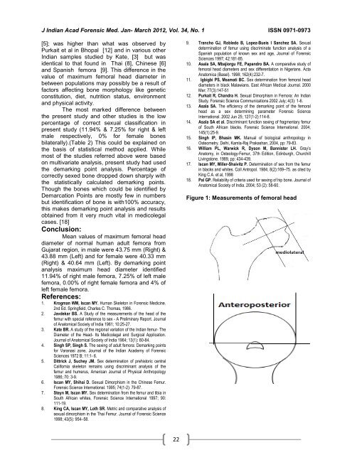
Figure 1: Measurements <strong>of</strong> femoral head