Indian Academy of Forensic Medicine (IAFM) - Official website of IAFM
Indian Academy of Forensic Medicine (IAFM) - Official website of IAFM
Indian Academy of Forensic Medicine (IAFM) - Official website of IAFM

You also want an ePaper? Increase the reach of your titles
YUMPU automatically turns print PDFs into web optimized ePapers that Google loves.
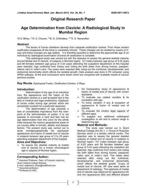
J <strong>Indian</strong> Acad <strong>Forensic</strong> Med. Jan- March 2012, Vol. 34, No. 1 ISSN 0971-0973<br />
Original Research Paper<br />
Age Determination from Clavicle: A Radiological Study in<br />
Mumbai Region<br />
*S.S. Bhise, **G. S. Chavan, **B. G. Chikhalkar, ***S. D. Nanandkar<br />
Abstract<br />
The bones <strong>of</strong> human skeletons develop from separate ossification centres. From these centers<br />
ossification progresses till the bone is completely formed. These changes can be studied by means <strong>of</strong> Xrays<br />
and these changes are age related. It is therefore possible to determine the approximate age <strong>of</strong> an<br />
individual by radiological examination <strong>of</strong> bones till ossification is complete.<br />
This radiological study was carried out with the objective to assess the general skeletal maturity<br />
around Medial end <strong>of</strong> clavicle, <strong>of</strong> subjects in Mumbai region. 131 males between age group <strong>of</strong> 9-25 years<br />
and 68 females between age group <strong>of</strong> 3-23 years attending the outpatient department <strong>of</strong> this hospital<br />
were selected. Age confirmed from history and noting the birth dates from driving license, passport,<br />
rations card or voter’s card. The cases were selected after ruling out the nutritional, developmental, and<br />
endocrinal abnormality which affects the skeletal growth. Data analysis was done in P4 computer using<br />
HPSS s<strong>of</strong>tware. At the end conclusions were drawn which are compared with available results <strong>of</strong> various<br />
previous studies.<br />
Key Words: Epiphyseal Fusion, Ossification Centres, X-Rays<br />
Introduction:<br />
Determination <strong>of</strong> the age <strong>of</strong> an individual<br />
from the appearance and the fusion <strong>of</strong> the<br />
ossification centres is a well accepted fact in the<br />
field <strong>of</strong> medical and legal pr<strong>of</strong>essions. Epiphysis<br />
<strong>of</strong> bones unites during age periods which are<br />
remarkably constant for a particular epiphysis.<br />
The determination <strong>of</strong> age presents a<br />
task <strong>of</strong> considerable importance from the viewpoint<br />
<strong>of</strong> the administration <strong>of</strong> justice. It is not<br />
possible to enunciate a hard and fast rule for<br />
age determination from this union for the whole<br />
India because the various geographical areas <strong>of</strong><br />
our country differ in climatic, dietetic and disease<br />
factors. The present study was carried out to<br />
study roentgenographically the epiphyseal<br />
appearance and fusion <strong>of</strong> medial end <strong>of</strong> clavicle<br />
in subjects between age group <strong>of</strong> 3 to 25 years<br />
attending outpatient department <strong>of</strong> this hospital.<br />
Aims and Objectives:<br />
To assess the skeletal maturity at medial<br />
end <strong>of</strong> clavicle for a known chronological<br />
age in subjects <strong>of</strong> Mumbai region.<br />
Corresponding Author:<br />
* Assistant Pr<strong>of</strong>essor, Dept. <strong>of</strong> <strong>Forensic</strong> medicine,<br />
Grant medical college Mumbai.<br />
E-mail: sadanand_bhise@rediffmail.com<br />
** Associate pr<strong>of</strong>essor<br />
***Pr<strong>of</strong>essor & HOD,<br />
7<br />
Do Comparative study <strong>of</strong> appearance &<br />
fusion <strong>of</strong> medial end <strong>of</strong> clavicle with known<br />
standards.<br />
To evaluate sex related variation & its<br />
correlation with age.<br />
To know variation if any & exception <strong>of</strong><br />
appearance & fusion <strong>of</strong> medial end <strong>of</strong><br />
clavicle.<br />
To evaluate the medico legal aspects <strong>of</strong><br />
different ages.<br />
To suggest any additional radiological<br />
investigation to aid and to reduce range in<br />
determining age.<br />
Material and Methods:<br />
The study was carried out in Grant<br />
Medical College and Sir J. J. Group <strong>of</strong> Hospitals<br />
Mumbai which is a tertiary referral centre. The<br />
objective was to assess the general skeletal<br />
maturity <strong>of</strong> medial end <strong>of</strong> clavicle in subjects in<br />
Mumbai region. 131 males between age group<br />
<strong>of</strong> 9-25 years and 68 females between age<br />
group <strong>of</strong> 3-23 years attending the outpatient are<br />
selected. Age confirmed from history and noting<br />
the birth dates from driving license, passports<br />
ration card or voter’s card. The cases were<br />
selected after ruling out the nutritional,<br />
developmental, and endocrinal abnormality<br />
which affects the skeletal growth. X-rays <strong>of</strong><br />
medial end <strong>of</strong> clavicle, AP view were taken at<br />
department <strong>of</strong> radiology. The epiphysis <strong>of</strong> medial<br />
end <strong>of</strong> clavicle was observed for appearance (A)<br />
and non appearance (NA) and different phases