NMPN_guidelines_2013.pdf

Create successful ePaper yourself
Turn your PDF publications into a flip-book with our unique Google optimized e-Paper software.
Nordic <strong>guidelines</strong> on the diagnosis and treatment of<br />
patients with Myeloproliferative Neoplasms<br />
The Nordic study group on myeloproliferative neoplasms (<strong>NMPN</strong>) is a pan-Nordic<br />
organization that has conducted Nordic clinical trials since 2001. <strong>NMPN</strong> decided in 2006 to<br />
write new <strong>guidelines</strong>, based on already existing national <strong>guidelines</strong> from the Nordic countries,<br />
Italy 1 and Great Britain. 2 The first version was published in 2007. The aim has been to write a<br />
document that can be used in all Nordic countries. We have strived to use evidence-based<br />
medicine, i.e. the conscientious, explicit, and judicious use of current best evidence in making<br />
decisions on our recommendations. The grading system employed in these <strong>guidelines</strong> is<br />
detailed on page 31. However, it should be stressed that few randomized controlled trials exist<br />
in the MPNs to support decision-making for individual patients. The <strong>guidelines</strong> are written for<br />
health professionals with a specialty or interest in hematology. They have now been updated<br />
in 2013, and are for the most part similar to those recently published by the European<br />
Leukemia Net. 3 This is the third update since our <strong>guidelines</strong> were first published.<br />
For the Nordic MPN Group, January 2013<br />
Christen Lykkegaard Andersen<br />
Björn Andreasson<br />
Hans Hasselbalch<br />
Malin Hultcrantz<br />
Håvar Knutsen<br />
Marie Lindgren<br />
Tove Skjelbakken<br />
Jan Samuelsson<br />
Thomas Stauffer Larsen<br />
1
Author disclosures (last three years)<br />
Christen Lykkegaard Andersen<br />
Björn Andreasson<br />
Hans Hasselbalch<br />
Malin Hultcrantz<br />
Håvar Knutsen<br />
Marie Lindgren<br />
Tove Skjelbakken<br />
Jan Samuelsson<br />
Thomas Stauffer Larsen<br />
Nothing to disclose<br />
Nothing to disclose<br />
Advisory Board /invited speaker for Novartis, Sanofi,<br />
and Glaxo Smith Kline<br />
Nothing to disclose<br />
Invited speaker for Novartis and Swedish Orphan<br />
Nothing to disclose<br />
Nothing to disclose<br />
Advisory board/consultant/invited speaker for Novartis,<br />
Swedish Orphan-Biovitrum, Shire, and Glaxo Smith<br />
Kline<br />
Research funding from Swedish Orphan Biovitrum<br />
2
Table of Content<br />
General introduction ............................................................................................................. 5<br />
Polycythemia vera .................................................................................................................. 5<br />
Diagnostic criteria ...................................................................................................................................................... 5<br />
Diagnostic algorithm for polycythemia vera .............................................................................................. 6<br />
Diagnostic work-up .................................................................................................................................................... 7<br />
Risk stratification in PV ........................................................................................................................................... 7<br />
High risk.........................................................................................................................................................................7<br />
Low risk..........................................................................................................................................................................7<br />
Clinical management of polycythemia vera................................................................................................. 7<br />
Goals of therapy in polycythemia vera ........................................................................................................... 7<br />
Summarized recommendations ......................................................................................................................... 7<br />
Phlebotomy ..................................................................................................................................................................8<br />
Aspirin therapy ...........................................................................................................................................................8<br />
Choice of cytoreductive therapy in PV ............................................................................................................ 8<br />
Interferon- .................................................................................................................................................................8<br />
Hydroxyurea ................................................................................................................................................................8<br />
Anagrelide .....................................................................................................................................................................9<br />
Busulphan .....................................................................................................................................................................9<br />
Radioactive phosphorus .........................................................................................................................................9<br />
JAK2 inhibitors............................................................................................................................................................9<br />
Evaluation of response and follow up ...........................................................................................................10<br />
Essential thrombocythemia............................................................................................... 11<br />
Diagnostic criteria ....................................................................................................................................................11<br />
Diagnostic algorithm in essential thrombocythemia...........................................................................12<br />
Diagnostic work-up ..................................................................................................................................................12<br />
Risk stratification in ET .........................................................................................................................................13<br />
High risk...................................................................................................................................................................... 13<br />
Low risk....................................................................................................................................................................... 13<br />
Clinical management of essential thrombocythemia...........................................................................13<br />
Goals of therapy in essential thrombocythemia .....................................................................................13<br />
Summarized recommendations .......................................................................................................................13<br />
Aspirin ......................................................................................................................................................................... 14<br />
Choice of cytoreductive therapy in ET ..........................................................................................................14<br />
Interferon-α............................................................................................................................................................... 14<br />
Hydroxyuea ............................................................................................................................................................... 14<br />
Anagrelide .................................................................................................................................................................. 15<br />
Busulphan .................................................................................................................................................................. 15<br />
Radioactive phosphorus ...................................................................................................................................... 15<br />
JAK2 inhibitors......................................................................................................................................................... 15<br />
Response monitoring in ET .................................................................................................................................15<br />
Practical considerations regarding cytoreductive therapy in PV and ET ................ 16<br />
Interferon.......................................................................................................................................................................16<br />
Hydroxyurea ................................................................................................................................................................16<br />
Anagrelide .....................................................................................................................................................................17<br />
Busulphan......................................................................................................................................................................17<br />
Radioactive phosphorus .......................................................................................................................................17<br />
Primary Myelofibrosis ........................................................................................................ 18<br />
General introduction...............................................................................................................................................18<br />
3
Diagnostic criteria ....................................................................................................................................................18<br />
Early “Prefibrotic” Myelofibrosis .....................................................................................................................19<br />
Diagnostic work-up of PMF .................................................................................................................................20<br />
Prognosis and Risk stratification in PMF ....................................................................................................20<br />
International Prognostic Scoring System (IPSS)....................................................................................... 20<br />
Dynamic International Prognostic Scoring System (DIPSS) and DIPSS plus................................ 20<br />
Clinical management of PMF ..............................................................................................................................22<br />
Goals of therapy in PMF .........................................................................................................................................22<br />
Cure of PMF via stem cell transplantation..................................................................................................22<br />
Treatment of Anemia in Primary Myelofibrosis .....................................................................................22<br />
Erythropoietin.......................................................................................................................................................... 23<br />
Danazol........................................................................................................................................................................ 23<br />
Glucocorticoids ........................................................................................................................................................ 23<br />
Thalidomide and thalidomide analogues..................................................................................................... 23<br />
Treatment of symptomatic splenomegaly and constitutional symptoms ...............................24<br />
Hydroxyurea ............................................................................................................................................................. 24<br />
Interferon- .............................................................................................................................................................. 24<br />
Jak2 inhibitors .......................................................................................................................................................... 24<br />
Splenectomy ............................................................................................................................................................... 25<br />
Splenic irradiation .................................................................................................................................................. 25<br />
Other less commonly used therapies in PMF ............................................................................................25<br />
Busulphan .................................................................................................................................................................. 25<br />
2-Chlorodeoxyadenosine (2-CdA)................................................................................................................... 26<br />
Anagrelide .................................................................................................................................................................. 26<br />
Irradiation of lungs and other sites ................................................................................................................ 26<br />
Avoiding thrombotic and bleeding complications.................................................................................26<br />
Evaluation of response and follow up ...........................................................................................................26<br />
Management of complications in MPNs .......................................................................... 27<br />
Acute thrombotic events and secondary prophylaxis .........................................................................27<br />
Bleeding ..........................................................................................................................................................................27<br />
Pruritus ...........................................................................................................................................................................28<br />
Elective surgical interventions..........................................................................................................................28<br />
Transformation to AML .........................................................................................................................................28<br />
Pregnancy ......................................................................................................................................................................28<br />
Pediatric MPN ....................................................................................................................... 30<br />
Evidence levels and recommendations grades ............................................................. 31<br />
References............................................................................................................................. 32<br />
4
General introduction<br />
The Philadelphia chromosome/bcr-abl negative myeloproliferative neoplasms (MPNs)<br />
represent a range of clonal hematological diseases with overlapping features. The ma in<br />
entities are polycythemia vera (PV), essential thrombocythemia (ET) and primary<br />
myelofibrosis (PMF) which are characterized by clonal excess hematopoiesis in one or more<br />
cell lines. They are associated with an elevated risk of arterial and venous thrombosis; many<br />
PV and ET patients have thrombosis at the time of diagnosis. Both PV and ET can progress to<br />
myelofibrosis and all three entities can transform into acute myeloid leukemia.<br />
Polycythemia vera<br />
Diagnostic criteria<br />
Males and females with hematocrit (Hct) > 0.52 and > 0.48 (well above 99 th percentile) for<br />
more than 2 months should be evaluated. The diagnostic work-up of PV will be reviewed<br />
here, for diagnostic work-up of erythrocytosis other than PV please see the 2009 version of<br />
the <strong>guidelines</strong> at www.nmpn.org. A diagnosis of PV should be made using WHO criteria. 4<br />
Revised WHO 2008 criteria for polycythemia vera (PV)<br />
Major criteria<br />
1. Hemoglobin > 185 g/L in men, >165 g/L in women (11,5/10,2 mmol/l), or haematocrit > 0.52 in<br />
men and > 0.48 in women, or other evidence of increased red cell volume*<br />
2. Presence of JAK2 V617F or other functionally similar mutation such as JAK2 exon 12 mutation<br />
Minor criteria<br />
1. Bone marrow biopsy showing hypercellularity for age with trilineage growth (panmyelosis) with<br />
prominent erythroid, granulocytic, and megakaryocytic proliferation<br />
2. Serum erythropoietin level below the reference range for normal<br />
3. Endogenous erythroid colony formation in vitro<br />
Diagnosis requires the presence of both major criteria and 1 minor criterion or the presence of the<br />
first major criterion together with 2 minor criteria.<br />
* Hemoglobin or hematocrit greater than 99th percentile of method-specific reference range for age,<br />
sex, altitude of residence or hemoglobin greater than 170 g/L in men, 150 g/L in women if<br />
associated with a documented and sustained increase of at least 20 g/L from an individual’s baseline<br />
value, or elevated red cell mass greater than 25% above mean normal predicted value.<br />
A diagnosis of PV can thus be made without bone marrow biopsy if both major criteria are<br />
fulfilled. Biopsy is however highly recommended since degree of fibrosis confers valuable<br />
prognostic information. 5 The JAK2 V617F mutation is present in at least 95 % of PV<br />
patients, 6-9 in later studies up to 98%. 10 The JAK2 V617F mutation is very seldom found in<br />
normal individuals (and if so at very low levels < 1 %), in patients with secondary<br />
erythrocytosis, and is rarely found in other hematologic disorders with the exception of MDS<br />
5
RARS-T. 11 If PV is suspected and the patient is negative for the JAK2 V617F mutation,<br />
further investigation with JAK2 exon 12 mutations should be carried out, if this analysis is<br />
available.<br />
PV should also be suspected in a patient with a Hct/Hb below the diagnostic threshold if this<br />
is combined with a PV-related feature, e.g. an arterial or venous thrombotic event, especially<br />
in young patients and/or atypical thrombosis (eg. splanchnic vein thrombosis and Budd-Chiari<br />
syndrome), aquagenic pruritus, erythromelalgia or other symptoms of acral ischemia,<br />
splenomegaly, leukocytosis, thrombocytosis or microcytosis.<br />
Patients with PV typically have a S-erythropoietin (S-EPO) that is subnormal or in the lower<br />
reference interval.<br />
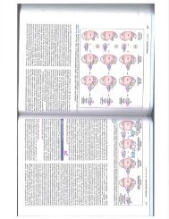
Diagnostic algorithm for polycythemia vera<br />
Hb > 185 g/L (men) and > 165 g/L<br />
(women) or Hct >0.52 (male) and<br />
>0.48 (female) or other evidence of<br />
increased red cell volume (see<br />
diagnostic criteria)<br />
PV related feature (aquagenic pruritus,<br />
erythromelagia, atypical thrombosis,<br />
splenomegaly, thrombocytosis or<br />
leukocytosis).<br />
Medical history and relevant physical<br />
investigation<br />
Full blood count<br />
JAK2 V617F and plasma<br />
erythropoietin (EPO)<br />
Screening for JAK2 V617F mutation<br />
JAK2 V617F positive<br />
JAK2 V617F negative<br />
JAK2 exon 12 mutation<br />
analysis<br />
(If not available proceed<br />
to bone marrow biopsy)<br />
Subnormal EPO<br />
Normal EPO<br />
Increased EPO<br />
Positive<br />
Bone marrow<br />
biopsy<br />
(recommended)<br />
Negative<br />
Bone<br />
marrow<br />
biopsy<br />
EPO in the lownormal<br />
range or<br />
clinical and<br />
laboratory signs<br />
of MPN present<br />
EPO normal<br />
and no clinical<br />
or laboratory<br />
signs of MPN<br />
present<br />
Diagnosis confirmed<br />
in the presence of a<br />
JAK2-mutation and/or<br />
a consistent<br />
histology.<br />
PV unlikely if no<br />
mutation and bone<br />
marrow biopsy not<br />
consistent w ith MPN.<br />
Follow up is<br />
recommended<br />
PV unlikely.<br />
Secondary<br />
polycythemia or<br />
pseudopolycythemia<br />
should be assessed<br />
PV unlikely.<br />
Evaluate<br />
secondary<br />
erythrocytosis.<br />
6
Diagnostic work-up<br />
Full blood count with differential count<br />
Iron status<br />
ASAT, ALAT, bilirubin, creatinine, uric acid, lactate dehydrogenase (LDH)<br />
S-erythropoietin<br />
JAK2 V617F mutation<br />
JAK2 exon 12 mutation in JAK2 V617F-negative patients, if available<br />
Bone marrow biopsy<br />
Physical examination including palpation of spleen<br />
Evaluation of cardiovascular risk factors<br />
Risk stratification in PV<br />
Polycythemia vera is associated with an excess mortality and reported median survival varies<br />
from 10 to 20 years in different studies. 12-14 In order to achieve the clinical goals listed below<br />
risk stratification of patients is essential.<br />
High risk<br />
Patients > 60 years or with previous thrombosis or platelets > 1500 x 10 9 /l. These patients<br />
should be treated with cytoreductive therapy. PV patients with isolated erythrocytosis i.e.<br />
normal white cell and platelet counts can initially be treated with phlebotomy alone, until<br />
leukocytosis or thrombocytosis occurs.<br />
Low risk<br />
Patients < 60 years, no previous thrombosis and platelets < 1500 x 10 9 /l. These patients<br />
should in general not receive cytoreductive therapy, but can be considered in certain situations<br />
(see below).<br />
Clinical management of polycythemia vera<br />
Goals of therapy in polycythemia vera<br />
• avoid first occurrence and/or recurrence of thrombotic and bleeding complications<br />
• reduce constitutional symptoms (weight loss, night sweats, fever, pruritus)<br />
• manage risk situations (e.g. pregnancy, surgery)<br />
• minimize the risk of acute leukemia and post-PV myelofibrosis<br />
Summarized recommendations<br />
Vigorous treatment of cardiovascular risk factors<br />
Phlebotomy to maintain a Hct < 0.45<br />
Aspirin 75 - 100 mg/day unless contraindicated<br />
Cytoreduction should be given to high-risk patients. The goal of therapy should be<br />
normalization of peripheral blood counts.<br />
Cytoreduction can also be considered in low risk patients:<br />
- with poor tolerance/high frequency of phlebotomies<br />
- with symptomatic or progressive splenomegaly<br />
- with other evidence of disease progression e.g. weight loss, night sweats<br />
- with progressive leukocytosis and/or thrombocytosis<br />
7
Stem cell transplantation is very seldom performed in PV and should be reserved for<br />
the occasional patient that does not respond to conventional cytoreductive therapy<br />
Phlebotomy<br />
The hematocrit should be maintained at less than 0.45 in all patients, as shown in a recent<br />
randomized trial. 15 There is currently no evidence to support a different level of Hct in males<br />
and females. Hemoglobin levels should not be used for decision-making regarding<br />
phlebotomy. Grade A recommendation, evidence level Ib. For explanation of grading<br />
system – see p. 31.<br />
Aspirin therapy<br />
Aspirin has been shown to reduce both arterial and venous thrombosis in PV 16 and should be<br />
given to all PV patients unless it is contraindicated. In Finland, 50mg tablets are available and<br />
the recommended dose is 100mg. In the other Nordic countries the recommended dose is<br />
75mg per day. Grade A recommendation, evidence level Ib<br />
Aspirin should not be given to patients with platelets > 1,500 x 10 9 /L due to an increased risk<br />
of bleeding, instead cytoreductive therapy should be initiated. In case of aspirin allergy,<br />
clopidogrel can be used but no there is so far no data on its efficacy in PV. Trials on<br />
clopidogrel in PV are ongoing. The combination of aspirin and anagrelide should in general<br />
be used with some caution due to an increased risk of bleeding 17 and is not recommended in<br />
patients with previous bleeding.<br />
Choice of cytoreductive therapy in PV<br />
• 60 years: 1 st line hydroxyurea or interferon- , 2 nd line anagrelide*<br />
• >75 years or with a short expected survival 1 st line hydroxyurea, 2 nd line intermittent<br />
busulfan, 3 rd line 32 P<br />
Combination therapy (hydroxyurea+anagrelide*, hydroxyurea+interferon ) can be an<br />
alternative second line therapy in fit patients if dose-limiting side effects occur with<br />
monotherapy.<br />
* anagrelide only if the indication for therapy is thrombocytosis<br />
Grade C recommendation, evidence level IV<br />
Interferon-<br />
Interferon- IFN- is theoretically superior for treating PV as molecular remissions can be<br />
achieved with IFN. 18-21 IFN does not increase the risk of leukemia. 3 It is along with<br />
ruxolitinib the most effective drug for PV related pruritus.<br />
Recommendation: IFN or pegylated IFN is the drug of choice in younger patients (
PV having been the subject of large randomized trials. 22,23 By itself it has very limited<br />
leukemogenic potential, if any. 24 However, long-term use of HU in PV has not been able to<br />
prevent lekemogenic transformation in 10-20 % of patients after 20 years of therapy in some<br />
trials. 25 HU is not recommended during pregnancy, and should be withdrawn at least 3<br />
months before conception both in males and females.<br />
Recommendation: Hydroxyurea is recommended as first-line cytoreductive therapy in PV in<br />
patients >60 years or younger patients that do not tolerate IFN. Grade A recommendation,<br />
evidence level Ib. The documented high leukemia transformation rate despite long term HU<br />
treatment (see above), 25 suggests that HU should be limited in patients below 60 years. Grade<br />
C recommendation, evidence level IV. For dosing and practical recommendations, see page<br />
16.<br />
Anagrelide<br />
Anagrelide is megakaryocyte specific and is therefore only effective in controlling the platelet<br />
count, and probably does not control progression of PV.<br />
Recommendation: Anagrelide may be used to control thrombocytosis in PV patients that<br />
cannot tolerate or do not respond to IFN or HU, and when HU is considered a less suitable<br />
alternative. Grade C recommendation, evidence level IV. Due to risk of cardiovascular<br />
events, cardiac arrhythmias, and cardiomyopathy, patients should have normal cardiac<br />
function. A recent report from Shire states that Anagrelide can give serious adverse cardiac<br />
events also in patients with normal cardiac function. The combination of aspirin and<br />
anagrelide should in general be used with some caution due to an increased risk of bleeding 17<br />
and is not recommended in patients with previous bleeding. Anagrelide should not be used<br />
during pregnancy.<br />
Busulphan<br />
Busulphan (BU) is an alkylating agent and can increase the leukemic transformation rate.<br />
Recommendation: BU should be reserved for patients 75 years or older, or for patients not<br />
tolerating HU, IFN or anagrelide. Grade B recommendation, evidence level III. Low dose<br />
intermittent busulfan is more efficacious in controlling PV than radioactive phosphorus. 26<br />
Grade A recommendation, evidence level Ib.<br />
Radioactive phosphorus<br />
Radioactive phosphorus (P 32 ) can control elevated blood counts in PV. Infrequent intermittent<br />
treatment is required and follow-up can therefore be minimized. It is valuable in older patients<br />
if compliance with continuous oral therapy is a problem.<br />
Recommendation: Due to the leukemogenic effect, P 32 use should be limited to patients older<br />
than 75 years where HU, IFN, BU or anagrelide are not suitable. 27 Grade A<br />
recommendation, evidence level Ib<br />
JAK2 inhibitors<br />
Since no JAK2 inhibitor has been studied extensively in PV to date, these drugs are at the<br />
present time experimental and should not be used outside of the context of clinical trials.<br />
9
Evaluation of response and follow up<br />
The goal of therapy should be normalized peripheral blood counts. Consider changing therapy<br />
in patients with resistance or intolerance to ongoing therapy and in patients not achieving<br />
treatment goals. The ELN have published criteria for hydroxyurea resistance and<br />
intolerance. 28<br />
Patients on phlebotomy alone should be monitored with complete blood counts at least every<br />
4 to 6 weeks. For recommendations regarding patients on cytoreductive therapy, see p. 17-18.<br />
There is no indication to use repeated bone marrow trephine biopsies for routine follow-up in<br />
PV, but is essential in assessing transformation to myelofibrosis or acute leukemia.<br />
Monitoring of molecular response, including sequential assessment of the JAK2 V617F allele<br />
burden is at the moment not recommended for routine clinical use.<br />
10
Essential thrombocythemia<br />
All patients with unexplained and persistent thrombocytosis above 450x10 9 /l should be<br />
investigated for the possibility of an MPN.<br />
Essential thrombocythemia (ET) is now diagnosed according to the 2008 WHO diagnostic<br />
criteria which differ from the former PVSG criteria in that some ET patients with fibrosis<br />
would according to the WHO be classified as early myelofibrosis and not as ET, 4 see also<br />
page 19). There is however no significant difference in the risk of thrombosis between the two<br />
entities, 29 and therefore no reason for not extending the existing clinical care<br />
recommendations to also apply to ET classified by WHO. It is of paramount importance that a<br />
diagnosis of ET is made using the 2008 WHO criteria, highlighting the absolute need of a<br />
bone marrow biopsy in the diagnostic workup of the patient.<br />
Diagnostic criteria<br />
Revised WHO 2008 criteria for essential thrombosis (ET)<br />
1. Sustained platelet count >450x10 9 /L<br />
2. Bone marrow biopsy specimen showing proliferation mainly of the<br />
megakaryocytic lineage with increased number of enlarged, mature<br />
megakaryocytes. No significant increase of left-shift of neutrophil granulopoiesis<br />
or erythropoiesis.<br />
3. Not meeting WHO criteria for polycythemia vera, primary myelofibrosis, BCR-<br />
ABL positive chronic myelogenous leukemia or myelodysplastic syndrome or<br />
other myeloid neoplasm.<br />
4. Demonstration of JAK2 V617F or other clonal marker, or in the absence of JAK2<br />
V617F, no evidence for reactive thrombosis.<br />
All four criteria must be met.<br />
Screening for JAK2 V617F mutation should be included in the initial work-up of all patients<br />
with suspected ET (positive in 50-60 % of ET patients 6-9 ). JAK2 negative patients should be<br />
screened for mutations in the thrombopoietin receptor MPL (W515L and W515K), which is<br />
positive in around 3% of JAK2 negative ET patients, 30,31 if this analysis is available.<br />
Even in the presence of a mutation in JAK2 or MPL, a diagnosis of ET requires exclusion of<br />
PV and PMF. It is important to differentiate ET from prefibrotic/early and overt PMF since<br />
there are significant differences in disease progression, leukemic transformation and<br />
survival. 29<br />
In the absence of a clonal marker (JAK2, MPL) it is important to rule out other myeloid<br />
malignancies and secondary causes of thrombocytosis such as connective tissue disease,<br />
malignant lymphomas, bleeding and iron deficiency.<br />
11
Diagnostic algorithm in essential thrombocythemia<br />
Sustained platelet count >450x10 9 /l without signs of reactive thrombocytosis (eg. iron<br />
deficiency, inflammatory disorders, chronic infections or non-myeloid malignancy).<br />
JAK2V617F mutation screening<br />
JAK2 V617F<br />
positive<br />
JAK2 V617F<br />
negative<br />
MPL mutation analysis<br />
if available<br />
MPL mutation<br />
positive<br />
MPL mutation<br />
negative<br />
Bone marrow biopsy<br />
Bone marrow<br />
biopsy<br />
Bone marrow histology<br />
not consistent with<br />
MPN<br />
ET or PMF<br />
depending on histology<br />
Bone marrow<br />
histology consistent<br />
with MPN<br />
Unlikely to be ET or other<br />
MPN.<br />
Consider work-up for CML<br />
(including BCR/ABL) or<br />
secondary thrombocytosis<br />
Diagnostic work-up<br />
Full blood count with differential count<br />
Iron status<br />
ASAT, ALAT, bilirubin, creatinine, uric acid, LDH<br />
S-erythropoietin<br />
JAK2 V617F mutation<br />
MPL mutation in JAK2 negative patients, if available<br />
Bone marrow biopsy<br />
BCR-ABL in JAK2 V617-negative patients if differentiation against chronic<br />
myelogenous leukemia is not clear<br />
Evaluation of cardiovascular risk factors<br />
Physical examination including palpation of spleen<br />
12
Risk stratification in ET<br />
The risk stratification system in ET is based on the assessment of risk of thrombosis, as the<br />
current therapy in ET is aimed at lowering the risk of thrombosis. Extreme thrombocytosis is<br />
also included in the risk stratification as it can be associated with acquired von Willebrand<br />
disease and bleeding tendency. In the absence of high-risk features, cytoreductive therapy is<br />
generally not recommended for patients with cardiovascular risk factors, but could be<br />
considered on a case-by-case basis. 32 ET has over the last decades been associated with a<br />
normal life expectancy is in most, 14,33 but not all, 12 studies. True ET diagnosed according to<br />
the 2008 WHO classification hos not been reported to affect the life expectancy of patients. 29<br />
New prognostic scoring systems have been presented, 34,35 based on additional risk factors<br />
such as high leukocyte count and cardiovascular risk factors, but until these are thoroughly<br />
validated, recommendations are based on the following risk groups.<br />
High risk<br />
ET patients > 60 years or with a history of previous thrombosis should be treated with<br />
cytoreductive therapy in order to prevent thrombosis.<br />
ET patients with a platelet count > 1,500 x 10 9 /L should be treated with cytoreductive therapy<br />
in order to avoid bleeding.<br />
Low risk<br />
ET patients < 60 years, no previous thrombosis and a platelet count < 1,500 x 10 9 /L should<br />
normally not be treated with cytoreductive therapy.<br />
Clinical management of essential thrombocythemia<br />
Goals of therapy in essential thrombocythemia<br />
Prevent first occurrence or recurrence of thrombotic and bleeding complications.<br />
Minimize the risk of acute leukemia and post-ET myelofibrosis<br />
Reduce constitutional symptoms (weight loss, night sweats, fever)<br />
Manage risk situations (e.g. pregnancy and surgery).<br />
Summarized recommendations<br />
Vigorous treatment of cardiovascular risk factors<br />
Aspirin 75 - 100 mg/day to selected patients, see below<br />
Cytoreduction should be given to high-risk patients. The goal of therapy should be<br />
normalization of peripheral blood counts.<br />
Cytoreduction can also be considered in low risk patients with uncontrolled<br />
cardiovascular risk factors<br />
Cytoreduction can also be considered in low risk patients with any of the below<br />
mentioned features. It is important to stress that before starting therapy these patients<br />
should be evaluated for eventual progression to myelofibrosis:<br />
- with symptomatic or progressive splenomegaly<br />
- with other evidence of disease progression e.g. weight loss, night sweats<br />
- with progressive leukocytosis and/or thrombocytosis<br />
Stem cell transplantation is almost never performed in ET.<br />
13
Aspirin<br />
Antiplatelet therapy with aspirin 75 mg per day is recommended, unless otherwise<br />
contraindicated, for some ET patients namely<br />
high risk ET patients<br />
low risk ET patients with one or more risk factor for cardiovascular disease<br />
low risk ET patients with microvascular symptoms (erythromelalgia)<br />
Aspirin is generally not recommended in low risk patients with no cardiovascular risk factors,<br />
but may be considered in low risk JAK2 positive patients since JAK2 positive patients may<br />
have a higher risk of thrombosis 36 and retrospective data indicate a lower thrombotic risk in<br />
aspirin-treated JAK2 positive patients. 37<br />
Patients with a platelet count > 1,500 x 10 9 /L should not be treated with aspirin due to<br />
increased bleeding risk, instead cytoreductive therapy should be initiated. ASA should be<br />
started again when platelets are stable < 1,000 x 10 9 /L.<br />
The combination of aspirin and anagrelide should in general be used with some caution due to<br />
an increased risk of bleeding, 17 and is not recommended in patients with previous bleeding.<br />
Choice of cytoreductive therapy in ET<br />
• 60 years: 1 st line hydroxyurea or interferon- , 2 nd line anagrelide*<br />
• >75 years or with a short expected survival 1 st line hydroxyurea, 2 nd line intermittent<br />
busulfan, 3 rd line 32 P<br />
Combination therapy (hydroxyurea+anagrelide*, hydroxyurea+interferon ) can be an<br />
alternative as second line therapy in fit patients if dose-limiting side effects occur with<br />
monotherapy.<br />
* anagrelide only if the indication for therapy is thrombocytosis<br />
Grade C recommendation, evidence level IV<br />
Interferon-α<br />
Interferon-α (IFN) treatment is well documented and safe in ET and is not considered<br />
leukemogenic or teratogenic. 1 Pegylated IFN given sc weekly has been shown to have equal<br />
efficacy as conventional IFN given three times weekly. 38,39 We have a long standing therapy<br />
tradition in the Nordic countries using IFN in ET, whereas the ELN does not recommend IFN<br />
in ET due to the lack of larger trials in ET. 3<br />
Recommendation: IFN is the recommended first line therapy in younger patients. It can be<br />
used in older patients if long-term use of HU is not suitable and in patients who do not<br />
tolerate HU. Grade B recommendation, evidence level III. IFN or pegylated IFN is the<br />
treatment of choice if cytoreductive therapy is indicated during pregnancy or when pregnancy<br />
is planned.<br />
Hydroxyuea<br />
Hydroxyurea (HU) is the best-documented therapy in ET and is recommended as a first-line<br />
therapy in the majority of ET patients. HU markedly reduces thrombotic complications<br />
compared to aspirin alone. 40 HU was more effective than anagrelide in reducing arterial<br />
14
thrombotic events in the PT1- trial. 17 HU should not be used during pregnancy or when<br />
pregnancy is planned.<br />
Recommendation: Hydroxyurea is recommended as a first-line cytoreductive therapy in ET<br />
patients > 60 years of age. Grade A recommendation, evidence level Ib.<br />
Anagrelide<br />
Anagrelide is not considered leukemogenic but was associated with a higher risk of bone<br />
marrow fibrosis in the PT-1 trial. 17 In the same trial anagrelide was more effective than HU in<br />
preventing venous thrombosis. Due to known cardiovascular side effects (palpitations most<br />
frequent), anagrelide is only recommended in patients without congestive heart failure or<br />
cardiac arrhythmias. Anagrelide should not be used during pregnancy or when pregnancy is<br />
planned, since there are no data available concerning effects on the fetus.<br />
Recommendation: Anagrelide can be used in patients where long-term use of IFN or HU is<br />
not suitable, and only when platelet reduction is the goal of therapy. Due to risk of<br />
cardiovascular events, cardiac arrhythmias, and cardiomyopathy, patients should have normal<br />
cardiac function. A recent report from Shire states that Anagrelide can give serious adverse<br />
cardiac events also in patients with normal cardiac function. Anagrelide should not be used<br />
during pregnancy. Grade B recommendation, evidence level III.<br />
Busulphan<br />
Busulphan (BU) is an alkylating agent given as intermittent treatment. BU effectively controls<br />
platelet count but is associated with an increased risk of progression to acute leukemia,<br />
particularly when used sequentially with hydroxyurea. BU is a second- or third-line agent and<br />
should be restricted to patients with short life expectancy.<br />
Recommendation: Intermittent BU treatment can be used in patients 75 years or older, or for<br />
patients where HU, IFN or anagrelide are not suitable. Grade B recommendation, evidence<br />
level IIb.<br />
Radioactive phosphorus<br />
Radioactive phosphorus (P 32 ) is an intermittent treatment effective at controlling platelet<br />
count. Few randomized studies have been conducted with P 32 , in a PVSG trial P 32 showed<br />
comparable effect with melphalan after one year from medication. 41 P 32 is associated with an<br />
increased risk of progression to acute leukemia, particularly when used sequentially with<br />
hydroxyurea. P 32 should be restricted to patients with short life expectancy and can be<br />
valuable in older patients if compliance with continuous oral therapy is a problem.<br />
Recommendation: P 32 treatment can be used in ET patients with short life expectancy where<br />
HU, IFN, BU or anagrelide are not suitable. Grade A recommendation, evidence level Ib.<br />
JAK2 inhibitors<br />
Since no JAK2 inhibitor has been studied extensively in ET to date, these drugs are at the<br />
present time experimental and should not be used outside of the context of clinical trials.<br />
Response monitoring in ET<br />
The goal of therapy should be normalized peripheral blood counts in patients that tolerate<br />
therapy. The ELN have published criteria for hydroxyurea resistance and intolerance. 28<br />
Change of therapy should be considered in patients with resistance or intolerance to ongoing<br />
15
therapy and in patients not achieving goals of treatment. There is currently no absolute<br />
evidence for a correlation between platelet levels < 400 and the reduced risk of thrombosis.<br />
Therefore, in patients that develop anemia on HU or IFN treatment consider lowering the dose<br />
and allowing a higher platelet number in order to avoid anemia.<br />
There is no indication for repeated bone marrow trephine biopsies in routine follow-up in ET,<br />
but is essential in assessing transformation to myelofibrosis or acute leukemia. Monitoring of<br />
molecular response, including sequential assessment of the JAK2 V617F allele burden is at the<br />
moment not recommended for clinical use.<br />
Practical considerations regarding cytoreductive therapy in PV and<br />
ET<br />
Interferon<br />
Interferon (IFN) suppresses growth of multipotent hematopoietic progenitor cells. Pegylated<br />
interferons, which are administered weekly, are today being used in most centers. The starting<br />
dose for pegylated interferon alfa-2a (Pegasys®) is 90 g subcutaneously once weekly. The<br />
large majority of patients respond to a dosage of 90 g once weekly. When the response is<br />
stable, controls every 4-8 weeks is sufficient. The starting dose of pegylated interferon alfa-2b<br />
(PegIntron®) is 0,5 g/kg subcutaneously once weekly. Responses are seen within 2-3<br />
months. If cell counts are still high after 3 months, the dose should be increased. Most<br />
patients respond to a dose between 0.5 and 0.75 µg/kg. When sufficient effect is reached,<br />
taper the dose to the lowest effective dose.<br />
Pegylated interferons can be associated with side effects, e.g. flu-like symptoms and<br />
psychiatric disorders. Therefore, interferons are contraindicated in patients with pre-existing<br />
psychiatric conditions. Flu-like symptoms may be transient and can be well controlled by<br />
paracetamol. Treatment should be stopped if patients develop psychiatric disorders.<br />
Hydroxyurea<br />
Hydroxyurea (HU) is a non-alkylating, non-specific myelosuppressive drug. The<br />
recommended starting dose is 500-1000 mg daily (15-20 mg/kg/day). Elderly patients (>70<br />
years old) are usually started on 500 mg/day. In situations when rapid platelet reduction is<br />
needed higher starting doses (1500-2000 mg/day) are recommended. Elevated leukocyte and<br />
platelet counts may decrease within days. Follow up with full blood count (FBC) is<br />
recommended every second week initially. The dose should be adjusted in order to achieve a<br />
stable platelet count of 200 – 400 x10 9 /L. When this is achieved, FBC controls every 4 to 8<br />
weeks is usually sufficient. The average dosage needed is approximately 10-14 tablets a week,<br />
lower in elderly patients.<br />
Leukopenia with neutrophil count of 1.0 – 1.5 x10 9 /L and anemia can occur during the first 3-<br />
6 months and is accepted when the goal is to reduce large splenomegaly. In some cases the<br />
neutropenia or anemia is dose limiting, but if the platelet count can be kept below 600 x10 9 /L,<br />
this can sometimes be acceptable.<br />
Hydroxyurea should be administered continuously. Interruptions in the treatment can result in<br />
abrupt rise in the platelet count and risk of thrombosis.<br />
16
Some patients will experience other significant side effects from HU, including<br />
gastrointestinal disturbance, skin pigmentation, mucocutaneous and leg ulcers. The latter can<br />
be seen in up to 10% of patients, and do not heal until HU is discontinued.<br />
Anagrelide<br />
Anagrelide acts on the post-mitotic phase of megakaryocyte development, showing a selective<br />
effect on megakaryocytes in vitro. Anagrelide (Xagrid®) capsules contains 0,5 mg and the<br />
starting dose is 1 capsule twice daily. FBC should be checked once a week during the first<br />
few weeks as the platelet count may fall abruptly. If needed, the dosage can be increased with<br />
one capsule (0.5 mg) after 1-2 weeks but the dosage should not be increased with more than<br />
one capsule/day per week. The average dosage is 2-2.5 mg daily divided in 2-3 doses to<br />
achieve a satisfactory platelet level between 200-400 x10 9 /l. The maximum dosage should not<br />
exceed 10 mg daily, or 3 mg in one single dose. Some patients with troublesome side effects<br />
(palpitations or gastrointestinal problems most frequent) may need the daily dosage divided in<br />
3-4 doses.<br />
Due to risk of cardiovascular events, cardiac arrhythmias, and cardiomyopathy, patients<br />
should have normal cardiac function. An ECG and an echocardiogram are recommended<br />
before Anagrelide is started. A recent report from Shire states that Anagrelide can give serious<br />
adverse cardiac events also in patients with normal cardiac function. The combination of<br />
aspirin and anagrelide should in general be used with some caution due to an increased risk of<br />
bleeding 17 and is not recommended in patients with previous bleeding. When the response is<br />
stable, controls every 4-8 weeks is sufficient. Anagrelide should not be used during<br />
pregnancy.<br />
Busulphan<br />
Busulphan is an alkylating agent that is given intermittently. Start with 2-4 mg daily until<br />
response (normally 2-6 weeks). A full blood cell count should be checked every week while<br />
the patient is on busulphan therapy. The leukocyte count may increase during the first 10 to<br />
15 days, this should not be interpreted as resistance to the drug and the dose should not be<br />
increased. If busulphan is started at 4 mg/day, the dose should be lowered to 2 mg/d when<br />
platelets start to decrease, typically within 2-6 weeks. Busulphan should be stopped when the<br />
platelet count reaches 400 x10 9 as platelets will continue to fall for another 2-3 weeks.<br />
Busulphan can be repeated when the platelet count rises above 400 x10 9 again.<br />
Radioactive phosphorus<br />
Radioactive phosphorus (P 32 ) is an orally given radioactive treatment that effectively controls<br />
hematopoiesis. P 32 is associated with an increased risk of progression to acute leukemia,<br />
particularly when used sequentially with hydroxyurea. Blood counts should be checked every<br />
other week until platelet counts have normalized, after that every 8-12 weeks. The dose of P 32<br />
is adjusted according to age, weight and previous treatment and is calculated by the<br />
radiotherapists.<br />
17
Primary Myelofibrosis<br />
General introduction<br />
Primary myelofibrosis (PMF) is characterized by progressive accumulation of connective<br />
tissue and endothelial proliferation in the bone marrow accompanied by extramedullary<br />
hematopoiesis with enlargement of the spleen and liver. PMF is associated with a significant<br />
excess mortality and the median survival of PMF patients is 5-6.5 years with a wide<br />
range. 12,42,43 Survival of patients with early PMF (see below) has been estimated to be longer<br />
than 10 years in a recent study. 44<br />
Diagnostic criteria<br />
Revised WHO criteria for primary myelofibrosis (PMF)<br />
Major criteria<br />
1. Presence of megakaryocyte proliferation and atypia*, usually accompanied by either reticulin<br />
and/or collagen fibrosis, or, in the absence of significant reticulin fibrosis, the megakaryocyte<br />
changes must be accompanied by an increased bone marrow cellularity characterized by<br />
granulocytic proliferation and often decreased erythropoiesis (ie, prefibrotic cellular-phase<br />
disease)<br />
2. Not meeting WHO criteria for PV, CML, MDS, or other myeloid neoplasm**<br />
3. Demonstration of JAK2617V>F or other clonal marker (e.g., MPL515W>L/K), or in the<br />
absence of a clonal marker, no evidence of bone marrow fibrosis due to underlying<br />
inflammatory or other neoplastic diseases***<br />
Minor criteria<br />
1. Leukoerythroblastosis<br />
2. Increase in serum lactate dehydrogenase level****<br />
3. Anemia****|<br />
4. Palpable splenomegaly****<br />
Diagnosis requires meeting all 3 major criteria and 2 minor criteria.<br />
*Small to large megakaryocytes with an aberrant nuclear/cytoplasmic ratio and hyperchromatic,<br />
bulbous, or irregularly folded nuclei and dense clustering.<br />
**Requires the failure of iron replacement therapy to increase hemoglobin level to the polycythemia<br />
vera range in the presence of decreased serum ferritin. Exclusion of polycythemia vera is based on<br />
hemoglobin and hematocrit levels. Red cell mass measurement is not required. Requires the absence<br />
of bcr-abl. Requires the absence of dyserythropoiesis and dysgranulopoiesis.<br />
***Secondary to infection, autoimmune disorder or other chronic inflammatory condition, hairy cell<br />
leukemia or other lymphoid neoplasm, metastatic malignancy, or toxic (chronic) myelopathies. It<br />
should be noted that patients with conditions associated with reactive myelofibrosis are not immune<br />
to primary myelofibrosis and the diagnosis should be considered in such cases if other criteria are<br />
met.<br />
****Degree of abnormality could be borderline or marked.<br />
18
Early “Prefibrotic” Myelofibrosis<br />
The concept of early “prefibrotic” myelofibrosis is increasingly recognized as the early phase<br />
of myelofibrosis in the biological continuum from early disease stage to the advanced burntout<br />
stage of myelofibrosis with myeloid metaplasia. Accordingly, early “prefibrotic” PMF<br />
was included in the revised 2008 World Health Organization (WHO) classification. 4 Early<br />
PMF is characterized by a hypercellular bone marrow with megakaryocytic, and in contrast to<br />
true ET, also granulocytic proliferation. Megakaryocytes show extensive tight clustering and<br />
condensed nuclei with clumped chromatin and abnormal nuclear-cytoplasmic ratio. Reticulin<br />
fibrosis is absent or minimal. 4 It is important to recognize that early PMF exhibits the three<br />
major criteria of the WHO classification shown above, but leukoerytroblastosis, splenomegaly<br />
and anemia is most often not present. Although most experienced pathologists acknowledge<br />
the diagnosis of early PMF, there is still some controversy around this concept as a separate<br />
disease entity and how to distinguish this from ET by distinct histopathological features has<br />
not been universally accepted. 45<br />
In clinical practice the occurrence of anemia, elevated leukocyte count or elevated LDH in<br />
“ET” patients should alert the clinician to reevaluate the diagnosis and rule out PMF. 46<br />
A large retrospective trial of 1104 patients diagnosed as having ET by PVSG criteria were<br />
reanalyzed by one of the authors of the WHO 2008 classification. In 891 patients (81 %) the<br />
diagnosis was confirmed as ET, whereas 180 patients (16 %) were reclassified as early PMF<br />
and 33 patients were not evaluable. Clinical follow-up of these patients showed no differences<br />
in thrombosis rates. In contrast, 10 and 15 year overall survival was markedly lower in early<br />
PMF, 76 vs. 89 % at 10 years and 59 vs. 80 % at 15 years, respectively. Survival in ET was<br />
not different from a sex- and age-standardized European population. Rates of transformation<br />
to acute leukemia and progression to overt PMF were clearly increased in early PMF. 29<br />
Further analyses of this cohort has shown that major bleeding was more common in early<br />
PMF compared to ET, especially in patients treated with aspirin. 47 Finally, in a substudy<br />
where 178 ET patients below 40 years of age at diagnosis were compared to 35 patients with<br />
early PMF, progression to overt PMF was more common in patients with early PMF and there<br />
was a trend towards more arterial thrombosis. 48 Transformation to leukemia was not observed<br />
during a median follow-up of 7.6 years.<br />
There is currently no evidence that treatment of early myelofibrosis is associated with<br />
prolonged survival. The majority of patients in the studies above received HU therapy.<br />
Whether other treatment modalities such as interferon can prevent or prolong time to overt<br />
PMF progression is unknown, but there are ongoing studies to address this question. For the<br />
time being patients with early MF without risk factors should not be treated with<br />
cytoreductive therapy outside study protocols. In patients with risk factors and an indication<br />
for cytoreductive treatment for platelet reduction we recommend the same treatment and<br />
treatment goals as for ET (see ET chapter).<br />
19
Diagnostic work-up of PMF<br />
Full blood count with differential count<br />
Iron status<br />
ASAT, ALAT, bilirubin, creatinine, uric acid, LDH<br />
S-erythropoietin<br />
JAK2 V617F mutation<br />
MPL mutation in JAK2 negative patients, if available<br />
BCR-ABL in JAK2V617-negative patients if differentiation against chronic<br />
myelogenous leukemia is not clear<br />
Bone marrow biopsy<br />
Bone marrow cytogenetics (in patients < 60 years to assess prognostic stratification<br />
and the indication for SCT)<br />
Physical examination including palpation of spleen<br />
Evaluation of cardiovascular risk factors<br />
Prognosis and Risk stratification in PMF<br />
Complications of PMF are common and contribute significantly to morbidity and mortality.<br />
Common complications are infections (20-60%) cardiovascular events (20-50 %),<br />
thromboembolic (10-40%), and hemorrhagic events (30%). Transformation to acute leukemia<br />
is seen in about 10-30 % of the patients. 36,42,49-51<br />
The JAK2 V617F -mutation is positive in 50-60% of PMF-patients 6-9 The prognostic value of<br />
the JAK2 mutation have in most studies not been shown to affect survival but a low allele<br />
burden has been associated with a poorer prognosis 52 However, this issue is controversial and<br />
further studies are required to delineate if JAK2-positive PMF-patients have a clinical<br />
phenotype and a prognosis that differ from those who are JAK2-negative.<br />
The previous Lille scoring system has been replaced by the international prognostic scoring<br />
system (IPSS) which later has been updated to the dynamic IPSS (DIPSS) and DIPSS plus.<br />
International Prognostic Scoring System (IPSS)<br />
The International prognostic scoring system (IPSS) was introduced by the International<br />
Working Group for Myelofibrosis Research and Treatment in 2009. 42 The IPSS is used for<br />
risk stratification at diagnosis. Patients are divided into four prognostic groups; low risk,<br />
intermediate-1 (Int-1), intermediate-2 (Int-2), and high risk, based on five risk factors, see<br />
table. These prognostic scores do not include early (prefibrotic) PMF and neither post-PV nor<br />
post-ET MF even though in clinical practice and in studies the two latter are also classified<br />
according to these systems.<br />
Dynamic International Prognostic Scoring System (DIPSS) and DIPSS plus<br />
The IPSS has been modified to dynamic IPSS (DIPSS) that can be used at any time during the<br />
course of the disease. 51 Recently DIPSS has been upgraded to DIPSS-plus by the<br />
incorporation of three additional independent risk factors, see table. 53 The eight DIPSS-plus<br />
risk factors define low (no risk factors), intermediate 1 (1 risk factor), intermediate 2 (2-3 risk<br />
factors), and high (≥ 4 risk factors) risk groups with median survivals of 15.4, 6.5, 2.9, and 1.3<br />
years, respectively. 53 Leukemic transformation was predicted by the presence of unfavorable<br />
20
karyotype (complex karyotype or sole or 2 abnormalities including +8, -7/7q-, i(17q), -5/5q-,<br />
inv(3), 12p-, or 11q23 rearrangement) or platelet count < 100 x 10 9 /L. 53,54<br />
We recommend the use of IPSS at diagnosis and DIPSS during follow-up. In order to quickly<br />
find new adverse prognostic factors, the patient’s DIPSS score should be evaluated at every<br />
visit. The DIPSS-plus is not yet in clinical use but can be useful in younger patients if there<br />
are insecurities regarding risk group and indication for allogeneic stem cell transplantation.<br />
Table 1. Prognostic factors included in the IPSS, DIPSS and DIPSS-plus. For scoring of the<br />
DIPSS-plus, see table 3.<br />
Risk factor IPSS DIPSS DIPSS-plus<br />
Age > 65 years 1 1 X<br />
Constitutional symptoms 1 1 X<br />
Hemoglobin < 10g/L 1 2 X<br />
Leukocyte count ≥ 25 × 10(9)/L 1 1 X<br />
Circulating blasts ≥ 1% 1 1 X<br />
Platelet count
Clinical management of PMF<br />
Patients with post-PV or post-ET MF should be treated according to the <strong>guidelines</strong> given for<br />
PMF below.<br />
Goals of therapy in PMF<br />
• Cure if possible, which means allogeneic stem cell transplantation when indicated<br />
• Treat anemia and other cytopenias when indicated<br />
• Reduce symptomatic splenomegaly<br />
• Reduce constitutional symptoms (weight loss, night sweats, fever, pruritus)<br />
• Avoid first occurrence or recurrence of thrombotic and bleeding complications<br />
• Manage risk situations (e.g. surgery)<br />
• Minimize the risk of acute leukemia<br />
Cure of PMF via stem cell transplantation<br />
Allogeneic stem cell transplant (SCT) is the only curative treatment in PMF, and SCT should<br />
be considered in all PMF patients at diagnosis. It is recommended in transplantable patients<br />
with Int-2 or high risk at diagnosis, and during follow-up of younger low/Int-1 patients that<br />
progress to a higher risk using DIPSS (or DIPSS plus). 3<br />
Mortality after SCT in PMF is significant and 5-year survival has been between 30% and 60%<br />
in different studies. 55-57 Outcome is better for patients with low risk disease, but due to the<br />
high toxicity, transplants should only be performed in patients with an expected survival of<br />
less than 5 years which includes patients with IPSS, DIPSS, or DIPSS plus risk score of Int-2<br />
or high risk. 55,58-62<br />
Patients above 45 years have a very poor survival on myeloablative conditioning. Introduction<br />
of reduced-intensity conditioning has significantly improved results in the higher age groups<br />
but results are still poorer for patients above 60 years of age due to the high transplant-related<br />
mortality. 57 Sorror index can be of value in selection of patients. 63,64 Results are similar for<br />
sibling donors and matched unrelated donors in the Nordic countries. 57<br />
Recommendation: Allo-SCT with myeloablative or reduced intensity conditioning is indicated<br />
in young (< 40 years of age) Int-2 or high-risk patients with PMF. Reduced intensity<br />
transplantation should be considered for patients aged of 40-60 (65) years with Int-2 or high<br />
risk at diagnosis or later during the course of the disease. Grade B recommendation,<br />
evidence level III.<br />
Treatment of Anemia in Primary Myelofibrosis<br />
Anemia in PMF is multifactorial and deficiency of iron, vitamin B12 and folic acid should<br />
always be ruled out before considering other therapies. As a general guideline,<br />
pharmacological treatment of anemia should be initiated at Hb levels approximately < 110g/L<br />
in symptomatic patients and should be considered in asymptomatic patients with Hb levels<br />
Erythropoietin<br />
Recombinant human erythropoietin has been shown to effectively increase the Hbconcentration<br />
in 20-60 % of PMF patients in non-randomized studies. 65 The starting dose is<br />
30.000 U once weekly, and may be increased to twice weekly in patients not responding after<br />
6 weeks of therapy. Darbepoietin-alpha administered once a week is equally effective, but<br />
more expensive, and the recommended dose is in the range 150-300ug/week. If no response is<br />
seen after 8 weeks of full dose EPO therapy should be discontinued. The goal is an Hb-level<br />
around 120 g/L in other hematological malignancies and is reasonable also in PMF. A higher<br />
Hb-level should be avoided in order to minimize the risk of thrombosis. A plasma<br />
erythropoietin below 125 U/L has been clearly associated with a higher probability of<br />
response. However, higher S-EPO levels do not preclude patients from responding. 65<br />
Recommendation: Erythropoietin is recommended as first-line therapy for treatment of<br />
anemia in PMF in patients. Grade B recommendation, evidence level III.<br />
Danazol<br />
Androgens stimulate bone marrow function and have been shown to improve Hb in about<br />
40% of the patients, in particular in those patients with only moderate splenomegaly and<br />
normal cytogenetics. In general, treatment with Danazol is well tolerated with only moderate<br />
toxicity. 66 Side effects include a slight increase in liver enzymes and androgenic side effects<br />
in female patients. Danazol is administered at a dose of 200mg x 3/day. Monitoring of liver<br />
function is recommended regularly (once monthly). Most patients respond within the first 2-3<br />
months but a subgroup of patients have a late response occurring about 6-8(-9) months after<br />
starting therapy. A synergistic effect between human recombinant erythropoietin and danazol<br />
has been recorded. 67 Danazol is easily available only in Denmark, in other countries only by<br />
individual license.<br />
Recommendation: Danazol is, if available, recommended as an alternative first-line therapy in<br />
the treatment of anemia in PMF. Grade B recommendation, evidence level III.<br />
Glucocorticoids<br />
Treatment with glucocorticoids is indicated in patients with Coombs positive immune<br />
hemolysis, but may also be effective in patients with anemia without overt hemolytic activity.<br />
In the latter situation, a staring dose of 30 mg prednisolone is recommended. Grade C<br />
recommendation, evidence level IV<br />
Thalidomide and thalidomide analogues<br />
Thalidomide can increase the Hb-level and decrease spleen size in PMF patients. Low-dose<br />
thalidomide (50mg/day) in combination with prednisolone can improve anemia in 20-30% of<br />
patients. 68 However, thalidomide is associated with non-hematological toxicity.<br />
Recommendation: Low-dose thalidomide (50mg/day) + prednisolone (1mg/kg for 2 weeks<br />
and afterwards tapering to the lowest dose maintaining an adequate Hb-concentration) is<br />
recommended for patients not responding to erythropoietin or danazol. In the rare patient<br />
harboring del(5q) lenalidomide should be considered. Grade B recommendation, evidence<br />
level III.<br />
23
Treatment of symptomatic splenomegaly and constitutional symptoms<br />
Hydroxyurea<br />
The efficacy and safety of hydroxyurea (HU) (0.5 g/2d – 2 g/d) in the treatment of PMF has<br />
been reported in several studies. 69-71 Whereas HU lowers elevated leukocyte and platelet<br />
counts within days, regression of an enlarged spleen may take several months. In some<br />
patients bone marrow fibrosis may regress during treatment with HU, although this finding<br />
has not been reproduced in most recent larger studies. 72<br />
Recommendation: Hydroxyurea is recommended as first-line cytoreductive therapy in older<br />
PMF patients not eligible for transplantation. Grade B recommendation, evidence level III.<br />
Interferon-<br />
Several studies have shown that interferon (IFN may be efficacious in PMF, in particular<br />
patients in the hyperproliferative stage of the disease. 73,74 In addition, IFN is not<br />
leukemogenic. IFN- 2 treatment may also be associated with regression of bone marrow<br />
fibrosis, especially in patients with lower grades of fibrosis. 75 However, in PMF patients with<br />
advanced fibrosis treatment with IFN- is associated with significant side effects and a high<br />
degree of discontinuation.<br />
Recommendation: IFN- is recommended as first-line therapy in patients < 60 years who are<br />
not candidates for transplantation. Patients should be in the hyperproliferative phase of the<br />
disease and not have extensive fibrosis. Grade B recommendation, evidence level III.<br />
Jak2 inhibitors<br />
Several JAK2 inhibitors have been tested but until now, ruxolitinib is the only one approved<br />
in the USA and in Europe. Ruxolitinib has been shown to reduce spleen volume by at least 35<br />
% in 40 % of patients with Int-2 or high risk disease. 76,77 Most patients experience very rapid<br />
relief in constitutional symptoms within a few days from start of therapy and the reduction of<br />
spleen size is usually seen within 2-6 weeks of therapy. Responses have been durable in the<br />
course of the two randomized phase III clinical trials, comparing ruxolitinib to placebo 76 or<br />
best available therapy. 77 So far there is no clear evidence that ruxolitinib can slow disease<br />
progression. However, a survival benefit has been reported in patients on ruxolitinib when<br />
compared to patients on placebo or best available therapy. 76,77 In one center occasional<br />
patients experienced rebound symptoms on discontinuation and the drug may therefore be<br />
tapered during a period of two weeks if discontinuation is not immediately necessary due to<br />
severe side effects. 78,79 This rebound phenomenon has not been seen in a larger patient<br />
population. 80 Ruxolitinib has not been studied in patients with low and intermediate-1 risk<br />
disease and is not recommended for patients in these disease stages.<br />
Recommendation: Ruxolitinib should be considered in patients with high or int-2 risk disease<br />
with marked splenomegaly or constitutional symptoms not being controlled by conventional<br />
drug therapy such as HU or interferon. Grade A recommendation, evidence level 1b.<br />
Ruxolitinib may also be considered in patients in need of fast relief of splenomegaly and<br />
symptoms prior to stem cell transplantation. The drug is currently being investigated in this<br />
setting in several trials.<br />
24
Splenectomy<br />
In addition to mechanical discomfort and early satiety, a massively enlarged spleen is<br />
associated with portal hypertension and a hyperdynamic portal flow, implying an increased<br />
risk of bleeding from the upper gastrointestinal tract. Furthermore, the enlarged spleen<br />
contributes to the development of anemia and thrombocytopenia consequent to pooling and<br />
sequestration of red blood cells and platelets. All these features of hypersplenism are<br />
alleviated by splenectomy with symptomatic improvement in most patients and a rise in Hbconcentration<br />
in about half of the patients. Thrombocytopenia is also improved in<br />
approximately 50% of patients. 81,82<br />
Accordingly, the main indications for splenectomy in PMF include, in addition to pronounced<br />
mechanical discomfort, are:<br />
- episodes of upper gastrointestinal bleeding secondary to portal hypertension (varices)<br />
- transfusion-dependent anemia<br />
Since the procedure is associated with significant morbidity (25-30%) and mortality (7-10%)<br />
conditioning and timing of the patient and expertize of the surgeon are of utmost<br />
importance. 81 There is no evidence in the literature to support the contention that splenectomy<br />
is followed by an increased risk of leukemic transformation. 83 Splenectomy prior to SCT in<br />
patients with huge spleens is a matter of debate.<br />
Recommendation: Splenectomy should be considered in patients with marked splenomegaly<br />
associated with repeated upper gastrointestinal bleeding episodes due to portal hypertension<br />
and/or cytopenias secondary to hemodilution, splenic pooling and sequestration of blood<br />
cells, not responsive to HU, IFN, or ruxolitinib. Grade B recommendation, evidence level<br />
III<br />
Splenic irradiation<br />
Several reports have documented that irradiation of the spleen may benefit symptomatic<br />
patients with huge spleens. However, the risk of ensuing prolonged and severe cytopenias is<br />
considerable, probably also due to an effect on circulating progenitor cells. The improvement<br />
of symptoms is in most patients but temporary lasting 6-8 months. Irradiation prior to<br />
splenectomy is associated with an increased risk of postoperative bleeding. 84 Irradiation<br />
should thus be reserved for patients not responsive to conventional therapy and who are not<br />
candidates for splenectomy. Grade B recommendation, evidence level III<br />
Other less commonly used therapies in PMF<br />
Busulphan<br />
Busulphan (BU) has previously been used extensively in the treatment of PMF. Low dose (2<br />
mg/day) BU is administered in repeated courses of 1-2 months at intervals of 3-6 months.<br />
Busulfan is leukemogenic. The sequential use of HU and BU is accompanied by a high risk of<br />
leukemic transformation (about 30%). 24,85 Combination therapy with BU and danazol has<br />
been reported to be well tolerated and can alleviate constitutional symptoms and increase Hblevels<br />
in selected patients. Grade B recommendation, evidence level III<br />
25
2-Chlorodeoxyadenosine (2-CdA)<br />
2-Chlorodeoxyadenosine (2-CdA) may be useful in symptomatic patients who do not tolerate<br />
other cytolytic agents. In particular, 2-CdA may be used in patients with progressive<br />
hepatomegaly and symptomatic leukocytosis and thrombocytosis following splenectomy. 86 It<br />
is administered at 0.05-0.1 mg/kg for 7 days monthly for up to five treatment cycles. Grade B<br />
recommendation, evidence level III<br />
Anagrelide<br />
Anagrelide (0,5 mg – 3 mg /d) may be used in PMF-patients with symptomatic<br />
thrombocytosis who do not tolerate other cytolytic agents due to side effects or the<br />
development of granulocytopenia without adequate control of the platelet count. This agent<br />
does not inhibit progression of myelofibrosis or the production of growth factors in PMF or<br />
essential thrombocythemia. 87,88 Due to risk of cardiovascular events, cardiac arrhythmias, and<br />
cardiomyopathy, patients should have normal cardiac function. A recent report from Shire<br />
states that Anagrelide can give serious adverse cardiac events also in patients with normal<br />
cardiac function. Anagrelide should not be used during pregnancy. Grade B<br />
recommendation, evidence level III<br />
Irradiation of lungs and other sites<br />
Irradiation of the lungs (whole-lung external beam radiotherapy in a single fraction of<br />
100 cGy) may induce marked clinical improvement and decrease in pulmonary artery systolic<br />
pressure in patients with pulmonary hypertension due to myeloid metaplasia.<br />
Symptomatic extramedullary hematopoiesis, other than the spleen and liver, may be seen in<br />
virtually all organs with infiltrates in the skin, peritoneum (ascites), pericardium (congestive<br />
heart failure/pericardial tamponade), pleura, lungs (pulmonary hypertension), brain, spinal<br />
cord and bone (granulocytic sarcomas). This can be treated with low dose irradiation.<br />
Avoiding thrombotic and bleeding complications<br />
Retrospective analyses indicate that the incidence of thrombotic complications seem similar in<br />
PMF and ET. 29,89 No prospective trials of platelet reducing agents or aspirin has been<br />
performed in PMF. We and others suggest that clinicians follow the <strong>guidelines</strong> given for ET<br />
regarding thrombosis and bleeding prevention in PMF.<br />
Evaluation of response and follow up<br />
The evaluation of PMF patients should be focused on the major clinical problem(s). Change<br />
of therapy should be considered in patients with resistance or intolerance to ongoing therapy<br />
and in patients not achieving treatment goals.<br />
Evaluation of full blood count is recommended at least on a weekly basis when new therapies<br />
are started that have a potential to lower blood counts, since PMF patients are especially<br />
susceptible to marrow suppression. There is no indication for repeated bone marrow trephine<br />
biopsies in routine follow-up in PMF, but bone marrow investigation is essential in assessing<br />
transformation to acute leukemia. Quantitative JAK2 V617F analysis is recommended in the<br />
setting of bone marrow transplantation to monitor residual disease. Outside of the SCT<br />
setting, monitoring of molecular response is at the moment not recommended for clinical use.<br />
26
Management of complications in MPNs<br />
Practical suggestions to guide clinical decisions in these settings remain largely empirical.<br />
Therefore the recommendations below follow recent experts’ consensus recommendations.<br />
Acute thrombotic events and secondary prophylaxis<br />
The risk of thromboembolic events in PV and ET varies in different studies. Risks as high as<br />
50% have been presented while in more recent studies the reported risks reach 30.7% and<br />
13.4% for ET and PV patients, respectively. 90<br />
Control of the Hct and platelet count should be optimized. In emergency situations, such as<br />
acute cerebrovascular complications or severe digital ischemia, acute platelet apheresis or<br />
hemodilution/erythrapheresis can be used in order to achieve a rapid reduction in blood<br />
counts. Since the effect is brief, cytoreductive therapy with preferably hydroxyurea should be<br />
started as soon as possible in patients not already on cytoreductive therapy. Apart from<br />
rapidly lowering the Hct and platelet levels, acute thrombotic events should be treated as in<br />
non-MPN patients.<br />
In a retrospective study, 235 PV and 259 ET patients were followed after an arterial (n=341)<br />
or venous (n=160) thrombotic event. Cytoreductive therapy reduced the incidence of<br />
rethrombosis in the entire cohort by 50% due to a marked reduction of arterial events. 91<br />
Significant prevention of rethrombosis was independently achieved in patients with previous<br />
venous thrombosis by both oral anticoagulants and antiplatelet drugs. Since no prospective<br />
trials exist, it remains unclear whether it is better to give a short course of warfarin or to<br />
continue with long-term therapy for secondary prevention of venous thromboembolism.<br />
Retrospective analyses indicate that the incidence of thrombotic complications seem similar in<br />
PMF and ET. 29,89<br />
Myeloproliferative neoplasms represent the commonest systemic cause of splanchnic vein<br />
thrombosis, including Budd-Chiari syndrome and portal vein thrombosis. 92 No evidencebased<br />
<strong>guidelines</strong> can be given regarding long-term therapy after splanchnic vein thrombosis,<br />
most clinicians tend to favor continued warfarin therapy if possible. Normalization of any<br />
abnormal blood counts is also important.<br />
Bleeding<br />
The most important cause of bleeding in ET and PV is acquired von Willebrand´s syndrome<br />
(AvWS) associated with high platelet counts (>1,500 x 10 9 /L). 93 Therefore, the most<br />
important therapeutic intervention to manage acute bleeding in the thrombocythemic patient is<br />
platelet reduction, and the recommended agent is hydroxyurea. Platelet apheresis is indicated<br />
when extreme thrombocytosis is accompanied by an urgent need to reduce platelet counts i.e.<br />
severe or life-threatening bleeding. 94<br />
In case of major bleeding, all use of aspirin or anticoagulants should be stopped at once.<br />
These drugs should also be avoided in patients with previous bleeding episodes and AvWS. 95<br />
In view of reports of thrombotic complications among non-hematological patients at high risk<br />
for thrombosis treated with desmopressin, this agent is not recommended. 96 The same goes<br />
for von Willebrand factor-containing plasma products. 97<br />
27
The combination of aspirin and anagrelide was in the PT-1 trial associated with an increased<br />
risk of bleeding; this combination should therefore be used with caution. 17,95<br />
Patients with PMF are at higher risk of bleeding than patients with ET. 47 No prospective trial<br />
of platelet reducing agents or aspirin has been performed in PMF. We and others suggest that<br />
clinicians follow the <strong>guidelines</strong> given for ET regarding prevention and treatment of<br />
thrombosis and bleeding in PMF.<br />
Pruritus<br />
Pruritus, typically aquagenic, can be a severe clinical problem in PV. Antihistamines may be<br />
of benefit. Several studies describe improvements with treatment with IFN. Selective<br />
serotonin re-uptake inhibitors can also lead to improvement of pruritus. Benefit has been<br />
shown with phototherapy using psoralen and ultraviolet A light. Ruxolitinib has been reported<br />
to have a good effect on pruritus, 76,77 and should be considered in patients with severe<br />
pruritus.<br />
Elective surgical interventions<br />
Patients with MPNs have both a high risk of thrombosis and a high risk of bleeding when<br />
undergoing surgery. 95 It is generally recommended to use cytoreductive agents in order to<br />
normalize blood counts before elective surgery, but this recommendation is not based on solid<br />
data from well-controlled studies. It has been shown that perioperative complications after<br />
splenectomy have decreased after prompt use of cytoreductive agents to counteract<br />
postsplenectomy thrombocytosis, implying the benefit of lowering elevated blood counts<br />
before invasive surgery. Aspirin should be withheld for at least one week before elective<br />
surgery. Patients should be monitored closely after major surgery regarding thrombotic<br />
complications, especially in the abdominal veins after splenectomy. 95<br />
Transformation to AML<br />
The results after conventional AML induction chemotherapy are dismal in patients developing<br />
AML after PV, ET or PMF, with a very short median survival. 24 Results are not significantly<br />
better than palliative therapy. If possible it is recommended that patients undergo allogene ic<br />
stem cell transplantation after induction chemotherapy or therapy with azacytidine. Such<br />
patients should be discussed with the transplant team in order to choose a proper induction<br />
therapy. As a rule, patients with highly proliferative leukemia will need conventional<br />
chemotherapy, whereas those characterized by cytopenia may be transplanted upfront or after<br />
induction therapy with azacytidine. In patients not eligible for transplantation, a phase II trial<br />
of azacytidine suggests that azacytidine may confer a better survival compared to<br />
conventional induction. 98<br />
Pregnancy<br />
There is only limited information in the medical literature about the management of MPNs in<br />
pregnancy. 99 The live birth rate is about 60% due to an overall incidence of first trimester<br />
miscarriage of 31-36% (about twice that expected) and an increased risk of intrauterine<br />
growth retardation, intrauterine death and stillbirth (8%). Major maternal complications are<br />
more uncommon and occur in approximately 8% of ET patients. 95,100<br />
28
Pregnant MPN patients should be followed in hematology centers with experience in handling<br />
pregnancies and in close collaboration with an obstetrical department. Therapeutic strategies<br />
for PV and ET in pregnancy are influenced by the patients’ disease status and prior obstetric<br />
history. If any of the following factors are present, the pregnancy is likely to be at a high risk<br />
of complication for the mother and/or fetus: 95<br />
Previous venous or arterial thrombosis in mother<br />
previous hemorrhage attributed to PV/ET<br />
previous pregnancy complication that may have been caused by PV/ET<br />
o significant ante- or postpartum hemorrhage<br />
o severe preeclampsia<br />
o unexplained recurrent first trimester loss (≥3)<br />
o intrauterine growth retardation (>5 percentile for gestation)<br />
o intrauterine death or stillbirth (no other cause identified)<br />
o placental abruption<br />
platelet count rising to >1,000x10 9 /l<br />
Therapeutic options include antithrombotic treatment, phlebotomy in PV and cytoreductive<br />
agents. The optimal Hct in pregnancy has yet to be established but the current<br />
recommendation is to maintain the Hct within the normal range appropriate for gestation. The<br />
increased plasma volume often results in a reduced Hct and platelet count during the second<br />
trimester. The levels rise again during the post-partum period contributing to an increased<br />
thrombotic risk during the first six weeks after delivery. Close monitoring of blood counts is<br />
important during this period. 100<br />
Low dose aspirin is safe and seems advantageous during pregnancy in ET. We recommend<br />
that in the absence of clear contraindications all PV and ET patients should be on aspirin<br />
75mg per day throughout the pregnancy. In planned pregnancies, aspirin should be started<br />
before conception to facilitate formation of placental circulation. 95 Starting on the day of the<br />
delivery, aspirin is substituted by a prophylactic dose of low molecular weight heparin<br />
(LMWH) which is given until six weeks after delivery. 100 Blood counts should be checked<br />
weekly to detect rebound thrombocythemia or polycythemia.<br />
If the mother or fetus is at high risk for complication (see risk stratification), the use of<br />
LMWH is indicated during the whole pregnancy and six weeks after delivery. The doses of<br />
LMWH that have been reported are dalteparin 5000 U or enoxaparin 40mg daily.<br />
To avoid teratogenic effects, hydroxyurea or anagrelide should be gradually withdrawn 3-6<br />
months prior to conception, also in fathers to be, and may be substituted with IFN if<br />
necessary.<br />
Cytoreductive therapy is recommended when the pregnancy is classified as high risk<br />
according to the criteria above, when platelet level is >1,000-1,500 x10 9 /l or when familial<br />
thrombophilia or cardiovascular factors are present. The drug of choice is IFN 101 and<br />
pegylated forms of IFN are also considered to be safe.<br />
Breast-feeding is safe with heparin, but contra-indicated with the cytoreductive agents<br />
including interferon.<br />
29
Pediatric MPN<br />
The incidence of different MPN in patients aged less than 20 years is so low that formal<br />
evidence based recommendations is impossible to give. This has recently been emphasized by<br />
the ELN. 3<br />
PV and PMF is more uncommon than ET, which in turn is 90-fold less frequent than in adults<br />
with an estimated annual incidence of 4 new ET cases per 10 million inhabitants. In children<br />
with ET a lower proportion of less than 1 in 3 JAK2 V617F mutated patients has been found.<br />
The importance of bone marrow histology in the diagnosis of childhood ET is also undefined<br />
at the present, 102 suggesting the need for improved and more specific criteria in childhood ET.<br />
In contrast, the WHO criteria for adult PV and PMF can be used in children. 103<br />
ELN recommends that cytoreductive therapy in children with PV and ET should be given as a<br />
last possibility. Therapy in children with PV and ET should be tailored according to the risk<br />
profile of the patient. 103,104 High risk in children is arbitrarily defined as previous life<br />
threatening major thrombotic or severe hemorrhagic complication. In such children therapy<br />
with interferon or HU is recommended as first line therapy by the ELN. In the Nordic region<br />
we are concerned with potential long-term leukemogenicity of HU and therefore recommend<br />
interferon as first-line therapy in PV. In ET anagrelide can be an alternative to interferon as<br />
first-line therapy.<br />
Low risk PV patients are recommended to be treated with phlebotomy to maintain a<br />
hematocrit < 0.45. Aspirin is warranted if they have microcirculatory symptoms or<br />
concomitant cardiovascular risk factors. Aspirin may also be given to patients without such<br />
problems based on the ECLAP study in adults, 16 but no formal studies have been performed<br />
of it´s efficacy in children.<br />
Low risk ET patients are given aspirin if they have microcirculatory symptoms or<br />
concomitant cardiovascular risk factors. In asymptomatic pediatric patients aspirin should be<br />
used with caution due to the risk of Reye’s syndrome. 104<br />
The natural course of PMF seems different from that in adults. Variable outcomes with either<br />
a fulminant course rapidly evolving to acute leukemia or on the other hand a relatively<br />
indolent course have been described. It is imperative that children with PMF are evaluated for<br />
stem cell transplant using the same risk score as in adults. Other palliative treatments also<br />
follow the recommendations given for adults.<br />
30
Evidence levels and recommendations grades<br />
Where possible and appropriate, recommendation grade (A, B and C) and evidence level (I-<br />
IV) are given (for definitions see Table 1). Grade A does not imply that a treatment is more<br />
recommendable than a grade B, but implies that the given recommendation regarding the use<br />
of a specific treatment is based on at least one randomized trial.<br />
Table 4.<br />
A) Levels of evidence<br />
Level Type of evidence<br />
Ia Evidence obtained from meta-analysis of randomized trials<br />
Ib Evidence obtained from at least one randomized controlled trial<br />
IIa<br />
Evidence obtained from at least one well-designed controlled study without<br />
randomization<br />
IIb<br />
Evidence obtained from at least one other type of well-designed quasi-experimental<br />
study<br />
III<br />
Evidence obtained from well-designed non-experimental descriptive studies, such as<br />
comparative studies, correlation studies and case control studies<br />
IV<br />
Evidence obtained from expert committee reports and/or clinical experiences of<br />
respected authorities<br />
B) Grades of recommendation<br />
Grade<br />
Evidence<br />
level<br />
Recommendation<br />
A Ia, Ib<br />
Required: At least one randomized controlled trial as part of the body of<br />
literature of overall good quality and consistency addressing specific<br />
recommendation<br />
B IIa, IIb, III<br />
Required: Availability of well-conducted clinical studies but no<br />
randomized clinical trials on the topic of recommendation<br />
C IV<br />
Required: Evidence obtained from expert committee reports or opinions<br />
and /or clinical experiences of respected authorities.<br />
Indicates absence of directly applicable studies of good quality<br />
31
References<br />
1. Barbui T, Barosi G, Grossi A, et al: Practice <strong>guidelines</strong> for the therapy of essential<br />
thrombocythemia. A statement from the Italian Society of Hematology, the Italian<br />
Society of Experimental Hematology and the Italian Group for Bone Marrow<br />
Transplantation. Haematologica 89:215-32, 2004<br />
2. McMullin MF, Bareford D, Campbell P, et al: Guidelines for the diagnosis,<br />
investigation and management of polycythaemia/erythrocytosis. British journal of<br />
haematology 130:174-95, 2005<br />
3. Barbui T, Barosi G, Birgegard G, et al: Philadelphia-Negative Classical<br />
Myeloproliferative Neoplasms: Critical Concepts and Management Recommendations<br />
From European LeukemiaNet. J Clin Oncol, 2011<br />
4. Swerdlow SH, Jaffe ES, International Agency for Research on Cancer., et al: WHO<br />
classification of tumours of haematopoietic and lymphoid tissues. Lyon, International<br />
Agency for Research on Cancer, 2008<br />
5. Barbui T, Thiele J, Passamonti F, et al: Initial bone marrow reticulin fibrosis in<br />
polycythemia vera exerts an impact on clinical outcome. Blood 119:2239-41, 2012<br />
6. Kralovics R, Passamonti F, Buser AS, et al: A gain-of-function mutation of JAK2 in<br />
myeloproliferative disorders. N Engl J Med 352:1779-90, 2005<br />
7. Levine RL, Wadleigh M, Cools J, et al: Activating mutation in the tyrosine kinase<br />
JAK2 in polycythemia vera, essential thrombocythemia, and myeloid metaplasia with<br />
myelofibrosis. Cancer Cell 7:387-97, 2005<br />
8. Baxter EJ, Scott LM, Campbell PJ, et al: Acquired mutation of the tyrosine kinase<br />
JAK2 in human myeloproliferative disorders. Lancet 365:1054-61, 2005<br />
9. James C, Ugo V, Le Couedic JP, et al: A unique clonal JAK2 mutation leading to<br />
constitutive signalling causes polycythaemia vera. Nature 434:1144-8, 2005<br />
10. Brecqueville M, Rey J, Bertucci F, et al: Mutation analysis of ASXL1, CBL,<br />
DNMT3A, IDH1, IDH2, JAK2, MPL, NF1, SF3B1, SUZ12, and TET2 in<br />
myeloproliferative neoplasms. Genes, chromosomes & cancer 51:743-55, 2012<br />
11. Malcovati L, Della Porta MG, Pietra D, et al: Molecular and clinical features of<br />
refractory anemia with ringed sideroblasts associated with marked thrombocytosis.<br />
Blood 114:3538-45, 2009<br />
12. Hultcrantz M, Kristinsson SY, Andersson TM, et al: Patterns of survival among<br />
patients with myeloproliferative neoplasms diagnosed in Sweden from 1973 to 2008: a<br />
population-based study. Journal of clinical oncology : official journal of the American<br />
Society of Clinical Oncology 30:2995-3001, 2012<br />
32
13. Tefferi A: Polycythemia vera and essential thrombocythemia: 2012 update on<br />
diagnosis, risk stratification, and management. Am J Hematol 87:285-93, 2012<br />
14. Cervantes F, Passamonti F, Barosi G: Life expectancy and prognostic factors in the<br />
classic BCR/ABL-negative myeloproliferative disorders. Leukemia 22:905-14, 2008<br />
15. Marchioli R, Finazzi G, Specchia G, et al: Cardiovascular Events and Intensity of<br />
Treatment in Polycythemia Vera. The New England journal of medicine, 2012<br />
16. Landolfi R, Marchioli R, Kutti J, et al: Efficacy and safety of low-dose aspirin in<br />
polycythemia vera. N Engl J Med 350:114-24, 2004<br />
17. Harrison CN, Campbell PJ, Buck G, et al: Hydroxyurea compared with anagrelide in<br />
high-risk essential thrombocythemia. N Engl J Med 353:33-45, 2005<br />
18. Kiladjian JJ, Cassinat B, Turlure P, et al: High molecular response rate of<br />
polycythemia vera patients treated with pegylated interferon alpha-2a. Blood<br />
108:2037-40, 2006<br />
19. Kiladjian JJ, Cassinat B, Chevret S, et al: Pegylated interferon-alfa-2a induces<br />
complete hematologic and molecular responses with low toxicity in polycythemia<br />
vera. Blood 112:3065-72, 2008<br />
20. Quintas-Cardama A, Kantarjian H, Manshouri T, et al: Pegylated interferon alfa-2a<br />
yields high rates of hematologic and molecular response in patients with advanced<br />
essential thrombocythemia and polycythemia vera. J Clin Oncol 27:5418-24, 2009<br />
21. Kiladjian JJ, Chomienne C, Fenaux P: Interferon-alpha therapy in bcr-abl-negative<br />
myeloproliferative neoplasms. Leukemia 22:1990-8, 2008<br />
22. Najean Y, Rain JD: Treatment of polycythemia vera: use of 32P alone or in<br />
combination with maintenance therapy using hydroxyurea in 461 patients greater than<br />
65 years of age. The French Polycythemia Study Group. Blood 89:2319-27, 1997<br />
23. Kiladjian JJ, Rain JD, Bernard JF, et al: Long-term incidence of hematological<br />
evolution in three French prospective studies of hydroxyurea and pipobroman in<br />
polycythemia vera and essential thrombocythemia. Seminars in thrombosis and<br />
hemostasis 32:417-21, 2006<br />
24. Bjorkholm M, Derolf AR, Hultcrantz M, et al: Treatment-Related Risk Factors for<br />
Transformation to Acute Myeloid Leukemia and Myelodysplastic Syndromes in<br />
Myeloproliferative Neoplasms. J Clin Oncol, 2011<br />
25. Kiladjian JJ, Chevret S, Dosquet C, et al: Treatment of Polycythemia Vera With<br />
Hydroxyurea and Pipobroman: Final Results of a Randomized Trial Initiated in 1980.<br />
J Clin Oncol, 2011<br />
26. Treatment of polycythaemia vera by radiophosphorus or busulphan: a randomized<br />
trial. "Leukemia and Hematosarcoma" Cooperative Group, European Organization for<br />
33
Research on Treatment of Cancer (E.O.R.T.C.). British journal of cancer 44:75-80,<br />
1981<br />
27. Berk PD, Goldberg JD, Silverstein MN, et al: Increased incidence of acute leukemia in<br />
polycythemia vera associated with chlorambucil therapy. N Engl J Med 304:441-7,<br />
1981<br />
28. Barosi G, Besses C, Birgegard G, et al: A unified definition of clinical<br />
resistance/intolerance to hydroxyurea in essential thrombocythemia: results of a<br />
consensus process by an international working group. Leukemia : official journal of<br />
the Leukemia Society of America, Leukemia Research Fund, U.K 21:277-80, 2007<br />
29. Barbui T, Thiele J, Passamonti F, et al: Survival and disease progression in essential<br />
thrombocythemia are significantly influenced by accurate morphologic diagnosis: an<br />
international study. J Clin Oncol 29:3179-84, 2011<br />
30. Pardanani AD, Levine RL, Lasho T, et al: MPL515 mutations in myeloproliferative<br />
and other myeloid disorders: a study of 1182 patients. Blood 108:3472-6, 2006<br />
31. Beer PA, Campbell PJ, Scott LM, et al: MPL mutations in myeloproliferative<br />
disorders: analysis of the PT-1 cohort. Blood 112:141-9, 2008<br />
32. Ruggeri M, Finazzi G, Tosetto A, et al: No treatment for low-risk thrombocythaemia:<br />
results from a prospective study. Br J Haematol 103:772-7, 1998<br />
33. Passamonti F, Rumi E, Pungolino E, et al: Life expectancy and prognostic factors for<br />
survival in patients with polycythemia vera and essential thrombocythemia. Am J Med<br />
117:755-61, 2004<br />
34. Barbui T, Finazzi G, Carobbio A, et al: Development and validation of an<br />
International Prognostic Score of thrombosis in WHO-Essential Thrombocythemia<br />
(IPSET-thrombosis). Blood, 2012<br />
35. Passamonti F, Thiele J, Girodon F, et al: A prognostic model to predict survival in 867<br />
World Health Organization-defined essential thrombocythemia at diagnosis: a study<br />
by the International Working Group on Myelofibrosis Research and Treatment. Blood<br />
120:1197-201, 2012<br />
36. Barbui T, Carobbio A, Cervantes F, et al: Thrombosis in primary myelofibrosis:<br />
incidence and risk factors. Blood 115:778-82, 2010<br />
37. Alvarez-Larran A, Cervantes F, Pereira A, et al: Observation versus antiplatelet<br />
therapy as primary prophylaxis for thrombosis in low-risk essential thrombocythemia.<br />
Blood 116:1205-10; quiz 1387, 2010<br />
38. Langer C, Lengfelder E, Thiele J, et al: Pegylated interferon for the treatment of high<br />
risk essential thrombocythemia: results of a phase II study. Haematologica 90:1333-8,<br />
2005<br />
34
39. Samuelsson J, Hasselbalch H, Bruserud O, et al: A phase II trial of pegylated<br />
interferon alpha-2b therapy for polycythemia vera and essential thrombocythemia:<br />
feasibility, clinical and biologic effects, and impact on quality of life. Cancer<br />
106:2397-405, 2006<br />
40. Cortelazzo S, Finazzi G, Ruggeri M, et al: Hydroxyurea for patients with essential<br />
thrombocythemia and a high risk of thrombosis. N Engl J Med 332:1132-6, 1995<br />
41. Murphy S, Rosenthal DS, Weinfeld A, et al: Essential thrombocythemia: response<br />
during first year of therapy with melphalan and radioactive phosphorus: a<br />
polycythemia Vera Study Group report. Cancer treatment reports 66:1495-500, 1982<br />
42. Cervantes F, Dupriez B, Pereira A, et al: New prognostic scoring system for primary<br />
myelofibrosis based on a study of the International Working Group for Myelofibrosis<br />
Research and Treatment. Blood 113:2895-901, 2009<br />
43. Cervantes F, Dupriez B, Passamonti F, et al: Improving survival trends in primary<br />
myelofibrosis: an international study. Journal of clinical oncology : official journal of<br />
the American Society of Clinical Oncology 30:2981-7, 2012<br />
44. Barosi G, Rosti V, Bonetti E, et al: Evidence that prefibrotic myelofibrosis is aligned<br />
along a clinical and biological continuum featuring primary myelofibrosis. PLoS One<br />
7:e35631, 2012<br />
45. Wilkins BS, Erber WN, Bareford D, et al: Bone marrow pathology in essential<br />
thrombocythemia: interobserver reliability and utility for identifying disease subtypes.<br />
Blood 111:60-70, 2008<br />
46. Carobbio A, Finazzi G, Thiele J, et al: Blood tests may predict early primary<br />
myelofibrosis in patients presenting with essential thrombocythemia. American<br />
journal of hematology, 2011<br />
47. Finazzi G, Carobbio A, Thiele J, et al: Incidence and risk factors for bleeding in 1104<br />
patients with essential thrombocythemia or prefibrotic myelofibrosis diagnosed<br />
according to the 2008 WHO criteria. Leukemia : official journal of the Leukemia<br />
Society of America, Leukemia Research Fund, U.K 26:716-9, 2012<br />
48. Barbui T, Thiele J, Carobbio A, et al: Disease characteristics and clinical outcome in<br />
young adults with essential thrombocythemia versus early/prefibrotic primary<br />
myelofibrosis. Blood 120:569-71, 2012<br />
49. Hasselbalch H: Idiopathic myelofibrosis: a review. European journal of haematology<br />
45:65-72, 1990<br />
50. Hasselbalch H: Idiopathic myelofibrosis: a clinical study of 80 patients. American<br />
journal of hematology 34:291-300, 1990<br />
51. Passamonti F, Cervantes F, Vannucchi AM, et al: A dynamic prognostic model to<br />
predict survival in primary myelofibrosis: a study by the IWG-MRT (International<br />
35
Working Group for Myeloproliferative Neoplasms Research and Treatment). Blood<br />
115:1703-8, 2010<br />
52. Guglielmelli P, Barosi G, Specchia G, et al: Identification of patients with poorer<br />
survival in primary myelofibrosis based on the burden of JAK2V617F mutated allele.<br />
Blood 114:1477-83, 2009<br />
53. Gangat N, Caramazza D, Vaidya R, et al: DIPSS plus: a refined Dynamic International<br />
Prognostic Scoring System for primary myelofibrosis that incorporates prognostic<br />
information from karyotype, platelet count, and transfusion status. J Clin Oncol<br />
29:392-7, 2011<br />
54. Tefferi A, Pardanani A, Gangat N, et al: Leukemia risk models in primary<br />
myelofibrosis: an International Working Group study. Leukemia 26:1439-41, 2012<br />
55. Ballen KK, Shrestha S, Sobocinski KA, et al: Outcome of transplantation for<br />
myelofibrosis. Biology of blood and marrow transplantation : journal of the American<br />
Society for Blood and Marrow Transplantation 16:358-67, 2010<br />
56. Guardiola P, Anderson JE, Bandini G, et al: Allogeneic stem cell transplantatio n for<br />
agnogenic myeloid metaplasia: a European Group for Blood and Marrow<br />
Transplantation, Societe Francaise de Greffe de Moelle, Gruppo Italiano per il<br />
Trapianto del Midollo Osseo, and Fred Hutchinson Cancer Research Center<br />
Collaborative Study. Blood 93:2831-8, 1999<br />
57. Abelsson J, Merup M, Birgegard G, et al: The outcome of allo-HSCT for 92 patients<br />
with myelofibrosis in the Nordic countries. Bone Marrow Transplant, 2011<br />
58. Kroger N, Holler E, Kobbe G, et al: Allogeneic stem cell transplantation after reducedintensity<br />
conditioning in patients with myelofibrosis: a prospective, multicenter study<br />
of the Chronic Leukemia Working Party of the European Group for Blood and<br />
Marrow Transplantation. Blood 114:5264-70, 2009<br />
59. Patriarca F, Bacigalupo A, Sperotto A, et al: Allogeneic hematopoietic stem cell<br />
transplantation in myelofibrosis: the 20-year experience of the Gruppo Italiano<br />
Trapianto di Midollo Osseo (GITMO). Haematologica 93:1514-22, 2008<br />
60. Bacigalupo A, Soraru M, Dominietto A, et al: Allogeneic hemopoietic SCT for<br />
patients with primary myelofibrosis: a predictive transplant score based on transfusion<br />
requirement, spleen size and donor type. Bone marrow transplantation 45:458-63,<br />
2010<br />
61. Gupta V, Hari P, Hoffman R: Allogeneic hematopoietic cell transplantation for<br />
myelofibrosis in the era of JAK inhibitors. Blood 120:1367-79, 2012<br />
62. McLornan DP, Mead AJ, Jackson G, et al: Allogeneic stem cell transplantation for<br />
myelofibrosis in 2012. British journal of haematology 157:413-25, 2012<br />
36
63. Sorror ML, Maris MB, Storb R, et al: Hematopoietic cell transplantation (HCT)-<br />
specific comorbidity index: a new tool for risk assessment before allogeneic HCT.<br />
Blood 106:2912-9, 2005<br />
64. Armand P, Gibson CJ, Cutler C, et al: A disease risk index for patients undergoing<br />
allogeneic stem cell transplantation. Blood 120:905-13, 2012<br />
65. Huang J, Tefferi A: Erythropoiesis stimulating agents have limited therapeutic activity<br />
in transfusion-dependent patients with primary myelofibrosis regardless of serum<br />
erythropoietin level. European journal of haematology 83:154-5, 2009<br />
66. Cervantes F, Alvarez-Larran A, Domingo A, et al: Efficacy and tolerability of danazol<br />
as a treatment for the anaemia of myelofibrosis with myeloid metaplasia: long-term<br />
results in 30 patients. British journal of haematology 129:771-5, 2005<br />
67. Hasselbalch HC, Clausen NT, Jensen BA: Successful treatment of anemia in<br />
idiopathic myelofibrosis with recombinant human erythropoietin. American journal of<br />
hematology 70:92-9, 2002<br />
68. Thapaliya P, Tefferi A, Pardanani A, et al: International working group for<br />
myelofibrosis research and treatment response assessment and long-term follow-up of<br />
50 myelofibrosis patients treated with thalidomide-prednisone based regimens.<br />
American journal of hematology 86:96-8, 2011<br />
69. Lofvenberg E, Wahlin A: Management of polycythaemia vera, essential<br />
thrombocythaemia and myelofibrosis with hydroxyurea. European journal of<br />
haematology 41:375-81, 1988<br />
70. Siragusa S, Vaidya R, Tefferi A: Hydroxyurea effect on marked splenomegaly<br />
associated with<br />
primary myelofibrosis: response rates and correlation with JAK2V617F allele burden.<br />
Presented at the American Society of Hematology 2009<br />
71. Martinez-Trillos A, Gaya A, Maffioli M, et al: Efficacy and tolerability of<br />
hydroxyurea in the treatment of the hyperproliferative manifestations of myelofibrosis:<br />
results in 40 patients. Annals of hematology 89:1233-7, 2010<br />
72. Lofvenberg E, Wahlin A, Roos G, et al: Reversal of myelofibrosis by hydroxyurea.<br />
European journal of haematology 44:33-8, 1990<br />
73. Hasselbalch HC, Kiladjian JJ, Silver RT: Interferon alfa in the treatment of<br />
Philadelphia-negative chronic myeloproliferative neoplasms. Journal of clinical<br />
oncology : official journal of the American Society of Clinical Oncology 29:e564-5,<br />
2011<br />
74. Ianotto JC, Kiladjian JJ, Demory JL, et al: PEG-IFN-alpha-2a therapy in patients with<br />
myelofibrosis: a study of the French Groupe d'Etudes des Myelofibroses (GEM) and<br />
37
France Intergroupe des syndromes Myeloproliferatifs (FIM). British journal of<br />
haematology 146:223-5, 2009<br />
75. Silver RT, Vandris K, Goldman JJ: Recombinant interferon-alpha may retard<br />
progression of early primary myelofibrosis: a preliminary report. Blood 117:6669-72,<br />
2011<br />
76. Verstovsek S, Mesa RA, Gotlib J, et al: A double-blind, placebo-controlled trial of<br />
ruxolitinib for myelofibrosis. N Engl J Med 366:799-807, 2012<br />
77. Harrison C, Kiladjian JJ, Al-Ali HK, et al: JAK inhibition with ruxolitinib versus best<br />
available therapy for myelofibrosis. N Engl J Med 366:787-98, 2012<br />
78. Tefferi A, Litzow MR, Pardanani A: Long-term outcome of treatment with ruxolitinib<br />
in myelofibrosis. N Engl J Med 365:1455-7, 2011<br />
79. Tefferi A, Pardanani A: Serious adverse events during ruxolitinib treatment<br />
discontinuation in patients with myelofibrosis. Mayo Clin Proc 86:1188-91, 2011<br />
80. Verstovsek S, Kantarjian HM, Estrov Z, et al: Long-term outcomes of 107 patients<br />
with myelofibrosis receiving JAK1/JAK2 inhibitor ruxolitinib: survival advantage in<br />
comparison to matched historical controls. Blood 120:1202-9, 2012<br />
81. Mesa RA, Nagorney DS, Schwager S, et al: Palliative goals, patient selection, and<br />
perioperative platelet management: outcomes and lessons from 3 decades of<br />
splenectomy for myelofibrosis with myeloid metaplasia at the Mayo Clinic. Cancer<br />
107:361-70, 2006<br />
82. Taner T, Nagorney DM, Tefferi A, et al: Splenectomy for Massive Splenomegaly:<br />
Long-Term Results and Risks for Mortality. Annals of surgery, 2012<br />
83. Barosi G, Ambrosetti A, Centra A, et al: Splenectomy and risk of blast transformation<br />
in myelofibrosis with myeloid metaplasia. Italian Cooperative Study Group on<br />
Myeloid with Myeloid Metaplasia. Blood 91:3630-6, 1998<br />
84. Elliott MA, Chen MG, Silverstein MN, et al: Splenic irradiation for symptomatic<br />
splenomegaly associated with myelofibrosis with myeloid metaplasia. British journal<br />
of haematology 103:505-11, 1998<br />
85. Nielsen I, Hasselbalch HC: Acute leukemia and myelodysplasia in patients with a<br />
Philadelphia chromosome negative chronic myeloproliferative disorder treated with<br />
hydroxyurea alone or with hydroxyurea after busulphan. American journal of<br />
hematology 74:26-31, 2003<br />
86. Faoro LN, Tefferi A, Mesa RA: Long-term analysis of the palliative benefit of 2-<br />
chlorodeoxyadenosine for myelofibrosis with myeloid metaplasia. European journal of<br />
haematology 74:117-20, 2005<br />
38
87. Yoon SY, Li CY, Mesa RA, et al: Bone marrow effects of anagrelide therapy in<br />
patients with myelofibrosis with myeloid metaplasia. British journal of haematology<br />
106:682-8, 1999<br />
88. Ejerblad E, Kvasnicka HM, Thiele J, et al: Diagnosis according to World Health<br />
Organization determines the long-term prognosis in patients with myeloproliferative<br />
neoplasms treated with anagrelide: Results of a prospective long-term follow-up.<br />
Hematology, 2012<br />
89. Carobbio A, Thiele J, Passamonti F, et al: Risk factors for arterial and venous<br />
thrombosis in WHO-defined essential thrombocythemia: an international study of 891<br />
patients. Blood 117:5857-9, 2011<br />
90. Papadakis E, Hoffman R, Brenner B: Thrombohemorrhagic complications of<br />
myeloproliferative disorders. Blood Rev, 2010<br />
91. De Stefano V, Za T, Rossi E, et al: Recurrent thrombosis in patients with<br />
polycythemia vera and essential thrombocythemia: incidence, risk factors, and effect<br />
of treatments. Haematologica 93:372-80, 2008<br />
92. Kiladjian JJ, Cervantes F, Leebeek FW, et al: The impact of JAK2 and MPL mutations<br />
on diagnosis and prognosis of splanchnic vein thrombosis: a report on 241 cases.<br />
Blood 111:4922-9, 2008<br />
93. Budde U, Schaefer G, Mueller N, et al: Acquired von Willebrand's disease in the<br />
myeloproliferative syndrome. Blood 64:981-5, 1984<br />
94. Greist A: The role of blood component removal in essential and reactive<br />
thrombocytosis. Therapeutic apheresis : official journal of the International Society for<br />
Apheresis and the Japanese Society for Apheresis 6:36-44, 2002<br />
95. Barbui T, Finazzi G: Special issues in myeloproliferative neoplasms. Curr Hematol<br />
Malig Rep 6:28-35, 2011<br />
96. Elliott MA, Tefferi A: Pathogenesis and management of bleeding in essential<br />
thrombocythemia and polycythemia vera. Current hematology reports 3:344-51, 2004<br />
97. van Genderen PJ, Leenknegt H, Michiels JJ: The paradox of bleeding and thrombosis<br />
in thrombocythemia: is von Willebrand factor the link? Seminars in thrombosis and<br />
hemostasis 23:385-9, 1997<br />
98. Thepot S, Itzykson R, Seegers V, et al: Treatment of progression of Philadelphianegative<br />
myeloproliferative neoplasms to myelodysplastic syndrome or acute myeloid<br />
leukemia by azacitidine: a report on 54 cases on the behalf of the Groupe Francophone<br />
des Myelodysplasies (GFM). Blood 116:3735-42, 2010<br />
99. Griesshammer M, Struve S, Harrison CM: Essential thrombocythemia/polycythemia<br />
vera and pregnancy: the need for an observational study in Europe. Semin Thromb<br />
Hemost 32:422-9, 2006<br />
39
100. Harrison CN, Robinson SE: Myeloproliferative disorders in pregnancy. Hematol<br />
Oncol Clin North Am 25:261-75, vii, 2011<br />
101. Griesshammer M, Struve S, Barbui T: Management of Philadelphia negative chronic<br />
myeloproliferative disorders in pregnancy. Blood reviews 22:235-45, 2008<br />
102. Teofili L, Giona F, Martini M, et al: The revised WHO diagnostic criteria for Phnegative<br />
myeloproliferative diseases are not appropriate for the diagnostic screening of<br />
childhood polycythemia vera and essential thrombocythemia. Blood 110:3384-6, 2007<br />
103. Barbui T: How to manage children and young adults with myeloproliferative<br />
neoplasms. Leukemia 26:1452-7, 2012<br />
104. Harrison CN, Bareford D, Butt N, et al: Guideline for investigation and management<br />
of adults and children presenting with a thrombocytosis. British journal of<br />
haematology 149:352-75, 2010<br />
40