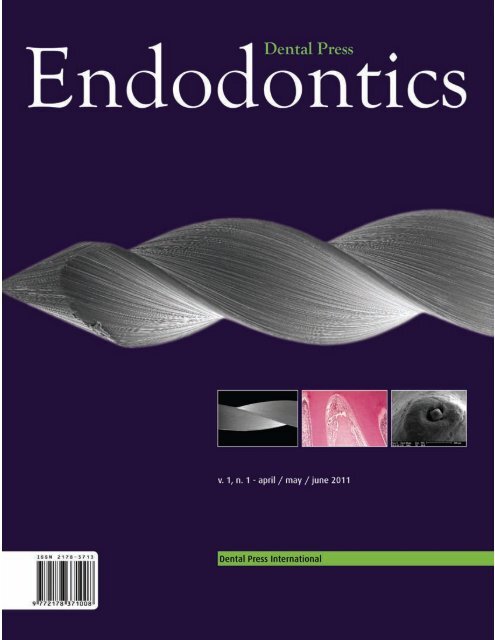
Dental Press
Dental Press
Dental Press
Create successful ePaper yourself
Turn your PDF publications into a flip-book with our unique Google optimized e-Paper software.
editorialRevolution in scientific informationMankind is experiencing constant change, whichcauses direct repercussions in its essence. The industrialrevolution was a remarkable event. The societywitnesses, at the present time, the revolution of informationin different segments. The speed and the wayin which this information has been promoted is fantastic.The scientific globalization encourages the differentlevels of an academic structure.The challenge of the moment requires a careful selectionof storage and a proper interpretation. Brazilis experiencing a very favorable moment in science,within which we can mention its quality and its acceptanceby the international community. Moreover,the stimulus to the development of a major project, tothe entire community that brings together endodonticsas a magna specialty, is born from a careful andwell structured programming. The realization of thisproject came from the opportunity afforded by Dr.Laurindo Furquim, publisher of <strong>Dental</strong> <strong>Press</strong>, with thecreation of the <strong>Dental</strong> <strong>Press</strong> Endodontics.Therefore, the challenge of disseminating endodonticscience is launched with the creation ofthe <strong>Dental</strong> <strong>Press</strong> Endodontics, which is composedby a team of renowned professors, researchers andspecialists in endodontics in Brazil and internationally.Endodontic scientific information certainly willhave a new vehicle facilitator and promoter, ableto improve clinical decisions supported by scientificevidence. The <strong>Dental</strong> <strong>Press</strong> Endodontics allow thereader to renew concepts and experience the revolutionin scientific informations.Carlos EstrelaEditor-in-chief© 2011 <strong>Dental</strong> <strong>Press</strong> Endodontics 3 <strong>Dental</strong> <strong>Press</strong> Endod. 2011 apr-june;1(1):3
contentsEndo in Endo14. Orthodontic treatment does not causepulpal necrosisAlberto ConsolaroOriginal articles21. A NiTi rotary instrument manufacturedby twisting: morphology and mechanicalpropertiesVictor Talarico Leal VieiraCarlos Nelson EliasHélio Pereira LopesEdson Jorge Lima MoreiraLetícia Chaves de Souza28. Effect of intracanal posts on dimensions ofcone beam computed tomography images ofendodontically treated teethCarlos EstrelaMike Reis BuenoJulio Almeida SilvaOlavo César Lyra PortoClaudio Rodrigues LelesBruno Correa Azevedo37. Efficacy of chemo-mechanical preparationwith different substances and the use of a rootcanal medication in dog’s teeth with inducedperiapical lesionFrederico C. MartinhoLuciano T. A. CintraAlexandre A. ZaiaCaio C. R. FerrazJosé F. A. AlmeidaBrenda P. F. A. Gomes46. In vitro determination of direct antimicrobialeffect of calcium hydroxide associated withdifferent substances against Enterococcusfaecalis strainsPaulo Henrique WeckwerthNatália Bernecoli SiquinelliAna Carolina Villas Bôas WeckwerthRodrigo Ricci VivanMarco Antonio Hungaro Duarte52. Analysis of forces developed during root canalfilling by different operatorsMaria Rosa Felix de Sousa G. GuimarãesHenner Alberto GomideMaria Antonieta Veloso C. de OliveiraJoão Carlos Gabrielli Biffi58. Root canal filling with calcium hydroxide pasteusing Lentullo spiral at different speedsMarili Doro DeonízioGilson Blitzkow SydneyAntonio BatistaCarlos Estrela
64. Location of the apical foramen and itsrelationship with foraminal file sizeRonaldo Araújo SouzaJosé Antônio Poli de FigueiredoSuely ColomboJoão da Costa Pinto DantasMaurício LagoJesus Djalma Pécora82. SEM and microbiological analysis of dirtof endodontic files after clinical use and itsinfluence on sterilization processMatheus Albino SouzaMárcio Luiz Fonseca MeninFrancisco MontagnerDoglas CecchinAna Paula Farina69. In vitro evaluation of shape changes in curvedartificial root canals prepared with two rotarysystemsBenito André Silveira MiranziAlmir José Silveira MiranziLuis Henrique BorgesMário Alfredo Silveira MiranziFernando Carlos Hueb MenezesRinaldo MattarThiago Assunção ValentinoCarlos Eduardo Silveira BuenoCase report87. Influence of cone beam computed tomographyon dens invaginatus treatment planningDaniel de Almeida DecurcioJulio Almeida SilvaRafael de Almeida DecurcioRicardo Gariba SilvaJesus Djalma Pécora77. The persistence of different calcium hydroxidepaste medications in root canals: an SEM studyHélio Katsuya OnodaGerson Hiroshi YoshinariKey Fabiano Souza PereiraÂngela Antonia Sanches Tardivo DelbenPaulo ZárateDanilo Mathias Zanello Guerisoli
Aquarium TM 2Case presentation softwareShow it. Share it.LingualBracketsTeethEruptionForsus ApplianceTAD Canine RetractionMandibular Advancement SurgeryExport movies to other programsIntuitive Interface • Stunning 3D Movies • Comprehensive Library •Personalized Images • Network-Ready • Export MoviesThe second generation of Aquarium brings greatly expanded content andcapabilities. New movies such as 3rd Molar Extraction, Lip Bumper, and LingualBraces make this interactive patient education software more relevant than ever.Record your own audio, export media, enlarge interface for easy viewing, andpersonalize your program with thematic skins. Aquarium movies are networkreadyand display beautifully on most monitors and resolutions. To learn more,visit www.renovatio3.com.br or contact us at comercial@renovatio3.com.br,fone: +55 11 3286-0300.© 2010 Dolphin Imaging & Management Solutions
Endo in EndoOrthodontic treatment does not cause pulpalnecrosisAlberto consolaroProfessor, Bauru <strong>Dental</strong> School (USP) and Postgraduate Professor of Ribeirão Preto <strong>Dental</strong>School (USP).Consolaro A. Orthodontic treatment does not cause pulpal necrosis. <strong>Dental</strong> <strong>Press</strong>Endod. 2011 apr-june;1(1):14-20.IntroductionThe dental pulp has an arborized vascular systemand its only blood source is represented by a delicateartery that penetrates the apical foramen. Seldomthere is a vascular communication of the pulp withthe periodontal ligament through the lateral canalsand accessories from the lateral and apical foramina.Connective tissue has many functions such asfilling in the spaces between the organs, ducts andother structures. Another important function of connectivetissue is the support of specialized cells inorgans such as liver, kidney, pancreas and glands. Inthese organs, the connective tissue support is calledstroma and the specialized cells parenchyma. Theconnective tissue can, besides filling and supporting,assume very specialized roles as in the dental pulp,which provides sensitivity and form dentin.There are different kinds of connective tissuessuch as fibrous, bone, cartilage, adipose and others.They are the only vascularized tissues and they getblood to nourish and keep the cells alive and functional.Just as the vessels, there is a conjunctive plotof neural threads.During compression and massage of soft tissuesof the body, major shifts in centimeters can occurwithout breakdown of connective tissue structures,especially of vessels and nerves. This elasticity of theconnective tissue is caused by the presence of collagenand elastic fibers in the extracellular matrix,especially when subjected to forces applied graduallyand slowly. When there are sudden movementsof conjunctive structures, there may be disruptionsof vessels and nerves. When this occurs bruises areformed clinically characterized by reddish-purplespots. These hematomas can occur after pinching,biting and hitting, i.e., sudden traumatic and intenseactions on the fibrous connective tissues.<strong>Dental</strong> traumatism, occlusal trauma andorthodontic movement: are different in theireffect the in periodontal tissueAlthough the terms traumatism and trauma accountfor the deleterious action of physical agents,as forces on the tissues, their characteristics are notalways equal in intensity and frequency. The dentaltraumatism can break vessels and promote ruptures,characterized by the application of sudden and intenseforces on the teeth. Differently from the occlusaltrauma, in which the forces are intense, smallextension, short duration, but constantly repeated.On the other hand, the forces of tooth movement arevery light, even the most intense and prolonged onesslowly applied on the teeth, so that they graduallydisappear within hours or days. In short, althoughthey are caused by forces, dental traumatism, occlusaltrauma and orthodontic movement are not alikeregarding the characteristics of the forces appliedand their effects on connective tissues.In dental traumatism the forces act abruptly withrupture of connective structures, including vesselsand nerves. When a force apparently light acts onthe tooth, depending on its angle of incidence and© 2011 <strong>Dental</strong> <strong>Press</strong> Endodontics 14<strong>Dental</strong> <strong>Press</strong> Endod. 2011 apr-june;1(1):14-20
Consolaro Alocation in which it acted, there may be a resultant offorces in the apical third of the tooth root with ruptureof the vascular and nerve bundle that enters the pulp.An example of dental traumatism is the concussion,with no clinically detected mobility and pain, if any,is easily controlled with common analgesics, lastingseveral hours or even 2 to 3 days. 2,3 Apparently, thetooth gets back to normal, but within time the pulpmay show its damage with the presence of calciummetamorphosis of the pulp or pulp aseptic necrosis,both clinically revealed by coronary darkening in anapparently healthy tooth.In occlusal trauma the death of cell and the structuralrupture are minimized by the quick length andrepetitiveness of the process, although it is for a longtime. In this case, there is no structural damage tovascular and nervous bundle of pulp, nor fast agingof the pulp. The periodontal lesions are light andsubtle. The periodontal structure must be acknowledgedas an example of an organization to receivethe strong forces of chewing. The periodontal fibersare organized in a space with an average thicknessof 0.25 mm, but even so during chewing the teeth donot touch the bone.In the orthodontic movement the forces appliedto the tooth structure, even the most intense, graduallydisappear in the surrounding tissues. The plasticityof the connective tissue of the periodontal ligament,plus the deflection capacity of the bone crestand the rotation that happens in the tooth socketpromote a slow and gradual adaptation of the surroundingtissues. The orthodontic movement is limitedto a maximum of 0.9 mm at the crown during thefirst hours 1 providing no conditions for the structuralrupture of vessels and nerves to happen.There should not be a comparison among thetissue effects induced by dental traumatism, occlusaland orthodontic movement, as they are differentsituations. In the apical third of root the inducedorthodontic tooth movement is confined practicallyto the compression of the periodontal ligament,because the bone deflection in the periapical boneis much smaller and the tooth hinge axis is nearthe apex. The forces are absorbed and dissipatedslowly, without rupturing vessels. Small movementsare naturally absorbed by fibrous and elastic connectivetissue.Another important information concerns the durationin which the orthodontic forces are active: 2 to4 days. After this time these forces are dissipated andthe reorganization of the periodontal structures beginswith resorption of the periodontal bone surface,cell migration for reorganization with the productionof new collagen fibers. 1 After 15 to 21 days the periodontalligament and other structures are ready fora new cycle of events by the reactivation of the orthodonticappliance. In other words, the induced toothmovement is achieved in cycles of 15 to 21 days, thetooth does not move all the time. In the orthodonticmovement forces are mitigated by the collagen andelastic fibers, without damaging the structures thatcarry blood and sensitivity to the pulp.With each activation period of orthodontic appliances— from 15 to 21 days — the periodontaltissues reorganize themselves and return to normal.The ultimate effects of orthodontic treatment on thestructures and position of teeth are the sum of all cyclesfrom 15 to 21 days. The forces and the effectswere not continuous and unceasing. Sometimes thequestion is: when there is rotation of the tooth aroundits long axis, as in giroversion, vascular and nervebundles get twisted around themselves, does it notcompromise the blood supply to the pulp? No, theyare not twisted, because the tissues reorganize themselvesin each period of 15 to 21 days, they return totheir normal position and relationship. When the newcycle of movement is established by a new activation,the vessels and nerves are in normal relationship withno change in their shape. Tissues constantly renewits structures, remodel and adapt themselves well tonew positions and structural relationships.Consolaro, 4 in his investigation of Masters in 2005,and Massaro et al 9 in 2009, examined microscopicallythe pulp of 49 first molars of rats under inducedtooth movement after 1, 2, 3, 4, 5, 6 and 7 days. Resorptionwas detected in the external surfaces of theroot, indicating the efficiency of the applied forces.However, no morphological changes was detectedin the pulp tissues (Figs 1-6).Synopsis for endodontists of the inducedtooth movement, or does intense force increasethe chance of pulpal necrosis by orthodonticmovement?© 2011 <strong>Dental</strong> <strong>Press</strong> Endodontics 15<strong>Dental</strong> <strong>Press</strong> Endod. 2011 apr-june;1(1):14-20
[ endo in endo ]Orthodontic treatment does not cause pulpal necrosisGDPPLABP D C PL ABP C ABFigure 1. Rat’s molar 7 days after been moved. P = pulp, D = dentin, C= cementum; PL = periodontal ligament, AB =alveolar bone, G = gum.(HE; 4X).Figure 2. Area of compression of the periodontal ligament (PL) of therat’s molar 7 days after been moved. The arrow indicates the direction ofthe applied force and the narrowing of the periodontal space. Despite thecompression of periodontal ligament, cells and fibers are present in thearea, as well as cementoblasts, osteoblasts and also the clasts (circles).The morphological pattern of normal dental pulp is highlighted (P). D =dentin, C = cementum; AB = alveolar bone. (HE, 25X).The orthodontic forces compress a certain segmentof the periodontal ligament, because the teethare bent on the alveolar bone crest or on the apicalthird on the opposite side (Figs 1 and 2). The compressedblood vessels reduce the amount of blood tothe cells of that local: they momentarily stop the productionand renewal of the extracellular matrix, includingcollagen; and get disorganized (Figs 3, 4 and 5).In some cases the cells migrate to surrounding areasstill vascularized. Only the extracellular matrixin some areas that have been strongly affected byhypoxia remains in the local. These areas turn intoa glassy aspect to the optical microscope and are,therefore, called hyaline areas (Fig 6).In this segment of the compressed periodontal ligamentand with reduction of blood support, there will© 2011 <strong>Dental</strong> <strong>Press</strong> Endodontics 16<strong>Dental</strong> <strong>Press</strong> Endod. 2011 apr-june;1(1):14-20
Consolaro AP D C PL ABFigure 3. Area of compression of the periodontal ligament (PL) of the rat’smolar 7 days after been moved. The larger arrow indicates the directionof the applied force. The small arrows indicate the cementoblasts, whichare absent in the area of pressure, indicating efficiency of the applied force.Despite the compression of periodontal ligament, cells and fibers arepresent in the area, as well as cementoblasts, osteoblasts and also theclasts (circles). It is important to notice the morphological pattern of normaldental pulp (P). D = dentin, C = cementum; AB = alveolar bone. (HE, 25X).Pbe an increased local production of cellular mediatorsproduced by metabolic stress and by the mild inducedinflammation. The periodontal ligament is alive, metabolicallyviable, with blood supply and with clasts sufficientlyactivated to resorb the periodontal bone surfaceof the tooth socket (Fig 5). The periodontal boneresorption occurs in front of the compressed periodontalligament and therefore it is nominated frontalbone resorption (Fig 5). Gradually, over few days, thetooth will be displaced to one side of the tooth socket,reoccupying its new place, and ligament cells restorethe average thickness of 0.25 mm. In the process, especiallyin the apical region, vascular rupture does notoccur in tissues that enter into the root canal. Fromthis normal restored relationship, the periodontal ligamentand surrounding tissues will be reorganized in afew days. After 15 to 21 days it is ready to reactivatethe appliance as the tissues return to normal.CTCCPDCTCTPDPLABACBFigure 4. A) Area of compression of the periodontal ligament (PL) of rat’s molar 7 days after been moved. The arrow indicates the direction of theapplied force. The clasts (CT) in the root surface indicate efficiency of the applied force. In B, there is the morphological pattern of normal dental pulpwith odontoblastic layer (small arrows). D = dentin, C = cementum; AB = alveolar bone. (HE, 40X).© 2011 <strong>Dental</strong> <strong>Press</strong> Endodontics 17<strong>Dental</strong> <strong>Press</strong> Endod. 2011 apr-june;1(1):14-20
[ endo in endo ]Orthodontic treatment does not cause pulpal necrosisThe displacement of the root apex is very smalland slow, the connective tissue is elastic enough towithstand much larger displacements. Besides havingelastic fibers, the extracellular matrix of connectivetissue display a gel between the cells and fibers,damping forces and applied displacements, withoutcell death and vascular rupture (Figs 1 and 2).When a very intense force, as the one applied tothe teeth that act as support for jaw expander appliance,acts on the tooth there will not be an effectivemovement of the tooth in the tooth socket. A veryintense force collapses the blood vessels, interruptsnormal vascularization in that periodontal segment.The local cells die, or, more often, flee to surroundingareas, including inflammatory and clast cells (Fig 6).Without blood supply there will be no cell activity inthe periodontal surface of the alveolar bone. That is,the compressed periodontal segment gets hyaline inthese conditions and without any cell activity (Fig 6).When the vascularization is restored due to thegradual dissipation of excessive force applied, theneighboring cells will change from center to the periphery,resorbing and remodeling the hyalinized areaof the periodontal ligament. Therefore, the tooth willPLCHHD C PL ABPDHABPLFigure 5. Area of compression of the periodontal ligament (PL) ofthe rat’s molar 6 days after been moved with typical frontal boneresorption. The larger arrow indicates the direction of the appliedforce. The small arrows indicate the cementoblasts. Despite thecompression of periodontal ligament, cells and fibers are present inthe area, as well as cementoblasts, osteoblasts and clasts (circles).D = dentin, C = cementum; AB = alveolar bone. (HE, 25X).Figure 6. Classic bone resorption at distance in the area of compressionof the periodontal ligament (PL) of the rat’s molar 3 days after been moved.The arrow indicates the direction of applied force and the narrowing of theperiodontal space. The area of compression of the periodontal ligamentwas hyalinized (H) without osteoblasts and cementoblasts. The clasts(circles) act at a distance from the compression area of the periodontalligament (PL). The morphological pattern of normal dental pulp ishighlighted (P). D = dentin, C = cementum; AB = alveolar bone. (HE, 25X).© 2011 <strong>Dental</strong> <strong>Press</strong> Endodontics 18<strong>Dental</strong> <strong>Press</strong> Endod. 2011 apr-june;1(1):14-20
Consolaro Anot move because the clasts are not in metabolicconditions of nutrition and with no metabolism to actin the periodontal surface of the alveolar bone. Theremodeling process of bone and hyaline area will bedone from the periphery to the center, including theunderlying part of the alveolar bone plate (Fig 6).The bone resorption process and reorganizationthat should take place in front of the compressionof the periodontal ligament, will take place at distance:bone resorption at distance, but it is undesirable(Fig 6). In this case, the tooth did not move neitherdisplaced minimally, thus can not have broken thevascular and nerve bundle.This explanation helps to understand why theteeth that act as anchoring for palatal expansion appliances,the strongest possible force to be appliedto a tooth, do not suffer necrosis neither pulp aging.In short, the more intense the orthodontic force appliedis, the smaller the chance of the tooth to move inits socket; and as consequence, there is no way to inferassociated pulp necrosis. Valadares Neto, 13 in his masterresearch in 2000 with me as advisor, analyzedthe effects of rapid maxillary expansion in the dentin-pulpcomplex in 12 adolescents. Using deviceslike Hass, he examined microscopically the entirelength of the pulp and dentin of 12 premolars rightafter the removal of the appliance with the expansionof the jaw established and other 12 premolarsafter 120 days from the removal of appliances. Other6 premolars of adolescents that did not undergoany orthodontic and/or orthopedic procedure wereused as control group. In every analyzed teeth thepulp-dentin complex was fully normal, without anymicroscopically detectable change.And when the pulp necrosis is diagnosedin sound teeth during the orthodontic treatment?Based on the above explanations, it is perfectlypossible to understand why orthodontic treatmentdoes not induce pulp necrosis nor accelerates itsaging. In all the cases in which pulp necrosis is detectedduring orthodontic treatment, the history ofdental traumatism must be recalled. Patients do notremember those concussions and small dental injuriesin children, but they occur daily. Small strokes,bump and home accidents can be seemingly innocent,but by concentrating forces at the apex theymay cause sudden and small displacements withrupture in the pulp vascular bundle. In many casesof dental traumatism, no coronary nor gingival damageor bleeding occur, but there may be aseptic pulpnecrosis. In some dental traumatism, there may besevere gingival damage and heavy bleeding, butwithout breaking the pulp vascular bundle.<strong>Dental</strong> concussion can also occur in the followingsituations: teeth that act as levers to support theextraction of adjacent teeth, small forceps beats inthe extraction of third molars, unerupted and pulledcanines luxation, laryngoscope trans-operative beatsduring general anesthesia, or even accidental bitesin cutlery, seeds or strange materials during feeding.There is no clinical, laboratorial or experimentalevidence to assign, although theoretically, the pulpalnecrosis as a result of orthodontic movement. 5-8,10,11,12When facing a situation like this, try to recall the historyof dental traumatism and do not assign pulpalnecrosis to the orthodontic movement.Final Considerations1. The aseptic pulp necrosis cannot be attributedclinically and experimentally to orthodonticmovement.2. In cases of pulpal necrosis during orthodontictreatment, the history of dental traumatismshould be researched, especially the lightertypes, such as concussion.3. In cases of very strong forces used in orthodonticand orthopedic treatment, tooth movementdoes not occur and displacement with ruptureof the pulp vascular bundle has even lesschance of happening.4. <strong>Dental</strong> traumatism, orthodontic tooth movementand occlusal trauma situations are totallydifferent from each other, although they arephysical events on the tissues. The biological effectsin each of these three situations are differentand specific and therefore not comparable.© 2011 <strong>Dental</strong> <strong>Press</strong> Endodontics 19<strong>Dental</strong> <strong>Press</strong> Endod. 2011 apr-june;1(1):14-20
[ endo in endo ]Orthodontic treatment does not cause pulpal necrosisReferences1. Consolaro A. Reabsorções dentárias nas especialidadesclínicas. 2ª ed. Maringá: <strong>Dental</strong> <strong>Press</strong>; 2005.2. Consolaro A. Inflamação e reparo. Maringá: <strong>Dental</strong> <strong>Press</strong>;2010.3. Consolaro A, Consolaro MFM-O. Controvérsias na Ortodontiae atlas de Biologia da movimentação dentária. Maringá: <strong>Dental</strong><strong>Press</strong>; 2008.4. Consolaro RB. Análise do complexo dentinopulpar em dentessubmetidos à movimentação dentária induzida em ratos[dissertação]. Bauru: Universidade de São Paulo; 2005.5. Derringer KA, Jaggers DC, Linden RW. Angiogenesis in humandental pulp following orthodontic tooth movement. J Dent Res.1996;75(10):1761-6.6. Derringer KA, Linden RW. Epidermal growth factor released inhuman dental pulp following orthodontic force. Eur J Orthod.2007;29(1):67-71.7. Grünheid T, Morbach BA, Zentner A. Pulpal cellular reactions toexperimental tooth movement in rats. Oral Surg Oral Med OralPathol Oral Radiol Endod. 2007;104(3):434-41.8. Junqueira LC, Carneiro J. Histologia básica. 11 a ed. Rio deJaneiro: Guanabara Koogan, 2008. 524 p.9. Massaro CS, Consolaro RB, Santamaria M Jr, Consolaro MF,Consolaro A. Analysis of the dentin-pulp complex in teethsubmitted to orthodontic movement in rats. J Appl Oral Sci.2009;17(sp. issue):35-42.10. Osborn JW, Ten Cate AR. Histologia dental avançada. 4 a ed.São Paulo: Quintessence; 1988.11. Pissiotis A, Vanderas AP, Papagiannoulis L. Longitudinal studyon types of injury, complications and treatment in permanenttraumatized teeth with single and multiple dental traumaepisodes. Dent Traumatol. 2001;23(4):222-5.12. Santamaria M Jr, Milagres D, Stuani AS, Stuani MBS, RuellasACO. Pulpal vasculature changes in tooth movement. Eur JOrthod. 2006;28(3):217-20.13. Valladares J Neto. Análise microscópica do complexodentinopulpar e da superfície radicular externa após aexpansão rápida da maxila em adolescentes [dissertação].Goiânia: Universidade Federal de Goiás; 2000.Contact address: Alberto ConsolaroE-mail: consolaro@uol.com.br© 2011 <strong>Dental</strong> <strong>Press</strong> Endodontics 20<strong>Dental</strong> <strong>Press</strong> Endod. 2011 apr-june;1(1):14-20
original articleA NiTi rotary instrument manufactured by twisting:morphology and mechanical propertiesVictor Talarico Leal Vieira, DDS, MSc 1Carlos Nelson Elias, MSc, PhD 1Hélio Pereira Lopes, PhD 2Edson Jorge Lima Moreira, DDS, MSc, PhD 3Letícia Chaves de Souza, DDS, MSc 1abstractObjectives: The surface morphology of TF ® endodonticinstruments was studied using stereomicroscopy and scanningelectron microscopy (SEM). Mechanical tests weredone for flexibility and microhardness. Methods: Fourtapers of TF ® files were used (0.04; 0.06; 0.08 and 0.10mm/mm). The stereomicroscopy associated with the AxioVision® program was used to measure the tip angle, thehelical angle, the taper and the tip diameter of the instruments.SEM was used to identify surface defects due to machiningand finishing. The flexibility and the microhardnesswere measured with bending and microhardness Vickerstests, respectively. Results and Conclusion: The analysisshowed that the manufacturer complied with the valuesrecommended by the ANSI/ADA standard number 28. TheSEM results showed many surface defects and a distortionof the instrument helix. It was observed that the instrumentflexibility changes with its taper. The forces to induce thephase transformation by stress on instruments with taper0.04; 0.06 and 0.08 mm/mm were 100 gf, 150 gf and 250gf, respectively. The values of Vickers microhardness of theinstruments are compatible with rotary instruments manufacturedby the machining process.Keywords: Endodontic instruments. NiTi alloy. R-phase.Materials characterization. Mechanical tests. NiTi manufacturingmethods.Vieira VTL, Elias CN, Lopes HP, Moreira EJL, Souza LC. A NiTi rotary instrument manufactured by twisting: morphology and mechanical properties. <strong>Dental</strong> <strong>Press</strong> Endod.2011 apr-june;1(1):21-7.1Department of Mechanical Engineering and of Materials, Military Institute of Engineering, Rio de Received: January 2011 / Accepted: February 2011Janeiro, RJ, Brazil.2Department of Endodontics, Estácio de Sá University, Rio de Janeiro/RJ, Brazil.3Department of Endodontics, UNIGRANRIO, Grande Rio University, Rio de Janeiro, RJ, Brazil. Correspondence address: Victor Talarico Leal VieiraRua Engenheiro Coelho Cintra 25/101, Ilha do GovernadorRio de Janeiro, RJ, Brazil - Zip Code: 21.920-420Email: victortalarico@yahoo.com.br.© 2011 <strong>Dental</strong> <strong>Press</strong> Endodontics 21<strong>Dental</strong> <strong>Press</strong> Endod. 2011 apr-june;1(1):21-7
[ original article ] A NiTi rotary instrument manufactured by twisting: morphology and mechanical propertiesIntroductionIn 1988, Walia et al 1 used a new metal alloy to manufactureendodontic instruments, the NiTi alloy. The instrumentsproduced with this alloy had a lower Youngmodulus than the instruments made with stainless steel,thus allowing the endodontic treatment of cases withlarge root curvatures. The use of instruments made withstainless steel could make the treatment more difficult.The first endodontic instruments of NiTi where manufacturedby a machining process using burs. With thedevelopment of new NiTi alloys, the study of the mechanismsinvolved in the phase transformation and bettercontrol of the microstructure, it was possible to developa new manufacturing method based on twisting. TheTF ® instruments (Twisted Files, California - USA) aremanufactured by twisting. This new generation of instrumentshas better clinical properties.In the present work the surface morphology ofendodontic instruments manufactured by twistingwas investigated and microhardness and flexibilitymeasurements were performed. These properties areimportant to understand the clinical behavior and todevelop new instruments.Materials and methodsMorphologyThe TF ® endodontic instruments (Twisted Files,California) used in this study has a length of 27 mm, atip diameter (D o) of 25 mm. Three different tapers wereused (0.04, 0.06 and 0.08 mm/mm).The tip angle, the tip length and the taper were determinedwith an optic microscope Zeiss with a pixeLINKcamera model PL- a662 and a light source Zeiss 1500LCD. The taper was determined with an amplificationof 1.6X. The other dimensions were quantified with anamplification of 5X. All dimensions of the instrumentswere determined with the program AxioVison 4.4 ® .Five instruments with each taper were investigated.Bending tests (at 45º)The bending tests were performed with an apparatusconnected to a universal material testing system EMICDL1000 (EMIC Equipment, Brazil). A 20 N load cellwas used to measure the force necessary to bend thetip of the instruments by 10 o , 20 o , 30 o and 45 o . The testswere performed according to ADA standard 28, withthe force applied 3 mm from the tip of the instrument.Vickers MicrohardnessFor microhardness testing, the instruments wereembedded in epoxy resin. The fixation cable was parallelto the recipient base with the purpose of keepingthe central longitudinal surface outside of the resin afterpolishing. The instruments were prepared with sandpaper200, 300, 400, 600 and 1200 and polished with aluminaparticles of 0.5 µm.The Vickers indentations were made with 100 gf during15 s using a microdurometer Bhueler model 1600-5300. Five indentations were made in the working partand five in the neck of each specimen.Scanning electron microscopy (SEM)Two instruments of each taper were submitted toSEM (JEOL, LSM 5800LV) to evaluate the morphologiesof the cutting edge, the tip and interface of the neckregion with the fixation cable.Statistical analysisThe data of the bending tests and the Vickers microhardnesswere analyzed statistically by the Kruskal-Wallis method and complemented with the Student-Newman-Keuls multiple comparison test to comparethe tapers. The microhardness at the neck region wascompared applying the Mann-Whitney test. The levelof significance of all analyses was 5%.ResultsThe results of the statistical analysis are shown inTables 1 and 2. The bending testing results are shown inTable 3. Figure 1 shows a mean curve obtained from 10bending tests performed in instruments with taper 0.06.The tests for other tapers showed similar curves.The curves show a slope change that is attributed toa phase transformation. The values of the forces necessaryto bend the instruments by 10 o , 20 o , 30 o and 45 o areshown in Table 2 and the forces necessary to inducephase transformation by stress are shown in Table 3.Statistical analysis (Kruskal-Wallis test) demonstratedthat there was a significant difference between instrumentswith different tapers (P < 0.00001). Then, the Student-Newman-Keulsmultiple comparison test revealedthat the instrument of taper 0.04mm/mm is more flexiblethan instruments of tapers 0.06 and 0.08 mm/mm. Moreover,the instrument of taper 0.06 mm/mm proved to bemore flexible than the instrument of taper 0.08 mm/mm.© 2011 <strong>Dental</strong> <strong>Press</strong> Endodontics 22<strong>Dental</strong> <strong>Press</strong> Endod. 2011 apr-june;1(1):21-7
Vieira VTL, Elias CN, Lopes HP, Moreira EJL, Souza LCThe Vickers microhardness average values at theneck region and at the working region of the instrumentsare shown in Table 4.The Vickers microhardness results for each taperwere submitted to the Mann-Whitney test and therewas no significant difference between the values in theneck region and in the working region for all instruments(p> 0.05).Table 1. Tip angle, Tip length (L) and taper of the instruments.Instrument Taper 0.04 Taper 0.06 Taper 0.08 Taper 0.10Tip angle 26.56 + 4.39 32.41 + 7.59 32.39 + 13.89 25.48 + 4.92L (mm) 0.24 + 0.011 0.25 + 0.013 0.24 + 0.007 0.26 + 0.012Taper 0.039 + 0.0029 0.061+ 0.0016 0.077 + 0.001 0.099 + 0.0022Table 2. Average values of the maximum forces to bend at 45º (gf) and respective standard deviations.Instrument Taper 0.04 Taper 0.06 Taper 0.0810 o 67.82 + 7.02 130.7 + 17.21 179.7 + 20.6220 o 92.26 + 4.36 183.9 + 16.17 295.2 + 26.2730 o 120.3 + 7.27 247.5 + 20.61 390.3 + 23.1545 o 131.7 + 9.43 263.6 + 23.18 400.7 + 23.88Table 3. Average forces for phase transformation by stress.Instrument TF 0.04 mm/mm TF 0.06 mm/mm TF 0.08 mm/mmAverage force 100 gf 150 gf 250 gfTable 4. Vickers microhardness of the instruments.Instrument HV neck region HV working region0.06 272.4 + 31.6 291.2 + 240.08 292.8 + 33.8 293 + 170.10 315.5 + 33.7 279 + 10.72.52Figure 1. Mean curve for taper 0.06 mm/mm TF ®files. The red line represents the elastic region, thegreen line phase represents the transformationregion and the dashed line the superelastic region.Force/100 (gf)1.510.50Elastic zonePhasetransformationzoneSuperelasticzone0 5 10 15 20Strain (mm)© 2011 <strong>Dental</strong> <strong>Press</strong> Endodontics 23<strong>Dental</strong> <strong>Press</strong> Endod. 2011 apr-june;1(1):21-7
[ original article ] A NiTi rotary instrument manufactured by twisting: morphology and mechanical propertiesThe microhardness values were also analyzed bythe Kruskal-Wallis test. The statistical analysis confirmedthat there was no significant difference amongthe groups (p=0.658). It is possible to conclude that theVickers microhardness is independent of the taper andinstrument region tested.Surface analysis showed manufacturing defects in allinstruments analyzed (Fig 2).Figure 2 shows grooves produced in manufacturingprocess. It is possible to see the drawing toolmarks along the longitudinal direction. All the sampleshad microcavities.100 µm100 µm100 µm50 µmFigure 2. TF ® instrument taper 0.04mm/mm images. A) Lateral cuttingedge showing shavings (magnification of 60×). B) Curvature at theedge inherent to the manufacturing process (magnification of 100×).C) Pores present at the instrument magnification of 500×). D) Presenceof burs (magnification of 300×). E) Junction of neck region to cable(magnification of 27×).500 µm© 2011 <strong>Dental</strong> <strong>Press</strong> Endodontics 24<strong>Dental</strong> <strong>Press</strong> Endod. 2011 apr-june;1(1):21-7
Vieira VTL, Elias CN, Lopes HP, Moreira EJL, Souza LCDiscussionIs important that the instrument tip has a goodfinishing and a transition angle that permits the introductionin the root canal. Small angles (less than 33º)can generate steps or deviations. The TF ® instrumentshave a progressive tip angle that allows introductionin regions with a substantial curvature without deformingthe canal, thus promoting a safe enlargement. Theround tip can be classified as smooth. 2 Due this configuration,it is likely that the instrument will not causedamage to the root.The instrument dimension D ois determinedby the diameter at the base of the tip basis, whichserves as a reference during introduction. The valueof D oand the taper permits to determine the workdiameter in a given canal region. A simple calculationcan be used to determine what instruments canbe used in sequence to perform an effective work.The diameter values of the tip bases (D o) found onthis study meet the ANSI/ADA standard number 28recommendation. In the present work, we observedthat the tip angles conform to the standard recommendations.According to Thompson 3 the phase transformationof NiTi alloys does not show macroscopic changeswhen the application of an external force changes themicrostructure. The bending tests showed a changein slope after an initial linear increase that resembledthe Hooke law. This change is attributed to an austenite-to-martensitephase transformation. This slopechange is in agreement with the results reported byThompson 3 . In the beginning the material is in theelastic region, at the end the material is in the superelasticregion and between the two regions the materialin the phase transformation region.In this study, we plotted the relation between theforce and the strain. Thompson 3 probably used awire in his experiments, so it was easy to calculatethe stress (σ), using the area of the specimen. In ourcase, since the shape of the file is very complex, it isimpossible to calculate the stress with any accuracy.However, since the stress is proportional to the force(σ = F/A), the shape of the curve is the same.Thompson 3 mentioned that the preparation of theroot canal promotes the martensite transformation bystress of NiTi alloy instruments. The stress level at whichthe phase transformation happens is not mentioned bythe authors, and was found by us to be different for eachinstrument taper. This is an important information forfuture studies and for clinical practice.According to Schäfer et al, 4 the cross section isthe main factor that affects the bending tests. This isreasonable, since that a larger area implies a largervolume of metal at the core of the instrument. In thepresent study, a factor that influenced the maximumforce to bend the instruments to 45º was the taper. Thetaper has the same influence as the cross section, forthe same reason. If the tip diameter is kept constant,a larger taper will promote less flexibility, as was observedin the tests.According to Miyai et al 5 and Hayashi et al, 6 the instrumentflexibility is influenced by a phase transformation.The R-phase or rhombohedral has a large memoryform effect and the Young modulus is lower than thatof austenite, so an instrument that goes through a martensitictransformation will be more flexible.Yahata et al 7 used the same scheme proposed byMiyai et al 5 to study the flexibility of annealed samples.However, differently from this study, the authors did notmeasure the average force for phase transformationwhen the instrument is submitted to stress.Other values found in this study were compatiblewith NiTi alloys. Lopes et al 8 found average values of345 HV in NiTi instruments (Files NiTi-flex). Serene etal 9 found values between 303 and 362 HV for the microhardnessof NiTi alloys used in the manufacture ofendodontic instruments. The average value found in thethe present work was 289 HV. This value is consistentwith others from the literature.In this work it was observed that the manufacturingprocess did not change the Vickers microhardness,probably because of the thermal treatment, that couldbe lower than the temperature of recrystalization proposedby Kuhn and Jordan. 10According to the manufacturer, 11 the absence ofother metal at the fixation cables avoids galvanic corrosion.The instrument is really formed by only one piece.However, galvanic corrosion should not be an importantproblem because of the low life in cycle at the clinic.It will be important only if the instrument remains instock for a long period of time in adverse conditions.According to Kim et al, 12 the TF ® instrumentspresent a significant resistance to fracture by rotating-bendingfatigue when compared with others© 2011 <strong>Dental</strong> <strong>Press</strong> Endodontics 25<strong>Dental</strong> <strong>Press</strong> Endod. 2011 apr-june;1(1):21-7
[ original article ] A NiTi rotary instrument manufactured by twisting: morphology and mechanical propertiesinstruments manufactured by the machining process,corroborating the results obtained by Gambarini etal 13 and Larsen et al. 14 This can be explained by thefact that machining produces perpendicular defectsthat favor nucleation and propagation of cracks.Even presenting good results in flexion-bendingfatigue tests, the TF ® files should have a better surfacefinishing, that would improve the clinical performanceconcerning durability in relation to thefracture. The surface morphology found at this workwas very similar to that found by Kim et al. 12 Despitethe eletropolishing, the surface is not completely flatand has machining marks from the manufacturingprocess. This observation corroborates the resultsof that study.ConclusionsBased on the results we concluded that:a) the dimensions of the TF ® files meet the ANSI/ADA standard number 28 recommendations;b) the files present many defects from the manufacturingprocess;c) the instrument flexibility decreased with increasingtaper;d) the phase transformation induced by stress averageforces to the TF ® files of taper 0.04; 0.06 and0.08 mm/mm where 100 gf, 150 gf and 250 gf,respectively, ande) the TF ® Vickers microhardness values were similarto those of NiTi rotary instruments manufacturedby the machining process.© 2011 <strong>Dental</strong> <strong>Press</strong> Endodontics 26<strong>Dental</strong> <strong>Press</strong> Endod. 2011 apr-june;1(1):21-7
Vieira VTL, Elias CN, Lopes HP, Moreira EJL, Souza LCReferences1. Walia H, Brantley WA, Gerstein H. An initial investigation of thebending and torsional properties of nitinol root canal files. J Endod.1988;14(7):346-51.2. Lopes HP, Siqueira JF Jr. Endodontia: biologia e técnica. 2ª ed. Riode Janeiro: Guanabara Koogan; 2007.3. Thompson SA. An overview of nickel–titanium alloys used indentistry. Int Endod J. 2000;33:297-310.4. Schäfer E, Dzepina A, Danesh G, Münster B. Bending properties ofrotary nickel-titanium instruments. Oral Surg Oral Med Oral PatholOral Radiol Endod. 2003;96:757-63.5. Miyai K, Ebihara A, Hayashi Y, Doi H, Suda H, Yoneyama T.Influence of phase transformation on the torsional and bendingproperties of nickel–titanium rotary endodontic instruments IntEndod J. 2006;39:119-26.6. Hayashi Y, Yoneyama T, Yahata Y, Miyai K, Doi H, Hanawa T,et al. Phase transformation behavior and bending properties ofhybrid nickel-titanium rotary endodontic instruments. Int Endod J2007;40(4):247-53.7. Yahata Y, Yoneyama T, Hayashi Y, Ebihara A, Doi H, Hanawa T, etal. Effect of heat treatment on transformation temperatures andbending properties of nickel–titanium endodontic instruments. IntEndod J. 2009;42:621-6.8. Lopes HP, Elias CN, Campos LC, Moreira EJL. Efeito da frequênciada rotação alternada na fratura de instrumentos tipo K de NiTi. RevBras Odontol. 2004;61(3-4):210-2.9. Serene TP, Adams JD, Saxena A. A nickel-titanium instruments:applications in endodontics. St. Louis: Ishyaku EuroAmerica; 1995.10. Kuhn G, Jordan L. Fatigue and mechanical properties of nickel–titanium endodontic instruments. J Endod. 2002;28(10):716-20.11. TF Technical Bulletin - Part No. 077-3140 Rev. A - 2008.12. Kim HC, Yum J, Hur B, Cheung GSP. Cyclic fatigue and fracturecharacteristics of ground and twisted nickel-titanium rotary files.J Endod. 2010;36(1):147-52.13. Gambarini G, Grande NM, Plotino G, Somma F, Garala M, De LucaM, et al. Fatigue resistance of engine-driven rotary nickel-titaniuminstruments produced by new manufacturing methods. J Endod.2008;34:1003-5.14. Larsen CM, Watanabe I, Glickman GN, He J. Cyclic fatigue analysisof a new generation of nickel titanium rotary instruments. J Endod.2009;35(3):401-3.© 2011 <strong>Dental</strong> <strong>Press</strong> Endodontics 27<strong>Dental</strong> <strong>Press</strong> Endod. 2011 apr-june;1(1):21-7
original articleEffect of intracanal posts on dimensions ofcone beam computed tomography images ofendodontically treated teethCarlos ESTRELA, DDS, MSc, PhD 1Mike Reis BUENO, DDS, MSc 2Julio Almeida SILVA, DDS, MSc 3Olavo César Lyra PORTO, DDS 4Claudio Rodrigues LELES, DDS, MSc, PhD 5Bruno Correa Azevedo, DDS, MSc 6abstractObjectives: This study evaluated the effect caused by intracanalposts (ICP) on the dimensions of cone beam computedtomography (CBCT) images of endodontically treatedteeth. Methods: Forty-five human maxillary anteriorteeth were divided into 5 groups: Glass-Fiber Post ® , CarbonFiber Root Canal ® , Pre-fabricated Post – Metal Screws ® ,Silver Alloy Post ® and Gold Alloy Post ® . The root canalswere prepared and filled; after that, the gutta-percha fillingwas removed, and the ICP space was prepared. The postcementation material was resin cement. CBCT scans wereacquired, and the specimens were sectioned in axial, sagittaland coronal planes. The measures of ICP were obtainedusing different 3D planes and thicknesses to determine thediscrepancy between the original ICP measurements andthe CBCT scan measurements. Results: One-way analysisof variance, Tukey and Kruskall-Wallis tests were usedfor statistical analyses. The significance level was set atα = 5%. CBCT scan ICP measurements were from 7.7%to 100% different from corresponding actual dimensions.Conclusion: Gold alloy and silver alloy posts had greatervariations (p>0.05) than glass fiber, carbon fiber and metalposts (p
Estrela C, Bueno MR, Silva JA, Porto OCL, Leles CR, Azevedo BCIntroductionRoot canal obturation is a major step in the lastphase of endodontic treatment, which is completedwith coronal restoration. However, endodonticallytreated teeth often have a substantial loss of dentalstructure and need an intracanal post. 11Several types of intracanal posts (ICP) have beenrecommended for dental reconstructions accordingto the analysis of important restoratives aspects: thepossibility of endodontic post failure, which may resultin loss of retention; the risk of root canal reinfectiondue to bacterial microleakage; the effect ofintracanal post length on apical periodontitis; theretentive effect of adhesive systems for the differenttypes of posts; the possibility of stress concentration;and the difference in modulus of elasticity betweenpost and dentin. 6,25Pathological and clinical findings, often supportedby radiographs, provide the basis for endodontictherapy protocols and treatments. Images, however,are necessary in all phases of endodontic treatment. 11Since the discovery of X-rays by Roentgen in 1895,radiology has witnessed the constant development ofnew technologies. The angle variations proposed byClark and the development of panoramic radiographyproduced novel applications in endodontics. Conebeam computed tomography (CBCT) has recentlyintroduced three-dimensional (3D) imaging into dentistry2,24 and brought benefits to specialties that hadnot yet enjoyed the advantages of medical CT dueto its lack of specificity. Computerized tomography(CT) is an important, nondestructive and noninvasivediagnostic imaging tool. 2,5,24,29CBCT produces 3D images of a structure becauseit adds a new plane: depth. Its clinical application ensureshigh accuracy and is useful in nearly all areasof dentistry. 2,5,8,9,10,24,29 However, dimensions misdiagnosesmay result from imaging artifacts. Metal or solidstructures (higher density materials) may producenonhomogeneous artifacts and affect image contrast.Concerns about diagnostic errors have motivatedauthors to study alternatives to correct for beamhardeningartifacts during image acquisition, imagereconstruction, or under other conditions. 11-25Jian and Hongnian 16 found that beam hardeningis caused by the polychromatic spectrum of the X-ray beam and that artifacts decrease image quality.Katsumata et al 18,19 reported that artifacts caused byhalation or saturation from an imaging sensor decreaseCT values on the buccal side of the jaws. Indental CBCT imaging, artifacts may change CT valuesof the soft tissues adjacent to the lingual and buccalsides of the jaws. The CT values of hard tissuestructures may also be similarly affected.CBCT images showing teeth with solid plastic ormetal ICP may project ghost images over the areassurrounding it and mask the actual root canal structures,which increases the risk of clinical misdiagnosis.Few studies investigated misdiagnosis in associationwith CBCT images and ICP. This study evaluated theeffect of original ICP on dimensional of CBCT imagesof endodontically treated teeth.Material and MethodsTooth preparationThis study is the continuation of a preliminaryevaluation of the effect of CBCT slice on the visualizationof endodontic sealers. The study sample comprised45 maxillary anterior teeth extracted for differentclinical reasons at the <strong>Dental</strong> Urgency Service ofthe Federal University of Goiás, School of Dentistry,Goiânia, Brazil. This study was approved by the EthicsCommittee of the Federal University of Goiás,Brazil. Preoperative radiographs of each tooth wereobtained to confirm the absence of calcified root canalsand internal or external resorption, and the presenceof a fully formed apex.The teeth were removed from storage in 0.2%thymol solution and were immersed in 5% sodiumhypochlorite (Fitofarma, Lt. 20442, Goiânia, GO,Brazil) for 30 min to remove external organic tissues.The crowns were removed to set the remainingtooth length to a standardized length of 13 mm fromthe root apex. After initial radiographs, standard accesscavities were prepared and the cervical third ofeach root canal was enlarged with ISO # 50 to # 90Gates-Glidden drills (Dentsply/Maillefer, Ballaigues,Switzerland). Teeth were prepared up to an ISO #50 K-File (Dentsply/Maillefer) 1 mm short of the apicalforamen. During instrumentation, the root canalswere irrigated with 3 ml of 1% NaOCl (Fitofarma) ateach change of file. Root canals were dried and filledwith 17% EDTA (pH 7.2) (Biodinâmica, Ibiporã, PR,Brazil) for 3 min to remove the smear layer.© 2011 <strong>Dental</strong> <strong>Press</strong> Endodontics 29<strong>Dental</strong> <strong>Press</strong> Endod. 2011 apr-june;1(1):28-36
[ original article ] Effect of intracanal posts on dimensions of cone beam computed tomography images of endodontically treated teethThe teeth were randomly allocated into 5 groupsaccording to the intracanal post material: Group 1 (n =9) - Pre-fabricated Glass-Fiber Post ® (White post DC ® ,FGM, Joinville, SC, Brazil); Group 2 (n = 9) - Pre-fabricatedCarbon Fiber Root Canal ® (Reforpost CarbonFiber RX, Angelus, Londrina, PR, Brazil); Group 3 (n =9) - Pre-fabricated Post – Metal Screws ® (ObturationScrews ® , FKG, Dentaire, La Chaux-de-Founds, Swiss);Group 4 (n = 9) – Silver Alloy Post ® (Silver Alloy laCroix ® , Rio de Janeiro, RJ, Brazil); Group 5 (n = 9)– Gold Alloy Post ® (Gold Alloy Stabilor G ® , Au-58.0,Pd-5.5, Ag-23.3, Cu-12.0, Zn trace, Ir trace; DeguDentBenelux BV, Hoorn, Netherlands). It was consideredas control the original specimen of each group.After root canal preparation was completed, allteeth were filled with AH PlusTM (Dentsply/Maillefer)and gutta-percha points, and prepared accordingto the manufacturer’s instructions and using aconventional lateral condensation technique. The diametersof the prefabricated posts used in Groups 1to 3 were compatible with the diameter of preparedroot canals. For Groups 4 and 5, silver and gold metalposts were fabricated after obtaining impressions ofthe root canals.The gutta-percha filling was removed and an intracanalpost space was prepared using Gates-Gliddendrills #2 to #3 (Dentsply/Maillefer) and Largodrill #1 (Dentsply/Maillefer) to achieve a post lengthof 8 mm and to leave at least 4 mm of filling materialin the apical third (Fig 1). The post cementationmaterial used was resin cement (RelyX Unicem,3M ESPE, Seefeld, Germany) strictly according tomanufacturer´s instructions.Images AnalysisCBCT scans were acquired to obtain 3D images.The teeth were placed on a plastic platform positionedin the center of a bucket filled with water tosimulate soft tissue, according to a model describedin previous studies. 18,26,28 CBCT images were acquiredwith a first generation i-CAT Cone Beam 3D imagingsystem (Imaging Sciences International, Hatfield, PA,USA). The volumes were reconstructed 0.2 mm isometricvoxels. The tube voltage was 120 kVp and thetube current, 3.8 mA. Exposure time was 40 seconds.Images were examined with the scanner’s proprietarysoftware (Xoran version 3.1.62; Xoran Technologies,Ann Arbor, MI, USA) in a PC workstation runningMicrosoft Windows XP professional SP-2 (MicrosoftCorp, Redmond, WA, USA) with an Intel ® Core 2Duo-6300 1.86 Ghz processor (Intel Corporation,USA), NVIDIA GeForce 6200 turbo cache videocard(NVIDIA Corporation, USA) and an EIZO - FlexscanS2000 monitor at 1600x1200 pixels resolution (EizoNanao Corporation Hakusan, Japan).Root sectioningAfter obtaining the CBCT scans, each specimenwas carefully sectioned in axial, sagittal or coronalplanes using an Endo Z bur (Dentsply/Maillefer) athigh speed rotation under water-spray cooling. Thecross-sectional slices for the axial plane were obtainedat 8 mm from the root apex; and for sagittaland coronal planes, the roots were sectioned longitudinallyalong the center of the root canal (Fig 1).Measurement of specimens and CBCT slicesThe CBCT scans of intracanal posts (ICP) weremeasured in the axial, sagittal or coronal planes. Allmeasurements were made at 8 mm from the rootapex (Fig 1). ICP measurements on axial slices weremade in the buccolingual direction; on sagittal slices,in the mesiodistal direction; and on coronal slices, inthe buccolingual direction. All teeth were measuredby two endodontic specialists using a 0.01-mm resolutiondigital caliper (Fowler/Sylvac Ultra-cal Mark IVEletronic Caliper, Crissier, Switzerland).To determine the discrepancy between originalICP values and CBCT values, all measurements weremade on the same axial, sagittal and coronal sites. Allthe CBCT measurements were acquired by two dentalradiology specialists using the measuring tool ofthe CBCT proprietary software (Xoran version 3.1.62;Xoran Technologies, Ann Arbor, MI, USA). CBCT dimensionswere reformatted using 0.2-, 0.6-, 1.0-, 3.0-and 5.0-mm slice thicknesses.The two calibrated examiners measured all thespecimens and CBCT images and evaluated ICPdimensions in the directions previously described.When a consensus was not reached, a third observermade the final decision.One-way analysis of variance (ANOVA), Tukeyand Kruskall-Wallis tests were used for statisticalanalyses. The level of significance was set at α = 5%.© 2011 <strong>Dental</strong> <strong>Press</strong> Endodontics 30<strong>Dental</strong> <strong>Press</strong> Endod. 2011 apr-june;1(1):28-36
Estrela C, Bueno MR, Silva JA, Porto OCL, Leles CR, Azevedo BCSagittal View Axial View Coronal View1 mm 1 mm4 mm 4 mm13 mm8 mm13 mm8 mm8 mm8 mmFigure 1. Schematic representation of sectioning root method and posts length, showing the sagittal, axial and coronal views.Table 1. Percentage (%) of original ICP dimension increase on CBCT scans according to slice thickness and planes for each type of endodonticmaterial (α=5%).Thickness/Plane Glass fiber Carbon fiber Metallic pre-fabricated Silver Gold0.2 mm/ Axial 16.70 7.70 50.00 100.00 73.300.2 mm/ Coronal 16.70 38.50 66.70 85.70 100.000.2 mm/ Sagittal 0.00 33.30 53.80 57.10 57.100.6 mm/ Axial 16.70 7.70 50.00 100.00 73.300.6 mm/ Coronal 16.70 38.50 66.70 85.70 100.000.6 mm/ Sagittal 0.00 33.30 53.80 57.10 57.101 mm/ Axial 16.70 -7.70 50.00 100.00 73.301 mm/ Coronal 16.70 38.50 66.70 85.70 100.001 mm/ Sagittal 0.00 33.30 53.80 57.10 57.103 mm/ Axial 16.70 -7.70 50.00 100.00 73.303 mm/ Coronal 16.70 38.50 50.00 85.70 84.603 mm/ Sagittal 0.00 16.70 38.50 57.10 42.905 mm/ Axial 16.70 -7.70 50.00 100.00 73.305 mm/ Coronal 16.70 38.50 50.00 71.40 84.605 mm/ Sagittal 0.00 16.70 23.10 57.10 42.90P value 0.001* 0.001* 0.001* 0.001* 0.001**Interaction between type of cut and slice thickness and type of post significantly by Kruskall Wallis test.© 2011 <strong>Dental</strong> <strong>Press</strong> Endodontics 31<strong>Dental</strong> <strong>Press</strong> Endod. 2011 apr-june;1(1):28-36
[ original article ] Effect of intracanal posts on dimensions of cone beam computed tomography images of endodontically treated teethResultsThe increase of ICP dimensions in CBCT imagesranged from 7.7% to 100% (Table 1). Differences weresignificant between glass fiber post, carbon fiber postand metal posts (Table 2). Figures 2-7 show the CBCTsagittal, axial and coronal views of the ICP. No significantdifferences were found when different slice thicknesseswere used.Table 2. Percentage (%) of original ICP dimension increase on CBCT scans in each group according to study variables (post, slices thickness andplanes) and statistic analysis (α=5%).FactorPosts*Thickness**Planes***GroupsGlass Fiber Carbon Fiber Metallic pre-fabricated Gold Silver11.13 D 21.20 C 51.54 B 72.85 A 79.98 A0.2 mm 0.6 mm 1 mm 3 mm 5 mm50.44 A 50.44 A 49.41 A 44.20 A 42.22 AAxial Coronal Sagittal47.69 A 58.38 A 35.95 BDifferent letters in horizontal demonstrate statistically significant difference with p
Estrela C, Bueno MR, Silva JA, Porto OCL, Leles CR, Azevedo BCPre-fabricated Post - Metal ScrewsSilver Alloy PostSagittalSagittal0.2 mmAxial0.6 mm1 mm3 mm5 mm0.2 mmAxial0.6 mm1 mm3 mm5 mm0.2 mm0.6 mm1 mm3 mm5 mm0.2 mm0.6 mm1 mm3 mm5 mmCoronalCoronal0.2 mm0.6 mm1 mm3 mm5 mm0.2 mm0.6 mm1 mm3 mm5 mmFigure 4. CBCT images of root canal filling with Pre-fabricated Post –Metal Screws in different slice thickness (0.2 mm, 0.6 mm, 1 mm, 3 mmand 5 mm) and planes (sagittal, axial and coronal).Figure 5. CBCT images of root canal filling with Silver Alloy Post indifferent slice thickness (0.2 mm, 0.6 mm, 1 mm, 3 mm and 5 mm) andplanes (sagittal, axial and coronal).SagittalGold Alloy PostFigure 6. CBCT images of root canal filling with Gold Alloy Post indifferent slice thickness (0.2 mm, 0.6 mm, 1 mm, 3 mm and 5 mm) andplanes (sagittal, axial and coronal).0.2 mm0.6 mm1 mm3 mm5 mmAxial0.2 mm0.6 mm1 mm3 mm5 mmCoronal0.2 mm0.6 mm1 mm3 mm5 mm© 2011 <strong>Dental</strong> <strong>Press</strong> Endodontics 33<strong>Dental</strong> <strong>Press</strong> Endod. 2011 apr-june;1(1):28-36
[ original article ] Effect of intracanal posts on dimensions of cone beam computed tomography images of endodontically treated teeth0.2 mm 0.6 mm 1 mm 3 mm 5 mmFigure 7. CBCT images of root canal filling with Gold Alloy Post, in different slice thickness (0.2 mm, 0.6 mm, 1 mm, 3 mm, and 5 mm) in coronalview showing metallic artifact in some slices.DiscussionThe 3D visualization of a tooth and oral structures usingCBCT imaging represents an impressive advance indentistry. In the past, 3D structures were superimposedon periapical radiographs; today, they may be perfectlyassessed using CBCT scans. 2,5,8,9,10,24,29 Periapicalradiographs are the standard method to evaluate rootcanal filling and ICP. However, several authors havedescribed their limitations. 8,9,10 At the same time, highdensity materials may produce image artifacts, whichmay limit interpretation, reduce image quality, and inducediagnostic errors conditions. 3,4,7,12,13,14,16-21,26,27,28Few studies have evaluated imaging artifacts associatedwith ICP. Our findings showed that dimensionalvalues measured on CBCT scans of gold and silver alloyposts are greater than the original specimen measurements(Tables 1 and 2). Beam hardening effectsmay be seen depending of the type of ICP. These resultshave important clinical implications, particularlywhen artifacts cover parts of the root and simulate ormask root pathologies. Therefore, the interpretation ofCBCT scans of teeth reconstructed with ICP must becautiously made, which justifies the use of periapicalradiographs as a reference for endodontic diagnoses.Clinical examinations should always be used as a supportto imaging diagnoses.CBCT measurement tools provide satisfactory informationabout linear distances within an anatomicvolume. 1,8,9,10,15,23,30 However, metal ICP may generateartifacts on reconstructed images, which may affectCBCT measurements. Our results did not show anysignificant differences between gold alloy and silver alloyposts; however, differences between metal, glassfiber and carbon fiber posts were significant (Table 2).The occurrence of imaging artifacts on CBCT scansof metal ICP should always be suspected becauseartifacts may limit image interpretation and characterizepotential risks of misdiagnosis. No significantdimensional differences were found in this studywhen different slice thicknesses were used (Table 2).CBCT reconstructions may have greater image dimensionalvalues, as well as lack of image homogeneityand definition. Other studies have already discussedsimilar findings. 1,4,7,15,17-20,30© 2011 <strong>Dental</strong> <strong>Press</strong> Endodontics 34<strong>Dental</strong> <strong>Press</strong> Endod. 2011 apr-june;1(1):28-36
Estrela C, Bueno MR, Silva JA, Porto OCL, Leles CR, Azevedo BCCBCT scans of endodontically treated teeth and ICPshould be carefully examined because of the higher densityof metal posts and their capacity to generate imageartifacts. Density artifacts affect diagnostic procedures, 28and beam hardening correction methods have alreadybeen evaluated. Artifacts appear as cupping, streaks,dark bands, or flare artifacts, and are associated withspecial absorption of low-energy photons. 4,7,16-21,26,27,28 Arecent study 3 suggested that the use of a harder energybeam during scanning may result in less artifact formation.The effects of beam hardening-induced cuppingartifacts may also be reduced by using beam filtration. 22Further studies should evaluate the clinical implicationsof metallic artifacts and the strategies tominimize them. Our results revealed that the dimensionsof gold-alloy and silver-alloy ICPs were greateron CBCT scan measurements than on the actualspecimen.AcknowledgmentsThis study was supported in part by grants fromthe National Council for Scientific and TechnologicalDevelopment (CNPq grants #302875/2008-5 andCNPq grants #474642/2009 to C.E.).References1. Anbu R, Nandini S, Velmurugan N. Volumetric analysis of root fillingsusing spiral computed tomography: an in vitro study. Int Endod J.2010;43:64-8.2. Arai Y, Tammisalo E, Iwai K, Hashimoto K, Shinoda K. Developmentof a compact computed tomographic apparatus for dental use.Dentomaxillofac Radiol. 1999;28(4):245-8.3. Azevedo B, Lee R, Shintaku W, Noujeim M, Nummikoski P. Influenceof the beam hardness on artifacts in cone-beam CT. Oral Surg OralMed Oral Pathol Oral Radiol Endod. 2008;105(4):e48.4. Barrett JF, Keat N. Artifacts in CT: recognition and avoidance.Radiographics. 2004;24(6):1679-91.5. Cotton TP, Geisler TM, Holden DT, Schwartz SA, Schindler WG.Endodontic applications of cone-beam volumetric tomography. JEndod. 2007;33:1121-32.6. Demarchi MG, Sato EF. Leakage of interim post and cores used duringlaboratory fabrication of custom posts. J Endod. 2002;28:328-9.7. Duerinckx AJ, Macovski A. Polychromatic streak artifacts incomputed tomography images. J Comput Assist Tomogr.1978;2(4):481-7.8. Estrela C, Bueno MR, Alencar AH, Mattar R, Valladares JNeto, Azevedo BC, et al. Method to evaluate inflammatory rootresorption by using Cone Beam Computed Tomography. J Endod.2009;35(11):1491-7.9. Estrela C, Bueno MR, Azevedo BC, Azevedo JR, Pécora JD. A newperiapical index based on cone beam computed tomography. JEndod. 2008; 34:1325-33.10. Estrela C, Bueno MR, Leles CR, Azevedo B, Azevedo JR. Accuracyof cone beam computed tomography and panoramic andperiapical radiography for detection of apical periodontitis. J Endod.2008;34:273-9.11. Estrela C, Bueno MR, Porto OCL, Rodrigues CD, Pécora JD.Influence of intracanal post on apical periodontitis identified by conebeam computed tomography. Braz Dent J. 2009;20:370-5.12. Haristoy RA, Valiyaparambil JV, Mallya SM. Correlation of CBCT grayscale values with bone densities. Oral Surg Oral Med Oral Pathol OralRadiol Endod. 2009;107(4):e28.13. Herman GT. Image reconstruction from projections: the fundamentalsof computerized tomography. New York: Academic Publishers; 1980.14. Hunter A, McDavid D. Analyzing the Beam Hardening Artifact inthe Planmeca ProMax. Oral Surg Oral Med Oral Pathol Oral RadiolEndod. 2009;107(4):e28-e29.15. Huybrechts B, Bud M, Bergmans L, Lambrechts P, Jacobs R. Voiddetection in root fillings using intraoral analogue, intraoral digital andcone beam CT images. Int Endod J. 2009;42:675-85.© 2011 <strong>Dental</strong> <strong>Press</strong> Endodontics 35<strong>Dental</strong> <strong>Press</strong> Endod. 2011 apr-june;1(1):28-36
[ original article ] Effect of intracanal posts on dimensions of cone beam computed tomography images of endodontically treated teeth16. Jian F, Hongnian L. Beam-hardening correction method based onoriginal sinogram for X-CT. Nucl Instrum Methods Phys Res Sect AAccel Spectrom Detect Assoc Equip. 2006; 556(1):379-85.17. Joseph PM, Spital RD. Method for correcting bone induced artifactsin computed tomography scanners. J Comput Assist Tomogr.1978;2(1):100-8.18. Katsumata A, Hirukawa A, Noujeim M, Okumura S, Naitoh M,Fujishita M, et al. Image artifact in dental cone-beam CT. Oral SurgOral Med Oral Pathol Oral Radiol Endod. 2006;101:652-7.19. Katsumata A, Hirukawa A, Okumura S, Naitoh M, Fujishita M, ArijiE, et al. Effects of image artifacts on gray-value density in limitedvolumecone-beam computerized tomography. Oral Surg Oral MedOral Pathol Oral Radiol Endod. 2007;104:829-36.20. Katsumata A, Hirukawa A, Okumura S, Naitoh M, Fujishita M, ArijiE, et al. Relationship between density variability and imaging volumesize in cone-beam computerized tomography scanning of themaxillofacial region: an in vitro study. Oral Surg Oral Med Oral PatholOral Radiol Endod. 2009;107:420-5.21. Ketcham A, Carlson WD. Acquisition, optimization and interpretationof X-ray computed tomography imagery: applications to thegeosciences. Comput Geosci. 2001;27(4):381-400.22. Meganck JA, Kozloff KM, Thornton MM, Broski SM, Goldstein SA.Beam hardening artifacts in micro-computed tomography scanningcan be reduced by X-ray beam filtration and the resulting images canbe used to accurately measure BMD. Bone. 2009;45(6):1104-16.23. Mischkowski RA, Pulsfort R, Ritter L, Neugebauer J, BrochhagenHG, Keeve E, et al. Geometric accuracy of a newly developedcone-beam device for maxillofacial imaging. Oral Surg Oral Med OralPathol Oral Radiol Endod. 2007 Oct;104(4):551-924. Mozzo P, Procacci C, Taccoci A, Martini PT, Andreis IA. A newvolumetric CT machine for dental imaging based on the cone-beamtechnique: preliminary results. Eur Radiol. 1998;8(9):1558-64.25. Naumann M, Sterzenbach G, Rosentritt M, Beuer F, FrankenbergerR. Is Adhesive cementation of endodontic posts necessary? JEndod. 2008;34:1006 -10.26. Noujeim M, Prihoda TJ, Langlais R, Nummikoski P. Evaluation ofhigh-resolution cone beam computed tomography in the detection ofsimulated interradicular bone lesions. Dentomaxillofac Radiol. 2009Mar;38(3):156-62.27. Ramakrishna K, Muralidhar K, Munshi P. Beam-hardening insimulated X-ray tomography. NDT&E International. 2006;39:449-57.28. Rao SP, Alfidi RJ. The environmental density artifact: abeam-hardening effect in computed tomography. Radiology.1981;141(1):223-7.29. Scarfe WC, Farman AG, Sukovic P. Clinical applications of conebeamcomputed tomograghy in dental practice. J Can Dent Assoc.2007;72(1):75-80.30. Sogur E, Baksi BG, Gröndahl HG. Imaging of root canal fillings:a comparison of subjective image quality between limited conebeamCT, storage phosphor and film radiography. Int Endod J.2007;40:179-85.© 2011 <strong>Dental</strong> <strong>Press</strong> Endodontics 36<strong>Dental</strong> <strong>Press</strong> Endod. 2011 apr-june;1(1):28-36
original articleEfficacy of chemo-mechanical preparationwith different substances and theuse of a root canal medication in dog’s teethwith induced periapical lesionFrederico C. Martinho, DDS, MSc 1Luciano T. A. Cintra, DDS, MSc, PhD 2Alexandre A. Zaia, DDS, MSc, PhD 3Caio C. R. Ferraz, DDS, MSc, PhD 3José F. A. Almeida, DDS, MSc, PhD 4Brenda P. F. A. Gomes, DDS, MSc, PhD 3AbstractObjectives: to evaluate the effect of instrumentation, irrigationwith different substances and the use of calcium hydroxideon bacterial load and microbiota profile in dog’s teethwith pulp necrosis and periapical lesion. Methods: Fifty fiveroot canals were divided into groups: I) Saline (SSL) (n=11);II) natrosol gel (n=11); III) 2.5% NaOCl (n=11); IV) 2%CHX-gel (n=11); V) 2% CHX-solution (n=11). Endodonticsamples were cultured, microorganisms counted and themicrobiota analyzed at different sampling times — s1, s2and s3. Results: At s1, the mean CFU counts ranged from5.5 x10 5 to 1.5 x 10 6 . These values dropped significantly ats2 (p
[ original article ] Efficacy of chemo-mechanical preparation with different substances and the use of a root canal medication in dog’s teeth with induced periapical lesionIntroductionApical periodontitis is an infectious disease causedby microorganisms colonizing the root canal system. 1One of the main goal in endodontic treatment is to eliminateor at least reduce the bacterial population withinroot canal to levels that are compatible with the healingprocess of periapical tissues. 2The antimicrobial efficacy of endodontic procedureshas been evaluated over a known numbers of bacteria inroot canals by culture 3,4,5,6,7 and molecular techniques. 8,9To an optimally disinfection of root canal system,endodontic treatment comprises both mechanicaland chemical phases. The first involves the action ofthe instruments in dentin walls combined to the flowand backflow of the irrigant solution. It acts primarilyon the main canal which harbors the largest numberof bacterial cells, assuming a prior role in the rootcanal disinfection. 3However, due to the anatomical complexities in rootcanal system 3,4,10 and the restricted action of the instrumentsin the main root canal, the mechanical phasedoes not eradicate bacteria from the entire root canalsystem 3,11,12 requiring a chemical phase, which involvesthe use of potent antimicrobial agents to act deeply indentin tubules. 3,4Several auxiliary chemical substances have beenproposed over the years to be used during chemo-mechanicalpreparation, but sodium hypochlorite (NaOCl)remains the most widely used one. Recently, chlorhexidinehas been tested as a potential substance. 5,9,13,14Most antimicrobial comparisons between the two auxiliarychemical substances are demonstrated by in vitrostudies 13,14,16-19 over a selected microorganism. Indeed,not only controversy exists among these studies butalso limitation in the reproducing models of the infection(mono-infection) must be considered.As a matter of fact, in vivo studies have also been inconsistentin their findings when comparing NaOCl andCHX; with NaOCl being more effective 9,20 or with nosignificant difference existing among them. 14,15The use of an inter-appointment root canal medication— calcium hydroxide [Ca(OH) 2] — has beenrecommended to help eliminate remaining bacteriastrategically located in the root canal system after chemo-mechanicalprocedures. 4,10,21,22,23While some studies 4,11,12,24 had reported a furtherbacterial load reduction after the placement of Ca(OH) 2,others 10,21,22 demonstrated an increase in the proportionof positive cultures and bacterial counts. Indeed, its effectivenessin significantly increasing bacterial load reductionand the number of negative culture after chemo-mechanicalprocedures in clinical practice has beendoubtful. 11,21,23,25Moreover, most in vivo studies 7,9,13,23 investigatingthe antibacterial effects of root canal procedures hadprovided only quantitative data, not determining its effecton the microbiota involved, which assumes specialrelevance to the establishment of therapeutic strategies.This clinical study was conducted to evaluate theeffect of instrumentation, irrigation with different substancesand the use of calcium hydroxide on bacterialload and microbiota profile in dog’s teeth with pulp necrosisand periapical lesion.Materials and MethodsRoot canal selectionFifty five root canals (5 single root-canal premolarsand 25 multiple root-canal premolars) from adultmongrel dogs were selected. Tooth shorter than 12 mmlength and/or incompletely formed apices was excluded.The animals were first anesthetized with intravenousinjection of 5% sodium thionembutal (10 mg/Kg bodyweight). An access opening was made with a high-speeddiamond bur under irrigation, the pulp tissues were removed,and apical foramen was standardizing in 0.20K-file diameter. Afterwards, the root canals remainedopen and exposed to the oral environment for 6 monthsto allow microbial contamination. An approval for thestudy protocol was obtained from the Ethics Committeeof the <strong>Dental</strong> School of Piracicaba.Microbial samplingAfter isolating the tooth with a rubber dam, thecrown and the surrounding structures were disinfectedwith 30% H 2O 2(v/v) for 30 s, followed by 2.5% Na-OCl for an additional 30 s. The disinfectant solutionswere inactivated with 5% sodium thiosulphate in orderto avoid interference with bacteriologic sampling. 9Then the sterility control samples were taken from thetoot surface with sterile paper points. An access cavitywas prepared with sterile high-speed diamond bursunder irrigation with sterile saline. Before entering thepulp chamber, the access cavity was disinfected bythe same protocol as above and new sterility control© 2011 <strong>Dental</strong> <strong>Press</strong> Endodontics 38<strong>Dental</strong> <strong>Press</strong> Endod. 2011 apr-june;1(1):37-45
Martinho FC, Cintra LTA, Zaia AA, Ferraz CCR, Almeida JFA, Gomes BPFAsamples were taken of the cavity surface and streakingit on blood agar plates. For the inclusion of the toothin the study, these control samples had to be negative.All subsequent procedures were performed aseptically.The pulp chamber were accessed with burs and rinsedwith sterile saline, which was aspirated with suction tips.The first root canal sample (s1) was taken as follows:five sterile paper points were placed for 1 minute periodinto each canal to the total length calculated from thepre-operative radiograph and then pooled in a steriletube containing 1 ml Viability Medium Göteborg Agar(VMGA III). Afterwards, the baseline samples (s1) weretransported to the laboratory within 15 minutes for microbiologicalprocedures.Clinical proceduresAfter accessing the pulp chamber and subsequent microbialsampling (s1), teeth were randomly divided intogroups according to the substances applied, as follows:I) saline solution (SSL) (n=11); II) natrosol gel (n=11);III) 2.5% NaOCl (n=11); IV) 2% CHX-gel (Endogel,Itapetininga, SP, Brazil) (n=11) and V) 2% CHX-solution(n=11). The pulp chamber was thoroughly cleanedwith substances from each group. A K-file size 10 or 15(Dentsply Maillefer, Ballaigues, Switzerland) was placedto the full length of the root canal calculated from thepre-operative radiographs. The coronal two-thirds ofeach canal was initially prepared using rotary files (GT ®rotary files size 20, 0.06 taper - Dentsply Maillefer, Ballaigues,Switzerland) at 350 rpm, 4 mm shorter than theestimated length. Gates-Glidden burs sizes 5, 4, 3 and 2(DYNA-FFDM, Bourges, France) were used in a crowndowntechnique reaching 6 mm shorter than the workinglength (1 mm from the radiographic apex). Afterwards,the working length was checked with a radiograph afterinserting a file in the canal to the estimated working length,confirmed by the apex locator (Novapex, Forum Technologies,Rishon le-Zion, Israel). The apical preparation wasperformed using K-files ranging from size 35-45, followedby a step back instrumentation, which ended after the useof three files larger than the last filed used for the apicalpreparation. The working time of the chemo-mechanicalprocedure was established at 20 minutes for all cases.In the CHX and natrosol gel groups, root canals wereirrigated with a syringe (27-gauge needle) containing 1ml of each substance before the use of each instrument,being immediately rinsed afterwards with 4 ml of salinesolution. Particulary, in NaOCl-group, the use of eachinstrument was followed by an irrigation of the canalwith 5 ml of 2.5% NaOCl solution. CHX activity was inactivatedwith 5 ml solution containing 5% Tween 80%and 0.07% (w/v) lecithin over a 1 min period. NaOClwas inactivated with 5 ml of sterile 5% sodium thiosulphateover a 1 min period. A second bacteriologicalsample was taken (s2), as previously described.After drying the canal with sterile paper points, allteeth were dressed with a thick mix of a paste of calciumhydroxide (Merck, Darmstad, Germany) with sterilesaline. The calcium hydroxide slurry was plugged in thecanals with a lentulo spiral. Radiographs were taken toensure proper placement of the calcium hydroxide inthe canal. The access cavity was restored with 2 mm ofCavit (3M <strong>Dental</strong> Products, St Paul, MN, USA) andFiltek Z250 (3M <strong>Dental</strong> Products), in order to preventcoronal microleakage. After 14 days, teeth were asepticallyaccessed under rubber dam isolation and the calciumhydroxide was removed by the use of the masterapical file and with sterile saline and careful filling thecanal with the master apical file. A third bacteriologicalsample (s3) was taken, as previously described.Culture techniqueThe transport medium containing the root canalsamplings was shaken thoroughly in a mixer inside ananaerobic chamber for 60 s (Vortex, Marconi, São Paulo,SP, Brazil). The transport medium contained glassbeads of 3 mm in diameter in order to facilitate mixingand homogenization of the sample prior to cultivation.Serial 10-fold dilutions were made up to 1:104 intubes containing Fastidious Anaerobe Broth (FAB, LabM, Bury, UK). Fifty µL of the serial dilutions 1:10 2 and10:10 4 were plated, using sterile plastic spreaders, into5% defibrinated sheep blood Fastidious Anaerobe Agar(FAA, Lab M), in which 1ml/l of hemin and 1ml/l ofvitamin K1 were added, so as to culture non-selectivelyobligate anaerobes. Plates were incubated anaerobically(80% N 2, 10% H 2, 10% CO 2) at 37 o C for 7 days(Peters LB 2002). Subsequently, 50 µL of each dilutionwere inoculated on BHI agar plates (Brain Heart Infusionagar, Oxoid, Basingstoke, UK), supplemented with5% sheep blood, and incubated aerobically (37º C, air)for 24 and 48 h. After incubation, the total CFU valuewas counted using a stereomicroscope at 16 x magnifications(Zeiss, Oberkoren, Germany).© 2011 <strong>Dental</strong> <strong>Press</strong> Endodontics 39<strong>Dental</strong> <strong>Press</strong> Endod. 2011 apr-june;1(1):37-45
[ original article ] Efficacy of chemo-mechanical preparation with different substances and the use of a root canal medication in dog’s teeth with induced periapical lesionMicrobial characterizationPreliminary characterization of microbial specieswere based on colony features (i.e. size, color, shape,height, lip, surface, texture, consistency, brightness andhemolysis) visualized under a stereoscopic lens (LambdaLet 2, Atto instruments Co., Hong Kong). Isolateswere purified by subculture. Gram-stained and gaseousrequirements were established by incubation for 2 daysunder aerobic and anaerobic environments.Based on microbial colony features, Gram-stain andgaseous requirements, it was possible to determine themicrobiota profile from root canals at different samplingsmoments (s1, s2, s3).StatisticsStatistical comparisons were made between allgroups (I-V) at the same samplings moments (s1, s2or s3) and between s1, s2 and s3 in each group usingthe Kruskall-wallis test for non-parametric data (CFUcounts, percentages of gram-positive rods and cocci,percentages of facultative and strict anaerobes species).When significant differences were found in the Kruskallwallistest, Mann-Whitney test was performed to demonstratewhere the differences were located. P-values0.05) (Table 1). These values dropped significantly asa result of root canal instrumentation (s2): GI) 1.6 x 10 4 ,GII) 1.4 x 10 4 , GIII) 7.6 x 10 2 , GIV) 3.2 x 10 2 and GV) 2.6x 10 3 (Table 1). At s2, statistically significant differenceswere found between all the mean CFU values (p0.05) (Table 1), as both substancesreduced almost 100% of the bacterial load (Fig 1).After application of Ca(OH) 2for 2 weeks (s3) bacterialmean CFU values dropped even lower than those atthe end of the first visit (s2): GI) 6.7 x 10 3 , GII) 5.3 x 10 3 ,GIII) 1.4 x 10 2 , GIV) 1.8 x 10 2 and GV) 1.2 x 10 3 (Table 1).Higher and significant percentage levels of bacterial loadreduction were found between s2 and s3 in group I (SSL),II (Natrosol-gel) and V (CHX-solution) (p 0.05).© 2011 <strong>Dental</strong> <strong>Press</strong> Endodontics 40<strong>Dental</strong> <strong>Press</strong> Endod. 2011 apr-june;1(1):37-45
Martinho FC, Cintra LTA, Zaia AA, Ferraz CCR, Almeida JFA, Gomes BPFANevertheless, no statistically significant difference inpercentage levels of bacterial load reduction was foundin groups III (NaOCl) and IV (CHX-gel) (Fig. 1) comparings2 and s3.In contrast to s2, at s3 no statistically significantdifference was found in bacterial load betweenCHX-solution (GV) and NaOCl (GIII) or CHX-gel(GIV) (p>0.05) (Table 1). Distribution in mean percentagevalues of bacterial load reduction after rootcanal instrumentation (s2) and after root canal medication(s3) are shown in Figure 1.A mixed microbiota, comprised predominantly byTable 1. Quantity bacterial of UFC in 55 root canals with necrotic pulp and periapical lesions induced in the initial samples (S1) after root canal instrumentation(s2) and after intracanal medication (S3).Saline solution (GI) Natrosol gel (GII) 2.5% NaOCl (GIII) 2% CHX-gel (GIV) 2% CHX-solution (GV)Samples s1 s2 s3 s1 s2 s3 s1 s2 s3 s1 s2 s3 s1 s2 s3H1 2.2 D 2.46 C 8.0 A 3.0 D 1.26 C 6.0 A 6.8 D 4.0 A 2.0 A 4.2 D 4.0 A 2.0 A 3.6 D 5.8 B 8.0 AH2 8.6 D 1.96 C 3.94 C 2.6 D 1.88 C 8.0 A 3.4 D 1.0 B 2.0 A 5.4 D 4.0 A 2.0 A 2.4 D 3.6 B 1.2 BH3 4.2 D 9.0 B 1.66 C 3.2 D 2.88 C 1.0 B 3.8 D 2.0 A 2.0 A 5.4 D 2.0 A 2.0 A 6.0 D 2.4 C 3.92 BH4 4.0 D 2.9 C 1.0 B 3.6 D 1.08 C 4.0 A 6.8 D 8.0 A 2.0 A 3.0 D 2.0 A 2.0 A 5.8 D 3.6 B 2.14 BH5 5.2 D 2.08 C 1.0 B 3.8 D 8.6 B 1.0 B 4.2 D 6.0 A 0 9.4 D 2.0 A 2.0 A 6.2 D 2.6 B 6.0 AH6 2.12 E 1.36 C 2.0 B 5.6 D 1.44 C 3.08 C 1.22 E 4.0 A 2.0 A 6.4 E 2.0 A 0 4.4 E 2.2 B 1.0 BH7 1.78 E 1.7 C 1.6 B 2.8 D 1.16 C 1.9 C 8.4 D 2.0 B 2.0 A 3.8 D 4.0 B 2.0 A 6.2 D 2.4B 1.0 BH8 3.0 D 1.84 C 2.4 B 1.52 E 2.04 C 2.0 B 4.8 E 1.2 B 2.0 A 4.6 C 2.0 B 2.0 A 5.0 D 1.8B 1.0 BH9 5.6 D 1.3 C 1.8 B 1.08 E 1.64 C 1.6 B 6.2 E 1.0 B 0 1.66 E 2.0 B 0 6.8 E 1.4B 4.0 AH10 1.64 E 9.0 B 5.6 B 4.2 D 4.6 B 1.8 B 1.22 E 4.0 A 2.0 A 1.28 E 8.0 A 6.0 A 6.2 E 2.4B 8.0 AH11 1.48 E 1.12 C 2.4 B 6.4 D 7.4 B 4.0 A 9.0 D 4.0 A 2.0 A 3.0 D 4.0 A 0 3.6 D 1.8B 4.0 BMean 9.3 D a* 1.6 C b 6.7 B e 5.5 D a 1.4 C c 5.3 B e 6.7 D a 7.6 A d 1.4 A d 6.4 D a 3.2 A d 1.8 A d 1.5 E a 2.6 B e 1.2 B dDifferent lowercase letters, in bold, represent differences in the statistical viewpoint (p < 0.05). A =10 2 , B =10 3 , C = 10 4 , D = 10 5 , E = 10 6 .Table 2. Frequency (on percentage mean values) of the profile of the microbiota of root canals with necrotic pulp and periapical lesion in the initialsamples (S1) after root canal instrumentation (s2) and after root canal medication (s3) according to the tested groups (GI, GII, GIII, GIV, GV).Gram-positivecocciGram-negativecocciGram-positiverodsGram-negativerodsStrictanaerobesFacultativeanaerobess1 s2 s3GI GII GIII GIV GV Mean GI GII GIII GIV GV Mean GI GII GIII GIV GV Mean100 81.8 90.9 90.9 100 92.7 72.7 81.8 81.8 81.8 81.8 79.98 79.98 72.7 72.7 45.5 100 76.427.3 72.7 63.6 36.4 72.7 54.54 54.5 18.2 27.3 0 45.5 29.1 29.1 27.3 0 0 0 7.2827.3 72.7 36.4 27.3 72.7 47.28 27.3 45.5 27.3 18.2 9.1 25.48 25.48 36.4 9.1 27.3 18.2 21.836.4 18.2 9.1 45.5 81.8 38.2 100 90.9 54.5 45.5 100 78.18 78.18 27.3 0 18.2 0 21.855.5 58.2 69.4 62.3 37.7 56.62 5.8 18.7 100 100 80 60.9 60.9 16.6 0 100 80 36.444.5 41.8 30.6 36.8 62.3 43.2 94.2 81.3 0 0 20 39.1 39.1 83.4 100 0 20 72.7© 2011 <strong>Dental</strong> <strong>Press</strong> Endodontics 41<strong>Dental</strong> <strong>Press</strong> Endod. 2011 apr-june;1(1):37-45
[ original article ] Efficacy of chemo-mechanical preparation with different substances and the use of a root canal medication in dog’s teeth with induced periapical lesionstrict anaerobe bacteria, was found in the baselinesamples (s1) (Table 2).At s1, Gram-positive cocci bacteria predominatedin all groups (GI, GII, GIII, GIV and GV). After chemo-mechanicalpreparation (s2), a high frequency ofGram-positive cocci and Gram-negative rods bacteriawere found. At s3, regardless the auxiliary chemicalsubstance applied during chemo-mechanical preparation,Gram-positive cocci bacteria predominated inall root canal samples (Table 2).The microbiota profile at different sampling times(s1, s2 and s3), according to the groups tested (GI,GII, GIII, GVI and GV) are shown in Table 2.DiscussionCulture procedure, used in this study, rather thancontemporary techniques (molecular methods) 8,9 isa reliable method to evaluate the antimicrobial efficacyof root canal procedures, due to its capacityto detect viable bacteria afterwards. Additionally,correlation between non-cultivable bacteria and afavorable treatment outcome had been developedover the years. 22,25,26Most infecting bacteria (more than 97%) wereremoved only by the mechanical instrumentationand the flow/back-flow of the irrigant solution (salinesolution). However, the addition of an auxiliarychemical substance exhibiting a potent antimicrobialactivity is required in order to promote a deeper disinfectionin dentin tubules. 3,4 Increased mean valuesin bacterial load reduction (almost achieving 100%)were found in teeth irrigated with 2.5% NaOCl or2.0% CHX, demonstrating their potent antimicrobialactivity against microorganisms involved in primaryroot canal infections.Bacterial load in infected root canals was reducedfrom 10 5 to 10 2 UFC/ml after chemo-mechanicalpreparation with either 2.5% NaOCl or 2% CHX-gel.Typical results were shown by Vianna et al 9 detectinga reduction from 10 5 to10 1 UFC/ml in the 2.5%NaOCl-group and from 10 5 to 10 2 UFC/ml in the 2%CHX-gel-group. Alike, Siqueira et al 15 reported a reductionfrom 10 5 to 10 3 UFC/ml in the 2.5% NaO-Cl-group and from 10 5 to 10 2 UFC/ml in the 0.12%CHX-gel-group.Regarding the antimicrobial activity, the presentstudy, in agreement with previous in vivo 14,15 and invitro studies, 13,16,19 showed no significant differencebetween the use of NaOCl and CHX-gel as an auxiliarychemical substance, even though a higher meanpercent value of bacterial load reduction was foundin teeth irrigated with 2.5% NaOCl. In contrast, Viannaet al 9 comparing in vivo the antibacterial efficacyof these two substances by molecular technique(RTQ-PCR) found 2.5% NaOCl to be more effectivethan 2% CHX-gel. However, the clinical significancein reducing bacterial DNA from infected root canalsafter chemo-mechanical procedures remains unclear,once dead cells may not implicate in the failure of theendodontic treatment.Overall, it is reasonable to assume that 2.5% Na-OCl and 2% CHX-gel have a potent antimicrobialactivity in clinical practice and the choice betweenthem should rely upon their particular and individualproperties. CHX-gel seem to posses a residual antimicrobialactivity that helps to prevent root canalreinfection. 27,28 In addition, its biocompatibility turnsit the choice for teeth with open apices 13 and for patientswho are allergic to bleaching solutions as Na-OCl. 27 However, its inability to dissolve pulp tissues(an important advantage of NaOCl) 29 is its downside.The antimicrobial activity of Ca(OH) 2medicationapplied for 14 days was notable in teeth irrigatedwith an inert substance (SSL-group and natrosol gelgroup).A significant increased reduction in the meanbacterial load was found in comparison with the valuesafter instrumentation — from 1.6 x 10 4 to 6.7 x10 3 CFU/ml in the SSL-group and 1.4 x 10 4 to 5.3 x10 3 CFU/ml in the natrosol-group. Nevertheless, itsefficacy in reducing bacteria load after chemo-mechanicalprocedures was consistent but not significantin teeth irrigated with a potent auxiliary chemicalsubstance — from 7.6 x 10 2 to 1.4 x 10 2 UFC/mlin 2.5% NaOCl-group and from 3.2 x 10 2 to 1.8 x 10 2UFC/ml in 2% CHX-gel.Even different periods of application of Ca(OH) 2have been found in the literature 4,6,23,25 most findingsin the mean bacterial load reduction from “positiveculture”canals (often ≅10 2 UFC/ml) are consistentwith our data after its use for 14 days, particularly inteeth irrigated with 2.5% NaOCl and 2% CHX-gel.Thus, the range in percent values of bacterial loadreduction found after the placement of Ca(OH) 2medication (97.42% to 99.90%) is also in agreement© 2011 <strong>Dental</strong> <strong>Press</strong> Endodontics 42<strong>Dental</strong> <strong>Press</strong> Endod. 2011 apr-june;1(1):37-45
Martinho FC, Cintra LTA, Zaia AA, Ferraz CCR, Almeida JFA, Gomes BPFAwith the ones previously reported by different authors(91.0-99.9%). 3,11,12After the placement of Ca(OH) 2medication for14 days, the number of root canals yielding negativeculture increased, whereas 4 ‘positive’ samplesshowed an increase in the number of CFUs valueswhen compared to s2. As a matter of fact, severalstudies 21,22,23,30 had demonstrated increasing valuesin bacterial counts after the use of Ca(OH) 2medication.This fact may be explained by the presence ofremained bacteria in the dentinal tubules that mayescape from the direct action of Ca(OH) 210and (re)infect the canal space; and the reduced action of theCa(OH) 2medication provided by the buffering effectof the dentine.It is reasonable to assume from the present studythat Ca(OH) 2medication has a low ability in vivo topromote a significant bacterial load reduction, particularlyin teeth irrigated with 2.5% NaOCl or 2%CHX-gel; and in helping eliminate bacteria in the majorityof the infected root canals. Therefore, its is applicationin clinical practice should not only be to itsantimicrobial activity but also to its other propertiessuch as the ability to change the pH of dentin andcementum, the ability to depolymerize bacterial LPSof gram-negative bacteria and its hygroscopic actionthat eliminates exudates.Overall, it is important to mention that the efficacyroot canal procedures are not due only to the antimicrobialproperties of the substances, but also to the susceptibilityof root canal flora involved. Therefore, theknowledge of endodontic microbiota and its susceptibilityto endodontic therapy is important to help achievingan optimal disinfection of the root canal system.Regardless the auxiliary substance applied (inertor not) during instrumentation, a predominance ofGram-positive cocci and Gram-negative rods bacteriawere found in the root canals, suggesting a nonselectivepressure performed by any of the chemicalsubstance tested (NaOCl or CHX). In contrast, afterthe use of Ca(OH) 2medication, a predominanceof Gram-positive cocci species was observed in all“positive” root canal samples. Such a critical findingmust be considered in clinical practice, since Grampositivecocci, particularly E. faecalis, is often implicatedin persistent root canal infections, due to itshigh level of resistance to calcium hydroxide.ConclusionIn conclusion, regardless the use of calcium hydroxideas a root canal medication, 2.5% NaOCl and2% CHX-gel demonstrated a potent antimicrobial activityagainst endododontic pathogens in vivo.AcknowledmentsWe would like to thank Fernanda BarrichelloTosello, Thais Mageste Duque and Geovania CaldasAlmeida. This work was supported by the Brazilianagencies FAPESP (07/58518-4, 08/58299-3,08/57551-0, 08/57954-8) & CNPq (3470820/2006-3;471631/2008-6; 302575/2009-0).© 2011 <strong>Dental</strong> <strong>Press</strong> Endodontics 43<strong>Dental</strong> <strong>Press</strong> Endod. 2011 apr-june;1(1):37-45
[ original article ] Efficacy of chemo-mechanical preparation with different substances and the use of a root canal medication in dog’s teeth with induced periapical lesionReferences1. Kakehashi S, Stanley HR, Fitzgerald RJ. The effects of surgicalexposures of dental pulps in germ-free and conventional rats.Oral Surg Oral Med Oral Pathol 1965 Sep;30(05):340-9.2. Takahashi K. Microbiological, pathological, inflammatory,immunological and molecular biological aspects of periradiculardisease. Int Endod J. 1998;31(5):311-25.3. Byström A, Sundqvist G. Bacteriologic evaluation of the efficacyof mechanical root canal instrumentation in endodontic therapy.Scand J Dent Res 1981;89(4):321-8.4. Byström A, Claesson R, Sundqvist G. The antibacterial effectof camphorated paramonochlorophenol, camphorated phenoland calcium hydroxide in the treatment of infected root canals.Endod Dent Traumatol 1985;1(5):170-5.5. Vianna ME, Horz HP, Conrads G, Zaia AA, Souza-Filho FJ,Gomes BP. Effect of root canal procedures on endotoxinsand endodontic pathogens. Oral Microbiol Immunol.2007;22(6):411-8.6. Manzur A, González AM, Pozos A, Silva-Herzog D, FriedmanS. Bacterial quantification in teeth with apical periodontitisrelated to instrumentation and different intracanal medications: arandomized clinical trial. J Endod. 2007;33:114-8.7. Martinho FC, Gomes BP. Quantification of endotoxins andcultivable bacteria in root canal infection before and afterchemomechanical preparation with 2.5% sodium hypochlorite. JEndod. 2008;34:268-72.8. de Souza CA, Teles RP, Souto R, Chaves MA, ColomboAP. Endodontic therapy associated with calcium hydroxideas an intracanal dressing: microbiologic evaluation by thecheckerboard DNA-DNA hybridization technique. J Endod.2005;31(2):79-83.9. Vianna ME, Horz HP, Gomes BPFA, Conrads G. In vivoevaluation of microbial reduction after chemo-mechanicalpreparation of human root canals containing necrotic pulp tissue.Int Endod J. 2006;39:484-92.10. Ørstavik D, Haapasalo M. Disinfection by endodontic irrigantsand dressings of experimentally infected dentinal tubules. EndodDent Traumatol. 1990;6(4):142-9.11. Shuping GB, Orstavik D, Sigurdsson A, Trope M. Reduction ofintracanal bacteria using nickel-titanium rotary instrumentationand various medications. J Endod. 2000;26(12):751-5.12. McGurkin-Smith R, Trope M, Caplan D, Sigurdsson A. Reductionof intracanal bacteria using GT rotary instrumentation, 5.25%NaOCl, EDTA, and Ca(OH) 2. J Endod. 2005;31:359-63.13. Jeansonne MJ, White RR. A comparison of 2.0% chlorhexidinegluconate and 5.25% sodium hypochlorite as antimicrobialendodontic irrigants. J Endod. 1994;20(6):276-8.14. Ercan E, Ozekinci T, Atakul F, Gul K. Antibacterial activity of2% chlorhexidine gluconate and 5.25% sodium hypochlorite ininfected root canal: in vivo study. J Endod. 2004;30:84-7.15. Siqueira JF, Rôças IN, Paiva SSM, Guimarães-Pinto T,Magalhães KM, Lima KC. Bacteriologic investigation of theeffects of sodium hypochlorite and chlorhexidine during theendodontic treatment of teeth with apical periodontitis. Oral SurgOral Med Oral Pathol Oral Radiol Endod. 2007;104(1):122-30.16. Heling I, Chandler NP. Antimicrobial effect of irrigant combinationswithin dentinal tubules. Int Endod J. 1998;31(1):8-14.17. Vianna ME, Gomes BPFA, Berber VB, Zaia AA, Ferraz CCR,Souza-Filho FJ. In vitro evaluation of the antimicrobial activity ofchlorhexidine and sodium hypochlorite. Oral Surg Oral Med OralPathol Oral Radiol Endod. 2004;97:79-84.© 2011 <strong>Dental</strong> <strong>Press</strong> Endodontics 44<strong>Dental</strong> <strong>Press</strong> Endod. 2011 apr-june;1(1):37-45
Martinho FC, Cintra LTA, Zaia AA, Ferraz CCR, Almeida JFA, Gomes BPFA18. Gomes BPFA, Ferraz CCR, Vianna ME, Berber VB, TeixeiraFB, Souza-Filho FJ. In vitro antimicrobial activity of severalconcentrations of sodium hypoclorite and chlorhexidinegluconate in the elimination of Enterococcus faecalis. Int EndodJ. 2001;34(6):424-8.19. Ruff ML, McClanahan SB, Babel BS. In vitro antifungal efficacy offour irrigants as a final rinse. J Endod. 2006;32(4):331-3.20. Ringle AM, Patterson SS, Newton CW, Miller CH, MulhermJM. In vivo evaluation of chlorhexidine gluconate solution andsodium hypochlorite solution as root canal irrigants. J Endod.1982;8(5):200-4.21. Peters LB, Winkelhoff AJ Van, Buijs JF, Wesselink PR. Effects ofinstrumentation, irrigation and dressing with calcium hydroxideon infection in pulpless teeth with periapical bone lesions. IntEndod J. 2002;35:13-21.22. Waltimo T, Trope M, Haapasalo M, Ørstavik D. Clinical efficacyof treatment procedures in endodontic infection control and oneyear follow-up of periapical healing. J Endod. 2005;31:863-6.23. Siqueira JF Jr. Guimarães-Pinto T, Rôças IN. Effects ofchemomechanical preparation with 2.5% sodium hypochloriteand intracanal medication with calcium hydroxide on cultivalebacteria in infected root canals. J Endod. 2007;33(7);800-5.24. Sjögren U, Fidgor D, Spandberg L, Sundqvist G. Theantimicrobial effect of calcium hydroxide as a short-termintracanal dressing. Int Endod J. 1991;24:119-25.25. Sjögren U, Figdor D, Persson S, Sundqvist G. Influence of infectionat the time of root filling on the outcome of endodontic treatment ofteeth with apical periodontitis. Int Endod J. 1997 Sep;30(5):297-306.26. Siqueira JF Jr, Rôças IN, Riche FN, Provenzano JC. Clinicaloutcome of the endodontic treatment of teeth with apicalperiodontitis using an antimicrobial protocol. Oral Surg Oral MedOral Pathol Oral Radiol Endod. 2008;106(5):757-62.27. Tanomaru M Filho, Leonardo MR, Silva LAB, Aníbal FF, FaccioliLH. Inflamatory response to different endodontic irrigantssolutions. Int Endod J. 2002;35(9):735-9.28. Kuruvilla JR, Kamath MP. Antimicrobial activity of 2.5% sodiumhypoclorite and 0.2% chlorhexidine gluconate separately andcombined, as endodontic irrigants. J Endod. 1998;24(7):472-6.29. Gordon TM, Dammato D, Christner P. Solvent effect of variousdilutions of sodium hypochlorite on vital and necrotic pulp tissue.J Endod 1981;7(10):466-9.30. Ørstavik D. Radiographic evaluation of apical periodontitis andendodontic treatment results: a computer approach. Int Dent J.1991;41(2):89-98.© 2011 <strong>Dental</strong> <strong>Press</strong> Endodontics 45<strong>Dental</strong> <strong>Press</strong> Endod. 2011 apr-june;1(1):37-45
original articleIn vitro determination of direct antimicrobial effectof calcium hydroxide associated with differentsubstances against Enterococcus faecalis strainsPaulo Henrique Weckwerth, DDS, MS, PhD 1Natália Bernecoli Siquinelli, PharmD 2Ana Carolina Villasbôas Weckwerth, DDS 3Rodrigo Ricci Vivan, DDS, MS 4Marco Antonio Hungaro Duarte, DDS, MS, PhD 5AbstractObjective: To determinate the direct antimicrobial effectsof Casearia sylvestris Swart (guaçatonga), propylene glycol,and of chlorhexidine associated to calcium hydroxidepaste against 40 Enterococcus faecalis strains isolated fromthe oral cavity when direct contact. Methods: After activation,the bacterial strains were suspended in sterile salineto 1.0 McFarland standard. The suspension was placedin direct contact with calcium hydroxide paste [Ca(OH) 2]+ pure propylene glycol, Ca(OH) 2+ chlorhexidine 1%in propylene glycol, and Ca(OH) 2+ guaçatonga extractin propylene glycol by covering paper points, previouslycontaminated for 3 minutes, with the different pastes. Antimicrobialactivity was evaluated at 6, 24, 48, 72 hours,and at 7 days. After the incubation period, the points wereremoved from the pastes and incubated in Letheen brothat 37 o C for 48 hours. Following that, 0.1ml of the Letheenbroth was transferred to tubes containing brain heart infusion(BHI) broth and incubated again at 37 o C for 48hours. Turbidity was observed in the medium. After that,M-Enterococcus agar plates were seeded with BHI brothfrom each tube and colony growth was assessed. Results:All the bacterial strains were inhibited by all pastes at theevaluated periods. Conclusions: It was concluded that theaddition of these substances to calcium hydroxide did notinterfere with its direct antimicrobial effect.Keywords: Environmental Microbiology. Enterococcus faecalis.Calcium hydroxide. Products with antimicrobial action.Weckwerth PH, Siquinelli NB, Weckwerth ACVB, Vivan RR, Duarte MAH. In vitro determination of direct antimicrobial effect of calcium hydroxide associated with differentsubstances against Enterococcus faecalis strains. <strong>Dental</strong> <strong>Press</strong> Endod. 2011 apr-june;1(1):46-51.1Microbiology Professor, Sagrado Coração University, Bauru, SP, Brazil.Received: January 2011 / Accepted: February 20112Graduate of the Sagrado Coração University, Bauru, SP, Brazil.3Microbiologist at the Lauro de Souza Lima Institute, Bauru, SP, Brazil.4Endodontics Professor, Sagrado Coração University, Bauru, SP, Brazil.5Endodontics Professor, Bauru <strong>Dental</strong> School, University of São Paulo, Bauru, SP, Brazil.Correspondence address: Marco Antonio Hungaro DuarteRua Anna Pietro Forte, 3-18 (lote A12), Residencial Villagio 1 - Bauru, SP, BrazilZip code: 17.018-820. E-mail: mhungaro@fob.usp.br© 2011 <strong>Dental</strong> <strong>Press</strong> Endodontics 46<strong>Dental</strong> <strong>Press</strong> Endod. 2011 apr-june;1(1):46-51
Weckwerth PH, Siquinelli NB, Weckwerth ACVB, Vivan RR, Duarte MAHIntroductionThe Enterococcus genus includes members previouslyclassified as Group D Streptococci due to thepresence of Group D cell wall antigen, a glycerolteichoic acid associated with the cytoplasmic membrane.Enterococci are normal inhabitants of the gastrointestinaltract and are found in lesser amounts inthe vagina and male urethra. 1These have become important pathogenic microorganismsin humans, mainly due to their resistanceto antimicrobial agents and to recently studied virulencefactors. 2These Gram-positive cocci are arranged as pairs orin small chains, and are very hard to differentiate fromstreptococci. They are facultative anaerobes that thriveat 35 o C, typically growing on the surface of blood agarplates as gamma-hemolytic cultures and on M-Enterococcusagar medium as deep-red or purplish colonies.Enterococci are tolerant to bile at 40% and can hydrolyzeesculin. Moreover, they are able to grow in thepresence of 6.5% sodium chloride, and can be distinguishedfrom bacteria in genus Staphylococcus by theirinability to produce catalase. 3Enterococcus faecalis are frequently found in root canalsafter failed endodontic therapy. 4,5,6Being highly resistant to several medications, theyare also among the few microorganisms that displayin vitro resistance to calcium hydroxide. This resistanceis related to a proton pump 7 or to biofilm formation.8 In an attempt to overcome this resistance,the addition of different substances to calcium hydroxidehas been proposed.One of the additives suggested is chlorhexidine, abiguanide. Calcium hydroxide-chlorhexidine paste hasshown better antimicrobial action in vitro, comparedwith calcium hydroxide paste with pure water. 9 Despiteits positive antimicrobial effect, this associationhas shown greater peroxide ion release, resulting ingreater tissue irritation. 10New alternatives have been proposed in endodontictherapy, including natural substances such as propolisand phytotherapeutic agents. One of these phytotherapeuticagents is Casearia sylvestris Sw infusion oralcoholic extract.This plant is native to Latin America, from Mexico toArgentina. It is found throughout Brazil, being particularlycommon in the state of São Paulo. It is popularlyknown as guaçatonga, erva de lagarto (“lizard’s herb”),vassitonga, bugre branco, among other names. Theword “guaçatonga” originated from the Tupi-Guarani(indigenous language), showing that this species wasknown by the native populations of Brazil. 11Guaçatonga extract has shown antiinflamatory 13and antimicrobial action. 14 However, no studies showingwhether the addition of phytotherapeutic agents tocalcium hydroxide paste interferes with its antimicrobialaction can be found in the scientific literature.With this in mind, the objective of the presentstudy was to evaluate the sensitivity of several Enterococcusfaecalis strains isolated from the oral cavityto direct contact with calcium hydroxide pastes associatedwith Casearia sylvestris Sw (guaçatonga) in propyleneglycol, calcium hydroxide with pure propyleneglycol, or calcium hydroxide and 1% chlorhexidine inpropylene glycol.MetodologyPreparation of the extractThe Casearia sylvestris Sw leaves used in this studywere collected at the Lageado farm, School of AgronomicalSciences - Unesp, in Botucatu, state of SãoPaulo, and identified at the herbarium of the SagradoCoração University (USC) - Bauru, São Paulo, Brazil.After harvesting and desiccation, the material wasfurther dehydrated in an air-circulating oven undercontrolled temperature until constant weight wasachieved. Following that, the leaves were trituratedin a knife mill and used to prepare the extract. Thedehydrated material was macerated in propylene glycol(extracting solution) following a ratio of 25 gramsof powder to 200 ml of extracting solution. The plantpowder remained in the extracting solution for eightdays, with sporadic agitation during that period. Theentire extraction process took place in an amber coloredcontainer (in order to prevent possible interferenceby light) and at room temperature (25ºC).Enterococccus StrainsForty E. faecalis strains from the USC Microbiologylaboratory bacterial library were used in the presentwork. These strains were cultured from bacterialsamples obtained from the oral cavity of patients seenat the USC School of Dentistry Endodontics clinic inBauru, Brazil.© 2011 <strong>Dental</strong> <strong>Press</strong> Endodontics 47<strong>Dental</strong> <strong>Press</strong> Endod. 2011 apr-june;1(1):46-51
[ original article ] In vitro determination of direct antimicrobial effect of calcium hydroxide associated with different substances against Enterococcus faecalis strainsAll strains had been frozen at -20 o C and were isolatedin M-Enterococcus agar medium (Difco ® ). Strainswere then identified following a standard identificationroutine described by Koneman et al. 1Activation of the strains was carried out on M-Entorococcusagar plates (Difco ® ) in an oven set at 36 o C for18-24 hours. Subsequently, colonies were suspended inBHI broth (Oxoid ® ) until complete turbidity of the mediumwas observed.Antimicrobial substances testedAll bacterial substances tested in this study werebased on calcium hydroxide P.A. paste (Table 1).The pastes were prepared by mixing 2 grams ofpowder to 70 drops of each corresponding vehicle, resultingin a mixture with toothpaste-like consistency afterspatulation. For each material tested, approximately12 grams of calcium hydroxide paste were manipulated.Table 1. Pastes used in the experiment.Ca(OH) 2+ guaçatonga extract in propylene glycolCa(OH) 2+ 1% guaçatonga solution in propylene glycolCa(OH) 2+ pure propylene glycolAssessment of the antimicrobial activityThe inoculum suspensions in BHI broth (Oxoid ® ),were diluted in 5 ml sterile saline to reach turbidity correspondingto 1 McFarland standard (3x10 8 cells/ml).For the antimicrobial activity test, 1,200 paperpoints (Tanari ® , Tanariman Ltda), previously sterilizedby autoclaving, were immersed in the experimentalbacterial suspensions for 3 minutes in order to achievecontamination. Following that, the paper points wereaseptically removed from the bacterial suspension anddistributed on the surface of sterile Petri dishes. Thepaper points were then covered by the different pastesbeing evaluated. The Petri dishes were covered andkept in an oven at 37 o C.At 6, 24, 48, 72 hours, and at 7 days, the paper pointswere removed from direct contact with the pastes andplaced in test tubes containing 4 ml sterile LetheenBroth (Difco ® ). The broth was incubated at 37 o C for 48hours and visually assessed for macroscopic turbidity.An inoculum containing 0.1 ml of Letheen broth wastransferred to a test tube with 4 ml BHI broth that hadbeen incubated under the same conditions. The BHI brothtest tubes with no evidence of turbidity were consideredas negative, and the ones displaying turbidity of the brothwere seeded on M-Enterococcus agar in order to determinewhether the bacterial strains remained viable.All the experimental procedures were conductedunder aseptic conditions with the aid of a laminar flowhood, and assays were performed in duplicate. One experimentwas carried out with a standard Enterococcusfaecalis ATCC 29212 strain.The pH of each paste was measured after manipulationand placement in deionized water, with the aidof a pH meter.ResultsThe pH values for the pastes were: 12.67 for the calciumhydroxide + 1% chlorhexidine, 12.62 for the calciumhydroxide + propylene glycol, and 12.60 for thecalcium hydroxide + Casearia sylvestris Sw extract.The assessment of antimicrobial activity for the threedifferent pastes at 6, 24, 48, 72 hours, and at 7 days postincubationshowed that all strains were inhibited in allperiods of evaluation (Table 2).DiscussonThe efficacy of Ca(OH) 2paste against E. faecalis andother microorganisms has been extensively discussed inthe scientific literature. 15-19The addition of chlorhexidine has conferred greaterantimicrobial efficacy to calcium hydroxide pastes usedfor disinfection of the dentin tubules. 7 However, Schäferet al 17 observed no increase in efficacy against E. faecalisby associating Ca(OH) 2with chlorexidine.Ercan et al, 18 in an in vitro experiment involving extractedteeth, revealed that 2% chlorhexidine gel was more efficientagainst E. faecalis and Candida albicans comparedto plain Ca(OH) 2or to Ca(OH) 2with 2% chlorexidine.Enterococcus faecalis needs to be maintained in directcontact with calcium hydroxide in order to be killed 20,21 .In the present work, the least amount of time Enterococcusfaecalis was kept in contact with the pastes was 6hours, and none of the strains survived.The results reported in this study for calcium hydroxidepaste with chlorhexidine are in agreement withEstrela et al, 21 who used similar methodology.© 2011 <strong>Dental</strong> <strong>Press</strong> Endodontics 48<strong>Dental</strong> <strong>Press</strong> Endod. 2011 apr-june;1(1):46-51
Weckwerth PH, Siquinelli NB, Weckwerth ACVB, Vivan RR, Duarte MAHTable 2. Antimicrobial action of the calcium hydroxide pastes against the different bacterial strains.Ca(OH) 2+ Propylene glycol Ca(OH) 2+ 1% Chlorexidine Ca(OH) 2+ Guaçatonga extract6h 24h 48h 72h 7d 6h 24h 48h 72h 7d 6h 24h 48h 72h 7d1 - - - - - - - - - - - - - - - - - - - - - - - - - - - - - -2 - - - - - - - - - - - - - - - - - - - - - - - - - - - - - -3 - - - - - - - - - - - - - - - - - - - - - - - - - - - - - -4 - - - - - - - - - - - - - - - - - - - - - - - - - - - - - -5 - - - - - - - - - - - - - - - - - - - - - - - - - - - - - -6 - - - - - - - - - - - - - - - - - - - - - - - - - - - - - -7 - - - - - - - - - - - - - - - - - - - - - - - - - - - - - -8 - - - - - - - - - - - - - - - - - - - - - - - - - - - - - -9 - - - - - - - - - - - - - - - - - - - - - - - - - - - - - -10 - - - - - - - - - - - - - - - - - - - - - - - - - - - - - -11 - - - - - - - - - - - - - - - - - - - - - - - - - - - - - -12 - - - - - - - - - - - - - - - - - - - - - - - - - - - - - -13 - - - - - - - - - - - - - - - - - - - - - - - - - - - - - -14 - - - - - - - - - - - - - - - - - - - - - - - - - - - - - -15 - - - - - - - - - - - - - - - - - - - - - - - - - - - - - -16 - - - - - - - - - - - - - - - - - - - - - - - - - - - - - -17 - - - - - - - - - - - - - - - - - - - - - - - - - - - - - -18 - - - - - - - - - - - - - - - - - - - - - - - - - - - - - -19 - - - - - - - - - - - - - - - - - - - - - - - - - - - - - -20 - - - - - - - - - - - - - - - - - - - - - - - - - - - - - -21 - - - - - - - - - - - - - - - - - - - - - - - - - - - - - -22 - - - - - - - - - - - - - - - - - - - - - - - - - - - - - -23 - - - - - - - - - - - - - - - - - - - - - - - - - - - - - -24 - - - - - - - - - - - - - - - - - - - - - - - - - - - - - -25 - - - - - - - - - - - - - - - - - - - - - - - - - - - - - -26 - - - - - - - - - - - - - - - - - - - - - - - - - - - - - -27 - - - - - - - - - - - - - - - - - - - - - - - - - - - - - -28 - - - - - - - - - - - - - - - - - - - - - - - - - - - - - -29 - - - - - - - - - - - - - - - - - - - - - - - - - - - - - -30 - - - - - - - - - - - - - - - - - - - - - - - - - - - - - -31 - - - - - - - - - - - - - - - - - - - - - - - - - - - - - -32 - - - - - - - - - - - - - - - - - - - - - - - - - - - - - -33 - - - - - - - - - - - - - - - - - - - - - - - - - - - - - -34 - - - - - - - - - - - - - - - - - - - - - - - - - - - - - -35 - - - - - - - - - - - - - - - - - - - - - - - - - - - - - -36 - - - - - - - - - - - - - - - - - - - - - - - - - - - - - -37 - - - - - - - - - - - - - - - - - - - - - - - - - - - - - -38 - - - - - - - - - - - - - - - - - - - - - - - - - - - - - -39 - - - - - - - - - - - - - - - - - - - - - - - - - - - - - -40 - - - - - - - - - - - - - - - - - - - - - - - - - - - - - -The antimicrobial action of calcium hydroxidearises from the release of hydroxyl ions with consequentpH increase, reaching 11 to 12.5. 22 Accordingto Siqueira-Júnior et al 23 the lethal effect of hydroxylions against bacterial cells is mainly due to the damageinflicted on their cytoplasmic membrane, proteindenaturation, and direct damage to DNA, althoughit is now clear that one of the crucial factors forE. faecalis survival in high pH is the presence of aproton pump that enables cytoplasmic homeostasis,even in extremely alkaline environments. 7 Enterococcusfaecalis strains have been found to survive in environmentswith pH as high as 10.5 to 11.0; pH valueshave to be greater than 11.5 in order to kill thesestrains. 24 In the present paper, all the pastes had pHgreater than 12.5 and were able to kill all strains.© 2011 <strong>Dental</strong> <strong>Press</strong> Endodontics 49<strong>Dental</strong> <strong>Press</strong> Endod. 2011 apr-june;1(1):46-51
[ original article ] In vitro determination of direct antimicrobial effect of calcium hydroxide associated with different substances against Enterococcus faecalis strainsIt is important to emphasize that the pH of calciumhydroxide pastes is generally higher than 11, and thatthe addition of several substances does not alter thesevalues. 25,26 However, within the dentin tubules, the pHmight not reach such high levels, 27,28 hence the suggestedassociation of different substances to the pasteswith the goal of enhancing the antimicrobial action, withpositive results. 9In this paper, Ca(OH) 2pastes in three different vehiclesdemonstrated great effectiveness against all E.faecalis strains after direct in vitro contact of the microorganismwith the paste. The addition of guaçatongadid not interfere with the antimicrobial action of calciumhydroxide, confirming that the presence of thissubstance did not alter the pH of the paste. Furtherexperiments should be carried out in order to demonstrate,both in vivo and in vitro (using extracted teeth)whether similar effect is observed. It is important totake into consideration that for calcium hydroxide tomaintain its ability to raise the pH within the dentintubules, the hydroxyl ions should diffuse throughoutdentin in high enough concentrations to exert buffereffect and consequently induce a drastic increase inthe local pH values.The guaçatonga essential oil has shown effective actionagainst Gram-positive bacteria such as Enterococcus,Micrococcus, Staphylococcus aureus, S. epidermidis 14 andBacillus cereus strains. 29Methods in which the pastes are diffused on the agarsurface, as described by Gomes et al, 15 or those involvingdirect contact with the paste, followed in the present workand others, 21 are susceptible to interference from severalvariables, namely differences in solubility and diffusion ofthe paste in the medium, the inoculum, pH of the agarcomponents, agar viscosity, incubation times and temperature,and metabolic activity of the microorganism inthe culture medium. All of these factors hinder the extrapolationof the results to a clinical setting, where other differentfactors may interfere with the antimicrobial actionof the paste against microorganisms in the dentin tubules.Therefore, it is unquestionable that future studies areneeded in order to determine whether guaçatonga extractin propylene glycol is able to enhance the efficacy of calciumhydroxide pastes against E. faecalis in extracted teethin vitro or in vivo, in actual clinical conditions. The discoveryof antimicrobial biocomponents derived from this plant,with activity against bacteria found in the oral microbiota,may lead to new therapeutic alternatives in Dentistry.References1. Koneman EW, Allen SD, Janda WM, Schreckenberger PC, Winn-Júnior WC. Diagnóstico microbiológico: texto e atlas colorido. 6ª ed.Rio de Janeiro: Guanabara Koogan; 2008.2. Kayaoglu G, Ørstavik D. Virulence factors of Enterococcus faecalis:relationship to endodontic disease. Crit Rev Oral Biol Med.2004;15:308-20.3. Murray PR, Rosenthal KS, Pfaller MA. Microbiologia médica. 5ª ed.Rio de Janeiro: Elsevier; 2006.4. Sundqvist G, Figdor D, Persson S, Sjögren U. Microbiologic analysisof teeth with failed endodontic treatment and the outcome ofconservative retreatment. Oral Surg Oral Med Oral Pathol Oral RadiolEndod. 1998;85(1):85-93.5. Pinheiro ET, Gomes BPFA, Ferraz CCR, Sousa ELR, Teixeira FB,Souza FJ Filho. Microorganisms from canals of root-filled teeth withperiapical lesions. Int Endod J. 2003;36(1):1-11.6. Röças IN, Siqueira JF Jr, Santos KRN. Association of Enterococcusfaecalis with different forms of periradicular diseases. J Endod.2004;30(5):315-20.7. Evans M, Davies JK, Sundqvist G, Figdor D. Mechanism involvedin the resistance of Enterococcus faecalis to calcium hydroxide. IntEndod J. 2002;35(3):221-8.8. Cháves de Paz LE, Bergenholtz G, Dáhlen G, Svensäter G.Response to alkaline stress by root canal bacteria in biofilms. IntEndod J. 2007;4(5):344-55.9. Evans MD, Baumgartner JC, Khemaleelakul S, Xia T. Efficacy ofcalcium hydroxide: chlorhexidine paste as an intracanal medication inbovine dentin. J Endod. 2003;29(5):338-9.10. Barbin LE, Saquy PC, Guedes DFC, Sousa-Neto MD, Estrela C,Pecora JD. Determination of para-chloroaniline and reactive oxygenspecies in chlorhexidine and chlorhexidine associated with calciumhydroxide. J Endod. 2008;34(12):1508-14.11. Teske M, Tentini AMM. Compêndio de fitoterapia. 4ª ed. Curitiba:Herbarium; 2001.12. Lorenzi H. Árvores brasileiras: manual de identificação e cultivo deplantas arbóreas nativas do Brasil. Nova Odessa: Plantarum; 1992.13. Esteves I, Souza IR, Rodrigues M, Cardoso LG, Santos LS, SertieJA, et al. Gastric antiulcer and anti-inflammatory activities of theessential oil from Casearia sylvestris Sw. J Ethnopharmacol.2005;101(1-3):191-6.14. Schneider NFZ, Moura NF, Colpo T, Flach A. Composição química eatividade antimicrobiana do óleo volátil de Casearia sylvestris Swart.Rev Bras Farm. 2006;87:112-14.15. Gomes BPFA, Ferraz CCR, Garrido FD, Rosalen PL, Zaia AA,Teixeira FB, et al. Microbial susceptibility to calcium hydroxide pastesand their vehicles. J Endod. 2002;28(11) 758-61.16. Cwikla SJ, Bélanger M, Guiguére S, Progulske-Fox A, VertucciFJ. Dentinal tubule disinfection using three calcium hydroxideformulations. J Endod. 2005;31:50-2.© 2011 <strong>Dental</strong> <strong>Press</strong> Endodontics 50<strong>Dental</strong> <strong>Press</strong> Endod. 2011 apr-june;1(1):46-51
Weckwerth PH, Siquinelli NB, Weckwerth ACVB, Vivan RR, Duarte MAH17. Schäfer E, Bössmann K. Antimicrobial efficacy of chlorhexidine andtwo calcium hydroxide formulations against Enterococcus faecalis.J Endod. 2005;31:53-6.18. Ercan E, Dalli M, Dülgergil T. In vitro assessment of the effectivenessof chlorhexidine gel and calcium hydroxide paste with chlorhexidineagainst Enterococcus faecalis and Candida albicans. Oral Surg OralMed Oral Pathol Oral Radiol Endod. 2006;102(2):e27-e31.19. Tanomaru JMG, Pappen FG, Tanomaru M Filho, Spolidório DMP, ItoIY. In vitro antimicrobial activity of different gutta-percha points andcalcium hydroxide pastes. Braz Oral Res. 2007;2(1):35-9.20. Byströn A, Claesson R, Sundqvist G. The antibacterial effect ofcamphorated paramonochlorophenol, camphorated phenol andcalcium hydroxide in the treatment of infected root canals. EndodDent Traumatol. 1985;1(5):170-5.21. Estrela C, Bammann LL, Pimenta FC, Pécora JD. Control ofmicroorganisms in vitro by calcium hydroxide pastes. Int Endod J.2001;34(5):341-5.22. Estrela C, Pimenta FC, Ito IY, Bammann LL. In vitro determinationof direct antimicrobial effect of calcium hydroxide. J Endod.1998;2(1):15-7.23. Siqueira-Júnior JF, Lopes HP. Mechanisms of antimicrobial activity ofcalcium hydroxide: a critical review. Int Endod J. 1999;32(5):361-9.24. McHugh PC, Zhang P, Michelek S, Eleazer PD. pH required to killEnterococcus faecalis in vitro. J Endod. 2004;30(4): 218-9.25. Pacios MG, de la Casa ML, de Bulacio MA, Lopez ME. Influence ofdifferent vehicles on the pH of calcium hydroxide pastes. J Oral Sci.2004;46(2):107-11.26. Yücel AÇ, Aksoy A, Ertas E, Güvenç D. The pH changes of calciumhydroxide mixed with six different vehicles. Oral Surg Oral Med OralPathol Oral Radiol Endod. 2007;103(5):712-7.27. Nerwich A, Figdor D, Messer HH. pH changes in root dentin over a4 week period following root canal dressing with calcium hydroxide.J Endod. 1993;19:302-6.28. Estrela C, Sydney GB, Pesce HF, Felippe O Júnior. Dentinal diffusionof hydroxyl ions of various calcium hydroxide pastes. Braz Dent J.1995;6(1):5-9.29. Alves TMA, Silva AF, Brandão M, Grandi TSM, Smânia EFA, Smânia-Júnior A, et al. Biological screening of brazilian medicinal plants. MemInst Oswaldo Cruz. 2000;95(3):367-73.© 2011 <strong>Dental</strong> <strong>Press</strong> Endodontics 51<strong>Dental</strong> <strong>Press</strong> Endod. 2011 apr-june;1(1):46-51
original articleAnalysis of forces developed during root canal fillingby different operatorsMaria Rosa Felix de Sousa G. GUIMARÃES, DDS, MSc 1Henner Alberto GOMIDE, DDS, MSc, PhD 2Maria Antonieta Veloso C. de OLIVEIRA, DDS, MSc 3João Carlos Gabrielli BIFFI, DDS, MSc, PhD 4AbstractObjectives: Endodontic procedures might contribute tothe development of vertical root fracture as well as otherlocalized defects such as craze lines or incomplete cracksin root dentine. The objective of this study was to evaluatethe maximum fracture resistance and the force producedby five different operators in lateral and verticalcondensation during root canal filling. Methods: 74 humanteeth, superior canines (SC) and inferior premolars(IPM) were selected. In order to determine the maximumfracture resistance during condensation, 24 teeth weresubmitted until failure to an axial compression load in auniversal testing machine. Fifty teeth were used in orderto measure the axial condensation force by means of adevice developed to simulate clinical working conditions.Results: Fracture resistance mean values in kg were: SC= 14.96±2.65 and IPM = 7.56±1.05. Mean values of forceapplied by each of the five operator in Kg were, respectively:2.49; 3.75; 2.24; 2.08 and 1.18. None of the operatorsachieved teeth’s maximum fracture resistance duringprocedures. Conclusions: Different behaviors amongfive professionals monitored were observed for the sametechnique of root canal filling. The increase in strengthduring condensation had no radiographic improvement ofroot canal filling. During the root canal filling, lateral andespecially vertical condensation, must be performed withreduced apical strength and pressure, avoiding excessiveand unnecessary stress to root dentin.Keywords: Lateral condensation technique. Root canalfilling. Condensation force.Guimarães MRFSG, Gomide HA, Oliveira MAVC, Biffi JCG. Analysis of forces developed during root canal filling by different operators. <strong>Dental</strong> <strong>Press</strong> Endod. 2011 apr-june;1(1):52-7.1Professor, Department of Endodontics, São Lucas <strong>Dental</strong> School, Porto Velho, RO, Brazil.2Retired Professor, Faculty of Mechanical Engineering, Federal University of Uberlândia,Uberlândia, MG, Brazil.3Department of Endodontics, Faculty of Dentistry, Federal University of Uberlândia, Uberlândia,MG, Brazil.4Professor, Department of Endodontics, Faculty of Dentistry, Federal University of Uberlândia,Uberlândia, MG, Brazil.Received: January 2011 / Accepted: February 2011Correspondence address: Maria Rosa Felix de Sousa Gomide GuimarãesAv. Pará, 1720, bloco 2B s/25, bairro UmuaramaZip code: 38.403-036 - Uberlândia/MG, BrazilE-mail: antocassia@hotmail.com© 2011 <strong>Dental</strong> <strong>Press</strong> Endodontics 52 <strong>Dental</strong> <strong>Press</strong> Endod. 2011 apr-june;1(1):52-7
Guimarães MRFSG, Gomide HA, Oliveira MAVC, Biffi JCGIntroductionEndodontic procedures may contribute to the developmentof the vertical root fracture as well as otherdefects such as fissures and incomplete cracks on rootdentin. 1 These located defects may have the potential todevelop fractures 2 and should, therefore, be prevented.Vertical root fracture is a clinical implication that maybe associated with endodontic treatment 3 and, being alongitudinal fracture, it extends throughout the entirethickness of dentine from the root canal to the periodontium.4 The prognosis is very unfavorable, resultingin tooth extraction or resection of the affected root. 3,4Several factors may be responsible for increasing theroot fracture risk; some may not be controlled by thedentist such as tooth structure’s reduced physical propertiescaused by physiological and pathological processes.5 But there are many other factors that may becontrolled during and after the endodontic treatment.Amongst others, we can list: access cavity and root canalpreparation, irrigation, obturation, post space preparation1,5 and coronal restoration. 5In vitro studies examined the effect of various obturationtechniques on endodontically treated teeth’sfracture resistance. 1,2,6-11 Greater forces occur when lateralcondensation obturation warm vertical compactionor thermomechanical compaction techniques areused, in comparison with the thermoplasticized condensationtechnique. 6,7 During lateral condensation,the use larger than # 25 spreaders caused a significantreduction on roots fracture resistance. 11 This is due tothe fact that the insertion of the spreader during obturationcan generate stresses within the root canal. 12However, even when thin spreaders are used duringlateral condensation, root surface craze lines occur. 2The pressure applied during lateral condensation isno sufficient to cause vertical root fracture, 10 but it canproduce a greater number of root dentin defects 12 thannoncompaction canal filling was used. 1This way, the present study evaluated the fractureresistance and the force produced by five different operatorsin lateral and vertical condensation during rootcanal filling.Material and MethodsSeventy-four freshly extracted single canals humanteeth (superior canines and inferior premolars) werestored in 10% aqueous formol solution. Teeth werehorizontally sectioned by means of a diamond disc(KG Sorensen, Barueri, SP, Brazil) under water cooling,near their cement-enamel junction. Then, the measuredroots were stored individually and their moisturewas maintained using a piece of gauze soaked inphysiological solution.Roots were embedded into Adesivo B Flexible EpoxiResin (Polipox, Interlagos, São Paulo, Brazil), parallel tothe walls of 25 mm- height PVC cylinders with an externaldiameter of 25 mm and an internal diameter of 21mm. Silicone was used to facilitate the positioning andfixation of the roots inside the cylinder.Passed 48 hours from the teeth´s inclusion, whichcorresponded to the period of resin’s final polymerization,the samples were randomly divided. Fifty sampleswere used to measure the axial force applied during lateraland vertical condensation. Five endodontists participatedin this study. The professionals were namedas A, B, C, D and E. Each professional received 10samples, being the 2 first used to calibrate the equipmentduring the monitoring of the lateral and verticalcondensation procedures according to each professional.The remaining 8 samples were obturated by thelateral and vertical condensation obturation. Aiming toreproduce dentists working conditions in their offices, adevice was specially developed for this study: a 60 cmmetallic stem was adapted to the universal testing machine(EMIC DL-2000, São José dos Pinhais, PR, Brazil)with a 20 kg load-cell in a way that the samples’ positionwas similar to that in the oral cavity (Fig 1A). Besidesthat, over this bar, a metallic support was used so thatthe professionals could rest their hands on it during theclinical procedures (Fig 1B). During the procedures, thegenerated forces were recorded by the testing machineM Test software and turned into graphs in order to analyzethe applied forces in Kg afterwards.Root canal instrumentation was carried out with theconcern of standardizing its dilatation, following the techniquedescribed by Goerig, Michelich and Schultz. 13 Aftercanals drying, the main gutta-percha cone was selected,in way that it presented a locking 1 mm short the radicularapex, matching the work length. A sealer based on zincoxide and eugenol (Endofill, Dentsply, Petrópolis, Brazil)was inserted into the canal using the main cone and itwas applied to the whole canal wall. After the positioningof the main cone, spaces were generated by meansof a finger spreader (Maillefer, Ballaigues, Switzerland)© 2011 <strong>Dental</strong> <strong>Press</strong> Endodontics 53 <strong>Dental</strong> <strong>Press</strong> Endod. 2011 apr-june;1(1):52-7
[ original article ] Analysis of forces developed during root canal filling by different operatorscompatible to the accessory cones used. During lateralcondensation, all the accessory cones were embeddedin sealer and inserted in each space, followed by a newcondensation successively, until obturation was completed.Excess filling material was removed by Paiva´spluggers (Golgran, São Paulo, Brazil) heated and heldvertical condensation.During the lateral and vertical condensation experimentaltests, the efforts made by the five operatorswere captured by the load-cell, transferred and saved(Fig 1B). From each condensation procedure, a graphwas obtained demonstrating the value and the behaviorof the load applied by the professional, as well asthe maximum load. All the tests were carried-out at acrosshead speed of 2 mm/min, with a working time ofapproximately 4 minutes. Data was analyzed allowingthe working profile of each operator to be established.Ten obturated teeth were radiographically evaluated(Agfa Dentus M2 Comfort <strong>Dental</strong> Film - SpeedGroup D - Agfa Gevaert N. V., Belgium). For the radiographicexamination, all teeth were removed fromtheir PVC cylinders and epoxy resin. Radiographswere taken from each tooth in the buccolingual andmesiodistal positions by means of an X-Ray machinecalibrated with an exposure time of 0.3 sec and a focaldistance of 8 cm from the roots.The 24 remaining samples were used to measure themaximum fracture resistance of the roots during lateralcondensation. Samples were submitted to a fractureresistance test using a finger spreader compatible tothe canal’s diameter as a load applying device coupledto the universal testing machine (EMIC DL-2000) at acrosshead speed of 2 mm/min until failure. Data wasanalyzed and displayed in graphs.ResultsDuring the mechanical tests, the applied forces weremonitored as the lateral and vertical condensation wasperformed, generating graphs that represent the behaviorand magnitude of the maximum force appliedduring tests. The mean fracture resistance values were:Superior canines = 14.96±2.65 and inferior premolars= 7.56±1.05 Kg. Mean values of the loading forces appliedby each operator were, respectively: 2.49 Kg; 3.75Kg; 2.24 Kg; 2.08 Kg and 1.18 Kg (Table 1).The difference between the five operators graphscould be verified, demonstrating the individual characteristicsof each professional (Fig 2).The radiographic image of the obturations performedby the all five professionals showed a satisfactoryquality, as a compact obturing mass, without voidscould be seen inside the root canals in all samples.ABFigure 1. Sample couple with the cylindrical device attached to the loadcellof the universal testing machine (A) and monitoring of the long-axisloading force applied during obturation (B).DiscussionThe comparative evaluation of the axial loadingforce applied during lateral and vertical condensation ofthis research aimed to know the magnitude of the forceand the load applying behavior of five endodontists,which used the same obturation technique. Using standardmechanical tests, similar to clinical conditions andwith samples coupled to the load cell, it was possible inthis study and former others to register the behavior ofeach professional, as in previous studies. 6,7,8The use of an electronic monitoring device fittedwith the mechanical testing machine, such as that developedin this study, in which the forces generatedduring the filling steps are recorded in real time andtransformed into graphs is of great value for teachingand enhancement of endodontics. For this device wasable to verify the pressure at the time of root canal filling,during insertion of the finger spreader in the lateralcondensation and the plugger in the vertical condensation.Graduate and undergraduate students learn with their use,© 2011 <strong>Dental</strong> <strong>Press</strong> Endodontics 54 <strong>Dental</strong> <strong>Press</strong> Endod. 2011 apr-june;1(1):52-7
Guimarães MRFSG, Gomide HA, Oliveira MAVC, Biffi JCGTable 1. Maximum loading forces applied by the professionals during root canal filling (kg) and their mean values (kg).Professional Maximum loading force Mean ValuesA 2.31 2.62 2.43 2.64 2.91 2.95 2.22 1.83 2.49B 4.09 4.20 3.98 4.10 3.95 4.10 2.75 2.85 3.75C 2.44 1.84 2.36 1.91 2.22 2.22 2.25 2.69 2.24D 1.60 1.68 1.85 2.88 2.19 1.95 2.36 2.17 2.08E 1.28 1.34 1.10 1.12 1.10 1.07 1.26 1.20 1.18Force (N)40.00A40.00Force (N)BVC32.00LCVC32.00LC24.0024.0016.0016.008.008.00.000.0001,6003,2004,800.000.0001,6003,2004,800C D EForce (N) Force (N) Force (N)40.00 40.00 40.0032.00 32.00 32.00VCLCLCVC24.00 24.00 24.00VC16.00 16.00 16.00LC8.00 8.00 8.00.000 .000 .000.000 1,600 3,200 4,800 .000 1,600 3,200 4,800.000 1,600 3,200 1,600Figure 2. Registering of the behavior and the maximum load force applied by the professionals during lateral (LC) and vertical condensation (VC).© 2011 <strong>Dental</strong> <strong>Press</strong> Endodontics 55 <strong>Dental</strong> <strong>Press</strong> Endod. 2011 apr-june;1(1):52-7
[ original article ] Analysis of forces developed during root canal filling by different operatorsadequate force of condensation in different techniquesof root canal filling, without generating excessive andunnecessary stress to root dentin.The results of this study demonstrated that there wasa variation in the load forces in magnitude as well as inconstancy applied by each of the five endodontists. Theloads averaged professionals A, C, D and E, are consistentwith the loads found in previous studies. 6,7,8The behaviors registered from professionals A and E,as shown in graphs were similar. A practically constantload force was maintained from the beginning of obturation,from the insertion of the first cones to its end,with vertical condensation of gutta-percha and theirgraphs presented a constant curve from the beginningto the end of the procedures. The professionals differedfrom each other regarding the magnitude of the forceapplied during the whole procedure. The mean value ofthe loading forces applied by professional A (2.49 Kg)was different from professional E (1.18 Kg). Regardingthe usage of finger spreaders, it could be verified thatboth professionals applied an apically directed pressure,inserting the spreader from 1 to 2 mm short the workinglength during lateral condensation.Professionals B and C also demonstrated similarbehavior regarding the distribution of the applied effortand not the magnitude of the loading force. Graphsshow an upward curve, revealing that this professionalsstarted obturation using a small amount of force whichwas increasing until the canals were completely filled.In relation to the magnitude of the applied load, a greatdifference could be verified amongst theses operators.The mean value of loading forces exercised by professionalB was 3.75 Kg and by professional C 2.24 Kg,both distributed in an increasing way.The graph generated from professional’s D behaviorduring lateral condensation was similar to professionals’A and E graphs, as a constant load was applied inthis stage of the procedure. In the moment of verticalcondensation, an increase of applied effort was verified,which was demonstrated by a peak in the curve,comparing to the force that had been previously exercised.This way, operator D ranged from a constant lowforce to a higher one during vertical condensation. Thisincrease of loading force during vertical condensationcould be also observed during the tests of operator B,but varying its mean values: 2.88 Kg for professional Dand 4.20 Kg for professional B.Investigating the maximum load force applied byfinger spreaders and capable of inducing root fracture,Holcomb, Pitts and Nicholls 9 observed the presenceof vertical fracture in teeth tested with a loading forceranging from 1.5 to 3.5 kg. These values are close tothe ones registered from the test of operator B. However,the groups of teeth tested this previous study 9 hadsmaller dimensions when compared to the teeth usedthis research, which could explain the fracturing of rootssubmitted to smaller forces.None of the five endodontists has reached the maximumfracture resistance load fracture because the pressureapplied during the lateral and vertical condensationwas insufficient. However, studies show that this techniqueof obturation may cause major defects in the rootdentin 12 than noncompaction canal filling was used. 1The most common defects are the fissure lines andcracks in the root dentin that can result after conclusionof endodontic treatment in vertical root fracture, 6,7,11 becausethe simply by forces applied to the root duringmastication 1 and additional treatments such as postspacepreparation. 2Each of the five endodontists examined demonstrateda different working profile when performing thesame obturation technique and taking in account thatthe radiographic images revealed a satisfactory andhomogenous obturation mass in all specimens. Facingthese results, it is recommended that during the processof lateral condensation, professionals apply a constantloading and reduced pressure in the apical direction,always respecting the limit of work and the space providedby the finger spreader. In the vertical condensation,in which we found three endodontists (B, C and D)within the highest values of force applied during the rootcanal filling, it is recommended using the plugger with areduced loading in the apical direction. This is becauseincrease of loading did not generate radiographic improvementin the final result of the filling, and can generated,especially in weakened or less dentinal structureroots, the appearance of defects such as fissure linesand/or incomplete cracks. 1 Following these recommendations,the professional will obtain a proper rootcanal filling, generating little stress on dentin structures.What is an important factor, as the vertical root fracturedo not occur instantly, but are, indeed a result of agradual diminishment of root structure coupled with theuse of force and pressure to root dentin. 1© 2011 <strong>Dental</strong> <strong>Press</strong> Endodontics 56 <strong>Dental</strong> <strong>Press</strong> Endod. 2011 apr-june;1(1):52-7
Guimarães MRFSG, Gomide HA, Oliveira MAVC, Biffi JCGReferences1. Tamse A. Iatrogenic vertical root fractures in endodontically treatedteeth. Endod Dent Traumatol. 1988;4(5):190-6.2. Hammad M, Qualtrough A, Silikas N. Effect of new obturatingmaterials on vertical root fracture resistance of endodonticallytreated teeth. J Endod. 2007;33(6):732-6.3. Shemesh H, Bier CAS, Wu M-K, Tanomaru-Filho M, WesselinkPR. The effects of canal preparation and filling on the incidence ofdentinal defects. Int Endod J. 2009;42(3):208-13.4. Wilcox LR, Roskelley C, Sutton T. The relationship of rootcanal enlargement to finger-spreader induced vertical rootfracture. J Endod 1997;23(8):533-4.5. Tang W, Wu Y, Smales RJ. Identifying and reducing risks forpotential fractures in endodontically treated teeth. J Endod.2010;36(4):609-17.6. Blum JY, Esber S, Micallef JP. Analysis of forces developedduring obturations. Comparison of three gutta-perchatechniques. J Endod. 1997;23(5):340-5.7. Blum JY, Machtou P, Micallef JP. Analysis of forces developedduring obturations. Wedging effect: Part II. J Endod.1998;24(4):223-8.8. Harvey TE, White JT, Leeb IJ. Lateral condensation stress in rootcanals. J Endod. 1981;7(4):151-5.9. Holcomb JQ, Pitts DL, Nicholls JI. Further investigation of spreaderloads required to cause vertical root fracture during lateralcondensation. J Endod. 1987;13(6):277-84.10. Lertchirakarn V, Palamara JE, Messe HH. Load and strainduring lateral condensation and vertical root fracture. J Endod.1999;25(2):99-104.11. Piskin B, Aydın B, Sarikanat M. The effect of spreader sizeon fracture resistance of maxillary incisor roots. Int Endod J.2008;41(1):54-9.12. Soros C, Zinelis S, Lambrianidis T, Palaghias G. Spreader loadrequired for vertical root fracture during lateral compactionex vivo: evaluation of periodontal simulation and fracture loadinformation. Oral Surg Oral Med Oral Pathol Oral Radiol Endod.2008;106(2):e64-e70.13. Goerig AC, Michelich RJ, Schultz HH. Instrumentation of rootcanals in molar using the step-down technique. J Endod.1982;8(12):550-4.© 2011 <strong>Dental</strong> <strong>Press</strong> Endodontics 57 <strong>Dental</strong> <strong>Press</strong> Endod. 2011 apr-june;1(1):52-7
original articleRoot canal filling with calcium hydroxide paste usingLentullo spiral at different speedsMarili Doro Deonízio, DDS, PhD 1Gilson Blitzkow Sydney, DDS, PhD 1Antonio Batista, DDS 1Carlos Estrela, DDS, PhD 2abstractObjective: This study analyzed the effectiveness of fillingthe root canal with calcium hydroxide paste using the Lentulospiral at different speeds. Methods: Thirty mandibularpremolars after root canal preparation were divided in threegroups. Calcium hydroxide paste was inserted in the rootcanals with a Lentulo spiral at 5,000 rpm (G1), 10,000 rpm(G2) and 15,000 rpm (G3). The optical density was determinedby the use of the digital radiography system Kodak<strong>Dental</strong> RGV-5000. Results: The highest optical densityobtained in the apical third was in G3 and in the middle andcervical third in G1. Statistical difference (Kruskal-Wallis -Anova) was observed (p0.05). Conclusion:Different speeds are necessary for the correct fillingof the root canal with calcium hydroxide paste. The 15,000rpm speed was more effective in filling the apical third and5,000 rpm speed was more effective in filling the cervicaland middle thirds.Keywords: Calcium hydroxide. Intracanal dressing.Root canal filling.Deonízio MD, Sydney GB, Batista A, Estrela C. Root canal filling with calcium hydroxide paste using Lentullo spiral at different speeds. <strong>Dental</strong> <strong>Press</strong> Endod. 2011 apr-june;1(1):58-63.1Department of Endodontics, School of Dentistry, Federal University of Paraná, Curitiba, PR, Brazil.2Department of Endodontics, School of Dentistry, Federal University of Goiás, Goiânia, GO, Brazil.Received: January 2011 / Accepted: February 2011Correspondence address: Gilson Blitzkow SydneyFederal University of Paraná - Department of EndodonticsRua da Glória 314, suite 23 - Zip code: 80.030060 - Curitiba/PR, BrazilE-mail: gsydney@bbs2.sul.com.© 2011 <strong>Dental</strong> <strong>Press</strong> Endodontics 58<strong>Dental</strong> <strong>Press</strong> Endod. 2011 apr-june;1(1):58-63
Deonízio MD, Sydney GB, Batista A, Estrela CIntroductionThe success of endodontic treatment is related todifferent factors like correct cleaning and shaping andsanitization of the root canals. 1 Biomechanical preparationusing instruments and irrigating solutions offers away to combat the endodontic microbiota. In this way,intracanal dressing increases the power of the sanitizationprocess. 2,3Calcium hydroxide is, currently, the most used intracanaldressing, due to its physical and chemical properties.It has a high pH, antibacterial activity, acts in thedegradation of bacterial lipopolysaccharides, induceshealing through the formation of hard tissue, and controlsradicular resorption. 3,4,5,6As ionic calcium hydroxide dissociation occurs, thepaste quantity to be placed within the root canal mustbe enough to supply hydroxyl and calcium ions overa period of time necessary for sanitization of the rootcanal system. 4,7,8,9 Its effectiveness is dependent on thedirect action between the paste and remaining microorganismsin the dentinal tubules. 10,11 To reach this goalthe root canal must be homogeneously and completelyfilled with the paste showing a tri-dimensionally densex-ray image. 6,8 Many times, ineffectively of calcium hydroxidecan be explained by the manner in witch it isplaced, ie, the canals are not filled in the middle andapical thirds. 9In general, its insertion is performed by using instrumentsand endodontic materials, such as K-files, reamers,absorbent paper points, gutta-percha cones, amalgamcarriers, McSpadden compactors, Lentulo spirals,ultrasonic and sonic files, ML syringe (SS White), and27G long needles. 3,4,5Cvek et al 12 proposed the use of an injection syringeor Lentulo spiral aided by lateral condensation. Webberet al 13 suggested the use of a plastic transporterto drive the paste into the root canal followed by aneffective vertical condensation. To Anthony and Senia14 the ideal way of calcium hydroxide filling is usingthe Lentulo spiral. Leonardo 15 recommended theuse of a special syringe with a long G-27 needle inthe Calen system. Sigurdsson et al 16 comparing theLentulo spiral, endodontic file and syringe, pointed tothe first, the best results accompanied by others authors.12,14 However, these findings have not been unanimous,and empty spaces have been identified in somestudies. 3,17,18 Estrela et al 3 analyzing the placement ofcalcium hydroxide in dog’s teeth obtained the lowestnumber of empty spaces when the paste was insertedusing a K-file, absorbent paper points and verticalpluggers, followed by the Lentulo spiral. Torres et al 19concluded that the radiodensity of the paste in curvedplastic resin block canals was significantly greater usinga Lentulo spiral only technique.But, there are two critical points with the Lentulo spiralfilling: the speed and the paste quantity inserted ateach time. Different speeds have not yet been studied.In the methodology used by Deveaux et al 18 the speedof 500 rpm was referred to. However, Rahde et al 20 andCaliskan et al 21 only refer to low and moderate speeds,without specifying it.The aim of this study was to verify the efficacy ofcalcium hydroxide filling with the Lentulo spiral at differentspeeds.Material and MethodsThirty lower premolars from the tooth bank of theFederal University of Paraná Dentistry School — byauthorization of the Research Ethics Committee ofthe Health Sciences Sector – CEP/SD registrationnumber 584.121.08.07; CAAE research protocol:2407.0.000.091-08 — were selected for this study.The teeth were classified by an average length of 20mm and the presence of a single root canal, confirmedthrough a mesiodistal and buccolingual radiography.The crown was maintained in order to reproduce theclinical conditions.Access was performed using a spherical diamondtippedhigh-speed drill nº. 1014 (KG Sorensen) andcompleted with nº 3205 (KG Sorensen). The workinglength was determined 1 mm from the anatomic apex,maintaining patency with a #10 K-fileRoot canal preparation was performed in a crowndowntechnique aided by a reciprocating angle TEP 4R-NSK. Teeth were instrumented to a # 50 master apicalfile 1 mm from the anatomic apex. The canals were irrigatedwith 1% sodium hypochlorite followed by 17%EDTA-T witch was left in place for 3 minutes to removesmear layer, followed by a final flush with sodium hypochlorite.The foramen was coated with a small piece ofwax to prevent calcium hydroxide extrusion.The specimens were randomly divided into 3 experimentalgroups. Calcium hydroxide paste wasprepared for each tooth by mixing 1 g of calcium© 2011 <strong>Dental</strong> <strong>Press</strong> Endodontics 59<strong>Dental</strong> <strong>Press</strong> Endod. 2011 apr-june;1(1):58-63
[ original article ] Root canal filling with calcium hydroxide paste using Lentullo spiral at different speedshydroxide P.A. (Merck Kgaa) lot 1020471000 and0,015 g of barium sulphate P.A. (Alphatec QuímicaFina: analytic reagent) lot 15559, in two drops of distilledwater until a toothpaste consistency.Lentulo # 40 spiral in a clockwise rotation wasinserted in the root canal always with a small pastequantity, at different speeds: G1 = 5,000 rpm; G2 =10,000 rpm; and G3 = 15,000 rpm, coupled to a 1:1angle in an Endo Plus electric motor (VK Driller Ltda,Jaguaré, São Paulo, Brazil). The Lentulo spiral wasinserted up to 3 mm short of the working length forfilling of the apical third. This procedure was repeated3 times, followed by condensation with an apical pluggerwhich diameter was compatible with that of theroot canal diameter 5 . For filling the middle and cervicalthirds, the spiral was 5 mm short, and used as describedabove. The extrusion of the calcium hydroxidepaste through access cavity, clinically determinedthe complete filling.To analyze the quality of root canal filling, the KodakDigital <strong>Dental</strong> Systems (RVG 5000- Eastman KodakCompany, Rochester, NY, USA), was used. It hasan electrical and optical sensor of 3 justaposed slides:a scintillation crystal, fiber optics, and a CCD (chargecoupled device), producing an electrical signal thatgenerates an image with a real image resolution of 14px/mm and resolution of 27.03 px/mm.A millimeter screen (Plexus odonto-technology,Gloucester, UK) was connected to a shield made oflight cardboard (2.0 cm by 1.5 cm) and fixed to thesensor in the digital system. It was kept connected tothe Rx device by means of a positioner in the digitalsystem (Rinn XCP - DS).The crown of each specimen was fixed to anEpendorf tube with ethyl cyanoacrylate. The tubewas cut at by using a carborundum disk, leaving it20 mm in length. Transversal grooves were made toobtain an insertion pathway in the casting materialmade of silicone Speedex putty (Coltène Swiss AG),used as a connection between the positioner and theRx tube.The radiographic apparatus (Spectro 70 X, Dabi-Atlante) was used with an electrical stabilizer (GnatusT-1. 200S 110 V.), 70 kVp and 7mA. The cylinderwas positioned perpendicularly at a distance of 5.0 cmand with an exposure time of 0.32 seconds. Opticaldensity values in pixels were obtained from the digitalimage capture using filter tools for clarity and densitometryanalysis from the digital system, following themillimeter ruler lines from the apical to the cervicalsubdivided into thirds at equidistant points. The dataobtained from the images of each of the specimens,before and after filling with calcium hydroxide pastewere registered. The pixels difference before and afterfilling were statistically analyzed by means of theKruskal-Wallis test (p
Deonízio MD, Sydney GB, Batista A, Estrela CThe calcium hydroxide paste was prepared with adistilled water base, because it is a hydrosoluble vehicle,which increases the effectiveness of calciumhydroxide. 3,24,25 Barium sulphate was used as a radiopaquesubstance to differentiate the optical densityof the calcium hydroxide from the dentine. The ratio ofbarium sulphate used to calcium hydroxide was 1:2. 13,17The insertion of the paste was performed using smallquantities at a time. When activated, the Lentulo spirallaunched the paste against the canal walls, and the useof a plugger allowed its condensation in all thirds.The speeds used were determined based on themaximum speeds possible in dental equipment(around 20,000 rpm). The higher the speed and thequantity of paste in the Lentulo, the greater the quantityof air that ends up being retained inside of theroot canal, generating air bubbles formation that donot allow the complete filling and, consequently, thedesired action. Thus, the speeds used in the studywere 15,000 rpm, 10,000 rpm, and 5,000 rpm, whichwere maintained constant through an electric motor(Driller – São Paulo, Brazil).Digital radiography today represents one of thegreat advances in imaging, allowing speed and simplicityin the capture of images with a significant reductionin exposure time and allowing standardization,high-quality analysis, besides becoming a viableand safe alternative for the results interpretation, conferringgreater diagnostic precision. The use of digitaltechnology besides being reproducible is a systemthat allows almost instant images of the structures tobe observed, without the need for chemical processingand with a reduced exposure time. 26The assessment of areas filled in the cervical, middle,1008060OPTICAL DENSITY40200-20-405000 r.p.m. - cervical5000 r.p.m. - middle5000 r.p.m. - apical1080 r.p.m. - cervical1080 r.p.m. - middle1080 r.p.m. - apical15000 r.p.m. - cervical15000 r.p.m. - middle15000 r.p.m. - apical± 1.96* Std. Dev.± 1.00* Std. Dev.MeanFigure 1. Optical density in the groups and thirds.© 2011 <strong>Dental</strong> <strong>Press</strong> Endodontics 61<strong>Dental</strong> <strong>Press</strong> Endod. 2011 apr-june;1(1):58-63
[ original article ] Root canal filling with calcium hydroxide paste using Lentullo spiral at different speedsand apical thirds was performed based in the numberof pixels (optical density) in the captured digital image.The millimeter screen used had the objective of servingas a measurement parameter before and after eachof the specimens was filled with paste, at equidistantpoints, both in the dentine and in the root canal. 4The higher the optical density the better the fillingof the root canal. The results obtained demonstratethat the middle third in G1 was better filled than G2and G3, and statistically significant (p
Deonízio MD, Sydney GB, Batista A, Estrela CReferences1. Schilder H. Cleaning and shaping the root canal. Dent Clin NorthAm. 1974;18(2):269-96.2. Bystron A, Sundquist G. Bacteriologic evaluation of the efficacyof mechanical root canal instrumentation in endodontic therapy.Scand J Dent Res. 1981;89(4):321-8.3. Estrela C, Mamede I Neto, Lopes H, Estrela CR, Pécora J. Rootcanal filling with calcium hydroxide using different techniques. BrazDent J. 2002;13(1):53-6.4. Estrela C. Endodontic science. São Paulo: Artes Médicas;2009. v. 1.5. Sydney G. Medicação intra-canal: estágio atual. In: Bottino MA.Livro do ano: clínica odontológica brasileira. São Paulo: ArtesMédicas; 2004. p. 131-61.6. Siqueira JF Jr, Uzeda M. Influence of different vehicles onthe antibacterial effects of calcium hydroxide. J Endod.1998;24(10):663-5.7. Dumsha TC, Gutmann JL. Clinical techniques for the placement ofcalcium hydroxide. Compend Contin Educ Dent 1985;6(7):482-9.8. Safavi KE, Nichols FC. Effect of calcium hydroxide on bacteriallipopolysaccharide. J Endod. 1993;19:76-8.9. Teixeira FB, Levin LG, Trope M. Investigation of pH at differentdentinal sites after placement of calcium hydroxide dressing bytwo methods. Oral Surg Oral Med Oral Pathol Oral Radiol Endod.2005;99:511-6.10. Estrela C, Pimenta FC, Ito IY, Mammann LL. In vitro determinationof direct antimicrobial effect of calcium hydroxide. J Endod.1998;24:15-7.11. Estrela C, Pimenta FC, Ito IY, Bammann LL. Antimicrobial evaluationof calcium hydroxide in infected dentinal tubules. J Endod.1999;25:416-8.12. Cvek M, Hollender L, Nord CE. Treatment of non-vital permanentincisor with calcium hydroxide. A clinical microbiological andradiological evaluation of treatment in one sitting of teeth withmature or immature root. Odontol Revy. 1976;27(2):93-108.13. Webber RT, Schwiebert KA, Cathey GM. A technique for placementof calcium hydroxide in the root canal system. J Am Dent Assoc.1981;103:417-21.14. Anthony DR, Senia S. The use of calcium hydroxide as temporarypaste fill. Tex <strong>Dental</strong> J. 1981;99(10):6-10.15. Leonardo MR. Endodoncia: Tratamiento de conductos radiculares.São Paulo: Artes Médicas; 2005. vol. 2 .16. Sigurdsson A, Stancill R, Madison S. Intracanal placementof calcium hydroxide: a comparison of techniques. J Endod.1992;18:367-70.17. Staehle HJ, Thomä C, Muller HP. Comparative in vitro investigationof different methods for temporary root canal filling with aqueoussuspension of calcium hydroxide. Endod Dent Traumatol.1997;13:106-12.18. Deveaux E, Dufour D, Boniface B. Five methods of calciumhydroxide intracanal placement: an in vitro evaluation. Oral SurgOral Med Oral Pathol Oral Radiol Endod. 2000;89:349-55.19. Torres CP, Apicella MJ, Yancich PP, Parker H. Intracanal placementof calcium hydroxide: a comparison of techniques, revisited. IntEndod J. 2004;30:225-7.20. Rahde N, Figueiredo JA, Oliveira EP. Influence of calcium hydroxidepoints on the quality of intracanal dressing filling. J Appl Oral Sci.2006;14(3):219-23.21. Caliskan M, Turkun M, Turkun L. Effect of calcium hydroxide as anintracanal dressing on apical leakage. Int Endod J. 1998; 31:173-7.22. Holland R, Otoboni A Filho, Souza V, Nery MJ, Bernabé PFE,Dezan E. A comparison o fone versus two appointment endodontictherapy in dog’s teeth with apical periodontitis. J Endod.2003;29;121-4.23. Simcock RM, Hicks ML. Delivery of calcium hydroxide: comparisonof four filling techniques. J Endod. 2006;32:680-2.24. Safavi KE, Nakayama TA. Influence of mixing vehicle on dissociationof calcium hydroxide in solution. J Endod. 2000;11:649-51.25. Holland R, Valle GF, Taintor JF, Ingle JI. Influence of bony resorptionon endodontic treatment. Oral Surg Oral Med Oral Pathol.1983;55:191-203.26. Kawauchi N, Bullen IRFR, Chinell LEM. Evaluation of the linearmeasurements by conventional radiographs indirect digital imagesin the endodontic treatment. J Appl Oral Sci. 2004;12:330-6.© 2011 <strong>Dental</strong> <strong>Press</strong> Endodontics 63<strong>Dental</strong> <strong>Press</strong> Endod. 2011 apr-june;1(1):58-63
original articleLocation of the apical foramen and its relationshipwith foraminal file sizeRonaldo Araújo Souza, DDS, MSc, PhD 1José Antônio Poli de Figueiredo, DDS, MSc, PhD 2Suely Colombo, DDS 1João da Costa Pinto Dantas, DDS, MSc 1Maurício Lago, DDS 1Jesus Djalma Pécora, DDS, MSc, PhD 3AbstractAim: This article analyzed the location of the apical foramenand its relationship with foraminal file size in maxillarycentral incisors. Methods: Eighty four human maxillarycentral incisors were used in this study. K-files of progressivelyincreasing diameters were inserted into the root canaluntil it got snugly fit and the tip was visible at the apicalforamen. The files were removed and teeth were crosssectioned10 mm from the root apex. The files were thenreinserted, fixed with a cyanoacrylate-based adhesive, andsectioned at the same level as the root. The root apiceswere examined using a scanning electron microscope set at140x magnification, the images were captured digitally andthe results were subjected to Chi-square test. Results: Itwas observed that 63 (75%) of the apical foramen emergedlaterally to the root apex and 21 (25%) coincided with theapex. The results presented statistically significant differences2 ג) =22.1; p=0.00). Conclusions: Lateral emergenceof the apical foramen is more common than coincidence ofthe foramen with the apex in maxillary central incisors. Thisanatomical characteristic may have influence on determinationof the foraminal file size.Keywords: Apical patency. Apical foramen. Endodonticinstruments.Souza RA, Figueiredo JAP, Colombo S, Dantas JCP, Lago M, Pécora JD. Location of the apical foramen and its relationship with foraminal file size. <strong>Dental</strong> <strong>Press</strong> Endod.2011 apr-june;1(1):64-8.1School of Dentistry, Bahiana School of Medicine and Public Health, Salvador, Bahia, Brazil.2Pontifical Catholic University of Rio Grande do Sul, Porto Alegre, Rio Grande do Sul, Brazil.3School of Dentistry of Ribeirão Preto, University of São Paulo, Ribeirão Preto, São Paulo,Brazil.Received: January 2011 / Accepted: February 2011Correspondence address: Ronaldo Araújo SouzaAv. Paulo VI, 2038/504, Ed. Villa Marta, Itaigara, Salvador/BA, BrazilZip code: 41.810-001. E-mail: ronaldoasouza@lognet.com.br© 2011 <strong>Dental</strong> <strong>Press</strong> Endodontics 64<strong>Dental</strong> <strong>Press</strong> Endod. 2011 apr-june;1(1):64-8
Souza RA, Figueiredo JAP, Colombo S, Dantas JCP, Lago M, Pécora JDIntroductionCorrelation between the presence of microorganismsin the cementum portion of root canal and thedevelopment of periapical lesions 2,7,10,13,14 suggests theneed for including instrumentation of this segment ofthe canal during endodontic therapy. 16Apical patency consists of the passive use of asmall size file through the apical constriction withoutenlarging it 3 and it is believed to promote cleaning ofcemental canal. 4,6,19According to Souza 16 , Hülsmann and Schäfer, 5 itseems unlikely that the cementum portion of the canalcan be cleaned by this procedure alone as it hasbeen suggested by some authors. 4,6,19 It may be necessaryto employ larger instruments, with diametersmore compatible with that of the cemental canal, inorder to exert some pressure against its walls. 16Considering that lateral emergence of the apicalforamen in relation to the root apex is a common occurrence,1,8,9,11,12,17,18 it is possible that the use of largerand less flexible instruments constitutes a challengefor the foraminal file.The goal of this study was to analyze the lateralopening of the apical foramen and its relationshipwith the size of the foraminal file in maxillary centralincisors.binding and the tip was visible at the apical foramen.Size was annotated and the file was removed.Teeth were cross-sectioned 10 mm from the rootapex with a double-face diamond disk (KG Sorensen,Cotia, Brazil) and the files were reintroduced up tothe foramen and fixed with cyanoacrylate-based adhesive.After the adhesive was set, the files were sectionedat the same level as the root.Roots were fixed in stubs and gold sputtered anda Scanning Electron Microscope (SEM) Philips XL-30 (Philips, Eindhoven, Netherland) was used at 140xmagnification. The images were digitally captured inorder to determine the position of the foramen in relationto the root apex and the results were subjectedto Chi-square test at 5% significance for comparisonof frequencies.ResultsIt was observed that 63 (75%) apical foramenpresented lateral emergence in relation to the rootapex and 21 (25%) coincided with the apex (Figs 1and 2). The results presented statistically significantdifferences 2 ג) =22.1; p=0.00). Table 1 shows the diameterand frequency of the files that bound at thecemental canal.Material and MethodsEighty four human maxillary central incisors withcomplete roots were obtained from the tooth bankat the School of Dentistry of the Bahiana School ofMedicine and Public Health. The criteria adoptedfor selection of the specimens were absence ofcomplex external anatomy, accentuated curvature,incomplete root formation and apical resorption,observed by means of direct examination and periapicalradiographs.After access and preparation of the pulp chamberwith a #3 carbide round bur (KG Sorensen, Cotia,Brazil) and Endo-Z bur (Maillefer, Ballaigues, Switzerland),canals were irrigated with 1 ml 2.5% sodiumhypochlorite and explored with a #15 K-file (FKGDentaire, La-Chaux-de-Fonds, Switzerland), inserteduntil the tip was visible at the apical foramen.After that, K-files (FKG Dentaire, La-Chaux-de-Fonds, Switzerland) of progressively larger diameterswere inserted with gentle watch-winding motion untilTable 1. Distribution, frequency, medium and median values of the filesthat bound in the cemental canal.File Number of canals X ± SD Median25 5 37±7.74 3530 2135 2140 1745 1050 755 260 1© 2011 <strong>Dental</strong> <strong>Press</strong> Endodontics 65<strong>Dental</strong> <strong>Press</strong> Endod. 2011 apr-june;1(1):64-8
[ original article ] Location of the apical foramen and its relationship with foraminal file sizeFigure 1. Apical foramen opening in relation to radicular apex.AB706050403020100Figure 2. Location of the apical foramen in relation to the root apex.A) Apical foramen emerging laterally. B) Apical foramen coinciding withthe root apex.Foraminal openingLateralAt the apexDiscussionThe endodontic literature has demonstrated theimportance of infection control for therapy success.10 Mechanical action of the instruments againstthe root canal walls has been shown as fundamentalto reach this aim.Due to the lack of instruments that adequately fitthe anatomy of the entire canal, instrumentation isnormally carried out using files of sequentially largerdiameters. Considering that mechanical action is animportant factor to achieve cleanliness of dentin portionof the root canal, instrumentation of the cementumportion should deserve the same considerations.In other words, the instrument should exert pressureagainst the cemental canal walls in order to effectivelyachieve cleanliness.Therefore, when instrumenting the cementumportion of the canal, using at least one file that bindsagainst its walls is more effective than relying on asmaller instrument. Probably, sequential use of largerdiameter instruments would contribute towardsgreater predictability of the results. 15,16In order to achieve adequate contact between theendodontic files and the foramen opening, it is possiblethat instruments 3 to 4 sizes larger than the one that initiallybound at the foramen should be employed.In the present study, 63 (75%) of the apical foramenemerged laterally to the root apex, while only 21(25%) coincided with the apex (Figs 1 and 2). Analysisof the data by the Chi-square test revealed statisticallysignificant differences 2 ג) =22.1; p=0.00).It should be kept in mind that in order to penetrateforamen emerging laterally, endodontic files have tobe pre-curved. In the present study, we encounteredno difficulties when exploring and accessing the apicalforamen with a #15 K-file (FKG Dentaire). However,as the diameter of the instruments increased, toidentify the instrument that better fit the apical foramen,it became progressively more difficult to penetrateinto the foramen.Once files with larger diameter are less flexible,these instruments may present more challenges forpenetration into laterally-emerging foramen, a frequentoccurrence in the present study. As observedin Table 1, the mean size of the files that bound atthe foramen was 37±7, which corresponds approximatelyto a #35 K-file. It may be challenging to instrumentthe cementum portion of some canals withinstruments 3 to 4 sizes greater than a # 35 file.Knowing that in necrotic teeth this segment ofcanal is infected, especially when periapical lesionsare present, instrumentation of this portion of the canalseems logical. Therefore, this step of treatment© 2011 <strong>Dental</strong> <strong>Press</strong> Endodontics 66<strong>Dental</strong> <strong>Press</strong> Endod. 2011 apr-june;1(1):64-8
Souza RA, Figueiredo JAP, Colombo S, Dantas JCP, Lago M, Pécora JDis subjected to the rules of instrumentation, particularlyto the recommendation that mechanical actionshould be ensured by physical contact of the fileswith the canal walls.Still, it is important to bear in mind that numbersin endodontics should be considered as references,and should not be viewed as absolute requirements.Regarding instrumentation of the dentinal canal, itsanatomy and the characteristics of the instrumentsemployed should guide the principles of root canalinstrumentation. Likewise, these same factors shouldbe considered when performing instrumentation ofthe cementum portion of the canal. In other words,this step of endodontic therapy should not follow rigidpre-established principles, but rather, each clinicalsituation should be individually examined.It should be remembered that it was not the aimof this study to analyze other anatomical aspects,such as the diameter of the apical foramen or itsdistance to the root apex. Our goal was solely toidentify the location of the foramen in relation to theroot apex.ConclusionWe concluded that lateral emergence of the apicalforamen is more common than foramen emergenceat the root apex in maxillary central incisors and thatthis anatomical characteristic may interfere with foraminalfile size determination. Further studies shouldbe carried out in order to analyze the location of theapical foramen and its relationship with foraminal filesize in other groups of teeth.© 2011 <strong>Dental</strong> <strong>Press</strong> Endodontics 67<strong>Dental</strong> <strong>Press</strong> Endod. 2011 apr-june;1(1):64-8
[ original article ] Location of the apical foramen and its relationship with foraminal file sizeReferences1. Arora S, Tewari S. The morphology of the apical foramen inposterior teeth in a North Indian population. Int Endod J. 2009Oct;42(10):930-9.2. Bergenholtz G, Spangberg L. Controversies in endodontics. CritRev Oral Biol Méd. 2004 Mar;15(2):99-114.3. Buchanan LS. Management of the curved root canal. J Calif DentAssoc. 1989 Apr;17(4):18-27.4. Flanders DH. Endodontic patency. How to get it. How to keep it.Why it is so important. NY State Dent J. 2002 68(3):30-2.5. Hülsmann M, Schäfer E. Apical patency: fact and fiction — amyth or a must? A contribution to the discussion. Endo (LondEngl) 2009;3(4):285-307.6. Lambrianidis T, Tosounidou E, Tzoanopoulou M. The effect ofmaintaining apical patency on periapical extrusion. J Endod.2001 Nov;27(11):696-8.7. Lin LM, Rosenberg PA, Lin J. Do procedural errorscause endodontic treatment failure? J Am Dent Assoc.2005;136(2):187-93.8. Marroquín BB, El-Sayed MAA, Willershausen-Zonnchen B.Morphology of the physiological foramen: I. Maxillary andmandibular molars. J Endod. 2004 May;30(5):321-8.9. Martos J, Ferrer-Luque CM, González-Rodríguez MP, CastroLAS. Topographical evaluation of the major apical foramen inpermanent human teeth. Int Endod J. 2009 Apr;42(4):329-34.10. Nair PNR. On the causes of persistent apical periodontitis: areview. Int Endod J. 2006 Apr;39(4):249-81.11. Ponce EH, Vilar Fernández JA. The cemento-dentino-canaljunction, the apical foramen, and the apical constriction: evaluationby optical microscopy. J Endod. 2003 Mar;29(3):214-9.12. Rahimi S, Shahi S, Yavari HR, Reyhani MF, Ebrahimi ME, Rajabi E.A stereomicroscopy study of root apices of human maxillary centralincisors and mandibular second premolars in an Iranian population.J Oral Sci. 2009;51(3):411-5.13. Ricucci D, Bergenholtz G. Histologic features of apical periodontitisin human biopsies. Endod Topics. 2004;8(1):68-87.14. Ricucci D, Siqueira JF Jr, Bate AL, Pitt Ford TR. Histologicinvestigation of root canal-treated teeth with apical periodontitis:a retrospective study from twenty-four patients. J Endod. 2009Apr;35(4):493-502.15. Souza RA. Clinical and radiographic evaluation of the relationbetween the apical limit of root canal filling and success inendodontics. Part 1. Braz Endod J. 1998 3(1 Pt 1):43-8.16. Souza RA. The importance of apical patency and cleaning of the apicalforamen on root canal preparation. Braz Dent J. 2006 17(1):6-9.17. Vertucci FJ. Root canal morphology and its relationship toendodontic procedures. Endod Topics. 2005;10(1):3-29.18. Williams CB, Joyce AP, Roberts S. A comparison between in vivoradiographic working length determination and measurement afterextraction. J Endod. 2006 Jul;32(7):624-7.19. Wu M-K, Dummer PMH, Wesselink PR. Consequences of andstrategies to deal with residual post-treatment root canal infection.Int Endod J. 2006;39(5):343-56.© 2011 <strong>Dental</strong> <strong>Press</strong> Endodontics 68<strong>Dental</strong> <strong>Press</strong> Endod. 2011 apr-june;1(1):64-8
original articleIn vitro evaluation of shape changes in curvedartificial root canals prepared with two rotary systemsBenito André Silveira Miranzi, MSc, PhD 1Almir José Silveira Miranzi, DDS 2Luis Henrique Borges, MSc, PhD 2Mário Alfredo Silveira Miranzi, DDS, MSc, PhD 3Fernando Carlos Hueb Menezes, DDS, MSc, PhD 4Rinaldo Mattar, DDS 1Thiago Assunção Valentino, MSc, PhD 5Carlos Eduardo Silveira Bueno, MSc, PhD 6AbstractThe aim of this in vitro experimental analysis was to comparethe changes in canal shape after the use of ProTaper UniversalNiTi rotary system, ProDesign system, and a hybridtechnique using both systems. A total of seventy-five simulatedroot canals were prepared and divided into five groups(n = 15). For Group 1, the ProTaper Universal System withapical preparation file F3 was used. For Group 2, ProDesignSystem with apical preparation using file 30/0.2 was used.For Group 3, ProTaper Universal System with apical preparationwith file F2 was applied. For Group 4, ProDesign Systemand ProTaper Universal System with apical preparationwith file F2 were applied. For Group 5, ProDesign Systemand ProTaper Universal System with apical preparation F1and F2 were used. All instrumentation was performed withthe help of Gates-Glidden drills #5, #4, #3, #2 and #1 accordingto crow-down preparation. The difference and thequotient the amount of removed resin were analyzed withinsix millimeters of the canal curvature, measured for both innerand outer walls. The amount of zip and elbow apical formationand mean final shape for each type tested were analyzed.Data were analyzed using parametric tests (ANOVA p
[ original article ] In vitro evaluation of shape changes in curved artificial root canals prepared with two rotary systemsIntroductionThe main goal of preparing root canals is to providecleanliness and shape, resulting in a surgically preparedcanal with tapered shape, seeking to preserve its originalanatomy. 1 This task is considerably difficult to beachieved in curved and narrow root canals, because thestainless steel files tend to straighten the canal curvature,causing aberrations which were described by Weineet al, 2 as zip, elbow and danger zones.The nickel-titanium (NiTi) rotary systems were designedto prepare root canals with marked curvatures.The ProDesign (Easy ® , Belo Horizonte, Brazil) systemis composed of rigid preparation files with high-cuttingefficiency to work in the straight part of the canal (0.7 taper#20 and 0.10 taper #35). The apical files have triplehelix and good flexibility (0.3 taper #20, 0.5 taper #15,0.4 taper #22, 0.4 taper #25 and 0.6 taper #20).ProTaper instruments (Dentsply Maillefer ® , Ballaigues,Switzerland) present innovative files concerningtaper variation (multitaper) of 3.5% to 19%. The techniquewhich is used for the system is the crow-downtechnique, and the system has three root canal shapingfiles (shaping SX, S1 and S2), of greater taper, and threeapical preparation files (finishing files) with different diameters:#20 (F1), #25 (F2) and #30 (F3). 3 Recently,Dentsply Maillefer ® (Ballaigues, Switzerland) mademodifications to the system and named it ProTaperUniversal. 4 Therefore, it was the goal of this study toassess the shape modifications of the simulated curvedcanals after using ProTaper Universal, ProDesign anda hybrid technique combining both rotary systems, aswell as the final mean shape for each case was also assessed.Materials and MethodsA total of 75 Endo-training resin blocks (DentsplyMaillefer ® , Ballaigues, Switzerland) with gradual curvaturesof about 40 degrees, according to the Schneider 5method were used in this study.Working lengthIn order to establish the working length (WL), a K-File #10 (Dentsply Maillefer ® , Ballaigues, Switzerland)was placed up to the apical end of each simulated rootcanal to determine patency (P). This was establishedby using the transparency of the resin blocks. For instrumentationsequence, 1 mm of this measure was reducedto determine the WL.Photographic ProceduresA total of two references were determined in theresin blocks for image superimposed before and afterthe preparation of the simulated root canals. India ink(Acrilex ® ) was inserted in the artificial root canals inorder to photograph them before and after preparation.The blocks were placed always in the same position,and photographed using a Nikon D7OS camerawith 60 mm macro lenses, 0,23 focal length, underfluorescent lighting attached to an LPL light stand,following the same subject-to-camera distance. In orderto quantify the transportations produced by theinstruments, a measured section was placed alongwith the resin blocks. After preparation, the blockswere photographed one more time, using the initialposition direction and the previously establishedsubject-to-camera distances. The photos were digitalizedand edited using (Photoshop 6.0; Adobe, San,Jose) and superimposed in order to analyze possiblemodifications.Preparation of simulated root canalsThe 75 blocks were randomly divided into fivegroups with 15 samples each and handled by a singleoperator, who had previous experience performingboth systems. Gates-Glidden drills (Dentsply Maillefer® , Ballaigues, Switzerland) #5, #4, #3, #2, and #1were used for all groups in the straight segment of theroot canal. Endo Easy SI (Easy ® , Belo Horizonte, Brazil)electric engine, started the files of both systems.For Protaper Universal Sx, S1, S2 and F3 instrumentsa speed of 300 rpm and a 3 N.cm torque were applied.Protaper Universal instruments F1 and F2 required300 rpm speed and 2 N.cm torque. For ProDesignfiles a chip inside the device was responsible forprogramming files sequence, speed and torque. Ateach instrument change canals were abundantly irrigatedwith 2 ml of distilled water (Pharmakon ® Uberaba,Brazil), along with 0.25 ml of bi-distilled glycerin(Farmax ® , Brazil), in order to lubricate the canal andmake the instrumentation easier in each block. A #10instrument was taken up to the patency to preventresin residues from accumulating. The blocks with artificialroot canals were fixed into a mini vice (Western® ) for easier handling. A dark-colored adhesivetape was placed to cover the preparation, simulatingthe clinical condition.© 2011 <strong>Dental</strong> <strong>Press</strong> Endodontics 70<strong>Dental</strong> <strong>Press</strong> Endod. 2011 apr-june;1(1):69-76
Miranzi BAS, Miranzi AJS, Borges LH, Miranzi MAS, Menezes FCH, Mattar R, Valentino TA, Bueno CESGroup 1 (n=15) — preparation with (NiTi) ProTaperUniversal:» File SX, working before curvature.» Gates-Glidden: 5, 4, 3, 2 and 1.» Files S1, S2, F1, F2 and F3 up to WL.Group 2 (n=15) — preparation with NiTi ProDesign:» Black (20/07) and green (35/10) files before curvature.» Gates-Glidden: 5, 4, 3, 2 and 1.» Files #1 20/0.3 (white), #2 15/0.5 (yellow), #322/04 (red), #4 25/0.4 (blue), #5 20/0.6 (green)and #6 20/0.7 (black) in the WL.» Apical preparation #30/02 (blue).Group 3 (n=15) — preparation with NiTi ProTaperUniversal:» File SX, working before curvature.» Gates-Glidden: 5, 4, 3, 2 and 1.» Files S1, S2, F1 and F2 up to WL.in milimeters, as a measure unit, with the measured sectionsplaced in the blocks as reference point. In the distanceicon, each millimeter was marked until it reacheda total of 6 milimeters before the apical end of the simulatedroot canal, coinciding with the end of the curvature(Fig 1). The amount of material removed was measuredin each milimeter of the curved segment (6 mm)both inside and outside, according to Uzun et al 6 (Fig 2).To calculate the difference, the following was defined:D (difference) = Do (outer resin removed) – Di (innerresin removed)The positive result meant the prevalence of outer andthe negative result meant prevalence of inner resin removed.The closer has come to zero, the more balanced6 mm5 mmGroup 4 (n=15) — preparation with NiTi ProTaperUniversal and ProDesign hybrid technique 1:» ProDesign Black (20/07) and Green (35/10) filesbefore curvature.» Gates-Glidden: 5, 4, 3, 2 and 1.» ProDesign files #1 20/0.3 (white), #2 15/0.5 (yellow),#3 22/04 (red), #4 25/0.4 (blue), #5 20/0.6(green) in the WL.» F2 ProTaper Universal in the WL.4 mm3 mm2 mm1 mmGroup 5 (n=15) — preparation with (NiTi) ProTaperUniversal and ProDesign hybrid technique 2:» ProDesign (Easy ® ) black (20/07) and green(35/10) files before curvature.» Gates-Glidden: 5, 4, 3, 2 and 1.» ProDesign (Easy ® ) files #1 20/0.3 (white), #215/0.5 (yellow), #3 22/04 (red), #4 25/0.4 (blue),#5 20/0.6 (green) in the WL.» F1 and F2 ProTaper Universal (Dentsply-Maillefer® ) in the WL.Figure 1. Values evaluated in this study.0 mmInner sideEvaluation methodsThe superimposed images were increased and evaluatedwith Image Tool 3.0, which measures distances,angles and areas of the images. It was initially calibratedFigure 2. Measuring of removed material, inner and outer sides, ateach level.Outer side© 2011 <strong>Dental</strong> <strong>Press</strong> Endodontics 71<strong>Dental</strong> <strong>Press</strong> Endod. 2011 apr-june;1(1):69-76
[ original article ] In vitro evaluation of shape changes in curved artificial root canals prepared with two rotary systemsthe preparation was, the further, positive or negative,the greater transportation, according to Hata et al. 7The quotient between inner and outer resin removedwas calculated. The highest value was placed in the numeratorand the lowest in the denominator. The mostbalanced preparation was that which was closer to 1.Aydin et al 8 indicate this systematic evaluation, howeverthey place the lowest number in the numerator andthe highest in the denominator. The superimposed wereanalyzed by two experienced raters, Endodontics Masters,who did not know to which group the preparationbelonged. A “masking” technique was used to verify theoccurrence of zip and elbow apical formation. The referencefigures were revealed by Thompson and Dummer.9 The removed resin means were used to generatea final mean of preparation for each group.ResultsNormality tests were carried to determine differences.The adoption of non-parametric Kruskal-Wallis testwas applied for levels 1, 2, 4, 5 and 6 mm, whereas theANOVA parametric test with Tukey’s test (Table 1) wasapplied for level 3 mm.We can observe through the mean values that outerremoved resin prevailed for all groups up to the thirdmillimeter. The remaining millimeters had greater innercurvature.Significant differences were observed for group 1 inlevels 3, 4 and 5. At levels 5 and 6, a significant innermaterial removed was observed for group 1 and 5.Normality tests were carried for quotients. Kruskal-Wallis non-parametric test was adopted for levels 1, 2,4, 5 and 6 mm and ANOVA parametric test with Tukey’stest was applied for level 3 mm (Table 2). Comparisonswere made at each level.We can observe values which are far from 1 for group1 in the three apical millimeters, except for the third millimeters.In the three remaining millimeters, we can observemore discrepant values for groups 1, 4 and 5.Based on inner and outer material removal at all levels,a final mean shape outline was made along with an exampleof the transference of means to Image Tool (Fig 3).Table 1. Statistical Inference, for compared differences at each level.Levels/ Groups 1 mm 2 mm 3 mm 4 mm 5 mm 6 mmGroup 1 0.0880 A 0.1887 A 0.1053 A,b -0.1213 A -0.2813 A -0.2540 AbGroup 2 0.0853 A 0.0840 B 0.0020 C -0.1393 A -0.1913 B -0.1347 CbGroup 3 0.0120 B 0.0713 Cb 0.0687 B -0.0387 B -0.1887 B -0.1933 BGroup 4 0.0007 B 0.1313 Ab 0.1220 A -0.0200 B -0.2080 B -0.2640 AGroup 5 0.0767 A 0.1253 Ab 0.0760 B -0.0760 A -0.2433 A -0.3127 ACapital letters different in columns indicate significant differences.Table 2. Statistical Inference, for quotients, compared at each level.Levels/Groups 1 mm 2 mm 3 mm 4 mm 5 mm 6 mmGroup 1 5.5647 A 4.1117 A 2.1205 Ab 5.3540 A 5.3539 A 4.0819 BcGroup 2 2.5853 B 2.4453 B 1.3703 C 2.7449 Ab 3.7659 B 2.4744 CGroup 3 1.9021 B 2.4570 B 1.9229 Ac 1.5195 Cd 3.8589 B 3.3282 CGroup 4 1.9317 B 2.9786 Ba 2.4726 A 1.2185 Dc 4.7617 Ab 7.2291 AGroup 5 2.0114 B 2.3212 B 1.6606 Bc 1.6145 Bd 4.8421 Ab 8.5153 ACapital letters different in columns indicate significant differences.© 2011 <strong>Dental</strong> <strong>Press</strong> Endodontics 72<strong>Dental</strong> <strong>Press</strong> Endod. 2011 apr-june;1(1):69-76
Miranzi BAS, Miranzi AJS, Borges LH, Miranzi MAS, Menezes FCH, Mattar R, Valentino TA, Bueno CESIIIIIIFigure 3. Schematic representation of the averagewear (internal and external) in 6 levels testedfor the five groups. There is greater internal andexternal transportation for groups I and V, internaltransportation for the group IV and wearmore balanced for groups II and III.IVVIn order to obtain inter-rater agreement, Kappatest was applied results value = 1 with very goodinter-rated agreement.The occurrence of zip and elbow apical formationwas also observed according to Table 3. Chi-squaretest was applied in order to verify the significance betweencomparisons. No significant differences wereobserved, but the amount of deformations in theProTaper Universal, group 1, was much higher thanthose of the other analyzed groups.It was observed that, when there was zip formation,mostly for group 1, the values for the differencebetween inner and outer resin removal, at 2 mm,were 0.25 and the quotient was 4. For group 2, thedifference was 0.15 and the quotient was 4. For group3, the difference was 0.21 and the quotient was 6. Forgroup 4, the difference was 0.23 and the quotientswere 5 and 6. For group 5, the difference was 0.16and the quotient was 3.DiscussionJust as observed in previous studies, the artificialroot canal methodology was introduced by Weine etal. 2 in order to analyze the preparation procedures ofroot canals. The use of simulated curved root canalsoffers a standardized condition of curvature angleand length, as well as the analysis of the previous andfinal shapes of preparation. 7-10© 2011 <strong>Dental</strong> <strong>Press</strong> Endodontics 73<strong>Dental</strong> <strong>Press</strong> Endod. 2011 apr-june;1(1):69-76
[ original article ] In vitro evaluation of shape changes in curved artificial root canals prepared with two rotary systemsTable3. Formation of zip and elbow.Formation ProTaper F3 ProDesign ProTaper F 2 ProDesign+F2 ProDesign F1+F2Zip 5 1 1 1 1Elbow 5 1 1 1 1Observed more aberration for group 1.We can observe in this in vitro study that, throughthe values of the material removal means and of thedifference between inner and outer, there was greaterouter removal in the three apical millimeters of thecurvature and, there was greater inner material removalto all groups in the three cervical millimetersof the curvature (Table 1). These results were supportedby other studies. 11,12,13 For the 2 mm level,greater transportations are observed for groups 1, 4and 5, significant in comparison with the other testedgroups. At this level, we observed that greater outermaterial removal and values distant from zero inducedthe occurrence of zip formation. For the 5 mmlevel, the greatest material removals were for groups1 and 5, which were significant in comparison withthe other tested groups, showing a strong tendencyfor perforation in inner curvature. For the 6 mm level,groups 1, 4 and 5 presented significant material removals,in comparison with the other groups, confirmingthe tendency of perforation. Better preparationsare observed for groups 2 and 3 in the prevention ofzip and perforation in inner curvature. Preparationswith greater potential for aberrations formation arefound in groups 1, 4 and 5 (Fig 3).Centering ability was quantified by obtaining thequotient between the highest and the lowest value.Results closer to 1 mean that the system is better atbalancing inner and outer material removal. Exceptfor the 6 mm level, we observed a longer distancefrom 1 for the ProTaper Universal system up to F3apical file (group 1). At this level, there was a greaterdistance for groups 4 and 5. For the 1 mm level, therewas a significant difference for groups 1 and 2. It ispossible to observe that the value for group 1 is twicethe value of group 2, showing reduced balance. Forthe 2 mm level, we can see the significance of group1 in comparison with the other groups. At 3 mm level,the significant preparations with longer distancefrom 1 were for groups 1 and 4. For 4 mm level, theleast centered group was group 1. For 5 mm level,there was also a significant unbalanced material removalfor group 1. At 6 mm level, there was greatersignificant level for groups 4 and 5. Therefore, Pro-Taper Universal system up to F3 instrument was thatwhich provided more irregular and less centeredpreparations. We can observe values closer to 1 forthe other groups, except for 5 mm and 6 mm levelsfor groups 4 and 5, which were maintaining preparationregularity (Table 2). ProTaper systems up toF2 instrument and ProDesign showed more centeredpreparations at all levels.Peters et al 14 (through the use of human teeth andCT scan), Iqbal et al 15 and Veltri et al 16 (through radiographicmethod), and Guelsow et al 17 (throughBramante et al 18 methodology) showed preparationswith low incidence of apical transportation for Pro-Taper system up to F3 file. A similar result was obtainedby Yun and Kim 19 in simulated root canals andby Ankrum et al 20 in extracted molars, showing innerremoved resin for the furcation area whereas. Schäferand Vlassis; 11 Yoshimine et al; 12 Uzun et al 21 conductedresearch using simulated root canals showing thatProTaper system provides a high occurrence of zipswhen taken up to F3 file. Schäfer and Vlassis 22 in asimilar study, but using human teeth and radiographicmethod before and after preparations, verified similarresults for ProTaper system.Loizides et al; 23 Zhang et al; 10 recommend a hybridtechnique using ProTaper and Hero (Micro-Mega ® )and show better results in “S”-shaped simulated rootcanals. They also observed better taper of preparations,due to the taper of ProTaper files F1 (#20diameter tip and 0.07 taper initially) and F2 (#25diameter tip and 0.08 taper initially). Setzer et al. 24 observedno differences in the combination of differentsystems in increasing the level of apical transport. Itwas proved that group 4, with hybrid technique, presentedregular shapes in the apical region and greatertaper than group 2 ProDesign using the apical preparation#30/0.2. These conditions favor cleanliness© 2011 <strong>Dental</strong> <strong>Press</strong> Endodontics 74<strong>Dental</strong> <strong>Press</strong> Endod. 2011 apr-june;1(1):69-76
Miranzi BAS, Miranzi AJS, Borges LH, Miranzi MAS, Menezes FCH, Mattar R, Valentino TA, Bueno CESand filling quality. Special attention must be paid todisplacement, at levels 5 and 6 mm, inner wall, togroups 1, 4 and 5, with tendency to form perforationin inner curved.Visual analysis showed high incidence of zip and elbowformation for ProTaper Universal when using F3file (group 1). This result is similar to those observedin other studies 8,11,12,17,22,25,26 Contrarily, Guelsow et al 17showed a low incidence of irregularities for ProTaper.It is important to be careful when transferring theseresults to patient preparation. Despite the countlessadvantages of artificial root canals, they do not simulatetheir complicated internal anatomy, mainly theflattening of roots in curved root canals. Cleanness isone of the factors which should be considered, sinceit cannot be observed in artificial root canals becausethey are made of resin, whereas human teeth root canalspresent such a complex anatomy.ConclusionIn conclusion, based on the adopted methodologyused and on the obtained results, we can concludethat: through the results of difference and quotient, agreater distance of reference values (0 to 1) was observedfor groups 1, 4 and 5. A larger number of zipand elbow formation was present in group 1. The removalresin means showed more regular mean configurationsfor groups 2 and 3.References1. Schilder H. Cleaning and shaping the root canal. Dent Clin NorthAm. 1974;18:269-96.2. Weine FS, Kelly RF, Lio PJ. The effect of preparation procedureson original canal shape and on apical foramen shape. J Endod.1975;1:255-63.3. Baumann MA. Nickel-titanium: options and challenges. Dent ClinAm. 2004;48(1):55-67.4. Unal GC, Maden M, Savgat A, Onur Orhan E. Comparativeinvestigation of 2 rotary nickel-titanium instruments: ProTaperuniversal versus ProTaper. Oral Surg Oral Med Oral Pathol OralRadiol Endod. 2009;107(6):886-92.5. Schneider SW. A Comparation of canal preparations in straightand curved root canals. Oral Surg Oral Med Oral Pathol.1971;32(2):271-5.6. Uzun Ö, Topuz Ö, Aydyn C, Alaçam T, Aslan B. Enlargingcharacteristics of four nickel-titanium rotary instrumentssystems under standardized conditions of operator-relatedvariables. J Endod. 2007;33(9):1117-20.7. Hata G-I, Uemura M, Kato AS, Imura N, Novo NF, Toda T.A comparison of shaping ability using ProFile, GT file, andFlex-R endodontics instruments in simulated canals. J Endod.2002;28(4):316-21.8. Aydin C, Inan U, Yasar S, Bulucu B, Tunca YM. Comparison ofshaping ability of RaCe and Hero Shaper instruments in simulatedcurved canals. Oral Surg Oral Med Oral Pathol Oral Radiol Endod.2008;105(3):e92-7.9. Thompson SA, Dummer PMH. Shaping ability of ProFile. 04 Taperseries 29 rotary nickel-titanium instruments in simulated rootcanals. Part 1. Int Endod J. 1997;30(1Pt 1):1-7.10. Zhang L, Luo H, Zhou X, Tan H, Huang D. The shaping effectof the combination of two rotary Nickel-Titanium instruments insimulated S-Shaped canals. J Endod. 2008;34(4):456-8.11. Schäfer E, Vlassis M. Comparative investigation of two rotarynickel-titanium instruments: ProTaper versus RaCe. Part1. Shaping ability in simulated curved canals. Int Endod J.2004;37(4):229-38.12. Yoshimine Y, Ono M, Akamine A. The shaping effects of threenickel-titanium rotary instruments in simulated S-shaped canals.J Endod. 2005;31(5):373-5.13. Ding-ming H, Hong-xia L, Cheung GSP, Lan Z, Hong T, XuedongZ. Study of the progressive changes in canal shape afterusing different instruments by hand in simulated S-shapedcanals. J Endod. 2007;33(8):986-9.14. Peters OA, Peters CI, Schönenberger K, Barbakow F. ProTaperrotary root canal preparation: effects of canal anatomy on finalshape analysed by micro CT. Int Endod J. 2003;36(2):86-92.15. Iqbal MK, Firic S, Tulcan J, Karabucak S, Kim S. Comparison ofapical transportation between Profile and ProTaper NiTi rotaryinstruments. Int Endod J. 2004;37:359-64.16. Veltri M, Mollo A, Pini PP, Ghelli LF, Balleri P. In vitro comparisonof shaping abilities of ProTaper and GT rotary files. J Endod.2004;30(3):163-6.© 2011 <strong>Dental</strong> <strong>Press</strong> Endodontics 75<strong>Dental</strong> <strong>Press</strong> Endod. 2011 apr-june;1(1):69-76
[ original article ] In vitro evaluation of shape changes in curved artificial root canals prepared with two rotary systems17. Guelsow A, Stamm O, Martus P, Kielbassa AM. Comparativestudy of six rotary nickel-titanium systems and handinstrumentation for root canal preparation. Int Endod J.2005;38(10):743-52.18. Bramante CM, Berbert A, Borges RP. A methodologyfor evaluation of root canal instrumentation. J Endod.1987;13(5):243-5.19. Yun HH, Kim SK. A comparison of the shaping abilities of 4 nckeltitaniumrotary instruments in simulated root canals. Oral SurgOral Med Oral Pathol Oral Radiol. 2003;95(2):228-33.20. Ankrum MT, Hartwell GR, Truitt JE. K3 Endo, ProTaper, andProFile systems: breakage and distortion in severely curved rootsof molars. J Endod. 2004;30(4):234-7.21. Uzun Ö, Topuz Ö, Aydyn C, Alaçam T, Aslan B. Enlargingcharacteristics of four nickel-titanium rotary instruments systemsunder standardized conditions of operator-related variables. J Endod.2007;33(9):1117-20.22. Schäfer E, Vlassis M. Study comparative two instruments ofnickel-titanium: ProTaper versus RaCe. Part 2. Efficiency shapingand cleaning in molars curved. Int Endod J. 2004;37:239-48.23. Loizides AI, Kakavetsos VD, Tzanetakis GN, Kontakiotis EG,Eliades G. A comparative study of the effects of two nickeltitaniumpreparation techniques on root canal geometry assessedby microcomputed tomography. J Endod. 2007;33(12):1455-9.24. Setzer FC, Kwon TK, Karabucak B. Comparison of apicaltransportation between two rotary file systems and two hybridrotary instrumentation sequence. J Endod. 2010;36(7):1226-9.25. Calberson CA, Deroose CAJG, Hommez GMG, Moor RJG.Shaping ability of ProTaper nickel-titanium files in simulated resinroot canals. Int Endod J. 2004;37(9):613-23.26. Sonntag D, Ott M, Kook K, Stachniss V. Root canal preparationwith the NiTi systems K3, Mtwo and ProTaper. Aust Endod J.2007;33(2):73-81.© 2011 <strong>Dental</strong> <strong>Press</strong> Endodontics 76<strong>Dental</strong> <strong>Press</strong> Endod. 2011 apr-june;1(1):69-76
original articleThe persistence of different calcium hydroxide pastemedications in root canals: an SEM studyHélio Katsuya Onoda, DDS 1Gerson Hiroshi Yoshinari, MSc, PhD 2Key Fabiano Souza Pereira, MSc, PhD 2Ângela Antonia Sanches Tardivo Delben, MSc, PhD 3Paulo Zárate, MSc, PhD 4Danilo Mathias Zanello Guerisoli, MSc, PhD 2AbstractIntroduction: There is a possibility of intracanal medicationremain in the root canal even after its removal priorto obturation. The present study aims to evaluate underscanning electron microscopy the persistence of residuesin the root canal from calcium hydroxide medicationsprepared with different vehicles. Methods: Thirtysixbovine incisors had their crowns removed, the rootcanals prepared and were assigned randomly to six differentexperimental groups, according to the intracanalmedication used. Group I (control) received no intracanalmedication, whereas root canals of Group II were filledwith P.A. calcium hydroxide. Group III received a mixtureof Ca(OH) 2and saline solution, in Group IV glycerin wasused as vehicle, and Groups V and VI received Ca(OH) 2mixed with propylene glycol or polyethylene glycol 400,respectively. After one week, medication was removed,roots were split and the canals observed under the scanningelectron microscope. Representative photomicrographsof the apical third of each experimental groupwere observed and analyzed quantitatively by means ofa grid, with results expressed in percentage of canal wallscovered by debris. Results: Statistical analysis (one-wayANOVA and Tukey’s post hoc test, α=0.05) revealedsignificant differences between groups, indicating higheramounts of Ca(OH) 2residues in the canals where propyleneglycol or polyethylene glycol were used as vehicles.The dentinal walls of the canals that received pure P.A.calcium hydroxide or its association to glycerin presentedamounts of debris similar to the control group. Conclusions:Ca(OH) 2P.A. based medications or its associationto glycerin allows an easier removal from the root canal.Keywords: Calcium hydroxide. Intracanal medication. Vehicles.Onoda HK, Yoshinari GH, Pereira KFS, Delben AAST, Zárate P, Guerisoli DMZ. The persistence of different calcium hydroxide paste medications in root canals: an SEMstudy. <strong>Dental</strong> <strong>Press</strong> Endod. 2011 apr-june;1(1):77-81.1Post-Graduation student, Federal University of Mato Grosso do Sul, Program for Health andDevelopment in the Midwest Region.2Discipline of Endodontics, Federal University of Mato Grosso do Sul.3Department of Physics, Federal University of Mato Grosso do Sul.4Discipline of Cariology, Federal University of Mato Grosso do Sul.Received: January 2011 / Accepted: February 2011Correspondence address: Danilo M. Zanello GuerisoliAv. Senador Filinto Müller, s/n, Campo Grande/MS, BrazilZip code: 79.076-000E-mail: danilo.zanello@uol.com.br© 2011 <strong>Dental</strong> <strong>Press</strong> Endodontics 77<strong>Dental</strong> <strong>Press</strong> Endod. 2011 apr-june;1(1):77-81
[ original article ] The persistence of different calcium hydroxide paste medications in root canals: an SEM studyIntroductionThe elimination of microorganisms in the root canalenvironment prior to obturation is of paramountimportance for predictable treatment of apical periodontitis,and literature demonstrates the necessityof using an intracanal dressing to achieve such goal 1-5 .Calcium hydroxide (Ca[OH] 2) has been used successfullyin endodontics as a microbicide agent, dueto its ionic effect observed by chemical dissociationinto calcium and hydroxyl ions. The last inhibits bacterialenzymes by acting on the cytoplasmic membraneof the bacteria, generating irreversible effects,while calcium activates tissue enzymes such as alkalinephosphatase, leading to a mineralizing effect. 2,4The use of Ca(OH) 2, however, is not limited to itsmicrobicide action. Other uses of this substance includeinhibition of tooth resorption 4,6,7 and inductionof repair by hard tissue formation, 4,8,9 which makes itsuse recommended in many clinical situations. 4 Currently,this chemical is considered the best medicamentto induce hard tissue deposition and promotehealing of vital pulpal and periapical tissues. 5The vehicle used with Ca(OH) 2to create a pastegrant chemical characteristics that will influence itsclinical handling during application and rate of ionicdissociation and diffusion. Some authors believe thathydrosoluble vehicles have better biological behavior(antimicrobial qualities and induction of tissuerepair), due to a higher ionic dissociation, whereasothers advocate the use of viscous or oily vehicles,since the alkaline properties of such pastes will onlybe exhausted after a longer period. 5,10,11,12Prior to obturation of the root canal system,though, calcium hydroxide must be completely removedin order to avoid failure of the treatment. 13Literature shows that this is a difficult, if not impossible,task. Margelos et al 14 have shown that it is necessaryto combine sodium hypochlorite (NaOCl) andethilenediamine tetracetic acid (EDTA) as irrigantswith hand instrumentation to improve the removalefficiency of Ca(OH) 2from root canal, but its completeelimination may not be achieved. Lambrianidiset al 15 found that even after irrigation with NaOCland EDTA, as well as reinstrumentation with a #25file, a considerable amount of calcium hydroxide(25 to 45%) from intracanal dressings remained attachedto the canal walls. Attempts of removing suchmedication with nickel-titanium rotary instruments,sonic or ultrasonic irrigation or citric acid instead ofEDTA also proved unsuccessful, given that Ca(OH) 2still remained in the root canal. 16-19The persistence of Ca(OH) 2in the root canal priorto obturation may lead to failure of the endodontictreatment by creation of voids in the root canalthat will not be properly filled, thus affecting apicalseal. 13,15,20,21 Even small amounts of Ca(OH) 2remainingin the root canal may obliterate dentinal tubulesaffecting sealer adhesion 20,22,23 or cause adversechemical reactions with the sealer, which may lead toan unpredictable prognosis. 14,20Since the vehicle used during the preparation ofthe calcium hydroxide-based intracanal dressing mayinterfere with its removal capacity, the purpose ofthis study is to evaluate under the scanning electronmicroscope the persistence of residues in the rootcanal from calcium hydroxide medications preparedwith saline solution, glycerin, propylene glycol 400 orpolyethileneglycol 400.Material and MethodsThirty-six bovine incisors with closed root apexeshad their crowns removed and root canals instrumentedup to a #50 master apical file according tothe step-back technique. Irrigation was performed using1 ml of 2.5% sodium hypochlorite between files,with a final flush of 1 ml 15% EDTA for 1 minutefollowed by 10 ml of distilled water. Specimens wererandomly assigned to six experimental groups, accordingto intracanal medication to be used, as follows:GI= no medication (control); GII= Ca(OH) 2P.A.powder (Synth, Diadema, SP, Brazil); GIII= Ca(OH) 2mixed with saline solution; GIV= Ca(OH) 2mixedwith glycerin (Synth, Diadema, SP, Brazil); GV=Ca(OH) 2mixed with propylene glycol 400 (Synth, Diadema,SP, Brazil); GVI= Ca(OH) 2mixed with polyethyleneglycol 400 (Synth, Diadema, SP, Brazil). Apediatric amalgam carrier was used in GII to placethe Ca(OH) 2powder inside the root canal, followingcompaction using a #2 Paiva endodontic condenser.The other groups had the root canals filled withthe aid of a Lentulo spiral bur (Maillefer, Ballaigues,Switzerland). Radiographs of the roots were obtainedboth in buccal-lingual and proximal views to assurethat the medication was homogeneous and no voids© 2011 <strong>Dental</strong> <strong>Press</strong> Endodontics 78<strong>Dental</strong> <strong>Press</strong> Endod. 2011 apr-june;1(1):77-81
Onoda HK, Yoshinari GH, Pereira KFS, Delben AAST, Zárate P, Guerisoli DMZwere produced during its introduction. The canalopenings were then sealed with Coltosol ® (Coltène,Whaledent, Switzerland).After storage for seven days at 37°C, 100% humidity,samples were irrigated 1 mm short of workinglength with 5 ml of 2.5% sodium hypochlorite alternatedwith 5 ml of 15% EDTA, using the masterapical file to reach the working length. A final irrigationwith saline solution was performed and sampleswere processed for observation under the scanningelectron microscope, at 500x magnification. Threerepresentative photomicrographs of the apical thirdof each sample were obtained and analyzed quantitativelyfor debris, with the aid of a 10x10 grid. Resultswere recorded as percentage of debris coveringthe root canal walls, and statistical analysis wasperformed (one-way ANOVA and Tukey’s post hoctest, α=0.05).ResultsTable 1 presents the average of debris found inall experimental groups, according to SEM observations.Statistical analysis (one-way ANOVA, α=0.05)revealed statistical significant differences betweengroups (p0.05).19.4% (±15.3%) cDiscussionCalcium hydroxide-based medications are routinelyused in endodontics to eradicate microorganismsfrom the root canal system, which due to itscomplex anatomy may lodge such pathogens evenafter careful instrumentation and irrigation, leadingto failure. 1-4 Various vehicles associated to Ca(OH) 2have been proposed, and a consensus seems still farfrom being reached. 5Most authors agree that such medication must beremoved from the root canal prior to filling since itmay interfere with the quality of the obturation, especiallythe apical seal, 13-23 while other studies indicatethat the persistence of Ca(OH) 2does not promote ahigher apical leakage. 24,25 However, Kim and Kim 20point out that these studies also noted that whencalcium hydroxide dressing was retained in the canal,apical leakage increased with time. The fact thatmethylene blue dye may suffer discoloration when incontact with Ca(OH) 2may also lead to false positiveresults, which might invalidate some of the previousfindings. 26,27The complete removal of Ca(OH) 2prior to obturationby the clinician is impossible to verify, sincethis material has the same radiographic aspect asthat of dentin. 23 According to previous studies, evenminute concentrations of Ca(OH) 2covering the rootcanal walls may interfere with the setting of zinc oxide-eugenolbased sealers. 13,20 Resin-based sealersalso may suffer adverse effects from such intracanalmedication. 21The present study evaluated the persistence ofCa(OH) 2medication in the root canal walls at a microscopiclevel. The choice of using bovine incisorswas due to their wide root canal, which would providea standardized, generous space for irrigation,thus creating the most favorable conditions possiblefor the medication removal. Anatomical complexitieswould retain mechanically more intracanal medication,19 which might lead to biased results. The choiceof using NaOCl and EDTA as irrigants and the masterapical file at the working length also constitutes anattempt to remove as much medication as possiblefrom the root canal walls.Results indicate that, despite such favorable conditions,Ca(OH) 2still persists inside the canal after itsremoval attempts. This is in agreement with previous© 2011 <strong>Dental</strong> <strong>Press</strong> Endodontics 79<strong>Dental</strong> <strong>Press</strong> Endod. 2011 apr-june;1(1):77-81
[ original article ] The persistence of different calcium hydroxide paste medications in root canals: an SEM studystudies that found the removal of calcium hydroxidebaseddressings extremely difficult or even impossible.13-21,23The use of pure Ca(OH) 2as intracanal dressing,although reported in some studies, 18,28 seem to beboth impractical clinically in narrow canals and notdesirable, since ionic diffusion would be minimal. Inthe present study, it was used merely as a control, toallow comparison with other formulations. Althoughthe lower persistence in the root canal system reportedin the results, the use of such medicationwithout a vehicle does not seem to be suitable ordesirable clinically.Propylene glycol or polyethylene glycol used asvehicles provide a viscous consistency to the paste,which facilitates the insertion in the root canal, leadingsome authors to prefer this formulation. The slowrelease of ions and resorption by the surroundingtissues are also among the qualities advocated. 5,11,12However, results suggest that removal of viscouspastes may be more difficult than other formulations,causing an excess of medication remaining at the apicallevel of the root canal. Similar findings were foundby Lambrianidis et al 15 and Nandini et al, 18 but usingcommercially available pastes based on methylcelluloseor silicone oil, respectively. Other authors foundno differences regarding Ca(OH) 2medication persistenceassociated to different vehicles. 23,28Association of Ca(OH) 2with saline solution showedto be easier to remove from the root canals than propyleneor polyethylene glycol, but still persisted ingreater amounts when compared to glycerin used asvehicle. Other studies may be necessary to understandthe reasons of the lower amounts of Ca(OH) 2found onthe Ca(OH) 2+ glycerin group (GIV).Conclusions1. Pure calcium hydroxide based medications orits association to glycerin allows an easier removalfrom the root canal.2. The association of Ca(OH) 2with polyethyleneglycol or propylene glycol 400 determines ahigher persistence of the medication inside thecanal prior to obturation.3. None of the intracanal medications could betotally removed from the root canals.© 2011 <strong>Dental</strong> <strong>Press</strong> Endodontics 80<strong>Dental</strong> <strong>Press</strong> Endod. 2011 apr-june;1(1):77-81
Onoda HK, Yoshinari GH, Pereira KFS, Delben AAST, Zárate P, Guerisoli DMZReferences1. Byström A, Sundqvist G. Bacteriologic evaluation of the efficacy ofmechanical root canal instrumentation in endodontic therapy. ScandJ Dent Res. 1981 Aug;89(4):321-8.2. Estrela C, Sydney GB, Bammann LL, Felippe O Junior. Mechanismof action of calcium and hydroxyl ions of calcium hydroxide on tissueand bacteria. Braz Dent J. 1995;6(2):85-90.3. Estrela C, Pimenta FC, Ito IY, Bammann LL. Antimicrobial evaluationof calcium hydroxide in infected dentinal tubules. J Endod. 1999Jun;25(6):416-8.4. Siqueira JF, Lopes HP. Mechanisms of antimicrobial activityof calcium hydroxide: a critical review. Int Endod J. 1999May;32(5):361-9.5. Fava LRG, Saunders WP. Calcium hydroxide pastes: classificationand clinical indications. Int Endod J. 1999 Apr;32(4):257-82.6. Trope M, Moshonov J, Nissan R, Buxt P, Yesilsoy C. Short vs longterm calcium hydroxide treatment of established inflammatory rootresorption in replanted dog teeth. Endod Dent Traumatol. 1995Jun;11(3):124-8.7. Tronstad L, Andreasen JO, Hasselgren G, Kristerson L, Riis I.pH changes in dental tissues after root canal filling with calciumhydroxide. J Endod. 1981 Jan;7(1):17-21.8. Krakow AA, Berk H, Gron P. Therapeutic induction of root formationin the exposed incompletely formed tooth with vital pulp. Oral SurgOral Med Oral Pathol. 1977 May;43(5):755-65.9. Walia T, Chawla HS, Gauba K. Management of wide open apices innon-vital permanent teeth with Ca(OH) 2paste. J Clin Pediatr Dent.2000 Fall;25(1):51-6.10. Estrela C, Pécora JD, Sousa-Neto MD, Estrela CR, Bammann LL.Effect of vehicle on antimicrobial properties of calcium hydroxidepastes. Braz Dent J. 1999;10(2):63-72.11. Leonardo MR, Silva LAB, Utrilla LS, Leonardo RT, Consolaro A.Effect of intracanal dressings on repair and apical bridging of teethwith incomplete root formation. Endod Dent Traumatol. 1993Feb;9(1):25-30.12. Simon ST, Bhat KS, Francis R. Effect of four vehicles on the pH ofcalcium hydroxide and the release of calcium ion. Oral Surg Oral MedOral Pathol. 1995 Apr;80(4):459-64.13. Ricucci D, Langeland K. Incomplete calcium hydroxide removal fromthe root canal: a case report. Int Endod J. 1997 Nov;30(6):418-21.14. Margelos J, Eliades G, Verdalis C, Palaghias G. Interaction of calciumhydroxide with zinc oxide eugenol type sealers: a potential clinicalproblem. J Endod. 1997 Jan;23(1):43-8.15. Lambrianidis T, Margelos J, Beltes P. Removal efficiency ofcalcium hydroxide dressing from the root canal. J Endod.1999Feb;25(2):85-8.16. Kuga MC, Tanomaru-Filho M, Faria G, Só MVR, Galletti T, BavelloJRS. Calcium hydroxide intracanal dressing removal with differentrotary instruments and irrigating solutions: a scanning electronmicroscopy study. Braz Dent J. 2010;21(4):310-4.17. Wiseman A, Cox TC, Paranjpe A, Flake NM, Cohenca N, JohnsonJD. Efficacy of sonic and ultrasonic activation for removal ofcalcium hydroxide from mesial canals of mandibular molars: amicrotomographic study. J Endod. 2011 Fev;37(2):235-8.18. Nandini S, Velmurugan N, Kandaswamy D. Removal efficiency ofcalcium hydroxide intracanal medicament with two calcium chelators:volumetric analysis using spiral CT, an in vitro study. J Endod. 2006Dec;32(12):1097-101.19. van der Sluis LWM, Wu MK, Wesselink PR. The evaluation of removalof calcium hydroxide paste from an artificial standardized groovein the apical root canal using different irrigation methodologies. IntEndod J. 2007 Jan;40(1):52-7.20. Kim SK, Kim YO. Influence of calcium hydroxide intracanalmedication on apical seal. Int Endod J. 2002 Jul;35(7):623-8.21. Böttcher DE, Hirai VH, Silva UX Neto, Grecca FS. Effect of calciumhydroxide dressing on the long-term sealing ability of two differentendodontic sealers: an in vitro study. Oral Surg Oral Med Oral PatholOral Radiol Endod. 2010 Sep;110(3):386-9.22. Çalt S, Serper A. Dentinal tubule penetration of root canal sealersafter root canal dressing with calcium hydroxide. J Endod. 1999Jun;25(6):431-3.23. da Silva JM, Andrade CV Junior, Zaia AA, Pessoa OF. Microscopiccleanliness evaluation of the apical root canal after using calciumhydroxide mixed with chlorhexidine, propylene glycol, or antibioticpaste. Oral Surg Oral Med Oral Pathol Oral Radiol Endod. 2011Feb;111(2):260-4.24. Porkaew P, Retief DH, Barfield RD, Lacefield WR, Soong S. Effects ofcalcium hydroxide paste as an intracanal medicament on apical seal.J Endod. 1990 Aug;16(8):369-74.25. Holland R, Alexandre AC, Murata SS, Dos Santos CA, Dezan EJúnior. Apical leakage following root canal dressing with calciumhydroxide. Endod Dent Traumatol. 1995 Dec;11(6):261-3.26. Kontakiotis EG, Wu MK, Wesselink PR. Effect of calcium hydroxidedressing on seal of permanent root filling. Endod Dent Traumatol.1997 Dec;13(6):281-4.27. Wu MK, Kontakiotis EG, Wesselink PR. Decoloration of 1%methylene blue solution in contact with dental filling materials. J Dent.1998 Sep;26(7):585-9.28. Balvedi RPA, Versiani MA, Manna FF, Biffi JCG. A comparison of twotechniques for the removal of calcium hydroxide from root canals. IntEndod J. 2010 Sep;43(9):763-8.© 2011 <strong>Dental</strong> <strong>Press</strong> Endodontics 81<strong>Dental</strong> <strong>Press</strong> Endod. 2011 apr-june;1(1):77-81
original articleSEM and microbiological analysis of dirt ofendodontic files after clinical use and your influenceon sterilization processMatheus Albino Souza, MSc 1Márcio Luiz Fonseca Menin, MSc, PhD 1Francisco Montagner, MSc, PhD 2Doglas Cecchin, MSc, PhD 3Ana Paula Farina, MSc, PhD 3AbstractObjective: The aim of this study was to assess the levelof cleaning of endodontic files after its use in root canalspreparation and their influence on the sterilization process.Methods: Fifty files were divided into two groups:one group of 25 files for analysis in scanning electron microscopy(SEM) for verification of cleaning and anothergroup of 25 files for microbiological analysis in thioglycolateand BHI after sterilization. Results: The resultsshowed that endodontic files had different degrees of dirton his active part through evaluation by scanning electronmicroscopy. The bacterial growth wasn’t detectedthrough microbiological test after sterilization. Conclusion:It was concluded that despite the significant presenceof dirt on endodontic files in their active part, thisdirt don’t interfere in the sterilization process.Keywords: Dirt. Endodontic files. Microbiological test.Scanning electron microscopy.Souza MA, Menin MLF, Montagner F, Cecchin D, Farina AP. SEM and microbiological analysis of dirt of endodontic files after clinical use and its influence on sterilizationprocess. <strong>Dental</strong> <strong>Press</strong> Endod. 2011 apr-june;1(1):82-6.1School of Dentistry, Pontificial Catholic University of Rio Grande do Sul, Porto Alegre, RS,Brazil.2School of Dentistry, Federal University of Rio Grande do Sul, Porto Alegre, RS, Brazil.3School of Dentistry of Piracicaba, University of Campinas, Piracicaba, SP, Brazil.Received: January 2011 / Accepted: February 2011Correspondence address: Matheus Albino SouzaAv. Ipiranga 6681 Building 6, room 507Zip code: 90.619-900 - Porto Alegre / RS, BrazilE-mail: matheus292@yahoo.com.br© 2011 <strong>Dental</strong> <strong>Press</strong> Endodontics 82<strong>Dental</strong> <strong>Press</strong> Endod. 2011 apr-june;1(1):82-6
Souza MA, Menin MLF, Montagner F, Cecchin D, Farina APIntroductionThe success of endodontic therapy is groundednot only in the correct diagnosis, but also the properplanning and technical implementation, and especiallyin caring for the maintenance of the aseptic chainduring the patients care.The endodontic instruments are used to removethe remnants of pulp tissue during the procedures ofcleaning and shaping of the root canal system. Theseinstruments can be recycled for reuse after its firstuse. In a large study was reported that 88% of generalpractitioners dentists in the UK re-process the endodonticfiles after use 1 .The mandatory conduct of biosecurity recognizesthat the endodontic instruments, to be reused, theymust go through a cleaning process before sterilization2 , since the presence of organic matter and/ordebris on the instruments may interfere with the sterilizationprocess. These organic compounds createsbarriers to protect the microorganisms, which mayprevent the penetration of the sterilizing agent 3 .The procedures of pre-cleaning and autoclave canbe used to sterilize endodontic instruments 4,5 . However,the complex architecture of endodontic filescould difficult these procedures 5 . <strong>Dental</strong> structuresand organic debris have been observed on the surfaceof rotary instruments, especially in the cracks 6 .According to a previous study, 66% of endodonticfiles retrieved dental general practitioners remainedvisibly contaminated 7 . Thus, there is the possibility ofcross contamination associated with the inability toproperly clean and sterilize them, and suggested thatthese instruments should be single-use devices.Therefore, the aim of this study was to determinethe presence of debris left on the surface of endodonticfiles after performing a cleaning process and analyzeits influence on the sterilization process.Materials and methodsFifty endodontic files K #25 were selected for thisstudy, regardless of their trademark. The sampleswere divided into two groups (n = 25) according tothe method of analysis, prior use and performance ofdisinfection protocol, according to Table 1.The endodontic instruments were obtained directlyfrom students of the School of Dentistry ofPontifical Catholic University of Rio Grande do SulTable 1. Distribution of samples in groups.Group Method N Prior use Clean SterilizationG1 SEM 25 Yes Yes YesG2 Culture 25 Yes Yes Yes(PUC-RS, Porto Alegre, Brazil). The cleaning protocolused consisted of brushing with chlorhexidine gluconate2% (Globomedia, Sacomã, SP, Brazil), washingin water and drying. Prior to analysis, sampleswere placed in plastic Eppendorf tubes (EppendorfAG, São Paulo, Brazil) and sterilized by autoclaving(Dabi Atlante, Ribeirão Preto, Brazil), for 30 minutesat a temperature of 120ºC.Analysis in Scanning Electron MicroscopyThe first group of endodontic files was removedfrom the Eppendorf plastic tubes with clinical tweezersand manipulated only by the cable, avoidingany contact of the active part of the instrument. Thecables of instruments were removed through a wirecutter and its metal rods, made by the blade and theintermediate portion, were fixed in stubs for furtherobservation.After this process, samples were taken to a scanningelectron microscope. The initial portion of theactive blade of each instrument was evaluated undermagnification of 150 X and 15 kV, recording the imagesfor each instrument.The images were evaluated by four examinersprevious calibrated by Kappa test for inter-examineragreement. A numeric score was assigned for eachimage, representing its degree of dirt for each instrument:1 = no residues in the file, 2 = file almost cleansurface, i.e., with low residue, 3 = surface file with anaverage amount of waste, and 4 = the surface of thefile with a large amount of waste.The data were submitted to Kruskal-Wallis test,using the mode to qualitative assessment on a significancelevel of 1%.Microbiological Analysis ofContamination of FilesAll procedures were performed under strict asepticconditions inside a laminar flow camera. Each endodonticfile was removed from the Eppendorf plastictube with a sterile tweezers and then introduced into© 2011 <strong>Dental</strong> <strong>Press</strong> Endodontics 83<strong>Dental</strong> <strong>Press</strong> Endod. 2011 apr-june;1(1):82-6
[ original article ] SEM and microbiological analysis of dirt of endodontic files after clinical use and its influence on sterilization processa glass tube containing BHI (Brain Heart Infusion, Himedia,Curitiba, PR, Brazil). Then it was removed andplaced in a test tube containing thioglycolate broth(Himedia, Curitiba, PR, Brazil). As a negative control,two tubes of BHI liquid and thioglycolate were used.These tubes didn’t receive samples. The positivecontrol was performed by inoculating strains of periodontalpathogens from clinical specimens and isolatesof Enterococcus spp. The tubes were incubatedin a microbiological stove, in the presence of oxygenat 37°C for 72 hours. The presence of microorganismswas confirmed by observing turbidity of the liquidculture medium after 24, 48 and 72 hours. Thenegative samples were those which do not lead tochange in the culture medium, whereas the positivesamples were those that caused the turbidity of it.To prove the sterility of files, after observingthe presence or absence of turbidity in liquid media,was made the inoculation in solid medium. A10µl aliquot of BHI was inoculated on the surfaceof the culture medium (agar plain), allowed to dryand incubated aerobically at 37ºC. The same procedurewas performed with sodium thioglycolate, butthe plates were incubated in microaerophilic by themethod of the candle flame.ResultsThe results showed that in group 1 the endodonticfiles showed different degrees of dirt after performingthe same cleaning protocol (Fig 1) providing, in mostcases, a surface with large quantities of waste, representedby score 4 (Graph 1).Moreover, the results didn’t show presence ofbacterial growth on the surface of endodontic files for24, 48 and 72 hours after incubation, both in BHI andin thioglycolate medium, except the positive controlwhere there was the presence of growth bacteria inall periods of compliance and in both culture media(Graphs 2 and 3).DiscussionThe endodontic instruments are used to removethe remnants of pulp tissue during the procedures ofcleaning and shaping of the root canal system. Theseinstruments are submitted by a cleaning process beforesterilization to be reused, with the aim of removalof organic matter and waste tissue in the instruments.ACDFigure 1. Scores for determining the amount of dirt on the surface ofendodontic files: A) Score 1 - no residues in the file; B) Score 2 - filealmost clean surface, i.e., presenting a small quantity of waste; C) Score3 - surface of the file with an average amount of waste and, D) Score4 - surface of the file with a large amount of waste.876543210Graph 1. Assessment of the degree of contamination of endodontic files.2520151050Graph 2. Presence / absence of bacterial growth on the surface ofendodontic files in culture media BHI/Time.2520151050Score 1BHI (-) BHI (+)TGC (-) TGC (+)Graph 3. Presence / absence of bacterial growth on the surface ofendodontic files in culture media Thioglycolate TGC/Time.BScore 2 Score 3 Score 424 hours 48 hours 72 hours24 hours 48 hours 72 hours© 2011 <strong>Dental</strong> <strong>Press</strong> Endodontics 84<strong>Dental</strong> <strong>Press</strong> Endod. 2011 apr-june;1(1):82-6
Souza MA, Menin MLF, Montagner F, Cecchin D, Farina APSeveral studies approach the cleaning techniquesof endodontic files, including brushing, enzymaticcleaners and ultrasonic aid. However, these methodsaren’t able to clean completely the instrument, leavingit free of any residue, although the best resultshave been obtained by combining the resources ofbrushing and ultrasonic. 2,8,9,10The ultrasonic cleaning has some advantagesover the manual, such as higher cleaning efficiency;reduces the aerosolization of infectious particlesreleased during the brushing; instruments with reducedincidence, increased cleaning, including removalof oxidation, better use of time and reductionof manual work. 3,11,12,13The files collected for this study were subjectedto cleaning by brushing performed by students of theSchool of Dentistry of Pontifical Catholic Universityof Rio Grande do Sul. SEM analysis demonstratedthat 20% of files were included on the score 1, 28% inscore 2, 20% in score 3 and 32% in score 4. This maybe related to the fact that the feature was not used toperform ultrasonic cleaning of endodontic files, showingthem a significant degree of dirt on their surfaces.Previous study states that the presence of organicmatter and/or debris on the instruments may interferewith the sterilization process, because it creates barriersto protect the microorganisms, which may preventthe penetration of the sterilizing agent. 3 However,these findings aren’t in agreement with the findings inour study, where was shown that, despite the presenceof dirt and organic matter on the surface of endodonticfiles, no bacterial growth was detected after the sterilizationprocess of them. This can be explained by theefficient sterilization process that is able to reduce andeliminate all forms of microbial content present on thesurfaces of endodontic instruments.Results similar to our study were found by previousstudy which compared the microbiological conditionsof files used by undergraduate students in sixSchools of Dentistry of Rio Grande do Sul. 14 The resultsshowed that 53 samples were sterile of a totalof 60 samples examined, whereas 7 were contaminated.The collected endodontic files obtained 100%of negative cultures only in two schools.According to the limitations of this study, was concludedthat despite a significant presence of dirt onthe surface of endodontic files after cleaning, this factordoesn’t influence the process of sterilizing them.© 2011 <strong>Dental</strong> <strong>Press</strong> Endodontics 85<strong>Dental</strong> <strong>Press</strong> Endod. 2011 apr-june;1(1):82-6
[ original article ] SEM and microbiological analysis of dirt of endodontic files after clinical use and its influence on sterilization processReferences1. Bagg J, Sweeney CP, Roy KM, Sharp T, Smith AJ. Crossinfection control measures and the treatment of patients at risk ofCreutzfeldt Jakob Disease in UK general dental practice. Br Dent J.2001;191(2):87-90.2. Queiroz MLP. Avaliação comparativa da eficácia de diferentestécnicas empregadas na limpeza das limas endodônticas[dissertation]. Canoas: Universidade Luterana do Brasil; 2001.3. Miller CH. Sterilization: disciplined microbial control. Dent Clin NorthAm. 1991;35(2):339-55.4. Filippini EF. Avaliação microbiológica e das condições de limpezade limas endodônticas novas, tipo K, de diferentes marcascomerciais [dissertation]. Canoas: Universidade Luterana doBrasil; 2003.5. Morrison A, Conrod S. <strong>Dental</strong> burs and endodontic files: are routinesterilization procedures effective? J Can Dent Assoc. 2009;7(1):39.6. Alapati SB, Brantley WA, Svec TA, Powers JM, Nusstein JM,Daehn GS. Scanning electron microscope observations of new andused nickel-titanium rotary files. J Endod. 2003;29(10):667-9.7. Smith A, Dickson M, Aitken J, Bagg J. Contaminated dentalinstruments. J Hosp Infect. 2002;5(3):233-5.8. Sousa SMG. Análise comparativa de quatro métodos de limpezade limas endodônticas durante o trans-operatório: estudopela microscopia eletrônica de varredura [dissertation]. Bauru:Universidade de São Paulo; 1994.9. Carmo AMR. Estudo comparativo de diferentes métodos delimpeza de limas endodônticas sobre microscopia eletrônica devarredura [dissertation]. Rio de Janeiro: Universidade Federal doRio de Janeiro; 1996.10. Figueiredo JAP. Eficácia das técnicas de limpeza de instrumentosendodônticos retentivos. Rev Paraense Odontol 1997;2:1-12.11. Spolyar JL, Johnson CG, Head R, Porath L. Ultrasonic colddisinfection. J Clin Orthod. 1986;20(12):852-3.12. Zelante F, Alvares S. Esterilização e desinfecção do instrumentale materiais utilizados na clínica endodôntica. In: Alvares S.Endodontia Clínica. 2ª ed. São Paulo: Santos; 1991.13. Schant ME. Biosecurity in endodontics. Rev Asoc Odontol Argen.1991;7(4):243-6.14. Mazzocato G. Avaliação microbiológica das limas endodônticasdos alunos de graduação de 6 faculdades de Odontologia do RS.Pesq Odontol Bras. 2002;16:94.© 2011 <strong>Dental</strong> <strong>Press</strong> Endodontics 86<strong>Dental</strong> <strong>Press</strong> Endod. 2011 apr-june;1(1):82-6
case reportInfluence of cone beam computed tomography ondens invaginatus treatment planningDaniel de Almeida DECURCIO, DDS, MSc, PhD 1Julio Almeida SILVA, DDS, MSc, PhD 1Rafael de Almeida DECURCIO, DDS, MSc 1Ricardo Gariba Silva, DDS, MSc, PhD 2Jesus Djalma PÉCORA, DDS, MSc, PhD 2AbstractThe achievement of endodontic success is associatedwith the accurate diagnosis. To establish the diagnostichypothesis based on periapical radiograph is a challengefor all different dentistry specialties. The visualization ofthree dimensional structures, available with cone beamcomputed tomography (CBCT), favors precise definitionof the problem and treatment planning. The aim of thismanuscript is to present a case report of dens invaginatustreatment planning changed by 3-D CBCT images. Thecomplete and dynamic visualization regarded the correctendodontic-periodontal structures, suggesting type 2dens invaginatus associated with radiolucent areas, andperiodontal compromising. The adequate examinationusing imaging exams should be always made in conjunctionwith the clinical findings. The accurate managementof CBCT images may reveal abnormality which is unableto be detected in periapical radiographs. The choice ofclinical therapeutics for these dental anomalies was influencedby CBCT views which showed bone destructionnot previously visible in initial periapical radiograph.Based on the necessity of extensive restorative treatment,the option of treatment was the extraction of this toothand oral rehabilitation.Keywords: Dens invaginatus. <strong>Dental</strong> anomaly. Conebeam computed tomography. Endodontic diagnosis.Decurcio DA, Silva JA, Decurcio RA, Silva RG, Pécora JD. Influence of cone beam computed tomography on dens invaginatus treatment planning. <strong>Dental</strong> <strong>Press</strong> Endod.2011 apr-june;1(1):87-93.1Faculty of Dentistry, Federal University of Goiás, Goiânia, GO, Brazil.Received: January 2011 / Accepted: February 20112Faculty of Dentistry, University of São Paulo, Ribeirão Preto, SP, Brazil.Correspondence address: Daniel de Almeida DecurcioFederal University of Goiás, Department of Stomatologic SciencesPraça Universitária s/n, Setor UniversitárioZip code: 74.605-220, Goiânia, GO / BrazilE-mail: danieldecurcio@gmail.com© 2011 <strong>Dental</strong> <strong>Press</strong> Endodontics 87<strong>Dental</strong> <strong>Press</strong> Endod. 2011 apr-june;1(1):87-93
[ case report ] Influence of cone beam computed tomography on dens invaginatus treatment planningIntroductionDens invaginatus is an anomaly of development(malformation) of teeth resulting from a infolding of dentalpapilla during tooth development. The affected toothshows a deep infolding of enamel and dentine startingfrom the foramen coecum or even the tip of cusps andwhich may extend deep into the root. 1Every tooth may be affected, but the maxillary lateralincisor is the most affected and the bilateral occurrenceis common. The root canal therapy may presentsevere difficulties and problems because of the complexanatomy of these teeth. 2-6 Since the second periodof 19 th century the dental literature has publishedabout this malformation with the following synonyms:dens in dens, invaginated odontome, dilated gestantodontome, dilated composite odontome, tooth inclusion,dentoid in dens. 1-7Hülsmann, 1 based on several studies, presents sevenpossibilities about aetiology of dens invaginatus malformationand these etiologies are controversial and stilltoday remains unclear. These theories about etiologyof dens invaginatus have been proposed to explainthese dental malformation: growth pressure of dentalarch results in buckling of the enamel organ; the invaginationresults from a focal failure of growth of theinternal enamel epithelium while the surrounding normalepithelium continues to proliferate and engulfs thestatic area; the invagination is a result of a rapid andaggressive proliferation of a part of the internal enamelepithelium invading the dental papilla; Oehlers 8 consideredthat distortion of the enamel organ during toothdevelopment and subsequent protrusion of a part of theenamel organ will lead to the formation of an enamellinedchannel ending at the cingulum or occasionallyat the incisal tip; the latest might be associated with irregularcrown form; a fusion of two tooth-germs; infection;traumatic dental injury; genetic factors. Alani andBishop 7 reported recently that the exact aetiology ofdens invaginatus is unknown although a genetic causeis probably the most likely factor.In many cases a dens invaginatus is detected byroutine radiograph examination, and it may be easilyoverlooked because of the absence of any significantclinical signal of the anomaly. This is unfortunate asthe presence of an invagination is considered to increasethe risk of caries, pulpal pathosis and periodontalinflammation. 2-6Cone beam computed tomography (CBCT) has permittedlately the third dimension into dentistry, beinga benefit to all the areas of dentistry, up to this timehad not used the advantages of medical CT, due tolack of specificity. CBCT is an important tool in diagnostic,with non-destructive and non-invasive characteristics,9,10 and this diagnosis tool allows visualizationof a three-dimensional image, in which a new plane hasbeen added: depth. Its clinical application allows highaccuracy and is directed towards nearly. 11-15This article discusses a case report in which the 2-Dradiography shows a standard aspect of type 2 dens invaginatusin peg shaped lateral incisor that in an initialmoment seems to be possible an endodontic treatment.The 3-D images had resulted in additional informationwhich had not been previously seen with the commonlyused 2-D radiography.Case ReportA 20-year-old man was referred to the clinical serviceof Faculty of Dentistry of Federal University of Goiás,in order to assess and clarify an oral health problem,and sporadic discomfort during mastication. The medicalhistory was negative for concomitant disease, andit was not contributory. Clinical examination revealedpresence of periodontal inflammation. There was nospontaneous symptom or edema in the teeth, but it wasdetected a large mobility in maxillary left lateral incisor.Vitality pulp test showed the dental pulp to be nonvital.Periapical radiographic revealed a type 2 dens invaginatus,associated with periapical radiolucency. Consideringthe discomfort of patient, periodontal, orthodonticsand endodontics problems, it was suggested to performCBCT imaging with i-CAT Cone Beam 3D imagingsystem (Imaging Sciences International, Hatfield, PA,USA). The volumes were reconstructed with isotropicisometricvoxels measuring 0.20 mm - 0.20 mm - 0.20mm. The tube voltage was 120 kVp and the tube current3.8 mA. Exposure time was 40 seconds. Images wereexamined with the scanner’s proprietary software (Xoranversion 3.1.62; Xoran Technologies, Ann Arbor, MI,USA) in a PC workstation running Microsoft WindowsXP professional SP-2 (Microsoft Corp, Redmond, WA,USA), with processor Intel ® CoreTM 2 Duo-6300 1.86Ghz (Intel Corporation, USA), NVIDIA GeForce 6200turbo cache videocard (NVIDIA Corporation, USA) andMonitor EIZO - Flexscan S2000, resolution 1600x1200© 2011 <strong>Dental</strong> <strong>Press</strong> Endodontics 88<strong>Dental</strong> <strong>Press</strong> Endod. 2011 apr-june;1(1):87-93
Decurcio DA, Silva JA, Decurcio RA, Silva RG, Pécora JDpixels (EIZO NANAO Corporation Hakusan, Japan).The maxillary left lateral incisor was focused and scanswere obtained in different planes (sagittal, coronal andaxial) of 0.2 mm thickness.In sagittal and axial CBCT images it may be observedthe presence of dens invaginatus type 2 ofOehlers, suggesting clearly infolding of the enameland dentine. Periapical radiolucency with presenceof the periapical bone cortical destruction was detectedin palatal surface, and loss of buccal bonecortical until apical third could be also visible (Fig1). Axial CBCT images in apical, middle and coronalthirds from dens invaginatus showed a central positioninto the tooth (Fig 1). Note apical, palatal andbuccal bone cortical destruction in CBCT imagesreconstructions (Fig 2).Figure 1. In sagittal and axial CBCT views it may be observed the presence of dens invaginatus type 2 of Oehlers. Periapical radiolucency with presenceof the periapical bone cortical destruction was detected in palatal surface, and loss of buccal bone cortical until apical third can be also visible.© 2011 <strong>Dental</strong> <strong>Press</strong> Endodontics 89<strong>Dental</strong> <strong>Press</strong> Endod. 2011 apr-june;1(1):87-93
[ case report ] Influence of cone beam computed tomography on dens invaginatus treatment planningFigure 2. CBCT images reconstructions. Note apical, palatal and buccal bone cortical destruction.© 2011 <strong>Dental</strong> <strong>Press</strong> Endodontics 90<strong>Dental</strong> <strong>Press</strong> Endod. 2011 apr-june;1(1):87-93
Decurcio DA, Silva JA, Decurcio RA, Silva RG, Pécora JDDiscussionIn many cases a dens invaginatus is detected by radiographexamination. Clinically, unusual crown morphology(peg shaped) or a deep foramen coecun maybe important to indicate the probability of a dens invaginatus.2 As the maxillary incisors are the most susceptibleteeth to present dens invaginatus and theseteeth should be investigated thorough clinical and radiographicexams. If one tooth is affected in a patient,the contra lateral tooth should be investigated. It is necessaryto acquire radiographs for the maxillary lateralincisors with peg shaped, because Siqueira et al 5 foundthat 10 per cent of these teeth may be associated withdens invaginatus.The most commonly used classification was proposedby Oehlers: 8 dens invaginatus Type I - an enamel-linedminor form occurring within the confines ofthe crown not extending beyond the amelocementaljunction; dens invaginatus Type II - an enamel-linedform which invades the root but remains confined as ablind sac (it may or may not communicate with dentalpulp); dens invaginatus Type III – a form which penetratesthrough the root perforating the apical areashowing a “second foramen” in the apical or periodontalarea. There is no immediate communication withthe pulp. The invagination may be completely lined byenamel and frequently cementum will be found liningthe invagination.As pulpal involvement of teeth with coronal invaginationsmay occur a short time after tooth eruption, theearly diagnosis is very important to instigated preventivetreatment. The Table 1 describes the summary of treatmentof different type of dens invaginatus observed indental literature. 4,6 Ridell et al 16 evaluated the prognosisfor pulp survival in the teeth with dens invaginatus subjectedto prophylactic invagination treatment. The dentalrecords of all patients referred to the Eastman <strong>Dental</strong> Institute,Stockolm, Sweed, with diagnosis of dens invaginatusbetween the years 1969-1997 were reviewed. Fiveteeth in 66 patients had been subjected to prophylacticinvagination treatment. The retrospective evaluationwas based on an examination of the radiographs availablefrom the follow-up. They founded: (a) patients with 1tooth affected (64.8%); with 2 teeth (29.7%), with 3 teeth(2.2%) and with 4 teeth (3.3%); (b) teeth with dens invaginatus- maxillary central incisors 13%, maxillary lateralincisors 85.5%, maxillary pre-molar 0.8% and maxillarymolars 0.8%; (c) dens invaginatus founded accordingto the Oehlers’ classification: 8 Type I (15.35%); Type II(79.4%) and Type III (5.3%). After prophylactic invaginationtreatment they observed 71% of success and a 9% offailure in an observation period of 6-128 months.Table 1. Summary of treatment of different type of dens invaginatus.Dens Invaginatus Characteristics Treatment observed in dental literatureType I(Oehler, 1957)An enamel invagination is confined within thecrown before enamelcemental junctPrevention and clinical and radiograph control;Application of sealant in invagination;Restoration of teethType II(Oehler, 1957)The invagination extends to the amelocementaljunction and may or may not present acommunication with dental pulpRestoration of invagination if dental pulp is normal;Endodontic therapy;Combined endodontic-surgical treatmentType III(Oehler, 1957)The enamel-lined invagination penetrates theentire root usually without a communicationwith dental pulpEndodontic therapy;Surgery therapy;Combined endodontic-surgical treatment;Extraction© 2011 <strong>Dental</strong> <strong>Press</strong> Endodontics 91<strong>Dental</strong> <strong>Press</strong> Endod. 2011 apr-june;1(1):87-93
[ case report ] Influence of cone beam computed tomography on dens invaginatus treatment planningHamasha and Alomari, 17 in Jordania, collected3024 radiographs from a random sample of 1660 patientsshowing 9377 teeth. A tooth was considered ashaving dens invaginatus if an infolding of a radiopaqueribbon-like structure equal in density to enamel wasseen extending from the cingulum into the root canal.The teeth with dens invaginatus were found in 49 subjectsout of 1660 subjects examined. The prevalencewas 2.95%. Bilateral dens invaginatus was seen in12 patients, whereas unilateral dens invaginatus wasdemonstrated in 37 patients. Maxillary lateral incisorwas the most common tooth affected with this condition,which represented 90% of cases.The introduction of CBCT brings the revolution ofinformation in health area, which have contributed inplanning, diagnosis, therapeutic and prognosis of severaldental alterations. 9-15 Radiographic image correspondsto a two-dimensional aspect of a three-dimensionalstructure, which had a potential to bring errors ofinterpretation. 18 The planning, diagnosis and prognosisof endodontic therapy involve the interpretation of images.New methods using CBCT scans to investigateapical periodontitis and root resorption and a new toolto use in several research areas are suggested. 12-16 Intwo articles recently published, the authors describe theuse of CT 7 and CBCT 17 in the management of the densinvaginatus. Patel 19 reported an interesting case withchronic periradicular periodontitis associated with aninfected invagination in an immature mandibular lateralincisor tooth. CBCT images showed absence of communicationbetween the invagination and the main rootcanal. The endodontic treatment was carried out on theinvagination and the root canal with a vital pulp wasleft untreated, thus allowing the tooth to mature and tocontinue its development.CBCT allows visualization of a three dimensionalimage, in which a new plane has been added: depth. Itsclinical application allows high accuracy and is directedtowards nearly all the areas of dentistry — surgery, implant,dentistry, orthodontics, endodontics, periodontics,temporomandibular dysfunction, image diagnosis,etc. The real view of the association of these indicatorswith the clinical aspects projects a fourth dimension,marked by the requirement of time and space. 20In the present case report, the real periapical bonecortical destruction was detected in palatal surface,and the loss of buccal bone cortical until apical thirdcan also be visible (Figs 1, 2 and 3). These aspects werenot visualized on 2-D initial periapical radiography. Infunction of periodontal conditions presented (high mobility,big bone loss in buccal, distal and palatal sides),and the necessity of extensive restorative treatment,the option of treatment was the extraction of this toothand oral rehabilitation.© 2011 <strong>Dental</strong> <strong>Press</strong> Endodontics 92<strong>Dental</strong> <strong>Press</strong> Endod. 2011 apr-june;1(1):87-93
Decurcio DA, Silva JA, Decurcio RA, Silva RG, Pécora JDReferences1. Hülsmann M. Dens invaginatus: aetiology, classification,prevalence, diagnosis, and treatment considerations. Int Endod J.1997;30:79-90.2. Pécora JD, Sousa-Neto MD, Costa WF. Dens invaginatus in amaxillary canine: an anatomic, macroscopic and radiographicstudy. Aust Endod Newsletter. 1992;18:12-21.3. Costa WF, Sousa MD Neto, Pécora JD. Upper molar dens indens. A case report. Braz Dent J. 1990;1:45-9.4. Sousa MD Neto, Zuccolotto WG, Saquy PC, Grandini SA, PécoraJD. Treatment of dens invaginatus in a maxillary canine. Casereport. Braz Dent J. 1991;2(2):147-50.5. Siqueira EL, Silva YTC, Leite AP, Pécora JD. Incidência deincisivos laterais coniformes. Rev Odonto. 1994;2(7):416-8.6. Pecora JD, Conrado CA, Zucolotto WG, Sousa MD Neto, SaquyPC. Root canal therapy of an anomalous maxillary central incisor:a case report. Dent Traumatol. 1993;9(6):260-2.7. Alani A, Bishop K. Dens invaginatus. Part 1: classification,prevalence and aetiology. Int Endod J. 2008;41(12 Pt 1):1123-36.8. Oehlers FA. Dens invaginatus. I. Variations of the invaginationprocess and associated anterior crown forms. Oral Surg Oral MedOral Pathol 1957;10:1204-18.9. Arai Y, Tammisalo E, Iwai K, Hashimoto K, Shinoda K.Development of a compact computed tomographic apparatus fordental use. Dentomaxillofac Radiol. 1999;28(4):245-8.10. Mozzo P, Procacci C, Taccoci A, Martini PT, Andreis IA. A newvolumetric CT macine for dental imaging based on the cone-beamtechnique: preliminary results. Eur Radiol. 1998;8(9):1558-64.11. Cotton TP, Geisler TM, Holden DT, Schwartz SA, SchindlerWG. Endodontic applications of cone-beam volumetrictomography. J Endod 2007;33(9):1121-32.12. Patel S, Dawood A, Pitt Ford T, Whaites E. The potentialapplications of cone beam computed tomography in themanagement of endodontic problems. Int Endod J. 2007;40:818-3.13. Estrela C, Bueno MR, Leles CR, Azevedo B, Azevedo JR.Accuracy of cone beam computed tomography and panoramicand periapical radiography for detection of apical periodontitis.J Endod. 2008;34(3):273-9.14. Estrela C, Bueno MR, Azevedo BC, Azevedo JR, PécoraJD. A new periapical index based on cone beam computedtomography. J Endod. 2008;34(11):1325-31.15. Estrela C, Bueno MR, Alencar AH, Mattar R, Valladares JNeto, Azevedo BC, et al. Method to evaluate inflammatory rootresorption by using Cone Beam Computed Tomography. J Endod.2009;35(11):1491-7.16. Ridell K, Mejáre I, Matsson L. Dens invaginatus: a retrospectivestudy of prophylactic invagination treatment. Int J Pediatric Dent.2001;11(2):92-7.17. Hamasha AA, Alomari QD. Prevalence of dens invaginatus inJordanian adults. Int Endod J. 2004;37(5):307-10.18. Bender IB. Factors influencing the radiographic appearance ofbone lesions. J Endod 1982;8(4):161-70.19. Patel S. The use of cone beam computed tomography in theconservative management of dens invaginatus: a case report. IntEndod J. 2010;43(8):707-13.20. Bueno MR, Estrela C. Cone beam computed tomography inendodontic diagnosis. In: Estrela C. Endodontic Science. 2ª ed.São Paulo: Artes Médicas; 2009. p.119-54.© 2011 <strong>Dental</strong> <strong>Press</strong> Endodontics 93<strong>Dental</strong> <strong>Press</strong> Endod. 2011 apr-june;1(1):87-93
Information for authors— <strong>Dental</strong> <strong>Press</strong> Endodontics publishes original research(e.g., clinical trials, basic science related to the biologicalaspects of endodontics, basic science relatedto endodontic techniques and case reports). Reviewarticles only for invited authors. Authors of potentialreview articles are encouraged to first contact theeditor during their preliminary development.— <strong>Dental</strong> <strong>Press</strong> Endodontics uses the PublicationsManagement System, an online system,for the submission and evaluation of manuscripts.To submit manuscripts please visit:www.dentalpressjournals.com.br/rdpendo— Please send all other correspondence to:<strong>Dental</strong> <strong>Press</strong> EndodonticsAv. Euclides da Cunha 1718, Zona 5Zip Code: 87.015-180, Maringá/PRPhone. (44) 3031-9818E-mail: artigos@dentalpress.com.br— The statements and opinions expressed by theauthor(s) do not necessarily reflect those of theeditor(s) or publisher, who do not assume any responsibilityfor said statements and opinions. Neitherthe editor(s) nor the publisher guarantee orendorse any product or service advertised in thispublication or any claims made by their respectivemanufacturers. Each reader must determine whetheror not to act on the information contained in thispublication. The <strong>Dental</strong> <strong>Press</strong> Endodontics and itssponsors are not liable for any damage arising fromthe publication of erroneous information.— To be submitted, all manuscripts must be originaland not published or submitted for publication elsewhere.Manuscripts are assessed by the editor andconsultants and are subject to editorial review. Authorsmust follow the guidelines below.— All articles must be written in English.GUIDELINES FOR SUBMISSIONOF MANUSCRIPTS— Manuscritps must be submitted via www.dentalpressjournals.com.br/rdpendo.Articles must be organizedas described below.1. Title Page— Must comprise the title in English, an abstract andkeywords.— Information about the authors must be provided ona separate page, including authors’ full names, academicdegrees, institutional affiliations and administrativepositions. Furthermore, the correspondingauthor’s name, address, phone numbers and e-mailmust be provided. This information is not madeavailable to the reviewers.2. Abstract— Preference is given to structured abstracts in Englishwith 250 words or less.— The structured abstracts must contain the followingsections: INTRODUCTION: outlining the objectivesof the study; METHODS, describing howthe study was conducted; RESULTS, describing theprimary results, and CONCLUSIONS, reporting theauthors’ conclusions based on the results, as well asthe clinical implications.— Abstracts in English must be accompanied by 3 to 5keywords, or descriptors, which must comply withMeSH.3. Text— The text must be organized in the following sections:Introduction, Materials and Methods, Results,Discussion, Conclusions, References and Figurelegends.— Texts must contain no more than 4,000 words, includingcaptions, abstract.— Figures and tables must be submitted in separatefiles (see below).— Insert the Figure legends also in the text documentto help with the article layout.4. Figures— Digital images must be in JPG or TIF, CMYK orgrayscale, at least 7 cm wide and 300 dpi resolution.— Images must be submitted in separate files.— In the event that a given illustration has been publishedpreviously, the legend must give full credit tothe original source.— The author(s) must ascertain that all figures are citedin the text.5. Graphs— Files containing the original versions of graphs mustbe submitted.— It is not recommended that such graphs be submittedonly in bitmap image format (not editable).— Drawings may be improved or redesigned by thejournal’s production department at the discretion ofthe Editorial Board.6. Tables— Tables must be self-explanatory and should supplement,not duplicate the text.— Must be numbered with Arabic numerals in the orderthey are mentioned in the text.— A brief title must be provided for each table.— In the event that a table has been published previously,a footnote must be included giving credit tothe original source.© 2011 <strong>Dental</strong> <strong>Press</strong> Endodontics 94<strong>Dental</strong> <strong>Press</strong> Endod. 2011 apr-june;1(1):94-6
Information for authors— Tables must be submitted as text files (Word or Excel,for example) and not in graphic format (noneditableimage).7. Copyright Assignment— All manuscripts must be accompanied by the followingwritten statement signed by the main author:“Once the article is published, the undersigned authorhereby assign all copyright of the manuscript[insert article title here] to <strong>Dental</strong> <strong>Press</strong> International.The undersigned author warrant that this isan original article and that it does not infringe anycopyright or other third-party proprietary rights, it isnot under consideration for publication by anotherjournal and has not been published previously, beit in print or electronically. I hereby sign this statementand accept full responsibility for the publicationof the aforesaid article.”— This copyright assignment document must bescanned or otherwise digitized and submittedthrough the website, along with the article.8. Ethics Committees— Articles must, where appropriate, refer to opinionsof the Ethics Committees.9. References— All articles cited in the text must appear in the referencelist.— All listed references must be cited in the text.— For the convenience of readers, references must becited in the text by their numbers only.— References must be identified in the text by superscriptArabic numerals and numbered in the orderthey are mentioned in the text.— Journal title abbreviations must comply with thestandards of the “Index Medicus” and “Index to<strong>Dental</strong> Literature” publications.— Authors are responsible for reference accuracy,which must include all information necessary fortheir identification.— References must be listed at the end of the text andconform to the Vancouver Standards (http://www.nlm.nih.gov/bsd/uniform_requirements.html).— The limit of 30 references must not be exceeded.— The following examples should be used:Articles with more than six authorsDe Munck J, Van Landuyt K, Peumans M, PoitevinA, Lambrechts P, Braem M, et al. A critical reviewof the durability of adhesion to tooth tissue: methodsand results. J Dent Res. 2005 Feb;84(2):118-32.Book chapterNair PNR. Biology and pathology of apical periodontitis.In: Estrela C. Endodontic science. SãoPaulo: Artes Médicas; 2009. v.1. p.285-348.Book chapter with editorBreedlove GK, Schorfheide AM. Adolescent pregnancy.2 nd ed. Wieczorek RR, editor. White Plains(NY): March of Dimes Education Services; 2001.Dissertation, thesis and final term paperDebelian GJ. Bacteremia and fungemia in patientsundergoing endodontic therapy. [Thesis]. Oslo -Norway: University of Oslo, 1997.Digital formatOliveira DD, Oliveira BF, Soares RV. Alveolar corticotomiesin orthodontics: Indications and effects ontooth movement. <strong>Dental</strong> <strong>Press</strong> J Orthod. 2010 Jul-Aug;15(4):144-57. [Access 2008 Jun 12]. Availablefrom: www.scielo.br/pdf/dpjo/v15n4/en_19.pdfArticles with one to six authorsVier FV, Figueiredo JAP. Prevalence of differentperiapical lesions associated with human teethand their correlation with the presence and extensionof apical external root resorption. Int Endod J2002;35:710-9.© 2011 <strong>Dental</strong> <strong>Press</strong> Endodontics 95<strong>Dental</strong> <strong>Press</strong> Endod. 2011 apr-june;1(1):94-6
Information for authors1. Registration of clinical trialsClinical trials are among the best evidence for clinical decisionmaking. To be considered a clinical trial a research projectmust involve patients and be prospective. Such patients mustbe subjected to clinical or drug intervention with the purposeof comparing cause and effect between the groups under studyand, potentially, the intervention should somehow exert an impacton the health of those involved.According to the World Health Organization (WHO), clinicaltrials and randomized controlled clinical trials should bereported and registered in advance.Registration of these trials has been proposed in order to(a) identify all clinical trials underway and their results since notall are published in scientific journals; (b) preserve the health ofindividuals who join the study as patients and (c) boost communicationand cooperation between research institutions andwith other stakeholders from society at large interested in aparticular subject. Additionally, registration helps to expose thegaps in existing knowledge in different areas as well as disclosethe trends and experts in a given field of study.In acknowledging the importance of these initiatives andso that Latin American and Caribbean journals may complywith international recommendations and standards, BIREMErecommends that the editors of scientific health journals indexedin the Scientific Electronic Library Online (SciELO) andLILACS (Latin American and Caribbean Center on HealthSciences) make public these requirements and their context.Similarly to MEDLINE, specific fields have been included inLILACS and SciELO for clinical trial registration numbers ofarticles published in health journals.At the same time, the International Committee of MedicalJournal Editors (ICMJE) has suggested that editors of scientificjournals require authors to produce a registration number atthe time of paper submission. Registration of clinical trials canbe performed in one of the Clinical Trial Registers validated byWHO and ICMJE, whose addresses are available at the IC-MJE website. To be validated, the Clinical Trial Registers mustfollow a set of criteria established by WHO.2. Portal for promoting and registering clinical trialsWith the purpose of providing greater visibility to validatedClinical Trial Registers, WHO launched its Clinical Trial SearchPortal (http://www.who.int/ictrp/network/en/index.html), aninterface that allows simultaneous searches in a number of databases.Searches on this portal can be carried out by enteringwords, clinical trial titles or identification number. The resultsshow all the existing clinical trials at different stages of implementationwith links to their full description in the respectivePrimary Clinical Trials Register.The quality of the information available on this portal isguaranteed by the producers of the Clinical Trial Registersthat form part of the network recently established by WHO,i.e., WHO Network of Collaborating Clinical Trial Registers.This network will enable interaction between the producers ofthe Clinical Trial Registers to define best practices and qualitycontrol. Primary registration of clinical trials can be performedat the following websites: www.actr.org.au (Australian ClinicalTrials Registry), www.clinicaltrials.gov and http://isrctn.org(International Standard Randomized Controlled Trial NumberRegister (ISRCTN). The creation of national registers is underwayand, as far as possible, the registered clinical trials will beforwarded to those recommended by WHO.WHO proposes that as a minimum requirement the followinginformation be registered for each trial. A unique identificationnumber, date of trial registration, secondary identities,sources of funding and material support, the main sponsor,other sponsors, contact for public queries, contact for scientificqueries, public title of the study, scientific title, countries ofrecruitment, health problems studied, interventions, inclusionand exclusion criteria, study type, date of the first volunteerrecruitment, sample size goal, recruitment status and primaryand secondary result measurements.Currently, the Network of Collaborating Registers is organizedin three categories:- Primary Registers: Comply with the minimum requirementsand contribute to the portal;- Partner Registers: Comply with the minimum requirementsbut forward their data to the Portal only through a partnershipwith one of the Primary Registers;- Potential Registers: Currently under validation by thePortal’s Secretariat; do not as yet contribute to the Portal.3. <strong>Dental</strong> <strong>Press</strong> Endodontics - Statement and NoticeDENTAL PRESS ENDODONTICS endorses the policiesfor clinical trial registration enforced by the World Health Organization- WHO (http://www.who.int/ictrp/en/) and the InternationalCommittee of Medical Journal Editors - ICMJE (#http://www.wame.org/wamestmt.htm#trialreg and http://www.icmje.org/clin_trialup.htm), recognizing the importanceof these initiatives for the registration and international disseminationof information on international clinical trials on anopen access basis. Thus, following the guidelines laid down byBIREME / PAHO / WHO for indexing journals in LILACS andSciELO, DENTAL PRESS ENDODONTICS will only acceptfor publication articles on clinical research that have receivedan identification number from one of the Clinical Trial Registers,validated according to the criteria established by WHOand ICMJE, whose addresses are available at the ICMJE websitehttp://www.icmje.org/faq.pdf. The identification numbermust be informed at the end of the abstract.Consequently, authors are hereby recommended to registertheir clinical trials prior to trial implementation.Yours sincerely,Carlos EstrelaEditor-in-Chief of <strong>Dental</strong> <strong>Press</strong> EndodonticsISSN 2178-3713E-mail: estrela3@terra.com.br© 2011 <strong>Dental</strong> <strong>Press</strong> Endodontics 96<strong>Dental</strong> <strong>Press</strong> Endod. 2011 apr-june;1(1):94-6
Sistema Endo-Eze ® AETSISTEMA OSCILATÓRIO DE PREPARO ENDODÔNTICOContra-Ângulo Endo-Eze ®• Movimento oscilatório de 30º que traz segurança contra desvios• Desenvolvido para ser usado em conjunto com o micromotor dacadeira odontológicaLimas Endo-Eze ®• Aço inoxidável• Desenho inovador que permite resistência e fl exibilidade• Disponível em 4 comprimentos: 17mm, 21mm, 24mm e 27mm»CorDiâmetro daPontaConicidadeAmarela #10 0.025Vermelha #13 0.035Azul #13 0.045Verde #13 0.060REMOÇÃO DE INTERFERÊNCIASEndo-Eze ® AET atua no achatamento presente no terço médio dosdentes, liberando as interferências para o livre acesso ao terço apical.Limpeza e preparo de todos ostipos de anatomia interna comsegurançaEvita desgaste excessivodentinário em áreas de riscowww.ultradent.com.brDESENVOLVENDO A SAÚDE ORAL GLOBALMENTE


