Dental Diseases of Llamas and Alpacas
Dental Diseases of Llamas and Alpacas
Dental Diseases of Llamas and Alpacas

Create successful ePaper yourself
Turn your PDF publications into a flip-book with our unique Google optimized e-Paper software.
DENTAL DISEASE IN LLAMAS AND ALPACAS<br />
Andy Niehaus, DVM, MS, Diplomate ACVS<br />
Farm Animal Surgery, The Ohio State University, Columbus, Ohio<br />
<strong>Dental</strong> disease is a common condition affecting llamas <strong>and</strong> alpacas. Abscessation <strong>of</strong> the<br />
teeth <strong>and</strong> the surrounding bone is the most common problem but other dental issues such as<br />
malocclusion, tooth fractures, uneven teeth, tooth overgrowth, worn teeth, <strong>and</strong> retained<br />
deciduous teeth also occur.<br />
Radiographic Imaging<br />
Radiographic imaging is the mainstay diagnostic modality to localize <strong>and</strong> diagnose dental<br />
disease in camelids. Due to the “busy” nature <strong>of</strong> the mouth <strong>and</strong> skull, excellent radiographic<br />
technique is necessary to obtain diagnostic images. Commuted tomography, intraoral images,<br />
<strong>and</strong> fistulograms are other techniques that may be employed to accurately diagnose <strong>and</strong> localize<br />
dental lesions.<br />
Sedation is very helpful to obtain good quality radiographs. Motion needs to be abolished<br />
<strong>and</strong> the animal needs to hold his or her mouth open to avoid superimposition <strong>of</strong> the m<strong>and</strong>ibular<br />
<strong>and</strong> maxillary arcades. Although other protocols exist, IV xylazine at 0.2 mg/kg for llamas <strong>and</strong><br />
0.3 mg/kg for alpacas is typically sufficient.<br />
Lateral, dorsoventral (DV), <strong>and</strong> 45- degree right <strong>and</strong> left oblique projections should be<br />
made. The DV projections should be made without a mouth gag; all other projections should be<br />
made with a mouth gag to separate the maxillary from the m<strong>and</strong>ibular arcades.<br />
The oblique projections are the most valuable to localize disease to specific teeth since<br />
they highlight specific arcades without summation <strong>of</strong> the opposite hemi-m<strong>and</strong>ibular arcade.<br />
Each oblique view highlights one m<strong>and</strong>ibular arcade <strong>and</strong> the opposite maxillary arcade. Properly<br />
marking the radiographs is <strong>of</strong> utmost importance. This is best achieved by placing both right <strong>and</strong><br />
left markers on the plate to indicate which hemi-m<strong>and</strong>ible <strong>and</strong> maxilla are being highlighted. 1<br />
A thin, well marginated, radiolucent periodontal ligament surrounds each tooth root <strong>and</strong><br />
should be visible on radiographs. The alveolar bone adjacent to that area should appear smooth<br />
<strong>and</strong> uniform. The most common radiographic abnormalities include abscessation around the<br />
tooth roots evident as a radiolucent pocket surrounding a tooth root, osteomyelitis affecting the<br />
alveolar bone evident as lysis <strong>and</strong> sclerosis <strong>of</strong> the adjacent alveolar bone, <strong>and</strong> sequestra.<br />
Tooth root abscessation<br />
<strong>Dental</strong> abscessation is the most common dental condition afflicting camelids. Although<br />
every tooth is a c<strong>and</strong>idate for developing an abscess, m<strong>and</strong>ibular teeth are 15 times more likely<br />
than maxillary teeth to be affected <strong>and</strong> cheek teeth are 14 times more likely to be affected than<br />
incisors or canines. 2 South American camelids are diagnosed with tooth root abscesses<br />
significantly more <strong>of</strong>ten than ruminants. Tooth root abscesses are most common in camelids<br />
around 5 years <strong>of</strong> age. 3,4<br />
Although the pathogenesis <strong>of</strong> tooth abscesses has not been established. Theories include<br />
mastication <strong>of</strong> rough <strong>and</strong> stemy forages leading to oral abrasions allowing ascension <strong>of</strong><br />
commensal organisms, premature breakage <strong>of</strong> the deciduous cap when the permanent teeth are<br />
erupting, fractured teeth, trimming <strong>of</strong> fighting teeth exposing the pulp cavity, decay <strong>of</strong> the<br />
infundibulum, <strong>and</strong> hematogenous spread <strong>of</strong> bacterial pathogens. Most organisms cultured from<br />
tooth abscesses are representatives <strong>of</strong> the normal oral flora. 5<br />
772<br />
1
Camelids with tooth abscesses usually present with a hard, bony swelling over the<br />
affected tooth. Reactive bone <strong>and</strong> osteomyelitis contribute to the swelling. The degree <strong>of</strong><br />
swelling is generally indicative <strong>of</strong> the chronicity <strong>of</strong> the disease. A draining tract may also be<br />
present in late stages <strong>of</strong> disease. Most animals eat normally <strong>and</strong> are not painful when<br />
masticating although pain on mastication <strong>and</strong> subsequent anorexia <strong>and</strong> weight loss are possible<br />
clinical signs. External palpation <strong>of</strong> the swelling is frequently painful.<br />
The history <strong>and</strong> clinical signs are usually sufficient to make a diagnosis but differentials<br />
include packing <strong>of</strong> cud within the mouth, m<strong>and</strong>ibular osteomyelitis without tooth involvement,<br />
s<strong>of</strong>t tissue abscess, salivary mucocele, <strong>and</strong> neoplasia. A good physical examination including a<br />
sedated oral exam should be performed. Definitive diagnosis is based on radiographic<br />
examination. If draining tracts are present, contrast fistulography may be a useful adjunctive<br />
technique. 6<br />
Generalized primary osteomyelitis (lumpy jaw) is another differential. It is difficult to<br />
clinically distinguish from tooth root abscesses. Since cases <strong>of</strong> tooth abscesses usually have<br />
some degree <strong>of</strong> osteomyelitis, radiographic appearance is also similar except the lytic areas are<br />
not centered around a specific tooth; rather a more generalized osteomyelitis is apparent.<br />
M<strong>and</strong>ibular sequestra are another differential. Non vital tooth roots can be thought <strong>of</strong> as<br />
sequestra in themselves <strong>and</strong> therefore present similarly to tooth root infections. Sequestra can be<br />
due to a previous m<strong>and</strong>ibular or tooth root infection, m<strong>and</strong>ibular fracture, or can be idiopathic.<br />
As with the other differentials, radiographic imaging is necessary to accurately diagnose <strong>and</strong><br />
localize.<br />
Successful treatment <strong>of</strong> tooth root abscesses can be achieved with either long term<br />
medical therapy, surgical therapy, or a combination. 3 Most tooth roots abscesses in chronic<br />
states <strong>of</strong> disease do not respond well to medical therapy alone <strong>and</strong> require surgery to remove the<br />
nidus for infection (tooth <strong>and</strong> tooth root). Surgical options include tooth extraction, tooth<br />
splitting, tooth root resection, root canal, <strong>and</strong> bone debridement. 7 These techniques are<br />
performed under general anesthesia.<br />
The author prefers a ventrolateral approach to the m<strong>and</strong>ible for tooth extraction. A 5 cm<br />
incision centered over the affected tooth is created incising through the skin, subcutaneous<br />
tissues, <strong>and</strong> periosteum. The periosteum is reflected dorsally exposing the m<strong>and</strong>ibular bone.<br />
After a pneumatic burr is used to remove the lateral alveolar plate <strong>of</strong> the m<strong>and</strong>ible, the tooth can<br />
be removed by freeing the periodontal ligament. The tooth is pried from its alveolar socket. The<br />
alveolus should be debrided <strong>and</strong> thoroughly flushed with a dilute antiseptic solution. If a large<br />
defect is created, it can be packed with dental wax, PMMA, or dental impression material to<br />
prevent feed material from becoming impacted. Establishing ventral drainage is an objective <strong>of</strong><br />
the procedure.<br />
Following surgery, the author packs the wound with an absorbable packing material to<br />
control hemorrhage. The defect is packed for at least a few days to control the loss <strong>of</strong> feed<br />
material <strong>and</strong> saliva. The packing should be changed on a daily basis <strong>and</strong> the surgical site lavaged<br />
with dilute antiseptic solution. The presence <strong>of</strong> packing within the defect prevents the defect<br />
from closing prematurely but may inhibit drainage.<br />
Maxillary tooth involvement is rare but if needed extraction can be accomplished in a<br />
similar manner. Closure <strong>of</strong> the skin incision is permitted because ventral drainage is now<br />
accomplished into the oral cavity. Tooth repulsion should be used with caution to avoid the risk<br />
<strong>of</strong> fracture.<br />
773<br />
2
Following extraction <strong>of</strong> the diseased tooth, the patient should be treated medically.<br />
Arcanobacter pyogenes is the most commonly isolated organism cultured from camelid tooth<br />
root abscesses, <strong>and</strong> penicillin, Cefti<strong>of</strong>ur <strong>and</strong> florfenicol are appropriate drugs based on culture<br />
<strong>and</strong> sensitivity data. 4 Non steroidal anti-inflammatory drugs are also appropriate to decrease<br />
inflammation <strong>and</strong> pain associated with the osteomyelitis <strong>and</strong> tooth extraction. Isoniazid is an<br />
antimicrobial agent that has good penetration into walled <strong>of</strong>f types <strong>of</strong> environments, <strong>and</strong> is useful<br />
in the treatment <strong>of</strong> camelid tooth abscesses.<br />
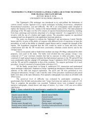
Antimicrobial <strong>and</strong> anti-inflammatory drugs used to treat tooth root infections<br />
Drug Dose Frequency Route Duration<br />
Procaine<br />
Penicillin G<br />
33,000 Units/kg Once Daily IM 14 days<br />
Cefti<strong>of</strong>ur 2 mg/kg Once Daily IV * / IM 14 days<br />
Florfenicol 20 mg/kg Every other day IM 3 treatments<br />
Isoniazid 10 mg/kg Once Daily Orally 30 days<br />
Flunixin 1 mg/kg Once – Twice IV / IM As needed<br />
meglumine<br />
Daily<br />
*<br />
Sodium Cefti<strong>of</strong>ur is the only cefti<strong>of</strong>ur formulation that can be given intravenously.<br />
The prognosis for cure following tooth extraction is good, <strong>and</strong> is not affected by the<br />
location or number <strong>of</strong> teeth affected. There is a higher mortality rate associated with animals<br />
presented in thin or emaciated conditions. 4<br />
Anesthesia for M<strong>and</strong>ibular Surgery<br />
General anesthesia is recommended for tooth extractions unless the abscess is in a<br />
chronic state which will permit an easy oral extraction. Anti-inflammatories <strong>and</strong> analgesics post<br />
operatively are recommended. Endotracheal intubation is highly recommended to prevent<br />
aspiration <strong>of</strong> the saliva, blood, <strong>and</strong> lavage that accumulates in the oral cavity during the surgical<br />
procedure.<br />
An alveolar (m<strong>and</strong>ibular) nerve block can supplement general anesthesia, <strong>and</strong> provides<br />
analgesia short term post operatively. Local anesthetic is injected at the m<strong>and</strong>ibular foramen<br />
(medial aspect <strong>of</strong> the ramus <strong>of</strong> the m<strong>and</strong>ible). This is located by palpation <strong>of</strong> the most prominent<br />
portion <strong>of</strong> the most caudoventral aspect <strong>of</strong> the m<strong>and</strong>ible immediately rostral to the ventral curve<br />
<strong>of</strong> the ramus. Full insertion <strong>of</strong> an 18 gauge 1.5 inch needle towards the zygomatic arch works<br />
well to approximate the level <strong>of</strong> the foramen in adult camelids. Alternatively injection into the<br />
mental foramen will allow backflow <strong>of</strong> local anesthetic <strong>and</strong> anesthetize the alveolar nerve. The<br />
mental foramen is <strong>of</strong>ten palpable 2-3 cm caudal to the incisors. The needle is inserted into the<br />
foramen <strong>and</strong> the injection is performed while placing a finger over the foramen to create a seal.<br />
Retained Deciduous Teeth<br />
Retained deciduous teeth occur when the deciduous incisors fail to fall out when the<br />
permanent teeth erupt. This is usually a cosmetic problem, although the potential exists to cause<br />
difficulty prehending. The retained deciduous teeth are smaller, are usually the more rostral<br />
located teeth, <strong>and</strong> may be loose while the permanent teeth are larger, located more caudal<br />
(lingual surface), <strong>and</strong> are usually more stable. 5<br />
774<br />
3
Method <strong>of</strong> removal is dependent on the how firmly they are attached. They may be<br />
easily extracted with minimal force or may require the use <strong>of</strong> a dental elevator. If more than<br />
mild force is needed for extraction, heavy sedation or a light plane <strong>of</strong> anesthesia will be<br />
necessary. Extraction <strong>of</strong> teeth that are firmly attached carries a risk <strong>of</strong> damage to the permanent<br />
tooth roots.<br />
Teeth Trimming<br />
There are multiple indications for trimming teeth in camelids. Overgrown m<strong>and</strong>ibular<br />
incisors can resulting in difficulty prehending food or can become a cosmetic blemish. The<br />
fighting teeth (upper <strong>and</strong> lower canines <strong>and</strong> the upper 3 rd incisors) are commonly trimmed in<br />
male llamas <strong>and</strong> alpacas. The incisors <strong>and</strong> fighting teeth can be trimmed with a cutting wheel<br />
attachment on a h<strong>and</strong> held rotary tool. An alternative to using a rotary tool to trim the incisors is<br />
to use obstetrical wire to saw the teeth.<br />
Premolars <strong>and</strong> molars that do not properly oppose the opposite arcade can become<br />
overgrown, which is common in cases <strong>of</strong> previous cheek tooth extraction. If mastication is<br />
impaired due to overgrowth, the teeth can be floated using small equine dental instruments.<br />
Enamel points on the buccal side <strong>of</strong> the maxillary check teeth <strong>and</strong> the lingual side <strong>of</strong> the<br />
m<strong>and</strong>ibular check teeth are normal because the m<strong>and</strong>ibular arcade does not sit as wide as the<br />
maxillary arcade, <strong>and</strong> should not be floated.<br />
References:<br />
1. Mattoon JS: Radiographic Technique for <strong>Dental</strong> Disease <strong>of</strong> Camelids, in Current<br />
Veterinary Care <strong>and</strong> Management <strong>of</strong> <strong>Llamas</strong> <strong>and</strong> <strong>Alpacas</strong>, Columbus, OH, pp 232-238<br />
2. Niehaus AJ, Anderson DE: Retrospective Study <strong>of</strong> Camelid Tooth Abscesses, inThe<br />
Ohio State University, 2007<br />
3. Cebra ML, Cebra CK, Garry FB: Tooth root abscesses in New World camelids: 23 cases<br />
(1972-1994). J Am Vet Med Assoc 209:819-822, 1996<br />
4. Niehaus AJ, Anderson DE: Tooth root abscesses in llamas <strong>and</strong> alpacas: 123 cases (1994-<br />
2005). J Am Vet Med Assoc 231:284-289, 2007<br />
5. Fowler ME: <strong>Dental</strong> Disease, in Medicine <strong>and</strong> surgery <strong>of</strong> South American camelids:<br />
llama, alpaca, vicuña, guanaco (ed 2nd). Ames, Iowa State University Press, 1998, pp<br />
112-120<br />
6. Thrall DE: Textbook <strong>of</strong> veterinary diagnostic radiology, in Textbook <strong>of</strong> veterinary<br />
diagnostic radiology (ed 3rd). Philadelphia, W. B. Saunders, 1998, pp 107-109<br />
7. Anderson DE: Tooth Root Abscesses, in Camelid Medicine, Surgery, <strong>and</strong> Reproduction,<br />
Columbus, Ohio, pp 87-88<br />
775<br />
4