CHEMICAL AND PHYSICAL PROCESSES OF DIGESTION
CHEMICAL AND PHYSICAL PROCESSES OF DIGESTION
CHEMICAL AND PHYSICAL PROCESSES OF DIGESTION

You also want an ePaper? Increase the reach of your titles
YUMPU automatically turns print PDFs into web optimized ePapers that Google loves.
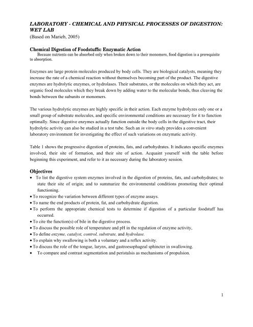
LABORATORY - <strong>CHEMICAL</strong> <strong>AND</strong> <strong>PHYSICAL</strong> <strong>PROCESSES</strong> <strong>OF</strong> <strong>DIGESTION</strong>:<br />
WET LAB<br />
(Based on Marieb, 2005)<br />
Chemical Digestion of Foodstuffs: Enzymatic Action<br />
Because nutrients can be absorbed only when broken down to their monomers, food digestion is a prerequisite<br />
to absorption.<br />
Enzymes are large protein molecules produced by body cells. They are biological catalysts, meaning they<br />
increase the rate of a chemical reaction without themselves becoming part of the product. The digestive<br />
enzymes are hydrolytic enzymes, or hydrolases. Their substrates, or the molecules on which they act, are<br />
organic food molecules which they break down by adding water to the molecular bonds, thus cleaving the<br />
bonds between the subunits or monomers.<br />
The various hydrolytic enzymes are highly specific in their action. Each enzyme hydrolyzes only one or a<br />
small group of substrate molecules, and specific environmental conditions are necessary for it to function<br />
optimally. Since digestive enzymes actually function outside the body cells in the digestive tract, their<br />
hydrolytic activity can also be studied in a test tube. Such an in vitro study provides a convenient<br />
laboratory environment for investigating the effect of such variations on enzymatic activity.<br />
Table 1 shows the progressive digestion of proteins, fats, and carbohydrates. It indicates specific enzymes<br />
involved, their site of formation, and their site of action. Acquaint yourself with the table before<br />
beginning this experiment, and refer to it as necessary during the laboratory session.<br />
Objectives<br />
• To list the digestive system enzymes involved in the digestion of proteins, fats, and carbohydrates; to<br />
state their site of origin; and to summarize the environmental conditions promoting their optimal<br />
functioning.<br />
• To recognize the variation between different types of enzyme assays.<br />
• To name the end products of protein, fat, and carbohydrate digestion.<br />
• To perform the appropriate chemical tests to determine if digestion of a particular foodstuff has<br />
occurred.<br />
• To cite the function(s) of bile in the digestive process.<br />
• To discuss the possible role of temperature and pH in the regulation of enzyme activity,<br />
• To define enzyme, catalyst, control, substrate, and hydrolase.<br />
• To explain why swallowing is both a voluntary and a reflex activity.<br />
• To discuss the role of the tongue, larynx, and gastroesophageal sphincter in swallowing.<br />
• To compare and contrast segmentation and peristalsis as mechanisms of propulsion.<br />
1
Table 1: Digestion and absorption of foodstuffs (from Maried, Fig. 39A.1)<br />
Path of absorption Foodstuff Enzyme(s) and source Site of action<br />
Carbohydrate<br />
absorption<br />
Product consumed Starch and disaccharides<br />
Lactose, Maltose,<br />
Sucrose<br />
End-product absorbed: Galactose, Glucose and<br />
Fructose<br />
Protein digestion<br />
Product consumed Proteins<br />
Salivary amylase Mouth<br />
Pancreatic amylase Small intestine<br />
Brush border enzymes Small intestine<br />
(lactase, maltase,<br />
sucrase, and dextrinase)<br />
Pepsin Stomach<br />
Large Polypeptides Pancreatic enzymes<br />
(trypsin, chymotrypsin<br />
and carboxypeptidase)<br />
Small polypeptides,<br />
small peptides<br />
End-product absorbed: Amino acids (some<br />
dipeptides and<br />
tripeptides)<br />
Fat digestion<br />
Product consumed Unemulsified fats<br />
End-product absorbed: Monoglycerides,<br />
glycerol, and fatty acids.<br />
Brush border enzymes<br />
(aminopeptidases,<br />
carboxypeptidase, and<br />
dipeptidases)<br />
Emulsification by<br />
detergent action of bile<br />
salts ducted in from<br />
liver<br />
Lumen of small<br />
intestine<br />
Brush border of small<br />
intestine.<br />
Small intestine<br />
Pancreatic lipase Small intestine<br />
2
Materials<br />
Part 1: Enzyme Action<br />
• Hot plates<br />
• IncubatorTest<br />
• test tube clamps<br />
Activity 2:<br />
• Test tubes + rack (7 tubes per team)<br />
• Spot plates<br />
• non-sterile 5 cc. pipettes (labelled)<br />
• droppers with bulbs (6)<br />
Activity 3:<br />
• Test tubes + rack (6 tubes per team)<br />
• pipettes (labelled)<br />
Activity 4:<br />
• 11 Disposable test tubes and rack<br />
• Bile salts (sodium taurocholate)<br />
(scoops)<br />
• pipettes (labelled)<br />
• Litmus cream (fresh cream to which<br />
powdered litmus is added to achieve a<br />
blue colour) (not a dropper bottle, use<br />
20 ml/team)<br />
Part 2: P hys ical Pr ocess es<br />
Activity 6:<br />
• Spring water (water pitcher)<br />
• Stethoscope<br />
• wax markers<br />
• 250-ml beaker<br />
• Chalkboard for recording class results<br />
• Lugol’s IKI (Lugol’s iodine) (dropper<br />
bottle)<br />
• Benedict’s solution (dropper bottle)<br />
• distilled water<br />
• alpha-amylase solution (20 cc / team)<br />
• 1% boiled starch solution, freshly prepared<br />
(20 cc / team)<br />
• 1% maltose solution (5cc X #team)<br />
• 1% Trypsin (20 cc X # teams)<br />
• 0.1% BAPNA solution (20 cc / team)<br />
• distilled water<br />
• 1% Pancreatin solution<br />
• Vegetable oil (5 cc/team)<br />
• dropper bottle of 0.1 M HCL<br />
• distilled water<br />
• paper cups<br />
• alcohol + gauze<br />
• stop watch<br />
3
Activities<br />
Activit y 1: Develop ing Hypoth eses for Experimen ts on Enzymatic D igest ion<br />
Work in groups according to instructors’ instructions, with each group taking responsibility for<br />
setting up and conducting one of the following experiments. In each of the digestive procedures being<br />
studied (starch, protein. and fat digestion) and in the amylase assay, you are directed to boil the contents<br />
of one or more test tubes. To do this, obtain a 250-ml beaker and a hot plate. Add about 125 ml of water<br />
to the beaker and bring to a boil. Place the specimen in the boiling water for the time (min.) specified in<br />
the directions.<br />
Upon completion of the experiments, each group should communicate its results to the rest of the class by<br />
recording them on the chalkboard. All members of the class should observe the controls (the specimens<br />
or standards against which experimental samples are compared) as well as the positive and negative<br />
examples of all experimental results. Additionally, all members of the class should be able to explain the<br />
tests used and the results observed and anticipated for each experiment.<br />
• Develop a hypothesis for each of the enzymatic digestions studied in Activities 2-4: starch, protein,<br />
and fat.<br />
• Write down your hypotheses, including your reasoning behind each hypothesis.<br />
• Carry out the following activities.<br />
4
Activity 2: Assessing Starch Digestion by Salivary Amylase<br />
In this experiment you will investigate the hydrolysis of starch to maltose by salivary amylase (the enzyme<br />
produced by the salivary glands and secreted into the mouth), so you’ll need to be able to identify the presence of<br />
starch and maltose to determine to what extent the enzymatic activity has occurred. Thus controls must be prepared<br />
to provide a known standard against which comparisons can be made. Starch decreases and sugar increases as<br />
digestion occurs, according to the following formula:<br />
Amylase<br />
Starch + Water � Maltose<br />
Table 2: Salivary Amylase Digestion of Starch<br />
1. Obtain a test tube rack, 7 test tubes, and a wax marking pencil. From supply area 1, obtain distilled water,<br />
maltose, amylase, and starch solutions.<br />
2. Prepare the controls (tubes 1A to 3A) and the experimental samples (tubes 4A to 6A).<br />
3. Mark each tube with a wax pencil and, load the tubes as indicated in Table 2, using 3 ml of each indicated<br />
substance.<br />
4. Place all tubes in a rack in the incubator or cold water bath for approximately 1 hour. Shake the rack gently from<br />
time to time to keep the contents evenly mixed.<br />
5. (While these tubes are incubating, proceed to Activity 6: Physical Processes: Mechanisms of Food Propulsion<br />
and Mixing.)<br />
6. Once the incubation period is over, you are ready to carry out the amylase assay.<br />
Amylase assay<br />
1. From supply area 1 obtain a spot plate and dropper bottles of Lugol’s IKI and Benedict’s solution. Set up your<br />
boiling water bath using a hot plate, and a 250-ml beaker.<br />
2. While the water is heating, mark the spot plate A (for amylase) and number six of its depressions 1 to 6 for<br />
sample identification.<br />
3. Pour about a drop of the sample from each of the tubes 1A-6A into the appropriately numbered spot. Into each<br />
sample droplet, place a drop of I Lugol’s IKI solution. A blue-black colour indicates the presence of starch and<br />
is referred to as a positive starch test. If starch is not present, the mixture will not turn blue, which is referred<br />
to as negative starch test. Record your results (+ for positive, - for negative) in the table and on the chalkboard.<br />
4. Into the remaining mixture in each tube, place 5 drops of Benedict's solution. Put each tube into the beaker of<br />
boiling water for about 5 minutes. If a green-to-orange precipitate forms, maltose is present, this is a positive<br />
sugar test. A negative sugar test is indicated by no colour change. Record your results in the table and on the<br />
chalkboard.<br />
5
Activit y 3: Protein digestion by tryp sin<br />
Trypsin, an enzyme produced by the pancreas, hydrolyzes proteins to small fragments (proteoses, peptones,<br />
and peptides). BAPNA (N-alpha-benzoyl-L-arginine-p-nitroanilide) is a synthetic trypsin substrate consisting of a<br />
dye covalently bound to an amino acid. Trypsin hydrolysis of BAPNA cleaves the dye molecule from the amino<br />
acid, causing the solution to change from colourless to bright yellow. Since the covalent bond between the dye<br />
molecule and the amino acid is the same as the peptide bonds that link amino acids together, the appearance of a<br />
yellow colour indicates the presence and activity of an enzyme that is capable of peptide bond hydrolysis. Because<br />
the colour change from clear to yellow is direct evidence of hydrolysis, additional tests are not required when<br />
determining trypsin activity using BAPNA.<br />
Table 3: Trypsin Digestion of Protein<br />
Assessing Protein Digestion by Trypsin<br />
1. Obtain 6 test tubes and a test tube rack, trypsin, BAPNA, distilled water and the appropriate labelled pipettes<br />
and bring them to your bench.<br />
2. Prepare the controls (tubes 1T and 2T) and the experimental samples (tubes 3T- to 6T), as shown in Table 3.<br />
3. Mark each tube with a wax pencil and load the tubes as indicated in the Table 3, using 3 ml of each indicated<br />
substance.<br />
4. Place all tubes in a rack in the cold water bath or incubator for approximately 1 hour. Shake the rack<br />
occasionally to keep the contents well mixed.<br />
5. While these tubes are incubating, proceed to Activity 6: Physical Processes: Mechanisms of Food Propulsion<br />
and Mixing.<br />
• Once the incubation period is over, you are ready to carry out the trypsin assay.<br />
Trypsin Assay<br />
Since BAPNA is a synthetic colourigenic (colour-producing substrate), the presence of yellow colour indicates<br />
a positive hydrolysis test; the dye molecule has been cleaved from the amino acid. If the sample mixture remains<br />
clear, no detectable hydrolysis has occurred. Record the negative hydrolysis test results in Table 3 and on the<br />
chalkboard.<br />
6
Activit y 4: Pancreatic Lipase Digestion of Fats and th e A ct ion of Bile<br />
The treatment that fats and oils go through during digestion in the small intestine is a bit more complicated than<br />
that of carbohydrates or proteins – pre-treatment with bile to physically emulsify the fats is required. Hence, two sets<br />
of reactions occur.<br />
First: Bile<br />
Fats/Oils �<br />
(Emulsification)<br />
Then: Lipase<br />
Fat/oil droplets �<br />
(Digestion)<br />
Minute fat/oil droplets<br />
Monoglycerides and fatty<br />
acids<br />
The term pancreatin describes the enzymatic product of the pancreas, which includes enzymes that digest proteins,<br />
carbohydrates, nucleic acids, and fats. It is used here to investigate the properties of pancreatic lipase, which<br />
hydrolyzes fats and oils to their component monoglycerides and two fatty acids (and occasionally to glycerol and<br />
three fatty acids).<br />
The fact that some of the end products of fat digestion (fatty acids) are organic acids that decrease the pH provides<br />
an easy way to recognize that digestion is ongoing or completed. You will be using a pH indicator called litmus blue<br />
to follow these changes; it changes from blue to pink as the test tube contents become acid.<br />
Fig 1: Emulsification action of bile<br />
Demonstrating the Emulsification Action of Bile<br />
Although bile, a secretory product of the liver is not an enzyme, it is important to fat digestion because of its<br />
emulsifying action (the physical breakdown of larger particles into smaller ones) on fats. Emulsified fats provide a<br />
larger surface area for enzymatic activity.<br />
NOTE: use disposable tubes with caps for this experiment.<br />
1. To demonstrate the action of bile on fats, prepare two test tubes and mark them 1E and 2E (fig.1).<br />
2. To tube 1E add 5 ml of water and 1 ml of vegetable oil.<br />
3. To tube 2E add 5 ml of water, 1 ml of vegetable, and a pinch of bile salts.<br />
4. Cap each tube, shake vigorously, and allow the tubes to stand at room temperature.<br />
5. After 10-15 minutes, observe both tubes.<br />
If emulsification has not occurred, the oil will be floating on the surface of the water. If emulsification has occurred,<br />
the fat droplets will be suspended throughout the water, forming an emulsion.<br />
In which tube has emulsification occurred?______________<br />
7
Assessing Fat Digestion by Lipase<br />
Table 4: Pancreatic Lipase Digestion of Fats.<br />
Prepare the controls (1L and 2L), and set up the experimental samples (3L to 6L and 4B to 6B) as illustrated in<br />
Table 4.<br />
1. Obtain 9 disposable test tubes with caps, a test tube rack, lipase, litmus cream, and water from supply area 3.<br />
2. Mark each tube with a wax pencil and load the tubes using 3 ml of each indicated solution.<br />
3. Place a scoop of bile salts in tubes 4B and 5B.<br />
4. Cover each tube with a cap and shake to mix the contents.<br />
5. Remove the caps and place all tubes in a rack in water bath or incubator for approximately 1 hour. Shake the<br />
test tube rack from time to time to keep the contents well mixed.<br />
6. (While these tubes are incubating, proceed to Activity 6: Physical Processes: Mechanisms of Food Propulsion<br />
and Mixing.)<br />
7. Once the incubation period is over, you are ready to carry out the trypsin assay.<br />
Lipase Assay<br />
The basis of this assay is a pH change that is detected by a litmus powder indicator. Alkaline or neutral<br />
solutions containing litmus are blue but will turn reddish in the presence of acid. Since fats are digested to fatty acids<br />
(organic acids) during hydrolysis, they lower the pH of the sample they are in. Litmus cream (fresh cream providing<br />
the fat substrate to which litmus powder was added) will turn from blue to pink if the solution is acid. Because the<br />
effect of hydrolysis is directly seen, additional assay reagents are not necessary.<br />
1. To prepare a colour control, add 0.1 N HCI drop by drop to tubes 1L and 2L (capping the tubes after each<br />
addition and shaking to mix) until the cream turns pink.<br />
2. Record the colour of the tubes in Table 4 and on the chalkboard.<br />
8
Activit y 5: Reporting Results an d Con clusion s<br />
1. Share your results with the class as directed in Activity 1<br />
2. Suggest additional experiments, and carry out experiments if time permits.<br />
3. Prepare a lab report for the experiments on digestion.<br />
Activit y 6: Ph ysical p roces ses: Mechanisms of Food Propulsion an d Mixing<br />
Although enzyme activity is a very important part of the overall digestion process, foods<br />
must also be processed physically (churning and chewing), and moved by mechanical means<br />
along the tract if digestion and absorption are to be completed. Just about any time organs exhibit<br />
mobility, muscles are involved, and movements of and in the gastrointestinal tract are no<br />
exception. Although we tend to think only of smooth muscles when visceral activities are<br />
involved, both skeletal and smooth muscles are involved in digestion. This fact is amply<br />
demonstrated by the simple activities that follow.<br />
Deglutition (Swallowing)<br />
Swallowing, or deglutition, which is largely the result of skeletal muscle activity, occurs in<br />
two phases: bucal (mouth) and pharyngeal-esophageal. The initial phase – the bucal – is<br />
voluntarily controlled and initiated by the tongue. Once begun, the process continues<br />
involuntarily in the pharynx and esophagus, through peristalsis, resulting in the delivery of the<br />
swallowed contents to the stomach.<br />
Observing Movements and Sounds of Digestion<br />
1. Obtain a pitcher of water, a stethoscope, a cup, an alcohol swab in preparation for making<br />
the following observations.<br />
2. While swallowing a mouthful of water, consciously note the movement of your tongue<br />
during the process. Record your observations.<br />
3. Repeat the swallowing process while your laboratory partner watches the externally visible<br />
movements of your larynx. (This movement is more obvious in a male, who has a larger<br />
Adam's apple.) Record your observations.<br />
What do these movements accomplish?<br />
Before donning the stethoscope your lab partner should clean the earpieces with an alcohol swab.<br />
Then, he or she should place the diaphragm of the stethoscope over your abdominal wall,<br />
approximately 2.5 cm below the xyphoid process and slightly to the left, to listen for sounds as<br />
you again take two or three swallows of water. There should be two audible sounds – one when<br />
the water splashes against the gastroesopheal sphincter and the second when the peristaltic wave<br />
of the esophagus arrives at the sphincter and the sphincter opens, allowing water to gurgle into<br />
9
the stomach. Determine, as accurately as possible, the time interval between these two sounds<br />
and record it below.<br />
Interval between arrival of water at the sphincter and the opening, of the sphincter: ______sec<br />
This interval gives a fair indication of the time it takes for the peristaltic wave to travel down the 25 cm-long<br />
esophagus. (Actually the time interval is slightly less than it seems, because pressure causes the sphincter to relax<br />
before the peristaltic wave reaches it.)<br />
Segmentation and Peristalsis<br />
Although several types of movements occur in the digestive tract organs, peristalsis and<br />
segmentation are most important as mixing and propulsive mechanisms. Peristaltic movements<br />
are the major means of propelling food through most of the digestive viscera. Essentially they are<br />
waves of contraction followed by waves of relaxation that squeeze foodstuffs through the<br />
alimentary canal, and they are superimposed on segmental movements.<br />
Segmental movements are local constrictions of the organ wall that occur rhythmically. They<br />
serve mainly to mix the foodstuffs with digestive juices and to increase the rate of absorption by<br />
continually moving different portions of the chyme over adjacent regions of the intestinal wall.<br />
However, segmentation is also an important means of food propulsion in the small intestine, and<br />
slow segmenting movements called haustral contractions are frequently seen in the large<br />
intestine.<br />
Sources<br />
Marieb, Elaine. 2005. Human anatomy and physiology laboratory manual, 8 th ed. Pearson.<br />
Exercise 39A, pp. 435-442<br />
Hickman, Cleveland P.Jr., Larry S. Roberts, Alland, Larson, & Helen I’Anson. 2004. Integrated<br />
principles of zoology. pp. 682-688.<br />
10