

impact o na sean one health - KVAC 2012 ....VET MED KKU thailand ...
impact o na sean one health - KVAC 2012 ....VET MED KKU thailand ...
impact o na sean one health - KVAC 2012 ....VET MED KKU thailand ...

You also want an ePaper? Increase the reach of your titles
YUMPU automatically turns print PDFs into web optimized ePapers that Google loves.
<strong>KVAC</strong> 13 th <strong>2012</strong><br />
The 13 th <strong>KKU</strong> Veteri<strong>na</strong>ry Annual Conference <strong>2012</strong><br />
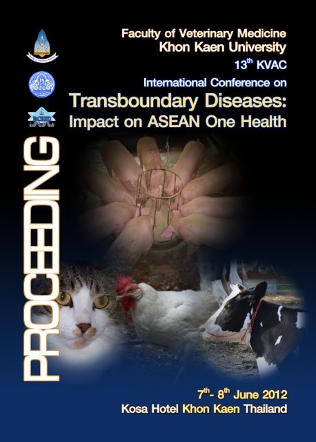
“Inter<strong>na</strong>tio<strong>na</strong>l Conference on Transboundary Diseases: Impact on ASEAN One Health”<br />
7-8 June <strong>2012</strong><br />
Kosa Hotel, Khon Kaen, Thailand<br />
PROCEEDING<br />
Organized by Faculty of Veteri<strong>na</strong>ry Medicine, Khon Kaen University, Thailand<br />
Edited by Jaruwan Kampa<br />
Weerapol Tawee<strong>na</strong>n<br />
Suthida Chanlun<br />
Ta<strong>na</strong>porn Asawapatta<strong>na</strong>kul
The 13 th <strong>KKU</strong> Veteri<strong>na</strong>ry Annual Conference <strong>2012</strong> (13 th <strong>KVAC</strong>, Khon Kaen, Thailand)<br />
“Inter<strong>na</strong>tio<strong>na</strong>l Conference on Transboundary Diseases: Impact on ASEAN One Health”, June<br />
7-8, <strong>2012</strong> / Organized by Faculty of Veteri<strong>na</strong>ry Medicine, Khon Kaen University, Thailand.<br />
1. Faculty of Veteri<strong>na</strong>ry Medicine<br />
2. Conference on Veteri<strong>na</strong>ry Medicine<br />
3. Human and Animal Health<br />
4. ASEAN<br />
I. Faculty of Veteri<strong>na</strong>ry Medicine, Khon Kaen University, Thailand<br />
II. Jaruwan Kampa<br />
III. Weerapol Tawee<strong>na</strong>n<br />
IV. Suthida Chanlun<br />
V. Ta<strong>na</strong>porn Asawapatta<strong>na</strong>kul<br />
All oral and poster presentation abstracts published in The 13 th <strong>KKU</strong> Veteri<strong>na</strong>ry Annual<br />
Conference <strong>2012</strong>: “Inter<strong>na</strong>tio<strong>na</strong>l Conference on Transboundary Diseases: Impact on ASEAN<br />
One Health proceeding pass a peer review process by the scientific committee of the<br />
conference. Proceeding did not require all authors of a research paper to sign the letter of<br />
submission, nor do they impose an order on the list of authors. Submission to the<br />
conference is obtained by scientific committee meaning that all the listed authors have<br />
agreed all of the contents. The corresponding (submitting) author is responsible for having<br />
ensured that this agreement has been reached, and for ma<strong>na</strong>ging all communication<br />
between the committee and all co-authors, before and after publication. Each author is<br />
responsible for the content and accuracy of the entire manuscript.<br />
©<strong>2012</strong> The Faculty of Veteri<strong>na</strong>ry Medicine, Khon Kaen University<br />
ISBN 978-616-223-189-6
Contents<br />
Welcome 1<br />
Committees 5<br />
General Information 8<br />
Scientific Program 11<br />
Invited Speakers 15<br />
Speaker’s Notes 35<br />
Oral Presentation 117<br />
Poster Presentation 129
Welcome Message from the President of Khon Kaen University<br />
Dear Distinguished Delegates,<br />
I am very pleased to warmly welcome you to Khon Kaen and to the Inter<strong>na</strong>tio<strong>na</strong>l<br />
Conference on “Transboundary Disease; Impact on ASEAN One Health” being held on 7-<br />
8 June <strong>2012</strong>.<br />
This conference is hosted by the Faculty of Veteri<strong>na</strong>ry Medicine, Khon Kaen University. I<br />
am very delightful for the breakthrough and expanding of the conference into an<br />
inter<strong>na</strong>tio<strong>na</strong>l event because presently we all need to live and join together as the<br />
partnerships.<br />
“ASEAN One Health” is major issue that needed a collaboration and cooperation<br />
between all 10 countries in South East Asia in all aspects of <strong>health</strong> care for humans and<br />
animals. Therefore, we all could build the strength of our ASEAN community by sharing<br />
our experience, knowledge and technology with each other during this conference.<br />
I would like to thank you the VM<strong>KKU</strong>, co-host organizations and all the private<br />
companies in making this conference successful.<br />
Warmest regards<br />
Associate Professor Dr.Kittichai Triratta<strong>na</strong>sirichai<br />
President of Khon Kaen University<br />
7 th -8 th June <strong>2012</strong><br />
Kosa Hotel, Khon Kaen, Thailand<br />
Faculty of Veteri<strong>na</strong>ry Medicine - Khon Kaen University - Khon Kaen - Thailand - Tel. +66 43 202404 - http://vet.kku.ac.th<br />
1
2<br />
Welcome Message from the Dean, Faculty of Veteri<strong>na</strong>ry Medicine, <strong>KKU</strong><br />
Dear Colleagues<br />
Thailand is known as a leading role in promoting regio<strong>na</strong>l partnership to control and<br />
prevent several diseases which harmful human and affect to animal production. Risk of<br />
emerging and re-emerging diseases at difference levels had been actively studied and<br />
research is going on. This is a time of increasing inter<strong>na</strong>tio<strong>na</strong>l trading and human<br />
mobility, therefore, update information and knowledge in trans-boundary diseases are<br />
important to share with scientists and veteri<strong>na</strong>rians who working together for a better<br />
living of people in ASEAN community.<br />
The <strong>KVAC</strong> <strong>2012</strong> is generated during 7 th -8 th June <strong>2012</strong> at Kosa Hotel, Khon Kaen, Thailand<br />
with the purpose of creating a platform for the development of <strong>one</strong> <strong>health</strong> research,<br />
exchange and contribute our knowledge for Human and animal <strong>health</strong> as well as quality<br />
of animal products. The conference will give a better understanding in information,<br />
policy and regulation of <strong>na</strong>tio<strong>na</strong>l and inter<strong>na</strong>tio<strong>na</strong>l levels among the ASEAN countries<br />
and in the Asian region. This conference also provide a companion animal session to<br />
veteri<strong>na</strong>rians to learn more and share knowhow with the speakers.<br />
I would like to express my appreciation to co-hosts, the Department of livestock<br />
Development (DLD) and Research and Diagnosis Center for Emerging Infectious Diseases<br />
(RCEID) as well as speakers, participants, organizing committee and sponsors. Welcome<br />
all of you to the “The Inter<strong>na</strong>tio<strong>na</strong>l Conference on Trans-boundary Diseases: Impact on<br />
ASEAN One Health “<br />
Best wishes,<br />
The 13 th <strong>KVAC</strong> Inter<strong>na</strong>tio<strong>na</strong>l Conference on<br />
Transboundary Diseases: Impact on ASEAN One Health<br />
Associate Professor Dr.Suneerat Aiumlamai<br />
Dean, Faculty of Veteri<strong>na</strong>ry Medicine<br />
Khon Kaen University<br />
Faculty of Veteri<strong>na</strong>ry Medicine - Khon Kaen University - Khon Kaen - Thailand - Tel. +66 43 202404 - http://vet.kku.ac.th
Welcome Message from Chairman of the 13 th <strong>KVAC</strong><br />
Dear Colleagues and Distinguished Delegates,<br />
It is my great honor to welcome you to the 13 th Inter<strong>na</strong>tio<strong>na</strong>l <strong>KVAC</strong> Conference, <strong>2012</strong><br />
hosted by the Faculty of Veteri<strong>na</strong>ry Medicine, Khon Kaen University. I wish you enjoy<br />
and have a wonderful experience during staying in Khon Kaen.<br />
This year the conference focuses on issues involving ASEAN One Health and other<br />
interesting aspects of small animal medicine, exotic and wildlife medicine, and<br />
infectious diseases. With a cooperation of the RCEID (Research and Diagnostic Center<br />
for Emerging Infectious Diseases), <strong>one</strong> session has been ma<strong>na</strong>ged for delegates who<br />
interested in infective endocarditis. The conference provides advance knowledge in<br />
diagnosis and current information and situation of the diseases both in human and<br />
animal.<br />
I would like to thank all committees, speakers, sponsors, co-host organizations and all<br />
delegates who support and contribute to the successful of the conference.<br />
Best Regards,<br />
Associate Professor Dr.Somboon Sangmaneedet<br />
Chairman of the Conference<br />
7 th -8 th June <strong>2012</strong><br />
Kosa Hotel, Khon Kaen, Thailand<br />
Faculty of Veteri<strong>na</strong>ry Medicine - Khon Kaen University - Khon Kaen - Thailand - Tel. +66 43 202404 - http://vet.kku.ac.th<br />
3
4<br />
The 13 th <strong>KVAC</strong> Inter<strong>na</strong>tio<strong>na</strong>l Conference on<br />
Transboundary Diseases: Impact on ASEAN One Health<br />
Welcome Message from Chairman, Scientific Comittees<br />
Dear Friends and Colleagues<br />
Welcome to Thailand, to Khon Kaen and to the 13 th <strong>KKU</strong> Veteri<strong>na</strong>ry Annual Conference.<br />
By the year 2015, all <strong>na</strong>tions in ASEAN Economic Community (AEC) shall be the regio<strong>na</strong>l<br />
economic integration which results not only on economic issue but also the <strong>health</strong> of<br />
ASEAN people. Therefore, we settled “Transboundary Diseases: Impact on ASEAN One<br />
Health” as the main theme of the 13 th <strong>KVAC</strong>.<br />
On behalf of Scientific Committee, we hope this conference would provide an<br />
impressive meeting place and pleasant atmosphere that will stimulate a lot of discussion<br />
and strengthening co-operation among ASEAN veteri<strong>na</strong>ry professions in fields of<br />
emerging diseases, livestock, wildlife/exotics and companion animals.<br />
We would like to thank for kindness of all of the invited speakers who are expertise in<br />
many interesting fields; Avian medicine, Food Safety, Disease Control and Epidemiology,<br />
Zoonoses, Epidemiology, Wildlife and Exotic, Livestock and Companion animals. Your<br />
contribution completes the theme of One Health. Presentations from young scientist<br />
also magnified the theme. We also take opportunity to thank our generous co-hosts<br />
and sponsors for making the meeting possible.<br />
Once again, it is great pleasure to welcome you to Khon Kaen!<br />
Assistant Professor Dr.Jaruwan Kampa<br />
Chairman, Scientific Committee<br />
Faculty of Veteri<strong>na</strong>ry Medicine - Khon Kaen University - Khon Kaen - Thailand - Tel. +66 43 202404 - http://vet.kku.ac.th
The 13 th <strong>KVAC</strong> Committee<br />
7 th -8 th June <strong>2012</strong><br />
Kosa Hotel, Khon Kaen, Thailand<br />
Scientific Advisory Committee<br />
1. President of Khon Kaen University<br />
2. Director General Department of Livestock Development<br />
3. Dean of the Faculty of Veteri<strong>na</strong>ry Medicine, Khon Kean University<br />
4. Director of Research and Diagnostic Center for Emerging Infectious Disease (RCEID)<br />
Organizing Committee<br />
1. Assoc.Prof.Dr.Somboon Sangmaneedet Chairman<br />
2. Assoc.Prof.Dr.Viraphong Lulitanond<br />
3. Assoc.Prof.Dr.Chuchat Kamollerd<br />
4. Assist.Prof.Dr.Sarthorn Porntrakulpipat<br />
5. Assist.Prof.Dr.Ekkachai Pattarapanwichien<br />
6. Assist.Prof.Dr.Prapaporn Tungtha<strong>na</strong>thanich<br />
7. Assist.Prof.Dr.Jaruwan Kampa<br />
8. Assist.Prof.Dr.Jatesada Jiwakanon<br />
9. Assist.Prof.Dr.Weerapol Tawee<strong>na</strong>n<br />
10.Assist.Prof.Dr.Naruepon Kampa<br />
11.Assist.Prof.Dr.Duangdaun Kaenkangploo<br />
12.Miss Ancharin Ounthaisong<br />
13.Mrs.Sombat Saengpol<br />
Scientific Committee<br />
1. Assist.Prof.Dr.Jaruwan Kampa Chairman<br />
2. Prof.Dr.Ulf Magnusson<br />
3. Assoc.Prof.Dr.Thaweesak Songserm<br />
4. Assoc.Prof.Dr.Fa<strong>na</strong>n Suksawat<br />
5. Assoc.Prof.Dr.Bongkot Noppon<br />
6. Assist.Prof.Dr.Sirikachorn Tangkawatta<strong>na</strong><br />
7. Assist.Prof.Dr.Weerapol Tawee<strong>na</strong>n<br />
8. Assist.Prof.Dr.Sarthorn Porntrakulpipat<br />
9. Assist.Prof.Dr.Chaiyapas Thamrongyoswittayakul<br />
10. Assist.Prof.Dr.Sompoth Weerakhun<br />
11. Prof.Dr.Orathai Pachirat<br />
12. Assoc.Prof.Dr.Wanlop Kaewkes<br />
13. Dr.Wises Namwat<br />
14. Dr.Kiatichai Faksri<br />
15. Dr.Tawatchai Pohuang<br />
16. Dr.Numfa Fungbun<br />
17. Mrs.Suthida Chanlun<br />
Information Committee<br />
1. Assist.Prof.Dr.Jatesada Jiwakanon Chairman<br />
2. Assist.Prof.Dr.Weerapol Tawee<strong>na</strong>n<br />
3. Assist.Prof.Dr.Suchat Watta<strong>na</strong>chai<br />
Faculty of Veteri<strong>na</strong>ry Medicine - Khon Kaen University - Khon Kaen - Thailand - Tel. +66 43 202404 - http://vet.kku.ac.th<br />
5
6<br />
The 13 th <strong>KVAC</strong> Inter<strong>na</strong>tio<strong>na</strong>l Conference on<br />
Transboundary Diseases: Impact on ASEAN One Health<br />
4. Mr.Pitakpong Maneeratta<strong>na</strong>rungroj<br />
5. Miss Chanida Chainn<br />
6. Mr.Yanyong Wangprecha<br />
7. Miss Rata<strong>na</strong> Daosawa<br />
8. Mrs.Bunserm Somboon<br />
9. Mrs.Prayoon Khamtat<br />
10. Mrs.Pris<strong>na</strong> Vichatham<br />
11. Mrs.Wan Buajan<br />
12. Mrs.Prapatson Thihta<br />
13. Miss Ancharin Ounthaisong<br />
Treasurer Committee<br />
1. Assoc.Prof.Dr.Chuchat Kamollerd Chairman<br />
2. Mrs.Phatchareeya Konchan<br />
3. Mrs.Suthathip Watta<strong>na</strong>chai<br />
4. Miss Nucha<strong>na</strong>rd Phonkhokkong<br />
5. Mr.Weera Suparuk<br />
Exhibition committee<br />
1. Assist.Prof.Dr.Naruepon Kampa Chairman<br />
2. Miss Supranee Stitammarat<br />
3. Mrs.Promporn Tongtieum<br />
4. Miss Rungarun Kulhintang<br />
5. Miss Pornthip Srichomchuen<br />
Banquet committee<br />
1. Assist.Prof.Dr.Prapaporn Tungtha<strong>na</strong>thanich Chairman<br />
2. Assoc.Prof.Dr.Bongkot Noppon<br />
3. Assist.Prof.Dr.Varaporn Sukolapong<br />
4. Dr.Nusara Suwan<strong>na</strong>chot<br />
5. Mrs.Yupadee Charoensawang<br />
Registration committee<br />
1. Assist.Prof.Dr.Duangdaun Kaenkangploo Chairman<br />
2. Assist.Prof.Dr.Arayaporn Macotpet<br />
3. Dr.Numfa Fungbun<br />
4. Dr.Suphannika Pnutthachalee<br />
5. Mrs.Sudarat Buatuan<br />
6. Mrs.Sombat Saengpol<br />
Ceremony and Reception committee<br />
1. Assist.Prof.Dr.Sirikachorn Tangkawatta<strong>na</strong> Chairman<br />
2. Assoc.Prof.Dr.Fa<strong>na</strong>n Suksawat<br />
3. Assist.Prof.Dr.Sarthorn Porntrakulpipat<br />
4. Assist.Prof.Dr.Weerapol Tawee<strong>na</strong>n<br />
5. Assist.Prof.Dr.Kochakorn Direksin<br />
Faculty of Veteri<strong>na</strong>ry Medicine - Khon Kaen University - Khon Kaen - Thailand - Tel. +66 43 202404 - http://vet.kku.ac.th
6. Dr.Aran Chanlun<br />
7. Dr.Trasida Ployngam<br />
8. Assist.Prof.Dr.Ranee Sachder<br />
9. Miss Sukumal Udom<br />
10. Miss Anongluk Manjai<br />
11. Miss Pawee<strong>na</strong> Sungseho<br />
12. Mr.Santi Kookrasang<br />
13. Miss Natchanok Kettongma<br />
7 th -8 th June <strong>2012</strong><br />
Kosa Hotel, Khon Kaen, Thailand<br />
Transportation committee<br />
1. Assist.Prof.Dr.Saijai Kongpechr Chairman<br />
2. Mrs.Yupadee Jaroensawang<br />
3. Mrs.Ta<strong>na</strong>korn Yanbuaban<br />
4. Mr.Amporn Krisornsri<br />
5. Mr.Pongpan Pongsapung<br />
Evaluation committee<br />
1. Assist.Prof.Dr.Ekkachai Pattarapanwichien Chairman<br />
2. Mrs.Aranya Singchomphoo<br />
3. Miss Nathapop Sechang<br />
4. Miss Aoythip Tha<strong>na</strong>nta<br />
Faculty of Veteri<strong>na</strong>ry Medicine - Khon Kaen University - Khon Kaen - Thailand - Tel. +66 43 202404 - http://vet.kku.ac.th<br />
7
8<br />
General Information<br />
Registration and Information desk- on the 2 th floor of Kosa Hotel<br />
Opening Hours<br />
Thursday 7 June <strong>2012</strong> 07.45-16.00<br />
Friday 8 June <strong>2012</strong> 07.45-12.00<br />
Conference Website: http://kvac.kku.ac.th<br />
Conference Venue<br />
Kosa Hotel<br />
250-252 Srichan Rd., Khon Kaen, 40000<br />
Ph<strong>one</strong>: +66 (43) 320 320<br />
Website: http://kosahotel.com<br />
Program Materials<br />
Your conference kit provided at registration counter will contain the conference<br />
program and proceeding CD and souvenir.<br />
Identification Badges<br />
Please wear your identification badge at all times while you are attending the<br />
conference and social events.<br />
Official Language<br />
The 13 th <strong>KVAC</strong> Inter<strong>na</strong>tio<strong>na</strong>l Conference on<br />
Transboundary Diseases: Impact on ASEAN One Health<br />
The official language of the conference is English.<br />
Faculty of Veteri<strong>na</strong>ry Medicine - Khon Kaen University - Khon Kaen - Thailand - Tel. +66 43 202404 - http://vet.kku.ac.th
Onsite Conference Services<br />
Onsite internet counter and computer access will be provided as a complimentary for<br />
all participants. Photocopying, fax, and teleph<strong>one</strong> services will be provided to participants<br />
on commercial basis.<br />
Coffee/Tea<br />
Coffee, tea and refreshment are included in the registration fee of participants and<br />
will be available throughout the conference at the assigned area during the breaks.<br />
Lunch<br />
Lunches are included in the registration fee of participants. Lunches will be served at<br />
12.00-13.00<br />
Liability and Insurance<br />
The Organizing Committee will not assure any responsibility for damages or injuries<br />
to person or property during the conference. It is recommended that participants should<br />
arrange their own perso<strong>na</strong>l travel and <strong>health</strong> insurance.<br />
Speaker-Ready Desk<br />
On 2 th floor of Kosa Hotel, we arrange a room for speakers for prepare their presentation. A<br />
desk for loading speaker’s presentation is in front of the room and is available from 07.45-<br />
16.00.<br />
Instruction for presenters<br />
Poster<br />
The posters are grouped by session and number. The number corresponds to the number in<br />
the abstract book. Poster should be displayed from Thursday 7 th June, 08.30 hours until<br />
Friday 8 th June, 16.00 hours. Posters should be mounted with double sided tape only which<br />
will be provided. Assistance and material for mounting the posters will be available, at the<br />
information desk. The authors of posters are expected to be available for answering<br />
questions during the poster session on 10.00-10.20, coffee beak time, Thursday.<br />
Oral presentations<br />
7 th -8 th June <strong>2012</strong><br />
Kosa Hotel, Khon Kaen, Thailand<br />
Time for your presentation is in the detailed of Oral presentation schedule in this<br />
proceeding.<br />
The conference language is English. The time schedule is tight and chairman will be strict on<br />
time, so please keep to the time allocated for you; 12 minutes talk and 3 minutes for<br />
question and discussion. If you aim at 10 minutes talk, you will be on the safe side.<br />
Faculty of Veteri<strong>na</strong>ry Medicine - Khon Kaen University - Khon Kaen - Thailand - Tel. +66 43 202404 - http://vet.kku.ac.th<br />
9
10<br />
The 13 th <strong>KVAC</strong> Inter<strong>na</strong>tio<strong>na</strong>l Conference on<br />
Transboundary Diseases: Impact on ASEAN One Health<br />
Power point presentation should be downloaded on the computer in Speaker-Ready Desk<br />
from 07.45-16.00. The operative system of the computer in the lecture rooms is Window 7.<br />
Please make sure that your presentation is compatible. It is not allowed to use individual<br />
computers for presentation.<br />
If you wish to provide your presentation after the conference by showing it on our official<br />
webpage, please inform our staff at the Speaker-Ready room about your decision. And if<br />
you do not want to, the files of your presentation will be deleted after the conference.<br />
However, please let us know your decision on your presentation day.<br />
Faculty of Veteri<strong>na</strong>ry Medicine - Khon Kaen University - Khon Kaen - Thailand - Tel. +66 43 202404 - http://vet.kku.ac.th
INTERNATIONAL CONFERENCE ON TRANSBOUNDARY DISEASES; IMPACT ON ASEAN ONE HEALTH<br />
07 June <strong>2012</strong><br />
Session 4 (Mongkut<br />
Thong):<br />
Oral scientific presentation<br />
(Eng)<br />
Session 3 (Mongkut Ngen):<br />
Zoonotic endocarditis<br />
(workshop)<br />
Session 2 (Mongkut Phet 2-3):<br />
Small animal, Exotic & Wildlife<br />
(Eng/Thai)<br />
Session 1 (Mongkut Phet 1):<br />
Infectious disease<br />
(Eng)<br />
Time<br />
7th-8th June <strong>2012</strong><br />
08.00-09.00<br />
Registration / Opening ceremony<br />
09.00-09.15<br />
Keynote Speech: Transboundary Diseases; Impact on ASEAN One Health<br />
09.15-10.15<br />
Dr.Wantanee Kalpravidh (FAO)<br />
Foresight of Transboundary Diseases in ASEAN community<br />
10.15-12.00<br />
Dr.R<strong>one</strong>llo C. Abila (OIE), Assoc.Prof.Kittisak Suwanyawisuth (<strong>KKU</strong>), Dr. Prapas Pinyocheep (DLD)<br />
Assoc.Prof.Dr.Suneerat Aiumlamai (Chairman)<br />
12.00-13.00 Lunch<br />
Rabies<br />
Vector-Borne Diseases in<br />
The Zoonotic Endocarditis Project in Vector Borne Vaccine<br />
Dr.Pornpitak Panlar<br />
Companion Animal<br />
Khon Kaen<br />
Preventable Disease<br />
(Bureau of Disease Control) Assoc.Prof.Dr.Fa<strong>na</strong>n Suksawasdi Moderator: Dr.Pawin Padungtod Dr.Ro<strong>na</strong>ld Enrique Morales<br />
(<strong>KKU</strong>)<br />
Opening and Introductory Remarks Vargas (MU)<br />
13.00-13.45<br />
Dr.Virat Klungboonkrong (<strong>KKU</strong>)<br />
Dr.Henry (Kip) Baggett (IEIP/GDD)<br />
1) The Zoonotic Endocarditis Project<br />
in Khon Kaen - Description and<br />
Kosa Hotel, Khon Kaen, Thailand<br />
Faculty of Veteri<strong>na</strong>ry Medicine - Khon Kaen University - Khon Kaen - Thailand - Tel. +66 43 202404 - http://vet.kku.ac.th<br />
11
12<br />
Scientific Presentation<br />
Updated Results Dr.George Watt<br />
(IEIP)<br />
2) Laboratory Aspects of Zoonotic<br />
Endocarditis in Khon Kaen<br />
Dr.Viraphong Lulitanond (<strong>KKU</strong>)<br />
Zoonotic Pathogens Causing Infective<br />
Endocarditis in Thailand<br />
Moderator: Dr Pawin Padungtod<br />
1) Current Concepts in Q Fever<br />
Dr.Gilbert Kersh (NCEZID)<br />
Lameness Exami<strong>na</strong>tion and<br />
Orthopedics Assessment<br />
Asst.Prof.Dr. Monchanok Vijarnsorn<br />
(KU)<br />
The 13 th <strong>KVAC</strong> Inter<strong>na</strong>tio<strong>na</strong>l Conference on<br />
Transboundary Diseases: Impact on ASEAN One Health<br />
3.45-14.30<br />
Scientific Presentation<br />
14.30-14.45 Coffee break / Poster Symposium<br />
Foot and Mouth Disease "Diagnosis and Treatment of Zoonotic Pathogens Causing Infective<br />
Dr.Sith Premasthira<br />
Common Orthopedic Problems" Endocarditis in Thailand<br />
(DLD)<br />
Moderator: Dr Pawin Padungtod<br />
and "Rehabilitation for<br />
2) An Overview of Streptococcus suis<br />
Orthopedic Patient: Tips and<br />
Infection in Thailand and Research<br />
Tricks"<br />
Findings<br />
Asst.Prof.Dr.Monchanok<br />
Mr.Anusak Kerdsin (NIH)<br />
Vijarnsorn (KU)<br />
3) An Overview of Bart<strong>one</strong>lla Infections<br />
in Thailand and Research Findings<br />
Dr.Sumalee Boonmar (IEIP/GDD)<br />
Q Fever Panel Discussion:<br />
4.45-16.30<br />
One Health Approach in Action<br />
Moderator: Dr.Pasakorn Akarasewi<br />
Panelist:<br />
Dr.Pravit Chumka<strong>sean</strong> (BOE)<br />
Dr.Teerasak Chuxnum (BOE)<br />
Dr.Mo<strong>na</strong>ya Ekathat (NIAH)<br />
Dr.Samuel Yingst (AFRIMS)<br />
Dr.Chien-Chung Chao (NMRC)<br />
Dr.Gilbert Kersh (NCEZID)<br />
Dr.Pawin Padungtod (IEIP,GDD)<br />
Faculty of Veteri<strong>na</strong>ry Medicine - Khon Kaen University - Khon Kaen - Thailand - Tel. +66 43 202404 - http://vet.kku.ac.th
08 June <strong>2012</strong><br />
Session 4 (Mongkut<br />
Thong):<br />
Oral scientific<br />
presentation<br />
(Eng)<br />
Session 3 (Mongkut Ngen):<br />
Zoonotic endocarditis<br />
(workshop)<br />
Session 2 (Mongkut Phet 2-3):<br />
Small animal, Exotic & Wildlife<br />
(Eng/Thai)<br />
Session 1 (Mongkut Phet 1):<br />
Infectious disease<br />
(Eng)<br />
Time<br />
08.00-09.00 Registration<br />
Cardiological Aspects of Zoonotic<br />
Endocarditis in Khon Kaen<br />
Moderator: Dr.George Watt (IEIP/GDD)<br />
One Health and the SEAOHUN Opening Remarks and Introduction<br />
09.00-09.45<br />
Network<br />
Dr.Susan Mal<strong>one</strong>y (Director, IEIP/GDD)<br />
Prof. Dr.Stanley Fenwick (Tufts) 1) Cardiological Findings in Zoonotic<br />
Endocarditis: The Khon Kaen<br />
Experience<br />
Avian Influenza<br />
Prof.Dr.Orathai Pachirat (<strong>KKU</strong>)<br />
Assoc.Prof.Dr.Thaweesak<br />
2) Current Status of Cardiovascular<br />
Songserm (KU)<br />
Surgery in Blood Culture Negative<br />
Endocarditis<br />
Emerging Zoonotic Diseases:<br />
Dr.Sompop Prathanee (<strong>KKU</strong>)<br />
Toward a One Health Approach for 3) Pathological Findings in Zoonotic<br />
09.45-10.30<br />
Research and Intervention<br />
Endocarditis: the Khon Kaen<br />
Prof.Dr.Bruce Wilcox (Tufts)<br />
Experience<br />
Dr.Anucha Paupairoj (<strong>KKU</strong>)<br />
7 th -8 th June <strong>2012</strong><br />
Kosa Hotel, Khon Kaen, Thailand<br />
Faculty of Veteri<strong>na</strong>ry Medicine - Khon Kaen University - Khon Kaen - Thailand - Tel. +66 43 202404 - http://vet.kku.ac.th<br />
Coffee break / Poster Symposium<br />
10.30-10.45<br />
13
14<br />
The 13 th <strong>KVAC</strong> Inter<strong>na</strong>tio<strong>na</strong>l Conference on<br />
Transboundary Diseases: Impact on ASEAN One Health<br />
Panel Discussion:<br />
Can the approach used in Khon Kaen<br />
be applied at other Thai sites and<br />
regio<strong>na</strong>lly to improve diagnosis,<br />
treatment and outcome of Infective<br />
Endocarditis?<br />
Introductory Remarks and Moderator<br />
Prof.Dr.Orathai Pachirat (<strong>KKU</strong>)<br />
Panelists: Cardiologists, Microbiologist,<br />
Immunologist, Pathologist<br />
Ophthalmic Drugs: General<br />
Consideration<br />
Assoc.Prof.Dr.Preenun<br />
Jitasombuti (<strong>KKU</strong>)<br />
"Zoonotic Helminthiases -<br />
Here and There -- "<br />
Prof.Dr.Yukifumi Nawa (<strong>KKU</strong>)<br />
10.45-12.15<br />
12.15-13.30 Lunch<br />
Laboratory Training: IFA Diagnosis of<br />
How to Ma<strong>na</strong>ge Infectious<br />
Bart<strong>one</strong>lla sp, Coxiella burnetii<br />
GMP GAP for Food Safety Diseases in Exotic pets<br />
13.30-14.15<br />
(Q Fever) and PCR identification of<br />
Dr.Sompiss Jullabutradee Asst.Prof.Dr.Sompoat Weerakul Streptococcus suis<br />
(<strong>KKU</strong>)<br />
Leader: Dr.Viraphong Lulitanond<br />
14.15-14.30 Coffee break<br />
Q Fever<br />
Dr.Gilbert Kersh (NCEZID)<br />
Acute Re<strong>na</strong>l Failure: Uri<strong>na</strong>ry Streptococcus suis<br />
GMP GAP for Food Safety<br />
Clues to Success<br />
Mr.Anusak Kerdsin (NIH)<br />
14.30-15.15<br />
Dr.Sompiss Jullabutradee<br />
Assoc.Prof.Dr.Rosama<br />
Bart<strong>one</strong>lla<br />
Pusoonthornthum (CU)<br />
Dr.Sumalee Boonmar (IEIP)<br />
Workshop Place: Faculty of Medicine,<br />
Khon Kaen University<br />
Current Treatment for<br />
Obstructed Cats<br />
Assoc.Prof.Dr.Rosama<br />
Pusoonthornthum (CU)<br />
Vet Public Health on Food<br />
Safety and Trend in the<br />
Future in Thailand<br />
Asst.Prof.Dr.Prapansak<br />
Chaveerach (<strong>KKU</strong>)<br />
Faculty of Veteri<strong>na</strong>ry Medicine - Khon Kaen University - Khon Kaen - Thailand - Tel. +66 43 202404 - http://vet.kku.ac.th<br />
15.15-16.00<br />
16.00-17.00 Awarding & Closing Ceremony
Invited Speaker
7 th -8 th June <strong>2012</strong><br />
Kosa Hotel, Khon Kaen, Thailand<br />
Dr. Wantanee Kalpravidh<br />
FAO Regio<strong>na</strong>l Coordi<strong>na</strong>tor, Food and Agricultural Office for Asia and the Pacific<br />
Keynote: Transboundary Diseases; Impact on ASEAN One Health<br />
Dr.Kalpravidh obtained her D.V.M. in 1986 from Faculty of Veteri<strong>na</strong>ry Sciences,<br />
Chulalongkorn University, Thailand and Ph.D. in veteri<strong>na</strong>ry epidemiology from College of<br />
Veteri<strong>na</strong>ry Medicine, University of Minnesota in 1993. She worked for the Thai<br />
Department of Livestock Development during the first ten years of her career. She then<br />
worked for Faculty of Veteri<strong>na</strong>ry Science and Chulalongkorn University and Betagro<br />
Holding, Ltd. before she joined the Food and Agriculture Organization of the United<br />
Nations in 2004 as Regio<strong>na</strong>l Coordi<strong>na</strong>tor until now. Over the years, she has been<br />
working on disease control planning and facilitating the coordi<strong>na</strong>tion among sectors at<br />
<strong>na</strong>tio<strong>na</strong>l and inter<strong>na</strong>tio<strong>na</strong>l levels.<br />
Faculty of Veteri<strong>na</strong>ry Medicine - Khon Kaen University - Khon Kaen - Thailand - Tel. +66 43 202404 - http://vet.kku.ac.th<br />
17
18<br />
The 13 th <strong>KVAC</strong> Inter<strong>na</strong>tio<strong>na</strong>l Conference on<br />
Transboundary Diseases: Impact on ASEAN One Health<br />
Dr. R<strong>one</strong>llo C. Abila<br />
OIE SRR SE Asia Sub-Regio<strong>na</strong>l Representative<br />
Foresight of Transboundary Diseases in ASEAN community<br />
Dr.R<strong>one</strong>llo C. Abila is the Sub-Regio<strong>na</strong>l Representative of the OIE Sub-Regio<strong>na</strong>l<br />
Representation for South East Asia based in Bangkok and also ma<strong>na</strong>ges the OIE South<br />
East Asia and Chi<strong>na</strong> Foot and Mouth Disease (SEACFMD) Campaign. He joined the OIE in<br />
June 2004 as Regio<strong>na</strong>l Coordi<strong>na</strong>tor of SEACFMD, and in September 2007 he was given<br />
additio<strong>na</strong>l task to ma<strong>na</strong>ge the OIE/AusAID Programme on Strengthening Veteri<strong>na</strong>ry<br />
Services in South East Asia until March 2009. In April 2009, he was appointed as the OIE<br />
Sub-Regio<strong>na</strong>l Representative concurrent to his position as Regio<strong>na</strong>l Coordi<strong>na</strong>tor of<br />
SEACFMD Campaign.<br />
Dr.Abila has a wide experience in ma<strong>na</strong>ging disease control programs. Before joining the<br />
OIE, he handled various posts in the Philippines’ Bureau of Animal Industry as head of<br />
the Epidemiology Section, Natio<strong>na</strong>l Veteri<strong>na</strong>ry Quarantine Services and Animal Health<br />
Division. Dr Abila is a graduate of Doctor of Veteri<strong>na</strong>ry Medicine (DVM) from the<br />
University of the Philippines and has a Master of Science in Tropical Veteri<strong>na</strong>ry<br />
Epidemiology from Free University of Berlin, Germany.<br />
Dr.Prapas Pinyocheep<br />
Chief of Bangkok Seaport Animal Quarantine Station<br />
Bureau of Disease Control and Veteri<strong>na</strong>ry Services, Department of Livestock<br />
Development<br />
Foresight of Transboundary Diseases in ASEAN community<br />
Faculty of Veteri<strong>na</strong>ry Medicine - Khon Kaen University - Khon Kaen - Thailand - Tel. +66 43 202404 - http://vet.kku.ac.th
Assoc.Prof.Dr.Kittisak Sawanyawisuth<br />
Faculty of Medicine, Khon Kaen University<br />
7 th -8 th June <strong>2012</strong><br />
Kosa Hotel, Khon Kaen, Thailand<br />
Foresight of Transboundary Diseases in ASEAN community<br />
Dr.Kittisak Sawanyawisuth is an Associate Professor in Inter<strong>na</strong>l Medicine, Department of<br />
Medicine, Faculty of Medicine, Khon Kaen University, Thailand. He received an M.D.<br />
from Faculty of Medicine, Khon Kaen University; M.A.S. (Clinical Research) from<br />
University of California, San Diego; and Ph.D. from University of Occupatio<strong>na</strong>l and<br />
Environmental Health, Japan. His research focuses on Angiostrongyliasis caused by<br />
Angiostrongylus cant<strong>one</strong>nsis. Currently, he has published more than 70 articles in<br />
inter<strong>na</strong>tio<strong>na</strong>l peer-reviewed jour<strong>na</strong>ls including the American Jour<strong>na</strong>l of Medicine, the<br />
green jour<strong>na</strong>l. He has been an exter<strong>na</strong>l peer reviewer for 17 inter<strong>na</strong>tio<strong>na</strong>l jour<strong>na</strong>ls. He<br />
also has been is also invited to be guest speakers and presenters in several inter<strong>na</strong>tio<strong>na</strong>l<br />
conferences.<br />
Faculty of Veteri<strong>na</strong>ry Medicine - Khon Kaen University - Khon Kaen - Thailand - Tel. +66 43 202404 - http://vet.kku.ac.th<br />
19
20<br />
The 13 th <strong>KVAC</strong> Inter<strong>na</strong>tio<strong>na</strong>l Conference on<br />
Transboundary Diseases: Impact on ASEAN One Health<br />
Assoc.Prof.Dr.Suneerat Aiumlamai<br />
Dean of Faculty of Veteri<strong>na</strong>ry Medicine, Khon Kaen University<br />
Chairman: Foresight of Transboundary Diseases in ASEAN community<br />
Assoc.Prof.Dr.Suneerat Aiumlamai graduated her Doctor of Veteri<strong>na</strong>ry Science degree<br />
from Chulalongkorn University, Thailand in 1987. Dr.Suneerat was working as Bovine<br />
Practiti<strong>one</strong>r, Head of Semen Production and Dairy Information at Dairy Farming<br />
Promotion Organization of Thailand (1987-1997). During the time, she had d<strong>one</strong> her<br />
Ph.D. in Theriogenology at the Swedish University of Agricultural Sciences (1991). At<br />
present Dr.Suneerat is an Associated Professor in Animal Reproduction and also Dean of<br />
Faculty of Veteri<strong>na</strong>ry Medicine, Khon Kaen University (2006-present). She also devoted<br />
her time on veteri<strong>na</strong>ry societies such as consultant of Thai Dairy Cooperative Association<br />
and Thai Holstein Association, Committee in Thai Veteri<strong>na</strong>ry Council, etc. In scientific<br />
part, she published many <strong>na</strong>tio<strong>na</strong>lly and inter<strong>na</strong>tio<strong>na</strong>lly papers on Dairy production,<br />
Rumi<strong>na</strong>nt reproduction, Milk quality and Mastitis. Her currently research studies focus<br />
on Feeding on dairy reproductive performance and milk quality and Reproductive<br />
diseases on dairy reproductive performance.<br />
Faculty of Veteri<strong>na</strong>ry Medicine - Khon Kaen University - Khon Kaen - Thailand - Tel. +66 43 202404 - http://vet.kku.ac.th
7 th -8 th June <strong>2012</strong><br />
Kosa Hotel, Khon Kaen, Thailand<br />
Dr.Pornpitak Panlar<br />
Bureau of General Communicable Disease, Department of Disease Control<br />
Ministry of Public Health, Thailand<br />
Rabies<br />
Dr.Pornpitak Panlar is a veteri<strong>na</strong>rian and the chief of public <strong>health</strong> emergency<br />
preparedness and response section (PHER), Department of Disease Control (DDC). He<br />
received his D.V.M. from Kasetsart University and M.PH. degree from Mahidol<br />
University . After working at DDC, he left to work as a exchange scholar at Rakuno<br />
Kakuen University, Japan on the veteri<strong>na</strong>ry Medicine Training Program by JVMA in 2002<br />
as well a visiting trainee in Hokkaido University on Hanta virus in 2002. On 2007, He has<br />
been trained in public <strong>health</strong> emergency at US CDC. He has experience in both public<br />
<strong>health</strong> and veteri<strong>na</strong>ry field in many disease control program such as Rabies, Avian<br />
Influenza, SARS and Pandemic Influenza. He is also participated in ASEAN + 3 Program<br />
on EID, RABIES.<br />
Currently, he is the chief of PHER, DDC. In this function he ma<strong>na</strong>ges the department<br />
strategies on Public Health Emergency preparedness and Response including<br />
reemerging and emerging zoonotic disease from 2010 - present.<br />
Faculty of Veteri<strong>na</strong>ry Medicine - Khon Kaen University - Khon Kaen - Thailand - Tel. +66 43 202404 - http://vet.kku.ac.th<br />
21
22<br />
The 13 th <strong>KVAC</strong> Inter<strong>na</strong>tio<strong>na</strong>l Conference on<br />
Transboundary Diseases: Impact on ASEAN One Health<br />
Dr.Sith Premashthira<br />
Bureau of Disease Control and Veteri<strong>na</strong>ry Services<br />
Department of Livestock Development<br />
Foot and Mouth Disease<br />
Dr.Sith Premasthira works in the position of Veteri<strong>na</strong>ry Officer at the Professio<strong>na</strong>l Level,<br />
Livestock Disease Control Section, Bureau of Disease Control and Veteri<strong>na</strong>ry Services,<br />
DLD. Dr. Premasthira received his education at Faculty of Veteri<strong>na</strong>ry Medicine, Kasetsart<br />
University (D.V.M.), Faculty of Tropical Medicine, Mahidol University (M.Sc.) and then<br />
from College of Veteri<strong>na</strong>ry Medicine and Biomedical Sciences, Colorado State University.<br />
His currently published article “Epidemiological simulation modeling and spatial a<strong>na</strong>lysis<br />
for foot-and-mouth disease control strategies: a comprehensive review” provides<br />
audience in depth of FMD.<br />
Faculty of Veteri<strong>na</strong>ry Medicine - Khon Kaen University - Khon Kaen - Thailand - Tel. +66 43 202404 - http://vet.kku.ac.th
Assoc.Prof.Dr.Fa<strong>na</strong>n Suksawat<br />
Faculty of Veteri<strong>na</strong>ry Medicine, Khon Kaen University<br />
7 th -8 th June <strong>2012</strong><br />
Kosa Hotel, Khon Kaen, Thailand<br />
Vector-Borne Diseases in Companion Animal<br />
Dr.Fa<strong>na</strong>n Suksawat is an Associate Professor in the Department of Veteri<strong>na</strong>ry<br />
Medicine, Faculty of Veteri<strong>na</strong>ry Medicine, Khon Kaen University. Dr.Fa<strong>na</strong>n received<br />
her D.V.M. degree from Kasetsart University in 1990 then completed her Ph.D. study<br />
in Comparative Biomedical Sciences from North Caroli<strong>na</strong> State University, USA. At<br />
present, Dr.Fa<strong>na</strong>n contribute her time, not only for teaching veteri<strong>na</strong>ry student at<br />
the <strong>KKU</strong>, but also on research of vector-borne disease among Thai pet owner,<br />
Veteri<strong>na</strong>ry professio<strong>na</strong>ls and companion animal.<br />
Faculty of Veteri<strong>na</strong>ry Medicine - Khon Kaen University - Khon Kaen - Thailand - Tel. +66 43 202404 - http://vet.kku.ac.th<br />
23
24<br />
The 13 th <strong>KVAC</strong> Inter<strong>na</strong>tio<strong>na</strong>l Conference on<br />
Transboundary Diseases: Impact on ASEAN One Health<br />
Assist.Prof.Dr.Monchanok Vijarnsorn<br />
Department of Companion Animal Clinical Science, Kasetsart University, Thailand<br />
Lameness Exami<strong>na</strong>tion and Orthopedics Assessment, Diagnosis and Treatment of<br />
Common Orthopedic Problems, and Rehabilitation for Orthopedic Patient: Tips and<br />
Tricks.<br />
Dr.Monchanok Vijarnsorn graduated with her D.V.M. degree from Kasetsart Unversity in<br />
the year of 1991. She received her master degree in the field of small animal<br />
orthopedics and her Ph.D. in the field of comparative orthopedics from Atlantic<br />
Veteri<strong>na</strong>ry College, University of Prince Edward Island, Ca<strong>na</strong>da. After returning from her<br />
study in the year of 2006, she has worked as an assistant professor in the Department of<br />
Companion Animal Clinical Science, Kasetsart University. Her works involve clinical<br />
teaching in small animal orthopedics for the veteri<strong>na</strong>ry student in both under and post<br />
graduate levels. Her research interests encompass small animal orthopedic surgery, gait<br />
a<strong>na</strong>lysis, implants and rehabilitation. She is <strong>one</strong> of the cofounder of the Center of<br />
Neurology, Orthopedics and Rehabilitation for Companion Animal of the Kasetsart<br />
University Veteri<strong>na</strong>ry Teaching Hospital. The center is aimed to provide an excellent<br />
clinical service and innovative research opportunity for the quality of life of small animal<br />
patients. At present Dr.Vijarnsorn is a director of the Kasetsart University Veteri<strong>na</strong>ry<br />
Teaching Hospital.<br />
Faculty of Veteri<strong>na</strong>ry Medicine - Khon Kaen University - Khon Kaen - Thailand - Tel. +66 43 202404 - http://vet.kku.ac.th
Dr.Ro<strong>na</strong>ld Enrique Morales Vargas<br />
Faculty of Tropical Medicine, Mahidol University<br />
7 th -8 th June <strong>2012</strong><br />
Kosa Hotel, Khon Kaen, Thailand<br />
Vector Borne Vaccine Preventable Disease<br />
Dr.Ro<strong>na</strong>ld Enrique Morales Vargas is an instructor at Department of Medical<br />
Entomology, Faculty of Tropical Medicine, Mahidol University, Thailand. He finished his<br />
Ph.D. in Medical Entomology from Institute for Tropical Medicine, Nagasaki University,<br />
Japan in 2002. Dr.Ro<strong>na</strong>ld Enrique Morales Vargas interested in research area of<br />
Geometric Morphometric, Bio-Ecology of Mosquito Borne Diseases, Surveillance of<br />
Emerging and Reemerging Arboviruses and Oxidative Stress in Mosquito Vectors. He is<br />
now joining in research projects of Morphometrics of Aedes aegypti and Aedes<br />
albopictus, Taxonomic and Morphological Diversity of Mosquito Fau<strong>na</strong> of Thailand,<br />
Dengue Virus strains Diversity (Quasispecies) in its vector mosquitoes and Climate<br />
Changes Effects on the Mosquito Vectors Dy<strong>na</strong>mics Populations.<br />
Assoc.Prof.Dr.Thaweesak Songserm<br />
Faculty of Veteri<strong>na</strong>ry Medicine, Kasetsart University<br />
Avian Influenza<br />
Associate Professor Dr.Thaweesak Songserm is an Associate Dean for Research, Head of<br />
the Center of Veteri<strong>na</strong>ry Research and Service and Head of the Center of Duck Health<br />
Science at the Faculty of Veteri<strong>na</strong>ry Medicine, Kasetsart University, Thailand where<br />
received his D.V.M. degree. His M.Sc. and Ph.D. degree were d<strong>one</strong> at the Utrecht<br />
University, The Netherlands. Dr.Thaweesak produced several scientific publications in<br />
Avian Pathology, Duck diseases and diagnosis, emerging infectious animal diseases.<br />
Faculty of Veteri<strong>na</strong>ry Medicine - Khon Kaen University - Khon Kaen - Thailand - Tel. +66 43 202404 - http://vet.kku.ac.th<br />
25
26<br />
The 13 th <strong>KVAC</strong> Inter<strong>na</strong>tio<strong>na</strong>l Conference on<br />
Transboundary Diseases: Impact on ASEAN One Health<br />
Prof.Dr.Yukifumi Nawa<br />
Faculty of Medicine, Khon Kaen University<br />
Zoonotic Helminthiases -Here and There<br />
Prof.Nawa graduated from, Faculty of Medicine, Kyoto University in 1970. He<br />
continued his Ph.D. at the Dept. of Immunology, John Curtin School of Medical<br />
Research, Australian Natio<strong>na</strong>l University, Canberra, Australia where got a Ph.D.<br />
degree awarded in 1978. After Ph.D. study, Prof.Nawa got a Postdoctoral Fellow,<br />
Department of Cell Biology, Faculty of Science, Auckland University, New Zealand. He<br />
was the Head of the department of Parasitology, Miyazaki Medical College, where he<br />
became the Professor. During the 2003-2007 he was the Director/Vice-President<br />
(Research & Planning), University of Miyazaki. After that Prof. Nawa hold a position<br />
of Invited Professor at the Faculty of Biochemical Sciences,Universitad Autonoma de<br />
Si<strong>na</strong>loa, Culiacan, Mexico. He was also an Invited Professor/Consultant for the<br />
Faculty of Tropical Medicine, Mahidol University, Thailand. From the 2011 up to<br />
present, Prof.Nawa is an Invited Professor/Consultant at the Faculty of Medicine,<br />
Khon Kaen University, Thailand. Prof.Nawa has several publications in fields of<br />
Mucosal Immunology, Immunodiagnosis for Parasitic Diseases and Food-borne<br />
parasitic zoonoses-seroepidemiology, molecular epidemiology. In the year 2002 and<br />
2009, Prof.Nawa received Koizumi Prize from Japanese Society of Parasitology and<br />
Katsurada Prize from Japan Association of Parasite Control, respectively. He has been<br />
Editor-in-Chief of Parasitology Inter<strong>na</strong>tio<strong>na</strong>l Jour<strong>na</strong>l and also editorial board<br />
members many inter<strong>na</strong>tio<strong>na</strong>l scientific jour<strong>na</strong>ls.<br />
Faculty of Veteri<strong>na</strong>ry Medicine - Khon Kaen University - Khon Kaen - Thailand - Tel. +66 43 202404 - http://vet.kku.ac.th
Dr.Sompiss Jullabutradee<br />
Ma<strong>na</strong>ging Director of G&S Agri Consultant Co., Ltd,<br />
7 th -8 th June <strong>2012</strong><br />
Kosa Hotel, Khon Kaen, Thailand<br />
GMP GAP for Food Safety<br />
Dr.Sompiss Jullabutradee recieved D.V.M. degree from the Faculty of Khon Kaen<br />
University, Thailand in 1993 and, then, in 2001 Executive M.BA. (Marketing and<br />
Fi<strong>na</strong>ncing) from Sripatum University. Dr.Sompiss finished her MSc in Veteri<strong>na</strong>ry Public<br />
Health, Joint program of <strong>VET</strong>-Chiang Mai University & Freie University. With high<br />
experience in the profession, she was the FAO <strong>na</strong>tio<strong>na</strong>l consultant on “Opportunities<br />
and constraints in implementing Good Hygiene Practices (GHP) and Good<br />
Manufacturing Practices (GMP) in abattoirs and meat processing enterprises” project<br />
and FAO Inter<strong>na</strong>tio<strong>na</strong>l consultant “Biosecurity Auditing training”, Dhaka and have been<br />
invited to present her fruitful knowledge in veteri<strong>na</strong>ry schools, many scientific meetings<br />
and conferences. At present, Dr.Sompiss work at SGS (Thailand) Co., Ltd as the subcontracted<br />
auditor, at the G&S Agriconsultants Co., Ltd as the MD (pre-auditing and<br />
customer audit for buyer from UK and EU (shrimp and meat producs) on BRC, IFS and<br />
other food safety standards) and TML Food and FACS as representative agent of NSF-<br />
CMi (UK certification body) (auditor certifying Red Tractor Assurance (ACP), Food safety<br />
training for poultry integrations and food processers).<br />
Faculty of Veteri<strong>na</strong>ry Medicine - Khon Kaen University - Khon Kaen - Thailand - Tel. +66 43 202404 - http://vet.kku.ac.th<br />
27
28<br />
The 13 th <strong>KVAC</strong> Inter<strong>na</strong>tio<strong>na</strong>l Conference on<br />
Transboundary Diseases: Impact on ASEAN One Health<br />
Assist.Prof.Dr.Prapansak Chaveerach<br />
Faculty of Veteri<strong>na</strong>ry Medicine, Khon Kaen University<br />
Vet Public Health on Food Safety and Trend in the Future in Thailand<br />
In 1993, Dr.Prapansak Chaveerach got his D.V.M. from the Faculty of Khon Kaen<br />
University and then finished the Ph.D. in Veteri<strong>na</strong>ry Public Health from the faculty of<br />
Veteri<strong>na</strong>ry Medicine, University of Utrecht, The Netherland, in 2002. In administrative<br />
tasks, Dr.Prapansak is the Assistant to the President of Khon Kaen university for<br />
Academic Affairs and also the Director of Master of Science program in Veteri<strong>na</strong>ry Public<br />
Health. His interesting research fields are Veteri<strong>na</strong>ry Epidemiology, Microbial control in<br />
Animal and Meat product (especially Campylobacter and Salm<strong>one</strong>lla), Zoonosis and Risk<br />
a<strong>na</strong>lysis on Food of animal origin.<br />
Faculty of Veteri<strong>na</strong>ry Medicine - Khon Kaen University - Khon Kaen - Thailand - Tel. +66 43 202404 - http://vet.kku.ac.th
Prof.Dr.Stan Fenwick<br />
Regio<strong>na</strong>l Technical Director, RESPOND Bangkok<br />
7 th -8 th June <strong>2012</strong><br />
Kosa Hotel, Khon Kaen, Thailand<br />
One Health and the SEAOHUN Network<br />
Prof.Dr.Stan Fenwick is B.V.MS. graduated from Glasgow University 1978 and received<br />
Masters in Aquatic Veteri<strong>na</strong>ry Studies and Masters in Tropical Veteri<strong>na</strong>ry Medicine form<br />
Stirling University (1980) and Edinburgh University (1981), respectively. During 1978-<br />
1980, he worked in a Dairy cattle practice in Somerset, UK then had been working as<br />
Senior Veteri<strong>na</strong>ry Field Officer, Yemen Arab Republic from 1982-1986. During 1986-<br />
2001, he was an Associate Professor, Microbiology and finished his PhD (1997) at<br />
Massey University and during 2001-2010, Professor, Veteri<strong>na</strong>ry Public Health, Murdoch<br />
University. From 2010-present he is the Regio<strong>na</strong>l Technical Director, RESPOND Bangkok,<br />
Thailand.<br />
Faculty of Veteri<strong>na</strong>ry Medicine - Khon Kaen University - Khon Kaen - Thailand - Tel. +66 43 202404 - http://vet.kku.ac.th<br />
29
30<br />
The 13 th <strong>KVAC</strong> Inter<strong>na</strong>tio<strong>na</strong>l Conference on<br />
Transboundary Diseases: Impact on ASEAN One Health<br />
Prof.Dr.Bruce A. Wilcox<br />
Department of Biomedical Sciences, Tufts University Cummings School of Veteri<strong>na</strong>ry<br />
Medicine<br />
Emerging Zoonotic Diseases: Toward a One Health Approach for Research and<br />
Intervention<br />
Bruce A. Wilcox currently holds the positions of Research Professor, Department of<br />
Biomedical Sciences, Tufts University Cummings School of Veteri<strong>na</strong>ry Medicine and<br />
Director of Integrative Health Research Programme at Mahidol University, Faculty of<br />
Public Health. Dr.Wilcox received his education at University of California at San Diego<br />
(A.B. and Ph.D.), Yale University (M.S.) and Stanford University (Post-Doctoral) earning<br />
degrees in biology, evolutio<strong>na</strong>ry ecology and population biology. Dr. Wilcox has held<br />
faculty positions and taught global <strong>health</strong> and disease ecology in the Departments of<br />
Public Health and Tropical Medicine at University of Hawaii, and environmental science<br />
and conservation biology in the Human Biology Program at Stanford University. He is a<br />
co-founder of the fields of conservation biology and of ecology & <strong>health</strong>, and is the first<br />
person to hold the position of Professor of Ecology and Health in a school of medicine in<br />
the United States. He is the founding Editor-in-Chief of the inter<strong>na</strong>tio<strong>na</strong>l Jour<strong>na</strong>l<br />
EcoHealth, led the establishment of the Inter<strong>na</strong>tio<strong>na</strong>l Association for Ecology and Health<br />
and served as its President at its establishment in 2004. His current research and<br />
educations interest is integrative <strong>health</strong> research methods and education, including,<br />
systems-based approaches for research and intervention for the control and prevention<br />
of emerging infectious diseases.<br />
Faculty of Veteri<strong>na</strong>ry Medicine - Khon Kaen University - Khon Kaen - Thailand - Tel. +66 43 202404 - http://vet.kku.ac.th
Assoc.Prof.Dr.Preenun Jitasombuti<br />
Faculty of Veteri<strong>na</strong>ry Medicine, Khon Kaen University<br />
7 th -8 th June <strong>2012</strong><br />
Kosa Hotel, Khon Kaen, Thailand<br />
Assoc.Prof.Dr.Preenun Jitasombuti graduated from Faculty of Veteri<strong>na</strong>ry Science<br />
Medicine, Chulaongkorn University in 1992. Then recieved MSc degree in veteri<strong>na</strong>ry<br />
Surgery in 1998. By experience and continuing educations aim at ophthalmology of<br />
companion animals, Dr.Preenun is the outstanding veteri<strong>na</strong>ry ophthalmologist in<br />
Thailand. He contributes time on educate veteri<strong>na</strong>ry students and veteri<strong>na</strong>ry<br />
practiti<strong>one</strong>rs not only in the field of ophthalmology but also how professio<strong>na</strong>l<br />
veteri<strong>na</strong>rian to be. At present, Dr.Preenun is an Associate Professor at the Department<br />
of Surgery and Theriogenology, Khon Kaen University and also the President of the Thai<br />
Society of Veteri<strong>na</strong>ry Ophthalmology Practiti<strong>one</strong>rs.<br />
Faculty of Veteri<strong>na</strong>ry Medicine - Khon Kaen University - Khon Kaen - Thailand - Tel. +66 43 202404 - http://vet.kku.ac.th<br />
31
32<br />
The 13 th <strong>KVAC</strong> Inter<strong>na</strong>tio<strong>na</strong>l Conference on<br />
Transboundary Diseases: Impact on ASEAN One Health<br />
Assist.Prof.Dr.Sompoth Weerakhun<br />
Faculty of Veteri<strong>na</strong>ry Medicine, Khon Kaen University<br />
How to Ma<strong>na</strong>ge Infectious Diseases in Exotic pets<br />
After graduation of DVM degree from Khon Kaen University (<strong>KKU</strong>),<br />
Assist.Prof.Dr.Sompoth Weerakhun has been a lecturer for wildlife, exotic and aquatic<br />
medicine for veteri<strong>na</strong>ry student at <strong>KKU</strong>. He was an Associate dean of student affairs<br />
(1996-2002). He also worked as an instructor for zoo and wildlife veteri<strong>na</strong>rians of<br />
Thailand by Smithsonian Institution Program (Annual Training course) during 1996-2000.<br />
In year 2008, he received his PhD degree from Nippon Veteri<strong>na</strong>ry and Life Science<br />
University, Japan. His present research is aimed at diseases; pathogen-host interaction<br />
and treatment; of fish, rabbit, sea-turtles, elephant, macaques and many wildlife and<br />
zoo animals.<br />
Faculty of Veteri<strong>na</strong>ry Medicine - Khon Kaen University - Khon Kaen - Thailand - Tel. +66 43 202404 - http://vet.kku.ac.th
Assoc.Prof.Dr.Rosama Pusoonthornthum<br />
Faculty of Veteri<strong>na</strong>ry Sciences, Chulalongkorn University<br />
7 th -8 th June <strong>2012</strong><br />
Kosa Hotel, Khon Kaen, Thailand<br />
Acute Re<strong>na</strong>l Failure: Uri<strong>na</strong>ry Clues to Success, Current Treatment for<br />
Obstructed Cats<br />
Dr.Pusoonthornthum received her D.V.M. (First-class honor) from the Faculty of<br />
Veteri<strong>na</strong>ry Science, Chulalongkorn University in 1987. She completed her Master of<br />
Science Degree in Physiology from the Faculty of Science, Mahidol University in<br />
1989. She became instructor of small animal medicine at the Faculty of Veteri<strong>na</strong>ry<br />
Science, Chulalongkorn University in 1990. In 1991, she went to further her study for<br />
her Ph.D. in Veteri<strong>na</strong>ry Medicine (Urology) at the University of Minnesota, USA<br />
under the supervision of Dr. Carl A. Osborne. She received Ph.D. in Veteri<strong>na</strong>ry<br />
Medicine from the University of Minnesota in January, 1996. Her thesis entitiled”<br />
Epizootic Evaluation of Feline Urolithiasis” was on the epidemiologic study for the<br />
cats’ uroliths in the United States. She is the member of Psi Zeta, the Fraternity of<br />
the Veteri<strong>na</strong>ry Medicine in the USA. After returning to Thailand, Dr.<br />
Pusoonthornthum became the director of the Small Animal Hospital, Faculty of<br />
Veteri<strong>na</strong>ry Science, Chulalongkorn University. She was active in both the clinical<br />
works and research in small animals. In 1996, she set up the special clinic for<br />
nephrology and urology at the Small Animal Hospital. Her research interest is on<br />
re<strong>na</strong>l failure of dogs and cats, feline lower uri<strong>na</strong>ry tract diseases and feline<br />
immunodeficiency virus infection. In 2003, she recieved “The Jaratsubsang<br />
Veteri<strong>na</strong>ry Practiti<strong>one</strong>r of the Year Award” from the Veteri<strong>na</strong>ry Practiti<strong>one</strong>r<br />
Association of Thailand (VPAT). Dr. Pusoonthornthum has published more than 70<br />
peer-reviewed papers, two books, book-chapters, and present papers in various<br />
inter<strong>na</strong>tio<strong>na</strong>l meeting. She is the member of the American Veteri<strong>na</strong>ry Medical<br />
Association, the American Association of Feline Practiti<strong>one</strong>r and Vice-President of<br />
the Thai Society of Feline Practiti<strong>one</strong>rs (TSOFP).<br />
Faculty of Veteri<strong>na</strong>ry Medicine - Khon Kaen University - Khon Kaen - Thailand - Tel. +66 43 202404 - http://vet.kku.ac.th<br />
33
Speaker’s Notes
7 th -8 th June <strong>2012</strong><br />
Kosa Hotel, Khon Kaen, Thailand<br />
Foresight of Transboundary Diseases in ASEAN Community:<br />
A Clinician Perspective<br />
Kittisak Sawanyawisuth, MD, MAS, Ph.D.<br />
Department of Medicine, Faculty of Medicine<br />
Khon Kaen University<br />
Email: kittisak@kku.ac.th<br />
Even though ASEAN countries are located in the same tropical area, there are so<br />
many different aspects such as gross domestic product (GDP), religions, cultures, or<br />
<strong>health</strong>care services. Both local people and visitors are needed to change living<br />
patterns when the merging of ASEAN countries occurs. People moving from their<br />
own country to another country need to change their life styles and learn how to live<br />
in the new environment. While, local people need to interact with visitors who are<br />
from different cultures and languages. Healthcare systems are also different among<br />
ASEAN countries and may need universal insurance coverage for all countries in the<br />
future. In addition, physicians in ASEAN countries may need to learn more regards of<br />
imported diseases from other countries. Conferences or travel medicine may need to<br />
be emphasized or established.<br />
Life expectancy in ASEAN countriesis quite comparable except Singapore, while GDPs<br />
are wide-range varied (1). Singaporeans have the longest life expectancy (78 years<br />
for male and 83 years for female), whereas Thai, Viet<strong>na</strong>mese, and Malaysian people<br />
have average life expectancy about 70 years for male and 75 years for female.<br />
Among ASEAN countries, Singapore has the highest GDP at 43,300 USD in 2006. The<br />
expenditure on <strong>health</strong> was high in Viet<strong>na</strong>me at 6.6%, while other countries such as<br />
Thailand, Singapore, or Malaysia spent about 3-4% of country’s GDP. Below is basic<br />
<strong>health</strong>care system in Thailand.<br />
In Thailand, the <strong>health</strong> insurance comprised of three systems; universal coverage,<br />
government coverage, and social security system. The majority of population have<br />
universal coverage which is the free and basic <strong>health</strong> insurance system. In 2010,<br />
there were 22,019 physicians in Thailand. The total number of Thai population in<br />
2010 was 63,701,703 people. The overall physician: population ratio was 1:2,893.<br />
This ratio was lowest in Bangkok (1:1,052) and <strong>one</strong> physician took care 4,947 people<br />
in the Northeastern part of Thailand.<br />
Top five causes of death in Thailand were cancer, accidents, heart disease,<br />
hypertension and stroke, and pneumonia, respectively (3). Generally, top five<br />
diseases may not be different among ASEAN countries. In Singapore, cancer is the<br />
leading cause of death (37%) in 2009, while heart disease, accident and stroke were<br />
the following orders (4). Infectious agents or sites of cancer may be different among<br />
countries.<br />
Cancer was the leading cause of death in 2009 in Thailand and the most common<br />
cancer is liver and bile duct liver. These two cancers are related to hepatitis B or C<br />
infection and liver fluke infection, respectively. Khon Kaen was renowned and<br />
crowned as the capital of cholangiocarcinoma or bile duct cancer. People get<br />
Faculty of Veteri<strong>na</strong>ry Medicine - Khon Kaen University - Khon Kaen - Thailand - Tel. +66 43 202404 - http://vet.kku.ac.th<br />
37
38<br />
The 13 th <strong>KVAC</strong> Inter<strong>na</strong>tio<strong>na</strong>l Conference on<br />
Transboundary Diseases: Impact on ASEAN One Health<br />
infected with liver fluke by eating raw freshwater fish (Figure 1) are at risk for<br />
cholangiocarcinoma. The prevalence of hepatitis B or C infection in Thailand was<br />
5.69% and 1.34% in 2011, respectively (3). There are other infectious diseases that<br />
may prevalent in Thailand such as parasitic infections, rabies, leptospirosis, and<br />
melioidosis. People who plan to work in specific area should get advice from travel<br />
medicine specialist.<br />
Some diseases may be exported to other countries for example eosinophilic<br />
meningitis or sudden cardiac death of Thai patients. Seventeen Thai labors were<br />
reported to have eosinophilic meningitis in Taiwan (5). They got infected by eating<br />
raw freshwater s<strong>na</strong>ils. The leading symptom is acute severe headache and physicians<br />
may misdiagnose as functio<strong>na</strong>l headache or migraine if they are not familiar of this<br />
disease. A total of 235 Thai male workers developed sudden unexpected death<br />
syndrome in Singapore between 1982 and 1990 (6). Most subjects were found death<br />
during the sleep. The cause of death is still unclear. Local people believe that a man’s<br />
life is taken by the female divorce ghost.<br />
Some diseases may also be imported from other countries. Malaria particularly in<br />
border of Thailand, Myanmar, and Cambodia is reported to have drug resistant (7).<br />
The rate of artemisinin-resistant malaria seems to be increasing along Thailand-<br />
Myanmar border (8). Therefore, public <strong>health</strong> policy may be needed to prevent the<br />
spreading of resistant Malaria. In addition, collaboration of ASEAN countries in<br />
<strong>health</strong>care should be emphasized to prevent the spreading of resistant Malaria.<br />
In conclusion, physicians in ASEAN countries should be alert for imported diseases<br />
from other ASEAN countries. Conferences, education, and collaboration in<br />
<strong>health</strong>care or travel medicine are crucial. Natio<strong>na</strong>l policy in <strong>health</strong>care should also be<br />
discussed.<br />
Figure 1. Dish made of raw fish (Koi) the local food in the Northeastern part of<br />
Thailand. This dish is a common source of liver fluke<br />
(source: http://en.wikipedia.org/wiki/Opisthorchis_viverrini)<br />
Faculty of Veteri<strong>na</strong>ry Medicine - Khon Kaen University - Khon Kaen - Thailand - Tel. +66 43 202404 - http://vet.kku.ac.th
7 th -8 th June <strong>2012</strong><br />
Kosa Hotel, Khon Kaen, Thailand<br />
References<br />
http://www.thaiwebsites.com/<strong>health</strong>care(2).asp<br />
http://bps.ops.moph.go.th/Statistic/2.3.4-52.pdf<br />
http://www.boe.moph.go.th/boedb/surdata/index.php<br />
http://www.moh.gov.sg/content/moh_web/home/Publications/information_papers<br />
/2010/trend_in_adult_mortalityinsingapore.html<br />
Tsai, H.C., Liu, Y.C., Kunin, C.M., Lee, S.S., Chen, Y.S., Lin, H.H., Tsai, T.H., Lin, W.R.,<br />
Huang, C.K., Yen, M.Y., and Yen, C.M. 2001. Eosinophilic meningitis caused<br />
by Angiostrongylus cant<strong>one</strong>nsis: report of 17 cases. Am J Med, 111: 109-114.<br />
Goh, K.T., Chao, T.C., Heng, B.H., Koo, C.C., Poh, S.C. 1993. Epidemiology<br />
of sudden unexpected death syndrome among Thai migrant workers<br />
in Singapore. Int J Epidemiol, 22: 88-95.<br />
http://www.who.int/csr/resources/publications/drugresist/malaria.pdf<br />
Phyo, A.P., Nkhoma, S., Stepniewska, K., Ashley, E.A., Nair, S., McGready, R., Ler<br />
Moo, C., Al-Saai, S., Dondorp, A.M., Lwin, K.M., Singhasivanon, P., Day, N.P.,<br />
White, N.J., Anderson, T.J., Nosten, F. <strong>2012</strong>. Emergence of artemisinin-<br />
resistant malaria on the western border of Thailand: a longitudi<strong>na</strong>l study.<br />
Lancet [Epub ahead of print]<br />
Faculty of Veteri<strong>na</strong>ry Medicine - Khon Kaen University - Khon Kaen - Thailand - Tel. +66 43 202404 - http://vet.kku.ac.th<br />
39
40<br />
The 13 th <strong>KVAC</strong> Inter<strong>na</strong>tio<strong>na</strong>l Conference on<br />
Transboundary Diseases: Impact on ASEAN One Health<br />
Towards the Elimi<strong>na</strong>tion of Rabies in the ASEAN Member States:<br />
what THAILAND forward step<br />
Pornpitak Panlar D.V.M., M.PH.<br />
Chief of Public Health Emergency Preparedness and Response<br />
Department of Disease Control<br />
Ministry of Public Health<br />
E-mail: ppanlar@yahoo.com<br />
Rabies is <strong>one</strong> of the oldest zoonotic diseases in medical history. Rabies is usually<br />
transmitted by bites of rabid animals mainly domestic dogs. However more than 95%<br />
of the 55,000 human rabies deaths estimated to occur annually in the world follow a<br />
rabid dog bite and are origi<strong>na</strong>ting from Africa and Asia. In Asia (OIE, 2011), more<br />
than 2.5 billion people are potentially exposed to rabies infection; each year, had<br />
estimated 8 million people receive treatment after being exposed to animals that are<br />
suspected of rabies. The dog remains the most important vector and reservoir of the<br />
disease and is responsible for almost all the reported human cases. The elimi<strong>na</strong>tion<br />
of dog rabies is the most cost-effective solution to elimi<strong>na</strong>te the rabies threat in Asia.<br />
The well - established dog registration and mass vacci<strong>na</strong>tion coverage program of at<br />
least 70% to 75% of the dog population in a country should be in place with<br />
vacci<strong>na</strong>tion of companion dogs in urban districts, gradual implementation of mass<br />
vacci<strong>na</strong>tion of rural dog, control measures consist of compulsory registration and,<br />
increased post-exposure treatment of dog-injured people and awareness education<br />
implementing will decrease the number of humans contacting the disease<br />
exp<strong>one</strong>ntially.<br />
Rabies remains a low priority for the global public <strong>health</strong> community. The burden of<br />
rabies falls mostly on poor rural communities and children in particular.Children in<br />
the 5-15 year age-group represent about 40% of people exposed to dog bites in<br />
rabies endemic areas (Gongal et al., 2011). The majority of bites that occur in<br />
children go unrecognized and unreported and, consequently, exposed children do<br />
not receive the benefit of timely and complete courses of post exposure prophylactic<br />
treatment.<br />
Epidemiological situation<br />
In Thailand, the number of deaths due to rabies decreased steadily, from 370 in 1980<br />
(0.78 per 100,000) to 185 in 1990 (0.33 per 100,000) 57 in 1998 (0.1 per 100,000)<br />
and 9 (0.027 per 100000 population) in 2008. Because of the public awareness, safe<br />
and potent vaccines for the protection in dog as well as for thepost-exposure<br />
prophylaxis (PEP) in human being and <strong>health</strong> care facilities are widely considered as<br />
the main factors leading to this decline.<br />
A total 23 human rabies cases were reported in 2009. The main route of<br />
transmission is through the bite of rabid dogs. Dogs are the primary source of human<br />
infection. Cats are an alter<strong>na</strong>te host and a secondary source of human exposure. The<br />
dog population in Thailand can be categorized into three groups: owned dogs;<br />
community dogs (living in public areas and fed by the population) which can be<br />
Faculty of Veteri<strong>na</strong>ry Medicine - Khon Kaen University - Khon Kaen - Thailand - Tel. +66 43 202404 - http://vet.kku.ac.th
7 th -8 th June <strong>2012</strong><br />
Kosa Hotel, Khon Kaen, Thailand<br />
reached by control measures under certain conditions; and ownerless strays which<br />
usually escape control programs. According to a dog population survey carried out in<br />
1999 in Bangkok, Thailand, there were approximately 650,000 dogs living in the area,<br />
20% of which were ownerless. The total estimated dog population was 6.7 million in<br />
Thailand in 1995. Stray or community dogs and unvacci<strong>na</strong>ted pet dogs are<br />
responsible for sustaining endemic rabies in Thailand.<br />
Burden of disease<br />
Thailand is moving towards low endemic status, Rabies is still a public <strong>health</strong><br />
problem in Thailand. The number of people exposed to animal bites varies from<br />
300,000 - 400,000 per year and it were estimated that 17,000 - 25,000 patients need<br />
application of rabies immunoglobulin due to third-degree exposure.<br />
Although the number of human rabies deaths is decreasing, an increase in the<br />
number of patients receiving post-exposure prophylaxis (PEP) has been observed in<br />
the past decade. As many as 400 000 patients received vacci<strong>na</strong>tion due to dog-bites.<br />
Dog-bites represent a huge fi<strong>na</strong>ncial burden to the government that must provide<br />
PEP to those who have been bitten. The Thai private and public sectors spend at<br />
least US$ 10 million per year on post exposure rabies prophylaxis. (WHO-SEARO,<br />
2011)<br />
Thailand have registered a decline in the number of human rabies deaths through<br />
implementation of outstanding ma<strong>na</strong>gement of dog bite victims by post-exposure<br />
prophylaxis (PEP) with modern cell-culture vaccines, and effective vaccine delivery<br />
systems.The reduction of human rabies deaths from 171 in 1991 to 22 in 2003<br />
seemed to reflect progress but 400,000 human required rabies PEP in 2003 and<br />
situation still remain in Thailand as 500,000 bite victims still required rabies PEP in<br />
2008 (Hemachud ha, 2005) .<br />
However, rabies is not yet controlled in the dog population and <strong>one</strong> of the major<br />
challenges is to enforce the Rabies Control Act and to achieve 70% of dog<br />
vacci<strong>na</strong>tion coverage to cut the transmission cycle of dog-to-dog rabies. A new<br />
strategic approach has been formulated to elimi<strong>na</strong>te human rabies which includes<br />
integrated rabies control at Sub-district level in dog population control with ensuring<br />
animal welfare, public awareness, especially of high risk groups, law enforcement for<br />
dog rabies control, community involvement in dog vacci<strong>na</strong>tion and dog-bite<br />
prevention campaigns and establishment of rabies-free areas (WHO, 2009).<br />
Elimi<strong>na</strong>tion of rabies is not possible without regio<strong>na</strong>l cooperation; no single country<br />
can maintain rabies-free status unless it is brought under control in neighboring<br />
countries.The ASEAN countries adopted the resolution to prevent and control rabies,<br />
with the goal of rabies elimi<strong>na</strong>tion by year 2020. There is also a growing concern and<br />
commitment for elimi<strong>na</strong>tion of human rabies by a number of <strong>na</strong>tio<strong>na</strong>l governments<br />
in the SE Asia Region. Regio<strong>na</strong>l Strategic set up agreement between ASEAN<br />
Secretariat and WHO/OIE for supply sufficient human and animal vaccines,<br />
immunoglobulin and other related supplies needed for prevention and control<br />
including facilitate sharing of legislation, good practices/ success stories, technical<br />
Faculty of Veteri<strong>na</strong>ry Medicine - Khon Kaen University - Khon Kaen - Thailand - Tel. +66 43 202404 - http://vet.kku.ac.th<br />
41
42<br />
The 13 th <strong>KVAC</strong> Inter<strong>na</strong>tio<strong>na</strong>l Conference on<br />
Transboundary Diseases: Impact on ASEAN One Health<br />
guidelines/ SOPs, <strong>na</strong>tio<strong>na</strong>l strategies, IEC materials on rabies control and prevention<br />
(ASEAN, 2008). Surveillance activities including laboratory diagnosis and disease<br />
surveillance for both human and animal <strong>health</strong> agencies still on focus, and dog<br />
population ma<strong>na</strong>gement, identify the laboratory needs and conduct training needs<br />
assessment of the countries (animal <strong>health</strong> and human <strong>health</strong>) for rabies. Advocate<br />
at regio<strong>na</strong>l level for allocation of budget to ensure sufficient supply of human and<br />
animal vaccines and immunoglobulin at <strong>na</strong>tio<strong>na</strong>l level.<br />
Evaluate on Surveillance Capacity and research on surveillance<br />
The lack of awareness and the lack of priority of rabies programs could represent<br />
barriers to the successful implementation of effective control measures. A “<strong>one</strong><br />
<strong>health</strong>” approach for controlling rabies, combining increased public awareness and<br />
World Rabies Day have brought this neglected disease to the attention of the<br />
scientific community, government authorities, the media and the public (AREB,<br />
2007). It is hoped that this will result in an increased collaboration between<br />
veteri<strong>na</strong>ry and human <strong>health</strong> authorities, and an involvement at all levels necessary<br />
for the control and elimi<strong>na</strong>tion of rabies in dogs.<br />
Combining activities both of human and animal <strong>health</strong> services in most developing<br />
countries, community representatives and other interested third parties involved in<br />
running a program must all agree on objectives, conditions and plans of action aimed<br />
at elimi<strong>na</strong>ting the disease and monitoring rabies safe areas. Community committees<br />
would play a crucial role in epidemic rabies control and prevention by providing<br />
more community participation, rather than depending on outside helps with an<br />
improved responsible dog ownership, are also key comp<strong>one</strong>nts for disease control<br />
(Sintu<strong>na</strong>wa et al., 2004). Human and animal vacci<strong>na</strong>tion services feasible and<br />
conserves fi<strong>na</strong>ncial resources when compared to single sector campaigns.<br />
The best practice among ASEAN country have been recognize such as The Bohol<br />
rabies prevention and eradication program in the Philippines with increasing<br />
awareness and education by involving the Ministry of Education of their country in<br />
every major rabies event or program (AREB, 2009) .<br />
We strongly recommend to establish the <strong>na</strong>tio<strong>na</strong>l rabies control program in every<br />
enzootic country in the world to ensure the continued political commitment, to<br />
active community participation with tailoring interventions of collaboration with<br />
<strong>na</strong>tio<strong>na</strong>l/ inter<strong>na</strong>tio<strong>na</strong>l organizations and especially to ensured the Alliance for<br />
Rabies control and Pre-exposure prophylaxis; WHO recommends that pre-exposure<br />
vacci<strong>na</strong>tion using for persons at increased risk of exposure to rabies virus, either by<br />
<strong>na</strong>ture of their residence, travel plans, or occupation, recognizing that in enzootic<br />
areas rabies should be consider.Lastly integration of rabies vaccine into the<br />
Expanded Program of Immunization (EPI) would facilitate access to the targeted<br />
population and minimize operation costs in future.<br />
Faculty of Veteri<strong>na</strong>ry Medicine - Khon Kaen University - Khon Kaen - Thailand - Tel. +66 43 202404 - http://vet.kku.ac.th
7 th -8 th June <strong>2012</strong><br />
Kosa Hotel, Khon Kaen, Thailand<br />
Reference<br />
ASEAN. 2008. Strenthening Cooperation and Information Sharing on Rabies among<br />
ASEAN plus Three Countries: Call for Action. Ha Long Viet Nam.<br />
Dodet, B. ARAB. 2007. Meeting report. Vaccine. 10:8647-8650.<br />
Dodet, B. ARAB. 2009. Report of the sixth AREB meeting,Manila, The Philippine,<br />
10-12 November 2009. Vaccine. 28:3265-3268.<br />
Gongal, G. and Wright, A.E. 2011. Human Rabies in the WHO Southeast Asia Region<br />
: Forward Steps for Elimi<strong>na</strong>tion. Advances in Preventive Medicine. Vol. 2011.<br />
1-5.<br />
Hemachudha, T. 2005. Rabies and dog population control in Thailand: Success or<br />
Failure. J Med Assoc Thai, 88 no.1 :120-123.<br />
OIE. 2011. Global Conference on Rabies Control :Towards Sustai<strong>na</strong>ble Prevention at<br />
the Source (abstract book). pp. 40 ,42 ,50. Incheon, Republic of Korea.<br />
Sintu<strong>na</strong>wa, C., Wacharapluesadee, S., Wilde H. and Hemachudha, T. 2004. Paradigm<br />
Shief in Rabies Control: A System Approach. J Med Assoc Thai,<br />
87 no.2 :1530-1533.<br />
WHO. 2009. Rabies in the South-East Asia Region.Report.<br />
Faculty of Veteri<strong>na</strong>ry Medicine - Khon Kaen University - Khon Kaen - Thailand - Tel. +66 43 202404 - http://vet.kku.ac.th<br />
43
44<br />
The 13 th <strong>KVAC</strong> Inter<strong>na</strong>tio<strong>na</strong>l Conference on<br />
Transboundary Diseases: Impact on ASEAN One Health<br />
Foot and Mouth Disease and ASEAN One Health<br />
Sith Premashthira, D.V.M., M.Sc., Ph.D.<br />
Bureau of Disease Control and Veteri<strong>na</strong>ry Services,<br />
Department of Livestock Development<br />
Ministry of Agriculture and Cooperatives<br />
E-mail: sith.prem@gmail.com<br />
Livestock animal diseases are a major constraint on economic growth, poverty<br />
reduction, and food security. Among the most important diseases that can damage<br />
the <strong>na</strong>tio<strong>na</strong>l economy and trade is Foot and mouth disease (FMD). FMD is a highly<br />
contagious viral disease in cloven-hoofed animals that may rapidly and unexpectedly<br />
spread in a country or across <strong>na</strong>tio<strong>na</strong>l boundaries. FMD control in South East Asia<br />
has been formally collaborated among member countries since 1994 with the<br />
establishment of the OIE Sub-commission for South-East Asia FMD campaign<br />
(SEAFMD). The regio<strong>na</strong>l coordi<strong>na</strong>tion unit for SEAFMD was established in Bangkok in<br />
1997 to ma<strong>na</strong>ge the related activities and coordi<strong>na</strong>tion among 8 member countries.<br />
In 2010, supported by OIE and ASEAN, the remaining ASEAN countries and Chi<strong>na</strong><br />
were participated in this collaboration that was re<strong>na</strong>med to the South-East Asia and<br />
Chi<strong>na</strong> Foot and Mouth Disease campaign (SEACFMD). After phase 1-3 (1997-2011) of<br />
the campaign (establishment, revision on strategic directions, and improvement on<br />
coordi<strong>na</strong>tion and partnership efforts), phase 4 (2011-2015) includes the lesson<br />
learned from previous phases, scientific developments, and socio-economic study in<br />
the disease control activities. In addition, the campaign includes other emerging<br />
infectious disease and zoonoses into the activities, since experience on FMD control<br />
in ASEAN serves as a model for regio<strong>na</strong>l coordi<strong>na</strong>tion. The SEACFMD 2020 roadmap<br />
to prevent, control and eradicate FMD was produced to provide a framework and<br />
direction to achieve FMD freedom with vacci<strong>na</strong>tion by 2020 and maintain FMD<br />
freedom in countries and z<strong>one</strong>s that have attained FMD-free status. The roadmap<br />
context is composed of changing dy<strong>na</strong>mics, key lessons, political support, animal<br />
movements, vacci<strong>na</strong>tion, zoning, biosecurity and traders, investment in maintain<br />
freedom, stakeholder engagement, legislation and expertise, research and<br />
development, and global disease control issues. The OIE/FAO progressive control<br />
pathway for FMD free z<strong>one</strong>s or countries was initiated to be a procedure to control<br />
and eradicate FMD. SEACFMD strategic directions are compatible with the global<br />
strategy because they have sought to ensure the consistency of regio<strong>na</strong>l with the<br />
global level.<br />
Faculty of Veteri<strong>na</strong>ry Medicine - Khon Kaen University - Khon Kaen - Thailand - Tel. +66 43 202404 - http://vet.kku.ac.th
7 th -8 th June <strong>2012</strong><br />
Kosa Hotel, Khon Kaen, Thailand<br />
Companion Animal Vector-Borne Diseases<br />
in One Health Concept<br />
Fa<strong>na</strong>n Suksawat, D.V.M., M.S. , Ph.D.<br />
Faculty of Veteri<strong>na</strong>ry Medicine,<br />
Khon Kaen University<br />
e-mail: jsvetmed@yahoo.com<br />
Abstract<br />
This lecture aims to promote awareness in recognizing vector borne diseases by<br />
human and veteri<strong>na</strong>ry medical professions. The major vector-borne diseases that are<br />
shared by man, domesticated companion animals in term of <strong>one</strong> <strong>health</strong> concept in<br />
Southeast Asia will be addressed. Recent investigations indicating various arthropod<br />
ectoparasitic vector-borne diseases caused by bacteria, protozoa, filariae, and<br />
rickettsiae including bart<strong>one</strong>llosis, rickettsiosis, ehrlichiosis, a<strong>na</strong>plasmosis,<br />
borreliosis, leishmaniosis, trypanosomiasis, dirofilariasis, lymphaticfilariasis, are<br />
prevalent in this region. Several arthropods have been known or suspected in the<br />
disease transmission, including flies, ticks, lice, sandflies and mosquitoes. The actual<br />
emergence of vector-borne diseases is possibly under estimated because stray dogs<br />
and cats are highly populated, and the popularity of pet ownership and popularity of<br />
pet importation are increasing. Up-to-date knowledge on vector-borne diseases in<br />
humans and animals and their public <strong>health</strong> implications will be reviewed.<br />
Companion animals in <strong>one</strong> <strong>health</strong><br />
Close physical association between human and animal diseases is a part of <strong>one</strong><br />
<strong>health</strong> concept. Lifestyle and environmental alterations including the rising in global<br />
human and animal movement play the major <strong>impact</strong>. Potential role of companion<br />
animals, and particularly domesticated dogs and cats, in <strong>one</strong> <strong>health</strong> remain relatively<br />
less taken into account in Southeast Asia. The majority of these animals may receive<br />
little veteri<strong>na</strong>ry attention and remain substantial reservoirs of zoonotic infection.<br />
Tropical climate, large populations of stray dogs and cats, and increasing popularity<br />
of pet ownership provide an ultimate environment for vector-borne disease<br />
transmission in this region.<br />
Companion animal vector-borne diseases in <strong>one</strong> <strong>health</strong><br />
The major vector-borne infectious diseases of dogs and cats that also infect man are<br />
several including, bart<strong>one</strong>llosis, rickettsiosis, ehrlichiosis, a<strong>na</strong>plasmosis, borreliosis,<br />
leishmaniosis, trypanosomiasis, dirofilariasis and lymphaticfilariasis. Transmission of<br />
the diseases varies on the pet lifestyle and several factors such as vacci<strong>na</strong>tion and<br />
parasite control, exposure to other domestic animals or wildlife. Only significant<br />
diseases are discussed as following:<br />
Bart<strong>one</strong>llosis<br />
Numerous Bart<strong>one</strong>lla species are newly worldwide emerging pathogens. Clinical<br />
manifestations in human beings and a variety of animals associated with Bart<strong>one</strong>lla<br />
spp. are Carrion's disease, trench fever, cat scratch disease, bacillary<br />
Faculty of Veteri<strong>na</strong>ry Medicine - Khon Kaen University - Khon Kaen - Thailand - Tel. +66 43 202404 - http://vet.kku.ac.th<br />
45
46<br />
The 13 th <strong>KVAC</strong> Inter<strong>na</strong>tio<strong>na</strong>l Conference on<br />
Transboundary Diseases: Impact on ASEAN One Health<br />
angiomatosis, peliosishepatis, chronic bacteremia, endocarditis, pericarditis,<br />
adenitis, lymphadenopathy and neurological disorders. Over 30 described species<br />
belong to the genus Bart<strong>one</strong>lla. Seventeen of these are currently considered as<br />
human pathogens. Seven zoonotic potential species have been isolated from dogs<br />
and cats. At least 5 species of Bart<strong>one</strong>lla are associated with human illnesses in<br />
Thailand: B. henselae, B. tamiae, B. tribocorum, B. elizabethae, and B.<br />
vinsoniiarupensis. Endocarditis and most other diseases are mostly reported from<br />
immunocompetent people. Ticks, fleas, lice, and mites are confirmed or proposed as<br />
transmission vectors. A variety of mammals, including rodents, rumi<strong>na</strong>nts,<br />
carnivores, reptiles, marine mammals and bats; represent as potential reservoir<br />
hosts for Bart<strong>one</strong>lla spp. Several arthropods have been known or suspected in<br />
Bart<strong>one</strong>lla transmission, including flies, ticks, lice, sandflies and mosquitoes.<br />
Rickettsiosis<br />
In Southeast Asia, scrub typhus caused by Orientia tsutsugamushi is medically<br />
significant, while spotted fever group (the flea-transmitted rickettsioses) caused by<br />
Rickettsia typhi, R. felis is not yet known. The role of dogs in these human infections<br />
is transporting infected ticks into the domestic environment and the risk to owners<br />
of removing infected ticks from their pets. The reservoir potential for domestic pets<br />
for R. typhi is not proven, but there is much recent interest in the emergence of R.<br />
felis which is found in dogs, cats and cat fleas.<br />
Ehrlichiosis and a<strong>na</strong>plasmosis<br />
A number of ehrlichiosis and a<strong>na</strong>plasmosis infections are shared by man and<br />
companion animals. From a public <strong>health</strong> standpoint, Ehrlichia canis, E. chaffeensis,<br />
A<strong>na</strong>plasma cytophagophila and E. ewingii have emerged as pathogens for both dogs<br />
and human beings. E. canis is not considered as significant zoonotic potential<br />
organism although human cases of infection are reported in Venezuela in 1996.<br />
Recent research indicates that dogs may serve as sentinels for human exposure to<br />
Ehrlichia species. Ehrlichia chaffeensis causes human monocytic ehrlichiosis and<br />
reports indicate simultaneous infection in dogs and humans. An Ehrlichia sp. closely<br />
related to or identical to A. phagocytophila can cause human granulocytic<br />
ehrlichiosis in humans and cause granulocytic ehrlichiosis in cats, dogsand horses. In<br />
many instances, however, identification of infection in domestic animals preceded<br />
recognition in humans.<br />
Borreliosis<br />
In Asia, borreliosis has a wider range of reservoirs, including lizards, birds, and<br />
mammals particularly rodents. It has been diagnosed throughout North America,<br />
Europe, and Asia and it may exist undiagnosed elsewhere.<br />
Leishmaniosis<br />
Leishmaniosis caused by Leishmania infantum and dogs are the major reservoir for<br />
human infection transmitted by sandflies. The disease is endemic in 88 countries on<br />
5 continents: Africa, Asia, Europe, North America and South America with a total of<br />
350 million people at risk. However, it is sporadic-Southeast Asia. L. tropica has been<br />
Faculty of Veteri<strong>na</strong>ry Medicine - Khon Kaen University - Khon Kaen - Thailand - Tel. +66 43 202404 - http://vet.kku.ac.th
7 th -8 th June <strong>2012</strong><br />
Kosa Hotel, Khon Kaen, Thailand<br />
reported in Southeast Asia. Borrelia burgdorferi (strain B. afzelii) antibodies were<br />
reported from Malaysian blood donors and patients in 2002.<br />
Conclusion<br />
Many countries in Southeast Asia are classified as tropical climate; rainy, warm,<br />
cloudy southwest monsoon (mid-May to September); dry, cool northeast monsoon<br />
(November to mid-March); and always hot and humid. Ecosystems are conducive for<br />
vector survival. One <strong>health</strong> challenge for vector-borne diseases is to collaborate<br />
partnership among <strong>health</strong> professions to develop strategies for ma<strong>na</strong>gement and<br />
elimi<strong>na</strong>tion of the diseases. To assess the diversity of potential vector-borne diseases<br />
along with the zoonotic significances, further inter<strong>na</strong>tio<strong>na</strong>l collaboration for<br />
zoonoses surveillance is crucial.<br />
References<br />
Day MJ. One <strong>health</strong>: the importance of companion animal vector-borne diseases.<br />
Parasit Vectors. 2011 Apr 13;4:49.<br />
Heppner DG, Wongsricha<strong>na</strong>lai C, Walsh DS, McDaniel P, Eamsila C,Hanson B,Paxton<br />
H.Human ehrlichiosis in Thailand. Lancet. 1997 Sep 13;350(9080):785-6.<br />
Hulinska D, Kurzova D, Drevova H, Votypka J. First detection of Ehrlichiosis<br />
detectedserologically and with the polymerase chain reaction in patients with<br />
borreliosis inthe Czech Republic. CasLekCesk. 2001 Mar 29;140(6):181-4.<br />
Lerdthusnee K, Nigro J, Monkan<strong>na</strong> T, Leepitakrat W, Leepitakrat S, Insuan<br />
S, Charoensongsermkit W, Khlaimanee N, Akkagraisee W, Chayapum K, J<strong>one</strong>s<br />
JW.Surveys of rodent-borne disease in Thailand with a focus on scrub typhus<br />
assessment.Integr Zool. 2008 Dec;3(4):267-73.<br />
Parola P,Cornet JP,SanogoYO,Miller RS,et al.Detection of Ehrlichia spp., A<strong>na</strong>plasma<br />
spp., Rickettsia spp., and other eubacteria in ticks from the Thai-Myanmar<br />
border and Viet<strong>na</strong>m. J Clin Microbiol. 2003 Apr;41(4):1600-8.<br />
Perez M, Rikihisa Y and Wen B. Ehrlichia canis-like agent isolated from a man in<br />
Venezuela: antigenic and genetic characterization. J Clin Microbiol.<br />
1996;34(9):2133-2139.<br />
Suksawat J, Xuejie Y, Hancock SI, Hegarty BC, Nilkumhang P, BreitschwerdtEB.<br />
Serologic and molecular evidence of coinfection with multiple vectorbornepathogens<br />
in dogs from Thailand. J Vet Intern Med. 2001 Sep-<br />
Oct;15(5):453-62.<br />
Suksawat J, Pitulle C, Arraga-Avarado Magrogal K, Hancock SI, Nilkumhang P,<br />
Breitschwerdt EB. Coinfection with three Ehrlichia spp. in dogs from Thailand<br />
andVenezuela with emphasis on consideration of 16 Sribosomal DNA. J Clin<br />
Microbiol. 2001 Jan;39(1):90-3<br />
Tay ST, Kamala<strong>na</strong>than M, Rohani MY. Borreliaburgdorferi (strain B. afzelii) antibodies<br />
among Malaysian blood donors and patients. Southeast Asian. J Trop Med<br />
Public Health. 2002 Dec;33(4):787-93.<br />
Faculty of Veteri<strong>na</strong>ry Medicine - Khon Kaen University - Khon Kaen - Thailand - Tel. +66 43 202404 - http://vet.kku.ac.th<br />
47
48<br />
The 13 th <strong>KVAC</strong> Inter<strong>na</strong>tio<strong>na</strong>l Conference on<br />
Transboundary Diseases: Impact on ASEAN One Health<br />
Diagnosis and Treatment of Common Orthopedic Problems<br />
Monchanok Vijarnsorn, D.V.M., M.Sc., Ph.D.<br />
Department of Companion Animal clinical Sciences<br />
Faculty of Veteri<strong>na</strong>ry Medicine, Kasetsart University<br />
e-mail: mvijarnsorn@yahoo.com<br />
Hip luxation<br />
Hip luxation is an acquired condition commonly found in small animal practice. Most of<br />
the hip luxations are traumatic origins (Schulz, 2007). The concurrent conditions, types<br />
of displacement, duration of luxation and extent of tissue damage should be taken into<br />
consideration when establishing a treatment plan. The possibly concurrent conditions<br />
include osteoarthritis of the hip, avulsion fracture of the femoral head and fractures<br />
involving acetabulum, epiphysis of femoral head, femoral neck and greater trochanter<br />
(Bordelon et al., 2005).The displacement incranio-dorsaldirection is the type of<br />
displacement that commonly occurs in small animal population. The ventral luxation<br />
ranks the second most common type of hip displacement. The patient’s weight bearing<br />
ability may be range from non-to minimal weight bearing. Weight bearing ability may be<br />
improved with chronicity. I<strong>na</strong>dequate treatment will result in osteoarthritis of the hip.<br />
The integrity of the hip can be achieved by functions of various tissue structures include<br />
the round ligament of the head of the femur, the joint capsule, the dorsal acetabular rim<br />
and the muscles supporting the hip (Bordelon et al., 2005) . The tearing of round<br />
ligament and joint capsule permit the luxation of femoral head. The extent of capsular<br />
tissue damage can be range from small rent to complete fraying of whole capsule<br />
(Schulz, 2007). The infolding capsular tissue may prevent repositioning the femoral head<br />
into acetabulum during closed reduction.<br />
Diagnosis of hip luxation relies on history, orthopedic and radiographic exami<strong>na</strong>tions.<br />
The lost of inverted triangle formed by 3 typical landmarks including iliac crest, greater<br />
trochanter and ischiatic tuberosity indicates the displacement of the hip. The thumb test<br />
is a specific maneuver for detection of hip luxation. The radiographs of the ventro-dorsal<br />
and lateral views are crucial for verifying hip luxation and other concurrent pathology<br />
(Piermattei et al., 2006). The closed reduction should be attempted in all cases prior<br />
surgical treatment if no concurrent condition exists (Bordelon et al., 2005). The closed<br />
reduction has to be performed under anesthesia. The epidural a<strong>na</strong>lgesia and pain<br />
medication may facilitate the reduction procedure. Generally, hip luxation should be<br />
corrected within 4-5 days post injury (Bordelon et al., 2005). After cranio-dorsal hip<br />
reduction, the Ehmer sling should be applied for a period of 7-10 days. The hobble sling<br />
is a method of choice to provide stabilization after the ventral hip reduction. The chance<br />
of success when using closed reduction technique is approximately 35-50% (Bordelon et<br />
al., 2005). The rate of success will be even lower in patient with poor conformation of<br />
the hip.<br />
Faculty of Veteri<strong>na</strong>ry Medicine - Khon Kaen University - Khon Kaen - Thailand - Tel. +66 43 202404 - http://vet.kku.ac.th
7 th -8 th June <strong>2012</strong><br />
Kosa Hotel, Khon Kaen, Thailand<br />
The open reduction of the hip is indicated when the closed reduction failed. The joint<br />
reconstruction techniques should be selected base on the extent of soft tissue damage.<br />
Those reconstructions include capsulorrhaphy and synthetic capsular techniques.The<br />
toggle pin placement is to construct the neoligament to replace the round ligament<br />
function. Either synthetic capsular reconstruction or toggle pin placement is very useful<br />
for the hip luxation with severe degree of capsular damage (Schulz, 2007). The<br />
translocation of greater trochanter in caudo-distal position may provide an additio<strong>na</strong>l<br />
stability for the hip. However the long term stability can be achieved through<br />
periarticular fibrosis.The excision arthoplasty is to remove the head and neck of the<br />
femur. The technique is more appropriate for the ma<strong>na</strong>gement of hip luxation with<br />
other pathologic conditions or the situation that the stability of the hip cannot be<br />
sustained. The success rate of open reduction is varied from 85-90% regardless of the<br />
reconstruction technique (Bordelon et al., 2005). The post operative ma<strong>na</strong>gement<br />
should be selected based on the stability achieved after repair and compliances of the<br />
owner and patient. The ma<strong>na</strong>gement after treatment using cage confinement with or<br />
without sling placement should be continued for 3-8 weeks.<br />
Legg-Calve-Perthes diseases<br />
Legg-Clave-Perthes disease is a developmental condition primarily found in miniature or<br />
toy breed dogs. This pathologic condition has also been known as Osteochondritis<br />
juvenilis or avascular necrosis of the femoral head. The disease process results in aseptic<br />
necrosis of the femoral head due to the interruption of blood supply to the femoral<br />
epiphysis (Gambardella, 1993). The condition mostly occurs at 4-11 months of ages<br />
before the closure of the proximal femoral physis. There was no gender predilection<br />
reported in canine population. The bilateral occurrences of 12-16% have been<br />
diagnosed in affected canine population (Demko and McLaughlin, 2005).<br />
The epiphyseal vascular system is a solely vascular architecture supplying the femoral<br />
epiphysis. These vessels enter the femoral head near insertion of the joint capsule. The<br />
disturbance of the femoral head vascularity may lead to oste<strong>one</strong>crosis of the femoral<br />
head. The underlying causesof the disease are still unknown however an autosomal<br />
recessive gene has been thought to be <strong>one</strong> of the predisposing factors of the disorder. It<br />
has been reported to be inherited disease in the Manchester Terriers (Vasseur et al.,<br />
1989). Other risk factors include hormo<strong>na</strong>l and metabolic imbalance, vascular<br />
abnormalities, trauma, and conformation abnormalities (Demko and McLaughlin, 2005).<br />
Synovitis or abnormal conformation of the limb may lead to the increase in intraarticular<br />
pressure. The intraarticular tampo<strong>na</strong>de cause the vein to collapse which in turn can<br />
compromise blood supply. In the early phase of oste<strong>one</strong>crosis, the subchondral b<strong>one</strong><br />
may still be able to support the articular cartilage without any presenting sign. When<br />
there is a progression of ischemic subchondral b<strong>one</strong> plate, the articular surface will<br />
collapse and the congruity between the femoral head and acetabular surface will be<br />
lost. The patients will demonstrate the signs of pain and lameness then the<br />
osteoarthritis will be subsequently developed (Schulz, 2007).<br />
Faculty of Veteri<strong>na</strong>ry Medicine - Khon Kaen University - Khon Kaen - Thailand - Tel. +66 43 202404 - http://vet.kku.ac.th<br />
49
50<br />
The 13 th <strong>KVAC</strong> Inter<strong>na</strong>tio<strong>na</strong>l Conference on<br />
Transboundary Diseases: Impact on ASEAN One Health<br />
The diagnosis of avascular necrosis of the femoral head is based on history, orthopedic<br />
and radiographic exami<strong>na</strong>tions. The manipulations of the affected hip will aggravate the<br />
painful response. The limited range of motion and joint crepitation can be detected<br />
upon joint manipulation. In chronic case, muscle atrophy may be observed. The<br />
radiographic exami<strong>na</strong>tion reveals the osteolytic foci associated with b<strong>one</strong> resorption at<br />
the femoral neck. The deformities including flattening of the femoral head and the<br />
collapse of the femoral neck are the appearances found in later stage of the disease<br />
process (Demko and McLaughlin, 2005).<br />
The conservative treatment using anti-inflammatory medication and exercise restriction<br />
may be successful treatment in small proportion of the cases. Most of the patients<br />
require surgical treatment. The excision arthroplasty of the femoral head and neck is the<br />
method of choice. The fragmentation, if presents should be removed. Thejoint capsule<br />
should be carefully preserved and reconstructed to ensure the stability of the joint<br />
(Schulz, 2007). After surgery, the patients should be encouraged to use their limbs. Nonsteroidal<br />
antiinflammatory medication should be prescribed for controlling pain and<br />
allowing the early limb function. Various rehabilitation modalities are recommended for<br />
ma<strong>na</strong>gement of pain, improvement of joint range of motion and restoration of overall<br />
limb function. The timely surgical intervention and appropriate rehabilitation protocol<br />
will yield a good to excellent prognosis (Demko and McLaughlin, 2005).<br />
Hip Dysplasia<br />
Canine hip dysplasia is a developmental disorder characterized by the laxity, subluxation<br />
and incongruity of the immature hip. The poor congruence leads to abnormal pressure<br />
distribution on the articular surface during gait cycle. This effect can mechanically<br />
induce cartilage damage and subchondral b<strong>one</strong> remodeling. If the condition sustains,<br />
the osteoarthritis of the hip will subsequently develop at varying degree of severity<br />
(Demko and McLaughlin, 2005). Most of the affected dogs elicit clinical signs of pain and<br />
lameness at the age of 4-10 months old. The disorder is mostly found in large breed<br />
dogs however, small breed dogs may also be affected. The etiology of canine hip<br />
dysplasia is multifactorial. Genetic predisposition, high nutritio<strong>na</strong>l plane, rapid growing<br />
and poor conformation of pelvic muscle mass are among the most important risk factors<br />
(Demko and McLaughlin, 2005).<br />
The patients maybe presented with the signs of lameness, morning stiffness, and<br />
reluctance to perform daily activities including running, climbing stair and jumping<br />
(Schulz, 2007). The typical bunny hopping and swaying gaits can be detected during<br />
lameness exami<strong>na</strong>tion (Piermattei at al., 2006). Joint crepitation, pain and limited range<br />
of motion can be detected upon joint manipulation. Atrophic muscle of the hind limb<br />
and hypertrophic muscle of the forelimb may be associated with weight shifting from<br />
hind to fore quarter. In skeletally immature patients, hip laxity can be assessed using the<br />
Ortolani and Barden tests (Piermattei et al., 2006). In older patients the laxity may no<br />
longer be a presenting sign owing to the development of periarticular fibrosis in the<br />
Faculty of Veteri<strong>na</strong>ry Medicine - Khon Kaen University - Khon Kaen - Thailand - Tel. +66 43 202404 - http://vet.kku.ac.th
7 th -8 th June <strong>2012</strong><br />
Kosa Hotel, Khon Kaen, Thailand<br />
later stage of the disease. The clinical signs of osteoarthritis will be prominent in chronic<br />
cases (Demko and McLaughlin, 2005). Radiographic exami<strong>na</strong>tion is a primary tool to<br />
evaluate the degenerative changes within the hip joint. The incongruity, subluxation and<br />
luxation can be revealed and the semi-objective measurement can be employed to<br />
gauge the varying degree of hip laxity including the measurements of Norberg angle and<br />
distraction index (Schulz, 2007). The radiographic appearances of the dysplastic hip can<br />
be characterized by the shallowness of acetabulum, the remodeling of femoral head and<br />
neck, the subchondral b<strong>one</strong> sclerosis and the formation of osteophyte at the joint<br />
margin (Demko and McLaughlin, 2005).<br />
The effective ma<strong>na</strong>gement of hip dysplasia can be achieved through pharmacological,<br />
non-pharmacological and surgical treatments. The conservative ma<strong>na</strong>gement is a<br />
combi<strong>na</strong>tion of pharmacological and non-pharmacological modalities including nonsteroidal<br />
antiinflammatory medication, the use of disease modifying agents, weight<br />
controlling program, and physical therapy (Schulz, 2007). The physical rehabilitation is<br />
very useful to decrease pain and increase muscle strength. The surgical interventions<br />
are selected based on the size and age of the patients, the progression of degenerative<br />
changes, the compliance of the owner and patients and the preference of the surgeon<br />
(Demko and McLaughlin, 2005). Juvenile pubic symphysiodesis is a procedure<br />
recommended to perform in young dogs at 3-4 months of age. The electrocauterization<br />
technique is directly employed on the pubic symphysis of immature patients causing the<br />
premature closure of pubic symphysis (Demko and McLaughlin, 2005). The procedure<br />
results in increased acetabular ventroversion which improve the coverage of the<br />
femoral head (Dueland et al., 2010). The application of juvenile pubic symphysiodesis is<br />
not beneficial for the canine patients over 6 months of age. The <strong>na</strong>rrowing of pelvic<br />
ca<strong>na</strong>l is the complication of this technique (Demko and McLaughlin, 2005). The triple<br />
pelvic osteotomy is a surgical procedure to reorient the pelvic conformation. The<br />
femoral head coverage is increased and the stability of the hip is regained (Schulz,<br />
2007). Good candidates for this procedure are the patients less than 10 months of age<br />
with a radiographic sign of hip laxity. The presence of osteoarthritic changes is a<br />
contraindication that preventsthe patient to become a candidate of this technique<br />
(Demko and McLaughlin, 2005). The study comparing the long-term effects of the<br />
juvenile pubic symphysiodesis and the triple pelvic osteotomy suggested the similar<br />
effects on hip joint conformation for both interventions. However neither procedure<br />
elimi<strong>na</strong>tes the laxity characteristic of hip dysplasia or the progression of degenerative<br />
joint disease (Manley et al., 2007).<br />
The total hip replacement is recommended when the patients reach the stage of severe<br />
osteoarthritis unresponsive to medical treatment. The procedure is to replace the hip<br />
with comp<strong>one</strong>nts of prosthetic implants. The cemented system was primarily developed<br />
and used for many decades (Schulz, 2007). The cementless canine modular prostheses<br />
have recently invented. The stabilization of the new implants relied on the biological<br />
osteointegration between prosthesis and b<strong>one</strong> (Bragdon et al., 2004). The procedure<br />
should be performed in skeletally mature dogs. The goals are to elimi<strong>na</strong>te pain and to<br />
Faculty of Veteri<strong>na</strong>ry Medicine - Khon Kaen University - Khon Kaen - Thailand - Tel. +66 43 202404 - http://vet.kku.ac.th<br />
51
52<br />
The 13 th <strong>KVAC</strong> Inter<strong>na</strong>tio<strong>na</strong>l Conference on<br />
Transboundary Diseases: Impact on ASEAN One Health<br />
restore the overall limb function (Schulz, 2007). The other alter<strong>na</strong>tive procedure is<br />
excision arthroplasty or femoral head and neck ostectomy. The development of the<br />
pseudoarthrosis supported by a strong fibrous joint capsule will allow the pain free<br />
ambulation. However the result from excision arthroplasty is favorable in smaller dogs<br />
weighing less than 20 kg (Demko and McLaughlin, 2005). The long-term study reported<br />
postoperative complications including limb shortening, muscle atrophy, reduced rangeof-motion,<br />
painful response upon passive movement of the hip, and joint crepitation<br />
(Off and Matis, 2010). The kinetic and kinematic gait a<strong>na</strong>lyses have revealedfunctio<strong>na</strong>l<br />
deficits that cannot be easily recognized by visualization (Off and Matis, 2010). The small<br />
dogs had better compensations of limb function when comparing to larger dogs.<br />
N<strong>one</strong>theless, the excision arthroplasty offers a practical and economical treatment to<br />
restore limb function and good quality of life. The prognosis after surgical treatment is<br />
generally good if the surgical technique and postoperative ma<strong>na</strong>gement are properly<br />
prescribed (Schulz, 2007).<br />
References<br />
Bordelon, JT. Reaugh, HF and Rochat, MC. (2005). Traumatic luxations of the appendicular<br />
skeletal.Veteri<strong>na</strong>ry Clinics of North America: Small Animal Practice, 35: 1169-1194.<br />
Bragdon, CR., Jasty, M., Greene, M., Rubash, HE.and Harris, WH. (2004). Biologic fixation of total<br />
hip implants: insights gained from a series of canine studies. Jour<strong>na</strong>l of B<strong>one</strong> and Joint<br />
Surgery (American volume), 86: 105-117.<br />
Demko J. and McLaughlin R. (2005). Developmental orthopedic disease.Veteri<strong>na</strong>ry Clinics of<br />
North America: Small Animal Practice, 35: 1111-1135.<br />
Dueland, RT., Adams WM., Patricelli, AJ., Linn KA. and Crump PM. (2010). Dueland RT, Adams<br />
WM, Patricelli AJ, Linn KA, Crump PM. Canine hip dysplasia treated by juvenile pubic<br />
symphysiodesis.part I: two year results of computed tomography and distraction index.<br />
Veteri<strong>na</strong>ry and Comparative Orthopaedics and Traumatology, 23: 306-317.<br />
Gambardella PC. 1993. Legg-Calve´-Perthes disease in dogs.In Disease mechanisms in small<br />
animal surgery. pp 804-807. Philadelphia, Pennsylvania USA, Lippincott Williams &<br />
Wilkins.<br />
Manley PA, Adams WM, Danielson KC, Dueland RT, Linn KA. 2007. Long-term outcome of<br />
juvenile pubic symphysiodesis and triple pelvic osteotomy in dogs with hip dysplasia.<br />
Jour<strong>na</strong>l of American Veteri<strong>na</strong>ry Medical Association, 230: 206-210.<br />
Off, W. and Matis, U. 2010. Excision arthroplasty of the hip joint in dogs and cats. Clinical,<br />
radiographic, and gait a<strong>na</strong>lysis findings from the Department of Surgery, Veteri<strong>na</strong>ry<br />
Faculty of the Ludwig-Maximilians-University of Munich, Germany. Veteri<strong>na</strong>ry and<br />
Comparative Orthopaedics and Traumatology, 23: 297-305.<br />
Piermattei, DL.,Flo,GL. and Decamp, CE. 2006. Orthopedic exami<strong>na</strong>tion and diagnostic tools. In<br />
Handbook of small animal orthopedics and fracture repair. pp 3-24. St. Louis, Missouri,<br />
USA, Saunders Elsevier.<br />
Schoulz, K. 2007. Diseases of the joints.In Small animal surgery. Pp 1143-1315. St. Louis,<br />
Missouri, USA, Mosby-Elsevier.<br />
Vasseur PB, Foley P, Stevenson S, Heitter D. 1989. Mode of inheritance of Perthes'disease in<br />
Manchester terriers. ClincalOrthopaedics and Related Research. 244:281-92.<br />
Faculty of Veteri<strong>na</strong>ry Medicine - Khon Kaen University - Khon Kaen - Thailand - Tel. +66 43 202404 - http://vet.kku.ac.th
7 th -8 th June <strong>2012</strong><br />
Kosa Hotel, Khon Kaen, Thailand<br />
Lameness Exami<strong>na</strong>tion and Orthopedic Assessment<br />
Monchanok Vijarnsorn, D.V.M., M.Sc., Ph.D.<br />
Department of Companion Animal clinical Sciences<br />
Faculty of Veteri<strong>na</strong>ry Medicine, Kasetsart University<br />
e-mail: mvijarnsorn@yahoo.com<br />
Lameness exami<strong>na</strong>tion and orthopedic assessment are the essential parts in diagnosis of<br />
musculoskeletal disorders. To achieve a good diagnostic accuracy, full and relevant<br />
history has to be obtained and general physical exami<strong>na</strong>tion has to be carried out<br />
(Piermattei et al., 2006). History taking may start with general questions that encourage<br />
the owners to describe the lameness characteristics, conditions and duration. Specific<br />
questions may be useful to rule in some particular diseases (Arthurs, 2011). The<br />
sequence of lameness exami<strong>na</strong>tion consists of 1) inspection, 2) palpation, 3)<br />
manipulation, 4) conduction of specific maneuvers, 5) formulating a differential<br />
diagnostic list and 6) arrangement of further investigations including diagnostic imaging,<br />
arthroscopy and laboratory tests (Piermattei et al., 2006). Ma<strong>na</strong>gement of orthopedic<br />
disorders is to understand the abnormality and to provide the treatment or advice<br />
necessary to restore the functio<strong>na</strong>l capacity of the musculoskeletal system. Such<br />
functio<strong>na</strong>l capacity may be subjectively or objectively evaluated (Hesbach, 2007) using<br />
several orthopedic assessment methods. The outcomes from appropriate orthopedic<br />
assessments will be helpful in establishing and refining an effective treatment plan<br />
(Hesbach, 2007).<br />
Pain is a crucial response that protects the body system. It is a sig<strong>na</strong>l that limits activities<br />
which will be potentially harmful to the comp<strong>one</strong>nts of the musculoskeletal system such<br />
as muscle, nerve, blood vessels, tendon, ligament, b<strong>one</strong> and articular cartilage. On the<br />
other hand, it is a practical tool for diagnosis and a good parameter for orthopedic<br />
assessment. Pain provides a starting point for the orthopedic exami<strong>na</strong>tion both by<br />
history and clinical finding. Pain results in decreased weight bearing ability of the limb<br />
and limited range of motion of the joints (McLaughlin, 2001). Although pain behaviors<br />
are complex, a behavior-based assessment tool for evaluating the severity of pai<strong>na</strong>nd<br />
subjective pain scales in dog have been developed and validated for clinical evaluation<br />
(Brown et al., 2007 and Hesbach, 2007). The decrease in pain level is an important<br />
indicator to prove the efficacy of treatment intervention including rehabilitation<br />
protocol (Hesbach, 2007). Muscle strength may be impaired by pain or denervation. Gait<br />
will eventually altered due to abnormality of overall function in the affected limb.<br />
Lameness is the result from the alteration in gait pattern.The examiners should make a<br />
conscious effort to include gait observation in the lameness exami<strong>na</strong>tion (Arthurs,<br />
2011).<br />
Faculty of Veteri<strong>na</strong>ry Medicine - Khon Kaen University - Khon Kaen - Thailand - Tel. +66 43 202404 - http://vet.kku.ac.th<br />
53
54<br />
The 13 th <strong>KVAC</strong> Inter<strong>na</strong>tio<strong>na</strong>l Conference on<br />
Transboundary Diseases: Impact on ASEAN One Health<br />
Inspection is to visualize the body structure and conformation for visible deformities or<br />
abnormalities in shape, posture and movement. Inspection should be performed either<br />
in standing position or during motion. The examiners can observe alignment and<br />
symmetry of the body segment in standing posture. Some clinical evidences including<br />
swelling and edema, bruise, muscle atrophy may be detected (Arthurs, 2011). Swelling<br />
that confined to the joint may indicate distension due to joint effusion, haemarthrosis or<br />
pyarthrosis. Localized area of swelling may suggests an obstruction of lymphatic or<br />
venous drai<strong>na</strong>ge, severe trauma, b<strong>one</strong> fracture, neoplastic condition or infection.<br />
Weight shifting from the affected limb to the contralateral or ipsilateral limb, or nonweight<br />
bearingposture may be observed at standing. Limb mala ligament such as genu<br />
varum, genu valgum, hyperextension or hyper flexion are deformities that commonly<br />
found in canine population. Reluctance to stand up or limb trembling may suggest<br />
weakness or pain conditions (Arthurs, 2011). The limb circumference can be measured<br />
using measure tape or tension-controlled measuring device. A significant difference in<br />
limb circumference between the affected limb comparing to the contralateral <strong>one</strong><br />
indicates muscle atrophy (Hesbach, 2007).<br />
Gait observation can be carried out at indoor corridoror outdoorsettings. The<br />
exami<strong>na</strong>tion should take place on the smooth and non-slippery floor which islong and<br />
wide enough for multiple stride a<strong>na</strong>lysis. Home videos taken by owner may be helpful to<br />
demonstrate the gait abnormalities that occur in familiar and comfortable environment<br />
(Arthurs, 2011). The gait observation usually employs at walking and trotting which<br />
represent the standard three- and two-beat gaits. The examiners should observe the<br />
gait abnormality and active range of motion of the joints. The affected limb(s) should be<br />
identified and severity of lameness should be determined. The examiners should<br />
subjectively assess weight bearing ability of the limbs and symmetry between the left<br />
and right limb pair. Some abnormal posture and movement including head bobbing, hip<br />
hiking, paddling or bunny hopping may be recognized during the gait observation. The<br />
head bobbing is consistent with the forelimb lameness while the hip hiking and bunny<br />
hopping are more likely to be associated with the hind limb lameness (Arthurs, 2011).<br />
The head bobbing is characterized by upward movement of the head when the affected<br />
or painful limb strikes the ground. The hip hiking is an upward motion ofthe hip to<br />
compensate weight bearing force when the affected hind limb strikes the ground and<br />
can be seen from the posterior position. The paddling movement of the forelimb can be<br />
detected during stair climbing. This paddling pattern is more consistent with the<br />
contracture of the infraspi<strong>na</strong>tous muscle (Piermattei et al., 2006).<br />
Gait observation can be objectively evaluated by using a more sophisticated system, the<br />
computer assisted kinetic or kinematic gait a<strong>na</strong>lysis (McLaughlin, 2001). Multiple gait<br />
variables can be obtained for quantitatively a<strong>na</strong>lysis of the abnormal gait pattern. The<br />
kinetic gait a<strong>na</strong>lysis measures variables derived from the limb when striking the ground.<br />
These variables include the 3 orthogo<strong>na</strong>l ground reaction forces and their associates<br />
such as limb loading rate, stance time and impulse (Evans et al., 2005). The kinematic<br />
gait a<strong>na</strong>lysis is the measurement of gait variables during the swing phase when the limb<br />
Faculty of Veteri<strong>na</strong>ry Medicine - Khon Kaen University - Khon Kaen - Thailand - Tel. +66 43 202404 - http://vet.kku.ac.th
7 th -8 th June <strong>2012</strong><br />
Kosa Hotel, Khon Kaen, Thailand<br />
lifting up from ground. The angular variables including angular velocity and acceleration<br />
will be quantitatively assessed by computer assisted video a<strong>na</strong>lysis (Torres et al., 2010).<br />
Both methods yield valuable and descriptive information for orthopedic research. In<br />
practice, the vertical ground reaction force is more relevant to weight bearing capacity.<br />
The practical method using force mapping platform is commercially available. This<br />
platform contains several force sensors that independently measure the weight bearing<br />
force of each limb (Lascelles et al., 2006). The changes of weight bearing force in<br />
respond to the injury or treatment can be objectively evaluated by stepping over the<br />
platform surface.<br />
Palpation is next important step that should be performed thoroughly on every limb. It<br />
is a process to examine the body by touching or pressing on the body area. The<br />
exami<strong>na</strong>tion should be carried out using a systematic approach (Piermattei et al., 2006).<br />
The sequence of exami<strong>na</strong>tion may start from the limb which is not likely to be<br />
u<strong>na</strong>ffected and the affected limb is the last <strong>one</strong> to evaluate (Arthurs, 2011). The initial<br />
palpation to identify the specific and recognizable structure or landmark will help<br />
determining the location of some structure that is not easy to identify. For example, the<br />
a<strong>na</strong>tomic landmarks of the hip are the ischial tuberosity, the greater trochanter of the<br />
femur and the dorsal iliac crest. These landmarks form the inverted triangular shape<br />
(Piermattei et al., 2006). This triangular shape will be lost in the hip dislocation cases.<br />
During palpation, the patients’ painful response when compressing the affected area<br />
(tenderness) should be recognized. The detection of the point of tenderness is very<br />
useful for localizing the lesion. One of important purposes of the palpation is to verify<br />
the continuity of a<strong>na</strong>tomic structures such as b<strong>one</strong> and tendon. The changes in<br />
temperature including increase in heat due to inflammation or asymmetrical coldness of<br />
the limb due to impaired circulation should be recognized during palpation.The<br />
palpation can be performed in standing posture. Joint effusion and asymmetric muscle<br />
mass may be easily detected. B<strong>one</strong> prominence will be obviously seen when muscle<br />
becomes atrophy. The deep palpation and assessment of flexibilities and tenderness of<br />
the vertebral column and paravertebral muscle should be performed from cervical to<br />
sacral spine (Arthurs, 2011). It is important to evaluate the neurological function by<br />
performing basic neurological exami<strong>na</strong>tion when conducting lameness exami<strong>na</strong>tion and<br />
orthopedic assessment (Piermattei et al., 2006). Generally, palpation and manipulation<br />
process can be carried out simultaneously. Both exami<strong>na</strong>tion processes should start<br />
from distal to proximal extremity (Arthurs, 2011). Some patients feel comfortable in<br />
lying position. The lateral recumbency would be more suitable position in those cases<br />
especially when performing joint manipulation and some specific test or maneuvers.<br />
The manipulation is aimed to detect the abnormalities of the joint during movement.<br />
Range of motion is amount of motion possible in each joint. The range of motion can be<br />
evaluated within 3 planes of movement: flexion/extension, abduction/adduction,<br />
inter<strong>na</strong>l/exter<strong>na</strong>l rotation (Hesbach, 2007). In normal condition, it is assumed to have<br />
pain free movement throughout the full range of motion. Irregular articular surface will<br />
produce grating sensation so called crepitation during the joint movement (Piermattei<br />
Faculty of Veteri<strong>na</strong>ry Medicine - Khon Kaen University - Khon Kaen - Thailand - Tel. +66 43 202404 - http://vet.kku.ac.th<br />
55
56<br />
The 13 th <strong>KVAC</strong> Inter<strong>na</strong>tio<strong>na</strong>l Conference on<br />
Transboundary Diseases: Impact on ASEAN One Health<br />
et al., 2006). The crepitation or click sound may also be produced by tissue moving over<br />
the bony prominence as found in the displacement of menisci (meniscal click).<br />
Assessment of the range of motion is essential part of orthopedic evaluation. The<br />
passive range of motion of the joint can be measured by using goniometer. Although the<br />
reference range of motion is documented for some certain breeds, but the data<br />
comparisons among different breed may not be appropriate due to the variations of<br />
body conformation within small animal population (Hesbach, 2007). It is more sensible<br />
to compare the values from those of contralateral limbs with the affected <strong>one</strong>s or the<br />
values from pre with post treatments. Restricted range of motion in a joint is nearly<br />
always due to mechanical causes including periarticular fibrosis or the contracture of<br />
musculotendinous unit (Piermattei et al., 2006). The decrease in range of motion may<br />
be an indicator of pain. The strength of muscle can be indirectly evaluated using<br />
morphometric measurement of the muscle mass. Other techniques such as<br />
ultrasonography, magnetic reso<strong>na</strong>nce image and computer tomography may be helpful<br />
in the estimation of the size of muscle. However those assessments are neither practical<br />
nor economical methods for routine work (Hesbach, 2007). The attempt to evaluate the<br />
range of motion and stability of the joint is not advisable if the broken b<strong>one</strong> is<br />
diagnosed.<br />
The stability of periarticular tissue, tendon and intra-articular ligament can be assessed<br />
using special tests which are the sets of maneuvers together with some knowledge of<br />
the a<strong>na</strong>tomy involved (Piermattei et al., 2006). Specific maneuvers for the exami<strong>na</strong>tion<br />
of the fore limb include drawer tests for detection of canio-caudal and medio-lateral<br />
instabilities of the carpal and shoulder joints (Arthurs, 2011). Initial diagnosis of the<br />
bicipital tendinitis can be made by perception of the pain in response to digital pressure<br />
applying on the bicipital tendon (Piermattei et al., 2006). This test may be facilitated by<br />
shoulder flexion and elbow extension maneuvers. The specific maneuvers for diagnosis<br />
of the stifle problems are the test for stability of patellar, cruciate and collateral<br />
ligament deficits. The cranial cruciate ligament rupture can be diagnosed using either<br />
cranial drawer test ortibial compression test. The test for medio-lateral stability of the<br />
stifle can be employed to confirm the collateral ligament rupture (Piermattei et al.,<br />
2006). The Barden hip lift and Ortolani tests are useful for detecting hip laxity in<br />
immature patients (Arthurs, 2011). The thumb test is to confirm luxation of the hip<br />
(Piermattei et al., 2006). Some of these tests may potentially induce pain response. It is<br />
highly recommended to perform those tests under sedation or light anesthesia. The<br />
information derived from thorough lameness exami<strong>na</strong>tion and orthopedic assessment<br />
will help identifying the affected limb and localizing the lesion(s). At the end of the<br />
exami<strong>na</strong>tionprocess, the examiners should be able to formulate a list of differential<br />
diagnosis (Arthurs, 2011). The radiographic exami<strong>na</strong>tion is required in most cases. At<br />
least two standard antero-posterior and lateral projections must be employed for<br />
diagnosis of the orthopedic disorders. Theoblique projections may be useful if standard<br />
viewsgive rise to confusion owing to the superimposition of several b<strong>one</strong> structures<br />
(Piermattei et al., 2006). The c<strong>one</strong>d-down localized view help focusing the x-ray beam<br />
within a <strong>na</strong>rrow window. This technique yield sufficient resolution so that the details of<br />
Faculty of Veteri<strong>na</strong>ry Medicine - Khon Kaen University - Khon Kaen - Thailand - Tel. +66 43 202404 - http://vet.kku.ac.th
7 th -8 th June <strong>2012</strong><br />
Kosa Hotel, Khon Kaen, Thailand<br />
structure and pathology such as joint effusion and hairline fracture can be observed.<br />
The stress radiographic technique is very useful to verify the tear of major ligament that<br />
supports and stabilizes the joint. The radiograph taken from the contralateral limb is<br />
very useful for comparison. It is indicated when there is some difficulty in interpreting<br />
the radiographs (Piermattei et al., 2006).<br />
Occasio<strong>na</strong>lly, additio<strong>na</strong>l tests may be required to confirm a strong impression<br />
demonstrating in the radiograph. These tests will allow a firm diagnosis to be concluded.<br />
In some situations when the lameness exami<strong>na</strong>tion fails to clarify the problem and<br />
localize the lesion, some additio<strong>na</strong>l tests may help identifying the area that should be<br />
concentrated upon (Samoy et al., 2008). These further investigation methods offer<br />
noninvasive diagnoses for the safety of the patients. Computer tomography (CT scan)<br />
can show the image of tissue slices in the median sagittal, parasagittal, coro<strong>na</strong>l<br />
andthetransverse planes. The CT scan also provides a greater range ofgrey-scale<br />
separation, thus allowing a greater differentiation of tissue types. Magnetic reso<strong>na</strong>nce<br />
image (MRI) is another tomographic imaging system that possessesa better ability to<br />
distinguish between different types of soft tissues (Marinoet al., 2010). They are of<br />
particular value in assessing neurological structures such as brain, spi<strong>na</strong>l cord, meniscal<br />
and ligamentous structures within joints. Scintigraphy is a functio<strong>na</strong>l imaging technique<br />
that involves intravenous injection of radionuclide such as technetium-99m tracer<br />
(Samoy et al., 2008). The technique is to evaluate uptake of technetium-99m by body<br />
tissue.The emission of gamma radiation from technetium-99m tracer in the body will be<br />
captured by a gamma camera system. The camera system connects to the computer<br />
work station that contains the software to generate the image representing the<br />
mapping of technetium-99m uptake amount and a<strong>na</strong>tomical position. It is very useful<br />
for identification of the inflammation site within the body including arthritis, hairline<br />
fracture and fragmented coronoid process (van Bruggen et al., 2010). Arthroscopic<br />
exami<strong>na</strong>tion is a direct visualization of the articular cartilage and intra-articular<br />
structures by endoscopic technique (Martini, 2003). The diagnosis of the lesion and<br />
treatment can be performed simultaneously. The laboratory test may be helpful in some<br />
cases especially in diagnoses of joint diseases. The routine synovial fluid a<strong>na</strong>lysis and the<br />
detection of biomarkers that indicate the presence of joint diseases are widely used in<br />
both human and veteri<strong>na</strong>ry medicine (Swan et al., 2002).<br />
References<br />
Arthurs G. (2011). Orthopedic exami<strong>na</strong>tion in dog 1.Thoracic limb.In practice, 33: 126-<br />
133.<br />
Arthurs G. (2011). Orthopedic exami<strong>na</strong>tion in dog 2.Pelvic limb.In practice, 33: 172-179.<br />
Brown DC., Boston RC., Coyne JC. and Farrar JT. (2007). Development and psychometric<br />
testing of an instrument designed to measure chronic pain in dogs with<br />
osteoarthritis. American Jour<strong>na</strong>l of Veteri<strong>na</strong>ry Research, 68: 631-7.<br />
Evans, R., Horstman, C. and Conzemius, M. (2005). Accuracy and optimization of force<br />
platform gait a<strong>na</strong>lysis in labradors with cranial cruciate disease evaluated at a<br />
walking gait.Veteri<strong>na</strong>ry Surgery,34: 445–449.<br />
Faculty of Veteri<strong>na</strong>ry Medicine - Khon Kaen University - Khon Kaen - Thailand - Tel. +66 43 202404 - http://vet.kku.ac.th<br />
57
58<br />
The 13 th <strong>KVAC</strong> Inter<strong>na</strong>tio<strong>na</strong>l Conference on<br />
Transboundary Diseases: Impact on ASEAN One Health<br />
Hesbach, AL. (2007). Techniques for objective outcome assessment. Clinical Techniques in<br />
Small Animal Practice, 22: 146-154.<br />
Lascelles, BDX., Roe, SC., Smith E., Reynolds, L., Markham, J., Marcellin-Little, D., Bergh,<br />
SM. and Budsberg, SC.(2006). Evaluation of a pressure walkway system for<br />
measurement of vertical limb forces in clinically normal dogs. American Jour<strong>na</strong>l<br />
of Veteri<strong>na</strong>ry Research, 67: 277–282.<br />
McLaughlin, RM. (2001). Kinetic and kinematic gait a<strong>na</strong>lysis in dogs. The Veteri<strong>na</strong>ry<br />
Clinics of North America.Small Animal Practice, 31: 193-201.<br />
Marino, DJ.andLoughin, CA. (2010). Diagnostic imaging of the canine stifle: a review.<br />
Veteri<strong>na</strong>ry Surgery, 39: 284–295.<br />
Martini, FM. (2003). Arthroscopy in the dog.Veteri<strong>na</strong>ry Research Communications, 27<br />
Suppl.1: 85–91.<br />
Piermattei, DL.,Flo,GL. and Decamp, CE. (2006) Orthopedic exami<strong>na</strong>tion and diagnostic<br />
tools. In Handbook of small animal orthopedics and fracture repair.pp 3-24. St.<br />
Louis, Missouri, USA, Saunders Elsevier.<br />
Samoy Y., Van Ryssen B., Van Caelenberg A., Gielen I., Van Vynckt D., Van Bree H., De<br />
Bacquer D. and Peremans K. (2008). Single-phase b<strong>one</strong> scintigraphy in dogs with<br />
obscure lameness.Jou<strong>na</strong>l of Small Animal Practice, 49: 444-50.<br />
Swan, A., Amer, H. and Dieppe, P. (2002). The value of synovial fluid assays in the<br />
diagnosis of joint disease: a literature survey.An<strong>na</strong>ls of the Rheumatic Diseases,<br />
61:493–498.<br />
Torres, BT.,Punke, JP., Fu, YC., Navik, JA.,Speas,AL., Sornborger, A. and Budsberg, SC.<br />
(2010). Comparison of canine stifle kinematic data collected with three different<br />
targeting models.Veteri<strong>na</strong>ry Surgery, 39: 504–512.<br />
vanBruggen LW., Hazewinkel HA., Wolschrijn CF., Voorhout G., Pollak YW. andBarthez<br />
PY. (2010) B<strong>one</strong> scintigraphy for the diagnosis of an abnormal medial coronoid<br />
process in dogs. Veteri<strong>na</strong>ry Radiology & Ultrasound, 51: 344-348.<br />
Faculty of Veteri<strong>na</strong>ry Medicine - Khon Kaen University - Khon Kaen - Thailand - Tel. +66 43 202404 - http://vet.kku.ac.th
7 th -8 th June <strong>2012</strong><br />
Kosa Hotel, Khon Kaen, Thailand<br />
Rehabilitation for Orthopedic Patients: Tips and Tricks<br />
Monchanok Vijarnsorn, D.V.M., M.Sc., Ph.D.<br />
Department of Companion Animal clinical Sciences<br />
Faculty of Veteri<strong>na</strong>ry Medicine, Kasetsart University<br />
e-mail: mvijarnsorn@yahoo.com<br />
In the year of 2008, the recommendations for ma<strong>na</strong>gement of hip and knee<br />
osteoarthritis have been developed and published by the Osteoarthritis Research<br />
Society Inter<strong>na</strong>tio<strong>na</strong>l (OARSI). Twenty-five recommendations have been generated<br />
based on a critical appraisal of new research evidences, guidelines, and the consensus<br />
opinions of multidiscipli<strong>na</strong>ry group of experts (Zhang et al., 2008). The OARSI<br />
recommendations for the treatment of hip and knee osteoarthritis consist of<br />
pharmacological, non-pharmacological and surgical modalities. Among those 25<br />
recommendations, rehabilitation regimen including appropriate muscle strengthening,<br />
range of motion exercises, aquatic therapy, thermal modalities, transcutaneous<br />
electrical neuromuscular stimulation (TENS) and acupuncture have been proposed with<br />
varying degrees of the strength of recommendation (Zhang et al., 2008).Not only<br />
patients with osteoarthritis but also the patients with fracture or patients with a high<br />
risk of fracture due to osteoporosis also benefit from effective rehabilitation program<br />
(Pfeifer et al., 2004). It has been known for a decade that the sense of propioception will<br />
decrease post surgically or post injury. Therefore the sensory ability should be<br />
reeducated to improve balance and strength (Edge-Hughes, 2007). The ultimate goals<br />
are to regain the normal function and to prevent the recurrence of injury.<br />
The physical rehabilitation relies on the accurate diagnosis and proper patient<br />
evaluationto establish an appropriate treatment plan. An effective team of small animal<br />
rehabilitation should consist of the rehabilitation-trained veteri<strong>na</strong>rian, the physical<br />
therapist, the veteri<strong>na</strong>ry technician or nurse, the client and the most important part of<br />
the team, the patient. The intervention should clearly demonstrate a patient-focused<br />
care. Establishment of therapeutic plan should emphasize on an outcome-oriented<br />
approach derives from an appropriate clinical judgment (Hesbach, 2007). The<br />
veteri<strong>na</strong>rian will perform most of the traditio<strong>na</strong>l diagnoses. When the traditio<strong>na</strong>l<br />
diagnoses have been made, the impairment or functio<strong>na</strong>l limitation should be focused.<br />
A good example among those circumstances is osteoarthritis of the hip. The<br />
impairments or functio<strong>na</strong>l limitations that should be taken into consideration are limited<br />
range of motion of the affected joint, painful response upon joint manipulation,<br />
decreased weight bearing capacity (of the affected limb) and diminished ability to<br />
perform daily life activities. The treatment protocol should be established in order to<br />
treat or meliorate those impairments. The relevant outcome measures have to be<br />
gauged regularly to ensure the effectiveness of the treatment protocol. The<br />
rehabilitation protocol may need to be adjusted according to the patient outcomes.<br />
Faculty of Veteri<strong>na</strong>ry Medicine - Khon Kaen University - Khon Kaen - Thailand - Tel. +66 43 202404 - http://vet.kku.ac.th<br />
59
60<br />
The 13 th <strong>KVAC</strong> Inter<strong>na</strong>tio<strong>na</strong>l Conference on<br />
Transboundary Diseases: Impact on ASEAN One Health<br />
The understandings of tissue inflammation and healing are crucial for treatments of<br />
tissue injury. Some rehabilitation techniques are beneficial for ma<strong>na</strong>ging inflammation<br />
and pain during the early phase of injury (Shumway 2007). Generally, during the first 24-<br />
48 hours after injury or post surgically, application of ice will help minimizing the<br />
inflammation responses thereby facilitating vasoconstriction. Cold application with or<br />
without pressure will reduce pain and control for swelling at the injured site. Physical<br />
modalities including low level laser therapy may be useful in the late stage of<br />
inflammation (day 3-5). The reparative phase will take place around day 5-21 and may<br />
occur for 15 weeks. The aims of rehabilitation at this phase are to promote the<br />
regeneration of connective tissue and vascular system. The architecture of newly<br />
formed fiber should be aligned in the direction that enhances the strength and flexibility<br />
of the musculoskeletal tissue (Edge-Hughes, 2007). The sense of propioception and body<br />
awareness should be restored. The co-ordi<strong>na</strong>tion of locomotor system has to be<br />
retrained to gain functio<strong>na</strong>l mobility of the whole body. The physical modalities for<br />
regeneration of the tissue encompass therapeutic exercises and some selected physical<br />
modalities such as therapeutic ultrasound, neuromuscular electrical stimulation (NMES),<br />
and low level laser therapy (Edge-Hughes, 2007). The targets of treatment of this phase<br />
can be extended to the last stage of healing process, the remodeling phase. The<br />
therapeutic exercises play their important roles in the latest phase to build up the<br />
strength, to enhance joint range of motion and to regain the propioception.<br />
The traditio<strong>na</strong>l modalities that have been used for centuries are cryotherapy and<br />
superficial heat therapy. The cryotherapy mainly exerts its effects via vasoconstriction<br />
which diminish blood flow to the injured area, decrease vascular permeability and<br />
minimize the cellular metabolism in response to acute inflammation (Heinrichs, 2004).<br />
The cryotherapy also provides a<strong>na</strong>lgesic effect by decreasing the sensory and motor<br />
nerve conduction velocity. A<strong>na</strong>lgesic effect of cold application can also be explained by<br />
gate control theory which the transmission of pain can be prevented at the spi<strong>na</strong>l cord<br />
level to the higher position by overstimulation of cold receptor. The simplest way to<br />
apply cryotherapy is the use of ice pack or cold pack. However the cold compression<br />
units are commercially available. Care should be taken when performing the cold<br />
application on sensitive skin to avoid cold-induced injury. The cryotherapy is more<br />
suitable to apply during the acute inflammatory phase of tissue healing for the<br />
reductions of edema and pain. The treatment time of 30-45 minutes and administration<br />
of 3-6 times a day to the injured part may be sufficient to provide therapeutic effects.<br />
The heat application is recommended to use when the inflammation subsides. The heat<br />
can induce the release of chemical mediators that may lead to vasodilation and<br />
relaxation of the smooth muscle (Heinrichs, 2004). The thermal effect from hot pack or<br />
thermal gel can cause vasodilation at superficial skin level. The therapeutic ultrasound<br />
may be more appropriate if deep thermal effect is required. Enzyme activities increase<br />
at the temperature of 45 degree Celsius. The oxygen-hemoglobin dissociation curve will<br />
shift to the right creating an optimum environment for tissue repair since oxygen is<br />
more available. Treatment duration and administration are similar to the cold<br />
application (Heinrichs, 2004).<br />
Faculty of Veteri<strong>na</strong>ry Medicine - Khon Kaen University - Khon Kaen - Thailand - Tel. +66 43 202404 - http://vet.kku.ac.th
7 th -8 th June <strong>2012</strong><br />
Kosa Hotel, Khon Kaen, Thailand<br />
Therapeutic ultrasound is a method usedfor promoting the healing of musculoskeletal<br />
tissue. The effects of this method can be described in 2 perspectives, thermal and nonthermal<br />
therapeutic ultrasound. The thermal mechanism is to generate heat that will<br />
increase cellular metabolic rate, enzyme activity, blood flow to the injured site, and<br />
viscoelasticity of the tissue. The non-thermal therapeutic ultrasound provides biological<br />
effects to stimulate cellular protein synthesis, to enhance regeneration and to reduce<br />
pain sensation. These effects are beneficial for reparative phase of the tissue healing if<br />
used properly. The ultrasound wave can penetrate into the tissue at varying degrees of<br />
depth depending on the frequency of the wave. Three MHz is rather superficial with the<br />
penetration depth of 0.5-2.0 cm. One MHz provides a better penetration as deep as 2-5<br />
cm. Some machine can generate the frequency of 2 MHz which is able to penetrate into<br />
tissue at the depth of 1-3 cm.In practice; 3-MHz setting is recommended for the<br />
treatment of superficial lesion while 1-MHz setting is more suitable for deeper lesion<br />
(Ca<strong>na</strong>pp, 2007). The other parameter setting is intensity which is a rate of energy<br />
delivered per area (Watt/cm 2 ). The high intensity means the temperature of the tissue<br />
will be increased faster. Generally coupling agents or ultrasound gel is required for a<br />
better contact between ultrasonic probe and skin. The movement of probe in figure 8 or<br />
circular manners at the speed of 4 cm per second is recommended. Statio<strong>na</strong>ry<br />
placement of the probe over the skin can induce hot spot or tissue damage therefore<br />
movement of probe is to prevent those complications. Contraindications of therapeutic<br />
ultrasound include the application overacute inflammation area of the incision or injury<br />
sites, neoplasia, preg<strong>na</strong>nt uterus, eye, etc. The therapeutic ultrasound may be used to<br />
treatmuscle and ligament sprain, synovitis, tendinitis, bursitis, adhesion, scar formation<br />
and contracture (Ca<strong>na</strong>pp, 2007).<br />
Low level laser used in rehabilitation belongs to class 3 with the wavelength range<br />
varied from 600-1000 nm. It is believed that the energy derived from low level laser will<br />
be absorbed into the cells being treated and will stored as ATP. The cells utilize ATP for<br />
protein synthesis in tissue regeneration and repair. The biological effects of low level<br />
laser have been reported to stimulate collagen synthesis, angiogenesis, cartilage and<br />
b<strong>one</strong> healing. The uses for reduction of inflammation and pain have also reported<br />
(Ca<strong>na</strong>pp, 2007). The power of energy delivered by low level laser equipment is<br />
expressed in a term of milliwatts of power. The equipment with high milliwatt capacity<br />
will takes less time to generate energy amount. The setting parameter for low level laser<br />
therapy is amount of energy delivered to an area unit or joule per square centimeter<br />
(J/cm 2 ). The varying therapeutic dosages have been documented for different purposes.<br />
The parameter settings for acute (24-72 hours post injury or surgery), subacute (more<br />
than 72 hours post injury or surgery) and chronic injury are recommended at the level<br />
less than 2, 3-4, and 5-8 J/cm 2 , respectively (Ca<strong>na</strong>pp, 2007). The indications for low level<br />
laser therapy include treatments of bursitis, tendinitis, capsulitis, arthritic conditions,<br />
tendon and ligament injury, muscle sprain and strain, and chronic wound. The<br />
applications of low laser therapy should be avoided in the neoplastic condition and<br />
Faculty of Veteri<strong>na</strong>ry Medicine - Khon Kaen University - Khon Kaen - Thailand - Tel. +66 43 202404 - http://vet.kku.ac.th<br />
61
62<br />
The 13 th <strong>KVAC</strong> Inter<strong>na</strong>tio<strong>na</strong>l Conference on<br />
Transboundary Diseases: Impact on ASEAN One Health<br />
preg<strong>na</strong>ncy. The other contraindications include applications near eye, thyroid and<br />
growth plate.<br />
The methods of electrical stimulation arewidely used in the rehabilitation for orthopedic<br />
or neurological patients. Transcutaneous electrical neuromuscular stimulation (TENS) is<br />
to pass low level electrical current transcutaneously via surface electrode to stimulate<br />
the nerve. The main purposes of TENS are to modify pain and inflammation. By this<br />
method, the pain threshold or pain pathway can be altered. The electrical currents can<br />
be optimized for each individual to alleviate acute pain and inflammation by delivering a<br />
high frequency of 80-150 Hz (Ca<strong>na</strong>pp, 2007). However a low frequency less than 10 Hz<br />
setting is more appropriate for chronic pain. The size of electrical stimulation padneeds<br />
to be selected based on the area treated and size of patient. Adequate amount of gel is<br />
required for conductance of electricity without any need for shaving the area of<br />
application.The concept of neuromuscular electrical stimulation (NMES) is to use the<br />
electrical current to sensitize the motor end plates and to allow nerve depolarization<br />
followed by muscle fiber activation and contraction. The aim is to enhance the ability of<br />
muscle contraction in order to regain muscle strength after injury. Multiple parameters<br />
may need to be adjusted including frequency, amplitude, pulse duration, ramp,<br />
waveform, on/off time and polarity of electrode. These parameter settings need to be<br />
modified for each patient to ensure the smooth, strong and comfortable contraction of<br />
muscle. One electrode should be placed at the point of insertion of the target muscle<br />
and the other electrode should be placed on motor pointof the muscle which usually<br />
situates at mid belly (Ca<strong>na</strong>pp, 2007). The beneficial effects of NMES have been studied.<br />
The NMES demonstrated its effects on skeletal muscle by increasing of the aerobicoxidative<br />
potential, augmentation of muscle fiber and enhance capillary perfusion.<br />
These effects will improve the performance of muscle in terms of increasing strength,<br />
muscle mass and oxidative activities.<br />
Therapeutic exercise is considered a crucial part of rehabilitation to improve function<br />
and performance of the locomotor system. It should be incorporated into rehabilitation<br />
program to restore the strength, endurance, balance, propioception, and flexibility of<br />
the patient after injury or post surgery. Not only the locomotor system will benefit from<br />
the exercise, the cardiovascular and pulmo<strong>na</strong>ry functions will be improved as well. At<br />
the early phase of the rehabilitation program, the protocol may consist of physical<br />
modalities such as cryotherapy, heat application, low level laser therapy, therapeutic<br />
ultrasound or electrical stimulator. When pain subsides, range of motion exercise may<br />
be started in order to restore the range of motion of the joint. Later on the other<br />
therapeutic exercises should be included in the treatment protocol. The prescription of<br />
appropriate type and duration of exercises should be varied according to the condition<br />
treated, age and <strong>health</strong> status of the patient. Each type of the therapeutic exercise has<br />
its specific goal of treatment. Range of motion exercises either passive or active <strong>one</strong>s<br />
will help improving the movement of the joints. Joint manipulation from flexion to<br />
extension in passive range of motion exercise should be performed gently and slowly.<br />
The pattern should mimic the <strong>na</strong>tural gait pattern which will be helpful in reeducating<br />
Faculty of Veteri<strong>na</strong>ry Medicine - Khon Kaen University - Khon Kaen - Thailand - Tel. +66 43 202404 - http://vet.kku.ac.th
7 th -8 th June <strong>2012</strong><br />
Kosa Hotel, Khon Kaen, Thailand<br />
neuromuscular function (Saunders, 2007). The therapist should observe any signs of<br />
discomfort and pain such as turning the head toward the therapist, moving the leg away<br />
or vocalization. The number of repetition and the frequency can be adjusted according<br />
to the condition of patient and affected joint. In general 15-20 repetitions per a set of<br />
exercise and 2-4 sets of exercise per day are considered to be sufficient (Millis et al.,<br />
2004). Stretching technique can be applied at the position of full flexion or extension<br />
when performing passive range of motion exercise. The aim of this technique is to<br />
stretch the connective tissue structures including muscle, musculotendinous unit and<br />
periarticular tissue to regain flexibility and extensibility. This will eventually allow a<br />
greater range of motion of the joint. Fibrotic adhesion, myostatic contracture and scar<br />
formation can be prevented and ma<strong>na</strong>ged by appropriate stretching exercise if timely<br />
prescribed. To perform static stretching technique, a therapist holds the joint in static<br />
manner for 15-30 seconds at the joint position that the target muscle and connective<br />
tissue are stretched. Balance exercise may start at the early stage when the patients can<br />
ambulate (Millis et al., 2004). At the initial phase of recovery, patients may be assisted<br />
to stand as long as their muscle can support. Once the strength is regained, 3-leg<br />
standing by holding <strong>one</strong> leg at a time for 10 seconds may enhance the strength of<br />
muscle (Saunders, 2007). Some devices may be useful for balance training such as<br />
rehabilitation disc which is unstable surface for balance training. The stabilization<br />
exercise can be performed through standing on a rehabilitation ball. The patient has to<br />
stabilize whole body on the ball while the therapist slowly and gently rocks the ball side<br />
to side or forward and backward. Several groups of muscles will be utilized during this<br />
activity. Not only the strength will be regained but also balance and propioception will<br />
be retrained as well. Weight bearing exercise can be d<strong>one</strong> through many activities such<br />
as leash walking, land tread mill exercise and standing on rocking platform or<br />
rehabilitation ball. By using these devices, the strength of the affected limb will be<br />
augmented due to weight shifting between the affected and u<strong>na</strong>ffected limb<br />
(contralateral limb). Aquatic therapy or hydrotherapy is <strong>one</strong> of the most useful and<br />
enjoyable rehabilitation activities for most canine patients. Buoyancy of the water is the<br />
force that supports the body making less weight bearing force on the limbs. Hydrostatic<br />
pressure from the water helps reducing edema. Movement in the water will enhance<br />
muscle strength and range of motion of the joints. Several orthopedic conditions are<br />
benefit from swimming including osteoarthritis, fracture (with inter<strong>na</strong>l fixation), muscle<br />
sprain, tendon or ligament strain (Millis et al., 2004). The swimming duration need to be<br />
adjusted according to the orthopedic condition and cardiovascular status of the<br />
patients. A set 2-5 minutes swimming can be performed several times a day in<br />
swimming pool or <strong>na</strong>tural lake or stream (Levine et al., 2004). However safety issues<br />
should be concerned. The patient should not be left u<strong>na</strong>ttended in water al<strong>one</strong>. Some<br />
dogs are reluctant to swim therefore more appropriate type of aquatic therapy should<br />
be selected. Walking on underwater treadmill is <strong>one</strong> of the best choices. The patients<br />
will gain muscle strength, better range of motion, cardiovascular endurance,<br />
propioceptive ability and weight bearing capacity from this activity. The depth of water<br />
can be adjusted to yield different amount of upward force derived from buoyancy effect<br />
of the water to support weight bearing. Standing in water at the level of the hock, the<br />
Faculty of Veteri<strong>na</strong>ry Medicine - Khon Kaen University - Khon Kaen - Thailand - Tel. +66 43 202404 - http://vet.kku.ac.th<br />
63
64<br />
The 13 th <strong>KVAC</strong> Inter<strong>na</strong>tio<strong>na</strong>l Conference on<br />
Transboundary Diseases: Impact on ASEAN One Health<br />
stifle and the hip weigh 91, 85 and 73% of the body weight respectively (Saunders,<br />
2007). The speed of the treadmill and walking time can be adjusted to control the<br />
intensity of exercise. Some underwater treadmills are equipped with jet stream which<br />
provides water current flow in the direction to resist the body movement. This<br />
resistance facilitates the strength and endurance training. When the locomotor function<br />
of the patient improves, the water level can be decreased and the speed can be<br />
increased to enhance weight bearing capacity, strength and endurance.<br />
In conclusion, the treatment protocol is the most important part of rehabilitation<br />
program. Understandings of the <strong>na</strong>ture of injury, wound healing and tissue repair, type<br />
of surgical treatment, anticipated result and prognosis, rehabilitation techniques and<br />
compliance of the client and patient are crucial factors to consider in establishment of<br />
an effective protocol (Adamson, 2004). The standard process of patient ma<strong>na</strong>gement<br />
consists of 5 elements including exami<strong>na</strong>tion, evaluation, diagnosis and prognosis. These<br />
5 elements should be performed thoroughly prior the development of specific<br />
rehabilitation protocol.Appropriate physical modalities and therapeutic exercises should<br />
be selected and incorporated into the treatment protocol based on the functio<strong>na</strong>l<br />
deficits and rehabilitation needs of the patient.<br />
References<br />
Adamson, CP., Levine, D., Millis DL. and Taylor RA. Putting it all together: principle of<br />
protocol development. In Canine rehabilitation and physical therapy. pp 426-<br />
432. St. Louis, Missouri, USA, Saunders.<br />
Ca<strong>na</strong>pp, DA. (2007). Select modalities. Clinical Techniques in Small Animal Practice, 22:<br />
160-165.<br />
Edge-Hughes, L. (2007). Hip and sacroiliac disease: selected disorders and their<br />
ma<strong>na</strong>gement in physical therapy. Clinical Techniques in Small Animal Practice,<br />
22: 183-194.<br />
Hesbach, AL. (2007) Techniques for objective outcome assessment. Clinical Techniques<br />
in Small Animal Practice, 22: 146-154.<br />
Levine, D., Rittenberry L. and Millis DL.(2004) Aquatic therapy.In Canine rehabilitation<br />
and physical therapy. pp 264-288. St. Louis, Missouri, USA, Saunders.<br />
Millis DL., Lewelling, A. and Hamilton S. (2004) Range-of-Motion and stretching exercise.<br />
In Canine rehabilitation and physical therapy. pp 228-243. St. Louis, Missouri,<br />
USA, Saunders.<br />
Pfeifer, M., Sa<strong>na</strong>ki, M., Geusens, P., Bo<strong>one</strong>n, S., Preisinger, E. and Minne, HW. (2004).<br />
Musculoskeletal rehabilitation in osteoporosis: a review. Jour<strong>na</strong>l of B<strong>one</strong> and<br />
Mineral Research, 19: 1208-1214.<br />
Saunders, DG. (2007). Therapeutic exercise. Clinical Techniques in Small Animal Practice,<br />
22: 155-159.<br />
Shumway R. (2007). Rehabilitation in the first hours after surgery.Clinical Techniques in<br />
Small Animal Practice, 22: 166-170.<br />
Faculty of Veteri<strong>na</strong>ry Medicine - Khon Kaen University - Khon Kaen - Thailand - Tel. +66 43 202404 - http://vet.kku.ac.th
7 th -8 th June <strong>2012</strong><br />
Kosa Hotel, Khon Kaen, Thailand<br />
Lohmander, LS. and Tugwell, P. (2008). OARSI recommendations for the ma<strong>na</strong>gement of<br />
hip and kneeosteoarthritis, Part II: OARSI evidence-based, expert consensus<br />
guidelines.Osteoarthritis Cartilage. 16:137-162.<br />
Faculty of Veteri<strong>na</strong>ry Medicine - Khon Kaen University - Khon Kaen - Thailand - Tel. +66 43 202404 - http://vet.kku.ac.th<br />
65
66<br />
The 13 th <strong>KVAC</strong> Inter<strong>na</strong>tio<strong>na</strong>l Conference on<br />
Transboundary Diseases: Impact on ASEAN One Health<br />
Zoonotic helminthiases -- here and there -- "<br />
Yukifumi Nawa, MD, Ph.D.<br />
Professor-emeritus, University of Miyazaki, Japan<br />
Invited Professor/Consultant<br />
Research Affairs Division, Faculty of Medicine, Khon Kaen University<br />
E-mail: yuki<strong>na</strong>wa@kku.ac.th, yukifumi<strong>na</strong>wa@gmail.com<br />
Introduction<br />
Many parasitic diseases are zoonotic in <strong>na</strong>ture. While some zoonotic helminthes are<br />
cosmopolitan parasite distributing worldwide, some others are living in a limited <strong>na</strong>rrow<br />
areas. In addition, even if the distribution of a parasite is comparable in the <strong>na</strong>tural life,<br />
the prevalence of human cases might be quite different due to the different behavior of<br />
the residents in relation to their socio-economical, cultural and educatio<strong>na</strong>l status.<br />
Moreover, due to globalization, the increase in number of travelers/immigrants has<br />
been causing global dissemi<strong>na</strong>tion of zoonotic helminthiases. Nowadays zoonotic<br />
helminthiases are emerging here, there and everywhere. Under such a situation,<br />
medicals and veteri<strong>na</strong>rians should share their knowledge and collaborate together to<br />
develop new cutting edges for prevention, control andtreatment of the disease.In this<br />
review, I would like to show just some examples of the emerging tasks of zoonotic<br />
helminthiases to be solved by Thai scientists. A few representative zoonotic<br />
helminthiases are listed and discussed in the alphabetical order.<br />
Angiostrongyliasis<br />
Angostrongyliasis can be seen where people prefer to eat freshwater s<strong>na</strong>ils. Taiwan and<br />
some Pacific islands including Hawaii have long been known endemic areas. Recently<br />
this disease is, emerging the coastal areas of mainland Chi<strong>na</strong> with several large<br />
outbreaks (Wang et al., 2002). This emergence is considered to be due to the recent<br />
introduction of HongK<strong>one</strong>se, Taiwanese or even Japanese dishes, which occasio<strong>na</strong>lly use<br />
raw s<strong>na</strong>il meats. May be along with the global warming and the expansion of cargo<br />
shipping, both the intermediate s<strong>na</strong>il hosts and the rodent fi<strong>na</strong>l hosts carrying this<br />
parasite are spreading from subtropical to climate z<strong>one</strong>. The mainland Japan used to be<br />
a non-endemic area of A. cant<strong>one</strong>nsis, this parasite can be seen in many cites in Japan.<br />
Recently Tokiwa et al. (<strong>2012</strong>) reported that, using small subunit (SSU) ribosomal (r) RNA<br />
and mitochondrial cytochrome c oxidase subunit I (coxI) gene sequences as molecular<br />
markers, they identified 2 distinct SSU genotypes (G1 and G2) and 8 distinct coxI<br />
haplotypes (ac1 to ac8). By phylogenetic a<strong>na</strong>lysis for coxI sequences, some isolates in<br />
Japan were related to Pacific isolates, and some related to those of mainland Chi<strong>na</strong>.<br />
Interestingly, some haplotypes found in Japan were not able to correlate to other Asian<br />
populations. Inter<strong>na</strong>tio<strong>na</strong>l collaboration is necessary to elucidate such orphan isolates.<br />
Moreover, Thailand has some minor and rare Angiostrongylus spp., A. siamensis and A.<br />
malaysiensis in wildlife. Their relation to other Angiostrongylus spp. and their<br />
pathogenicity to humans should be explored in future.<br />
Faculty of Veteri<strong>na</strong>ry Medicine - Khon Kaen University - Khon Kaen - Thailand - Tel. +66 43 202404 - http://vet.kku.ac.th
7 th -8 th June <strong>2012</strong><br />
Kosa Hotel, Khon Kaen, Thailand<br />
Dilofilariasis<br />
Dilofilariasis is an accidental infection with the larval stage of Dilofilaria spp., which is<br />
transmitted by mosquitoes. Two species, D. immitis and D. repens are known as the<br />
major human pathogen. While D. immitis infection is common in Asia and North America,<br />
D. repens infection is common in Europe and South Asia. As its common <strong>na</strong>me “dog<br />
heart worm, Dirofilaria adult worms lodge in the pulmo<strong>na</strong>ry artery to the right ventricle<br />
of dogs. In humans, D. immitis larvae also mainly found in the pulmo<strong>na</strong>ry artery.<br />
However, D. repens is found mainly in the subcutaneous connective tissue of humans.<br />
Including both species, the infection rate in dogs is estimated to be 5-30% in dogs with<br />
the predomi<strong>na</strong>nce in developing countries. In Thailand, recently Boonyapakorn et al.<br />
(2008) reported that the estimated prevalence of dirofilariasis in dogs in Chiang Mai is<br />
18.2% (15-21%; 95% CI). When we looked at the global prevalence of human pulmo<strong>na</strong>ry<br />
dirofilariasis, the results is astonishing with the extreme predomi<strong>na</strong>nce in Japan and US<br />
compared to the other countries. In Thailand, only few human cases (including<br />
cutaneous dirofilariasis by D. repens) have been reported. The situation is much the<br />
same in other Asian countries.<br />
Diphylobothriasis<br />
Diphylobothriasis is an infection with a fish tapeworm, Diphylobothriumlatum (in<br />
Europe) or D. nihonkaiense (in Japan) by eating uncooked/semi-cooked fillet of<br />
salmonoid fishes as “Sushi” or “Sashimi”. Infective stage of the parasites, prelocercoids,<br />
reside in the muscles of the intermediate host salmonoid fishes and, once they were<br />
ingested by humans or some mammals like bears, they become adult worms of 4-5 m<br />
length in the intestine of the hosts. Because of the distribution of the intermediate host<br />
fish, diphylobothriasis is primarily a disease of cold climate areas. However, along with<br />
the popularization of “Sushi” and “Sashimi” and the development of cold chain<br />
transportations, diphylobothriasis is no longer a disease of north, because, sporadic<br />
cases have been found recently in India, Malaysia, Ind<strong>one</strong>sia and Taiwan. Even a<br />
possible case of a Japanese boy infected with D. nihonkaiense in Switzerland was<br />
reported (Shimizu et al. 2008). Since the importation and consumption of raw salmon<br />
dishes is increasing in Thailand, it is not surprising that diphylobothriasis case(s) could<br />
happen right now in this country.<br />
G<strong>na</strong>thostomiasis<br />
This disease is primarily known as the representative of cutaneous larva migrans (CLM)<br />
caused by several species of genus G<strong>na</strong>thostoma. In Asia, G. spinigerum is the major<br />
pathogen to cause human diseases. Although this species distributed widely in Asia,<br />
patients have been found mainly in Thailand and Japan where people prefer to eat<br />
freshwater fish in raw. In addition, G. hispidum, G. doloresi and G. nipponicum are<br />
distributed in Asian countries and are proven to be pathogenic to humans to cause larva<br />
migrans. However, human infections with those 3 species were only found in Japan, but<br />
nowhere else (Nawa 1995). In Thailand, G. hispidum and G. doloresi were proven to<br />
distribute in the wildlife, but the confirmed human cases of infection with those species<br />
have never been reported. Moreover, in Thailand, two rare G<strong>na</strong>thostoma species, G.<br />
Faculty of Veteri<strong>na</strong>ry Medicine - Khon Kaen University - Khon Kaen - Thailand - Tel. +66 43 202404 - http://vet.kku.ac.th<br />
67
68<br />
The 13 th <strong>KVAC</strong> Inter<strong>na</strong>tio<strong>na</strong>l Conference on<br />
Transboundary Diseases: Impact on ASEAN One Health<br />
malayanum and G. viet<strong>na</strong>micumwere recorded in the past. Neither their pathogenicity<br />
to humans nor their molecular phylogenetic status among genus G<strong>na</strong>thostoma remains<br />
to be solved.<br />
In terms of clinical aspects, over 100 cases of CNS involvement have been recorded in<br />
Thailand, but the CNS g<strong>na</strong>thostomiasis is extremely rare in other countries albeit<br />
g<strong>na</strong>thostomiasis is endemic in many countries (Katchanov et al. 2011). In contrast, ocular<br />
g<strong>na</strong>thostomiasis have been reported from several countries like Japan, Mexico and India<br />
with the almost comparable numbers of cases seen in Thailand (Nawa et al. 2010). The<br />
discrepancy of the epidemiology of CNS and ocular g<strong>na</strong>thostomiasis needs some<br />
expla<strong>na</strong>tion.<br />
Sparganosis<br />
Sparganosis is <strong>one</strong> of the unique and common zoonotic helminthiasis in Asia. The<br />
patients are mainly found in Chi<strong>na</strong>, Japan, Korea and Thailand. In Thailand, over 50<br />
cumulative cases have been reported (A<strong>na</strong>ntaphruti et al. 2011). In terms of the clinical<br />
manifestations, subcutaneous tissues are the preferential sites affected. However, due<br />
to the <strong>na</strong>ture of larva migrans, varous organs/tissues can be affected. Interestingly,<br />
ocular sparganosis is prominent in Chi<strong>na</strong> and Thailand, whereas cutaneous cases are<br />
predomi<strong>na</strong>nt in Japan and Korea. One important reason for this is that as a part of<br />
traditio<strong>na</strong>l medicine, frog muscles have been commonly used as the poultice in Chi<strong>na</strong> as<br />
well as Thailand. In these countries, direct invasion of the parasite through<br />
ocular/periocular soft tissue is the major cause of infection. In Viet<strong>na</strong>m, large numbers<br />
of such cases have been reported in the 1930s but n<strong>one</strong> in the recent years. Whether<br />
this is a true figure or just a lack of statistical data should be checked carefully.<br />
Toxocariasis<br />
This is the most representative zoonotic helminthiasis to create keywords of ocular larva<br />
migrans (OLM) and visceral larva migrans (VLM). Infection occurs by ingesting eggs or<br />
larvae of dog and/or cat ascarids, Toxocaracanis or T. cati. Infection rates in dogs/cats<br />
are still high even in the developed countries because of the difficulties of control of this<br />
parasite due to their unique lifecycle of transplacental and/or colostrum infections.<br />
Generally, toxocariasis is considered as the disease of children infected by pet puppies.<br />
However, the situation in Japan is quite different from other countries where the peak<br />
prevalence was seen at the middle age people (Akao et al., 2007). By retrospective study,<br />
the majority of those Japanese toxocariasis cases were supposed to be infected by<br />
eating raw chicken/bovine liver as “Sashimi”. In Thailand, the prevalence of Toxocara in<br />
dogs is much higher than Japan with the prevalence of 3.4% in adult dogs and 37.5 % in<br />
puppies (Rojekittikhun et al., 1998). Nevertheless, human toxocariasis cases are only<br />
rarely reported in this country, in spite that human cases of dog hookworm infection<br />
have been more frequently reported (Inpankaew et al., 2007).<br />
Faculty of Veteri<strong>na</strong>ry Medicine - Khon Kaen University - Khon Kaen - Thailand - Tel. +66 43 202404 - http://vet.kku.ac.th
7 th -8 th June <strong>2012</strong><br />
Kosa Hotel, Khon Kaen, Thailand<br />
Conclusion<br />
This is just a quick glance of zoonotic helminthiasis in and outside of Thailand.<br />
Nevertheless, you will be able to see some key issues to develop new cutting edge for<br />
further research work. Epidemiological studies often reveal the significant difference of<br />
the prevalence of human cases in spite of equally high endemic areas. The factors affect<br />
such variance is quite variable from disease to disease. From the access to the<br />
diagnostic systems to the human behavior, we should examine carefully with the<br />
multidiscipli<strong>na</strong>ry collaboration. In particular, close collaboration between medicals and<br />
veteri<strong>na</strong>rians are necessary for elucidate the real figures of zoonotic helminthiasis.<br />
References<br />
Akao, N. and Ohta, N. (2007) Toxocariasis in Japan. ParasitolInt 56: 87-93.<br />
A<strong>na</strong>ntaphruti, M.T., Nawa, Y. and Vanvanitchai, Y. (2011) Human sparganosis in<br />
Thailand: An overview. Acta Trop. 118:171–176.<br />
Boonyapakorn, C., Srikitjakarn, L., Morakote, N., Hoerchner, F. (2008) The epidemiology<br />
of Dirofilaria immitis infection in outpatient dogs at Chiang Mai University<br />
Small Animal Hospital, Thailand. Southeast Asian J Trop Med Publ Health.<br />
39(1): 33-8.<br />
Inpankaew, T., Traub, R., Thompson, R.C., Suktha<strong>na</strong>, Y. (2007) Canine parasitic zoonoses<br />
in Bangkok temples. Southeast Asian J Trop Med Publ Health. 38: 247-55.<br />
Katchanov, J., Sawanyawisuth, K., Chotmongkol, V. andNawa, Y. (2011)<br />
Neurog<strong>na</strong>thostomiasis:a neglected parasitosis of nervous system. Emerg Infect<br />
Dis 17(7):1174-80.<br />
Nawa, Y., Katchanov, J., Yoshikawa, M., Rojekittikhun, W., Dekumyoy, P., Kusolusuk, T.<br />
andWatta<strong>na</strong>kulpanich, D. (2010) Ocular G<strong>na</strong>thostomiasis: A comprehensive<br />
review. J Trop Med Parasitol 33: 77-86.<br />
Nawa Y. (1991) Historical review and current status of g<strong>na</strong>thostomiasis in Asia.Southeast<br />
Asian J Trop Med Public Health. 1991 Dec;22 Suppl:217-9. Review.<br />
Rojekittikhun, W., Nuamtanong, S., A<strong>na</strong>ntaphruti, M.T., Pubampen, S., Maipanich, W.,<br />
Visedsuk, K. (1998) Toxocara and G<strong>na</strong>thostoma among stray canines in<br />
Bangkok.Southeast Asian J Trop Med Publ Health. 29: 744-7.<br />
Shimizu, H., Kawakatsu, H., Shimizu, T., et al. (2008) Diphyllobothriasis nihonkaiense:<br />
Possibly acquired in Switzerland from imported Pacific salmon. Inter<strong>na</strong>l<br />
Medicine 47: 1359-1362.<br />
Tokiwa, T., Haru<strong>na</strong>ri, T., Tanikawa, T.et al., (<strong>2012</strong>) Phylogenetic relationships of rat<br />
lungworm, Angiostrongylus cant<strong>one</strong>nsis, isolated from different geographical<br />
regions revealed widespread multiple lineages. ParasitolInt (in press,<br />
E-pubaheaddoi : 10.1016/j.parint.<strong>2012</strong>.02.005).<br />
Wang, X-T., Huang H-J., Dong Q-Q., Lin Y., Wang Z-M., Li, F-Q., Nawa Y. and Yoshimura, K.<br />
(2002) A clinical study of Eosinophilic meningoencephalitis caused by<br />
angiostrongyliasis. Chin Med J 115: 1312-1315.<br />
Faculty of Veteri<strong>na</strong>ry Medicine - Khon Kaen University - Khon Kaen - Thailand - Tel. +66 43 202404 - http://vet.kku.ac.th<br />
69
70<br />
The 13 th <strong>KVAC</strong> Inter<strong>na</strong>tio<strong>na</strong>l Conference on<br />
Transboundary Diseases: Impact on ASEAN One Health<br />
Food Safety as EssentialVeteri<strong>na</strong>ry Practice next decade<br />
Prapansak Chaveerach, D.V.M., Ph.D.<br />
Faculty of Veteri<strong>na</strong>ry Medicine<br />
Khon Kaen University<br />
e-mail: chaveerach@kku.ac.th<br />
Increasing food consumption number has been demonstrated for all countries. Due<br />
to the increasing number of people in the world, it is more urgent task on the<br />
livestock production to serve the need. While the veteri<strong>na</strong>ry task on the safe food is<br />
very important to certify, inspection and assessment of risk at farm level untilfi<strong>na</strong>l<br />
products. Meat is an important nutrition of diet of many people and is responsible<br />
for a significant number of foodborne disease outbreaks in developed and<br />
developing counties. Residues of all used drugs in the livestock and farm are<br />
responsible to cause <strong>health</strong> concern due to food contami<strong>na</strong>nt. It is well known that<br />
food of animal origin is served for all. At the beginning, livestock farming is taken<br />
care by veteri<strong>na</strong>ry profession to make sure that the <strong>health</strong>y animal has been reared<br />
and send to the slaughtering until becoming the safety food products for<br />
consumption. From farm to table concept is important to prevent the hazard<br />
substance or agents contami<strong>na</strong>ted into the food. From the meaning given by WHO,<br />
veteri<strong>na</strong>ry public <strong>health</strong> is a comp<strong>one</strong>nt of public <strong>health</strong> activity devoted to the<br />
application of professio<strong>na</strong>l veteri<strong>na</strong>ry skill, knowledge and resources for the<br />
protection and improvement of human <strong>health</strong> (WHO/FAO, 1975).It is obvious that<br />
food of animal origin is closely related to veteri<strong>na</strong>ry task.<br />
Development of Veteri<strong>na</strong>ry Public Health (Van K<strong>na</strong>pen F, 2002).<br />
In the phase 1, the beginning of 1900 the need of food and transportation was need<br />
obviously. Then veteri<strong>na</strong>ry job could focus on the cure or elimi<strong>na</strong>te a particular<br />
disease like Rinderpest and Anthrax. Food of animal origin produced by farmer was<br />
objective to security for the country and utilization benefit of transportation. Most of<br />
the time drugs were used to cure and treat the diseases. The agriculture pattern was<br />
found in small scale using self-resources. This phase may be called Food security<br />
because the food particularly served for man, energy and power mainly.<br />
Then, in the phase 2, around the mid of 1900, meat inspection has been implement<br />
to control zoono-parasitic disease like Trichinellosis, Echinoccosis and so on. The<br />
zoonotic disease outbreak came across the particular countries, so it has been<br />
revealed that the traditio<strong>na</strong>l meat inspection and the quality inspection of carcass<br />
and product and was huge pronounced in developed countries. The regulation and<br />
law in many developed countries has been started at the early 1950 and implement<br />
on the food chain effectively. Veteri<strong>na</strong>ry intervention methods, aim to control and<br />
prevention disease at farm, have been intensively focused to use ofdrug and<br />
vacci<strong>na</strong>tion regime. The result has been demonstrated that the risk of particular<br />
disease has been decrease significantly. However, the emerging disease like<br />
Campylobacter, Salm<strong>one</strong>lla, bird flu, BSE has recently been making people fear and<br />
very concern at the <strong>na</strong>tio<strong>na</strong>l and global level. Also biotechnology on <strong>health</strong> science<br />
has been developed to allow on the veteri<strong>na</strong>ry diagnosis and control the disease has<br />
Faculty of Veteri<strong>na</strong>ry Medicine - Khon Kaen University - Khon Kaen - Thailand - Tel. +66 43 202404 - http://vet.kku.ac.th
7 th -8 th June <strong>2012</strong><br />
Kosa Hotel, Khon Kaen, Thailand<br />
been d<strong>one</strong> successfully. It has been called in the term of Food safety. What kind of<br />
food that the people need to consume during this period of time? The food could be<br />
safe in principle, looking good, <strong>health</strong>y and fi<strong>na</strong>lly feel wealthy. The quality<br />
guarantee process on the food chain has been developed such as HACCP, GAP, GMP<br />
and risk assessment. So, in the food chain as veteri<strong>na</strong>ry task, a local veteri<strong>na</strong>ry,<br />
veteri<strong>na</strong>ry diagnosis at farm and animal hospital or meat inspection, veteri<strong>na</strong>ry<br />
clinical pathology on diagnosis has been concern. People in developed countries<br />
reveal trend to accept the consumption of safe food under the certain measurement<br />
previously. Food safety campaigns from government authority in many countries<br />
have been doing to push and implementation eventually in the <strong>na</strong>tio<strong>na</strong>l and<br />
inter<strong>na</strong>tio<strong>na</strong>l trade market.<br />
In the phase 3, since now, increasing population, economics, education and high<br />
demandof safe meat makes change of the pattern of animal husbandry and behavior<br />
of meat consumer. However, in this period the use of drug and chemical in the<br />
agriculture or livestock in order to prophylaxis and prevent the disease has been<br />
intensively operation. However, the demand of people really needs to have <strong>health</strong>y<br />
food, happy animal, friendly environment. By the results of researching,<br />
biotechnology on molecular and detection method has been developed so fast. Also<br />
the environment protection and animal welfare is very concern in the modern<br />
livestock. So, safe food is not enough for the consumer. From now on, trust,<br />
confidential and guarantee on the food products is strongly committed for<br />
communication. In many countries, the control system and prevention caution such<br />
HACCP, GMP, GAP and risk assessment plays an important role on the primary<br />
production level throughout the global trade market.This phase 3 is leading the<br />
modern animal husbandry, innovation of food production process, legislation and<br />
identification of animals at farm, good farming practice, good veteri<strong>na</strong>ry practice,<br />
monitoring and surveillance and certification system. These results effect on human<br />
<strong>health</strong> directly. Therefore, veteri<strong>na</strong>ry education curriculum has been focus more on<br />
public <strong>health</strong> of veteri<strong>na</strong>ry school in Europe (Lipman LJ and van K<strong>na</strong>pen F., 2009).<br />
With intensive husbandry and high demand quality and quantity of meat, veteri<strong>na</strong>ry<br />
task on this phase has been slightly change into global work skill as epidemiologist,<br />
risk a<strong>na</strong>lyst and cooperate with <strong>health</strong> scientist but the veteri<strong>na</strong>ry task in phase 2,<br />
slaughterhouse unit, pathologist, meat inspection and so on is still need to be<br />
completed the certification. Adaptation of gover<strong>na</strong>nce authority has been set in<br />
order to meet the inter<strong>na</strong>tio<strong>na</strong>l standard. However, the cultural practice influenced<br />
by multidiscipli<strong>na</strong>ry, lifestyle, the way of eating, behaviors, education, career and<br />
economics plays an importance role to select the food of consumer.Recently, food is<br />
not only nourishment but also does traditio<strong>na</strong>l cuisine and social occasion. These<br />
influence on people behavior to accept or choose the variety of food, not only a safe<br />
food. It was found that consumer perception on food risk is difference from food<br />
authority and stakeholder (Ellen van Kleef et al., 2006). Food acceptance may be<br />
activity of the commitment between consumer and stakeholders. However, it may<br />
be also become argumenton the trade barrier at inter<strong>na</strong>tio<strong>na</strong>l level, if it has not been<br />
clearly understood and well preparation. By general veteri<strong>na</strong>ry task on food safety is<br />
empower to responsibility of human <strong>health</strong>. Therefore, the certain veteri<strong>na</strong>ry<br />
profession in the food process should present the transparency, h<strong>one</strong>st and ethical.<br />
Faculty of Veteri<strong>na</strong>ry Medicine - Khon Kaen University - Khon Kaen - Thailand - Tel. +66 43 202404 - http://vet.kku.ac.th<br />
71
72<br />
The 13 th <strong>KVAC</strong> Inter<strong>na</strong>tio<strong>na</strong>l Conference on<br />
Transboundary Diseases: Impact on ASEAN One Health<br />
Moreover, there has been concern about the transgenic food, GMO, residues<br />
toxicant food and risk of hazard the bacteria. Hence, in veteri<strong>na</strong>ry school, specific<br />
topic on epidemiology, meat inspection and risk assessment on food chain should be<br />
emphasis on the veteri<strong>na</strong>ry curriculum.<br />
Challenge on Public Health<br />
Foodborne disease by contami<strong>na</strong>tion of Salm<strong>one</strong>lla, Campylobacter, Listeria and<br />
others (viral and parasitic protozoa) is global human <strong>health</strong> problem causing illness<br />
and economics loss. Increasing of number of illness in developed and developing<br />
countries is found.WHO estimated the mortality of foodborne disease, killed 2.2<br />
million people annually, most of them were children. A huge inter<strong>na</strong>tio<strong>na</strong>l trade and<br />
tourist is risk of distribution of the disease. Experienced of BSE (mad cow disease)<br />
and dioxin in Europe has been demonstrated that the disease could be easily spread<br />
and caused panic atmosphere. Drug and chemical used in the livestock that could<br />
release to environment around the farm and may expose into food chain. Surface<br />
water and waste product from farm is harbor to be the source of contami<strong>na</strong>tion.<br />
Veteri<strong>na</strong>ry Public Health Task<br />
Foodborne pathogen commonly comes from animal origin and environment which is<br />
easily contami<strong>na</strong>ted into the food. Salm<strong>one</strong>lla, Campylobacter, Shigella,<br />
Staphylococus, Vibrio, Listeria, E. coli O157:H7 are caused human diarrhea showing<br />
severe clinical sign particularly in young, elderly and immunosuppressive people.<br />
Most bacteria have been found in environment and non-clinical sing animal. For<br />
example, Salm<strong>one</strong>lla and Campylobacter are the important pathogen on food chain<br />
causing human diarrhea, typhoid fever, severe gastroenteritis after consumption of<br />
the contami<strong>na</strong>ted food. It is known that poultry is heavily contami<strong>na</strong>ted with<br />
pathogen bacteria like Salm<strong>one</strong>lla and Campylobacter while chicken show no clinical<br />
sign. It is assume that progeny could then pass the pathogen into young broilers via<br />
eggs. The bacteria stay in the broiler and be shed in to the environment. There are<br />
full of the bacteria in their gut after the transmission in the flock. The prevalence of<br />
Salm<strong>one</strong>lla and Campylobacter in chickens is high in industry purpose. From the<br />
point of view veteri<strong>na</strong>ry intervention, without using antibiotic in feed, the strict<br />
biosecurity measurement on farm, it is opportunity to discover the best of choice to<br />
concern. The study showed that high biosecurity on farm could reduce the<br />
prevalence of Salm<strong>one</strong>lla or Campylobacter in broiler farm. Moreover, freepathogen<br />
chicken breeder has also been practiced in many countries. Alter<strong>na</strong>tive<br />
measurement such as probiotic, prebiotic, vacci<strong>na</strong>tion and bacteriocin use to avoid<br />
residues and unwanted substanceare onward to be developed in the field. There is<br />
demonstrated that hygiene ma<strong>na</strong>gement and prevention method of farm level is<br />
current veteri<strong>na</strong>ry administration to cove with the pathogen bacteria.<br />
The purpose of the use of chemical, antibiotic, horm<strong>one</strong> and artificial substance, in<br />
farm animal is to prophylaxis and growth promoter. A ton of intensive drug use,<br />
penicillin and tetracycline in feed animal in pig or chicken every year is found. It may<br />
cause residues in meat if withdrawal time before slaughterhouse is not meet. It also<br />
created a breeding ground for multidrug resistant bacteria, superbug. However, non<br />
contami<strong>na</strong>nt chemical in meat is indeed preferable for consumer. The regulation on<br />
Faculty of Veteri<strong>na</strong>ry Medicine - Khon Kaen University - Khon Kaen - Thailand - Tel. +66 43 202404 - http://vet.kku.ac.th
7 th -8 th June <strong>2012</strong><br />
Kosa Hotel, Khon Kaen, Thailand<br />
the control of these forbidden substances in the Europe Union to monitor certain<br />
substance in live animal and animal products is compulsory (MilagroReig and Fidel<br />
Toldrá, 2007). In practicing, veteri<strong>na</strong>ry authority needs a responsible farmer or<br />
industrial part, feed and food technologist to work closely in operating under the<br />
strict regulation and law. To prohibit using of veteri<strong>na</strong>ry drug, it may cause low<br />
productivity and disease outbreak in animal but itis challenge veteri<strong>na</strong>ry professio<strong>na</strong>l<br />
to look for the possible intervention and control regime at farm. A<strong>na</strong>lytical measure<br />
for residue a<strong>na</strong>lysis of veteri<strong>na</strong>ry drugs and growth promoting agents in food<br />
product process is valuable for the control as well. New test technology should be<br />
developed on accuracy in order toavoid the dispute on the trade. Instruments<br />
method as HPLC, GC, or bioassay and the combine of bioassay and immunoassay<br />
demonstrating high sensitivity and specificity result have been recognized to be the<br />
high standard detection method of the veteri<strong>na</strong>ry drug and other toxicant from<br />
environment samples (Codex). However, resulting new toxicant has been recognized,<br />
the consequence in food on human <strong>health</strong> is <strong>na</strong>tion and inter<strong>na</strong>tio<strong>na</strong>l implication.<br />
In phase 3, by economics status, fear of residues in meat, new concept of<br />
consumption concerning about safe food of animal origin is spread globally by<br />
increasing number of travelling and inter<strong>na</strong>tio<strong>na</strong>l trade. On the other hand, if<br />
contami<strong>na</strong>ted food with harmful virus or bacteria has been distributed to other<br />
counties, the public <strong>health</strong> could be consequence worst. Obviously, local livestock<br />
with specific disease somewhere could have been caused disease outbreak across<br />
the world. For example, bird flu, H5N1, is mortal virus, carried by broiler chicken<br />
could be spread rapidly form the curtain country to others. Therefore, the animal<br />
should have been reared under hygiene circumstance at the farm at all time. To<br />
achieve the goal, the good cooperation among professio<strong>na</strong>l is need. Industry sector,<br />
veteri<strong>na</strong>ry profession, farmer, medical, epidemiologist and food technologist are<br />
responsible of food safety.<br />
Also, the use of antibiotic or a<strong>na</strong>bolic substance in feed of animal husbandry is<br />
prohibited though industrial and developing countries by laws and regulations.<br />
Under intensive livestock, animal can produces cortisol horm<strong>one</strong> accumulated in<br />
meat or other organs which could be harmful after the human consumption for a<br />
period of time. Then, a good ma<strong>na</strong>gement on husbandry following the role of animal<br />
welfare could reduce the stress of the animal given low level of the cortisol<br />
horm<strong>one</strong>. The role of animal welfare has been focus on companion and livestock<br />
animal to give animal free of 5 domains, freedom from discomfort, hunger and<br />
thirst, fear and distress, pain and injury and <strong>na</strong>tural behavior. In many counties, the<br />
ethical husbandry has been announce and accomplished. There may be on the trade<br />
argument in near future. Moreover, becoming equality inter<strong>na</strong>tio<strong>na</strong>l standard there<br />
will strongly change a veteri<strong>na</strong>ry profession direction on the way of veteri<strong>na</strong>ry<br />
practice.<br />
For epidemiological purpose, to control and prevention the disease on current<br />
veteri<strong>na</strong>ry practice of a large scale and intensive husbandry makes it complication.<br />
Consumer demand next decade, food acceptance not only safe food but also worth<br />
nutrient of <strong>health</strong>y (food is wealthy), will help a veteri<strong>na</strong>ry, producer agricultural<br />
Faculty of Veteri<strong>na</strong>ry Medicine - Khon Kaen University - Khon Kaen - Thailand - Tel. +66 43 202404 - http://vet.kku.ac.th<br />
73
74<br />
The 13 th <strong>KVAC</strong> Inter<strong>na</strong>tio<strong>na</strong>l Conference on<br />
Transboundary Diseases: Impact on ASEAN One Health<br />
industry and farmer to response of guarantee and certify the quality food product.<br />
The whole process of food will be monitored and surveillance. Many quality certified<br />
systems have been experienced such as GAP, GMP, HACCP, ISO and so on,<br />
inter<strong>na</strong>tio<strong>na</strong>lly. The argument of these resulting regulations on inter<strong>na</strong>tio<strong>na</strong>l trade<br />
may occur. This quality assurance may be inter-trade barrier near future. Although<br />
the diversity of location, business scale, plan pattern the safe food is the equal<br />
opportunity for individual consumer, generally risk assessment basedon scientific<br />
evidence of specific topic will be widely acceptable.<br />
Thailand Condition<br />
By diversity of biological, suitable climate, ma<strong>na</strong>ged land, Thailand is potential<br />
harbor for agriculture. High technology on animal husbandry and disease control,<br />
reliability slaughter unit and potential labor is globally accounted for the exported<br />
food country. In particular poultry exporter, Thailand is the <strong>one</strong> producing chicken<br />
meat for world consumption. Under the quality base, Thai food producerhas been<br />
obligatory followed by the regulations. Veteri<strong>na</strong>ry task on epidemiology should<br />
pursue on the quantitative method to prevent and control the disease successfully.<br />
For example, guarantee system need to be good preparing ma<strong>na</strong>ged data in the food<br />
process correctly and veteri<strong>na</strong>ry intervention need to use of free antibiotic and<br />
unwanted chemical in animal feed. It is challenge to discover a new strategy to<br />
control the disease, in particular burden disease like Salm<strong>one</strong>lla and Campylobacter<br />
causing human <strong>health</strong> and economics. Using epidemiological study based on risk<br />
assessment result, if the prevalence of Salm<strong>one</strong>lla in chicken is completed lower<br />
than 1 percentage, it could adequately control by other methods. In contrary,<br />
Campylobacter presents in the chicken cecum in high number but not yet such as<br />
method could control the bacteria successfully. New strategy focusing on the<br />
combi<strong>na</strong>tion of high biosecurity and other potential methods may cove with the<br />
bacteria. In spite of no currently success method to control the bacteria, risk a<strong>na</strong>lyst<br />
could have been in process to conduct the possible risk on the consumption of the<br />
poultry for the trade.<br />
However, social and behavior factor has been influence on the way of lifestyle<br />
regards to foodborne disease. By general people of western countries, rich people<br />
are risk of mild intesti<strong>na</strong>l disease because of their lifestyle, show to prefer to have<br />
raw food or mishandling of food while poverty experience with serious condition<br />
causing life-threatening such as infant diarrhea, typhoid fever, fluke infection and<br />
fi<strong>na</strong>lly high mortalityin young people, elderly, immunosuppressive people is indeed<br />
found. In Thailand, the data on rich and poor people related to foodborne disease is<br />
unknown. It is possible that privileged have trended to buy a safer food.<br />
Organic livestock under <strong>na</strong>tural environment, animal welfare regime and free from<br />
the use of chemical in animal feedcould create the food of animal origin for<br />
consumption safer. These husbandry farms have been increased in number of<br />
Europe and somewhere of USA area. However, when animal get back on ground<br />
during rearing period, it is a high opportunity to get the disease and dangerous<br />
parasitic pathogens from the environment as well. Therefore, the cure using drug<br />
and chemical direct to the animal may be u<strong>na</strong>voidable although the vacci<strong>na</strong>tion has<br />
Faculty of Veteri<strong>na</strong>ry Medicine - Khon Kaen University - Khon Kaen - Thailand - Tel. +66 43 202404 - http://vet.kku.ac.th
7 th -8 th June <strong>2012</strong><br />
Kosa Hotel, Khon Kaen, Thailand<br />
been d<strong>one</strong>. Clearly, the safe food of organic animal should be similar to the animal<br />
reared by the intensive husbandry. So, many regulations and laws as above should<br />
be followed rigorously.<br />
Conclusion<br />
Development of veteri<strong>na</strong>ry task is demonstrated in 3 phases followed by the purpose<br />
of food production as food security, food safety and food acceptance. Decision<br />
making of people on consumption of food of animal origin is bear to have safe food,<br />
<strong>health</strong>y animal and friendly environment. The quality assurance is very important to<br />
communication. Certification and risk assessment will play a role on the food process<br />
in order to trade globally. Concerning practice of veteri<strong>na</strong>ry profession is needed to<br />
focus on the quantitative epidemiology, risk a<strong>na</strong>lysis and multidiscipli<strong>na</strong>ry work.<br />
References<br />
Codex Alimentarius Commission. Joint FAO/WHO Food Standards Programme,<br />
Residues of veteri<strong>na</strong>ry drugs in food, Volume 3.<br />
Ellen van Kleef, Lynn J. Frewer, George M. Chryssochoidis, Julie R. Houghton, Sara<br />
Korzen-Bohr, Tha<strong>na</strong>ssis Krystallis, Jesper Lassen, UwePfenning and Gene<br />
Rowe. 2006. Perceptions of food risk ma<strong>na</strong>gement among key stakeholders:<br />
Results from a cross-European study. Appetite. 47(1); 46-63.<br />
Lipman LJ, van K<strong>na</strong>pen F. 2009. Integrating the issues of global and public <strong>health</strong> into<br />
the veteri<strong>na</strong>ry education curriculum: a European perspective. Rev Sci Tech.<br />
Aug;28(2):745-51.<br />
MilagroReig and Fidel Toldrá. 2007 Veteri<strong>na</strong>ry drug residues in meat: Concerns and<br />
rapid methods for detection.<br />
http://dx.doi.org/10.1016/j.meatsci.2007.07.029<br />
Van K<strong>na</strong>pen F. 2002. Veteri<strong>na</strong>ry public <strong>health</strong>: past, present, and future. Vet Q. Apr;<br />
22(2):61-2.<br />
Faculty of Veteri<strong>na</strong>ry Medicine - Khon Kaen University - Khon Kaen - Thailand - Tel. +66 43 202404 - http://vet.kku.ac.th<br />
75
76<br />
The 13 th <strong>KVAC</strong> Inter<strong>na</strong>tio<strong>na</strong>l Conference on<br />
Transboundary Diseases: Impact on ASEAN One Health<br />
Emerging and Reemerging Infectious Diseases: Biocomplexity as an<br />
Interdiscipli<strong>na</strong>ry Paradigm<br />
(EcoHealth Jour<strong>na</strong>l 2005,p244-257)<br />
Bruce A. Wilcox 1 and Rita R. Colwell 2<br />
1 Division of Ecology and Health, Department of Tropical Medicine, Medical Microbiology, and<br />
Pharmacology, John A. Burns School of Medicine,University of Hawaii, Honolulu, HI 96826<br />
2 Institute for Advanced Computer Studies, College of Computer, Mathematical and Physical<br />
Sciences, University of Maryland, College Park, MD 20742<br />
e-mail: wilcox.bruce@gmail.com<br />
Abstract<br />
Understanding factors responsible for reemergence of diseases believed to have<br />
been controlled and outbreaks of previously unknown infectious diseases is <strong>one</strong> of<br />
the most difficult scientific problems facing society today. Significant knowledge gaps<br />
exist for even the most studied emerging infectious diseases. Coupled with failures in<br />
the response to the resurgence of infectious diseases, this lack of information is<br />
embedded in a simplistic view of pathogens and disconnected from a social and<br />
ecological context, and assumes a linear response of pathogens to environmental<br />
change. In fact, the <strong>na</strong>tural reservoirs and transmission rates of most emerging<br />
infectious diseases primarily are affected by environmental factors, such as<br />
seaso<strong>na</strong>lity or meteorological events, typically producing nonlinear responses that<br />
are inherently unpredictable. A more realistic view of emerging infectious diseases<br />
requires a holistic perspective that incorporates social as well as physical, chemical,<br />
and biological dimensions of our planet’s systems. The notion of biocomplexity<br />
captures this depth and richness, and most importantly, the interactions of human<br />
and <strong>na</strong>tural systems. This article provides a brief review and a synthesis of<br />
interdiscipli<strong>na</strong>ry approaches and insights employing the biocomplexity paradigm and<br />
offers a social–ecological approach for addressing and garnering an improved<br />
understanding of emerging infectious diseases. Drawing on findings from studies of<br />
cholera and other examples of emerging waterborne, zoonotic, and vectorborne<br />
diseases, a ‘‘blueprint’’ for the proposed interdiscipli<strong>na</strong>ry research framework is<br />
offered which integrates biological processes from the molecular level to that of<br />
communities and regio<strong>na</strong>l systems, incorporating public <strong>health</strong> infrastructure and<br />
climate aspects.<br />
Keywords: emerging infectious diseases, complexity, disease ecology,<br />
global environmental change, climate variability<br />
Introduction<br />
Understanding the mechanisms that underlie newly emerging and reemerging<br />
infectious diseases (EID) is <strong>one</strong> of the most difficult scientific problems facing society<br />
today. EIDs are diseases that have recently increased in incidence or in geographic or<br />
host range (e.g., tuberculosis, cholera, malaria, dengue fever, Japanese encephalitis,<br />
West Nile fever, and yellow fever), diseases caused by new variants assigned to<br />
Faculty of Veteri<strong>na</strong>ry Medicine - Khon Kaen University - Khon Kaen - Thailand - Tel. +66 43 202404 - http://vet.kku.ac.th
7 th -8 th June <strong>2012</strong><br />
Kosa Hotel, Khon Kaen, Thailand<br />
known pathogens (e.g., HIV, new strains of influenza virus, SARS, drug resistant<br />
strains of bacteria, Nipah virus, Ebola virus, hantavirus pulmo<strong>na</strong>ry syndrome, and<br />
avian influenza virus), and bacteria newly resistant to antibiotics, notably the<br />
multiple resistant strains that render the armamentarium of antibiotics useless<br />
(Smolinski et al., 2003).<br />
Fundamental questions persist concerning molecular mechanisms and specific<br />
cellular processes involved in pathogenesis, as well as transmission dy<strong>na</strong>mics and<br />
epidemiology, of pathogens that cause some of the most studied of the reemerging<br />
infectious diseases, such as tuberculosis, malaria, and cholera. Newly emerging<br />
diseases caused by entirely novel or previously unrecognized pathogens, such as<br />
HIV/AIDS, SARS, and hantavirus, or those whose modes of transmission are currently<br />
under study, as in the case of Ebola and Nipah, represent yet another significant<br />
challenge. Certainly the mechanisms or processes of disease emergence involve<br />
factors in addition to those at molecular and cellular levels. These include climate,<br />
rainfall, ocean and air circulation patterns, and extreme weather events, as well as<br />
the ecology of the pathogens’ reservoirs and vectors, <strong>na</strong>mely those factors<br />
associated with larger-scale mechanisms and the dy<strong>na</strong>mic behavior of ecosystems in<br />
which parasite (pathogen) and host relationships are embedded (Horwitz and<br />
Wilcox, 2005). Still other factors are involved, and must be identified, if a truly<br />
holistic framework is to be constructed that incorporates factors related to human<br />
and societal mechanisms.<br />
Demographic and social changes, along with associated environmental alterations,<br />
and even the efforts to control disease, have contributed to the severity of the<br />
problem of EIDs (Wilcox and Gubler, 2005). The use of antimicrobials, pesticides, and<br />
biological controls predictably are effecting changes in pathogens, hosts, and<br />
ecological systems, and often unwittingly facilitating disease emergence or<br />
reemergence (Lederberg et al., 1992; Gubler, 1998; Burroughs et al., 2003; Knobler<br />
et al., 2003; Smolinski et al., 2003). Antibiotic resistant Strepococcus A and E. coli<br />
0:157 are prime examples. Pathogens and their hosts, including humans, reproduce,<br />
grow, and adapt in an environmental context, devastatingly exemplified by the avian<br />
influenza threat (chickens, ducks, pigs, and humans in close confines). This context is<br />
most accurately captured using a holistic or systems perspective, considering subsystems<br />
at different levels of organization—those at lower levels embedded within<br />
those at successively higher levels — including social as well as physical, chemical,<br />
and biological comp<strong>one</strong>nts.<br />
This view, applied to the extraordi<strong>na</strong>ry depth and richness of living systems,<br />
spanning the scale of microbial genomes to the regio<strong>na</strong>l ecosystems populated by<br />
humans and reservoir species, evoked the term biocomplexity (Colwell, 1998).<br />
Several investigators, including social scientists, conceived and elaborated on similar<br />
themes using different terminology. Ecological and social scientists working on<br />
ecosystem and <strong>na</strong>tural resources ma<strong>na</strong>gement challenges refer to ‘‘social–ecological<br />
systems’’ (Berkes and Folke, 1998; Berkes et al., 2003) or ‘‘human and <strong>na</strong>tural<br />
systems’’ (Gunderson and Holling, 2002). The contraction of social–ecological<br />
systems, ‘‘socioecological systems,’’ has been used to describe this same systems<br />
Faculty of Veteri<strong>na</strong>ry Medicine - Khon Kaen University - Khon Kaen - Thailand - Tel. +66 43 202404 - http://vet.kku.ac.th<br />
77
78<br />
The 13 th <strong>KVAC</strong> Inter<strong>na</strong>tio<strong>na</strong>l Conference on<br />
Transboundary Diseases: Impact on ASEAN One Health<br />
perspective, stressing coupled human–<strong>na</strong>tural systems and complexity theory, in the<br />
context of <strong>health</strong> and emerging infectious diseases (Waltner-Toews, 2001). The<br />
hyphe<strong>na</strong>ted or contracted terms share with biocomplexity an emphasis on the<br />
interaction of humans and <strong>na</strong>ture as a complex system, and arguably embrace what<br />
is fundamentally the same paradigm. For convenience in this article, we will refer to<br />
‘‘human–<strong>na</strong>tural systems perspective’’ as synonymous with ‘‘biocomplexity.’’ We<br />
also note these ideas, including those associated with ‘‘eco-epidemiology’’<br />
expressing the need for a broadened concept of causality in epidemiology (Kaufman<br />
and Poole, 2000), are part of a larger emerging paradigm called ‘‘post-normal<br />
science’’ by some investigators (Ravetz, 1999).<br />
In this article, we draw from our own research and the results of a recent meeting<br />
entitled Social–Ecological Systems and Emerging Infectious Diseases, that was part of<br />
the Natio<strong>na</strong>l Institutes of Health Roadmap initiative, Research Teams of the Future,<br />
the purpose of which was to examine EIDs through the lens of this new paradigm.<br />
The objective of this meeting was to facilitate interdiscipli<strong>na</strong>ry research on the<br />
problem of emerging and reemerging infectious diseases. Our aim here is to provide<br />
a short review and synthesis of these interdiscipli<strong>na</strong>ry approaches and insights<br />
emerging from the meeting and reported in the recent literature. We use the case of<br />
cholera, the current scientific understanding of which currently provides a basis for<br />
the most complete human–<strong>na</strong>tural systems model of any EID, and a complementary<br />
model based on zoonotic and vectorborne EIDs in general. Together, with two<br />
published EID case studies from this meeting, a framework is developing that has<br />
significant potential for explaining the phenomenon of global infectious disease<br />
emergence. We believe the approach has significant bearing on the interdiscipli<strong>na</strong>ry<br />
methodology of emerging infectious disease research, and suggests future research<br />
directions, as well as prospects for ma<strong>na</strong>ging infectious disease emergence.<br />
New EID Research Paradigm<br />
Two complementary views or general models of emerging infectious disease have<br />
developed, based on the human <strong>na</strong>tural systems perspective: <strong>one</strong> centered on and<br />
illustrated by the case of cholera and the other which builds on the accumulating<br />
cases of mainly reemerging zoonotic and vectorborne diseases. Cholera is historically<br />
the most studied infectious disease, since the discovery of its etiology established<br />
the theory of communicable diseases and the field of epidemiology (Snow, 1855).<br />
The relatively recent discovery (Huq et al., 1983; Colwell, 1996) of the connection of<br />
cholera with the <strong>na</strong>tural environment and ecological processes has dramatically<br />
broadened the scope of cholera research. With a few exceptions, zoonotic and<br />
vectorborne diseases are, in general, readily understood as having links with the<br />
<strong>na</strong>tural environment. However, the connection between their epidemiology and<br />
ecosystem dy<strong>na</strong>mics or processes, not to mention coupled human–<strong>na</strong>tural system<br />
behavior, is only now beginning to be appreciated. Some EIDs like SARS, Ebola, even<br />
HIV/AIDS, and the more recent and disturbingly potentially disastrous avian<br />
influenza (Aldhous and Tomlin, 2005; Osterholm, 2005) are diseases that effectively<br />
origi<strong>na</strong>te as zoonotic parasites or pathogens whose transmission cycles can become<br />
completely uncoupled from their animal reservoirs.<br />
Faculty of Veteri<strong>na</strong>ry Medicine - Khon Kaen University - Khon Kaen - Thailand - Tel. +66 43 202404 - http://vet.kku.ac.th
7 th -8 th June <strong>2012</strong><br />
Kosa Hotel, Khon Kaen, Thailand<br />
Biocomplexity and the case of cholera<br />
The most significant challenge in what has come to be called sustai<strong>na</strong>bility science<br />
(Kates et al., 2001), is reaching across and connecting disciplines toward<br />
understanding the complex chemical, biological, and social interactions in the<br />
systems comprising our planet. In pointing this out, the term ‘‘biocomplexity’’ was<br />
introduced to capture the depth and richness and interaction of society and <strong>na</strong>tural<br />
systems (Colwell, 1998). The coining of this term embraces a wide variety of goals<br />
and phenome<strong>na</strong> that Pickett et al. (2005) summarized to include:<br />
(a) links across the sciences;<br />
(b) the linkage of biological and physical processes;<br />
(c) the wide scope of various methodological approaches;<br />
(d) the inherent complexity of the Earth, including global scales and the<br />
human comp<strong>one</strong>nts of systems;<br />
(e) environmental problem solving;<br />
(f) a foundation in systems and chaos theories; and<br />
(g) the creation of order in <strong>na</strong>ture.<br />
The imperative for such a paradigm can be described in terms of the global context<br />
that now frames all <strong>health</strong> issues, and specifically in the case of <strong>one</strong> infectious<br />
disease, <strong>na</strong>mely cholera, and the environment (Colwell, 2004). The world has<br />
become so integrated and global (with inter<strong>na</strong>tio<strong>na</strong>l arrivals per year in the U.S.<br />
al<strong>one</strong> in the hundreds of millions), that the notion that a disease can be completely<br />
eradicated has become simplistic. Infectious disease is a moving target, and as the<br />
climate, as well as other sources of <strong>na</strong>tural or anthropogenic environmental<br />
variation changes, any disease that has an environmentally sensitive stage, reservoir,<br />
or vector will be affected. Thus, the simplistic view must be expanded to recognize<br />
that neither ecosystems nor the pathogens that live in them respond linearly to<br />
environmental changes. Moreover, many environmental events such as climate<br />
change and seaso<strong>na</strong>lity, but particularly associated meteorological and<br />
oceanographic events, are inherently unpredictable by present models. The<br />
expanding field of global climate research must include the human dimension, i.e.,<br />
infectious diseases. Incorporating a prediction based on sig<strong>na</strong>ls from climate models<br />
into <strong>health</strong> measures can thereby provide new opportunities for proactive, rather<br />
than reactive, approaches to public <strong>health</strong>.<br />
Cholera provides possibly the best example of how our understanding of an<br />
emerging infectious disease has evolved from a linear reductionist model focused on<br />
oral– fecal transmission of a waterborne bacterium and a human host, to a vastly<br />
more complex, yet accurate ecological model of an infectious disease. This model<br />
includes global weather patterns, aquatic reservoirs, bacteriophages, zooplankton,<br />
the collective behavior of surface attached cells, an adaptable genome, and the deep<br />
sea, together with the bacterium and its host (Huq et al., 1983; Colwell, 1996;<br />
Faruque and Nair, 2002; Merrell et al., 2002; Huq et al., 2003; Lipp et al., 2003; Louis<br />
et al., 2003; Zampini et al., 2003; Espeland et al., 2004; Gil et al., 2004; Huq et al.,<br />
2005).<br />
Faculty of Veteri<strong>na</strong>ry Medicine - Khon Kaen University - Khon Kaen - Thailand - Tel. +66 43 202404 - http://vet.kku.ac.th<br />
79
80<br />
The 13 th <strong>KVAC</strong> Inter<strong>na</strong>tio<strong>na</strong>l Conference on<br />
Transboundary Diseases: Impact on ASEAN One Health<br />
A study of the coast of Peru has provided new insights in this regard. Here, cholera<br />
surfaced in 1991 after a century of absence in Latin America. Cholera has recurred in<br />
Peru since then, following a seaso<strong>na</strong>l pattern, with the greatest number of cases in<br />
summer (June–March) in Lima and other major cities along the coast. The detection<br />
of V. cholerae followed both ambient and sea surface temperature increases and<br />
coincided with, or preceded, annual outbreaks of cholera in summer (Huq and<br />
Colwell, 2003; Lipp et al., 2003). Off the Peruvian coast, there was a significant<br />
correlation between cholera incidence and elevated sea surface temperature from<br />
October 1997–June 2000, which included the 1997–1998 El NinÕ event (Gil et al.,<br />
2004). This link suggests that an early-warning system for cholera risk could be<br />
established for Peru and neighboring countries. Although not related directly, both El<br />
NinÕ events and cholera outbreaks have increased since the 1970s. This pattern has<br />
emerged in both Peruvian waters and the Bay of Bengal. Sea surface temperature<br />
and height, as well as plankton blooms, can be remotely sensed and thus used to<br />
forecast outbreaks of cholera (Colwell, 1996).<br />
In addition to laboratory and satellite studies, social science has contributed to our<br />
understanding of cholera. Sari cloth, available even in the poorest household, was<br />
found to be useful for removing the cholera bacteria attached to particulates and<br />
plankton from water for households, including drinking water, in Bangladesh.<br />
Laboratory studies showed that old sari cloth folded at least four times filtered out<br />
more than 99% of the V. cholera attached to plankton. A 3-year study in 65 villages<br />
in Matlab, Bangladesh, demonstrated the incidence of cholera was roughly half<br />
among those who used sari filters compared to the control (Colwell et al., 2003).<br />
The full picture of cholera from a human–<strong>na</strong>tural systems perspective has clearly<br />
come together recently, based on the sea surface temperature (SST) and sea surface<br />
height (SSH) correlations with disease outbreaks (Colwell, 1996; Lobitz et al., 2000;<br />
Pascual et al., 2000). The linkage of disease cycles over a four decade time series<br />
with regio<strong>na</strong>l climate, rainfall, river discharge, and flooding has now been<br />
demonstrated (Koelle et al., 2005). This body of research establishes the linkages<br />
across time and space scales in the dy<strong>na</strong>mics of the regio<strong>na</strong>l system, the pathogen,<br />
and nonlinear human susceptibility levels. The picture that has emerged is that of a<br />
causal chain involving regio<strong>na</strong>l climatic patterns, river basin rainfall variability, river<br />
discharge and flooding, and transmission variability. The interannual variability<br />
shows a strong correspondence to climate patterns at long periods (for over 7 years<br />
for monsoon rains and Brahmaputra river discharge) and at shorter periods (under 7<br />
years for flood extent in Bangladesh, sea surface temperatures in the Bay of Bengal,<br />
and the El NinÕ Southern Oscillation). Calkin and Colwell [unpublisheddata] have<br />
developed a predictive model that has provenuncannily accurate (Fig. 1).<br />
Faculty of Veteri<strong>na</strong>ry Medicine - Khon Kaen University - Khon Kaen - Thailand - Tel. +66 43 202404 - http://vet.kku.ac.th
7 th -8 th June <strong>2012</strong><br />
Kosa Hotel, Khon Kaen, Thailand<br />
Figure 1.Time series comparison of predicted and observed cholera incidence in<br />
Matlab, Bangladesh, 1998–2002. Predicted Cholera Rate is calculated based on sea<br />
surface temperature and sea surface height data measured by satellite remote<br />
sensing data, and chlorophyll. Cholera Incidence Rate is actual incidence. Based on<br />
Calkin and Colwell [unpublished data].<br />
Comparison with leptospirosis and other waterborne EIDS<br />
Another globally reemerging waterborne disease, leptospirosis, the subject of <strong>one</strong> of<br />
three methodological case studies during in the recent Social–Ecological Systems and<br />
Emerging Infectious Diseases meeting (Lewis, 2005; Vinetz et al., 2005), could not be<br />
more dissimilar to cholera in its biology and in the epidemiological and basic<br />
research attention received. Yet striking similarities with cholera are surfacing that<br />
suggest its emergence follows a similar pattern of coupled human–<strong>na</strong>tural system<br />
dy<strong>na</strong>mics. Outbreaks commonly are associated with seaso<strong>na</strong>l rainfall and floods, and<br />
like cholera, according to local knowledge held by taro farmers (Vinetz et al., 2005),<br />
with drought conditions. If confirmed, this would lend further support to a disease<br />
emergence model incorporating the effects of anthropogenic environmental change<br />
on the hydroecology of <strong>na</strong>tural drai<strong>na</strong>ge basins (local watersheds or river basins),<br />
contributing to decreased ecological resilience and, thereby, an increased frequency<br />
of extreme floods and droughts (Kaneshiro et al., 2005; [Wilcox et al., unpublished<br />
data]). Even without an increase in climate variability predicted to occur with<br />
anthropogenic global climate change, the interaction of <strong>na</strong>tural rainfall variability<br />
with land use changes (e.g., increasing impervious surface area associated with<br />
urbanization) can explain, and is predicted to contribute to, both increased flood and<br />
drought conditions. Either appears to promote pathogen spread and survival in the<br />
environment, host–host transmission, higher prevalence in host reservoirs, increased<br />
environmental contami<strong>na</strong>tion by pathogenic bacteria shed by hosts, and the<br />
increased risk of human exposure as a causal chain.<br />
This expla<strong>na</strong>tion of how climate patterns and rainfall variability interact with land<br />
and ocean processes to increase both the pathogen reservoir ‘‘pool’’ and the<br />
frequency of human contact with pathogens, may be generally applicable to other<br />
emerging infectious diseases caused by waterborne pathogens (Campylobacter,<br />
Cryptosporidium, Cyclosporidium, norovirus, rotavirus, and even Escherichia coli).<br />
Human activity contributes through mechanisms linked across vastly different time<br />
and space scales: human behavior and exposure to contami<strong>na</strong>ted water in the<br />
household, village, or city; river basin, drai<strong>na</strong>ge basin, or catchment ma<strong>na</strong>gementespecially<br />
related to agriculture-affecting water quality, pathogen survival, and<br />
transport; and global climate change potentially causing more extreme weather<br />
conditions, including storm and rainfall variability in general (Curriero et al., 2001).<br />
Faculty of Veteri<strong>na</strong>ry Medicine - Khon Kaen University - Khon Kaen - Thailand - Tel. +66 43 202404 - http://vet.kku.ac.th<br />
81
82<br />
The 13 th <strong>KVAC</strong> Inter<strong>na</strong>tio<strong>na</strong>l Conference on<br />
Transboundary Diseases: Impact on ASEAN One Health<br />
Human mediated transport of these pathogens and their variants can be added as a<br />
global scale mechanism. Expressing these relationships, which produce nonlinear<br />
behavior, including the abrupt and as yet unpredictable changes in microorganisms,<br />
their ecology, and the infectious diseases they cause, is very difficult. However, a<br />
human–<strong>na</strong>tural systems perspective has begun to provide some insights into how to<br />
approach the problem of at least predicting the circumstances that facilitate disease<br />
emergence. Combining what cholera has taught us with the model of reemerging<br />
zoonotic and vectorborne diseases described below may provide the basis for a<br />
general theory of infectious disease emergence.<br />
Model of zoonotic and vectorborne EIDS<br />
A human–<strong>na</strong>tural systems perspective also has been used to explain zoonotic and<br />
vectorborne EIDs (Wilcox and Gubler, 2005). A theoretical framework or ‘‘blueprint’’<br />
depicted in Figure 2, describes disease emergence as involving biological processes<br />
operating on the scale of molecules and cells to that of coupled, regio<strong>na</strong>l-scale,<br />
human–<strong>na</strong>tural systems. Yet it stresses the demographic and social drivers of<br />
regio<strong>na</strong>l environmental change spreading geographically across the globe, as well as<br />
the failure of disease control and public <strong>health</strong> policy. Urbanization, agricultural<br />
intensification, and habitat loss and alteration, in particular, driven by population<br />
growth and consumption, characterize the model. In general, this blueprint stresses<br />
the role of regio<strong>na</strong>l environmental change described by land use transformations<br />
and their drivers (population, technological capacity, and sociocultural organization).<br />
These transformations have acted in concert with i<strong>na</strong>dequate or i<strong>na</strong>ppropriate<br />
policies or methods of vector control and disease prevention that have unwittingly<br />
promoted disease emergence. The changes taking place (at the second and third<br />
levels in Fig. 2) affecting pathogen and host ecology and evolution (e.g., selection for<br />
insecticide and antimicrobial resistance) include public <strong>health</strong> agencies’ actions, or<br />
i<strong>na</strong>ction. These ecological-evolutio<strong>na</strong>ry dy<strong>na</strong>mics are micro-scale in time and space.<br />
However, the cumulative effect of these microscale processes involving pathogen<br />
and host adaptation, and range expansion (or reexpansion), ultimately can produce<br />
regio<strong>na</strong>l and even global consequences.<br />
This blueprint places more of an equal emphasis on human and <strong>na</strong>tural system<br />
behavior. In particular, it emphasizes the interaction of regio<strong>na</strong>l environmental<br />
change and declining public <strong>health</strong> infrastructure that has occurred on a dramatic<br />
scale in tropical developing regions in recent decades. Thus, in the same period and<br />
places in which the most significant human–environmental transformations have<br />
been taking place in recent history, divestment in public <strong>health</strong> infrastructure,<br />
including in effective hygiene and disease control measures, has also been occurring.<br />
This is an elaboration on the argument that institutio<strong>na</strong>l factors, combined with<br />
dramatic changes in demographic and social conditions, including an exp<strong>one</strong>ntial<br />
increase in global transport, are responsible for much of the global emerging<br />
infectious disease problem (Gubler, 1989; 1998; 2002a,b). In fact, this view is<br />
complementary with that described for cholera and leptospirosis above. But it<br />
emphasizes more strongly the role of regio<strong>na</strong>l environmental change largely<br />
associated with urbanization, agricultural intensification, and <strong>na</strong>tural habitat<br />
alteration, rather than the role of climate variability (presently observed or that<br />
Faculty of Veteri<strong>na</strong>ry Medicine - Khon Kaen University - Khon Kaen - Thailand - Tel. +66 43 202404 - http://vet.kku.ac.th
7 th -8 th June <strong>2012</strong><br />
Kosa Hotel, Khon Kaen, Thailand<br />
predicted due to anthropogenic global climate change). Geographic shifts in vectors,<br />
effects of temperature change on pathogens and transmission rates, and other<br />
factors predicted by global climate change models (including increased climate<br />
variability) potentially will magnify disease emergence already caused by these<br />
human–<strong>na</strong>tural system dy<strong>na</strong>mics, again an argument for the holistic perspective.<br />
Figure 2. Blueprint illustrating environmental factors associated with emergence of<br />
disease. Regio<strong>na</strong>l environmental change, which is influenced significantly by<br />
population growth, resource consumption, and waste generation, plays an important<br />
role in the emergence of infectious disease, especially in tropical developing regions.<br />
Associated land use and transformation of resource production (urbanization,<br />
agricultural expansion and intensification, and <strong>na</strong>tural habitat alteration), have<br />
produced changes in ecological systems, notably in landscapes and, in turn, their<br />
<strong>na</strong>tural communities and ultimately in their pathogen, animal host, and human<br />
populations. Thus, the altered ‘‘host-pathogen’’ dy<strong>na</strong>mics facilitate novelty,<br />
including exchange of genetic material among pathogens, resulting in rapid<br />
adaptations by the pathogens and more frequent generation of novel pathogen<br />
variants. Some will be more virulent, infective, and/or capable of enhanced<br />
transmission, contributing to disease reemergence or emergence. Factors related to<br />
public <strong>health</strong> infrastructure and climate variability, and their interactions with<br />
regio<strong>na</strong>l environmental change, also contribute significantly to disease emergence.<br />
In addition to <strong>na</strong>tural climate variability and climate shifts, the climate change<br />
contribution of global warming may well contribute further to disease emergence.<br />
Faculty of Veteri<strong>na</strong>ry Medicine - Khon Kaen University - Khon Kaen - Thailand - Tel. +66 43 202404 - http://vet.kku.ac.th<br />
83
84<br />
The 13 th <strong>KVAC</strong> Inter<strong>na</strong>tio<strong>na</strong>l Conference on<br />
Transboundary Diseases: Impact on ASEAN One Health<br />
Pathology of Disease Control<br />
The emphasis on i<strong>na</strong>dequate or i<strong>na</strong>ppropriate institutio<strong>na</strong>l actions (or i<strong>na</strong>ctions)<br />
coupled with the response of vectors, pathogens, and ecological systems (which in<br />
many cases are believed to provide <strong>na</strong>tural control mechanisms), is particularly well<br />
suited to the application of the body of theory derived from the social ecological<br />
systems and resilience literature, developed mainly to explain failures in <strong>na</strong>tural<br />
resource ma<strong>na</strong>gement systems. This theory, centered on the so-called adaptive cycle<br />
has been used to help explain why, in most cases, such ma<strong>na</strong>gement efforts have<br />
failed (e.g., most ma<strong>na</strong>ged fisheries have either collapsed or have been over-fished,<br />
and efforts to control floods or pests frequently have resulted in worse floods or<br />
pest outbreaks). It also has been applied, in a very limited but promising degree, to<br />
the problem of infectious disease emergence (Holling, 1986;Holling et al., 2002).<br />
Other investigators (Waltner-Toews, 2001) have also described the imperative to<br />
consider EIDsfrom a complex, coupled human–<strong>na</strong>tural systems perspective.<br />
In fact, a ‘‘syndrome’’ has been described, called the‘‘pathology of regio<strong>na</strong>l<br />
development and ecosystem ma<strong>na</strong>gement,’’in which a causal chain of events is<br />
precipitated by human action, followed by <strong>na</strong>tural system reaction, etc. (Gunderson<br />
et al., 1995). This could be extended to emerging infectious diseases and be termed<br />
‘‘the pathology of infectious disease control.’’ The basic features of the new<br />
synthesis by Holling (2001) and Holling et al. (2002) aremost readily explained in<br />
terms of the definitions of the core concepts involved (Table 1). A remarkable<br />
feature ofthis ‘‘model’’ is its demonstration of how disease emergence results from<br />
the interaction of variables on vastly different time and space scales, as alluded to<br />
above (Fig. 2). While pathogen life cycles and human infection events, for example,<br />
act as ‘‘fast variables’’ with periodicities on the scale of days, weeks, or less, regio<strong>na</strong>l<br />
environmental and global climate change represent ‘‘slow variables’’ with<br />
periodicities on the scale of decades or longer. These variables interact through the<br />
mediation of cross-scale influences, for example, floods or drought, and their<br />
increased severity because of the loss of ecological resilience arising from<br />
anthropogenic land use change. The regio<strong>na</strong>l environmental change model of<br />
disease emergence and the ecological resilience model noted above, that<br />
incorporates the role of extreme weather events, readily integrate into a single<br />
general model that largely explains the current global EID phenomenon. It may<br />
provide a basis for developing future sce<strong>na</strong>rios, including those incorporating an<br />
increased frequency of extreme meteorological events predictable from<br />
anthropogenic global temperature increase.<br />
Faculty of Veteri<strong>na</strong>ry Medicine - Khon Kaen University - Khon Kaen - Thailand - Tel. +66 43 202404 - http://vet.kku.ac.th
7 th -8 th June <strong>2012</strong><br />
Kosa Hotel, Khon Kaen, Thailand<br />
EIDs, emerging infectious diseases; SES, social–ecological systems.<br />
Holling (1986), and subsequently Holling et al. (2002), used this theoretical<br />
framework to explain the resurgence of malaria in Africa. Janssen and Martens<br />
(1997) took a similar approach, developing a model simulating malaria prevalence,<br />
taking into account adaptation of the vector and parasite and adaptive ma<strong>na</strong>gement<br />
in the application of insecticide according to mosquito abundance. However, the<br />
‘‘adaptive cycle’’ of Holling et al. (2002) reflects the lack of adaptive ma<strong>na</strong>gement. It<br />
metaphorically describes a chain of events characteristic of coupled human–<strong>na</strong>tural<br />
systems whereby the social and economic environment changed (more development<br />
became possible in areas protected from malaria by vector control programs),<br />
followed by a <strong>na</strong>rrowing of the agencies’ goal (socioeconomic initially), from the<br />
agencies’ objective to spray insects and reduce mosquito populations and treat the<br />
human population, to that of distributing insecticide or antimalarial drugs. However,<br />
the persistent reduction in mosquitoes led to gradual increases in the number of<br />
people susceptible to malaria and mosquitoes resistant to insecticide. The objective<br />
Faculty of Veteri<strong>na</strong>ry Medicine - Khon Kaen University - Khon Kaen - Thailand - Tel. +66 43 202404 - http://vet.kku.ac.th<br />
85
86<br />
The 13 th <strong>KVAC</strong> Inter<strong>na</strong>tio<strong>na</strong>l Conference on<br />
Transboundary Diseases: Impact on ASEAN One Health<br />
to control the vector and disease succeeded, until the system itself evolved to a less<br />
resilient state producing, paradoxically, less control of the vector and disease, ending<br />
up with a return of episodic outbreaks of malaria.<br />
Reemergence of Dengue as a Human–Natural System Phenomenon<br />
The subject of case study during the recent Social–Ecological Systems and Emerging<br />
Infectious Diseases meeting, dengue is a classic reemerging infectious disease from<br />
which much can be learned by using a human–<strong>na</strong>tural systems perspective. Although<br />
new infections are reported annually on a lesser scale (approximately 50 million)<br />
than cholera and malaria, dengue has become <strong>one</strong> of the most troubling global<br />
infectious diseases, because it is reemerging at a faster rate, with a more severe<br />
form (dengue hemorrhagic fever), than perhaps any other. As summarized by Spiegel<br />
et al. (2005) and described previously (Gubler, 1997; Pinheiro and Corber, 1997),<br />
dengue fever and dengue hemorrhagic fever (DF/DHF) has g<strong>one</strong> from being an<br />
insignificant <strong>health</strong> problem 20 years ago to <strong>one</strong> of the world’s most important<br />
global public <strong>health</strong> problems today. Clearly, the interaction of key policy changes in<br />
vector control strategy with dramatic changes in demographic and social conditions<br />
accompanying unplanned urbanization can be concluded to be responsible for<br />
reemergence of DF/ DHF in the Americas (Gubler, 2002a).<br />
An arboviral (arthropod transmitted) virus, transmission of dengue is dependent<br />
entirely on the mosquito Aedes egypti; herein lies the fundamental biological factor<br />
contributing to this pathogen’s success. Outside its <strong>na</strong>tive range in Africa from which<br />
it spread (and ultimately the dengue virus with it) centuries ago, A. egypti’s<br />
adaptability to domestic environments, including its capability of readily breeding<br />
inside dwellings, makes the disease difficult to control in the absence of integrated<br />
‘‘top-down’’ and ‘‘bottom-up’’ vector control programs, beginning virtually at the<br />
neighborhood level. A. egypti had been, in fact, all but elimi<strong>na</strong>ted from Latin America<br />
by the 1970s, but its geographic distribution has since re-expanded and now extends<br />
from Paraguay to the U.S.–Mexico border. While many governments and agencies<br />
carried out ineffective, if any, vector control programs, the mosquito’s habitat<br />
(human settlements) and the virus’ host population (humans) grew exp<strong>one</strong>ntially.<br />
Ironically, reducing dengue to its pre-1970’s level will require a vastly greater effort<br />
than before. This time, not only will it require a much higher level of planning and<br />
coordi<strong>na</strong>tion of vector control efforts, but many of the same challenges facing global<br />
sustai<strong>na</strong>bility: increased rates of human migration, driven by poverty and political<br />
instability, and sprawling and overcrowded cities, where unscreened housing, dense<br />
residential areas with large numbers of household breeding sites (e.g., nonbiodegradable<br />
containers), and absence of waste ma<strong>na</strong>gement, sewer and water<br />
systems, produce ideal conditions for mosquito breeding.<br />
Examining the history of DF/DHF and a number of country-level programs in the<br />
Americas and Asia and using Biocomplexity and Emerging Infectious Disease 251a<br />
social-ecological system perspective, Spiegel et al. (2005) identified a number of<br />
important elements of successful control programs. Without going into detail, but<br />
relevant to this discussion, is the fact that the elements identified, using this<br />
approach, largely reflect what would have been required to avoid the system failure<br />
Faculty of Veteri<strong>na</strong>ry Medicine - Khon Kaen University - Khon Kaen - Thailand - Tel. +66 43 202404 - http://vet.kku.ac.th
7 th -8 th June <strong>2012</strong><br />
Kosa Hotel, Khon Kaen, Thailand<br />
that is suggested by‘‘pathology of disease control.’’ That is, adaptability, both of<br />
programs to control the mosquito vector’s changing behavior and to educate the<br />
public, addressing local toregio<strong>na</strong>l scale in the control efforts and the capacity to<br />
learn from experience, were found to be critical elements absent from the failed<br />
programs and key in the successful programs. Barriers (defined in Table 1) to their<br />
development and sustai<strong>na</strong>ble implementation include lack of interdiscipli<strong>na</strong>ry<br />
cooperation, reinforced by differences in valuesheld by the different players and the<br />
failure to take into account the complexities of coupled ecological and socialsystems.<br />
Toward a general theory<br />
The case of cholera, along with the prelimi<strong>na</strong>ry studies of the disease ecology of<br />
leptospirosis, and the cases of malaria and dengue fever, all share a number of<br />
features. First, their resurgence generally was not predicted, and can be<br />
characterized by what social-ecological systems theory calls ‘‘surprise’’ the<br />
unexpected result (Table 1) of the abrupt, nonlinear or discontinuous behavior<br />
characteristic of ecosystems arising, for example, from interaction between key<br />
variables that operate at distinctly different scales-a faster variable interacting with<br />
slower variables. Spatially contagious processes, such as epidemics that spread<br />
geographically, occur only when there is an interaction among a trigger, a pathogen<br />
jumping to a new intermediate host, or a human infection event (a fast variable),<br />
and a sufficiently large and connected population of susceptible hosts or humans (a<br />
slow variable). Thus, demographic, social, and landscape transformations occurring<br />
on the scale of a regio<strong>na</strong>l system over a period of decades or more, interact with<br />
changes in host–parasite/pathogen dy<strong>na</strong>mics that occur on the scale of a single<br />
watershed or catchment area, with a periodicity of days or months.<br />
How these cross-scale mechanisms produce regio<strong>na</strong>l or global-scale disease<br />
emergence patterns is outside the realm of conventio<strong>na</strong>l epidemiology or a<strong>na</strong>lytical<br />
approaches generally. Such cross-scale processes are, however, characteristic of<br />
ecosystems (Holling, 1992) and coupled human–<strong>na</strong>tural systems (Gunderson and<br />
Holling, 2002; Berkes et al., 2003), whose dy<strong>na</strong>mics are influenced by the interaction<br />
of variables and dy<strong>na</strong>mics operating on vastly different time and space scales that<br />
involve <strong>na</strong>tural processes discontinuously distributed, as shown in Figure 3. An<br />
essential comp<strong>one</strong>nt of this dy<strong>na</strong>mic, not easily conveyed visually (at least within the<br />
graphical representation of time and space scales), is that of resilience as a variable,<br />
both in the human and <strong>na</strong>tural sub-systems comprising the human–<strong>na</strong>tural<br />
environment (or social–ecological system) as a whole. This can be envisi<strong>one</strong>d as the<br />
increased or diminished capacity of the system to absorb <strong>impact</strong>s of periodic events,<br />
whose timing and location is inherently unpredictable, at least at the level of current<br />
scientific understanding and available models. For example, deforestation lessens<br />
the capacity of a watershed or river basin to absorb storm events and prevent<br />
excessive runoff that is laden with nutrients and/or pathogens contributing to<br />
disease emergence. Similarly, <strong>na</strong>rrowly focused and inflexible bureaucracies are<br />
u<strong>na</strong>ble to detect and learn from such events, so as to be able to respond with<br />
appropriate measures that either address the problem of lost resilience in the<br />
<strong>na</strong>tural environment or increased vulnerability of human populations to infections,<br />
outbreaks, or epidemics-as the pathogen expands from a local toward a global scale,<br />
Faculty of Veteri<strong>na</strong>ry Medicine - Khon Kaen University - Khon Kaen - Thailand - Tel. +66 43 202404 - http://vet.kku.ac.th<br />
87
88<br />
The 13 th <strong>KVAC</strong> Inter<strong>na</strong>tio<strong>na</strong>l Conference on<br />
Transboundary Diseases: Impact on ASEAN One Health<br />
via cross-scale influences. The spread of waterborne pathogens by flooding and<br />
other pathogens by human migration, transport, or other spreading modalities, is<br />
illustrated by the widening plume in the center of the space-time graph (Fig. 3).<br />
Figure 3.Time and space scales of demographic units, disease processes, and<br />
environmental events. Climate variability and meteorological events, with distinct<br />
periodicities and occurring on specific geographic scales, interact with the dy<strong>na</strong>mics<br />
of the pathogen–host (human populations in this example) that occur on similar<br />
scales. For example, a local windstorm or rainstorm can disperse mosquitoes or<br />
waterborne pathogens within a locality. Factors, such as social behavior and specific<br />
environmental conditions that are characteristic for a given human population, can<br />
determine whether or not a disease agent will flourish in an environment and be<br />
transmitted among hosts, including humans, in a particular settlement.<br />
Meteorological events that affect the environment, such as seaso<strong>na</strong>l monsoon rains<br />
or the El NinÕ Southern Oscillation (ENSO), which occur on a larger scale in time and<br />
space, can induce drought or flood conditions. The result can be the spread of a<br />
disease agent(s), setting the stage for epidemics, the occurrence of which will, in<br />
turn, depend on human population factors, such as human behavior and the level of<br />
herd immunity. Additio<strong>na</strong>l variables can facilitate disease emergence, i.e., increased<br />
connectivity created by urbanization, increased rate of regio<strong>na</strong>l migration, and global<br />
transport; and/or decreased social and ecological system resilience, resulting, for<br />
example, in more severe and larger scale <strong>impact</strong>s of the meteorological event.<br />
Connecting climate change and the effects on human <strong>health</strong><br />
The complexity and uncertainty associated with global climate change and its effect<br />
on disease incidence and distribution have proven challenging from both the<br />
research and policy standpoints. As a public <strong>health</strong> issue this can, unfortu<strong>na</strong>tely, be<br />
misconstrued as suggesting a lack of consensus among experts that the potential<br />
human <strong>health</strong> consequences-though as yet not clearly demonstrated-are of<br />
significant concern (Colwell et al., 1998). Uncertainty, due both to an incomplete<br />
picture of how climate and pathogens interact as well as the complexity inherent in<br />
human–<strong>na</strong>tural systems, has been a major factor. Moreover, few longitudi<strong>na</strong>l<br />
disease databases are long enough in time to study long-term trends required to<br />
Faculty of Veteri<strong>na</strong>ry Medicine - Khon Kaen University - Khon Kaen - Thailand - Tel. +66 43 202404 - http://vet.kku.ac.th
7 th -8 th June <strong>2012</strong><br />
Kosa Hotel, Khon Kaen, Thailand<br />
monitor <strong>health</strong> effects of climate change. However, the progress made in<br />
understanding cholera’s reemergence and that of zoonotic and vectorborne EIDs<br />
generally, has been significant and has, to some extent, begun to reduce uncertainty.<br />
Most modeling of the effects of climate change have been d<strong>one</strong> with the focus on<br />
malaria (e.g., Martens et al., 1999; Rogers and Randolf, 2000), the only zoonotic and<br />
vectorborne EID for which adequate climate change or variability models and<br />
surveillance data of sufficient resolution were available (van Lieshout et al., 2004), at<br />
least until recently (e.g., Hales et al., 2002; Brownstein et al., 2005). Though at first<br />
considered too simplistic (Rogers and Randolph, 2000), the malaria models have<br />
attempted to incorporate an increasing realism, taking into account altitudi<strong>na</strong>l<br />
instead of latitudi<strong>na</strong>l shifts al<strong>one</strong>, and the adaptive capacity of public <strong>health</strong><br />
infrastructure, in addition to socioeconomic conditions (Haines and Patz, 2004; van<br />
Lieshout et al., 2004). The MARA (Mapping Malaria Risk in Africa) project is <strong>one</strong> such<br />
example. Derived maps of malaria suitability (based on climate) were compared with<br />
previously collected, independent data including surveys, site visits, parasite rates,<br />
and spleen rates gathered in several East African countries and providing a more<br />
thorough validation than past climate-malaria modeling (MARA/AMRA, 1998).<br />
Excluding any increase in population, an increase of 16%–28% in person-month<br />
exposure to malaria risk by year 2100 was determined (Tanser et al., 2003). In spite<br />
of these improvements and the availability of increasingly high resolution geographic<br />
data, the reliability of projections for vectorborne diseases, in general, has been<br />
hampered by the complex and nonlinear dy<strong>na</strong>mics of the relationship between the<br />
vector population and disease prevalence. This is a result of the interactions<br />
between transmission rates, the rate of development and duration of temporary<br />
acquired immunity, and the age structure of the population, for which the field data<br />
are too limited to resolve (Rogers et al., 2002).<br />
However, even with such data, the i<strong>na</strong>bility of present modeling approaches to<br />
capture the uncertainty, nonlinearity, and the cross-scale <strong>na</strong>ture of pathogen–<br />
vector– human host relationships—and the ecosystems in which they are<br />
embedded—represents a major challenge. Nor can current modeling approaches<br />
account for locally or regio<strong>na</strong>lly abrupt climatic shifts that have proven societally<br />
disruptive in the past (Alley et al., 2003). Predicting the timing, spatial scale, and<br />
intensity of such shifts and their effects on transmission rates, host susceptibility,<br />
socioeconomic conditions, and public <strong>health</strong> infrastructure defies conventio<strong>na</strong>l<br />
a<strong>na</strong>lytical modeling. However, some projections for malaria have incorporated<br />
measures of ‘‘adapative capacity’’ based on expert opinion (van Lieshout et al.,<br />
2004). This notion has received recent attention, due to the coastal disasters in<br />
South Asia and the U.S., producing qualitative indicators of resilience (Adger et al.,<br />
2005). These potentially are quantifiable and could at least provide a basis for<br />
assessment and predictions of local- and regio<strong>na</strong>l- scale social–ecological resilience<br />
in response to the effects of abrupt change.<br />
Although progress awaits better and more field surveillance data and modeling<br />
methods that combine the power of multivariate statistical a<strong>na</strong>lysis with the realism<br />
of complex systems thinking, a significant amount of ecological theory and empirical<br />
Faculty of Veteri<strong>na</strong>ry Medicine - Khon Kaen University - Khon Kaen - Thailand - Tel. +66 43 202404 - http://vet.kku.ac.th<br />
89
90<br />
The 13 th <strong>KVAC</strong> Inter<strong>na</strong>tio<strong>na</strong>l Conference on<br />
Transboundary Diseases: Impact on ASEAN One Health<br />
data exists that link anthropogenic environmental change and pathogen emergence.<br />
Moreover, the validity and robustness of some of the key relationships, including the<br />
quantitative expressions describing them, are beyond dispute. For example, the<br />
mechanisms involved and the relationship of species extinction to habitat loss, in<br />
general (Rosenzweig, 1995), and the case of tropical forest, in particular (Laurence<br />
and Bierregaard, 1997), are relatively well understood, empirically demonstrated,<br />
and follow quantitative rules that are among the most thoroughly documented in<br />
ecology. The effect of climate change on habitat in terms of latitudi<strong>na</strong>l and<br />
altitudi<strong>na</strong>l shifts resulting in the contraction in the areal extent of many types of<br />
habitat is virtually certain. So, too, is the corresponding reduction in the sizes of local<br />
and regio<strong>na</strong>l populations of vulnerable wildlife species. These direct <strong>impact</strong>s on<br />
<strong>na</strong>tural ecological communities, along with the effects of urbanization and<br />
agricultural intensification shown in Figure 2, undoubtedly will affect pathogen<br />
emergence in ecosystems through multiple feedback loops linking ecosystem<br />
disturbance, local extinction, and disease (Harvell et al., 2002). Ecosystem-focused<br />
infectious disease research will require monitoring and surveillance that integrate<br />
wildlife and human disease (Deem et al., 2005).<br />
The decline or local extinction of species also diminishes ecosystem resilience<br />
(Chapin et al., 1997; 2005;Elmqvist et al., 2003), thus adding to their vulnerability to<br />
climate variability. A causal link between the present global warming trend and more<br />
extreme meteorological events remains to be established statistically. However, the<br />
interactionof the increased number and intensity of tropical storms that has been<br />
recently demonstrated (Webster et al.,2005), with the increased vulnerability of<br />
developed landscapes to floods and drought, has undeniably contributed to disease<br />
emergence.<br />
Conclusions: the challenge of forging a new paradigm<br />
The metaphor of biocomplexity was inspired by the interdiscipli<strong>na</strong>ry imperative, not<br />
only to integrate more completely the <strong>na</strong>tural sciences, but also to understand the<br />
role that <strong>na</strong>tural and social system interactions play in the dy<strong>na</strong>mics of our planet’s<br />
systems and how these influence sustai<strong>na</strong>bility. The coupled human-<strong>na</strong>tural systems<br />
perspective embodied in these ideas is fundamental to the development of a science<br />
of sustai<strong>na</strong>bility. As described by Kates et al. (2001), it requires addressing core<br />
questions, such as how these interactions can be incorporated into emerging models<br />
and conceptualizations of long-term trends in environment and development. What<br />
determines the vulnerability or resilience for particular types of ecosystems and<br />
human livelihoods? How can meaningful ‘‘limits’’ or ‘‘boundaries’’ be defined to<br />
provide effective warning of conditions beyond which <strong>na</strong>ture–society systems incur<br />
significantly increased risk?<br />
No issue could be a more fundamental measure of sustai<strong>na</strong>bility than public <strong>health</strong>,<br />
and the increasing emergence and reemergence of infectious diseases globally is<br />
possibly the world’s most challenging public <strong>health</strong> problem today. Yet this problem<br />
is incomprehensible without a vastly broadened research perspective, if not an<br />
entirely new paradigm—<strong>one</strong> that is encompassed by biocomplexity and new<br />
conceptual frameworks provoking new theories and models. The body of theory<br />
Faculty of Veteri<strong>na</strong>ry Medicine - Khon Kaen University - Khon Kaen - Thailand - Tel. +66 43 202404 - http://vet.kku.ac.th
7 th -8 th June <strong>2012</strong><br />
Kosa Hotel, Khon Kaen, Thailand<br />
associated with newliterature on social–ecological systems and resilience, as well as<br />
other schools of thought employing a human–<strong>na</strong>tural system perspective show<br />
promise for advancing interdiscipli<strong>na</strong>ry agenda for research and practice aimed at<br />
controlling infectious diseases.<br />
This noble goal will not be reached easily and will require science and education<br />
initiatives that cross discipli<strong>na</strong>ry as well as institutio<strong>na</strong>l, societal, and cultural<br />
boundaries (e.g. Kaneshiro et al., 2005), much like the case of HIV/AIDS. Clearly,<br />
without appreciating the complex dy<strong>na</strong>mic between social and ecological processes<br />
so readily apparent for the diseases discussed here, and employing the related<br />
frameworks and perspectives, we will forego the ability to gain insights into the<br />
underlying causes of the recent historical upsurge in emerging infectious diseases.<br />
Acknowledgments<br />
This work was supported by conference grants from the U.S. Public Health Service,<br />
R13TW007300 NIH Roadmap initiative ‘‘Research Teams of the Future’’ and the<br />
Henry M. Jackson Foundation for the Advancement of Military Medicine<br />
Telemedicine and Advanced Technology Research Center (TATRC) (USAMRAA<br />
Emerging Infectious Disease Conference-05357001) (Principal Investigator, B.A.W.).<br />
References<br />
Adger WN, Hughes TP, Folke C, Carpenter SR, Rockstrom J (2005) Social-ecological<br />
resilience to coastal disasters. Science 309:1036–1039<br />
Aldhous P, Tomlin S (2005) Avian flu special: avian flu: are we ready? Nature 435:399<br />
Alley RB, Marotzke J, Nordhaus WD, Overpeck JT, Peteet DM, Pielke RA Jr, et al.<br />
(2003) Abrupt climate change. Science 299:2005–2010<br />
Berkes F, Colding J, Folke C (2003) Navigating through Socialecological Systems:<br />
Building Resilience for Complexity and Change, Cambridge, UK: Cambridge<br />
University Press<br />
Berkes F, Folke C (1998) Linking social and ecological systems for resilience and<br />
sustai<strong>na</strong>bility. In: Linking Social and EcologicalSystems: Ma<strong>na</strong>gement Practices<br />
and Social Mechanisms forBuilding Resilience, Berkes F, Colding J, Folke C<br />
(editors),Cambridge, UK: Cambridge University Press, pp 1–24<br />
Brownstein JS, Holford TR, Fish D (2005) Effect of climate change on Lyme disease<br />
risk in North America.EcoHealth 2:38–46<br />
Burroughs T, Knobler SL, Lederberg J (editors) (2002) The Emergence of Zoonotic<br />
Diseases: Understanding the Impact onAnimal and Human Health, Washington,<br />
DC: Institute ofMedicine The Natio<strong>na</strong>l Academy Press<br />
Chapin FS, Walker B, Hobbs RJ, Hooper DU, Lawton JH, SalaOE, et al. (1997)<br />
Bioticcontrol over the functioning of ecosystems.Science 277:500–504<br />
Chapin FS, Eviner VT, Talbot LM, Wilcox BA, Magness DR,Brewer CA, et al. (2005)<br />
Disease effects on landscape and regio<strong>na</strong>l systems: a resilience framework. In:<br />
Infectious Disease Ecology: Effects of Ecosystems on Diseases and of Diseases<br />
onEcosystems, Ostfeld R, Keesing F, Eviner VT (editors), in press<br />
Colwell RR (1996) Global climate and infectious disease: the cholera paradigm.<br />
Science 274:2025–2031<br />
Faculty of Veteri<strong>na</strong>ry Medicine - Khon Kaen University - Khon Kaen - Thailand - Tel. +66 43 202404 - http://vet.kku.ac.th<br />
91
92<br />
The 13 th <strong>KVAC</strong> Inter<strong>na</strong>tio<strong>na</strong>l Conference on<br />
Transboundary Diseases: Impact on ASEAN One Health<br />
Colwell RR (1998) Balancing the complexity of planet’s living systems: a twenty-first<br />
century task for science. BioScience48:786–787<br />
Colwell RR (2004) Infectious disease and environment: cholera asa paradigm for<br />
waterborne disease. Inter<strong>na</strong>tio<strong>na</strong>l Microbiology7:285–289<br />
Colwell RR, Epstein PR, Gubler D, May<strong>na</strong>rd N, McMichael AJ,Patz JA, et al. (1998)<br />
Climate change and human <strong>health</strong>.Science279:963<br />
Colwell RR, Huq A, Islam MS, Aziz KMA, Yunus M, Huda KhanN, et al. (2003)<br />
Reduction of cholera in Bangladeshi villages by simple filtration. Proceedings of<br />
the Natio<strong>na</strong>l Academy of SciencesUSA 100:1051–1055<br />
Curriero FC, Patz JA, Rose JB, Lele S (2001) A<strong>na</strong>lysis of the association between<br />
extreme precipitation and waterborne disease outbreaks in the United States,<br />
1948–1994. AmericanJour<strong>na</strong>l of Public Health 91:1194–1199<br />
Deem SL, Eviner VO, Ward JR, Wilcox BA (2005) Research frontiers in infectious<br />
disease ecology: an interdiscipli<strong>na</strong>ry agenda for a changing world. In: Infectious<br />
Disease Ecology: Effectsof Ecosystems on Diseases and of Diseases on<br />
Ecosystems,Ostfeld R, Keesing F, Eviner VT (editors), in press<br />
Elmqvist T, Folke C, Nystro¨m M, Peterson G, Bengtsson J, WalkerB, et al. (2003)<br />
Response diversity, ecosystem change, andresilience. Frontiers of Ecology and<br />
Environment 1:488–494<br />
Espeland EM, Lipp EK, Huq A, Colwell R (2004) Polylysogeny and prophage induction<br />
in secondary infection in Vibriocholerae. Environmental Microbiology 6:760–<br />
763<br />
Faruque SM, Nair GB (2002) Molecular ecology of toxigenic Vibrio cholerae.<br />
Microbiology and Immunology 46:59–66<br />
Gil AI, Louis VR, Rivera IN, Lipp E, Huq A, La<strong>na</strong>ta CF, et al.(2004) Occurrence and<br />
distribution of Vibrio cholerae in the coastal environment of Peru.<br />
Environmental Microbiology 6:699–706<br />
Gubler DJ (1989) Aedes aegypti and Aedes aegypti-borne disease control in the<br />
1990’s: top down or bottom up.American Jour<strong>na</strong>lof Tropical Medicine and<br />
Hygiene 40:571–578<br />
Gubler D (1997) Dengue and dengue hemorrhagic fever: its history and resurgence<br />
as a global public <strong>health</strong> problem. In:Dengue and Dengue Hemorrhagic Fever,<br />
Gubler D, Kuno G(editors), Wallingford, UK: CAB Inter<strong>na</strong>tio<strong>na</strong>l, pp 1–21<br />
Gubler DJ (1998) Resurgent vector-borne diseases as a global <strong>health</strong> problem.<br />
Emerging Infectious Diseases 4:442-450<br />
Gubler DJ (2002a) Epidemic dengue/dengue hemorrhagic fever asa public <strong>health</strong>,<br />
social and economic problem in the 21 st century.Trends in Microbiology<br />
10:100–103<br />
Gubler DJ (2002b) The global emergence/resurgence of arboviral diseases as public<br />
<strong>health</strong> problems. Archives of Medical Research33:330–342<br />
Gubler DJ (2004) The changing epidemiology of yellow fever and dengue, 1900 to<br />
2003: full circle? Comparative Immunology and Microbiology of Infectious<br />
Diseases 27:319–330<br />
Gunderson LH (2003) Adaptive dancing: interactions between social resilience and<br />
ecological crises. In: Navigating Socialecological Systems: Building Resilience for<br />
Complexity and Change, Berkes F, Colding J, Folke C (editors), Cambridge,<br />
UK:Cambridge University Press, pp 33–52<br />
Faculty of Veteri<strong>na</strong>ry Medicine - Khon Kaen University - Khon Kaen - Thailand - Tel. +66 43 202404 - http://vet.kku.ac.th
7 th -8 th June <strong>2012</strong><br />
Kosa Hotel, Khon Kaen, Thailand<br />
Gunderson LH, Holling CS (2002) Pa<strong>na</strong>rchy: Understanding Transformations in<br />
Human and Natural Systems,Washington,DC: Island Press<br />
Gunderson LH, Holling CS, Light SS (1995) Barriers and Bridgesto the Renewal of<br />
Ecosystems and Institutions,New York:Columbia University Press<br />
Haines A, Patz JA (2004) Health effects of climate change. JAMA291:99–103<br />
Hales S, de Wet N, Maindo<strong>na</strong>ld J, Woodward A (2002) Potential effect of population<br />
and climate changes on global distribution of dengue fever: an empirical<br />
model. The Lancet 360:830–834<br />
Harvell CD, Mitchell CE, Ward JR, Altizer S, Dobson AP, OstfeldRS, et al. (2002)<br />
Climate warming and disease risks for terrestrial and marine biota. Science<br />
296:2158–2162<br />
Holling CS (1986) The resilience of terrestrial ecosystems: local surprise and global<br />
change. In: Sustai<strong>na</strong>ble Development of the Biosphere, Clark WC, Munn RE<br />
(editors), Cambridge, UK:Press Syndicate of the University of Cambridge, pp<br />
292–320<br />
Holling CS (1992) Cross-scale morphology, geometry, and dy<strong>na</strong>mics of ecosystems.<br />
Ecological Monographs 62:447–502<br />
Holling CS (2001) Understanding the complexity of economic, ecological, and social<br />
systems. Ecosystems 4:390–405<br />
Holling CS, Gunderson LH (2002) Resilience and adaptive cycles.In: Pa<strong>na</strong>rchy:<br />
Understanding Transformations in Human and Natural System, Gunderson<br />
LH, Holling CS (editors), Washington,DC: Island Press, pp 25–62<br />
Holling CS, Gunderson LH, Peterson GD (2002) Sustai<strong>na</strong>bility and pa<strong>na</strong>rchies. In:<br />
Pa<strong>na</strong>rchy: Understanding Transformations in Human and Natural System,<br />
Gunderson LH, Holling CS (editors),Washington, DC: Island Press, pp 63–102<br />
Horwitz P, Wilcox BA (2005) Parasites, ecosystems and sustai<strong>na</strong>bility: an ecological<br />
and complex systems perspective. Inter<strong>na</strong>tio<strong>na</strong>l Jour<strong>na</strong>l for Parasitology<br />
35:725–732<br />
Huq A, Colwell RR (2003) Direct detection of Vibrio cholerae and ctxA in Peruvian<br />
coastal water and plankton by PCR. Applied Environmental Microbiology<br />
69:3676–3680<br />
Huq A, Small EB, West PA, Huq MI, Rahman R, Colwell RR(1983) Ecological<br />
relationships between Vibrio cholerae and planktonic crustacean copepods.<br />
Applied and Environmental Microbiology 45:275–283<br />
Huq A, Sack RB, Nizam A, Longini IM, Nair GB, Ali A, et al.(2005) Critical factors<br />
influencing the occurrence of Vibrio cholerae in the environment of<br />
Bangladesh. Applied and Environmental Microbiology 71:4645–4654<br />
Kaneshiro KY, Chinn P, Duin KN, Hood AP, Maly K, Wilcox BA(2005) Hawai‘i’s<br />
mountain-to-sea ecosystems: social ecologicalmicrocosms for sustai<strong>na</strong>bility<br />
science and practice. EcoHealth 2(DOI: 10.1007/s10393-005-8779-z, this issue)<br />
Kates RW, Clark WC, Corell R, Hall JM, Jaeger CC, Lowe I, et al.(2001) Sustai<strong>na</strong>bility<br />
science. Science 292:641–642<br />
Kaufman JS, Poole C (2000) Looking back on ‘‘Causal Thinking inthe Health Sciences.’’<br />
Annual Review of Public Health 21:101–119<br />
Faculty of Veteri<strong>na</strong>ry Medicine - Khon Kaen University - Khon Kaen - Thailand - Tel. +66 43 202404 - http://vet.kku.ac.th<br />
93
94<br />
The 13 th <strong>KVAC</strong> Inter<strong>na</strong>tio<strong>na</strong>l Conference on<br />
Transboundary Diseases: Impact on ASEAN One Health<br />
Knobler SL, Lemon SM, Najafi M, Burroughs T (editors) (2003)The Resistance<br />
Phenomenon in Microbes and Infectious Disease Vectors: Implications for<br />
Human Health and Strategies for Containment. Workshop Summary, Institute<br />
of Medicine, Washington,DC: The Natio<strong>na</strong>l Academy Press<br />
Koelle K, Rodo X, Pascual M, Yunus M, Mostafa G (2005) Refractory periods and<br />
climate forcing in cholera dy<strong>na</strong>mics. Nature 436:696–700<br />
Janssen MA, Martens WJM (1997) Modeling malaria as a complexadaptive system.<br />
Artificial Life 3:213–237<br />
Laurence WF, Bierregaard RO Jr (editors) (1997) Tropical Forest Rem<strong>na</strong>nts: Ecology,<br />
Ma<strong>na</strong>gement, and Conservation of Fragmented Communities, Chicago:<br />
University of Chicago Press<br />
Lederberg J, Shope R, Oaks SC (editors) (1992) Emerging Infections: Microbial Threats<br />
to Health in the United States, Institute of Medicine, Washington, DC: The<br />
Natio<strong>na</strong>l Academy Press<br />
Lewis ND (2005) Is the social–ecological framework useful in understanding<br />
infectious diseases? The case of HIV/AIDS.EcoHealth 2 (DOI: 10.1007/s10393-<br />
005-8477-x, this issue)<br />
Lipp EK, Rivera ING, Gil AI, Espeland EM, Choopun N, LouisVR, et al. (2003) Detection<br />
of Vibrio cholerae and ctxAin Peruvian coastal waters and plankton by PCP.<br />
Applied and Environmental Microbiology 69:3676–3680<br />
Louis VR, Russek-Cohen E, Choopun N, Rivera ING, Gangle B,Jiang SC, et al. (2003)<br />
Predictability of Vibrio cholerae in Chesapeake Bay. Applied and Environmental<br />
Microbiology 69:2773–2785<br />
Lobitz B, Beck L, Huq A, Wood B, Fuchs G, Faruque ASG, etal. (2000) Climate and<br />
infectious disease: use of remote sensing for detection of Vibrio cholerae by<br />
indirect measurement. Proceedings of the Natio<strong>na</strong>l Academy of Sciences<br />
USA97:1438–1443<br />
MARA/AMRA (1998) Towards an Atlas of Malaria Risk in Africa:First Technical Report<br />
of the Collaboration, Durban, South Africa: MARA/AMRA<br />
Martens P, Kovats RS, Nijhof S, de Vries P, Livermore MTJ, Bradley DJ, et al. (1999)<br />
Climate change and future populations at risk of malaria. Global Environmental<br />
Change 9:S89–S107<br />
Merrell DS, Butler SM, Qadri F, Dolganov NA, Alam A, CohenMB, et al. (2002) Hostinduced<br />
epidemic spread of the cholera bacterium. Nature 417:642–645<br />
Osterholm MT (2005) Preparing for the next pandemic. Foreign Affairs 84:24–37<br />
Pascual M, Rodo´ X, Ellner SP, Colwell RR, Bouma MJ (2000) Cholera dy<strong>na</strong>mics and El<br />
NinÕ-Southern Oscillation. Science 289:1766–1769<br />
Pinheiro FP, Corber SJ (1997) Global situation of dengue and dengue hemorrhagic<br />
fever and its emergence in the Americas. World Health Statistics Quarterly<br />
50:161<br />
Ravetz JR (1999) What is post-normal science? Futures 31:647–653<br />
Rogers DJ, Randolph SE (2000) The global spread of malaria in a future, warmer<br />
world. Science 289:1763–1766<br />
Rogers DJ, Randolph SE, Snow RW, Hay SI (2002) Satellite imagery in the study and<br />
forecast of malaria. Nature 415:710–715<br />
Rosenzweig ML (1995) Species Diversity in Space and Time, Cambridge, UK:<br />
Cambridge University Press<br />
Faculty of Veteri<strong>na</strong>ry Medicine - Khon Kaen University - Khon Kaen - Thailand - Tel. +66 43 202404 - http://vet.kku.ac.th
7 th -8 th June <strong>2012</strong><br />
Kosa Hotel, Khon Kaen, Thailand<br />
Smolinski M, Hamburg MA, Lederberg J (2003) Microbial Threatsto Health:<br />
Emergence, Detection, and Response, Institute of Medicine, Washington, DC:<br />
The Natio<strong>na</strong>l Academy Press<br />
Snow J (1855) On the Mode of Communication of Cholera, London:John Churchill<br />
Spiegel J, Bennett S, Hattersley L, Hayden MH, Kittayapong P, Nalim S, et al. (2005)<br />
Barriers and bridges to prevention and control of dengue: the need for a<br />
social–ecological approach. EcoHealth 2 (DOI: 10.1007/s10393-005-8388-x, this<br />
issue)<br />
Tanser FC, Sharp B, le Sueur D (2003) Potential effect of climate change on malaria<br />
transmission in Africa. The Lancet 362:1792–1798<br />
Van Lieshout M, Kovats RS, Livermore MTJ, Martens P (2004) Climate change and<br />
malaria: a<strong>na</strong>lysis of the SRES climate and socio-economic sce<strong>na</strong>rios. Global<br />
Environmental Change14:87–99<br />
Vinetz JM, Wilcox BA, Aguirre A, Gollin LX, Katz AR, Fujioka RS,et al. (2005) Beyond<br />
discipli<strong>na</strong>ry boundaries: leptospirosis as a model of incorporating<br />
transdiscipli<strong>na</strong>ry approaches to understand infectious disease emergence.<br />
EcoHealth 2 (DOI: 10.1007/s10393-005-8638-y, this issue)<br />
Waltner-Toews D (2001) An ecosystem approach to <strong>health</strong> and its applications to<br />
tropical and emerging diseases. Cad SaudePublica 17(Suppl):7–22; discussion<br />
23–36<br />
Webster PJ, Holland GJ, Curry JA, Chang H-R (2005) Changes intropical cycl<strong>one</strong><br />
number, duration, and intensity in a warmingenvironment. Science 309:1844–<br />
1846<br />
Wilcox BA, Gubler DJ (2005) Disease ecology and the global emergence of zoonotic<br />
pathogens. Environmental Health and Preventive Medicine 10:263–272<br />
Zampini M, Canesi L, Betti M, Ciacci C, Tarsi R, Gallo G, et al. (2003) Role for<br />
mannose-sensitive hemagglutinin in promoting interactions between Vibrio<br />
cholerae El Tor and mussel hemolymph. Applied and Environmental<br />
Microbiology 69:5711–5715<br />
Faculty of Veteri<strong>na</strong>ry Medicine - Khon Kaen University - Khon Kaen - Thailand - Tel. +66 43 202404 - http://vet.kku.ac.th<br />
95
96<br />
The 13 th <strong>KVAC</strong> Inter<strong>na</strong>tio<strong>na</strong>l Conference on<br />
Transboundary Diseases: Impact on ASEAN One Health<br />
• Administration routes<br />
• Topical<br />
• Systemic<br />
• Injection<br />
• Factor considerations<br />
• Location of lesion<br />
• Owner compliance<br />
• Semipermeable barriers<br />
• Properties of drug<br />
Ophthalmic Drugs: General Considerations<br />
Preenun Jitasombuti, D.V.M., M.Sc.<br />
Department of Surgery and Theriogenology<br />
Faculty of Veteri<strong>na</strong>ry Medicine, Khon Kaen University<br />
e-mail: preenun@kku.ac.th<br />
• Location of the lesion<br />
• Anterior segment: cornea, conjunctiva, ant.Chamber, iris, Post.Chamber<br />
and lens<br />
• Posterior segment: vitreous, ciliary body, choroid, reti<strong>na</strong>, sclera<br />
• Orbit area: zygomatic salivary gr., extraocular muscle, cranial nerve,<br />
lacrimal gr., vascular system<br />
• Owner compliance<br />
• Time & ability<br />
• Ma<strong>na</strong>geable pet?<br />
• Painful condition<br />
• Treatment instruction***<br />
• Semipermeable barrier<br />
• Cornea barrier<br />
• Conjunctiva barrier<br />
• Sclera barrier<br />
• Blood-ocular barrier<br />
• Ophthalmic drugs<br />
•Antimicrobial drugs<br />
• Antibiotics<br />
- Topical<br />
- Systemic<br />
- Subconjunctival<br />
• Antiviral drugs<br />
- Topical<br />
- Systemic<br />
Faculty of Veteri<strong>na</strong>ry Medicine - Khon Kaen University - Khon Kaen - Thailand - Tel. +66 43 202404 - http://vet.kku.ac.th
• Antifungal agents<br />
- Topical<br />
- Systemic<br />
- Subconjunctival<br />
7 th -8 th June <strong>2012</strong><br />
Kosa Hotel, Khon Kaen, Thailand<br />
•Anti-inflammatory and immunosuppressive<br />
• Topical anti-inflammatory agents<br />
• Systemic anti-inflammatory agents<br />
• Subconjunctival anti-inflammatory agents<br />
•Glaucoma medication<br />
• Topical<br />
- Parasympatomimetics<br />
- Adrenergics<br />
- Adrenergics antagonists<br />
- Carbonic anhydrase inhibitors<br />
- Prostaglandin agonists<br />
• Systemic ocular hypotensive medications<br />
Faculty of Veteri<strong>na</strong>ry Medicine - Khon Kaen University - Khon Kaen - Thailand - Tel. +66 43 202404 - http://vet.kku.ac.th<br />
97
98<br />
The 13 th <strong>KVAC</strong> Inter<strong>na</strong>tio<strong>na</strong>l Conference on<br />
Transboundary Diseases: Impact on ASEAN One Health<br />
•Cataract medication<br />
• N-Acetyl-L- Carnosine<br />
•Lacrimomimetic agents and artificial tears<br />
• Artificial tear<br />
• Cyclosporine<br />
• Tacrolimus<br />
•Sedative drug<br />
• Dogs<br />
- Diazepam 0.28 mg/kg + KETAMINE 5.5 mg/kg<br />
- Tiletamine/Zolazepam 6.6-9.9 mg/kg<br />
• Cats<br />
- Diazepam 0.22-0.44 mg/kg + KETAMINE 1-5 mg/kg<br />
- Tiletamine/Zolazepam 6 mg/kg<br />
•Mydriatics<br />
•Topical anesthetics<br />
“USE <strong>MED</strong>ICATIONS RESPONSIBLY”<br />
Faculty of Veteri<strong>na</strong>ry Medicine - Khon Kaen University - Khon Kaen - Thailand - Tel. +66 43 202404 - http://vet.kku.ac.th
7 th -8 th June <strong>2012</strong><br />
Kosa Hotel, Khon Kaen, Thailand<br />
Acute Re<strong>na</strong>l Injury: Uri<strong>na</strong>ry Clues to Success<br />
Rosama Pusoonthornthum, D.V.M., M.Sc., Ph.D.<br />
Department of Veteri<strong>na</strong>ry Medicine,<br />
Faculty of Veteri<strong>na</strong>ry Science, Chulalongkorn University<br />
e-mail: trosama71@hotmail.com<br />
Acute re<strong>na</strong>l injury (ARI) or acute re<strong>na</strong>l failure (ARF) is defined as a severe and sudden<br />
decline in glomerular filtration rate (GFR) usually with signs of uremia. ARI can be<br />
due to pre-re<strong>na</strong>l, re<strong>na</strong>l, and post-re<strong>na</strong>l causes. Pre-re<strong>na</strong>l ARI is a result of<br />
hypovolemia and decreased re<strong>na</strong>l perfusion, which is usually reversible but can lead<br />
to irreversible re<strong>na</strong>l failure. Post-re<strong>na</strong>l ARI is a result of bilateral ureteral obstruction<br />
or urethral obstruction, which is also usually reversible but can lead to irreversible<br />
re<strong>na</strong>l failure. Intrinsic ARI is a primary insult to or a failure of nephrons caused by<br />
toxins, ischemia, infectious agents, or acute on chronic failure. Animals experiencing<br />
ARI, except for those that have pre-existing chronic re<strong>na</strong>l failure, are usually of<br />
normal body weight and condition and clinical signs of uremia are severe. Often<br />
animals in ARI are oliguric or anuric, but polyuric ARI does occur. Kidneys may be<br />
normal or enlarged and painful. Laboratory evaluation usually reveals a normal<br />
hematocrit, azotemia, hyperphosphatemia, acidosis, hyperkalemia, and<br />
i<strong>na</strong>ppropriately dilutes urine. Active sediment may be found on microscopic<br />
exami<strong>na</strong>tion of urine.<br />
The abrupt decline in re<strong>na</strong>l function occurs because of several mechanisms including<br />
1) afferent arteriolar vasoconstriction in response to decreased circulating volume;<br />
2) damaged glomerular surface area and filtration properties; 3) vasodilatation of<br />
efferent arteriole; 4) tubular back leak of lumi<strong>na</strong>l substances; and 5) tubular<br />
obstruction due to cell swelling, crystals, casts, and/or cellular debris.<br />
Causes of ARF include:<br />
Nephrotoxicants Substances associated with ARI include aminoglycosides,<br />
angiotensin-converting enzyme inhibitors, NSAID's, cis-platinum, amphotericin B,<br />
and ethylene glycol. These result in altered re<strong>na</strong>l hemody<strong>na</strong>mics or in direct<br />
nephrotoxicity, which causes ARI.<br />
Hypercalcemia Hypercalcemia may cause or be a result of re<strong>na</strong>l failure. ARI is caused<br />
by re<strong>na</strong>l vasoconstriction, tubular dysfunction, and nephrocalcinosis. Causes should<br />
be explored in an animal with hypercalcemia and ARI. Treatment involves<br />
ma<strong>na</strong>gement of ARI and lowering of serum calcium concentration with saline<br />
diuresis and furosemide.<br />
Pyel<strong>one</strong>phritis Acute pyel<strong>one</strong>phritis may cause ARI if the bacterial infection is<br />
bilateral. It is usually characterized by acute febrile illness associated with large<br />
painful kidneys. In addition to ma<strong>na</strong>gement of ARI, antimicrobials should be<br />
administered for 4-6 weeks.<br />
Faculty of Veteri<strong>na</strong>ry Medicine - Khon Kaen University - Khon Kaen - Thailand - Tel. +66 43 202404 - http://vet.kku.ac.th<br />
99
100<br />
The 13 th <strong>KVAC</strong> Inter<strong>na</strong>tio<strong>na</strong>l Conference on<br />
Transboundary Diseases: Impact on ASEAN One Health<br />
Leptospirosis ARI may occur in animals with leptospirosis without hepatic<br />
involvement. The organism induces acute interstitial nephritis. Treatment involves<br />
ma<strong>na</strong>ging ARI and treating with penicillin followed by doxycycline for the carrier<br />
state.<br />
Neoplasia Lymphoma is the most common cause of neoplastic associated ARI,<br />
especially in cats. For neoplasia to cause ARI, it must involve both kidneys.<br />
Approximately 50% of cats are FeLV positive. Treatment involves ma<strong>na</strong>ging ARI and<br />
chemotherapy; however, the prognosis is guarded and survival is shorter in cats that<br />
are FeLV positive.<br />
Uri<strong>na</strong>lysis<br />
Apart from its potential to aid discovery of non-uri<strong>na</strong>ry disease (e.g., diabetes<br />
mellitus), a complete uri<strong>na</strong>lysis is needed to screen for evidence of subclinical<br />
excretory pathway disorders (e.g., bacterial uri<strong>na</strong>ry tract infection, urolithiasis), as<br />
well as to provide the necessary context for proper interpretation of the results of<br />
tests (e.g., indices of proteinuria and azotemia) that will be used to screen for early<br />
re<strong>na</strong>l disease or failure. Subclinical excretory pathway disorders not only occur with<br />
sufficient frequency to warrant efforts to detect and treat them in their own right,<br />
they can incite re<strong>na</strong>l disease and lead to re<strong>na</strong>l failure.<br />
Urine sediment evaluation can alert the clinician to importantproblems while the<br />
patient is still asymptomatic (i.e., ammonium biurate crystals, pyuria in a diabetic<br />
patient). Also, there are some diseases for which early detection may lead to better<br />
survival (ie, protein losing nephropathy).<br />
When collecting a free-catch urine sample, a mid-stream sample is preferred. The<br />
initial urine that is voided will contain cells, bacteria, and debris from the urethra<br />
and vulva or prepuce, and may not be a representative sample. Because female dogs<br />
are usually positi<strong>one</strong>d with the vulva close to the ground when uri<strong>na</strong>ting, a shallow<br />
container (such as a piepan) may be helpful. The sample container should be<br />
clea<strong>na</strong>nd free of detergent to avoid interference with the biochemical a<strong>na</strong>lysis.<br />
Samples collected from midstream into a sterile container may have false positive<br />
urine culture results from exter<strong>na</strong>l contami<strong>na</strong>nts (i.e., hair on prepuce or<br />
hindquarters). Urine samples brought in by owners are rarely suitable for urine<br />
culture. Quantitative a<strong>na</strong>lysis should be performed on all free-catch samples<br />
submitted for bacterial culture.<br />
Obtaining a free-catch midstream urine sample from a cat can be a difficult task.<br />
Removing the litter from the litter boxor replacing it with no<strong>na</strong>bsorbent material<br />
[NoSorb beads(Catco, Inc. Cape Coral, FL), plastic packing material, unpopped<br />
popcorn, etc.] may allow collection of a suitablesample.<br />
Manual expression of the bladder to facilitate obtaining a free-catch sample is rarely<br />
successful in awake animals and may induce vesiculoureteral reflux at pressures<br />
necessary to induce voiding. If the urine is infected, this introduces the risk of<br />
causing ascending pyel<strong>one</strong>phritis.<br />
Faculty of Veteri<strong>na</strong>ry Medicine - Khon Kaen University - Khon Kaen - Thailand - Tel. +66 43 202404 - http://vet.kku.ac.th
7 th -8 th June <strong>2012</strong><br />
Kosa Hotel, Khon Kaen, Thailand<br />
Uri<strong>na</strong>ry Catheterization<br />
Male dogs are placed in lateral recumbency for catheterization. The penis is<br />
extruded from the prepuce, and the tip cleaned with dilute chlorhexiderm solution<br />
to remove any debris or discharge. Sterile lubricant is placed on the tip of the uri<strong>na</strong>ry<br />
catheter, which is then placed in the urethral orifice and the catheter advanced into<br />
the bladder.<br />
Most female dogs need chemical restraint to allow uri<strong>na</strong>ry catheterization. Lidocaine<br />
gel can be placed in the vagi<strong>na</strong>l vault by syringe (without needle) to facilitate<br />
catheter placement without chemical restraint in some dogs. The urethral papilla is<br />
located on the floor of the vagi<strong>na</strong> ca<strong>na</strong>l, and is recognizable as a subtle deviation on<br />
the floor. The lubricated uri<strong>na</strong>ry catheter is passed just under the finger on the<br />
urethral papilla, which helps direct the catheter down and into the urethra (Fig 2).<br />
Both male and female cats usually need chemical restraint to allow uri<strong>na</strong>ry<br />
catheterization. A 3.5 to 6 French catheter can be used in most female cats. After<br />
passing a uri<strong>na</strong>ry catheter to obtain a urine sample, the first few milliliters of urine<br />
should be discarded, as that is the portion most likely to have debris from the<br />
urethra. A second aliquot can then be obtained for uri<strong>na</strong>lysis and quantitative<br />
culture if desired. The risk of creating a uri<strong>na</strong>ry tract infection from catheterization is<br />
higher in female dogs compared with males.<br />
Cystocentesis<br />
Cystocentesis is the best method for obtaining urine samples for bacterial culture<br />
and can be performed on awake animals. A 1 inch, 22-gauge needle is preferred, and<br />
for very large or obese dogs, a 1.5 to2-inch needle may be required.Iatrogenic<br />
hematuria can occur if the need is in contact with the opposite bladder wall. If the<br />
UA is being performed to monitor hematuria, a free catch sample may be preferable<br />
to cystocentesis or catheterization.<br />
Urine Color<br />
Normal urine is typically transparent and yellow on visual inspection. Concentrated<br />
urine samples may appear amber colored and dilute urine may be a lighter yellow<br />
color. However, urine color should NEVER be used to replace USG assessment.<br />
When a patient has abnormal urine color, a thorough investigation into diet,<br />
medications, and environment should be undertaken. The most commonly observed<br />
abnormal urine colors in dogs and cats are red, brown, and black with hematuria,<br />
hemoglobinuria, myoglobinuria and bilirubinuria being the most common causes.<br />
The serum color may provide information about the cause of alterations in the urine<br />
color. Patients with bilirubinuria will typically have hyperbilirubinemia, although<br />
bilirubinuria without hyperbilirubinemia can occur in normal male dogs. Cats with<br />
bilirubinuria in absence of hyperbilirubinemia should have serial urine and<br />
biochemical assessment to monitor for the development of icterus. If normal serum<br />
color is present, an ammonium sulfate test should be performed for myoglobin.<br />
Faculty of Veteri<strong>na</strong>ry Medicine - Khon Kaen University - Khon Kaen - Thailand - Tel. +66 43 202404 - http://vet.kku.ac.th<br />
101
102<br />
The 13 th <strong>KVAC</strong> Inter<strong>na</strong>tio<strong>na</strong>l Conference on<br />
Transboundary Diseases: Impact on ASEAN One Health<br />
Like color, clarity of urine can be affected by urine concentration. A wellconcentrated<br />
urine sample is more likely to be turbid than a dilute sample. Other<br />
things that can increase turbidity include white blood cells (WBC), red blood cells<br />
(RBC), crystals, bacteria, mucus, lipid, and contami<strong>na</strong>ting materials.<br />
Urine Volume<br />
The daily volume of uri<strong>na</strong>tion reported by an owner is an important determi<strong>na</strong>nt of<br />
which portion of the uri<strong>na</strong>ry tract should be investigated. A patient that has disease<br />
limited to the lower uri<strong>na</strong>ry tract will present with signs of stranguria (difficult<br />
uri<strong>na</strong>tion) or pollakiuria (abnormally frequent uri<strong>na</strong>tion) that could easily be<br />
mistaken for polyuria (excessive volume of urine) by an owner. Diseases of the<br />
uri<strong>na</strong>ry bladder or urethra al<strong>one</strong> do not cause polyuria. The presence of polyuria<br />
would imply that either the upper uri<strong>na</strong>ry tract is thesource of the problem (ie,<br />
kidneys) or there is another metabolic problem causing the excessive uri<strong>na</strong>tion (ie,<br />
diabetes mellitus, Cushing’s disease, etc.). It is often difficult for owners to discern<br />
between pollakiuria and polyuria. The easiest way to get around this would be to<br />
have the owner quantitate water consumption for a few days. A normal dog should<br />
drink 1 oz per pound per day.<br />
Urine specific gravity<br />
Specific gravity is defined as the ratio of the weight of a volume of liquid to the<br />
weight of an equal volume of distilled water. Recent studies have documented that<br />
specific gravity is not affected by storage.It is accepted that a dog with normal re<strong>na</strong>l<br />
function will have a USG > 1.030 and a cat > 1.035.<br />
Hyposthenuria is defined as a USG
7 th -8 th June <strong>2012</strong><br />
Kosa Hotel, Khon Kaen, Thailand<br />
Biochemical A<strong>na</strong>lysis<br />
Urine dipstick a<strong>na</strong>lysis is a convenient method of assessing several biochemical<br />
aspects of urine. Urine pH can give an indication about re<strong>na</strong>l disease (ie, tubular<br />
acidosis) or systemic acid-base status, and urine pH affects the solubility of several<br />
types of uri<strong>na</strong>ry crystals.<br />
Blood, Hemoglobin Myoglobin<br />
The heme portion of hemoglobin and myoglobin acts as a peroxidase and can cause<br />
a color change on a reagent test strip. Most test strips candetect 5 to 20 RBC/mL or<br />
0.015 to 0.062 mg/dL of free hemoglobin (equivalent to 5-20 RBC/mL). About 0.5 mL<br />
blood per liter of urine (approx. 2500 RBC/mL) is necessary for visual detection of<br />
hematuria. Up to 5 RBC/mL of urine is considered normal in people. False positive<br />
reactions may occur if the sample is contami<strong>na</strong>ted with oxidizing agents in<br />
disinfectants. False negative reactions may occur if RBCs have settled to the bottom<br />
of the sample, leading to a disparity between the dipstick and sediment evaluation.<br />
RBCs will lyse in dilute (USG
104<br />
The 13 th <strong>KVAC</strong> Inter<strong>na</strong>tio<strong>na</strong>l Conference on<br />
Transboundary Diseases: Impact on ASEAN One Health<br />
tubular disorders, such as acute tubular necrosis, pyel<strong>one</strong>phritis, Fanconi’s<br />
syndrome, or primary re<strong>na</strong>l glucosuria, can lead to normoglycemic glucosuria.<br />
Ket<strong>one</strong><br />
The primary ket<strong>one</strong>s encountered in small animals are acetoacetic acid, acet<strong>one</strong>, and<br />
ß-hydroxybutyric acid. There agents are sensitive to the effects of moisture, heat,<br />
and light, so the container should be kept closed when not in use. Ket<strong>one</strong>s are<br />
produced from fatty acid metabolism. Poorly controlled diabetes mellitus is the most<br />
common reason for their development, but starvation, a high protein and low<br />
carbohydrate diet, and some drugs can also cause ketonuria.<br />
Bilirubin<br />
When hemoglobin is degraded, the heme portion is converted to bilirubin, which is<br />
conjugated in the liver and excreted in the bile. Some conjugated bilirubin escapes to<br />
the circulation and is filtered by the glomerulus and appears in the urine. The kidney<br />
can metabolize hemoglobin to bilirubin and secrete it in dogs but not in cats. The<br />
practical sensitivity of bilirubin reagent sticks is reported to be 0.4 to 0.8 mg/dL.<br />
Small amounts of bilirubin are normal in dogs, but bilirubinuria is never normal in<br />
cats. Bilirubinuria tends to be higher with extrahepatic biliary obstruction compared<br />
with intrahepatic disease.<br />
Urobilinogen<br />
Conjugated bilirubin is excreted into bile then degraded by intesti<strong>na</strong>l bacteria into<br />
urobilinogen. Most of the urobilinogen is then oxidized to urobilin, but a small<br />
amount is reabsorbed and excreted in the urine. Urobilinogen is unstable and stored<br />
urine samples yield i<strong>na</strong>ccurate results. Urobilinogen is a normal finding.<br />
Nitrite<br />
Nitrite is not a reliable marker of bacteriuria in small animals.<br />
Leukocytes<br />
In dogs, the test is specific, but not sensitive, meaning the test fails to detect many<br />
cases of pyuria, but a positive test is an indicative of pyuria. WBCs may be present<br />
from infection or inflammation. Because of the high false positive rate in cats, this<br />
test is not useful in this species.<br />
Protein<br />
The tests that detect excess amounts of protein in the urine do not in themselves<br />
provide information about where in the uri<strong>na</strong>ry tract or how the protein entered the<br />
uri<strong>na</strong>ry space. Lesions that generate hemorrhage or exudation into the urine may<br />
cause plasma proteins, including albumin, to be found in the urine regardless of<br />
whether the lesions are located in the kidneys or the excretory pathway. The<br />
hallmark of such sources of proteinuria is that the hemorrhage, inflammation, or<br />
both, cause a coincident finding of sufficient hematuria, pyuria, or both, to account<br />
for the proteinuria. Consequently, the first step in interpreting any positive test for<br />
albuminuria or proteinuria needs to be evaluation of the results of a sediment<br />
Faculty of Veteri<strong>na</strong>ry Medicine - Khon Kaen University - Khon Kaen - Thailand - Tel. +66 43 202404 - http://vet.kku.ac.th
7 th -8 th June <strong>2012</strong><br />
Kosa Hotel, Khon Kaen, Thailand<br />
exami<strong>na</strong>tion, preferably of the same urine specimen. Similarly, evaluation of the<br />
coincident urine specific gravity is a crucial first step in determining whether any<br />
increase in serum creatinine concentration that is observed might have an entirely<br />
prere<strong>na</strong>l cause.<br />
Persistent re<strong>na</strong>l proteinuria is an important marker of CKD in dogs and cats. Because<br />
of its importance, the ACVIM has published a consensus statement about the<br />
assessment and ma<strong>na</strong>gement of proteinuria in dogs and cats. Several different<br />
methods to detect proteinuria are useful. These include semiquantitative tests<br />
performed by conventio<strong>na</strong>l uri<strong>na</strong>lysis, determi<strong>na</strong>tion of UPC ratio, and assay of urine<br />
albumin concentration. Each of these methods has its place in veteri<strong>na</strong>ry practice,<br />
n<strong>one</strong> of these methods entirely replaces the others, and they can be used in a<br />
complementary fashion.<br />
Because proteinuria and albuminuria have numerous possible causes, they must be<br />
assessed appropriately to determine their implications in affected animals. This<br />
assessment involves localization of the origin of the proteinuria as well as<br />
determi<strong>na</strong>tion of its persistence and magnitude. Because persistent re<strong>na</strong>l proteinuria<br />
usually indicates presence of CKD, which sometimes is a progressive disorder, its<br />
detection identifies dogs and cats that have increased risk for adverse <strong>health</strong><br />
outcomes. At a minimum, urine tests for proteinuria should consist of a complete<br />
uri<strong>na</strong>lysis that includes conventio<strong>na</strong>l semiquantitative evaluations of protein<br />
concentration. Because false-positive dipstick colorimetric test reactions commonly<br />
occur in highly concentrated or highly alkaline (pH > 7.5) dog and cat urine<br />
specimens, satisfactory test methods are either a dipstick colorimetric test, with<br />
positive reactions confirmed by a sulfosalicylic acid (SSA) turbidometric test, or a SSA<br />
turbidometric test al<strong>one</strong>. Alter<strong>na</strong>tively, a species-specific, point-of-care test (i.e.,<br />
Heska E.R.D. Test) or a quantitative ELISA assay could be used to confirm the<br />
presence of albuminuria in the face of a positive dipstick result. All positive<br />
reactions, regardless of urine specific gravity, should prompt a follow-up evaluation<br />
of some kind. Reliance on dipstick tests al<strong>one</strong> is not recommended due to the low<br />
specificity of positive reactions (i.e., high frequency of false-positive results).<br />
For animals with a true-positive semiquantitative test for protein in a complete<br />
uri<strong>na</strong>lysis that does not reflect concomitant uri<strong>na</strong>ry hemorrhage or inflammation,<br />
evaluation of the UPC is the most logical next step. Usually the UPC confirms the<br />
proteinuria and provides an index of the magnitude of proteinuria for comparison<br />
with subsequent values as monitoring continues.<br />
Urine dipsticks can give a semiquantitative estimate of proteinuria. The color<br />
indicator (tetrabromophenol blue) is more sensitive to albumin than to globulins,<br />
and most urine protein is albumin. Results are reported as trace (10 mg/dL),1 + (30<br />
mg/dL), 2 + (100 mg/dL), 3+ (300 mg/dL), or 4+ (1000 mg/dL). Very alkaline or highly<br />
concentrated urine may cause false positive results. The sulfosalicylic acid<br />
turbidimetric test precipitates protein and can be used to confirm positive dipstick<br />
results.<br />
Faculty of Veteri<strong>na</strong>ry Medicine - Khon Kaen University - Khon Kaen - Thailand - Tel. +66 43 202404 - http://vet.kku.ac.th<br />
105
106<br />
The 13 th <strong>KVAC</strong> Inter<strong>na</strong>tio<strong>na</strong>l Conference on<br />
Transboundary Diseases: Impact on ASEAN One Health<br />
Because of minor species differences in albumin, there are different kits for dogs and<br />
cats. In people, the gold standard is the 24-hour protein excretion, calculated by<br />
a<strong>na</strong>lyzing an aliquot of all urine produced over a 24-hour period. Collecting a<br />
complete 24-hour urine sample is cumber some in animals. The urine<br />
protein:creatinine ratio (UPC) accounts for the variations in urine volume throughout<br />
the day by means of the creatinine concentration. UPC values correlate well to 24<br />
hour urine protein excretion in dogs and cats. Values below 0.5 are considered<br />
normal in no<strong>na</strong>zotemic dogs and cats, and values above 0.5 in azotemic dogs and 0.4<br />
in azotemic cats should prompt intervention. Glomerular proteinuria is caused by<br />
damage to the glomerular filtration barrier and is the most common and most<br />
serious type of proteinuria.<br />
Summary<br />
The uri<strong>na</strong>lysis may help to differentiate ischemia, toxic, and infective causes<br />
of acute re<strong>na</strong>l injury, thereby guiding the therapeutic approach. Early diagnosis of<br />
acute re<strong>na</strong>l injury is critical to success in its ma<strong>na</strong>gement. The presence of a urine<br />
low specific gravity or abnormal materials in the urine may be suggestive of the<br />
presence of acute re<strong>na</strong>l injury. In particular, proteinuria (including<br />
microalbuminuria), glucosuria, cylinduria, and enzymuria are useful diagnosis tests.<br />
Faculty of Veteri<strong>na</strong>ry Medicine - Khon Kaen University - Khon Kaen - Thailand - Tel. +66 43 202404 - http://vet.kku.ac.th
Table 1.Re<strong>na</strong>l failure due to primary re<strong>na</strong>l causes.<br />
Ischemia<br />
Severe pre-re<strong>na</strong>l causes<br />
Re<strong>na</strong>l vascular thrombosis<br />
Re<strong>na</strong>l parenchymal injury<br />
Nephroliths<br />
Immune-mediated<br />
Glomerul<strong>one</strong>phritis<br />
Interstitial nephritis<br />
Systemic lupus glomerul<strong>one</strong>phritis<br />
Neoplastic<br />
Lymphoma<br />
Hemangiosarcoma<br />
Polycystic<br />
Toxins<br />
Aminoglycoside antibiotics<br />
Polymyxin B<br />
Sulfo<strong>na</strong>mides<br />
Cephaloridine<br />
Tetracyclines<br />
Amphotericin B<br />
Thiacetarsamide<br />
Non-steroidal anti-inflammatory drugs<br />
Methoxyflurane<br />
Carbon tetrachloride<br />
Ethylene glycol<br />
Heavy metals<br />
Myoglobin<br />
Hemoglobin and red cell stroma<br />
Compost ingestion<br />
7 th -8 th June <strong>2012</strong><br />
Kosa Hotel, Khon Kaen, Thailand<br />
References<br />
Bartges, J.W. (2006).There’s Nothing Cute about Acute Re<strong>na</strong>l Failure. Proceedings of<br />
Atlantic Coast Veteri<strong>na</strong>ry Conference.<br />
Reine, N.J, Langston, C.E. (2004).Uri<strong>na</strong>lysis Interpretation: How to Squeeze Out the<br />
Maximum Information from a Small Sample. Clinical Technique in Small<br />
Animal Practice. 56:2-9.<br />
Rieser, T.M. (2005). Monitoring For and Prevention of Acute Re<strong>na</strong>l Failure in the<br />
Critically Ill. Proceedings of Inter<strong>na</strong>tio<strong>na</strong>l Veteri<strong>na</strong>ry Emergency and Critical<br />
Care Symposium.<br />
Faculty of Veteri<strong>na</strong>ry Medicine - Khon Kaen University - Khon Kaen - Thailand - Tel. +66 43 202404 - http://vet.kku.ac.th<br />
107
108<br />
The 13 th <strong>KVAC</strong> Inter<strong>na</strong>tio<strong>na</strong>l Conference on<br />
Transboundary Diseases: Impact on ASEAN One Health<br />
Current Treatment for Obstructed Cats<br />
Rosama Pusoonthornthum, D.V.M., M.Sc., Ph.D.<br />
Department of Veteri<strong>na</strong>ry Medicine,<br />
Faculty of Veteri<strong>na</strong>ry Science, Chulalongkorn University<br />
e-mail: trosama71@hotmail.com<br />
A blocked cat is a cat with urethral obstruction, most often a male due to the<br />
tapered distal urethra. The most commonly seen cause of urethral obstruction are<br />
plugs that form secondary to underlying feline interstitial cystitis (FIC; <strong>one</strong> of the<br />
causes of feline lower uri<strong>na</strong>ry tract disease; FLUTD); calculi and tumors are also seen<br />
but less commonly. The clinical signs of obstruction are very similar to the signs seen<br />
with unobstructed cystitis, so checking whether the bladder is full or empty should<br />
be a priority on exami<strong>na</strong>tion. If a female presents obstructed, the possibility of<br />
neoplasia rises sharply.<br />
Blocked cats are the most common condition seen in practice that is life threatening.<br />
Success rates for unblocking and survival can and should approach 100%. This<br />
depends upon prompt and accurate assessment of each case, careful stabilization<br />
and optimal treatment. Nevertheless, the majority can survive and go on to a full<br />
and happy life.<br />
Unfortu<strong>na</strong>tely the optimal ma<strong>na</strong>gement of the cat with urethral obstruction is not<br />
treating properly by certain problems and popular misconceptions. The most<br />
important things to consider optimizing success rates for the treatment of<br />
obstructed cats are:<br />
• An immediate and accurate assessment of the major body system<br />
abnormalities of the cat<br />
• Measurement of electrolyte and acid base status<br />
• Electrocardiographic monitoring<br />
• Optimal initial stabilization prior to unblocking:<br />
� Aggressive fluid therapy is necessary and can be administered safely<br />
despite the obstruction<br />
� Use any isotonic crystalloid fluid<br />
� Calcium gluco<strong>na</strong>te is an extremely useful drug and should be used in<br />
preference to insulin/dextrose or bicarbo<strong>na</strong>te for hyperkalaemia<br />
• Perform cystocentesis to decompress uri<strong>na</strong>ry bladder.<br />
• Optimise your technique for urethral catheterization to increase success<br />
rates and avoid urethral trauma.<br />
• Don't use baseline azotemia as a prognostic indicator. Cats with high<br />
creatinine that return to normal within 12-36 hours<br />
• Place intravenous catheter and commence intravenous fluid therapy (0.9%<br />
NaCl as hyperkalaemia is common, but Lactate Ringer solution is acceptable).<br />
• More specific treatments for hyperkalaemia (such as soluble insulin and<br />
glucose) are not always necessary, and very careful monitoring is required if<br />
they are used.<br />
Faculty of Veteri<strong>na</strong>ry Medicine - Khon Kaen University - Khon Kaen - Thailand - Tel. +66 43 202404 - http://vet.kku.ac.th
7 th -8 th June <strong>2012</strong><br />
Kosa Hotel, Khon Kaen, Thailand<br />
• Relieve bladder distension - this can be d<strong>one</strong> by catheterization if the cat is<br />
stable enough for anesthesia (rare at the time of initial presentation). More<br />
prolonged stabilization over a period of a couple of hours is more commonly<br />
required; in this circumstance the bladder should be decompressed by<br />
percutaneous aspiration (60 ml syringe, extension set, three-way tap and 22gauge<br />
intravenous catheter; the stylet is withdrawn as soon as the catheter<br />
enters the bladder).<br />
• Carefully monitor response to treatment.<br />
Technique for Catheterization<br />
1. 3 to 4 Fr cat catheter is selected (a new type with end holes rather than side<br />
holes is being developed and will make unblocking easier) and lubricated with<br />
sterile water-soluble jelly (K-Y jelly).<br />
2. Perineal area and penile tip cleaned with very dilute chlorhexidine solution<br />
(clipped beforehand if required).<br />
3. Assistant extrudes penis from prepuce and catheter is introduced. Gentle<br />
intermittent flushing with sterile saline solution may help alleviate the<br />
obstruction.<br />
4. In some cases the tip is so obstructed that the catheter cannot be introduced<br />
into the urethra at all. In these cases, a sterile lacrimal catheter or small<br />
intravenous catheter (with the stylet removed!) can be used, and is advanced<br />
into the urethral orifice and then withdrawn and flushed. This is repeated;<br />
each time a small amount of plug is removed, until the catheter can then be<br />
introduced freely.<br />
5. Massage of the penile tip and/or urethral massage per rectum can help<br />
alleviate the obstruction.<br />
6. As the catheter is advanced, care is taken to keep advancing along the<br />
midline. Once the catheter has passed 2–3 cm in, the penis is allowed back<br />
into the prepuce. The prepuce is then grasped, and pulled caudally and<br />
dorsally. This helps to straighten out the urethra and makes iatrogenic<br />
damage less likely. Repeated flushing with sterile saline may be required.<br />
7. Once the catheter is in the bladder, the bladder is emptied and urine<br />
collected for bacteriology, specific gravity and sediment exami<strong>na</strong>tion.<br />
8. The bladder is then repeatedly flushed with sterile saline to remove as much<br />
of the debris as possible.<br />
9. Debate exists about whether to suture the catheter in for 24–36 hours to<br />
allow ongoing drai<strong>na</strong>ge.<br />
10. Ongoing intravenous fluid therapy and monitoring are then required, along<br />
with a<strong>na</strong>lgesia and spasmolytic medication to encourage voluntary uri<strong>na</strong>tion.<br />
In recurrent cases, further bladder and urethral imaging, with ultrasonography and<br />
contrast radiography, is indicated to rule out other causes of obstruction.<br />
Ongoing treatment for underlying FIC must be addressed, as treatment for this will<br />
reduce the chance of recurrent episodes.<br />
Faculty of Veteri<strong>na</strong>ry Medicine - Khon Kaen University - Khon Kaen - Thailand - Tel. +66 43 202404 - http://vet.kku.ac.th<br />
109
110<br />
The 13 th <strong>KVAC</strong> Inter<strong>na</strong>tio<strong>na</strong>l Conference on<br />
Transboundary Diseases: Impact on ASEAN One Health<br />
Aftercare<br />
After re-establishing uri<strong>na</strong>ry output diagnostic investigations can be undertaken to<br />
determine the cause of the obstruction. The volume of the urine output should be<br />
closely monitored and the urine sediment should be examined daily for evidence of<br />
infection. If the urine output fails to rise, or falls to
7 th -8 th June <strong>2012</strong><br />
Kosa Hotel, Khon Kaen, Thailand<br />
How to Ma<strong>na</strong>ge Infectious Diseases in Exotic Pets<br />
Sompoth Weerakhun, D.V.M., Ph.D.<br />
Department of Medicine, Faculty of Veteri<strong>na</strong>ry Medicine<br />
Khon Kaen University<br />
e-mail: somwee@kku.ac.th<br />
Update infectious disease in exotic pets<br />
Recently, Thailand has a trend of large increasing exotic pets.The most common<br />
exotic pets are rabbit. The numerous of pet rabbits like the third companion<br />
compared dog and cat, followed by other small pocket pets, birds, reptiles and<br />
or<strong>na</strong>mental fish. These animals almost are imported, farmed and some wild-caught<br />
(Weerakhun, 2011). The retrospective studies in animal clinics in central Bangkok<br />
and Khon Kaen province suspected infectious disease of bacteria, virus, fungus and<br />
parasites. The most of cases of showed diarrhea from bacterial and protozoan<br />
gastrointesti<strong>na</strong>l tract infections, such as Escherichia coli, Campylobacter, Giardia,<br />
Spironucleus, Spirochetes and Eimeria (Weerakhun, 2011). In current year, diseased<br />
rabbits due to infectious disease were retrospective studied (N=2877) that they were<br />
divided to <strong>health</strong>y, surgical requested, skin diseases, gastrointesti<strong>na</strong>l tract infection,<br />
respiratory tract infection and other unidentified as 22.38% (N=624), 12.45%<br />
(N=347), 9.29% (N=259), 35.4% (N=987), 15.64% (N=436) and 4.84% (N=135),<br />
respectively (Woralar, unpublished data).<br />
Rabbit gastrointesti<strong>na</strong>l syndromes (RGIS) was investigated that 48% of pet rabbits<br />
were gastrointesti<strong>na</strong>l diseases compared with other diseases. Dysbiosis was highly<br />
detected for more than 25% of all RGIS.Young rabbit (
112<br />
The 13 th <strong>KVAC</strong> Inter<strong>na</strong>tio<strong>na</strong>l Conference on<br />
Transboundary Diseases: Impact on ASEAN One Health<br />
infected by Bordetella bronchiseptica, Salm<strong>one</strong>llosis was common in reptiles and also<br />
dermatophytosis and exter<strong>na</strong>l parasites infestation were common in rabbit and<br />
other small mammals (Weerakhun, 2011). Toxoplasma and hookworm infested in<br />
non-human primates and rodents. Inter<strong>na</strong>l parasites and exter<strong>na</strong>l parasites always<br />
harmfully damage in wildlife-caught (Weerakhun, 2011). These example data show<br />
that infectious diseases are common diseases in exotic pets, harmfully damage and<br />
they are also reservoir hosts.<br />
So, how we ma<strong>na</strong>ge infectious diseases of these animals, such as imported pets,<br />
farmed or wildlife-caught.<br />
Ma<strong>na</strong>gement infectious disease: Aim to ma<strong>na</strong>ge risk associated with infectious<br />
disease.<br />
Procedure detail: Adapted from DECS, (2011)<br />
1. Identified potential sources of infection. The practiti<strong>one</strong>rs must know well<br />
about the pathogens or diseases which be studying:<br />
a. Definition of the causative agents: Clostridium spp. is an obligate<br />
a<strong>na</strong>erobic, spore-forming, gram-positive and rod shape. Clostridium<br />
piliforme and C. spiroforme cause acute hemorrhagic typhlocolitis due<br />
to dysbiosis condition in rabbit and rodents (Jepson, 2009: Meredith<br />
and Crossley, 2002)<br />
b. Incidence and prevalence: Prevalence rate of Pasteurella multocida<br />
caused diseased rabbits of respiratory tract and was 33.3%.<br />
Incidence of Pseudomo<strong>na</strong>s aeruginosa pneumonitis among patient<br />
rabbits about 178 cases per 1000 rabbits. Mortality rate of respiratory<br />
infectious disease in rabbit was 43% (Woralar, unpublished data).<br />
c. Microbiology: P. multocida remains the most commonly pathogen of<br />
respiratory infection in rabbit and other small mammals. Other<br />
pathogens, such as P. aeruginosa and Staphylococcus aureus have<br />
been reported to cause pneumonitis in the same patient with high<br />
incidence rate.<br />
d. Transmission mode and pathophysiology: Pathogenetic mechanism in<br />
the disease could help us to observe risk factor, for example,<br />
inhalation of pathogen agents is probably the most important<br />
transmission mode of pneumonia whereas direct inoculation rare<br />
frequency occurs. Also, history of animal feeding could help to check<br />
risk factors, such as the low fiber-diet induces gastrointesti<strong>na</strong>l<br />
hypomotility and dysbiosis in rabbit (Weerakhun, 2011).<br />
e. History and clinical signs: Complete history taking and physical<br />
exami<strong>na</strong>tion, for example, Guiney pig who kept with rabbit is risk to<br />
Bordetella bronchiseptica (Jepson, 2009). Non-human primates<br />
expose to birds is risk for Chlamydophilla infection (Weerakhun,<br />
2011). History of the animals is importance to investigate the<br />
diseases, for example, practiti<strong>one</strong>rs should know how the owners<br />
keep its; where, with other animals, feeding, sanitation. Where the<br />
pet were bought, farm, wild-caught, market or pet shops. Who are<br />
the owners’ contacts after buy animals or find the problem of it, such<br />
Faculty of Veteri<strong>na</strong>ry Medicine - Khon Kaen University - Khon Kaen - Thailand - Tel. +66 43 202404 - http://vet.kku.ac.th
7 th -8 th June <strong>2012</strong><br />
Kosa Hotel, Khon Kaen, Thailand<br />
as vet, seller, farmer or nothing that help to evaluate the education<br />
level for well keep animals (Weerakhun, <strong>2012</strong>).<br />
f. Diagnostic testing for infectious diseases of exotic pets: Large diversity<br />
of exotic pets is also large details for diagnostic protocols; for<br />
example, all birds with suspected infectious diseases must observed in<br />
O2 cage for clinical signs and abnormal behavior, then coprology<br />
studied and fecal smear for gram stain. Radiography, complete blood<br />
count, serum chemistry profiles must take when patients are not<br />
severe. Severely ill will be cloa<strong>na</strong>l or cloacal swabbed for bacterial or<br />
fungal culture, also viral identification technique. Radiographic finding<br />
and pattern could tentative identified the group of pathogens,<br />
military always caused by acid-fast bacilli and fungi, whereas<br />
interstitial pattern might be viruses, Chlamydia or Mycoplasma. So,<br />
practiti<strong>one</strong>rs should know for addition routine check for the diseases<br />
of special pets.<br />
g. Study through clinical research, record, consultation, review<br />
literatures and case reports, etc.<br />
2. Risk assessment; evaluate the risk of infection from above (1), such as<br />
transmission modes, consider risk factors, frequency of exposure, experience<br />
of the owners, kinds of pet shop and markets, environment factors and<br />
seaso<strong>na</strong>l risk.<br />
a. For example, young rabbits opportunist dysbiosis with high incidence<br />
on some month as when Kaset’s Fairs provided. Young rabbit (
114<br />
The 13 th <strong>KVAC</strong> Inter<strong>na</strong>tio<strong>na</strong>l Conference on<br />
Transboundary Diseases: Impact on ASEAN One Health<br />
3. Risk control; to elimi<strong>na</strong>te or minimize the risk of infectious diseases.<br />
a. Treatment base on the pathogens, pathophysiology and clinical signs,<br />
drug susceptibility, severity index and support needs.<br />
b. Prevention;<br />
i. Good hygiene and reduce contami<strong>na</strong>tion<br />
ii. Elimi<strong>na</strong>te risk factors or avoid to contact the carrier<br />
iii. Vacci<strong>na</strong>tion, determine the need for vacci<strong>na</strong>tion and<br />
availability of vaccine<br />
iv. Education pet owners, related staffs and veteri<strong>na</strong>rians, some<br />
training for them in risk area and seaso<strong>na</strong>l<br />
v. Safe work practices to diminish environment contami<strong>na</strong>ted<br />
and spread of the diseases<br />
vi. Standard protocols providing and confidentiality recording<br />
vii. Quarantined new coming pets, wildlife-caught and patient in<br />
house or safe area<br />
viii. Severely patient or contact by harmful pathogens might need<br />
to hospital admission and observe<br />
ix. Survey the infectious disease and provide risk assessment<br />
form if need<br />
x. Introduce policies if need<br />
4. Monitoring, evaluation and review:<br />
a. Network or consultative system providing<br />
b. Conference and semi<strong>na</strong>r attend and also individual review, case<br />
research and report<br />
c. Recording<br />
d. Staff training<br />
e. Surveying or monitoring by diagnostic tools and statistics<br />
For successfully ma<strong>na</strong>ge for infectious diseases of exotic pets, practiti<strong>one</strong>rs need to<br />
prepare your facilities for special pets, such as special outpatient department,<br />
inpatient ward with large diversity of animals, knowledge on biology and veteri<strong>na</strong>ry<br />
sciences for each pets that you observe, some equipments for them, special<br />
nutrition and recovery formula, special drugs, well physical examine and laboratory<br />
testing and well treatment (Weerakhun, <strong>2012</strong>). Moreover, history and medical<br />
recording and clinical researching are importance for well risk control.<br />
References<br />
Butsurin, A., Weerakhun, S., Limaroon, S., Wata<strong>na</strong>wongwiboon, P., Woralar, R.,<br />
Roungreungkolkit, J.,Siriporn, D. and Sang a-roon, S. (<strong>2012</strong>). Diagnosis and<br />
treatment of rabbit gastrointesti<strong>na</strong>l syndrome.pp 38-40. VPAT Regio<strong>na</strong>l<br />
Veteri<strong>na</strong>ry Congress <strong>2012</strong> (VRVC <strong>2012</strong>): Towards <strong>one</strong> <strong>health</strong>, <strong>one</strong> standard.<br />
Impact Muang Thong Thani, Thailand.<br />
DECS; Department of Education and Children’s Service.(2011). Infectious disease<br />
control procedure.pp 4-13.Government of South Australia.<br />
Jepson, L. (2009). Exotic Animal Medicine: A Quick Reference Guide. p 578.Saunders<br />
Elsevier, Philadelphia, USA.<br />
Faculty of Veteri<strong>na</strong>ry Medicine - Khon Kaen University - Khon Kaen - Thailand - Tel. +66 43 202404 - http://vet.kku.ac.th
7 th -8 th June <strong>2012</strong><br />
Kosa Hotel, Khon Kaen, Thailand<br />
Keeble, E. (2011). Encephalitozoonosis in rabbits – what we do and don’t know.In<br />
practice. 33:426-435.<br />
Meredith, A. and Crossley, D.A. (2002).Rabbit.In BSAVA Manual of Exotic Pets 4 th<br />
edition. (ed. By A. Meredith and S. Redrobe). pp 76-92.BSAVA, Gloucester,<br />
UK.<br />
Sikong, N., Singse<strong>na</strong>, K., Pakpiboon, P., Yinhanmingmongkol, C and Arya, N.<br />
(<strong>2012</strong>).Serological survey of Encephalitozooncuniculi in pet rabbits in<br />
Thailand. pp 50-52. VPAT Regio<strong>na</strong>l Veteri<strong>na</strong>ry Congress <strong>2012</strong> (VRVC <strong>2012</strong>):<br />
Towards <strong>one</strong> <strong>health</strong>, <strong>one</strong> standard. Impact Muang Thong Thani, Thailand.<br />
Weerakhun, S. (2011).Infectious diseases of exotic pets. pp. 153-158. The 12 th <strong>KKU</strong><br />
Veteri<strong>na</strong>ry Annual Conference 2011:The inter<strong>na</strong>tio<strong>na</strong>l conference on global<br />
issues influencing human and animal <strong>health</strong> for ASEAN. Faculty of Veteri<strong>na</strong>ry<br />
Medicine, Khon Kaen University, Thailand.<br />
Weerakhun, S. (<strong>2012</strong>). How to set up ward for rabbit and rodents: tricks and tips. pp<br />
135-140. VPAT Regio<strong>na</strong>l Veteri<strong>na</strong>ry Congress <strong>2012</strong> (VRVC <strong>2012</strong>): Towards <strong>one</strong><br />
<strong>health</strong>, <strong>one</strong> standard. Impact Muang Thong Thani, Thailand.<br />
Woralar, R., Weerakhun, S., Chukanhom, K., Tangkhawatta<strong>na</strong>, S., Suwan<strong>na</strong>chot, N.<br />
and Kaenjumpa, P. (2011). Prelimi<strong>na</strong>ry study of Pasteurella multocida<br />
infection of respiratory tract system in rabbits.pp. 228.The 12 th <strong>KKU</strong><br />
Veteri<strong>na</strong>ry Annual Conference 2011:The inter<strong>na</strong>tio<strong>na</strong>l conference on global<br />
issues influencing human and animal <strong>health</strong> for ASEAN. Faculty of Veteri<strong>na</strong>ry<br />
Medicine, Khon Kaen University, Thailand.<br />
Woralar, R., Weerakhun, S., Juruwatta<strong>na</strong>dilok, P., Churuwatta<strong>na</strong>dilok., P and<br />
Kaenchampa, P. (<strong>2012</strong>). Nasal flora of <strong>health</strong>y rabbits and drugs sensitivity.<br />
pp 70-73. VPAT Regio<strong>na</strong>l Veteri<strong>na</strong>ry Congress <strong>2012</strong> (VRVC <strong>2012</strong>): Towards <strong>one</strong><br />
<strong>health</strong>, <strong>one</strong> standard. Impact Muang Thong Thani, Thailand.<br />
Faculty of Veteri<strong>na</strong>ry Medicine - Khon Kaen University - Khon Kaen - Thailand - Tel. +66 43 202404 - http://vet.kku.ac.th<br />
115
Oral presentation
7 th -8 th June <strong>2012</strong><br />
Kosa Hotel, Khon Kaen, Thailand<br />
Oral presentation<br />
Detection of Toxoplasma gondii with the New Limit Detection<br />
by Quantitative Polymerase Chain Reaction<br />
Chavaphat Suviriyapaisal 1,3,4 , Yaowaluk Suktha<strong>na</strong> 2 , Aongart Mahittikorn 2 ,<br />
Ruenruthai Udonsom 2 and Suvit Limawongpranee 1,4 *<br />
1 Department of Parasitology, Faculty of Medicine Siriraj Hospital,<br />
2 Department of Protozoology, Faculty of Tropical Medicine,<br />
3 Department of Immunology, Faculty of Medicine Siriraj Hospital,<br />
4 Faculty of Graduate Studies, Mahidol University, Bangkok, Thailand 10700<br />
ABSTRACT<br />
Toxoplasmosis is <strong>one</strong> of widespread zoonotic protozoa disease that occurs in both<br />
animals and humans. It is leading cause of abortion in newborn and blindness and<br />
brain abscess in immunodeficiency patient. Traditio<strong>na</strong>l molecular assays are often<br />
difficult to perform, especially for the early diagnosis of Toxoplasma gondii (T.<br />
gondii) infections. The objective of this present study develop and evaluate lower<br />
limit detection, sensitivity and specificity of Quantitative Polymerase Chain Reaction<br />
(qPCR) technique with a new set of primers targeting the B1 gene to reproducibly<br />
detect T. gondii. The result showed that, the detection of T. gondii DNA by qPCR<br />
with a detection limit at 0.01 tachyzoite/µl or 10 ag/µl and qPCR were 100% both<br />
sensitivity and specificity. The specificity of the qPCR was confirmed by using<br />
genomic DNA from related parasites, including genomic DNA of Entamoeba<br />
histolytica, Escherichia coli, Neospora caninum, Cryptosporidium spp. Cryptococcus<br />
neoforman, Giardia duode<strong>na</strong>lis, Klebsiella spp., Mycobacterium tuberculosis, Feline<br />
DNA, Canine DNA, Swine DNA, Bovine DNA and Human DNA. Our result<br />
demonstrated increased sensitivity and lower limit detection of the B1 gene in qPCR<br />
assay are highlighted.<br />
Keywords: Lower limit detection, Toxoplasma gondii, Quantitative polymerase chain<br />
reaction<br />
*Corresponding author e-mail: Suvit.lim@mahidol.ac.th<br />
Faculty of Veteri<strong>na</strong>ry Medicine - Khon Kaen University - Khon Kaen - Thailand - Tel. +66 43 202404 - http://vet.kku.ac.th<br />
119
120<br />
The 13 th <strong>KVAC</strong> Inter<strong>na</strong>tio<strong>na</strong>l Conference on<br />
Transboundary Diseases: Impact on ASEAN One Health<br />
Oral presentation<br />
Serological and Molecular Survey on Bart<strong>one</strong>lla spp.<br />
among Veteri<strong>na</strong>ry Professio<strong>na</strong>ls<br />
Nathatai Wa<strong>na</strong>chalerm 1 *, Fa<strong>na</strong>n Suksawat 2 , Orathai Pachirat 3 ,<br />
M.Y. Kosoy 4 , Soawalak Sripakdee 5 , Anusak Serdsin 5 ,<br />
Surang Dejsirilerk 5 , Pithai Kanbutra 6 and Pitaya Papirom 7<br />
1 Graduate student, 2 Department of Medicine, Faculty of Veteri<strong>na</strong>ry Medicine,<br />
3 Department of Cardiology Medicine, Faculty of Medicine, Khon Kaen University, Khon Kaen,<br />
Thailand, 40002, 4 Centers for Disease Control and Prevention, Fort Collins, Colorado, USA,<br />
5 Department of Medical Sciences, Ministry of Public Health, Nontaburi,<br />
Thailand, 6 Department of Veteri<strong>na</strong>ry Teaching Hospital, 7 Department of Pathobiology,<br />
Faculty of Veteri<strong>na</strong>ry Medicine, Khon Kaen University, Khon Kaen, Thailand 40002<br />
ABSTRACT<br />
Bart<strong>one</strong>lla spp., the causative agents of bart<strong>one</strong>llosis, is Gram-negative bacteria that<br />
periodically infect erythrocytes. Human bart<strong>one</strong>llosis is mostly zoonotic. At least 5<br />
species of Bart<strong>one</strong>lla are associated with human illnesses in Thailand: B. henselae, B.<br />
tamiae, B. tribocorum, B. elizabethae, and B. vinsoniiarupensis. Endocarditis,<br />
pari<strong>na</strong>ud occuloglandula syndrome, bacillary angiomatosis, peliosishepatis,<br />
septicemia, neuropathy and lymphadenopathy are mostly reported from<br />
immunocompromise people. Ticks, fleas, lice, and mites are confirmed or proposed<br />
as transmission vectors. Veteri<strong>na</strong>ry professio<strong>na</strong>ls are at risk due to the regularity in<br />
contact with Bart<strong>one</strong>lla spp. reservoirs. Therefore, objectives of this study were to<br />
investigate Bart<strong>one</strong>lla spp. in human volunteers who work at veteri<strong>na</strong>ry clinics and<br />
to evaluate risk of Bart<strong>one</strong>lla spp. for veteri<strong>na</strong>ry professio<strong>na</strong>ls based on a<strong>na</strong>lysis of<br />
their samples. Blood samples from 63 volunteers, 28 men and 35 women were<br />
collected. Indirect fluorescent assay (IFA) was used for detection of specific<br />
antibodies and polymerase chain reaction (PCR) was used for Bart<strong>one</strong>lla DNA<br />
detection. N<strong>one</strong> of serum samples was reactive to B. henselae, B. quinta<strong>na</strong>, B.<br />
clarridgeiae, and B. vinsoniiarupensis antigens and all samples were PCR negative.<br />
Negative result may be i<strong>na</strong>ppropriate sampling time, unresponsive immune systems<br />
in the studied group and/or good practice and hygiene in Thai veteri<strong>na</strong>ry<br />
professio<strong>na</strong>ls. Actual infection should be further determined to estimate the<br />
professio<strong>na</strong>l prevalence among veteri<strong>na</strong>rians in different parts of Thailand.<br />
Keywords: Bart<strong>one</strong>lla spp., Indirect fluorescent assay, Polymerase chain reaction,<br />
Veteri<strong>na</strong>ry professio<strong>na</strong>l<br />
*Corresponding author e-mail: wa<strong>na</strong>chalerm@yahoo.com<br />
Faculty of Veteri<strong>na</strong>ry Medicine - Khon Kaen University - Khon Kaen - Thailand - Tel. +66 43 202404 - http://vet.kku.ac.th
7 th -8 th June <strong>2012</strong><br />
Kosa Hotel, Khon Kaen, Thailand<br />
Oral presentation<br />
Detection and Characterization of Bart<strong>one</strong>lla species<br />
in Western Australia<br />
Gunn Kaewmongkol 1,2 *, Sarawan Kaewmongkol 3,4 , Hali<strong>na</strong> Burmej 1 ,<br />
Mark D. Bennetta 1 , Patricia A. Fleming 1 , Peter J. Adams 1 ,<br />
Adrian F. Wayne 5 , U<strong>na</strong> Ryan 1 , Peter J. Irwin 1 and Stanley G. Fenwick 1<br />
1 School of Veteri<strong>na</strong>ry and Biomedical Sciences, Murdoch University, South Street,<br />
Murdoch 6150, Western Australia, Australia, 2 Faculty of Veteri<strong>na</strong>ry Medicine, 3 Faculty of<br />
Veteri<strong>na</strong>ry Technology, Kasetsart University, Bangkok, Thailand,<br />
4 Center for Agricultural Biotechnology (AG-BIO/PEDRO-CHE), Thailand,<br />
5 Science Division, Department of Environment and Conservation, Manjimup,<br />
Western Australia 6258, Australia<br />
ABSTRACT<br />
Previous studies on Bart<strong>one</strong>lla species in Australia have been confined to mammalian hosts,<br />
including humans, cats, <strong>na</strong>tive rodents and eastern grey kangaroos. However, little is known<br />
about the status of bart<strong>one</strong>llae in arthropod vectors, which is essential in understanding the<br />
transmission dy<strong>na</strong>mics of the organisms. Ticks and fleas were collected from both wild and<br />
domestic animals from various locations in Australia. All ectoparasites were screened for<br />
Bart<strong>one</strong>lla species using newly designed nested-PCRs targeting the gltA gene and the ITS<br />
region. Multilocus sequence a<strong>na</strong>lysis (MLSA) of the 16SrRNA, gltA, ftsZ and rpoB genes and<br />
the ITS region was applied to identify and confirm the status of all Bart<strong>one</strong>lla species<br />
identified in this study. The MLSA of the COI and 18S rRNA genes of flea vectors harbouring a<br />
diversity of Bart<strong>one</strong>lla species were a<strong>na</strong>lysed to characterize the genetic diversity in the flea<br />
vectors and to elucidate vector-parasite associations. A phylogenetic a<strong>na</strong>lysis of the 5<br />
concate<strong>na</strong>ted loci identified 3 novel Bart<strong>one</strong>lla species in flea vectors from marsupials in<br />
Western Australia. Candidatus Bart<strong>one</strong>lla antechini was detected in fleas (Acanthopsylla<br />
jordani) from mardos (Antechinus flavipes; yellow-footed antechinus). Candidatus Bart<strong>one</strong>lla<br />
woyliei was detected in fleas (Pygiopsylla hilli), from brush-tailed bettongs (Bettongia<br />
penicillata; woylies), and Candidatus Bart<strong>one</strong>lla bandicootii was detected in Pygiopsylla<br />
tunneyi fleas from western barred bandicoots (Perameles bougainville). The grouping of<br />
marsupial-derived Bart<strong>one</strong>lla species confirmed the existence of a marsupial cluster of<br />
Bart<strong>one</strong>lla species in Australia, which appears to have evolved separately to Bart<strong>one</strong>lla<br />
species in other mammals. The detection of the zoonotic B. henselae and B. clarridgeiae in<br />
red foxes and their fleas (Ctenocephalides felis), indicated that red foxes could be an<br />
important reservoir of Bart<strong>one</strong>lla infections for other animals and humans in the same<br />
location. Bart<strong>one</strong>lla henselae and B. clarridgeiae DNA were also detected from fleas<br />
collected from pet cats in the same area. The genetic association of these zoonotic<br />
Bart<strong>one</strong>lla species detected in wildlife and pet animals has demonstrated and confirmed the<br />
distribution of zoonotic Bart<strong>one</strong>lla species in fleas from both wild and domestic animals in<br />
this region. The genetic clustering of Bart<strong>one</strong>lla species and flea vectors with their Australian<br />
fau<strong>na</strong> hosts suggests co-evolution of hosts, fleas and Bart<strong>one</strong>lla species in Australia. In<br />
conclusion, the close association between Australian fau<strong>na</strong>, Australian fleas and Bart<strong>one</strong>lla<br />
species suggests adaptation by Bart<strong>one</strong>lla species to a specific ecological niche, comprised of<br />
specific mammalian hosts and specific flea vectors in particular environments.<br />
Keywords: Australia, Bart<strong>one</strong>lla species, Co-evolution, Fleas, Native animals<br />
*Corresponding author e-mail: gunn_kaew@yahoo.com<br />
Faculty of Veteri<strong>na</strong>ry Medicine - Khon Kaen University - Khon Kaen - Thailand - Tel. +66 43 202404 - http://vet.kku.ac.th<br />
121
122<br />
The 13 th <strong>KVAC</strong> Inter<strong>na</strong>tio<strong>na</strong>l Conference on<br />
Transboundary Diseases: Impact on ASEAN One Health<br />
Oral presentation<br />
Microbiological Quality of Farm Waters in View of Indicator Organisms<br />
Sirinya Weruwa<strong>na</strong>rak 1* , Pairat Sornphlaeng 2 and Bongkot Noppon 2<br />
1 Master student for Veteri<strong>na</strong>ry Public Health program,<br />
2 Department of Veteri<strong>na</strong>ry Public Health, Faculty of Veteri<strong>na</strong>ry Medicine,<br />
Khon Kaen University, Khon Kaen, Thailand 40002<br />
ABSTRACT<br />
The study aimed at examining the microbiological quality of water at the broiler farm<br />
with regards to Escherichia coli and fecal coliform contami<strong>na</strong>tion. These bacteria are<br />
normally used as indicator for the safety evaluation of waters and foods. In total, 360<br />
water samples were collected from main tank, drinking lines, pond and water from<br />
evaporative systems during 3 consecutive seasons in September, 2011 to April, <strong>2012</strong>.<br />
The AOAC approved method i.e. 3M Petrifilm TM E. coli/Coliform Count Plates, was<br />
used for E. coli, fecal coliform and total coliform isolation and enumeration. Results<br />
showed that the highest detection of E. coli was found in rainy season (45.00%,<br />
54/120) and the lowest detection was encountered in winter (31.66%, 38/120).<br />
However, it was not the case for fecal coliform where the highest detection was<br />
found in summer (70.83%, 85/120) and the lowest detection was in winter (44.16%,<br />
53/120). In addition, the highest contami<strong>na</strong>tion of total coliform was noted in rainy<br />
season (45.00%, 54/120) while the lowest contami<strong>na</strong>tion was found in winter<br />
(29.17%, 35/120). In summary, the range of E. coli, fecal coliform and total coliform<br />
contami<strong>na</strong>tion was between 31.66-45.00%, 44.16-70.83%, and 29.17-45.00%,<br />
respectively. This study indicated that water at broiler farms are possibly acted as a<br />
mean to distribute the hazardous organism to enter the live birds and may be<br />
transferred to human after consumption of improperly cooked broiler meats.<br />
Keywords: Escherichia coli, Fecal coliform, Total coliform, Petrifilm<br />
*Corresponding author e-mail: wa<strong>na</strong>rak_si@yahoo.co.th<br />
Faculty of Veteri<strong>na</strong>ry Medicine - Khon Kaen University - Khon Kaen - Thailand - Tel. +66 43 202404 - http://vet.kku.ac.th
7 th -8 th June <strong>2012</strong><br />
Kosa Hotel, Khon Kaen, Thailand<br />
Oral presentation<br />
Surveillance of Salm<strong>one</strong>lla Contami<strong>na</strong>tion in Water and Litter Samples<br />
at Broiler Farms, Khon Kaen Province of Thailand<br />
Parichat Kubkaew 1 *, Kwankate Kanistanon 2 and Bongkot Noppon 3<br />
1 2<br />
Ph.D. candidate, Graduate school, Department of Veteri<strong>na</strong>ry Physiology,<br />
3 Oral presentation<br />
Surveillance of Salm<strong>one</strong>lla Contami<strong>na</strong>tion in Water and Litter Samples<br />
at Broiler Farms, Khon Kaen Province of Thailand<br />
Parichat Kubkaew<br />
Department of Veteri<strong>na</strong>ry Public Health, Faculty of Veteri<strong>na</strong>ry Medicine,<br />
Khon Kaen University, Khon Kaen, Thailand 40002<br />
ABSTRACT<br />
Surveillance of salm<strong>one</strong>llosis has been conducted in many parts of Thailand. Reports<br />
have shown the relationship between Salm<strong>one</strong>lla spp. in broilers farms and public<br />
<strong>health</strong>. Limited studies were conducted for monitoring the farm water quality. This<br />
study therefore aimed at monitoring water quality at farm level. Water samples<br />
were collected from main tank, drinking lines, waste or pond water and water from<br />
evaporation systems. Samplings were also extended to litter samples at the broiler<br />
farm. Sample collections were conducted during 3 consecutive seasons in<br />
September, 2011 to April, <strong>2012</strong>. Ten broiler farms located in Khon Kaen were<br />
included in this study. Each sample was taken when birds are at their 1-15 and 16-35<br />
days of age starting up on their arrival. A total of 400 samples were processed.<br />
Salm<strong>one</strong>lla spp. were isolated and identified following the ISO 6579:2002 guideline.<br />
Results showed that Salm<strong>one</strong>lla spp. contami<strong>na</strong>tion was found at 1.66% (1/60) in<br />
main tank water, 7.22% (13/180) in drinking water, 6.66% (4/60) in pond or waste<br />
water and 16.66% (10/60) in evaporation system water. In addition, litter samples<br />
were 32.50% (13/40) contami<strong>na</strong>ted with Salm<strong>one</strong>lla spp. in both age ranges. The<br />
overall contami<strong>na</strong>tion was 10.25% (41/400) from all sample types. Contami<strong>na</strong>tion of<br />
Salm<strong>one</strong>lla in all sample types showed that broilers aged 16-35 days are more<br />
pruned to have high detection rate (13.00%, 26/200) compared with younger birds<br />
(7.50%,15/200). This study indicated the improper ma<strong>na</strong>gement of water supply at<br />
farm level. This finding noted that the present of Salm<strong>one</strong>lla spp. in water and litter<br />
samples may also be transferrable to individual broiler, and eventually consumers.<br />
Therefore, farmers should have a proper control of the farm water quality in order to<br />
prevent cross contami<strong>na</strong>tion which may lead to salm<strong>one</strong>llosis in human.<br />
1 *, Kwankate Kanistanon 2 and Bongkot Noppon 3<br />
1 2<br />
Ph.D. candidate, Graduate school, Department of Veteri<strong>na</strong>ry Physiology,<br />
3 Department of Veteri<strong>na</strong>ry Public Health, Faculty of Veteri<strong>na</strong>ry Medicine,<br />
Khon Kaen University, Khon Kaen, Thailand 40002<br />
ABSTRACT<br />
Surveillance of salm<strong>one</strong>llosis has been conducted in many parts of Thailand. Reports<br />
have shown the relationship between Salm<strong>one</strong>lla spp. in broilers farms and public<br />
<strong>health</strong>. Limited studies were conducted for monitoring the farm water quality. This<br />
study therefore aimed at monitoring water quality at farm level. Water samples<br />
were collected from main tank, drinking lines, waste or pond water and water from<br />
evaporation systems. Samplings were also extended to litter samples at the broiler<br />
farm. Sample collections were conducted during 3 consecutive seasons in<br />
September, 2011 to April, <strong>2012</strong>. Ten broiler farms located in Khon Kaen were<br />
included in this study. Each sample was taken when birds are at their 1-15 and 16-35<br />
days of age starting up on their arrival. A total of 400 samples were processed.<br />
Salm<strong>one</strong>lla spp. were isolated and identified following the ISO 6579:2002 guideline.<br />
Results showed that Salm<strong>one</strong>lla spp. contami<strong>na</strong>tion was found at 1.66% (1/60) in<br />
main tank water, 7.22% (13/180) in drinking water, 6.66% (4/60) in pond or waste<br />
water and 16.66% (10/60) in evaporation system water. In addition, litter samples<br />
were 32.50% (13/40) contami<strong>na</strong>ted with Salm<strong>one</strong>lla spp. in both age ranges. The<br />
overall contami<strong>na</strong>tion was 10.25% (41/400) from all sample types. Contami<strong>na</strong>tion of<br />
Salm<strong>one</strong>lla in all sample types showed that broilers aged 16-35 days are more<br />
pruned to have high detection rate (13.00%, 26/200) compared with younger birds<br />
(7.50%,15/200). This study indicated the improper ma<strong>na</strong>gement of water supply at<br />
farm level. This finding noted that the present of Salm<strong>one</strong>lla spp. in water and litter<br />
samples may also be transferrable to individual broiler, and eventually consumers.<br />
Therefore, farmers should have a proper control of the farm water quality in order to<br />
prevent cross contami<strong>na</strong>tion which may lead to salm<strong>one</strong>llosis in human.<br />
Keywords: Broiler farms, Litters, Monitoring, Salm<strong>one</strong>lla spp., Water<br />
Keywords: Broiler farms, Litters, Monitoring, Salm<strong>one</strong>lla spp., Water<br />
*Corresponding author e-mail: botun_vet66@hotmail.com<br />
*Corresponding author e-mail: botun_vet66@hotmail.com<br />
Faculty of Veteri<strong>na</strong>ry Medicine - Khon Kaen University - Khon Kaen - Thailand - Tel. +66 43 202404 - http://vet.kku.ac.th<br />
123
124<br />
The 13 th <strong>KVAC</strong> Inter<strong>na</strong>tio<strong>na</strong>l Conference on<br />
Transboundary Diseases: Impact on ASEAN One Health<br />
Oral presentation<br />
A Comparison of Two Lameness Detection Methods in Sows:<br />
A Search for the Best Method<br />
Nitipong Homwong 1, 2 * and John Deen 1<br />
1 College of Veteri<strong>na</strong>ry Medicine, University of Minnesota, Minnesota, USA 55108,<br />
2 Faculty of Agriculture at KamphaengSaen, Kasetsart University,<br />
NakhonPathom, Thailand 73140<br />
ABSTRACT<br />
Lameness scoring systems are based on simple numerical scales. Therefore, the<br />
lameness scoring relies on subjective judgment. The difficulty in observing lameness<br />
due to hiding behavior in sows is like to contribute to a problem of attributing<br />
consistent scores. As a result, they may exhibit normal behavior to disguise their<br />
lameness from detection. This may also apply when lameness detection tests are<br />
conducted. This reaction, hence, can present difficulties for diagnosticians and<br />
researchers in accurately detection of lameness in sows. The objective of this casecontrol<br />
study was to compare the agreement and reliability of two methods used in<br />
detecting lameness in sows. In this study, sows were observed twice. They were<br />
observed during <strong>one</strong> 5-AM feeding session and recorded as being lame (1) or nonlame<br />
(0) for total 70 sows. Forty-eight of the sows were randomly selected from this<br />
gestation barn. Half were lame and the other half non-lame. This chosen group was<br />
used as “gold standard” for lame detection in this study. They were then moved<br />
individually through the walkway. Another observation was held with the same<br />
trained person recording the lame condition as previously, blinded to ID. For the<br />
second observation of detecting lamness, the sensitivity was 62.5% (95%CI[43.13-<br />
81.86]) and the specitivity was 95.8% (95%CI[87.84-100.00]). The agreement was<br />
referred to as Kappa statistic (1 indicated as perfect agreement beyond the chance,<br />
0.8 substantial, 0.6 moderate, 0.4 fair, 0.2 slight and 0.0 poor or no agreement).<br />
Based on this a<strong>na</strong>lysis, Cohen's Kappa for 2 rating by equal weight was 0.583 (95% CI<br />
[0.353-0.814], s.e.=0.118, p
7 th -8 th June <strong>2012</strong><br />
Kosa Hotel, Khon Kaen, Thailand<br />
Oral presentation<br />
Prevalence of Parasitic Infections of Dairy Cattle in<br />
Smallholder Dairy Farms in Khon Kaen Province, Thailand<br />
Aran Chanlun 1 *, Chaiyapas Thamrongyoswittayakul 1 ,<br />
Ma<strong>na</strong>ssa<strong>na</strong>n Borisutpeth 1 , Pitaya Papirom 1 , Pithai Kanbutra 1 ,<br />
Suthida Chanlun 1 , Pranpreeya Kammee 1 and Nawapat Kaewvisethong 1<br />
1 Faculty of Veteri<strong>na</strong>ry Medicine, Khon Kaen University, Khon Kaen, Thailand 40002<br />
ABSTRACT<br />
The objective of the present study was to investigate the prevalence of parasitic<br />
infections, i.e. intesti<strong>na</strong>l parasites, blood parasites and ectoparasites, of dairy cattle<br />
in the smallholder farms, Khon Kaen province, Thailand. Thirteen dairy farms were<br />
randomly selected for investigations during March and April <strong>2012</strong>. Two hundred and<br />
eighty-seven (60.2%) of 477 cattle were found ectoparasites by physical exami<strong>na</strong>tion<br />
and 59.5% (284/477) were infested with lice at either the brush of the tail or the skin<br />
of the vulva. Lice infestations were found in most farms (12/13) while no tick was<br />
detected in all farms. In additions, fecal and EDTA blood samples were also collected<br />
from 210 cattle for exami<strong>na</strong>tions of the intesti<strong>na</strong>l and blood parasites, respectively,<br />
by using the routine methods for the laboratory diagnosis. One hundred and<br />
nineteen (56.7%) of 210 fecal samples were found the intesti<strong>na</strong>l parasites and<br />
strongyle nematode infections were the principal problems of cattle in all farms,<br />
ranging from 4.4% to 100%. Overall prevalence of blood parasite infections was 11%<br />
(23/210) and A<strong>na</strong>plasma margi<strong>na</strong>le, Theileria spp. and microfilaria of round worms<br />
were found at 4.8% (10/210), 3.8% (8/210) and 2.4% (5/210), respectively. It was<br />
concluded that parasitic infections were highly prevalent in dairy cattle in the<br />
smallholder farms. However, the distribution of infections did not vary much with<br />
age. The programs for controlling parasitic infections in the farms need to be revised<br />
and evaluated to increase the productivity of the farms.<br />
Keywords: Blood parasite, Dairy cattle, Ectoparasites, Prevalence, Smallholder farm<br />
*Corresponding author e-mail: aran_jan@kku.ac.th<br />
Faculty of Veteri<strong>na</strong>ry Medicine - Khon Kaen University - Khon Kaen - Thailand - Tel. +66 43 202404 - http://vet.kku.ac.th<br />
125
126<br />
The 13 th <strong>KVAC</strong> Inter<strong>na</strong>tio<strong>na</strong>l Conference on<br />
Transboundary Diseases: Impact on ASEAN One Health<br />
Oral presentation<br />
Seroprevalence of Bovine Viral Diarrhea in Cattle<br />
in 4 Livestock Regions<br />
Kriwon Hongyuntarachai 1 *, Arun Chungrachang 1 and Panpilai Sekasiddhi 2<br />
1 Saraburi Artificial Insemi<strong>na</strong>tion and Biotechnology Research Center, Saraburi 18260<br />
2 The Bureau of Biotechnology in Livestock Production, Department of Livestock Development,<br />
Pathumthani, Thailand 12000<br />
ABSTRACT<br />
A prevalence study of Bovine Viral Diarrhea (BVD) by detecting BVDV antibodies and<br />
antigens in cattle was conducted. Blood and ear notch tissue samples were collected<br />
from female crossbred Holstein-Friesian in Livestock Region(LR) 1,2,3 and 7 of<br />
Thailand. The blood samples were screened for antibodies against BVDV using an<br />
ELISA test kit :PrioCHECK®BVDVAb while ear notch tissue samples were tested for<br />
BVDV antigens by PrioCHECK® AG PI focus. Data were a<strong>na</strong>lyzed and results were<br />
shown in 4 categories: (1.) number of farms, (2.) number of female cattle, (3.) farms<br />
with and without abortion history and (4.) status of female cattle (cow, heifer and<br />
calf). Results of BVDV antibodies prevalence showed category 1: among the 108<br />
farms, LR7 had highest seroprevalence of 83.9% whereas LR1,2,3 were 62.5%,<br />
53.6% and 72%, respectively. Category 2: the total of 3,380 cattle, LR7 also had<br />
highest seroprevalence of 30.21% while LR1,2,3 were 14.97%, 2.86% and 20.05%,<br />
respectively. Category 3: seroprevalence of all farms with history of abortion was<br />
71.43% while farms without abortion history was 64.44%. Category 4:<br />
seroprevalence of all cows, heifers and calves were 20.70%, 10.88% and 10.36%,<br />
respectively. Results of BVDV positive antigens prevalence showed category 1: from<br />
89 farms, LR1,2,3,7 were 0%, 4.5%, 5.6% and 6.9%, respectively. Category 2: from<br />
2,679 cattle, LR1,2,3,7 showed 0%, 0.2%, 0.2%, and 0.2%, respectively. Category 3:<br />
positive antigens of farms with abortion history was 6.45%. Category 4: positive<br />
antigens prevalence of all cows, heifers and calves were 0.11%, 0.52% and 0%,<br />
respectively. This study provides beneficial information for dairy cattle reproductive<br />
herd <strong>health</strong> ma<strong>na</strong>gement planning and BVD prevention program<br />
Keywords: Bovine viral diarrhea, Cattle<br />
*Corresponding author e-mail: khongbell@gmail.com<br />
Faculty of Veteri<strong>na</strong>ry Medicine - Khon Kaen University - Khon Kaen - Thailand - Tel. +66 43 202404 - http://vet.kku.ac.th
7 th -8 th June <strong>2012</strong><br />
Kosa Hotel, Khon Kaen, Thailand<br />
Oral presentation<br />
Dy<strong>na</strong>mic Population of Macaca fascicuaris in Kreo’s Cave,<br />
Semarang Residence After Developing of Jatibarang Dam<br />
Pudji Astuti 1,2 *, Asmarani Kusumawati 1,2 , Arif Setiawan 2 ,<br />
Luthfiralda Sjahfirdi 3 , Hera Maheshwari 4 and FX. Wagiman 2<br />
1 Faculty of Veteri<strong>na</strong>ry Medicine, GadjahMada University, Yogyakarta,<br />
2 Research Center For Ma<strong>na</strong>gement of Biological Resources, GadjahMada University,<br />
3 Faculty of Mathematics and Natural Science, University of Ind<strong>one</strong>sia, Depok,<br />
4 Faculty of Veteri<strong>na</strong>ry Medicine, Bogor Agricultural University, Bogor, Ind<strong>one</strong>sia<br />
ABSTRACT<br />
Kreo cave is tourism site in Semarang which is popular for “Macaca fascicularis” as<br />
their icon. In the other side, Macaca is <strong>one</strong> of primate that has a potency to<br />
distribute of zoonotic disease. In this moment, the preservation effort of Macaca<br />
becomes even more intensive due to developing of Jatibarang dam. The activities of<br />
Jatibarang dam are believed to affect number of population, daily range and<br />
behavior of Macaca fascicularis. The aims of this study are to determine dy<strong>na</strong>mic<br />
number of individual so that prediction rate of reproduction and population density<br />
would be known. Cencus of Macaca fascicualris has been started from April 2010<br />
where dam activity has been started and repeated in March 2011 and March <strong>2012</strong>.<br />
In this research, method of Track and Count was used to estimated the number of<br />
population. During 10 days survey it has been identified two groups of Longtailed<br />
macaque in Kreo-Cave Tourism-site with the <strong>na</strong>me of Parkiran’s and Kreo’s group. In<br />
the first cencus (year 2010), there were a total of 201 indiviadual which were contain<br />
of 134 individual from Kreo and 67 from Parkiran. In the second year, rate of<br />
reproduction has been increased in amount of 11% with the composition of 144<br />
individual for Kreo and 90 individual for Parkiran. In the third year, the number of<br />
Macaca fascicularis was 144 individual for Kreo and 75 individual for Parkiran so that<br />
the total was 219 individual. Up to now, habitat has been disappeared for 70% so<br />
that it would be worried if the animals will expand to the local communities, destroy<br />
and attack of their planting, also zoonotic disease will be spread significantly. Ten<br />
from 17 feeding tree has disappeared, most of sleeping tree has also g<strong>one</strong>. Rate of<br />
density is getting increase start from 11.48 individual/Ha in 2010 to 41.69<br />
individual/Ha in March <strong>2012</strong>. It has been concluded that although animals have lost<br />
most of their habitat, rate reproduction of Macaca fascicularis still has been running<br />
well eventhough number of juvenile was getting decrease. To inhibit their expansion<br />
to the local communities, feeding must be given from the authorities every day and<br />
replanting of sleeping and feeding tree must be started from now.<br />
Keywords: Kreo cav, Macaca fascicularis, Population<br />
*Corresponding author e-mail: pastuti2@yahoo.com<br />
Faculty of Veteri<strong>na</strong>ry Medicine - Khon Kaen University - Khon Kaen - Thailand - Tel. +66 43 202404 - http://vet.kku.ac.th<br />
127
128<br />
The 13 th <strong>KVAC</strong> Inter<strong>na</strong>tio<strong>na</strong>l Conference on<br />
Transboundary Diseases: Impact on ASEAN One Health<br />
Oral presentation<br />
Incomplete CIDR Device Retention During Induction of Oestrous<br />
in Dairy Heifers<br />
Suvaluk Seesupa 1 *, Nawarat Pha-obnga 1 and Suneerat Aiumlamai 2<br />
1 Graduate student, 2 Department of Surgery and Theriogenology,<br />
Faculty of Veteri<strong>na</strong>ry Medicine, Khon Kaen University, Khon Kaen, Thailand 40002<br />
ABSTRACT<br />
CIDR is an intravagi<strong>na</strong>l progester<strong>one</strong> device, is widely used for oestrous<br />
synchronization in dairy cattle which occasio<strong>na</strong>lly pulled off from the vagi<strong>na</strong> during<br />
the synchronization protocol. The objective of this study was to report the estrous<br />
synchronization outcome of an incomplete CIDR device retention in dairy heifers.<br />
Thirty-two cycling crossbred Holstein-Friesian dairy heifers were received a CIDR<br />
combined with 20 µg of GnRH agonist on day 0 and were injected with 500 µg of<br />
prostaglandin F2α a<strong>na</strong>logue on day 7 before the CIDR was removed 24 hrs later.<br />
Heifers in which the device was pulled off, the sanitized device was reinserted<br />
immediately. Oestrous detection was performed by visual observation 4 times daily<br />
during 24-72 hr after CIDR was removed. Heifers with the device remaining in vagi<strong>na</strong><br />
for 8 days were defined as the complete retention group, while those having the<br />
device pulled off were defined as the incomplete retention group. Data from the<br />
complete and incomplete retention groups were a<strong>na</strong>lysed using descriptive<br />
statistics, Generalized Estimating Equations and Mann-Whitney U test. The result<br />
found that 31.25% (10/32) of heifers with incomplete retention of CIRD in which<br />
78.13% (25/32) of heifers showed sign of oestrous. No significant difference in age,<br />
body weight and body condition score between incomplete and complete retention<br />
groups was found. Percentage of heifers showed sign of oestrous in the incomplete<br />
retention group was significantly lower (p0.05) between<br />
these two groups and vaginitis was found only in the complete retention group<br />
(9.06%; 2/22). In conclusion, the results showed that this CIDR protocol was<br />
effectively used for estrous synchronization. However, an incomplete retention of<br />
CIDR in vagi<strong>na</strong> could be diminished the successful estrous synchronization program.<br />
Keywords: CIDR, Oestrous synchronization, Dairy heifers<br />
*Corresponding author e-mail: suvalukse@kku.ac.th<br />
Faculty of Veteri<strong>na</strong>ry Medicine - Khon Kaen University - Khon Kaen - Thailand - Tel. +66 43 202404 - http://vet.kku.ac.th
Poster presentation
7 th -8 th June <strong>2012</strong><br />
Kosa Hotel, Khon Kaen, Thailand<br />
Poster presentation<br />
Case study: Kidney St<strong>one</strong>s in Perineal Urethrostomy<br />
Red Kalahari Goat<br />
Sarawut Sringam 1 *, Patchanee Sringam 2 and Sirikachorn Tangkawatta<strong>na</strong> 3<br />
1 Department of Surgery and Theriogenology, 2 Department of Physiology,<br />
3 Department of Pathobiology, Faculty of Veteri<strong>na</strong>ry Medicine, Khon Kaen University,<br />
Khon Kaen, Thailand 40002<br />
ABSTRACT<br />
A 65 kg-2.5-year-old male Red Kalahali goat had difficulty in uri<strong>na</strong>ting since last 4<br />
months. Uri<strong>na</strong>ry calculi was diagnosed and perineal urethrostomy was performed.<br />
The buck returned to the Veteri<strong>na</strong>ry Teaching Hospital, Faculty of Veteri<strong>na</strong>ry<br />
Medicine, Khon Kaen University with the clinical signs of depression, anorexia,<br />
abdomi<strong>na</strong>l pain, i<strong>na</strong>bility to stand and oliguria for 1 week. Uri<strong>na</strong>ry catheterization,<br />
fluid replacement, antibiotic, vitamin and antipyretic drugs were prescribed during 6<br />
days of treatment. His complete blood cell count indicated the presence of<br />
leukocytosis, neutrophilia, eosinopenia and monocytosis. The creatinine and<br />
aspartate aminotransferase increased. Uri<strong>na</strong>ry a<strong>na</strong>lysis revealed hematuria and<br />
pyuria together with bacteriuria, hyposthenuria, proteinuria and struvite crystalluria.<br />
The post-mortem exami<strong>na</strong>tion revealed the hemopurulent cystitis with turbid urine,<br />
the multiple necrosis on the re<strong>na</strong>l cortex and a plenty of various size of st<strong>one</strong>s in<br />
re<strong>na</strong>l pelvis. The findings indicated that the kidney st<strong>one</strong>s and infection were the<br />
major causes of death. The prevention of urolithiasis should be the best choice.<br />
Keywords: Goat, Kidney st<strong>one</strong>s, Urethrostomy<br />
*Corresponding author e-mail: sarsri@kku.ac.th<br />
Faculty of Veteri<strong>na</strong>ry Medicine - Khon Kaen University - Khon Kaen - Thailand - Tel. +66 43 202404 - http://vet.kku.ac.th<br />
131
132<br />
The 13 th <strong>KVAC</strong> Inter<strong>na</strong>tio<strong>na</strong>l Conference on<br />
Transboundary Diseases: Impact on ASEAN One Health<br />
Poster presentation<br />
A Comparative Study on the Efficacy of Two Toltrazuril Anticoccidial<br />
Drugs for Coccidiosis Control in Meat Goats<br />
Niorn Rata<strong>na</strong>pob 1 *, Pipat Arunvipas 1 and Waraphon Phimpraphai 2<br />
1 Department of Large Animal and Wildlife Clinical Science,<br />
2 Department of Veteri<strong>na</strong>ry Public Health, Faculty of Veteri<strong>na</strong>ry Medicine,<br />
Kasetsart University, Kamphaengsaen Campus, Thailand 73140<br />
ABSTRACT<br />
Coccidiosis can cause the losses in goat industry. However, many coccidiostats can<br />
also be used to alleviate the losses from coccidial infection. Toltrazuril is <strong>one</strong> of the<br />
most effective coccidiostats that has been used in domestic animals, including goat.<br />
There are many brand <strong>na</strong>mes of drug which have toltrazuril as an active ingredient<br />
but differed in manufactured sources and prices. This study was conducted to<br />
compare the treatment efficacy of two brand <strong>na</strong>mes of toltrazuril anticoccidial drug.<br />
Twenty free range goats in a meat goat farm in Chai<strong>na</strong>t province were systematically<br />
allocated into two groups. Toltrazuril A and B were used in this study as a single oral<br />
dose: group 1 received toltrazuril A 20 mg/kg BW and group 2 received toltrazuril B<br />
20 mg/kg BW. Fecal samples were collected per rectum before and after treatment<br />
at day 0, 7, 14, 21 and 28. Numbers of oocyst were detected by Modified Mc Master<br />
technique with a sensitivity of 50 oocysts per gram of feces. Student s t-test was<br />
conducted to compare log-transformed oocyst number between two groups at each<br />
time of sample collection. Means of oocysts per gram of feces in group 1 and 2 were<br />
76,856 and 64,835, 9,911 and 2,505, 25,344 and 16,985, 6,655 and 11,400, and<br />
44,127 and 12,785 at day 0, 7, 14, 21 and 28, respectively. Log-transformed means of<br />
oocysts per gram of feces between group 1 and 2 were not different statistically at<br />
all sampling times. Therefore, cost-effectiveness is a factor that should be concerned<br />
in choosing toltrazuril for coccidiosis control in meat goat.<br />
Keywords: Coccidiosis, Efficacy, Goat, Toltrazuril<br />
*Corresponding author e-mail: fvetnor@ku.ac.th<br />
Faculty of Veteri<strong>na</strong>ry Medicine - Khon Kaen University - Khon Kaen - Thailand - Tel. +66 43 202404 - http://vet.kku.ac.th
7 th -8 th June <strong>2012</strong><br />
Kosa Hotel, Khon Kaen, Thailand<br />
Poster presentation<br />
Relationship between Serum Urea Nitrogen and<br />
Reproductive Performance in Dairy Cows<br />
Pachjarawan Sukteab 1 , Chalong Wachirapakorn 2<br />
and Suneerat Aiumlamai 3 *<br />
1 Graduate Student, Department of Surgery and Theriogenology,<br />
Faculty of Veteri<strong>na</strong>ry Medicine, 2 Department of Animal Science, Faculty of<br />
Agriculture, 3 Department of Surgery and Theriogenology, Faculty of Veteri<strong>na</strong>ry Medicine,<br />
Khon Kaen University, Khon Kaen, Thailand 40002<br />
ABSTRACT<br />
The current study was conducted to determine the relationship between serum urea<br />
nitrogen (SUN) and reproductive performance of dairy cows in smallholder dairy<br />
farms of Waritchaphum dairy cooperative in SakonNakhon province, Thailand. A<br />
total of 184 Holstein crossbred dairy cows (>75 %) from 34 randomly selected<br />
farms were used in this study. Feeding and reproductive ma<strong>na</strong>gement were similar<br />
in all herds, veteri<strong>na</strong>ry and insemi<strong>na</strong>tion services were provided by the dairy<br />
cooperative. The diets were 10.90±3.28% crude protein, the concentrate was given<br />
twice daily during milking and the forages were given free choice. The average<br />
lactation number and days in milk of the cows were 3.05±2.12 and 170.79±84.91,<br />
respectively. Blood samples were collected twice from each cow in 50 days apart<br />
during the study period and the mean SUN from each cow was used. All breeding<br />
records were collected from each cow to be a<strong>na</strong>lyzed for the fertility indices which<br />
were days of calving to the first insemi<strong>na</strong>tion interval (CFS), days of calving to<br />
conception interval (CC) and numbers of service per conception (SC). The results<br />
showed that 28.80 % and 17.93 % of the cows had SUN level 20 mg/dl,<br />
respectively. Although, the correlation between SUN concentration and any of the<br />
fertility indices were not statistically significant (p>0.05), slightly negative correlation<br />
of SUN and fertility indices was found. Only 8.15 % of the cows had an acceptable<br />
reproductive performance in which the CFS, CC and SC were less than 60, 85 and 1.5,<br />
respectively and the mean SUN of these cows was 13.97±6.43 mg/dl. In conclusion,<br />
the decrease in fertility of the cows with SUN >20 mg/dl or SUN
134<br />
The 13 th <strong>KVAC</strong> Inter<strong>na</strong>tio<strong>na</strong>l Conference on<br />
Transboundary Diseases: Impact on ASEAN One Health<br />
Poster presentation<br />
Effect of Taxol on In Vitro Developmental Competence of<br />
Bovine Oocytes<br />
Chatree Chum<strong>na</strong>ndee 1�� and Saksiri Sirisathein 2<br />
1 Graduate Student of PhD Program in Interdiscipli<strong>na</strong>ry Veteri<strong>na</strong>ry Science,<br />
2 Department of Surgery and Theriogenology, Faculty of Veteri<strong>na</strong>ry Medicine,<br />
Khon Kaen University, Khon Kaen, Thailand 40002<br />
ABSTRACT<br />
Taxol (paclitaxel) is a microtubule stabilizing agent, commonly used as an anti-cancer<br />
drug. In recent years, Taxol has been used to treat mammalian oocytes prior to<br />
vitrification. The prelimi<strong>na</strong>ry study was conducted to determined effect of Taxol on<br />
in vitro developmental competence of bovine oocytes. The cumulus oocyte<br />
complexes (COCs) were divided into 2 groups; group 1; non-treated with Taxol<br />
(control), group 2; treated with Taxol. After follicular aspiration, the COCs was in<br />
vitro matured in TCM-199 with Earle s salt medium supplemented with 7.5 mM<br />
HEPES, 1 g/L PVA, 0.45 mM pyruvate, 5.3 mM sodium lactate, 0.2 mM cyteamine, 0.2<br />
mM cyteine, 0.25 mM glutamine, 25 mM NaHCO3, 1000 g/L gentamicin, 5% fetal calf<br />
serum and 0.025 Armour Unit/mL follicle stimulating horm<strong>one</strong>. The COCs were<br />
cultured at 38.6°C, 5% CO2 for 22-24 h. For Taxol treated group, COCs were<br />
incubated in the TCM-199 with 1 �M Taxol for 15 minutes before in vitro<br />
fertilization. Then, the COCs from both groups were co-incubated with spermatozoa<br />
at a concentration of 2�10 6 sperm/mL in the TALP medium (110 mM NaCl, 3.2 mM<br />
KCl, 0.4 mM NaH2PO4, 2.0 mM CaCl22H2O, 0.5 mM MgCl26H2O, 5.0 mM glucose,<br />
1mg/mL PVA, 3.35 mM penicillamine, 0.05 g/L gentamicin, 0.4 mM pyruvate, 13.0<br />
mM lactate, 5.0 mM HEPES and 25mM NaHCO3) at 38.6°C, 5% CO2 for 16-18 h. The<br />
presumptive zygotes were cultured at 38.6°C, 5% CO2 and 5% O2 in the SOF medium<br />
(103.7 mM NaCl, 7.16 mM KCl, 1.19 mM KH2PO4, 1.71 mM CaCl22H2O, 0.5 mM<br />
MgCl26H2O, 3 g/L PVA, 10 mM HEPES, 0.40 mM threonine, 0.1 mM NEA, 0.05 g/L<br />
gentamicin, 0.33 mM pyruvate, 3.33 mM lactate and 25 mM NaHCO3) with 0.05 mM<br />
glutamine until 72 hour post insemi<strong>na</strong>tion (hpi). The embryos with at least 4 cells<br />
were cultured in the SOF medium with 1.5 mM glucose and 0.5 mM citrate until 144<br />
hpi, then in the TCM-199 medium with 2% FCS until day 8. The results showed that<br />
the percentages of fertilization and blastocyst were not different between the<br />
control group and the Taxol group (90.4 vs 84.9, and 34.6 vs 24.5%, respectively)<br />
(p>0.05), as well as the cell number of blastocyst in both groups (117.33�25.18 and<br />
111.50�8.42, respectively) (p>0.05). In conclusion the results indicated that Taxol<br />
treatment, under the above condition, had no toxicity on developmental<br />
competence of bovine oocytes.<br />
Keywords: Bovine oocyte, Taxol, Toxicity<br />
*Corresponding author e-mail: chatree@npu.ac.th<br />
Faculty of Veteri<strong>na</strong>ry Medicine - Khon Kaen University - Khon Kaen - Thailand - Tel. +66 43 202404 - http://vet.kku.ac.th
7 th -8 th June <strong>2012</strong><br />
Kosa Hotel, Khon Kaen, Thailand<br />
Poster presentation<br />
The Effect of Corynebacterium cutis Lysate (Ultra-corn ® )<br />
to Control Somatic Cell Count in Dairy Cows<br />
Kanograt Suchalerm 1 , Chadaporn Sirirungsakulwong 1 ,<br />
Suriya Lamsida 1 and Chaiwat Jarassaeng 2 *<br />
1 6 th year student, 2 Department of Surgery and Theriogenology,<br />
Faculty of Veteri<strong>na</strong>ry Medicine, Khon Kaen University, Khon Kaen, Thailand 40002<br />
ABSTRACT<br />
The aim of this study was to evaluate the effectiveness of repeated inoculations of a<br />
Corynebacterium cutis lysate (Ultra-Corn ® ) in a commercial dairy farm at Phimai<br />
district Nakhon Ratchasima province. The study was d<strong>one</strong> in randomly selected 20<br />
dairy cows with SCC over 250,000 cells/ml. Cows were divided randomly into two<br />
groups and treated group received 3 weekly 20 mg/100 kg of Ultra-Corn ® injected<br />
intra-muscularly while the control group received distilled water instead. Milk was<br />
collected a week before first injection then followed entire period for 8 weeks of<br />
observation and collected milk for SCC a<strong>na</strong>lysis by using Fossomatic ® . The result<br />
showed no significant differences between the treatment and control groups of<br />
cows. In this study, the immuno-stimulant effect of Ultra-Corn ® , a Corynebacterium<br />
cutis lysate could not decreased SCC in case of infection of Streptococcus uberis.<br />
Keywords: Corynebacteriam cutis, Dairy cows, Somatic cell count, Subclinical mastitis<br />
*Corresponding author e-mail: chajar@kku.ac.th<br />
Faculty of Veteri<strong>na</strong>ry Medicine - Khon Kaen University - Khon Kaen - Thailand - Tel. +66 43 202404 - http://vet.kku.ac.th<br />
135
136<br />
The 13 th <strong>KVAC</strong> Inter<strong>na</strong>tio<strong>na</strong>l Conference on<br />
Transboundary Diseases: Impact on ASEAN One Health<br />
Poster presentation<br />
Determi<strong>na</strong>tion of Allis<strong>one</strong>lla histaminiformans in Dairy Cows with<br />
Rumen Acidosis induced by Feeding High Protein Concentrate<br />
using a real-time PCR Technique<br />
Nirattisai Chaisaen 1 , Chaiyapas Thamrongyoswittayakul 1 *,<br />
Rittichai Pilachai 2 , Aran Chanlun 1 and Chalong Wachirapakorn 3<br />
1 Faculty of Veteri<strong>na</strong>ry Medicine, Khon Kaen University, Khon Kaen, Thailand 40002,<br />
2 Faculty of Technology, UdonThani Rajabhat University, UdonThani, Thailand<br />
41000, 3 Faculty of Agriculture, Khon Kaen University, Khon Kaen, Thailand 40002<br />
ABSTRACT<br />
Allis<strong>one</strong>lla histaminiformans, known as gram negatively and acid tolerant bacteria in<br />
rumen, relied solely on histidine decarboxylation for their energy derivation for<br />
growth, thus producing hitamine. The study was conducted to determine the A.<br />
histaminiformans population in dairy cows rumi<strong>na</strong>l fluid in acidosis condition<br />
induced by feeding high protein concentrate (22.9% dry matter) using a real-time<br />
polymerase chain reaction approach. Six rumen-fistulated, non-preg<strong>na</strong>nt, dry<br />
Holstein cows with an average 449.3±53.4 kg of body weight were assigned in<br />
completely randomized design during a 28-day period (21-day prelimi<strong>na</strong>ry and 7-day<br />
experimental periods). The cows were fed twice daily by equally <strong>one</strong> halves of a<br />
concentrate:roughage ratio (87.5 : 12.5), subsequently measured rumen pH and<br />
collected rumen fluid pre-(T0) and 8-hour post-morning feeding (T8) in day 0 (D0), 3<br />
(D3) and 7 (D7). Rumi<strong>na</strong>l population of A. histaminiformans were compared and<br />
specifically detected by FAM/TAM Taqman probe, which was designed according to<br />
a partial sequence of 16S rRNA of A. histaminiformans. Results indicated that feeding<br />
cows with high protein diet induced rumen acidosis with a significantly reduced<br />
rumen pH in T8 of D3 and D7 (4.86 and 5.16, p=0.003). The population of A.<br />
histaminiformans was significantly increased (p
7 th -8 th June <strong>2012</strong><br />
Kosa Hotel, Khon Kaen, Thailand<br />
Poster presentation<br />
Bulk Tank Milk Investigation of BVDV Infection<br />
in an Extensive Dairy Population<br />
Tassanee Wetchapan 1 , Watcharapon Promsut 1 , Wilasinee Cha<strong>na</strong>pea 1 ,<br />
Suneerat Aiumlamai 2 and Jaruwan Kampa 3,4 *<br />
1 5 th year students, 2 Department of Surgery and Theriogenology, 3 Department of<br />
Pathobiology, 4 Research Group for Preventive Technology in Swine and Rumi<strong>na</strong>nt Disease,<br />
Faculty of Veteri<strong>na</strong>ry Medicine, Khon Kaen University, Khon Kaen, Thailand 40002<br />
ABSTRACT<br />
Bovine viral diarrhoea virus (BVDV) causes significant losses in reproduction and,<br />
subsequently, herd s production in dairy herds. Bulk tank milk of 532 dairy herds<br />
located in an extensive dairy raising area in the central part of Thailand, were<br />
investigated the virus prevalence by using an indirect ELISA kit. By detection of<br />
specific anti-BVDV antibodies, 25.9% of herds (138/532) had positive results. The<br />
prevalence was higher than the previous investigations on 1992 and 1997 which<br />
were 4.4% and 15.8%, respectively. Although most of the herds, 74.1%, were free-<br />
BVDV infection, however, those herds were at risk of losses in the near future if any<br />
biosecurity measures had no plan to be applied.<br />
Keywords: Bovine Viral Diarrhoea Virus, Dairy cattle, Prevalence<br />
*Corresponding author e-mail: jarpat@kku.ac.th<br />
Faculty of Veteri<strong>na</strong>ry Medicine - Khon Kaen University - Khon Kaen - Thailand - Tel. +66 43 202404 - http://vet.kku.ac.th<br />
137
138<br />
The 13 th <strong>KVAC</strong> Inter<strong>na</strong>tio<strong>na</strong>l Conference on<br />
Transboundary Diseases: Impact on ASEAN One Health<br />
Poster presentation<br />
Prevalence and Risk Factors of Brucellosis in Domestic Rumi<strong>na</strong>nts in<br />
Salakphra Wildlife Sanctuary, Kancha<strong>na</strong>buri Province, Thailand<br />
Nantawan Yatbantoong 1 *, Pipat Arunvipas 1 , Sathaporn Jittapalapong 2 ,<br />
Ratta<strong>na</strong>wat Chaiyarat 3 , Prawut Prampree 4 and Samart Prasittiphol 5<br />
1 Faculty of Veteri<strong>na</strong>ry Medicine, Kasetsart University, Kamphaengsaen Campus,<br />
Nakhon Pathom, Thailand 73140, 2 Faculty of Veteri<strong>na</strong>ry Medicine, Kasetsart University,<br />
Bangkhen, Thailand 10900, 3 Faculty of Environment and Resource Studies, Mahidol<br />
University, NakhonPathom, Thailand 73170, 4 Salakphra Wildlife Sanctuary, Kancha<strong>na</strong>buri,<br />
Thailand 71190, 5 Department of Livestock Development, Kancha<strong>na</strong>buri, Thailand 71000<br />
ABSTRACT<br />
The number of livestock such as cattle and goat raised in the protected area in<br />
Thailand has increased continually. Once the livestock get diseases, the spreading of<br />
pathogen to wildlife in the area may occur. Brucellosis is <strong>one</strong> of a chronic infectious<br />
disease in domestic and wild animals. It is also a zoonotic disease which is important<br />
to public <strong>health</strong>. A survey of brucellosis infection and risk factors of brucella<br />
seropositivity in domestic rumi<strong>na</strong>nts was carried out in a buffer z<strong>one</strong> of Salakphra<br />
Wildlife Sanctuary (SLP), Kancha<strong>na</strong>buri Province. SLP is located in the Western Forest<br />
Complex (WEFCOM) which is <strong>one</strong> of the largest protected forest area in Thailand.<br />
This study was performed between April 2009 and November 2010. Three hundred<br />
and thirty three blood samples from 48 herds were randomly selected. Data on<br />
husbandry practices were collected by question<strong>na</strong>ires for risk factors investigation.<br />
Sera were screened for anti-Brucella antibodies using Rose Bengal Test (RBT) and<br />
ELISA technique was used as a confirmatory test. Logistic regression was used to<br />
a<strong>na</strong>lyze factors to be associated with brucellosis infection. The results showed that<br />
17 samples (5.11%) had antibody against Brucella abortus. Cattle over two years<br />
were 36.5 times more likely to be seropositive than young cattle (p=0.03). A herd<br />
with time of livestock raising over ten years was 38.4 times greater odds for being<br />
seropositive than of the herd with time of livestock raising less than ten years<br />
(p
7 th -8 th June <strong>2012</strong><br />
Kosa Hotel, Khon Kaen, Thailand<br />
Poster presentation<br />
Inhibitory Effect of Plant Essential Oils against Malassezia<br />
pachydermatis isolated from Dogs with Malassezia dermatitis<br />
Ta<strong>na</strong>porn Asawapatta<strong>na</strong>kul 1 , Ratta<strong>na</strong>porn Leesing 2 ,<br />
Watcharee Khunkitti 3 and Jareerat Aiemsaard 1*<br />
1 Faculty of Veteri<strong>na</strong>ry Medicine, 2 Faculty of Science , 3 Faculty of Pharmaceutical Science,<br />
Khon Kaen University, Khon Kaen, Thailand 40002<br />
ABSTRACT<br />
This study was performed to investigate the anti-Malassezia activity of betel vine<br />
(Piper betel Linn.), clove [Syzygiumaromaticum(L.) Merr. & Perry] and lemongrass oil<br />
(Cymbopogoncitratus Stapf.) against Malassezia pachydermatis isolated from dogs<br />
with Malassezia dermatitis. The screening of antifungal activity of the essential oils<br />
were evaluated using disc diffusion and broth microdilution methods. By the disc<br />
diffusion assay, Malassezia pachydermatis showed the most susceptibility to the<br />
betel vineoil (inhibition z<strong>one</strong> at 2% = 21.00±3.126 mm). In addition, the results of<br />
broth microdilution assay indicated that the Malassezia pachydermatis was very<br />
susceptible to betel vineoil, with minimum inhibitory concentrations/minimum<br />
fungicidal concentrations (MICs/MFCs) ranging from 0.03 to 2 µl/ml. Thus, the oil<br />
may be a useful alter<strong>na</strong>tive for treating Malassezia-associated diseases.<br />
Keywords: Anti-Malassezia activity, Essential oil, Malassezia pachydermatis<br />
*Corresponding author e-mail: Ajaree@kku.ac.th<br />
Faculty of Veteri<strong>na</strong>ry Medicine - Khon Kaen University - Khon Kaen - Thailand - Tel. +66 43 202404 - http://vet.kku.ac.th<br />
139
140<br />
The 13 th <strong>KVAC</strong> Inter<strong>na</strong>tio<strong>na</strong>l Conference on<br />
Transboundary Diseases: Impact on ASEAN One Health<br />
Poster presentation<br />
Study of Gastro-intesti<strong>na</strong>l Transit Time in Rabbit<br />
using Barium Sulphate<br />
Ta<strong>na</strong>pol Ampratchar 1 , Nopporn Surdprakhon 1 , Machima Polyeam 1 ,<br />
Sompoat Weerakhun 1 and Naruepon Kampa 1 *<br />
1 Faculty of Veteri<strong>na</strong>ry Medicine, Khon Kaen University, Khon Kaen, Thailand 40002<br />
ABSTRACT<br />
Gastro-intesti<strong>na</strong>l (GI) tract problems such as gas, stasis and obstruction are common<br />
problem in rabbits. The diagnosis can be sometimes made based on the physical<br />
exami<strong>na</strong>tion and the symptoms of rabbits. Radiography, a useful tool for evaluating<br />
the GI tract problem has widely been used in exotic animals including rabbits to<br />
confirm the diagnosis. However, the information of GI transit time of rabbits has not<br />
been studied intensively. In this study, the GI transit time of normal rabbits has been<br />
studied using barium sulphate. Ten <strong>health</strong>y rabbits age between 1-1.5 years were<br />
used. All rabbits were fed the same food and were allowed to drink water ad lib.<br />
Food were fasted 3 hours prior the experiment. Each rabbit was given barium<br />
sulphate mixed with syrup at 5 ml/kg, approximately. The rabbit was radiographed<br />
immediately after swallowing barium sulphate and every 15 mins (15, 30, 45, 60 and<br />
75 mins) and after that every 30 minute (105, 135, 165, 195, 225 and 255 mins). All<br />
radiographs of each rabbit were interpreted for gastrointesti<strong>na</strong>l transit time a<strong>na</strong>lysis.<br />
The average of GI transit time of barium sulphate which appears to be seen in<br />
caecum was 1 hour and 52.5 mins ranging between 1 hour and 3.15 hour. The<br />
different of transit time may caused by many factors such as different volume<br />
content in GI tract, stress and individual variation of each rabbit. Values obtained<br />
from this study might be useful in helping the diagnosis of GI problem.<br />
Keywords: Gastro-intesti<strong>na</strong>l transit time, Rabbit, Barium sulphate<br />
*Corresponding author e-mail: <strong>na</strong>ruepon.kampa@gmail.com<br />
Faculty of Veteri<strong>na</strong>ry Medicine - Khon Kaen University - Khon Kaen - Thailand - Tel. +66 43 202404 - http://vet.kku.ac.th