diagnóstico bacteriológico de la bartonelosis humana o enfermedad ...
diagnóstico bacteriológico de la bartonelosis humana o enfermedad ...
diagnóstico bacteriológico de la bartonelosis humana o enfermedad ...

You also want an ePaper? Increase the reach of your titles
YUMPU automatically turns print PDFs into web optimized ePapers that Google loves.
Instituto Nacional <strong>de</strong> Salud<br />
Calle Cápac Yupanqui 1400, Lima 11, Perú<br />
Teléfono: (0511) 471-9920 Fax: (0511) 471-0779<br />
Correo electrónico: revme<strong>de</strong>x@ins.gob.pe<br />
Página web: www.ins.gob.pe<br />
INSTITUTO NACIONAL DE SALUD<br />
DIAGNÓSTICO BACTERIOLÓGICO DE LA BARTONELOSIS HUMANA O ENFERMEDAD DE CARRIÓN<br />
MINISTERIO DE SALUD<br />
INSTITUTO NACIONAL DE SALUD<br />
DIAGNÓSTICO BACTERIOLÓGICO<br />
DE LA BARTONELOSIS HUMANA<br />
O ENFERMEDAD DE CARRIÓN<br />
Lima, 2006
Ministerio <strong>de</strong> Salud<br />
DIAGNÓSTICO BACTERIOLÓGICO DE LA BARTONELOSIS HUMANA<br />
MINISTERIO DE SALUD<br />
INSTITUTO NACIONAL DE SALUD<br />
DIAGNÓSTICO BACTERIOLÓGICO<br />
DE LA BARTONELOSIS HUMANA O<br />
ENFERMEDAD DE CARRIÓN<br />
Lima, 2006<br />
1<br />
ELABORACIÓN:<br />
Bióloga G<strong>la</strong>dis Ventura Egúsquiza<br />
Biólogo Carlos P. Padil<strong>la</strong> Rojas<br />
Centro Nacional <strong>de</strong> Salud Pública<br />
Instituto Nacional <strong>de</strong> Salud<br />
INSTITUTO NACIONAL DE SALUD
DIAGNÓSTICO BACTERIOLÓGICO DE LA BARTONELOSIS HUMANA<br />
Catalogación hecha por el Centro <strong>de</strong> Información y Documentación<br />
<strong>de</strong>l Instituto Nacional <strong>de</strong> Salud (INS)<br />
Ventura Egúsquiza, G<strong>la</strong>dis; Padil<strong>la</strong> Rojas, Carlos P.<br />
Diagnóstico <strong>bacteriológico</strong> <strong>de</strong> <strong>la</strong> Bartonelosis <strong>humana</strong> o <strong>enfermedad</strong> <strong>de</strong><br />
Carrión / E<strong>la</strong>borado por G<strong>la</strong>dis Ventura Egúsquiza y Carlos P. Padil<strong>la</strong> Rojas.<br />
— Lima: Ministerio <strong>de</strong> Salud, Instituto Nacional <strong>de</strong> Salud, 2006.<br />
52 p. : 14.8 x 21 cm.<br />
1. INFECCIONES POR BARTONELLA / <strong>diagnóstico</strong> 2. PERÚ<br />
I. Ventura Egúsquiza, G<strong>la</strong>dis<br />
II. Padil<strong>la</strong> Rojas, Carlos P<br />
III. Instituto Nacional <strong>de</strong> Salud (Perú)<br />
IV. Perú. Ministerio <strong>de</strong> Salud<br />
ISBN 9972-857-57-3<br />
Hecho el Depósito Legal en <strong>la</strong> Biblioteca Nacional <strong>de</strong>l Perú Nº: 2006-8865<br />
© Ministerio <strong>de</strong> Salud, 2006<br />
Av. Sa<strong>la</strong>verry cuadra 8 s/n, Jesús María, Lima, Perú<br />
Teléfono: (511) 431-0410<br />
© Instituto Nacional <strong>de</strong> Salud, 2006<br />
Cápac Yupanqui 1400, Jesús María, Lima-Perú<br />
Teléfono: (511) 471-9920 Fax: (511) 471-0979<br />
Correo eléctrónico: revme<strong>de</strong>x@ins.gob.pe<br />
Página web: www.ins.gob.pe<br />
Publicación aprobada con R.J. Nº<br />
Portada: Frotis sanguíneo con 100% <strong>de</strong> hematíes infectados. Giemsa X 1000. INS<br />
Se autoriza su reproducción total o parcial, siempre y cuando se cite <strong>la</strong> fuente.<br />
INSTITUTO NACIONAL DE SALUD<br />
2
DIAGNÓSTICO BACTERIOLÓGICO DE LA BARTONELOSIS HUMANA<br />
CONTENIDO<br />
RESOLUCIÓN JEFATURAL ....................................................................................... 5<br />
INTRODUCCIÓN ........................................................................................................ 7<br />
SECCIÓN 1: BIOSEGURIDAD .................................................................................... 9<br />
1.1.AGENTE INFECCIOSO ......................................................................................... 10<br />
1.2.PELIGROS PARA LA SALUD ............................................................................... 10<br />
1.3.EPIDEMIOLOGÍA ................................................................................................... 10<br />
1.4.DISEMINACIÓN ..................................................................................................... 10<br />
1.5.VIABILIDAD .......................................................................................................... 10<br />
1.6.DATOS GENERALES ........................................................................................... 11<br />
1.7.RIESGOS EN EL LABORATORIO ........................................................................ 11<br />
1.8.PRECAUCIONES ................................................................................................... 11<br />
1.9.MANEJO DE MATERIAL EN CASO DE ACCIDENTES .......................................... 12<br />
SECCIÓN 2: PROCEDIMIENTO DE OBTENCIÓN DE MUESTRA ............................ 13<br />
2.1.OBTENCIÓN DE MUESTRAS ................................................................................ 13<br />
2.1.1. Objetivo<br />
2.1.2. Tipos <strong>de</strong> muestra<br />
2.2. CONDICIONES GENERALES .............................................................................. 13<br />
2.3. OBTENCIÓN DE MUESTRAS DE SANGRE ......................................................... 14<br />
2.3.1. Errores comunes al preparar los frotis sanguíneos<br />
2.3.2. Riesgos re<strong>la</strong>cionados con <strong>la</strong> punción venosa<br />
2.4. OBTENCIÓN DE MUESTRAS DE TEJIDO (BIOPSIAS) DE ERUPCIONES<br />
O VERRUGAS ...................................................................................................... 17<br />
2.4.1. Objetivo<br />
2.4.2. Materiales para <strong>la</strong> obtención <strong>de</strong> biopsias<br />
2.4.3. Procedimientos<br />
SECCIÓN 3: CONSERVACIÓN Y TRANSPORTE DE LAS MUESTRAS<br />
Y CULTIVOS .............................................................................................................. 18<br />
3.1.OBJETIVO ............................................................................................................ 18<br />
3.2. CONDICIONES ESPECÍFICAS ............................................................................. 18<br />
3.2.1. Muestras <strong>de</strong> sangre y biopsias para pruebas molecu<strong>la</strong>res<br />
3.2.2. Remisión <strong>de</strong> cultivos<br />
3.2.3. Información básica que <strong>de</strong>be acompañar a <strong>la</strong>s muestras<br />
SECCIÓN 4: DIAGNÓSTICO BACTERIOLÓGICO ................................................... 22<br />
4.1.DIAGNÓSTICO DIRECTO MEDIANTE COLORACIÓN DEL FROTIS<br />
SANGUÍNEO CON COLORANTE GIEMSA .................................................................. 23<br />
4.1.1. Objetivo<br />
4.1.2. Materiales para coloración<br />
4.2.PROCEDIMIENTOS DE LA COLORACIÓN DE LA MUESTRA SANGUÍNEA ......... 24<br />
4.2.1. Coloración sobre varil<strong>la</strong> <strong>de</strong> vidrio<br />
4.2.2. Coloración mediante <strong>la</strong> inversión <strong>de</strong> <strong>la</strong> lámina<br />
3<br />
INSTITUTO NACIONAL DE SALUD
DIAGNÓSTICO BACTERIOLÓGICO DE LA BARTONELOSIS HUMANA<br />
4.3 OBSERVACIÓN Y LECTURA MICROSCÓPICA DE LOS<br />
FROTIS SANGUÍNEOS ......................................................................................... 28<br />
4.4. ERRORES DURANTE LA LECTURA DE LÁMINAS ............................................. 32<br />
4.5.DIAGNÓSTICO MEDIANTE CULTIVOS ................................................................. 33<br />
4.5.1. Condición específica <strong>de</strong> <strong>la</strong> muestra<br />
4.5.2. Materiales para el cultivo<br />
4.5.3. Preparación <strong>de</strong> <strong>la</strong> muestra sanguínea<br />
4.5.4. Procedimiento para el cultivo en p<strong>la</strong>cas<br />
4.5.5. Procedimiento para el cultivo en medio bifásico<br />
4.5.6. Lectura <strong>de</strong> cultivos<br />
4.5.7. Subcultivos<br />
4.5.8. I<strong>de</strong>ntificación <strong>de</strong> Bartonel<strong>la</strong> bacilliformis<br />
4.6. CRIO CONSERVACION DE LAS CEPAS ............................................................. 42<br />
SECCIÓN 5: BIBLIOGRAFÍA ..................................................................................... 43<br />
ANEXOS<br />
ANEXO 1 PREPARACIÓN DE REACTIVOS Y COLORANTE GIEMSA .................... 45<br />
ANEXO 2 PREPARACIÓN DE MEDIOS DE CULTIVO MONOFÁSICO ..................... 47<br />
ANEXO 3 PREPARACIÓN DE MEDIOS DE CULTIVO BIFÁSICO .............................. 48<br />
ANEXO 4 PREPARACIÓN DEL MEDIO GEL DE FASES .......................................... 50<br />
ANEXO 5 PREPARACIÓN DEL MEDIO DE CRIOCONSERVACIÓN DE CEPAS ........ 51<br />
INSTITUTO NACIONAL DE SALUD<br />
4
DIAGNÓSTICO BACTERIOLÓGICO DE LA BARTONELOSIS HUMANA<br />
5<br />
INSTITUTO NACIONAL DE SALUD
DIAGNÓSTICO BACTERIOLÓGICO DE LA BARTONELOSIS HUMANA<br />
INSTITUTO NACIONAL DE SALUD<br />
6
DIAGNÓSTICO BACTERIOLÓGICO DE LA BARTONELOSIS HUMANA<br />
INTRODUCCIÓN<br />
La <strong>enfermedad</strong> <strong>de</strong> Carrión o Bartonelosis <strong>humana</strong> es una <strong>enfermedad</strong><br />
infecciosa cuyo agente etiológico es <strong>la</strong> Bartonel<strong>la</strong> bacilliformis, que es<br />
transmitida por <strong>la</strong> picadura <strong>de</strong> mosquitos <strong>de</strong>l género Lutzomyia.<br />
La <strong>enfermedad</strong> presenta una primera fase hemática, anémica o febril, con<br />
una letalidad alta cuando el tratamiento no es administrado oportunamente;<br />
<strong>la</strong> segunda fase es <strong>la</strong> histioi<strong>de</strong> o verrucosa que aparece varios meses<br />
<strong>de</strong>spués. Entre ambas fases, se presenta una fase interca<strong>la</strong>r asintomática<br />
que pue<strong>de</strong> durar <strong>de</strong> una a tres semanas o varios meses. Esta <strong>enfermedad</strong><br />
es endémica en algunas regiones <strong>de</strong>l Perú, Ecuador y Colombia.<br />
Existen indicios <strong>de</strong> que <strong>la</strong> Bartonelosis era conocida por culturas<br />
precolombinas <strong>de</strong> Perú y Ecuador, por <strong>la</strong> presencia <strong>de</strong> lesiones que<br />
semejan a <strong>la</strong> fase verrucosa <strong>de</strong> <strong>la</strong> <strong>enfermedad</strong>, en huacos <strong>de</strong> cerámica<br />
antropomórfica y en monolitos <strong>de</strong> <strong>la</strong> cultura preinca Huay<strong>la</strong>s encontrados<br />
en el <strong>de</strong>partamento <strong>de</strong> Ancash. Durante <strong>la</strong> conquista, los españoles<br />
sufrieron una <strong>enfermedad</strong> con <strong>la</strong> característica <strong>de</strong> verruga sangrante y<br />
mortal, siendo diezmados por el<strong>la</strong>. Durante <strong>la</strong> República, se registró una<br />
grave epi<strong>de</strong>mia mientras se construía el ferrocarril central.<br />
Existen zonas endémicas en Ancash, Cusco, Cajamarca, Amazonas, Lima,<br />
Piura, La Libertad, Huancavelica, Huánuco, Ayacucho y Junín. Durante el<br />
Fenómeno el Niño <strong>de</strong> los años 1997-1998, se produjeron cambios<br />
climatológicos que incrementaron <strong>la</strong> <strong>de</strong>nsidad <strong>de</strong>l vector en varios valles<br />
interandinos y se presentaron rebrotes <strong>de</strong>spués <strong>de</strong> muchos años en los<br />
valles <strong>de</strong> Cañete-Yauyos en Lima, Pataz en Trujillo, Quil<strong>la</strong>bamba en Cusco<br />
y en zonas don<strong>de</strong> no había reportes anteriores <strong>de</strong> <strong>enfermedad</strong> como el<br />
Valle <strong>de</strong>l Urubamba en el Cusco.<br />
El año 2002 se presentó otro brote, en una zona no endémica, en <strong>la</strong> localidad<br />
<strong>de</strong> Cañaris, <strong>de</strong>partamento <strong>de</strong> Lambayeque. Durante el 2004 se han<br />
presentado casos no autóctonos en Madre <strong>de</strong> Dios; y entre marzo y abril<br />
<strong>de</strong>l 2005, se presentó un brote <strong>de</strong>spués <strong>de</strong> muchos años en Huarochirí –<br />
Lima Este; y recientemente en setiembre <strong>de</strong> 2006 se presentó un caso<br />
fatal en una persona que visitó <strong>la</strong> cuenca <strong>de</strong> Santa Eu<strong>la</strong>lia<br />
La localización, <strong>diagnóstico</strong>, confirmación y tratamiento <strong>de</strong> los casos<br />
agudos <strong>de</strong> esta <strong>enfermedad</strong>, se constituyen en activida<strong>de</strong>s fundametales<br />
<strong>de</strong> un sistema <strong>de</strong> vigi<strong>la</strong>ncia. En este sentido, siendo el <strong>diagnóstico</strong><br />
7<br />
INSTITUTO NACIONAL DE SALUD
DIAGNÓSTICO BACTERIOLÓGICO DE LA BARTONELOSIS HUMANA<br />
<strong>bacteriológico</strong> necesario para confirmar <strong>la</strong> sospecha clínica, se requiere <strong>la</strong><br />
estandarización y <strong>de</strong>finirse <strong>la</strong>s pautas para <strong>la</strong> correcta obtención <strong>de</strong><br />
muestras <strong>de</strong> sangre y realización <strong>de</strong>l frotis sanguíneo, cultivo, ais<strong>la</strong>miento<br />
e i<strong>de</strong>ntificación <strong>de</strong> <strong>la</strong> Bartonel<strong>la</strong> bacilliformis.<br />
El Instituto Nacional <strong>de</strong> Salud pone a disposición este manual <strong>de</strong><br />
procedimientos orientado al <strong>diagnóstico</strong> <strong>de</strong> <strong>la</strong> <strong>enfermedad</strong> <strong>de</strong> Carrión con<br />
el objeto <strong>de</strong> fortalecer <strong>la</strong> capacidad diagnóstica <strong>de</strong> los establecimientos <strong>de</strong><br />
salud.<br />
INSTITUTO NACIONAL DE SALUD<br />
8
1.1. AGENTE INFECCIOSO<br />
NOMBRE: Bartonel<strong>la</strong> bacilliformis<br />
CLASIFICACIÓN<br />
DIAGNÓSTICO BACTERIOLÓGICO DE LA BARTONELOSIS HUMANA<br />
SECCIÓN 1<br />
BIOSEGURIDAD<br />
PHYLUM BXII. Proteo bacteria<br />
CLASE I. Alpha proteo bacteria<br />
ORDEN VI. Rhizobiales<br />
FAMILIA II Bartonel<strong>la</strong>ceae<br />
GENERO Bartonel<strong>la</strong><br />
ESPECIE bacilliformis<br />
SINÓNIMOS: Bartonelosis <strong>humana</strong>, fiebre <strong>de</strong> La Oroya, <strong>enfermedad</strong> <strong>de</strong><br />
Carrión, verruga peruana, anemia grave <strong>de</strong> Carrión<br />
CARACTERÍSTICAS: Bacilos Gram negativos; móviles con f<strong>la</strong>gelos po<strong>la</strong>res.<br />
Figura 1. Bartonel<strong>la</strong> baciliformis con f<strong>la</strong>gelos unipo<strong>la</strong>res. Microscopía electrónica.<br />
Cortesía: Dr. Luis So<strong>la</strong>no. Instituto <strong>de</strong> Medicina Tropical «Daniel Alci<strong>de</strong>s Carrión»,<br />
Universidad Nacional Mayor <strong>de</strong> San Marcos.<br />
9<br />
INSTITUTO NACIONAL DE SALUD
DIAGNÓSTICO BACTERIOLÓGICO DE LA BARTONELOSIS HUMANA<br />
1.2. PELIGROS PARA LA SALUD<br />
Patogenicidad: Caracterizada por dos formas clínicas diferentes: (a) <strong>la</strong><br />
anemia febril (fiebre <strong>de</strong> <strong>la</strong> Oroya) manifestada por fiebre irregu<strong>la</strong>r, anemia<br />
grave, linfoa<strong>de</strong>nopatía generalizada y <strong>de</strong>lirio; (b) <strong>la</strong> erupción dérmica (verruga<br />
peruana) caracterizada por nódulos pequeños tipo hemangioma,<br />
acompañada por dolores muscu<strong>la</strong>res y articu<strong>la</strong>res, pue<strong>de</strong> ser precedida<br />
por <strong>la</strong> fiebre <strong>de</strong> <strong>la</strong> Oroya.<br />
1.3. EPIDEMIOLOGÍA<br />
Limitada a los valles interandinos <strong>de</strong> Perú, Ecuador y el sudoeste <strong>de</strong><br />
Colombia don<strong>de</strong> el vector está presente.<br />
RANGO DE HOSPEDEROS: Humanos.<br />
DOSIS INFECTANTE: No conocida.<br />
MODO DE TRANSMISIÓN: Por <strong>la</strong> picadura <strong>de</strong> mosquitos <strong>de</strong>l género<br />
Lutzomyia, o por transfusión sanguínea.<br />
PERÍODO DE INCUBACIÓN: Usualmente <strong>de</strong> 16 a 22 días, ocasionalmente<br />
<strong>de</strong> tres a cuatro meses.<br />
COMUNICABILIDAD: No se ha documentado transmisión <strong>de</strong> persona a<br />
persona, <strong>la</strong> sangre <strong>de</strong>l paciente permanece infecciosa para el mosquito<br />
por varios meses <strong>de</strong>spués <strong>de</strong> <strong>la</strong> <strong>enfermedad</strong>.<br />
1.4. DISEMINACIÓN<br />
RESERVORIO: Humanos<br />
ZOONOSIS: No<br />
VECTORES: Mosquitos <strong>de</strong>l género Lutzomyia.<br />
1.5. VIABILIDAD<br />
SUSCEPTIBILIDAD A ANTIMICROBIANOS: Susceptible a penicilina,<br />
estreptomicina, ciprofloxacino, cloramfenicol, tetraciclina, azitromicina.<br />
RESISTENCIA A ANTIMICROBIANOS: Acido nalidíxico, se ha encontrado<br />
resistencia in vitro a penicilina, ampicilina, tetraciclina y vancomicina.<br />
INSTITUTO NACIONAL DE SALUD<br />
10
DIAGNÓSTICO BACTERIOLÓGICO DE LA BARTONELOSIS HUMANA<br />
SUSCEPTIBILIDAD A LOS DESINFECTANTES: Susceptible a los<br />
<strong>de</strong>sinfectantes comunes como el etanol 70%, el hipoclorito <strong>de</strong> sodio 1%, y<br />
el formal<strong>de</strong>hído 2%.<br />
INACTIVACION FÍSICA: Sensible al calor.<br />
SOBREVIDA FUERA DEL HUESPED: Pue<strong>de</strong> sobrevivir en agua <strong>de</strong> caño a<br />
temperatura ambiente hasta por siete días.<br />
1.6. DATOS GENERALES<br />
VIGILANCIA: Buscar síntomas, <strong>de</strong>mostrar <strong>la</strong> presencia <strong>de</strong>l microorganismo<br />
en sangre o en <strong>la</strong>s lesiones <strong>de</strong> <strong>la</strong> piel.<br />
PRIMEROS AUXILIOS / TRATAMIENTO: Terapia antibiótica<br />
INMUNIZACIÓN: Ninguna.<br />
PROFILAXIS: Ninguna.<br />
1.7. RIESGOS EN EL LABORATORIO<br />
INFECCIONES ADQUIRIDAS EN EL LABORATORIO: Un caso reportado<br />
hasta 1988; infección <strong>de</strong> un médico al momento <strong>de</strong> hacer una transfusión<br />
a un paciente anémico (Rebagliati, 1940).<br />
FUENTES / MUESTRAS: Sangre, lesiones <strong>de</strong> <strong>la</strong> piel.<br />
RIESGOS PRIMARIOS: Inocu<strong>la</strong>ción parenteral acci<strong>de</strong>ntal, contacto con<br />
mosquitos infectados (medio ambientales o en el <strong>la</strong>boratorio).<br />
RIESGOS ESPECIALES: Ninguno.<br />
1.8. PRECAUCIONES<br />
REQUERIMIENTOS DE CONTENCIÓN: Nivel <strong>de</strong> bioseguridad II, aplicable<br />
a <strong>la</strong>s prácticas, equipos y edificaciones para realizar <strong>la</strong>s activida<strong>de</strong>s que<br />
tengan material infeccioso o potencialmente infeccioso.<br />
ROPA DE PROTECCIÓN ADECUADA: Mandil <strong>de</strong> <strong>la</strong>boratorio con manga<br />
<strong>la</strong>rga y puños cerrados; guantes cuando se trabaje con material infeccioso.<br />
OTRAS PRECAUCIONES: Evitar <strong>la</strong> inocu<strong>la</strong>ción acci<strong>de</strong>ntal y seguir reg<strong>la</strong>s<br />
generales <strong>de</strong> seguridad en el uso <strong>de</strong> agujas.<br />
11<br />
INSTITUTO NACIONAL DE SALUD
DIAGNÓSTICO BACTERIOLÓGICO DE LA BARTONELOSIS HUMANA<br />
1.9. MANEJO DE MATERIAL EN CASO DE ACCIDENTES<br />
DERRAMES: Permitir que los aerosoles se <strong>de</strong>positen; utilizando ropa <strong>de</strong><br />
protección, cubrir cuidadosamente el <strong>de</strong>rrame con papel absorbente y<br />
aplicar una solución <strong>de</strong> hipoclorito <strong>de</strong> sodio al 1% empezando en el<br />
perímetro y terminando en el centro; permitir un tiempo <strong>de</strong> contacto<br />
a<strong>de</strong>cuado (30 minutos) antes <strong>de</strong> limpiar.<br />
ELIMINACIÓN: Descontaminar todos los <strong>de</strong>sechos antes <strong>de</strong> eliminarlos,<br />
por esterilización en autoc<strong>la</strong>ve, <strong>de</strong>sinfección química o incineración.<br />
ALMACENAMIENTO: En contenedores sel<strong>la</strong>dos que estén apropiadamente<br />
etiquetados, en un <strong>la</strong>boratorio <strong>de</strong> bioseguridad <strong>de</strong> nivel II.<br />
Nota: Aunque <strong>la</strong> información, opiniones y recomendaciones <strong>de</strong> seguridad se han<br />
compi<strong>la</strong>do <strong>de</strong> fuentes que se consi<strong>de</strong>ran confiables, no aceptamos responsabilidad<br />
por <strong>la</strong> precisión, suficiencia, confiabilidad o por cualquier pérdida o daño resultante<br />
<strong>de</strong>l uso <strong>de</strong> esta información. La aparición <strong>de</strong> nuevos riesgos es frecuente y esta<br />
información pue<strong>de</strong> no estar completamente al día.<br />
INSTITUTO NACIONAL DE SALUD<br />
12
DIAGNÓSTICO BACTERIOLÓGICO DE LA BARTONELOSIS HUMANA<br />
SECCIÓN 2<br />
PROCEDIMIENTOS DE OBTENCIÓN DE MUESTRAS<br />
2.1. OBTENCIÓN DE MUESTRAS<br />
La efectividad y éxito <strong>de</strong>l <strong>diagnóstico</strong> <strong>bacteriológico</strong> <strong>de</strong>pen<strong>de</strong> en gran medida<br />
<strong>de</strong> <strong>la</strong> obtención y el transporte oportuno <strong>de</strong> <strong>la</strong>s muestras al <strong>la</strong>boratorio, por<br />
esta razón el equipo <strong>de</strong> salud involucrado <strong>de</strong>be enten<strong>de</strong>r <strong>la</strong> naturaleza<br />
crítica <strong>de</strong> mantener <strong>la</strong> calidad <strong>de</strong> <strong>la</strong>s muestras durante todo el proceso.<br />
2.1.1. Objetivo<br />
Describir los procedimientos para <strong>la</strong> obtención <strong>de</strong> muestras <strong>de</strong> sangre<br />
para el <strong>diagnóstico</strong> directo mediante coloración Giemsa, ais<strong>la</strong>miento<br />
bacteriano y pruebas molecu<strong>la</strong>res.<br />
2.1.2. Tipos <strong>de</strong> muestra<br />
a) Para el <strong>diagnóstico</strong> directo:<br />
• Frotis sanguíneo <strong>de</strong> sangre periférica en láminas portaobjetos.<br />
b) Para ais<strong>la</strong>miento - cultivo:<br />
• Sangre total con anticoagu<strong>la</strong>nte en tubos al vacío con citrato <strong>de</strong> sodio,<br />
heparina o EDTA.<br />
• Sangre total inocu<strong>la</strong>da directamente al medio <strong>de</strong> cultivo bifásico<br />
(inmediatamente <strong>de</strong>spués <strong>de</strong> <strong>la</strong> extracción <strong>de</strong> <strong>la</strong> muestra<br />
sanguínea).<br />
• Biopsias <strong>de</strong> <strong>la</strong>s lesiones, colocadas en recipiente estéril.<br />
c) Para <strong>diagnóstico</strong> histopatológico:<br />
• Biopsias <strong>de</strong> <strong>la</strong>s lesiones en formol al 10%<br />
2.2. CONDICIONES GENERALES<br />
2.2.1. Todas <strong>la</strong>s muestras <strong>de</strong> sangre <strong>de</strong>ben ser consi<strong>de</strong>radas como<br />
potencialmente patógenas, por eso <strong>de</strong>ben ser manipu<strong>la</strong>das tomando <strong>la</strong>s<br />
medidas <strong>de</strong> protección y tratadas como altamente infecciosas, para evitar<br />
posibles contagios <strong>de</strong> enfermeda<strong>de</strong>s como hepatitis viral B, C, VIH, etc. Se<br />
<strong>de</strong>be usar guantes durante todo el procedimiento <strong>de</strong> obtención <strong>de</strong> muestras.<br />
13<br />
INSTITUTO NACIONAL DE SALUD
DIAGNÓSTICO BACTERIOLÓGICO DE LA BARTONELOSIS HUMANA<br />
2.2.2. Elegir el lugar correcto para obtener <strong>la</strong> muestra, usando una técnica<br />
aséptica que evite <strong>la</strong> contaminación <strong>de</strong> <strong>la</strong> muestra con flora normal.<br />
2.2.3. Obtener suficiente cantidad <strong>de</strong> muestra <strong>de</strong> sangre (5mL) para<br />
asegurar el ais<strong>la</strong>miento <strong>de</strong>l germen y evitar los resultados falsos negativos.<br />
2.2.4. Obtener <strong>la</strong>s muestras antes <strong>de</strong> <strong>la</strong> administración <strong>de</strong> algún agente<br />
antimicrobiano. Si <strong>la</strong> muestra ha sido tomada <strong>de</strong>spués <strong>de</strong> haber iniciado<br />
terapia antibiótica, el <strong>la</strong>boratorio <strong>de</strong>be ser informado al respecto.<br />
2.2.5. Las muestras se colocan en un recipiente secundario apropiado<br />
para su transporte al <strong>la</strong>boratorio para evitar cualquier <strong>de</strong>rrame, y por lo<br />
tanto los riesgos que <strong>de</strong> ello se <strong>de</strong>rivan.<br />
2.2.6. Luego <strong>de</strong> ser obtenidas, enviar <strong>la</strong>s muestras al <strong>la</strong>boratorio tan pronto<br />
como sea posible.<br />
2.2.7. Las muestras <strong>de</strong>ben conservarse en forma a<strong>de</strong>cuada.<br />
2.2.8. El paciente <strong>de</strong>be ser informado en forma c<strong>la</strong>ra y sencil<strong>la</strong> <strong>de</strong> acuerdo<br />
con su grado <strong>de</strong> instrucción, sobre los procedimientos que se van a realizar<br />
con <strong>la</strong>s muestras biológicas que <strong>de</strong> él se han obtenido.<br />
2.3. OBTENCIÓN DE MUESTRAS DE SANGRE<br />
2.3.1. Procedimiento<br />
Para <strong>la</strong> obtención <strong>de</strong> muestras <strong>de</strong> frotis sanguíneo y sangre total seguir <strong>la</strong>s<br />
indicaciones <strong>de</strong>l Manual <strong>de</strong> procedimientos <strong>de</strong> <strong>la</strong>boratorio para <strong>la</strong> obtención<br />
y envío <strong>de</strong> muestras, Norma Técnica Nº 15.<br />
2.3.2. Errores comunes al preparar <strong>la</strong>s muestras <strong>de</strong> frotis sanguíneo.<br />
Se <strong>de</strong>ben seguir <strong>la</strong>s siguientes indicaciones para evitar errores:<br />
• No colocar <strong>la</strong> cantidad a<strong>de</strong>cuada <strong>de</strong> sangre en <strong>la</strong> lámina. El tamaño <strong>de</strong><br />
<strong>la</strong> gota para hacer el frotis sanguíneo, i<strong>de</strong>almente es una gota simi<strong>la</strong>r a<br />
aquel<strong>la</strong> formada cuando se hace una gota con una aguja hipodérmica<br />
Nº 21.<br />
- Si <strong>la</strong> gota es muy gran<strong>de</strong> el extendido será muy grueso, se formará<br />
una pelícu<strong>la</strong> con varias capas superpuestas <strong>de</strong> célu<strong>la</strong>s, <strong>la</strong> coloración<br />
quedará oscura y durante el examen no se podrán observar <strong>la</strong>s<br />
célu<strong>la</strong>s que están por <strong>de</strong>bajo <strong>de</strong> otras y que podrían estar infectadas.<br />
INSTITUTO NACIONAL DE SALUD<br />
14
DIAGNÓSTICO BACTERIOLÓGICO DE LA BARTONELOSIS HUMANA<br />
- En caso contrario si <strong>la</strong> gota es muy pequeña, se obtendrá un frotis<br />
<strong>de</strong> distribución pobre y no homogénea <strong>de</strong> <strong>la</strong>s célu<strong>la</strong>s, por lo tanto no<br />
se examinará <strong>la</strong> cantidad a<strong>de</strong>cuada <strong>de</strong> glóbulos rojos disminuyendo<br />
<strong>la</strong> posibilidad <strong>de</strong> encontrar célu<strong>la</strong>s infectadas.<br />
• No usar láminas con grasa (sin <strong>la</strong>var) para hacer el frotis. En estas<br />
láminas <strong>la</strong> sangre se esparcirá en forma irregu<strong>la</strong>r, lo que dificultará <strong>la</strong><br />
lectura.<br />
• No usar láminas mal enjuagadas. Las láminas con algún rastro <strong>de</strong><br />
<strong>de</strong>tergente pue<strong>de</strong>n alterar el colorante en el momento <strong>de</strong> <strong>la</strong> tinción.<br />
• No usar láminas con bor<strong>de</strong> astil<strong>la</strong>do o irregu<strong>la</strong>r para hacer el frotis. En<br />
este caso <strong>la</strong> sangre se esparcirá irregu<strong>la</strong>rmente, el frotis no será<br />
homogéneo, y se formarán muchas co<strong>la</strong>s dificultando <strong>la</strong> lectura.<br />
• No guardar <strong>la</strong>s muestras ina<strong>de</strong>cuadamente. Al <strong>de</strong>jar <strong>la</strong>s muestras <strong>de</strong><br />
frotis expuestas al ambiente externo, <strong>la</strong> presencia <strong>de</strong> insectos, polvo,<br />
entre otros, podrían dañar <strong>la</strong> muestra.<br />
• No preparar <strong>la</strong>s muestras sobre láminas rayadas. En este caso también<br />
se producen extendidos irregu<strong>la</strong>res y no homogéneos <strong>de</strong> <strong>la</strong> muestra <strong>de</strong><br />
sangre.<br />
• No colorear <strong>la</strong>s muestras mucho tiempo <strong>de</strong>spués <strong>de</strong> haber<strong>la</strong>s obtenido.<br />
Esto dificulta <strong>la</strong> coloración <strong>de</strong> <strong>la</strong> lámina y por lo tanto se tienen resultados<br />
insatisfactorios.<br />
2.3.3. Riesgos re<strong>la</strong>cionados con <strong>la</strong> punción venosa<br />
• Sangrado excesivo.<br />
• Desmayo o sensación <strong>de</strong> mareo.<br />
• Hematoma.<br />
• Punciones múltiples para localizar <strong>la</strong>s venas: el tamaño <strong>de</strong> <strong>la</strong>s venas<br />
varía <strong>de</strong>pendiendo <strong>de</strong> un paciente a otro y <strong>de</strong> una parte <strong>de</strong>l cuerpo a<br />
otra, por tal razón obtener muestras <strong>de</strong> sangre <strong>de</strong> algunas personas<br />
pue<strong>de</strong> ser más dificultosa que en otras.<br />
• Infección local (flebitis).<br />
15<br />
INSTITUTO NACIONAL DE SALUD
DIAGNÓSTICO BACTERIOLÓGICO DE LA BARTONELOSIS HUMANA<br />
2.4. OBTENCIÓN DE MUESTRAS DE TEJIDOS MEDIANTE BIOPSIAS DE<br />
ERUPCIONES O VERRUGAS<br />
2.4.1 Objetivo<br />
Describir los procedimientos para <strong>la</strong> obtención <strong>de</strong> muestras <strong>de</strong> tejidos<br />
para cultivo.<br />
2.4.2. Materiales para <strong>la</strong> obtención <strong>de</strong> biopsias<br />
• Ficha <strong>de</strong> información básica.<br />
• Fichas <strong>de</strong> consentimiento informado.<br />
• Sacabocados <strong>de</strong>rmatológico (punch), <strong>de</strong> diferentes diámetros, según<br />
el tamaño <strong>de</strong> <strong>la</strong> lesión: #3, #4, #5, #6 y #8.<br />
• Frascos estériles con formol al 10%.<br />
2.4.3. Procedimientos<br />
Las muestras <strong>de</strong> biopsias se obtienen <strong>de</strong> <strong>la</strong>s verrugas <strong>de</strong>l paciente, <strong>de</strong> <strong>la</strong><br />
siguiente manera:<br />
• I<strong>de</strong>ntificar <strong>la</strong> verruga en <strong>la</strong> que se realizará el procedimiento, no <strong>de</strong>be<br />
tener infección secundaria.<br />
• Realizar <strong>la</strong> asepsia <strong>de</strong> <strong>la</strong> lesión y <strong>de</strong> 3 a 4 cm alre<strong>de</strong>dor <strong>de</strong>l área afectada.<br />
Técnicas estándar.<br />
• Colocar un campo fenestrado para evitar <strong>la</strong> contaminación.<br />
• Infiltrar <strong>la</strong> lesión con xilocaína al 5% sin epinefrina, se utilizará <strong>de</strong> 0,5 a<br />
1mL por verruga, se hace un habón en <strong>la</strong> base <strong>de</strong> <strong>la</strong> verruga y se infiltra<br />
<strong>de</strong>rmis y tejido celu<strong>la</strong>r subcutáneo.<br />
• Se toma el sacabocados a<strong>de</strong>cuado al tamaño <strong>de</strong> <strong>la</strong> lesión, lo i<strong>de</strong>al es<br />
que cubra toda <strong>la</strong> verruga siempre y cuando su localización lo permita.<br />
• Cuando <strong>la</strong> lesión es en <strong>la</strong> cara, sólo se tomará una muestra pequeña<br />
con punch # 3.<br />
• La muestra se toma mediante movimientos rotatorios y ejerciendo una<br />
presión sostenida pero suave, calcu<strong>la</strong>ndo que se haya alcanzado el<br />
tejido celu<strong>la</strong>r subcutáneo (TCSC).<br />
INSTITUTO NACIONAL DE SALUD<br />
16
DIAGNÓSTICO BACTERIOLÓGICO DE LA BARTONELOSIS HUMANA<br />
• Luego <strong>la</strong> muestra se retira con una pinza quirúrgica estéril, si <strong>la</strong> muestra<br />
estuviera fijada al TCSC se pue<strong>de</strong> utilizar una tijera quirúrgica para<br />
liberar<strong>la</strong>.<br />
• Cortar <strong>la</strong> biopsia en dos, una para patología y otra para estudios<br />
<strong>bacteriológico</strong>s.<br />
• Colocar <strong>la</strong>s biopsias para cultivo y pruebas molecu<strong>la</strong>res en un frasco<br />
estéril con tapa segura y mantener<strong>la</strong> en refrigeración. Enviar <strong>la</strong>s<br />
muestras lo más pronto posible (máximo tres días).<br />
• Colocar <strong>la</strong>s biopsias para estudios <strong>de</strong> anatomía patológica en frascos<br />
conteniendo formol al 10%. Conservar<strong>la</strong>s y enviar<strong>la</strong>s a temperatura<br />
ambiente.<br />
• No se necesita realizar sutura para <strong>la</strong> hemostasia, esta se realiza por<br />
compresión.<br />
• Luego <strong>de</strong> que haya cesado el sangrado se coloca sobre <strong>la</strong> lesión un<br />
antibiótico en crema y se cubre con una gasa estéril.<br />
• Se <strong>de</strong>be realizar curaciones diarias en el lugar <strong>de</strong> <strong>la</strong> toma <strong>de</strong> muestra<br />
para evitar sobreinfección.<br />
Figura 2. Obtención <strong>de</strong> muestra <strong>de</strong> tejido por biopsia con sacabocado.<br />
17<br />
INSTITUTO NACIONAL DE SALUD
DIAGNÓSTICO BACTERIOLÓGICO DE LA BARTONELOSIS HUMANA<br />
3.1. OBJETIVO<br />
INSTITUTO NACIONAL DE SALUD<br />
SECCIÓN 3<br />
CONSERVACIÓN Y TRANSPORTE DE LAS<br />
MUESTRAS Y CULTIVOS<br />
Describir los procedimientos y condiciones para <strong>la</strong> conservación y transporte<br />
<strong>de</strong> <strong>la</strong>s muestras para <strong>diagnóstico</strong> <strong>de</strong> <strong>la</strong> <strong>enfermedad</strong> <strong>de</strong> Carrión.<br />
3.2. CONDICIONES ESPECÍFICAS<br />
3.2.1. Muestras <strong>de</strong> sangre y biopsias<br />
• Las muestras <strong>de</strong> sangre y tejidos preferentemente <strong>de</strong>ben ser<br />
conservadas a -20 ºC o en cajas <strong>de</strong> teknopor conteniendo hielo seco, y<br />
enviadas al <strong>la</strong>boratorio antes <strong>de</strong> los siete días. De no tener esas<br />
condiciones, se pue<strong>de</strong>n mantener en refrigeración <strong>de</strong> 4 - 8 ºC hasta por<br />
cinco días.<br />
• Los tubos (contenedores primarios) con <strong>la</strong>s muestras <strong>de</strong> sangre, se<br />
envuelven individualmente con papel absorbente o en bolsa plástica,<br />
colocándolos <strong>de</strong>ntro <strong>de</strong> un contenedor secundario (recipiente <strong>de</strong> plástico<br />
o metal).<br />
• Entre los contenedores primarios y el secundario, colocar <strong>la</strong>na o algodón<br />
en cantidad suficiente para absorber el contenido completo <strong>de</strong> todos<br />
los contenedores primarios en caso <strong>de</strong> filtraciones o rotura <strong>de</strong>l<br />
contenedor primario.<br />
• Colocar <strong>de</strong>ntro <strong>de</strong>l contenedor terciario (caja <strong>de</strong> teknopor) al contenedor<br />
secundario ro<strong>de</strong>ado <strong>de</strong> hielo seco. De no tener hielo seco poner bloques<br />
refrigerantes.<br />
• Cerrar y sel<strong>la</strong>r herméticamente el último empaque.<br />
• Adjuntar <strong>la</strong> ficha <strong>de</strong> información básica y una lista <strong>de</strong> los contenidos en<br />
una bolsa plástica, adherirlo al contenedor terciario.<br />
• Colocar una etiqueta o rótulo sobre el último empaque, <strong>de</strong>l tamaño o<br />
dimensiones suficientes, que permita colocar todos los datos. Los datos<br />
o marcas <strong>de</strong>ben ser visibles y legibles.<br />
18
DIAGNÓSTICO BACTERIOLÓGICO DE LA BARTONELOSIS HUMANA<br />
DESTINO - DIRECCIÓN<br />
TELÉFONO<br />
NOMBRE DEL RESPONSABLE DEL TRANSPORTE<br />
MATERIAL INFECCIOSO - RIESGO BIOLÓGICO<br />
URGENTE MATERIAL BIOLÓGICO PERECIBLE<br />
EN CASO DE FUGAS O DAÑOS NOTIFICAR A LAS AUTORIDADES<br />
SANITARIAS<br />
3.2.2. Remisión <strong>de</strong> cultivos<br />
• Enviar al Instituto Nacional <strong>de</strong> Salud los frascos <strong>de</strong> cultivos con<br />
crecimiento <strong>de</strong> colonias sospechosas a Bartonel<strong>la</strong> bacilliformis, para<br />
reais<strong>la</strong>miento, confirmación e i<strong>de</strong>ntificación <strong>de</strong> especie.<br />
• El envío <strong>de</strong> los cultivos se hace a temperatura ambiente.<br />
• Los frascos <strong>de</strong>ben ser rotu<strong>la</strong>dos con plumón in<strong>de</strong>leble (fecha <strong>de</strong><br />
obtención <strong>de</strong> <strong>la</strong> muestra, nombre <strong>de</strong>l paciente y código).<br />
• Los frascos <strong>de</strong> cultivo se envuelven en material absorbente como<br />
algodón o papel que permita absorber y amortiguar el material<br />
infeccioso en caso <strong>de</strong> golpes o rotura <strong>de</strong>l frasco y colocados <strong>de</strong>ntro <strong>de</strong><br />
un envase plástico duro con tapa hermética. Luego en un contenedor<br />
TERCIARIO como el teknopor.<br />
• Enviar <strong>la</strong> ficha <strong>de</strong> información básica respectiva en un sobre, y éste<br />
<strong>de</strong>ntro <strong>de</strong> una bolsa plástica, con los datos <strong>de</strong>l <strong>de</strong>stinatario.<br />
• El contenedor terciario <strong>de</strong>be marcarse seña<strong>la</strong>ndo <strong>la</strong> orientación <strong>de</strong>l<br />
emba<strong>la</strong>je (figura 3), también se <strong>de</strong>ben colocar etiquetas indicando el<br />
tipo <strong>de</strong> riesgo.<br />
• Rotu<strong>la</strong>r <strong>la</strong> caja térmica con los siguientes datos:<br />
DESTINO - DIRECCIÓN<br />
TELÉFONO<br />
NOMBRE DEL RESPONSABLE DEL TRANSPORTE<br />
MATERIAL INFECCIOSO - RIESGO BIOLÓGICO<br />
URGENTE MATERIAL BIOLÓGICO PERECIBLE<br />
EN CASO DE FUGAS O DAÑOS NOTIFICAR A LAS AUTORIDADES<br />
SANITARIAS<br />
19<br />
INSTITUTO NACIONAL DE SALUD
DIAGNÓSTICO BACTERIOLÓGICO DE LA BARTONELOSIS HUMANA<br />
INSTITUTO NACIONAL DE SALUD<br />
Figura 3. Emba<strong>la</strong>je con señal <strong>de</strong> orientación.<br />
20
DIAGNÓSTICO BACTERIOLÓGICO DE LA BARTONELOSIS HUMANA<br />
3.2.3. Ficha <strong>de</strong> información básica que <strong>de</strong>be acompañar a <strong>la</strong>s muestras<br />
enviadas.<br />
Hospital o Establecimiento <strong>de</strong> Salud:<br />
Región:<br />
Nombre <strong>de</strong>l paciente:<br />
Sexo: M ( ) F ( ) Fecha <strong>de</strong> Nacimiento:<br />
Proce<strong>de</strong>ncia: Departamento: Provincia :<br />
Distrito: Localidad :<br />
Lugar y fecha probable <strong>de</strong> exposición:<br />
Diagnóstico presuntivo:<br />
Observaciones:<br />
USO DE ANTIBIÓTICO DENTRO DE LA ÚLTIMA SEMANA<br />
Recuerda uso <strong>de</strong> antibiótico: Si ( ) No ( ) No sabe ( )<br />
Si es “Si”. Qué antibiótico: Fecha <strong>de</strong> inicio <strong>de</strong>l tto:<br />
Número <strong>de</strong> dosis: Fecha última dosis:<br />
MUESTRA ENVIADA<br />
Tipo <strong>de</strong> muestra: frotis ( ) sangre ( ) suero ( ) otro:<br />
Fecha <strong>de</strong> obtención: ___/___/____ Inicio <strong>de</strong> <strong>enfermedad</strong>: ____/____/____<br />
Examen solicitado: Cultivo ( ) Frotis sanguíneo ( ) PCR ( ) Biopsia ( ) otro:<br />
NOMBRE DEL MEDICO: FIRMA:<br />
RESULTADO DE LABORATORIO DEL FROTÍS SANGUÍNEO<br />
Laboratorio :<br />
Muestra recibida:<br />
Estado <strong>de</strong> <strong>la</strong> lámina:<br />
Fecha <strong>de</strong> recepción:<br />
Resultado:<br />
Porcentaje <strong>de</strong> parasitemia:<br />
Fecha <strong>de</strong> envío a <strong>la</strong>boratorio <strong>de</strong> referencia:<br />
Nombre <strong>de</strong>l Responsable: Firma:<br />
21<br />
INSTITUTO NACIONAL DE SALUD
DIAGNÓSTICO BACTERIOLÓGICO DE LA BARTONELOSIS HUMANA<br />
INSTITUTO NACIONAL DE SALUD<br />
SECCIÓN 4<br />
DIAGNÓSTICO BACTERIOLÓGICO<br />
4.1. COLORACIÓN DEL FROTIS SANGUÍNEO CON GIEMSA<br />
La coloración <strong>de</strong> Giemsa es una modificación <strong>de</strong> <strong>la</strong> coloración <strong>de</strong><br />
Romanowsky, está disponible en cualquier <strong>la</strong>boratorio y nos permite<br />
<strong>de</strong>tectar los glóbulos rojos infectados por Bartonel<strong>la</strong> bacilliformis en sus<br />
formas baci<strong>la</strong>res, cocobaci<strong>la</strong>res o cocoi<strong>de</strong>s. La muestra <strong>de</strong>be ser fijada<br />
con metanol antes <strong>de</strong>l proceso <strong>de</strong> tinción.<br />
4.1.1. Objetivo<br />
Describir los procedimientos técnicos <strong>de</strong> tinción <strong>de</strong>l frotis sanguíneo<br />
mediante el método <strong>de</strong> <strong>la</strong> coloración Giemsa.<br />
4.1.2. Materiales para <strong>la</strong> Coloración<br />
• Colorante Giemsa (stock).<br />
• Solución <strong>de</strong> trabajo (colorante diluido).<br />
• Metanol.<br />
• Varil<strong>la</strong>s <strong>de</strong> vidrio para coloración.<br />
• Ban<strong>de</strong>ja.<br />
• Frascos gotero.<br />
• Papel <strong>de</strong> filtro.<br />
• Pinza.<br />
• Embudo.<br />
• Reloj.<br />
• Tampón fosfato.<br />
4.2. PROCEDIMIENTO DE LA COLORACIÓN DE LA MUESTRA SANGUÍNEA<br />
• El frotis sanguíneo <strong>de</strong>be estar seco antes <strong>de</strong> <strong>la</strong> fijación, se pue<strong>de</strong> acelerar<br />
el secado empleando calor suave como el generado por una lámpara.<br />
Evitar utilizar <strong>de</strong>masiado calor porque esto dificulta <strong>la</strong> coloración.<br />
22
DIAGNÓSTICO BACTERIOLÓGICO DE LA BARTONELOSIS HUMANA<br />
Figura 4. Muestras <strong>de</strong> frotis sanguíneo para coloración.<br />
• Fijar el frotis sanguíneo sumergiendo <strong>la</strong> lámina en un frasco con metanol<br />
por tres segundos. Sacarlo y <strong>de</strong>jar secar o colocar <strong>la</strong> lámina sobre una<br />
varil<strong>la</strong> <strong>de</strong> vidrio y cubrir <strong>la</strong> superficie con metanol y <strong>de</strong>jar que se evapore<br />
por completo (Figura 5).<br />
Figura 5. Fijación <strong>de</strong> <strong>la</strong> muestra <strong>de</strong> frotis sanguíneo.<br />
• Preparar el colorante diluido o solución <strong>de</strong> trabajo inmediatamente antes<br />
<strong>de</strong> iniciar <strong>la</strong> coloración (ver anexo 1) y asegurarse que el volumen<br />
preparado sea suficiente para <strong>la</strong>s muestras que se va a colorear (aprox.<br />
1 mL por lámina). Se <strong>de</strong>be evaluar y ajustar el colorante <strong>de</strong> trabajo en<br />
función al estado y características <strong>de</strong>l colorante madre.<br />
23<br />
INSTITUTO NACIONAL DE SALUD
DIAGNÓSTICO BACTERIOLÓGICO DE LA BARTONELOSIS HUMANA<br />
• Filtrar <strong>la</strong> solución <strong>de</strong> trabajo con papel filtro (Figura 6).<br />
Figura 6. Filtración <strong>de</strong> <strong>la</strong> solución <strong>de</strong> trabajo con papel filtro.<br />
4.2.1. Coloración sobre varil<strong>la</strong>s <strong>de</strong> vidrio<br />
Es <strong>la</strong> forma más rápida y se pue<strong>de</strong> colorear hasta diez láminas al mismo<br />
tiempo.<br />
• Colocar <strong>la</strong> varil<strong>la</strong> <strong>de</strong> vidrio sobre un <strong>la</strong>vatorio o recipiente, para facilitar <strong>la</strong><br />
eliminación <strong>de</strong> los líquidos usados en <strong>la</strong> coloración.<br />
• Se colocan <strong>la</strong>s láminas con los frotis sanguíneos, previamente fijados<br />
sobre <strong>la</strong> varil<strong>la</strong>, separadas una <strong>de</strong> otra para po<strong>de</strong>r manipu<strong>la</strong>r<strong>la</strong>s con<br />
seguridad.<br />
INSTITUTO NACIONAL DE SALUD<br />
24
DIAGNÓSTICO BACTERIOLÓGICO DE LA BARTONELOSIS HUMANA<br />
Figura 7. Muestras <strong>de</strong> frotis sanguíneo fijadas para colorear.<br />
• Se cubre <strong>la</strong> extensión con el colorante <strong>de</strong> trabajo diluido 1:10 por 15<br />
minutos. (La dilución y el tiempo <strong>de</strong> coloración se ajusta <strong>de</strong> acuerdo<br />
con <strong>la</strong>s características <strong>de</strong>l colorante preparado).<br />
Figura 8. Coloración <strong>de</strong> muestras <strong>de</strong> frotis sanguíneo sobre varil<strong>la</strong>s.<br />
• Descartar el colorante y <strong>la</strong>var <strong>la</strong> lámina a chorro suave y continuo <strong>de</strong><br />
agua <strong>de</strong> caño hasta que arrastre todo exceso <strong>de</strong> colorante.<br />
25<br />
INSTITUTO NACIONAL DE SALUD
DIAGNÓSTICO BACTERIOLÓGICO DE LA BARTONELOSIS HUMANA<br />
INSTITUTO NACIONAL DE SALUD<br />
Figura 9. Lavado <strong>de</strong> lámina.<br />
• Colocar <strong>la</strong>s láminas inclinadas en una gradil<strong>la</strong> para que escurra el<br />
agua y <strong>de</strong>jar secar a temperatura ambiente.<br />
• Proteger el frotis <strong>de</strong>l polvo.<br />
4.2.2. Coloración mediante lámina invertida<br />
Este método se utiliza para colorear varias láminas al mismo tiempo y para<br />
evitar o disminuir el precipitado <strong>de</strong>l colorante sobre el frotis sanguíneo.<br />
Para realizar este procedimiento se usa una ban<strong>de</strong>ja especial <strong>de</strong> coloración<br />
<strong>de</strong> material acrílico o <strong>de</strong> vidrio.<br />
• Colocar a ban<strong>de</strong>ja <strong>de</strong> coloración sobre una superficie p<strong>la</strong>na <strong>de</strong><br />
preferencia cerca <strong>de</strong> un <strong>la</strong>vatorio (Figura 10).<br />
• Colocar <strong>la</strong>s láminas fijadas con metanol que se van a colorear en forma<br />
invertida, es <strong>de</strong>cir, con <strong>la</strong> muestra <strong>de</strong>l frotis hacia abajo.<br />
• Entre lámina y lámina <strong>de</strong>be haber una distancia tal que permita correr o<br />
agregar el colorante a cada una <strong>de</strong> <strong>la</strong>s láminas.<br />
• Vaciar el colorante <strong>de</strong> trabajo a <strong>la</strong> altura <strong>de</strong> cada lámina sobre <strong>la</strong> ban<strong>de</strong>ja<br />
y <strong>de</strong>jar fluir el colorante por <strong>de</strong>bajo <strong>de</strong> cada una <strong>de</strong> el<strong>la</strong>s (Figura 11).<br />
26
DIAGNÓSTICO BACTERIOLÓGICO DE LA BARTONELOSIS HUMANA<br />
Figura 10. Ban<strong>de</strong>ja <strong>de</strong> coloración con <strong>la</strong>s láminas colocadas en posición invertida.<br />
A<br />
Figura 11. Proceso <strong>de</strong> coloración mediante el método <strong>de</strong> lámina invertida (A-C).<br />
27<br />
C<br />
B<br />
INSTITUTO NACIONAL DE SALUD
DIAGNÓSTICO BACTERIOLÓGICO DE LA BARTONELOSIS HUMANA<br />
• En esta forma <strong>de</strong> coloración, el precipitado <strong>de</strong>l colorante se va al fondo<br />
<strong>de</strong> <strong>la</strong> ban<strong>de</strong>ja quedando <strong>la</strong> superficie en contacto con el frotis, libre <strong>de</strong><br />
precipitado.<br />
• Dejar colorear por 15 - 20 minutos<br />
• Descartar el colorante y <strong>la</strong>var <strong>la</strong> lámina a chorro suave y continuo <strong>de</strong><br />
agua <strong>de</strong> caño hasta que arrastre todo el exceso <strong>de</strong> colorante.<br />
• En este procedimiento el colorante <strong>de</strong> <strong>la</strong> ban<strong>de</strong>ja pue<strong>de</strong> ser reusado<br />
hasta por tres veces.<br />
• Colocar <strong>la</strong>s láminas inclinadas en una gradil<strong>la</strong> para que escurra el<br />
agua y <strong>de</strong>jar secar a temperatura ambiente.<br />
4.3. OBSERVACIÓN Y LECTURA MICROSCÓPICA DE LOS FROTIS<br />
SANGUÍNEOS<br />
Para <strong>la</strong> lectura <strong>de</strong> <strong>la</strong>s láminas es importante conocer el manejo y limitaciones<br />
<strong>de</strong>l microscopio y mantenerlo en buen estado.<br />
En <strong>la</strong> lectura se <strong>de</strong>be tener en cuenta:<br />
a) Índice <strong>de</strong> bacteriemia<br />
b) Forma <strong>de</strong> <strong>la</strong>s bacterias<br />
En función a estos parámetros se <strong>de</strong>ben emitir los resultados para el<br />
pronóstico médico.<br />
• Se lee <strong>de</strong> 50 a 100 campos por lámina con <strong>la</strong> muestra <strong>de</strong> frotis<br />
sanguíneo a 1000 aumentos (objetivo <strong>de</strong> 100x) con aceite <strong>de</strong> inmersión,<br />
buscando <strong>la</strong> presencia <strong>de</strong> formas cocobaci<strong>la</strong>res, baci<strong>la</strong>res o cocoi<strong>de</strong>s<br />
que se encuentran <strong>de</strong>ntro <strong>de</strong> los hematíes.<br />
• Un frotis sanguíneo tiene tres partes cabeza, cuerpo y co<strong>la</strong>; entre el<br />
cuerpo y <strong>la</strong> co<strong>la</strong> generalmente se encuentra una so<strong>la</strong> capa <strong>de</strong> célu<strong>la</strong>s<br />
y los hematíes están separados, buscar esta zona para <strong>la</strong> lectura <strong>de</strong> <strong>la</strong><br />
muestra.<br />
• Las formas cocoi<strong>de</strong>s se encuentran en pares o ca<strong>de</strong>nas. Si se observa<br />
un solo hematíe con forma cocoi<strong>de</strong> es preferible revisar toda <strong>la</strong> lámina,<br />
es posible que no sea <strong>la</strong> forma cocoi<strong>de</strong> sino algún artefacto.<br />
• En un frotis positivo, se observan hematíes <strong>de</strong>formados y muchas veces<br />
toman <strong>la</strong> forma ameboi<strong>de</strong>.<br />
INSTITUTO NACIONAL DE SALUD<br />
28
DIAGNÓSTICO BACTERIOLÓGICO DE LA BARTONELOSIS HUMANA<br />
• El resultado <strong>de</strong> <strong>la</strong> lectura <strong>de</strong>l frotis sanguíneo se expresa en porcentaje<br />
<strong>de</strong> hematíes infectados, para lo cual se cuenta 100 hematíes y se<br />
<strong>de</strong>termina cuántos están infectados.<br />
• Si se observa que <strong>la</strong>s célu<strong>la</strong>s infectadas son más <strong>de</strong>l 50%, se leerán<br />
50 campos.<br />
• Si se observa que <strong>la</strong>s célu<strong>la</strong>s infectadas son menos <strong>de</strong>l 50%, se leerán<br />
100 campos.<br />
• Si se observa que el porcentaje <strong>de</strong> hematíes infectados es menor a<br />
5%, se <strong>de</strong>be leer toda <strong>la</strong> lámina.<br />
• En el reporte se <strong>de</strong>be colocar <strong>la</strong>s formas bacterianas observadas, ya<br />
sea baci<strong>la</strong>r, cocobaci<strong>la</strong>r o cocoi<strong>de</strong>.<br />
• En <strong>la</strong> fase aguda <strong>de</strong> <strong>la</strong> <strong>enfermedad</strong> se observan predominantemente<br />
formas baci<strong>la</strong>res <strong>de</strong> color rojizo o violeta, separadas o en grupos <strong>de</strong>ntro<br />
<strong>de</strong> los hematíes; con menos frecuencia se ven formas cocoi<strong>de</strong>s.<br />
Figura 12. Frotis sanguíneo con hematíes infectados con formas cocoi<strong>de</strong>s y baci<strong>la</strong>res.<br />
Giemsa X1000.<br />
29<br />
INSTITUTO NACIONAL DE SALUD
DIAGNÓSTICO BACTERIOLÓGICO DE LA BARTONELOSIS HUMANA<br />
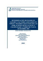
Figura 13. Frotis sanguíneo con 100% <strong>de</strong> hematíes infectados. Giemsa X 1000.<br />
Figura 14. Frotis sanguíneo con 100% <strong>de</strong> hematíes infectados. Giemsa X 1000.<br />
INSTITUTO NACIONAL DE SALUD<br />
30
DIAGNÓSTICO BACTERIOLÓGICO DE LA BARTONELOSIS HUMANA<br />
Figura 15. Frotis sanguíneo con 100% <strong>de</strong> hematíes infectados. Giemsa X 1000.<br />
Figura 16. Frotis sanguíneo con 40% <strong>de</strong> hematíes infectados con formas cocoi<strong>de</strong>s<br />
<strong>de</strong> B. bacilliformis. Giemsa X 1000.<br />
31<br />
INSTITUTO NACIONAL DE SALUD
DIAGNÓSTICO BACTERIOLÓGICO DE LA BARTONELOSIS HUMANA<br />
4.4. ERRORES DURANTE LA LECTURA DE LÁMINAS<br />
Los errores más frecuentes son confundir <strong>la</strong>s formas cocoi<strong>de</strong>s con punteado<br />
básofilo, precipitados <strong>de</strong>l colorante, célu<strong>la</strong>s <strong>de</strong> Heinz o con reticulocitos en<br />
los cuales se colorea el núcleo.<br />
La lectura también pue<strong>de</strong> ser obstaculizada en el caso <strong>de</strong> extendidos<br />
gruesos, que se colorean en tono oscuro y no se observan c<strong>la</strong>ramente <strong>la</strong>s<br />
formas cocoi<strong>de</strong>s.<br />
Figura 17. Frotis sanguíneo grueso, hematíes infectados con formas cocoi<strong>de</strong>s y<br />
baci<strong>la</strong>res. Giemsa X 1000.<br />
INSTITUTO NACIONAL DE SALUD<br />
32
DIAGNÓSTICO BACTERIOLÓGICO DE LA BARTONELOSIS HUMANA<br />
Figura 18. Frotis sanguíneo grueso con 100% <strong>de</strong> hematíes infectados<br />
con formas cocoi<strong>de</strong>s. Giemsa X 1000.<br />
4.5. DIAGNÓSTICO MEDIANTE CULTIVO<br />
El cultivo <strong>de</strong> <strong>la</strong>s muestras sanguíneas es importante para confirmar <strong>la</strong><br />
sospecha clínica, y con fines epi<strong>de</strong>miológicos.<br />
Figura 19. Medio <strong>de</strong> cultivo <strong>de</strong> agar Columbia con sangre <strong>de</strong> carnero.<br />
33<br />
INSTITUTO NACIONAL DE SALUD
DIAGNÓSTICO BACTERIOLÓGICO DE LA BARTONELOSIS HUMANA<br />
El ais<strong>la</strong>miento <strong>de</strong> Bartonel<strong>la</strong> bacilliformis se pue<strong>de</strong> realizar en los siguientes<br />
medios:<br />
• En agar Columbia con sangre <strong>de</strong> carnero al 10% en p<strong>la</strong>cas Petri.<br />
• En medio <strong>de</strong> cultivo bifásico distribuido en frascos, constituido por una<br />
fase sólida <strong>de</strong> agar Columbia con sangre <strong>de</strong> carnero al 10% y extracto<br />
<strong>de</strong> levadura al 0,1% y una fase líquida con medio RPMI suplementado<br />
con suero fetal bovino; una alternativa para <strong>la</strong> fase líquida es el medio<br />
RPMI sin suplemento o infusión cerebro corazón.<br />
• El mayor porcentaje <strong>de</strong> ais<strong>la</strong>mientos (70-80%) se logra en <strong>la</strong>s dos<br />
primeras semanas <strong>de</strong> incubación, en <strong>la</strong> tercera y cuarta semana se<br />
obtiene el resto (20-25%).<br />
4.5.1. Condición específica <strong>de</strong> <strong>la</strong> muestra<br />
• La muestra <strong>de</strong> sangre no <strong>de</strong>be <strong>de</strong> tener más <strong>de</strong> siete días <strong>de</strong> haber<br />
sido obtenida.<br />
• La muestra <strong>de</strong>be ser obtenida antes <strong>de</strong>l tratamiento con antibióticos.<br />
4.5.2. Materiales para el cultivo<br />
• Jeringa <strong>de</strong>scartable con aguja.<br />
• Alcohol <strong>de</strong> 70°.<br />
• Algodón.<br />
• Guantes estériles.<br />
• Contenedor <strong>de</strong> material contaminado.<br />
• Estufa <strong>de</strong> 28 a 29°C.<br />
• Pipetas <strong>de</strong> transferencia <strong>de</strong>scartables estériles.<br />
• Tubos al vacío conteniendo <strong>la</strong> muestra <strong>de</strong> sangre total.<br />
• Medios <strong>de</strong> cultivo.<br />
4.5.3. Preparación <strong>de</strong> <strong>la</strong> muestra sanguínea<br />
• Para incrementar el ais<strong>la</strong>miento <strong>de</strong> B. bacilliformis <strong>de</strong> <strong>la</strong> sangre es<br />
conveniente lisar los eritrocitos por medios mecánicos como el <strong>de</strong><br />
centrifugación <strong>de</strong> <strong>la</strong> muestra.<br />
4.5.4. Procedimiento para el cultivo en p<strong>la</strong>cas<br />
Colocar en <strong>la</strong> cabina <strong>de</strong> seguridad el material necesario para el cultivo <strong>de</strong><br />
<strong>la</strong> muestra sanguínea.<br />
INSTITUTO NACIONAL DE SALUD<br />
34
DIAGNÓSTICO BACTERIOLÓGICO DE LA BARTONELOSIS HUMANA<br />
• Dejar que los medios <strong>de</strong> cultivo tomen temperatura ambiente.<br />
• Rotu<strong>la</strong>r con el código <strong>de</strong>l paciente <strong>la</strong>s p<strong>la</strong>cas con agar Columbia sangre.<br />
• Desinfectar con alcohol <strong>de</strong> 70º <strong>la</strong> tapa <strong>de</strong> jebe <strong>de</strong>l tubo al vacío que<br />
contiene <strong>la</strong> muestra.<br />
• Extraer <strong>la</strong> muestra con una jeringa, a través <strong>de</strong> <strong>la</strong> tapa <strong>de</strong> jebe o con<br />
pipeta <strong>de</strong> transferencia.<br />
• Inocu<strong>la</strong>r <strong>de</strong> 0,5 a 1mL <strong>de</strong> sangre sobre el agar Columbia, <strong>de</strong>jando que<br />
<strong>la</strong> sangre fluya sobre <strong>la</strong> superficie <strong>de</strong>l medio, luego cerrar <strong>la</strong> p<strong>la</strong>ca.<br />
• Colocar <strong>la</strong> p<strong>la</strong>ca con <strong>la</strong> muestra sembrada en bolsas <strong>de</strong> polietileno <strong>de</strong><br />
baja <strong>de</strong>nsidad autosel<strong>la</strong>bles para mantener <strong>la</strong> humedad <strong>de</strong>l medio <strong>de</strong><br />
cultivo que <strong>de</strong>bido al <strong>la</strong>rgo período <strong>de</strong> incubación se seca y para evitar<br />
contaminación con hongos saprofitos.<br />
• Incubar el cultivo a 28 ºC hasta por 45 días.<br />
En el caso que el <strong>la</strong>boratorio no tenga <strong>la</strong>s facilida<strong>de</strong>s i<strong>de</strong>ales para po<strong>de</strong>r<br />
realizar el cultivo sin correr el peligro <strong>de</strong> contaminarlo, se <strong>de</strong>be usar un<br />
mechero <strong>de</strong> gas que dé a su alre<strong>de</strong>dor un amplio radio (espacio) <strong>de</strong><br />
esterilidad o, <strong>de</strong> lo contrario se <strong>de</strong>be utilizar el medio <strong>de</strong> cultivo bifásico.<br />
Figura 20. Siembra <strong>de</strong>l medio en p<strong>la</strong>cas <strong>de</strong> agar Columbia con sangre <strong>de</strong> carnero.<br />
35<br />
INSTITUTO NACIONAL DE SALUD
DIAGNÓSTICO BACTERIOLÓGICO DE LA BARTONELOSIS HUMANA<br />
INSTITUTO NACIONAL DE SALUD<br />
Figura 21. Sel<strong>la</strong>do <strong>de</strong> <strong>la</strong>s p<strong>la</strong>cas en bolsas <strong>de</strong> polietileno.<br />
4.5.5. Procedimiento para el cultivo en medio bifásico:<br />
• Colocar en <strong>la</strong> mesa <strong>de</strong> trabajo <strong>la</strong>s muestras <strong>de</strong> sangre total, contenidas<br />
en tubos al vacío con anticoagu<strong>la</strong>nte.<br />
• Dejar que los medios tomen temperatura ambiente.<br />
• Levantar el diafragma y <strong>de</strong>sinfectar <strong>la</strong> tapa <strong>de</strong> jebe <strong>de</strong>l frasco con medio<br />
<strong>de</strong> cultivo, con alcohol <strong>de</strong> 70º o alcohol yodado.<br />
• Extraer con una jeringa <strong>la</strong> sangre total <strong>de</strong>l tubo que contiene <strong>la</strong> muestra<br />
<strong>de</strong> sangre.<br />
• Inmediatamente, inocu<strong>la</strong>r a través <strong>de</strong>l tapón <strong>de</strong> jebe 0,5 a 1mL <strong>de</strong> sangre<br />
al frasco con medio <strong>de</strong> cultivo bañando <strong>la</strong> fase sólida.<br />
• Si el medio <strong>de</strong> cultivo se encuentra en frascos con tapa rosca, <strong>de</strong>stapar<br />
e inocu<strong>la</strong>r <strong>la</strong> muestra <strong>de</strong> sangre con una pipeta <strong>de</strong> transferencia estéril.<br />
Este procedimiento <strong>de</strong>be realizarse cerca <strong>de</strong> un mechero.<br />
• Se <strong>de</strong>ja reposar el frasco inclinado hasta que <strong>la</strong> sangre se impregne en<br />
el agar por 30 minutos.<br />
• Descartar <strong>la</strong> aguja y <strong>la</strong> jeringa en un contenedor resistente a <strong>la</strong>s<br />
punturas.<br />
• Limpiar con un algodón empapado con alcohol <strong>la</strong> tapa <strong>de</strong>l frasco.<br />
• Incubar el cultivo a 28-29 °C hasta 45 días.<br />
36
4.5.6. Lectura <strong>de</strong> cultivos<br />
Cultivos en p<strong>la</strong>cas:<br />
DIAGNÓSTICO BACTERIOLÓGICO DE LA BARTONELOSIS HUMANA<br />
• Examinar los cultivos visualmente con luz transmitida o con una lupa,<br />
todos los días hasta por 45 días.<br />
• Si se ve <strong>de</strong>sarrollo <strong>de</strong> colonias entre <strong>la</strong>s 24 y 72 horas, no se trata <strong>de</strong> B.<br />
bacilliformis. Efectuar un examen microscópico <strong>de</strong> <strong>la</strong>s colonias, realizar<br />
el frotis <strong>de</strong> una colonia y teñir<strong>la</strong> con colorante Gram para <strong>de</strong>terminar <strong>la</strong><br />
forma bacteriana, y si son Gram positivas o negativas. I<strong>de</strong>ntificar <strong>la</strong><br />
bacteria mediante pruebas bioquímicas y serológicas; podría tratarse<br />
<strong>de</strong> una bacteria que esté causando una complicación al paciente, por<br />
lo que es necesario i<strong>de</strong>ntificar<strong>la</strong> y realizar el antibiograma<br />
correspondiente.<br />
• En el ais<strong>la</strong>miento primario <strong>la</strong>s colonias <strong>de</strong> B. bacilliformis se <strong>de</strong>sarrol<strong>la</strong>n<br />
entre cuatro días a seis semanas <strong>de</strong> incubación, crecen lentamente.<br />
Son colonias pequeñas <strong>de</strong> apenas 1 mm <strong>de</strong> diámetro, translúcidas<br />
tipo rocío o b<strong>la</strong>nquecinas. Realizar el examen <strong>de</strong>l frotis <strong>de</strong> una colonia<br />
y teñir<strong>la</strong> con colorante Gram para observar <strong>la</strong> forma bacteriana.<br />
Bartonel<strong>la</strong> bacilliformis colorea muy débilmente con <strong>la</strong> tinción <strong>de</strong> Gram.<br />
• Realizar un subcultivo <strong>de</strong> <strong>la</strong>s colonias sospechosas.<br />
• Los cultivos pue<strong>de</strong>n ser crioconservados en medio <strong>de</strong> infusión cerebro<br />
corazón con 10% <strong>de</strong> glicerol a -70 ºC, en el Laboratorio <strong>de</strong> Referencia<br />
Regional o Nacional (punto 4.4.).<br />
Figura 22. Cultivo puro en p<strong>la</strong>cas con agar Columbia<br />
con sangre <strong>de</strong> cinco a seís días <strong>de</strong> incubación.<br />
37<br />
INSTITUTO NACIONAL DE SALUD
DIAGNÓSTICO BACTERIOLÓGICO DE LA BARTONELOSIS HUMANA<br />
Figura 23. Cultivo puro en p<strong>la</strong>cas con agar Columbia con sangre <strong>de</strong> siete<br />
días <strong>de</strong> incubación.<br />
Figura 24. Cultivo puro en p<strong>la</strong>cas con agar Columbia con sangre <strong>de</strong><br />
nueve días <strong>de</strong> incubación.<br />
INSTITUTO NACIONAL DE SALUD<br />
38
Cultivos en medio bifásico:<br />
DIAGNÓSTICO BACTERIOLÓGICO DE LA BARTONELOSIS HUMANA<br />
• En el caso <strong>de</strong>l medio bifásico, bañar <strong>la</strong> fase sólida <strong>de</strong>l medio con <strong>la</strong><br />
fase líquida inclinándo<strong>la</strong> suavemente cada cuatro o cinco días.<br />
• Si a <strong>la</strong>s 24 ó 48 horas se nota <strong>de</strong>sarrollo <strong>de</strong> colonias sobre el p<strong>la</strong>no<br />
inclinado <strong>de</strong> <strong>la</strong> fase sólida o se nota turbi<strong>de</strong>z o lisis <strong>de</strong> los hematíes en<br />
<strong>la</strong> fase líquida, realizar un subcultivo en p<strong>la</strong>cas <strong>de</strong> agar sangre y Mac<br />
Conkey, y proce<strong>de</strong>r a <strong>la</strong> i<strong>de</strong>ntificación <strong>de</strong>l microorganismo; no se trata<br />
<strong>de</strong> B. bacilliformis, pue<strong>de</strong> ser una infección sobreagregada.<br />
• Si se observa crecimiento <strong>de</strong> colonias, turbi<strong>de</strong>z o hemólisis <strong>de</strong>spués<br />
<strong>de</strong> seis a siete días <strong>de</strong> incubación, realizar un subcultivo en p<strong>la</strong>cas con<br />
agar Columbia sangre.<br />
• Si no se observa crecimiento <strong>de</strong> colonias o turbi<strong>de</strong>z <strong>de</strong>spués siete u<br />
ocho días, realizar subcultivos ciegos a los 15 días.<br />
• Los cultivos <strong>de</strong>ben seguir incubándose hasta por 45 días.<br />
• Si se carece <strong>de</strong> una incubadora <strong>de</strong> <strong>la</strong> temperatura indicada, mantener<br />
los frascos <strong>de</strong> cultivo a temperatura ambiente y enviarlos tan pronto<br />
como sea posible al Laboratorio <strong>de</strong> Referencia Regional o al Laboratorio<br />
<strong>de</strong> Referencia <strong>de</strong> Bartonel<strong>la</strong> <strong>de</strong>l INS.<br />
Figura 25. Cultivos en medio bifásicos.<br />
39<br />
INSTITUTO NACIONAL DE SALUD
DIAGNÓSTICO BACTERIOLÓGICO DE LA BARTONELOSIS HUMANA<br />
4.3.7. Subcultivos<br />
INSTITUTO NACIONAL DE SALUD<br />
Figura 26. Observación <strong>de</strong> colonias en medio bifásico.<br />
• Los subcultivos <strong>de</strong>ben realizarse en cabina <strong>de</strong> bioseguridad.<br />
• Desinfectar <strong>la</strong> tapa <strong>de</strong>l frasco <strong>de</strong> hemocultivo con alcohol 70 º.<br />
• Con una jeringa <strong>de</strong> tuberculina estéril, extraer 200 µL <strong>de</strong> <strong>la</strong> fase líquida<br />
<strong>de</strong>l medio e inocu<strong>la</strong>r en <strong>la</strong> superficie <strong>de</strong> p<strong>la</strong>cas con agar Columbia<br />
sangre y colocar una gota sobre una lámina para hacer un frotis y<br />
coloración Gram.<br />
• Los subcultivos también pue<strong>de</strong>n realizarse en frascos con medio<br />
bifásico.<br />
• Observar <strong>la</strong> lámina coloreada con Gram y buscar formas bacterianas.<br />
• Incubar los cultivos a 28-29 ºC y observar hasta por 45 días.<br />
• Observar los cultivos con luz transmitida todos los días.<br />
• En el caso que se haya realizado <strong>la</strong> siembra primaria en p<strong>la</strong>cas con<br />
agar Columbia y se noten colonias sospechosas, pue<strong>de</strong> hacer un cultivo<br />
usando un asa <strong>de</strong> siembra, coger una colonia por puntura, se pue<strong>de</strong><br />
replicar en otra p<strong>la</strong>ca con agar Columbia, o se pue<strong>de</strong> inocu<strong>la</strong>r<strong>la</strong> en<br />
frascos con tapa rosca conteniendo medio bifásico.<br />
40
Lectura <strong>de</strong> los subcultivos<br />
DIAGNÓSTICO BACTERIOLÓGICO DE LA BARTONELOSIS HUMANA<br />
Después <strong>de</strong> varios pasajes <strong>de</strong> cultivos o subcultivos sucesivos, el<br />
crecimiento <strong>de</strong> <strong>la</strong>s colonias es más rápido y se hacen visibles <strong>de</strong>spués <strong>de</strong><br />
tres a cuatro días <strong>de</strong> incubación. Generalmente, <strong>la</strong>s colonias crecen entre<br />
los 4 y 14 días; sin embargo, <strong>de</strong>be observarse el subcultivo hasta los 45<br />
días.<br />
4.5.8. I<strong>de</strong>ntificación <strong>de</strong> Bartonel<strong>la</strong> bacilliformis<br />
La i<strong>de</strong>ntificación <strong>de</strong>l género se realiza por:<br />
• Las condiciones requeridas para el <strong>de</strong>sarrollo bacteriano, como:<br />
1.La temperatura <strong>de</strong> crecimiento, entre 28 - 29 ºC.<br />
2.El período <strong>de</strong> incubación, <strong>de</strong>sarrol<strong>la</strong> <strong>de</strong>s<strong>de</strong> los 4 hasta 45 días.<br />
3.Desarrol<strong>la</strong> sólo en medios enriquecidos.<br />
4.El ais<strong>la</strong>miento primario es lento.<br />
• No produce hemólisis en agar Columbia con sangre <strong>de</strong> carnero.<br />
• Son Gram negativos.<br />
• Las colonias son pequeñas, puntiformes y translúcidas, <strong>de</strong> bor<strong>de</strong><br />
entero; otras son ligeramente más gran<strong>de</strong>s b<strong>la</strong>nquecinas, <strong>de</strong> bor<strong>de</strong><br />
entero y se adhieren a <strong>la</strong> superficie <strong>de</strong>l agar, esta adherencia es una<br />
característica fenotípica <strong>de</strong> algunas colonias. La morfología <strong>de</strong> <strong>la</strong> colonia<br />
y <strong>la</strong>s características <strong>de</strong> crecimiento probablemente estén re<strong>la</strong>cionadas<br />
con <strong>la</strong> patogenicidad <strong>de</strong>l microorganismo.<br />
• No crecen en medio <strong>de</strong> agar Mac Conkey o agar nutritivo.<br />
• No fermentan los azúcares, son bioquímicamente inertes.<br />
• No crece a 37 ºC.<br />
• Son oxidasa negativa, cata<strong>la</strong>sa positiva, ureasa e indol negativo.<br />
• Son móviles, lo que se observa en el medio <strong>de</strong> gel <strong>de</strong> fases, forman<br />
una banda <strong>de</strong> <strong>de</strong>sarrollo bacteriano <strong>de</strong> aproximadamente 6 mm, con<br />
migración <strong>de</strong>l cultivo <strong>de</strong>s<strong>de</strong> el trazo <strong>de</strong> inocu<strong>la</strong>ción hacia <strong>la</strong>s pare<strong>de</strong>s<br />
<strong>de</strong>l tubo.<br />
41<br />
INSTITUTO NACIONAL DE SALUD
DIAGNÓSTICO BACTERIOLÓGICO DE LA BARTONELOSIS HUMANA<br />
5.4. CRiOCONSERVACIÓN DE LAS CEPAS<br />
Los ais<strong>la</strong>mientos que tengan características compatibles a B. bacilliformis<br />
<strong>de</strong>ben ser almacenados bajo condiciones <strong>de</strong> crioconservación.<br />
Procedimiento:<br />
INSTITUTO NACIONAL DE SALUD<br />
Figura 27. Medio gel <strong>de</strong> fases.<br />
a. Si el subcultivo es en p<strong>la</strong>ca: Si se tienen subcultivos positivos y con<br />
colonias <strong>de</strong> un solo tipo, coger con una asa <strong>de</strong> siembra <strong>la</strong>s colonias<br />
(cosechar) e inocu<strong>la</strong>r<strong>la</strong>s en crioviales con medio infusión cerebro corazón<br />
con glicerol 10%.<br />
b. Si el subcultivo es en medio bifásico: Coger con una pipeta estéril <strong>de</strong><br />
0,5 - 2 mL <strong>de</strong> <strong>la</strong> fase líquida <strong>de</strong>l medio e inocu<strong>la</strong>r<strong>la</strong>s en crioviales.<br />
c. Las cepas <strong>de</strong>ben guardarse por duplicado o más a -20 o -70 ºC.<br />
42
DIAGNÓSTICO BACTERIOLÓGICO DE LA BARTONELOSIS HUMANA<br />
SECCIÓN 5<br />
BIBLIOGRAFÍA<br />
Birtles R, Fry NK, Ventosil<strong>la</strong> P, Cáceres AG, Sanchez E, Vizcarra H, et al.<br />
I<strong>de</strong>ntification of Bartonel<strong>la</strong> bacilliformis genotypes and their relevance to<br />
epi<strong>de</strong>miological investigations of human bartonellosis. J Clin Microbiol<br />
2002 ; 40(10): 3606-12.<br />
An<strong>de</strong>rson BE, Neuman A. Bartonel<strong>la</strong> spp. as emerging human pathogens.<br />
Clin Microbiol Rev 1997; 10(2): 203-19.<br />
Colichón AH, Colichón A, So<strong>la</strong>no L. La Bartonel<strong>la</strong> bacilliformis en el medio<br />
<strong>de</strong> fases. Arch Peru Pat Clin 1971; 25(1): 15-32.<br />
Cuadra M, Cuadra AM. Enfermedad <strong>de</strong> Carrión: inocu<strong>la</strong>ciones <strong>de</strong> seres<br />
humanos con Bartonel<strong>la</strong> bacilliformis, una revisión. An Fac Med 2000; 61(4):<br />
289-94.<br />
Dehio C. Molecu<strong>la</strong>r and cellu<strong>la</strong>r basis of Bartonel<strong>la</strong> pathogenesis. Ann Rev<br />
Microbiol 2004; 58: 365-90.<br />
Ellis BA, Rotz LD, Leake JA, Samalvi<strong>de</strong>s F, Bernable J, Ventura G, et al. An<br />
outbreak of acute bartonellosis (Oroya fever) in the Urubamba region of<br />
Peru, 1988. Am J Trop Med Hyg 1998; 6(12): 344-49.<br />
Garrity G, Winters M, Searles D. Taxonomic Outline of the Procaryotic Genera.<br />
En: Garrity GM, Holt JN. Bergey’s Manual of Systematic Bacteriology, 2 nd<br />
edition. Berlin: Springer-Ver<strong>la</strong>g KG; 2001.<br />
Huarcaya E, Chinga E, Chavez J, Chauca J, L<strong>la</strong>nos-Cuentas A, Maguiña C,<br />
et al. Influencia <strong>de</strong>l fenómeno <strong>de</strong> El Niño em <strong>la</strong> epi<strong>de</strong>miología <strong>de</strong> <strong>la</strong><br />
Bartonelosis <strong>humana</strong> en los <strong>de</strong>partamentos <strong>de</strong> Ancash y cusco entre 1996<br />
y 1999. Rev Med Hered 2004; 15(1): 4-10.<br />
Instituto Nacional <strong>de</strong> Salud. Manual <strong>de</strong> procedimientos. Bioseguridad en<br />
<strong>la</strong>boratorios <strong>de</strong> ensayo, biomédicos y clínicos. 3 ra ed. Lima: INS; 2005.<br />
Serie <strong>de</strong> Normas Técnicas Nº 18.<br />
Instituto Nacional <strong>de</strong> Salud. Manual <strong>de</strong> procedimientos <strong>de</strong> <strong>la</strong>boratorio para<br />
el <strong>diagnóstico</strong> histopatológico. Lima: INS; 1997. Serie <strong>de</strong> Normas Técnicas<br />
N º24.<br />
Instituto Nacional <strong>de</strong> Salud. Manual <strong>de</strong> procedimientos <strong>de</strong> electroforesis<br />
para proteínas y ADN. Lima: INS; 2003. Serie <strong>de</strong> Normas Técnicas Nº 38.<br />
43<br />
INSTITUTO NACIONAL DE SALUD
DIAGNÓSTICO BACTERIOLÓGICO DE LA BARTONELOSIS HUMANA<br />
Maguiña C. Bartonelosis o <strong>enfermedad</strong> <strong>de</strong> Carrión. Lima: Editorial AFA Import<br />
SA; 1998.<br />
Minnick MF, Mitchell SJ, McAllister SJ. Cell entry and pathogenesis of<br />
Bartonel<strong>la</strong> infections. Trends Microbiol 1996; 4(9): 343-47.<br />
Perú, Ministerio <strong>de</strong> Salud. Doctrina, normas y procedimientos para el control<br />
<strong>de</strong> <strong>la</strong> Bartonelosis o <strong>enfermedad</strong> <strong>de</strong> Carrión en el Perú. Lima: MINSA; 1998.<br />
Public Health Agency of Canada. Bartonel<strong>la</strong> bacilliformis [página <strong>de</strong><br />
internet]. Otawa: Office of Laboratory Security, Material Safety Data Sheets,<br />
Infectious Sustances; 2001. Fecha <strong>de</strong> acceso: enero <strong>de</strong>l 2005. Disponible<br />
en: www.phac-aspc.ge.ca/msds-ftss/msds16e.html.<br />
Rebagliatti R. Verruga Peruana (Enfermedad <strong>de</strong> Carrión) Lima; Imprenta<br />
Torres Aguirre; 1940.<br />
Saettone-León A. Verruga Peruana. Dermatol Peru 2004; 14(1-2): 121-33.<br />
Vil<strong>la</strong>seca P, Padil<strong>la</strong> C, Ventura G, Samalvi<strong>de</strong>s F, Yañez H, Chevarria L, et al.<br />
Importancia <strong>de</strong> <strong>la</strong> Lutzomia peruensis en <strong>la</strong> transmisión <strong>de</strong> <strong>la</strong> <strong>enfermedad</strong><br />
<strong>de</strong> Carrión en el Valle Sagrado <strong>de</strong> los Incas, Urubamba-Cusco, Perú. Rev<br />
Med Exp 1999; 16(1-2): 28-30.<br />
INSTITUTO NACIONAL DE SALUD<br />
44
DIAGNÓSTICO BACTERIOLÓGICO DE LA BARTONELOSIS HUMANA<br />
ANEXO 1<br />
PREPARACIÓN DE REACTIVOS Y COLORANTE GIEMSA<br />
1. PREPARACIÓN DE LA SOLUCIÓN ALCOHÓLICA GIEMSA (solución madre)<br />
Fórmu<strong>la</strong>:<br />
Colorante Giemsa en polvo, certificado 0,75 g<br />
Alcohol metílico puro. 65 mL<br />
Glicerina pura 35 mL<br />
Preparación:<br />
• A un frasco <strong>de</strong> vidrio <strong>de</strong> color ámbar conteniendo per<strong>la</strong>s <strong>de</strong> vidrio <strong>de</strong><br />
diámetro no mayor <strong>de</strong> 5 mm, se agrega <strong>la</strong> glicerina; luego se adiciona<br />
el colorante Giemsa, se va agitando. Finalmente, se adiciona el alcohol<br />
metílico.<br />
• Agitar el frasco intensamente varias veces al día, durante tres días como<br />
mínimo.<br />
• Extraer diariamente una pequeña muestra <strong>de</strong>l colorante preparado,<br />
filtrar<strong>la</strong> a través <strong>de</strong> papel filtro # 2, y probar con frotis sanguíneos recién<br />
preparados.<br />
• Cuando los elementos <strong>de</strong> <strong>la</strong> sangre aparezcan con sus colores<br />
apropiados, entonces el colorante está listo para ser utilizado.<br />
2. PREPARACIÓN DEL DILUYENTE (solución amortiguadora)<br />
El diluyente es una solución amortiguadora y se usa en <strong>la</strong> preparación <strong>de</strong>l<br />
colorante diluido o solución <strong>de</strong> trabajo.<br />
Las sustancias amortiguadoras actúan como un adaptador que inactiva<br />
<strong>de</strong>ntro <strong>de</strong> un margen limitado cantida<strong>de</strong>s variables <strong>de</strong> ácido o álcali.<br />
Fórmu<strong>la</strong>:<br />
Ortofosfato disodico anhidro (Na HPO ) 2 4 4 g<br />
Ortofosfato monopotásico (KH PO ) 2 4 5 g<br />
Preparación:<br />
Mezc<strong>la</strong>r bien, y disolver 1g <strong>de</strong> <strong>la</strong> mezc<strong>la</strong> en un litro <strong>de</strong> agua <strong>de</strong>sti<strong>la</strong>da y<br />
ajustar el pH a 7,2.<br />
45<br />
INSTITUTO NACIONAL DE SALUD
DIAGNÓSTICO BACTERIOLÓGICO DE LA BARTONELOSIS HUMANA<br />
3. PREPARACIÓN DEL COLORANTE DILUIDO O SOLUCIÓN DE TRABAJO<br />
Fórmu<strong>la</strong>:<br />
Solución madre Giemsa. 0,05 mL<br />
Solución amortiguadora o diluyente. 0,95 mL<br />
Preparación:<br />
En una probeta <strong>de</strong> vidrio se aña<strong>de</strong> <strong>la</strong> solución madre Giemsa al 5% con <strong>la</strong><br />
solución amortiguadora.<br />
Una forma más rápida <strong>de</strong> preparar el colorante diluido es agregando una<br />
gota <strong>de</strong> solución madre por mL <strong>de</strong> solución amortiguadora.<br />
Después <strong>de</strong> preparado el colorante madre, se <strong>de</strong>be <strong>de</strong>terminar el tiempo<br />
<strong>de</strong> coloración que pue<strong>de</strong> ser entre 15 - 20 minutos y <strong>la</strong> dilución en <strong>la</strong> que se<br />
<strong>de</strong>be preparar el colorante diluido, estas dos características se <strong>de</strong>ben<br />
ajustar en función al estado <strong>de</strong>l colorante madre.<br />
Recomendaciones:<br />
• Mantener el frasco <strong>de</strong> vidrio color ámbar, que contiene <strong>la</strong> solución madre<br />
<strong>de</strong> Giemsa bien cerrado y en un lugar fresco, seco y protegido <strong>de</strong> <strong>la</strong> luz<br />
so<strong>la</strong>r directa para evitar <strong>la</strong> vo<strong>la</strong>tilización <strong>de</strong>l solvente y <strong>la</strong> oxidación <strong>de</strong>l<br />
colorante.<br />
• El material <strong>de</strong> vidrio como probetas, pipetas, cubetas y embudos<br />
empleados en <strong>la</strong> preparación <strong>de</strong> los reactivos y colorantes <strong>de</strong>ben estar<br />
limpios y secos.<br />
• El frasco <strong>de</strong> vidrio usado en <strong>la</strong> preparación <strong>de</strong>l colorante <strong>de</strong>be ser <strong>la</strong>vado<br />
con <strong>de</strong>tergente y enjuagado hasta que no que<strong>de</strong>n residuos <strong>de</strong> éste, ya<br />
que pue<strong>de</strong> alterar el pH <strong>de</strong>l colorante y estropearlo.<br />
• No se <strong>de</strong>be añadir agua a <strong>la</strong> solución madre Giemsa, porque <strong>la</strong> mínima<br />
cantidad <strong>de</strong> ésta <strong>de</strong>teriora el colorante.<br />
• No agitar el frasco <strong>de</strong>l colorante antes <strong>de</strong> utilizarlo porque se suspen<strong>de</strong>n<br />
pequeños cristales <strong>de</strong> éste que no han sido disueltos, y se quedan en<br />
los frotis sanguíneos durante el proceso <strong>de</strong> coloración formando<br />
precipitados.<br />
• Debe <strong>de</strong>scartarse el colorante diluido no utilizado.<br />
INSTITUTO NACIONAL DE SALUD<br />
46
DIAGNÓSTICO BACTERIOLÓGICO DE LA BARTONELOSIS HUMANA<br />
ANEXO 2<br />
PREPARACIÓN DE MEDIOS DE CULTIVO MONOFÁSICO<br />
Para preparar los medios <strong>de</strong> cultivo, <strong>la</strong> sangre <strong>de</strong> carnero <strong>de</strong>be ser estéril.<br />
Esto se comprueba mediante el cultivo <strong>de</strong> una muestra <strong>de</strong> sangre <strong>de</strong> carnero<br />
en medios <strong>de</strong> cultivo <strong>de</strong> agar soya tripticase y agar Mac Conkey; incubar por<br />
24 y 48 horas.<br />
MEDIO MONOFÁSICO:<br />
Fórmu<strong>la</strong>:<br />
Fase sólida<br />
• Medio comercial agar Columbia<br />
• Sangre <strong>de</strong> carnero <strong>de</strong>sfibrinada 10%<br />
• Extracto <strong>de</strong> levadura 0,1%<br />
Procedimiento:<br />
• Disolver 44 g <strong>de</strong>l medio <strong>de</strong>shidratado y 1 g <strong>de</strong> extracto <strong>de</strong> levadura en<br />
1000 mL <strong>de</strong> agua <strong>de</strong>sti<strong>la</strong>da, mezc<strong>la</strong>r hasta obtener una solución<br />
homogénea. Calentar agitando hasta <strong>la</strong> ebullición para disolver<br />
completamente, mantener a un pH 7,4.<br />
• Esterilizar a 121 °C durante 15 minutos.<br />
• Cuando el medio se encuentra a 50 °C, añadir en forma aséptica 10%<br />
<strong>de</strong> sangre <strong>de</strong>sfibrinada <strong>de</strong> carnero, agitar suavemente para obtener<br />
una mezc<strong>la</strong> uniforme y repartir en p<strong>la</strong>cas Petri.<br />
• Dejar que solidifique y contro<strong>la</strong>r esterilidad.<br />
• Almacenar hasta por 25 días en refrigeración.<br />
47<br />
INSTITUTO NACIONAL DE SALUD
DIAGNÓSTICO BACTERIOLÓGICO DE LA BARTONELOSIS HUMANA<br />
INSTITUTO NACIONAL DE SALUD<br />
ANEXO 3<br />
PREPARACIÓN DE MEDIOS DE CULTIVO BIFÁSICO<br />
Para preparar los medios <strong>de</strong> cultivo, <strong>la</strong> sangre <strong>de</strong> carnero <strong>de</strong>be ser estéril.<br />
Esto se comprueba mediante el cultivo <strong>de</strong> una muestra <strong>de</strong> sangre <strong>de</strong> carnero<br />
en medios <strong>de</strong> cultivo <strong>de</strong> agar soya tripticase y agar Mac Conkey; incubar por<br />
24 y 48 horas.<br />
MEDIO BIFÁSICO<br />
Fórmu<strong>la</strong>:<br />
Fase sólida<br />
• Agar Columbia<br />
• Sangre <strong>de</strong> carnero <strong>de</strong>sfibrinada 10%<br />
• Extracto <strong>de</strong> levadura 0,1%<br />
Fase Líquida<br />
• RPMI 1640 con 10% <strong>de</strong> suero bovino fetal (<strong>la</strong> casa comercial Gibco<br />
distribuye el medio <strong>de</strong> RPMI por litro).<br />
Procedimiento:<br />
• Disolver 44 g <strong>de</strong>l medio <strong>de</strong>shidratado y 1 g <strong>de</strong> extracto <strong>de</strong> levadura en<br />
1000 mL <strong>de</strong> agua <strong>de</strong>sti<strong>la</strong>da, mezc<strong>la</strong>r hasta obtener una solución<br />
homogénea. Calentar agitando hasta <strong>la</strong> ebullición para disolver<br />
completamente, mantener a un pH 7,4.<br />
• Esterilizar a 121 °C durante 15 minutos.<br />
• Cuando el medio se encuentra a 50 °C, añadir en forma aséptica 10%<br />
<strong>de</strong> sangre <strong>de</strong>sfibrinada <strong>de</strong> carnero. Agitar suavemente para obtener<br />
una mezc<strong>la</strong> uniforme y repartir en frascos <strong>de</strong> 50 mL.<br />
• Inclinar los frascos sobre una <strong>de</strong> <strong>la</strong>s pare<strong>de</strong>s <strong>de</strong>l frasco y <strong>de</strong>jar que se<br />
solidifique el medio en p<strong>la</strong>no inclinado.<br />
• Contro<strong>la</strong>r su esterilidad durante 72 horas.<br />
• Preparar <strong>la</strong> fase líquida <strong>de</strong>l medio <strong>de</strong> acuerdo con <strong>la</strong> cantidad <strong>de</strong> frascos<br />
que va a preparar.<br />
• El pH <strong>de</strong>l medio RPMI 1640 <strong>de</strong>be <strong>de</strong> ser 7,4.<br />
• Al medio <strong>de</strong> RPMI añadir el 10% <strong>de</strong> SBF estéril.<br />
48
DIAGNÓSTICO BACTERIOLÓGICO DE LA BARTONELOSIS HUMANA<br />
• Añadir 3 mL <strong>de</strong> <strong>la</strong> fase líquida a cada frasco que contiene <strong>la</strong> fase sólida,<br />
este procedimiento <strong>de</strong>be ser realizado en condiciones <strong>de</strong> esterilidad.<br />
• Colocar el precinto <strong>de</strong> metal al frasco.<br />
• Contro<strong>la</strong>r su esterilidad por 24 horas.<br />
• Almacenar en refrigeración hasta por 25 días.<br />
49<br />
INSTITUTO NACIONAL DE SALUD
DIAGNÓSTICO BACTERIOLÓGICO DE LA BARTONELOSIS HUMANA<br />
INSTITUTO NACIONAL DE SALUD<br />
ANEXO 4<br />
PREPARACIÓN DE MEDIO GEL DE FASES<br />
A. CALDO PARA LA GELIFICACIÓN<br />
Bacto tryptosa 10 g<br />
Cloruro <strong>de</strong> sodio 7 g<br />
Agua <strong>de</strong>sti<strong>la</strong>da 1000 ml<br />
Disolver los ingredientes en agua <strong>de</strong>sti<strong>la</strong>da, ajustar a pH 7,0 - 7,2.<br />
Esterilizar a 15 lb X 15 minutos.<br />
B. GEL DE FASES<br />
A 90 mL <strong>de</strong>l caldo <strong>de</strong> gelificación se le aña<strong>de</strong> 1,5 mL <strong>de</strong> ClCa + al 2%<br />
(esterilizado por filtración).<br />
Añadir 10 mL <strong>de</strong> p<strong>la</strong>sma <strong>de</strong> carnero.<br />
Repartir inmediatamente 5 mL <strong>de</strong>l caldo p<strong>la</strong>sma en tubos con agar sangre.<br />
Dejar en posición vertical por 24 horas, luego llevar a 37 ºC para su<br />
gelificación y control <strong>de</strong> esterilidad<br />
50
DIAGNÓSTICO BACTERIOLÓGICO DE LA BARTONELOSIS HUMANA<br />
ANEXO 5<br />
PREPARACIÓN DE MEDIO DE CRIOCONSERVACIÓN DE CEPAS<br />
CALDO INFUSIÓN CEREBRO CORAZÓN (BHI)<br />
• Preparar 100 mL <strong>de</strong>l medio según <strong>la</strong>s indicaciones <strong>de</strong>l fabricante y<br />
luego añadir 10% <strong>de</strong> glicerol, repartir en crioviales y esterilizar por calor<br />
húmedo a 15 lb por 15 minutos.<br />
• Almacenar en refrigeración.<br />
51<br />
INSTITUTO NACIONAL DE SALUD
CEPREDIM<br />
SE TERMINÓ DE IMPRIMIR<br />
EN EL MES DE SETIEMBRE DE 2006,<br />
POR ENCARGO DEL INSTITUTO NACIONAL DE SALUD,<br />
EN LOS TALLERES GRÁFICOS DEL<br />
CENTRO DE PRODUCCIÓN EDITORIAL E IMPRENTA DE<br />
LA UNIVERSIDAD NACIONAL MAYOR DE SAN MARCOS<br />
JR. PARURO 119. LIMA 1.<br />
TELÉFONO: 619-7000 ANEXOS: 6011, 6015/ FAX: 6009<br />
E-MAIL: CEPEDIT@UNMSM.EDU.PE<br />
TIRAJE: 1000 EJEMPLARES