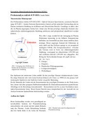
Zeitliche Einordnung Kreide
Zeitliche Einordnung Kreide
Zeitliche Einordnung Kreide

Erfolgreiche ePaper selbst erstellen
Machen Sie aus Ihren PDF Publikationen ein blätterbares Flipbook mit unserer einzigartigen Google optimierten e-Paper Software.
CASE REPORTFigure 2. This was the wound after the firstapplication of maggot therapy. At this stage therewas noticeable reduction in the size of the woundbed although extensive areas of slough were stillpresent. The margins of the pressure ulcers areshowing signs of epithelialisation. The skin stillremains red and inflamed due to a large amountof exudate from the wound.Figure 3. At this stage there was a hugeimprovement in the wound. Only one sloughy arearemained, the wounds had reduced considerablyin size and less exudate was noted. There wasnoticeable regeneration of new tissue and thesurrounding skin was less inflamed.Figure 4. Following the last application of maggotsthe wound on one side of the sacrum wascompletely healed, the other area had reduced insize to 2cm x 2cm and the depth of the woundwas approximately 2cm.tramadol (100mg) slow release morningand night and tramadol 50mg four timesa day.Due to the extent of the pressure ulcer onthe patient’s sacrum and the fact that theskin was necrotic in some areas and sloughyin others the district nurses suggested theuse of maggot therapy. LarvE BioFOAMdressing was ordered as the patient wasnot keen on the idea of free-range maggotsbeing placed in the wound.ZooBiotic Ltd’s (Bridgend) product LarvEBioFOAM has maggots contained in foampouches. The dressing makes the patientfeel more at ease as they do not worryabout the free-range maggots escapingor worrying that the the maggots havenot been removed when the treatment iscompleted. The pouches are easy to place inthe wound and also easy to remove.84 Wounds UK, 2009, Vol 5, No 1Maggot therapyThe first application of maggot therapywas in February 2008. The condition of thewound at this stage was very poor. Therewere large necrotic areas, the wound wasalso sloughy in other areas and exudinglarge amounts of offensive smelling exudatewhich was also damaging the surroundingskin. At this time the wound measuredabout 7cm x 4cm in one area and 4cm x3cm in the other. At this stage it was difficultto judge the depth of the wound due tothe necrotic tissue and also the amount ofslough present in the wound bed.After only one application of the maggottherapy which remained in situ for fourdays, with the outer dressing changed on adaily basis, the improvement in the woundwas easily noticeable. Most of the necrotictissue had been lifted and the sloughy areaswere reduced in size. On examination therewere obvious signs of epithelialisation at themargins of the wound (Figure 2). When themaggot therapy was removed conventionalmethods of debridement were used usingsilver dressings to remove the slough butthis was a slow process and the patientbecame increasingly more anxious abouthow long he would be confined to bed.The application of maggots had moved asignificant amount of the slough presentin the wound. Further consultation tookplace with the district nurses and the nursespecialist from Zoobiotic Ltd. At this stageeveryone agreed that the wound wouldindeed benefit from further treatmentsof maggot therapy. During the time thistreatment was being used and with thepatient’s consent regular photographs ofhis wound were taken. This allowed thedocumentation of the progress but alsoshowed the patient how well his wound