
Erfolgreiche ePaper selbst erstellen
Machen Sie aus Ihren PDF Publikationen ein blätterbares Flipbook mit unserer einzigartigen Google optimierten e-Paper Software.
RESEARCH ARTICLE<br />
<strong>Biology</strong> Open (2015) 00, 1-8 doi:10.1242/bio.012195<br />
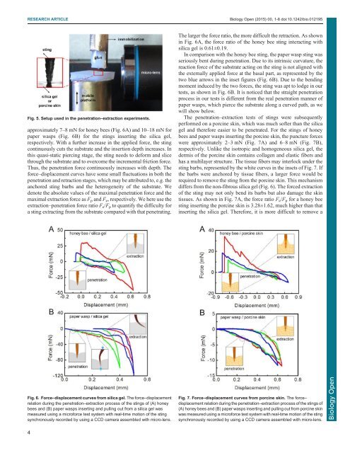
Fig. 5. Setup used in the penetration–extraction experiments.<br />
approximately 7–8 mN for honey bees (Fig. 6A) and 10–18 mN for<br />
paper wasps (Fig. 6B) for the stings inserting the silica gel,<br />
respectively. With a further increase in the applied force, the sting<br />
continuously cuts the substrate and the insertion depth increases. In<br />
this quasi-static piercing stage, the sting needs to deform and slice<br />
through the substrate and to overcome the incremental friction force.<br />
Thus, the penetration force continuously increases with depth. The<br />
force–displacement curves have some small fluctuations in both the<br />
penetration and retraction stages, which may be attributed to, e.g. the<br />
anchored sting barbs and the heterogeneity of the substrate. We<br />
denote the absolute values of the maximal penetration force and the<br />
maximal extraction force as F p and F e , respectively. We here use the<br />
extraction–penetration force ratio F e /F p to quantify the difficulty for<br />
a sting extracting from the substrate compared with that penetrating.<br />
The larger the force ratio, the more difficult the retraction. As shown<br />
in Fig. 6A, the force ratio of the honey bee sting interacting with<br />
silica gel is 0.61±0.19.<br />
In comparison with the honey bee sting, the paper wasp sting was<br />
seriously bent during penetration. Due to its intrinsic curvature, the<br />
reaction force of the substrate acting on the sting is not aligned with<br />
the externally applied force at the basal part, as represented by the<br />
two blue arrows in the inset figures (Fig. 6B). Due to the bending<br />
moment induced by the two forces, the sting was apt to lodge in our<br />
tests, as shown in Fig. 6B. It is noticed that the straight penetration<br />
process in our tests is different from the real penetration manner of<br />
paper wasps, which pierce the substrate along a curved path, as we<br />
will show below.<br />
The penetration–extraction tests of stings were subsequently<br />
performed on a porcine skin, which was much softer than the silica<br />
gel and therefore easier to be penetrated. For the stings of honey<br />
bees and paper wasps inserting the porcine skin, the puncture forces<br />
were approximately 2–3 mN (Fig. 7A) and 6–8 mN (Fig. 7B),<br />
respectively. Unlike the isotropic and homogeneous silica gel, the<br />
dermis of the porcine skin contains collagen and elastic fibers and<br />
has a multilayer structure. The tissue fibers may interlock under the<br />
sting barbs, represented by the white curves in the insets of Fig. 7. If<br />
the barbs were anchored by tissue fibers, a larger force would be<br />
required to remove the sting from the porcine skin. This mechanism<br />
differs from the non-fibrous silica gel (Fig. 6). The forced extraction<br />
of the sting may not only bend its barbs but also damage the skin<br />
tissues. As shown in Fig. 7A, the force ratio F e /F p for a honey bee<br />
sting inserting the porcine skin is 3.28±1.62, much higher than that<br />
inserting the silica gel. Therefore, it is more difficult to remove a<br />
Fig. 6. Force–displacement curves from silica gel. The force–displacement<br />
relation during the penetration–extraction process of the stings of (A) honey<br />
bees and (B) paper wasps inserting and pulling out from a silica gel was<br />
measured using a microforce test system with real-time motion of the sting<br />
synchronously recorded by using a CCD camera assembled with micro-lens.<br />
Fig. 7. Force–displacement curves from porcine skin. The force–<br />
displacement relation during the penetration–extraction process of the stings of<br />
(A) honey bees and (B) paper wasps inserting and pulling out from porcine skin<br />
was measured using a microforce test system with real-time motion of the sting<br />
synchronously recorded by using a CCD camera assembled with micro-lens.<br />
<strong>Biology</strong> Open<br />
4