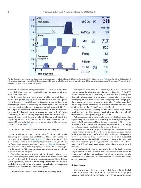
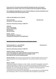
30 R. Breitkreutz et al. / Trends in Anaesthesia and Critical Care 32 (2020) 13e32 Fig. 10. Photograph represents a real-life example (ventilator display from Dr€ager Primus) within clinical anaesthesia. For details of a-g, see Fig 9. Note that only at the beginning of (g), the numeric capnometry result (dashed square upper right) pops up only 12 seconds after commencing mbag-PPV at a ventilation frequency of 12 per minute. The x-axis writespeed may be equivalent to 6.5 cm per second. auscultation, and it was merged into block 2, because it can be done in parallel with capnometry and addresses the question of main stem intubation only. To illustrate this comparison, we provide the workflows as stacked bar graphs (Fig. 8). Note that the time to decision upon a result depends on the different confirmation methods. Regarding capnometry, a result is depending on completion of ETT insertion, time span until ventilations can commence and curve visibility (i.e. air sample measurement, different in sidestream and mainstreamcapnography). For anterior neck ultrasound exam style #1 (postintubation) the result merely depends on the duration of the ultrasound exam itself. In exam style #2 (during intubation) it is depending on the time point of the ETT advancement to the ultrasound beam only, but not on the completion of the intubation process as a whole (Fig. 8). - Capnometry vs. anterior neck ultrasound exam style #1 We considered as the starting point for time tracking the beginning of reservoir bag ventilations. The positive signal for capnometry is delayed (Figs. 9 and 10) already in normal ETT placements. However, in an emergency scenario or CPR, additional confusion over an unsecure result can occur [68e70]. Moreover, if no clear values have been displayed, as it would be in esophageal misplacements or CPR, capnometry or auscultation results and time to decision will be delayed. Thereafter, in a normal conduct of an RSI with tracheal placement, all further parts of the completion were analysed until the end of the first and third positive capnometry curve. Nevertheless, when capnometry and auscultation are performed in parallel, both workflow tracks of either capnometry/auscultation or ultrasound exam style #1 seem to be almost equally fast. - Capnometry vs. anterior neck ultrasound exam style #2 Divergent to exam style #1, in exam style #2, we considered as a starting point for time tracking the end of insertion of the ETT, before withdrawal of the inlay/stylet. Because this is exactly the time point that anterior neck ultrasound can stop the process when identifying an advancement into the deep pharynx and esophagus, there would be no need to wait for a complete “double tract sign” by the supervisor. Thereafter, all further workflow details of the confirmation in block 2 und 3 were considered. Regardless whether waiting for the first positive capnometry result or until the third, anterior neck ultrasound exam, style #2 will be faster than capnometry/auscultation. Taken together, ultrasound can be considered at least as good as capnometry for the purpose of detecting an esophageal misplacement in both exam styles. Nevertheless in exam style #2 it will be markedly faster. The obvious reason in behind is that capnometry/ auscultation require ventilations to obtain a decision. However, in this ideal approach, no repeated measures, mixed teams, noise etc. are modeled. It would be common sense that in real life ventilation and confirmation will last longer and especially in the confusion and insecurity whether there is a suspected esophageal or endobronchial intubation. It thus can be presumed that exam times in capnometry/auscultation and the decision to reinsert the ETT will even take longer, when there is not a normal finding. Although real-life data are not available yet for both capnometry/auscultation and anterior neck ultrasound exam styles to support this opinion, our workflow model can be seen as hypothesis for an ideal definition. Clinical data will have to show the safety of the procedures. 5. Conclusion Our main message for emergencies is that there is the method of a post-intubation check in order to rule out or in esophageal misplacement without the necessity of ventilation. A second exam
R. Breitkreutz et al. / Trends in Anaesthesia and Critical Care 32 (2020) 13e32 31 style for continuous anterior neck ultrasound observation during intubations is available. Moreover confirmation of esophageal misplacement seems to be faster than with capnometry. Considering the evidence and limitations of focused anterior neck ultrasound procedures in emergencies, training within scenarios or a defined clinical context is recommended. The methods should be readily accessible for emergency and critical care practitioners. Acknowledgment This work is dedicated to WINFOCUS, a non-profit organization that supports science and exchange in the field of point-of-care ultrasound and thereby to its expert group of the International Consensus on point-of-care Lung Ultrasound 2012 [44]. Appendix A. Supplementary data Supplementary data to this article can be found online at www. yumpu.com/en/SonoABCD. References [1] G. Ma, S.R. Hayden, T.C. Chan, et al., Using ultrasound to visualize and confirm endotracheal intubation [SAEM meeting abstract], Acad. Emerg. Med. 6 (1999) 515. [2] M.J. Drescher, F.U. Conard, N.E. Schamban, Identification and description of esophageal intubation using ultrasound, Acad. Emerg. Med. 7 (2000) 722e725, https://doi.org/10.1111/j.1553-2712.2000.tb02055.x. [3] H.-C. Chou, W.-P. Tseng, C.-H. Wang, et al., Tracheal rapid ultrasound exam (T.R.U.E.) for confirming endotracheal tube placement during emergency intubation, Resuscitation 82 (2011) 1279e1284, https://doi.org/10.1016/ j.resuscitation.2011.05.016. [4] M.S. Kristensen, Ultrasonography in the management of the airway, Acta Anaesthesiol. Scand. 55 (2011) 1155e1173, https://doi.org/10.1111/j.1399- 6576.2011.02518.x. [5] K.E. You-Ten, N. Siddiqui, W.H. Teoh, M.S. Kristensen, Point-of-care ultrasound (POCUS) of the upper airway, Can. J. Anaesth. (2018), https://doi.org/10.1007/ s12630-018-1064-8. [6] K.E. You-Ten, D.T. Wong, X.Y. Ye, et al., Practice of ultrasound-guided palpation of neck landmarks improves accuracy of external palpation of the cricothyroid membrane, Anesth. Analg. 127 (2018) 1377e1382, https://doi.org/ 10.1213/ANE.0000000000003604. [7] A. Maniere-Ezvan, J.M. Duval, P. Darnault, Ultrasonic assessment of the anatomy and function of the tongue, Surg. Radiol. Anat. 15 (1993) 55e61, https://doi.org/10.1007/bf01629863. [8] M. Singh, K.J. Chin, V.W.S. Chan, et al., Use of sonography for airway assessment: an observational study, J. Ultrasound Med. 29 (2010) 79e85, https:// doi.org/10.7863/jum.2010.29.1.79. [9] S.L. Werner, R.A. Jones, C.L. Emerman, Sonographic assessment of the epiglottis, Acad. Emerg. Med. 11 (2004) 1358e1360, https://doi.org/10.1197/ j.aem.2004.05.033. [10] T. Beale, J. Rubin, Laryngeal ultrasonography, in: Orloff L (Hrsg) Head and Neck Ultrasonography, Plural Publishing, San Diego, 2008. S 183e202. [11] A. Bozzato, J. Zenk, F. Gottwald, et al., Der Einfluss der Schildknorpelossifikation bei der Larynxsonografie, Laryngo-Rhino-Otol. 86 (2007) 276e281, https://doi.org/10.1055/s-2006-945029. [12] C. Gourin, L. Orloff, Normal head and neck ultrasound anatomy, in: Orloff L (Hrsg) Head and Neck Ultrasonography, Plural Publishing, San Diego, 2008. S 39e68. [13] J.C. Gomez-Tamayo, et al., Inter-rater and intra-rater reliability of the airway diameter measured by sonography, J. Ultrasound 21 (1) (2018) 35e40, 6. [14] S. Falcetta, S. Cavallo, V. Gabbanelli, et al., Evaluation of two neck ultrasound measurements as predictors of difficult direct laryngoscopy: a prospective observational study, Eur. J. Anaesthesiol. 35 (2018) 605e612, https://doi.org/ 10.1097/EJA.0000000000000832. [15] J.S. Fulkerson, H.M. Moore, T.S. Anderson, R.F. Lowe, Ultrasonography in the preoperative difficult airway assessment, J. Clin. Monit. Comput. 31 (2017) 513e530, https://doi.org/10.1007/s10877-016-9888-7. [16] C. Petrisor, D. Dîrzu, S. Tranca, et al., Preoperative difficult airway prediction using suprahyoid and infrahyoid ultrasonography derived measurements in anesthesiology, Med. Ultrasonogr. 21 (2019) 83e88, https://doi.org/10.11152/ mu-1764. [17] J. Pinto, L. Cordeiro, C. Pereira, et al., Predicting difficult laryngoscopy using ultrasound measurement of distance from skin to epiglottis, J. Crit. Care 33 (2016) 26e31, https://doi.org/10.1016/j.jcrc.2016.01.029. [18] J. Wu, J. Dong, Y. Ding, J. Zheng, Role of anterior neck soft tissue quantifications by ultrasound in predicting difficult laryngoscopy, Med. Sci. Monit. 20 (2014) 2343e2350, https://doi.org/10.12659/MSM.891037. [19] N.K. Yadav, P. Rudingwa, S.K. Mishra, S. Pannerselvam, Ultrasound measurement of anterior neck soft tissue and tongue thickness to predict difficult laryngoscopy - an observational analytical study, Indian J. Anaesth. 63 (2019) 629e634, https://doi.org/10.4103/ija.IJA_270_19. [20] S. Adhikari, W. Zeger, C. Schmier, et al., Pilot study to determine the utility of point-of-care ultrasound in the assessment of difficult laryngoscopy, Acad. Emerg. Med. 18 (2011) 754e758, https://doi.org/10.1111/j.1553- 2712.2011.01099.x. [21] Fulkerson Airway Sonography fails to detect difficult laryngoscopy in an adult veterenal surgical population, Trends Anaesthesiol. Crit. Care 29 (2019) 26e34, https://doi.org/10.1016/j.tacc.2019.07.003. [22] M.S. Kristensen, W.H. Teoh, O. Graumann, C.B. Laursen, Ultrasonography for clinical decision-making and intervention in airway management: from the mouth to the lungs and pleurae, Insights Imag. 5 (2014) 253e279, https:// doi.org/10.1007/s13244-014-0309-5. [23] A. Sustic, Role of ultrasound in the airway management of critically ill patients, Crit. Care Med. 35 (2007) S173eS177, https://doi.org/10.1097/ 01.CCM.0000260628.88402.8A. [24] Cook TM, Woodall N, Frerk C, Fourth national audit project (2011) major complications of airway management in the UK: results of the fourth national audit project of the royal college of anaesthetists and the difficult airway society. Part 1: anaesthesia. Br. J. Anaesth. 106:617e631. https://doi.org/10. 1093/bja/aer058. [25] C. Frerk, V.S. Mitchell, A.F. McNarry, et al., Difficult Airway Society 2015 guidelines for management of unanticipated difficult intubation in adults, Br. J. Anaesth. 115 (2015) 827e848, https://doi.org/10.1093/bja/aev371. [26] M.S. Kristensen, W.H. Teoh, S.S. Rudolph, et al., A randomised cross-over comparison of the transverse and longitudinal techniques for ultrasoundguided identification of the cricothyroid membrane in morbidly obese subjects, Anaesthesia 71 (2016) 675e683, https://doi.org/10.1111/anae.13465. [27] M.S. Kristensen, W.H. Teoh, S.S. Rudolph, Ultrasonographic identification of the cricothyroid membrane: best evidence, techniques, and clinical impact, Br. J. Anaesth. 117 (Suppl 1) (2016) i39ei48, https://doi.org/10.1093/bja/aew176. [28] G. Yıldız, E. G€oksu, A. Şenfer, A. Kaplan, Comparison of ultrasonography and surface landmarks in detecting the localization for cricothyroidotomy, Am. J. Emerg. Med. 34 (2016) 254e256, https://doi.org/10.1016/j.ajem.2015.10.054. [29] K. Curtis, M. Ahern, M. Dawson, M. Mallin, Ultrasound-guided, Bougie-assisted cricothyroidotomy: a description of a novel technique in cadaveric models, Acad. Emerg. Med. 19 (2012) 876e879, https://doi.org/10.1111/j.1553- 2712.2012.01391.x. [30] G. Ma, D.P. Davis, J. Schmitt, et al., The sensitivity and specificity of transcricothyroid ultrasonography to confirm endotracheal tube placement in a cadaver model, J. Emerg. Med. 32 (2007) 405e407, https://doi.org/10.1016/ j.jemermed.2006.08.023. [31] T.L. Slovis, R.L. Poland, Endotracheal tubes in neonates: sonographic positioning, Radiology 160 (1986) 262e263, https://doi.org/10.1148/ radiology.160.1.3520649. [32] D.T. Raphael, F.U. Conard, Ultrasound confirmation of endotracheal tube placement, J. Clin. Ultrasound 15 (1987) 459e462, https://doi.org/10.1002/ jcu.1870150706. [33] J. Chenkin, C.J.L. McCartney, T. Jelic, et al., Defining the learning curve of pointof-care ultrasound for confirming endotracheal tube placement by emergency physicians, Crit. Ultrasound J. 7 (2015) 14, https://doi.org/10.1186/s13089- 015-0031-7. [34] S. Lahham, J. Baydoun, J. Bailey, et al., A prospective evaluation of transverse tracheal sonography during emergent intubation by emergency medicine resident physicians, J. Ultrasound Med. 36 (2017) 2079e2085, https://doi.org/ 10.1002/jum.14231. [35] E.H. Chou, E. Dickman, P.-Y. Tsou, et al., Ultrasonography for confirmation of endotracheal tube placement: a systematic review and meta-analysis, Resuscitation 90 (2015) 97e103, https://doi.org/10.1016/j.resuscitation.2015.02.013. [36] J. Soar, J.P. Nolan, B.W. B€ottiger, et al., European resuscitation council guidelines for resuscitation 2015: section 3. Adult advanced life support, Resuscitation 95 (2015) 100e147, https://doi.org/10.1016/j.resuscitation.2015.07.016. [37] P.M. Zechner, R. Breitkreutz, Ultrasound instead of capnometry for confirming tracheal tube placement in an emergency? Resuscitation 82 (2011) 1259e1261, https://doi.org/10.1016/j.resuscitation.2011.06.040. [38] O. Adi, T.W. Chuan, M. Rishya, A feasibility study on bedside upper airway ultrasonography compared to waveform capnography for verifying endotracheal tube location after intubation, Crit. Ultrasound J. 5 (2013) 7, https:// doi.org/10.1186/2036-7902-5-7. [39] S. Abbasi, D. Farsi, M.A. Zare, et al., Direct ultrasound methods: a confirmatory technique for proper endotracheal intubation in the emergency department, Eur. J. Emerg. Med. 22 (2015) 10e16, https://doi.org/10.1097/ MEJ.0000000000000108. [40] N. Siddiqui, E. Yu, S. Boulis, K.E. You-Ten, Ultrasound is superior to palpation in identifying the cricothyroid membrane in subjects with poorly defined neck landmarks: a randomized clinical trial, Anesthesiology 129 (2018) 1132e1139, https://doi.org/10.1097/ALN.0000000000002454. [41] S. Karacabey, E. Sanrı, E.G. Gencer, O. Guneysel, Tracheal ultrasonography and ultrasonographic lung sliding for confirming endotracheal tube placement: speed and Reliability, Am. J. Emerg. Med. 34 (2016) 953e956, https://doi.org/ 10.1016/j.ajem.2016.01.027. [42] B. Weaver, M. Lyon, M. Blaivas, Confirmation of endotracheal tube placement after intubation using the ultrasound sliding lung sign, Acad. Emerg. Med. 13