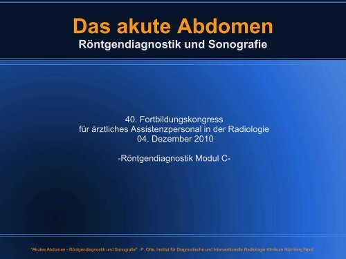
Das akute Abdomen Röntgendiagnostik und Sonografie
Das akute Abdomen Röntgendiagnostik und Sonografie
Das akute Abdomen Röntgendiagnostik und Sonografie

Sie wollen auch ein ePaper? Erhöhen Sie die Reichweite Ihrer Titel.
YUMPU macht aus Druck-PDFs automatisch weboptimierte ePaper, die Google liebt.
<strong>Das</strong> <strong>akute</strong> <strong>Abdomen</strong><br />
<strong>Röntgendiagnostik</strong> <strong>und</strong> <strong>Sonografie</strong><br />
40. Fortbildungskongress<br />
für ärztliches Assistenzpersonal in der Radiologie<br />
04. Dezember 2010<br />
-<strong>Röntgendiagnostik</strong> Modul C-<br />
"Akutes <strong>Abdomen</strong> - <strong>Röntgendiagnostik</strong> <strong>und</strong> <strong>Sonografie</strong>" P. Otte, Institut für Diagnostische <strong>und</strong> Interventionelle Radioloigie Klinikum Nürnberg Nord
� „Akutes Abomen“, was ist das?<br />
� Diagnostisches Vorgehen,<br />
Woran orientiere ich mich?<br />
� Konventionelle <strong>Röntgendiagnostik</strong>,<br />
Welche Anforderung?<br />
Was sehe ich?<br />
� Ultraschall,<br />
Was sieht der Arzt?<br />
Welche Einschränkungen?<br />
"Akutes <strong>Abdomen</strong> - <strong>Röntgendiagnostik</strong> <strong>und</strong> <strong>Sonografie</strong>" P. Otte, Institut für Diagnostische <strong>und</strong> Interventionelle Radioloigie Klinikum Nürnberg Nord
Begriffsbestimmung:<br />
<strong>Das</strong> „Akute <strong>Abdomen</strong>“<br />
� Ist kein eigenständiges Krankheitsbild<br />
� Bezeichnet einen Symptomenkomplex, wobei die Beschwerden in<br />
den Bauchraum lokalisiert werden<br />
� Es bestehen starke bis stärkste Bauchschmerzen < 24 St<strong>und</strong>en<br />
� Meist verb<strong>und</strong>en mit einer Bauchdeckenspannung (Peritonismus)<br />
<strong>und</strong> einer Störung der Darmmotilität (Peristaltikstörung) vor<br />
� Potentiell lebensbedrohliche Situation<br />
� Häufig Operation unumgänglich<br />
"Akutes <strong>Abdomen</strong> - <strong>Röntgendiagnostik</strong> <strong>und</strong> <strong>Sonografie</strong>" P. Otte, Institut für Diagnostische <strong>und</strong> Interventionelle Radioloigie Klinikum Nürnberg Nord
Begriffsbestimmung:<br />
<strong>Das</strong> „Akute <strong>Abdomen</strong>“<br />
Aber auch...<br />
� Ursache außerhalb des Bauchraumes möglich<br />
� Nicht immer Operation!<br />
Auch konservative Therapie oft ausreichend<br />
� Beschwerden mit spontaner Besserung ohne definierbare Ursache<br />
"Akutes <strong>Abdomen</strong> - <strong>Röntgendiagnostik</strong> <strong>und</strong> <strong>Sonografie</strong>" P. Otte, Institut für Diagnostische <strong>und</strong> Interventionelle Radioloigie Klinikum Nürnberg Nord
aus J. Mössner, „Akutes <strong>Abdomen</strong>“, Internist 2005 Nr. 46:974–981<br />
"Akutes <strong>Abdomen</strong> - <strong>Röntgendiagnostik</strong> <strong>und</strong> <strong>Sonografie</strong>" P. Otte, Institut für Diagnostische <strong>und</strong> Interventionelle Radioloigie Klinikum Nürnberg Nord
Häufigste Ursachen des <strong>akute</strong>n <strong>Abdomen</strong> (ca. 90%):<br />
� <strong>akute</strong> Appendizitis (35–50%)<br />
� <strong>akute</strong> Cholezystitis oder Gallenkolik (10–15%)<br />
� Ileus oder Divertikulitis (10–25%)<br />
� eine Perforation eines Ulkus des Magens oder des Duodenums (7%)<br />
� <strong>akute</strong> Pankreatitis (5%)<br />
� Myokardinfarkt (5%)<br />
� Nierenkolik (2,5%)<br />
"Akutes <strong>Abdomen</strong> - <strong>Röntgendiagnostik</strong> <strong>und</strong> <strong>Sonografie</strong>" P. Otte, Institut für Diagnostische <strong>und</strong> Interventionelle Radioloigie Klinikum Nürnberg Nord
Merke:<br />
Nicht jeder Patient mit starken Bauchschmerzen ist<br />
sterbenskrank, kann es aber sein!!!<br />
"Akutes <strong>Abdomen</strong> - <strong>Röntgendiagnostik</strong> <strong>und</strong> <strong>Sonografie</strong>" P. Otte, Institut für Diagnostische <strong>und</strong> Interventionelle Radioloigie Klinikum Nürnberg Nord
Wie komme ich diagnostisch<br />
möglichst schnell zur Klärung der<br />
Ursache?<br />
"Akutes <strong>Abdomen</strong> - <strong>Röntgendiagnostik</strong> <strong>und</strong> <strong>Sonografie</strong>" P. Otte, Institut für Diagnostische <strong>und</strong> Interventionelle Radioloigie Klinikum Nürnberg Nord
Diagnostik des Akuten <strong>Abdomen</strong><br />
� Klinische Untersuchung<br />
� Labordiagnostik<br />
Bildgebende Diagnostik<br />
� konventionelle <strong>Röntgendiagnostik</strong><br />
Ultraschall<br />
� Computertomografie<br />
Angiografie, Kernspintomografie, Durchleuchtung<br />
"Akutes <strong>Abdomen</strong> - <strong>Röntgendiagnostik</strong> <strong>und</strong> <strong>Sonografie</strong>" P. Otte, Institut für Diagnostische <strong>und</strong> Interventionelle Radioloigie Klinikum Nürnberg Nord
Diagnostik des Akuten <strong>Abdomen</strong><br />
� Klinische Untersuchung<br />
� Labordiagnostik<br />
Basisdiagnostik<br />
Bildgebende Diagnostik<br />
� konventionelle <strong>Röntgendiagnostik</strong><br />
Ultraschall<br />
� Computertomografie<br />
Angiografie, Kernspintomografie, Durchleuchtung<br />
Erweiterte Diagnostik<br />
"Akutes <strong>Abdomen</strong> - <strong>Röntgendiagnostik</strong> <strong>und</strong> <strong>Sonografie</strong>" P. Otte, Institut für Diagnostische <strong>und</strong> Interventionelle Radioloigie Klinikum Nürnberg Nord<br />
~80%<br />
~20%
Bildgebende Verfahren<br />
Welche?<br />
Wann?<br />
"Akutes <strong>Abdomen</strong> - <strong>Röntgendiagnostik</strong> <strong>und</strong> <strong>Sonografie</strong>" P. Otte, Institut für Diagnostische <strong>und</strong> Interventionelle Radioloigie Klinikum Nürnberg Nord
Es gibt derzeit keinen von der<br />
empfohlenen Leitlinienalgorithmus zur<br />
Abklärung des Akuten <strong>Abdomen</strong><br />
Die "Leitlinien" der Wissenschaftlichen Medizinischen Fachgesellschaften sind systematisch entwickelte Hilfen für Ärzte<br />
zur Entscheidungsfindung in spezifischen Situationen. Sie beruhen auf aktuellen wissenschaftlichen Erkenntnissen <strong>und</strong><br />
in der Praxis bewährten Verfahren <strong>und</strong> sorgen für mehr Sicherheit in der Medizin, sollen aber auch ökonomische<br />
Aspekte berücksichtigen. Die "Leitlinien" sind für Ärzte rechtlich nicht bindend <strong>und</strong> haben daher weder<br />
haftungsbegründende noch haftungsbefreiende Wirkung. http://www.leitlinien.net<br />
"Akutes <strong>Abdomen</strong> - <strong>Röntgendiagnostik</strong> <strong>und</strong> <strong>Sonografie</strong>" P. Otte, Institut für Diagnostische <strong>und</strong> Interventionelle Radioloigie Klinikum Nürnberg Nord
Standardindikationen zur<br />
Röntgenübersichtsaufnahme des <strong>Abdomen</strong><br />
aus C.-M. Reng/S. Grüne „Akutes <strong>Abdomen</strong>“, Intensivmed 2010 Nr. 47:225–234<br />
"Akutes <strong>Abdomen</strong> - <strong>Röntgendiagnostik</strong> <strong>und</strong> <strong>Sonografie</strong>" P. Otte, Institut für Diagnostische <strong>und</strong> Interventionelle Radioloigie Klinikum Nürnberg Nord
American College of Radiology<br />
ACR Appropriateness Criteria<br />
http://www.acr.org/SecondaryMainMenuCategories/quality_safety/guidelines.aspx<br />
"Akutes <strong>Abdomen</strong> - <strong>Röntgendiagnostik</strong> <strong>und</strong> <strong>Sonografie</strong>" P. Otte, Institut für Diagnostische <strong>und</strong> Interventionelle Radioloigie Klinikum Nürnberg Nord
"Akutes <strong>Abdomen</strong> - <strong>Röntgendiagnostik</strong> <strong>und</strong> <strong>Sonografie</strong>" P. Otte, Institut für Diagnostische <strong>und</strong> Interventionelle Radioloigie Klinikum Nürnberg Nord
"Akutes <strong>Abdomen</strong> - <strong>Röntgendiagnostik</strong> <strong>und</strong> <strong>Sonografie</strong>" P. Otte, Institut für Diagnostische <strong>und</strong> Interventionelle Radioloigie Klinikum Nürnberg Nord
1. Konventionelle<br />
<strong>Röntgendiagnostik</strong><br />
� Röntgenthoraxaufnahme in Standardtechnik<br />
� <strong>Abdomen</strong>übersichtsaufnahme in Rücken- <strong>und</strong> Linksseitenlage, ggfs.<br />
Behelfsaufnahmen<br />
"Akutes <strong>Abdomen</strong> - <strong>Röntgendiagnostik</strong> <strong>und</strong> <strong>Sonografie</strong>" P. Otte, Institut für Diagnostische <strong>und</strong> Interventionelle Radioloigie Klinikum Nürnberg Nord
1. Konventionelle<br />
<strong>Röntgendiagnostik</strong><br />
� Röntgenthoraxaufnahme in Standardtechnik<br />
zum Erfassen thoracaler Pathologien, welche die<br />
Schmerzsymptomatik in den Bauchraum lokalisieren (z.B. basale<br />
Pneumonie, Herzinsuffizienz bei Hinterwandinfarkt u.ä.)<br />
� <strong>Abdomen</strong>übersichtsaufnahme in Rücken- <strong>und</strong> Linksseitenlage, ggfs.<br />
Behelfsaufnahmen<br />
"Akutes <strong>Abdomen</strong> - <strong>Röntgendiagnostik</strong> <strong>und</strong> <strong>Sonografie</strong>" P. Otte, Institut für Diagnostische <strong>und</strong> Interventionelle Radioloigie Klinikum Nürnberg Nord
1. Konventionelle<br />
<strong>Röntgendiagnostik</strong><br />
� Röntgenthoraxaufnahme in Standardtechnik<br />
� <strong>Abdomen</strong>übersichtsaufnahme in Rücken- <strong>und</strong><br />
Linksseitenlage, ggfs. Behelfsaufnahmen<br />
"Akutes <strong>Abdomen</strong> - <strong>Röntgendiagnostik</strong> <strong>und</strong> <strong>Sonografie</strong>" P. Otte, Institut für Diagnostische <strong>und</strong> Interventionelle Radioloigie Klinikum Nürnberg Nord
Auszug aus der Leitlinie der B<strong>und</strong>esärztekammer zur Qualitätssicherung in der<br />
<strong>Röntgendiagnostik</strong> „<strong>Abdomen</strong>“<br />
Ärztliche Qualitätsanforderungen<br />
Bildmerkmale<br />
� Darstellung des <strong>Abdomen</strong>s vom Zwerchfell bis<br />
zum Beckenboden, evtl. in zwei Aufnahmen.<br />
� Darstellung der Weichteilschatten <strong>und</strong> lumbalen<br />
Fettlinien.<br />
� Darstellung des seitlichen Psoasrandes.<br />
� Darstellung der Nierenkonturen.<br />
� Darstellung des unteren Leberrandes<br />
� Darstellung der Verteilung von Gas <strong>und</strong> Flüssigkeit<br />
im Magen-Darmkanal inclusive der Darmwand,<br />
Peritonealraum sowie retro- <strong>und</strong> extraperitoneal.<br />
� Darstellung von verkalkten Strukturen<br />
� Ausreichende Darstellung der mitabgebildeten<br />
Knochen.<br />
Aufnahmetechnische Qualitätsanforderungen<br />
Aufnahmetechnik<br />
� Aufnahmespannung: 80 - 100 kV<br />
(LSL Aufnahmespannung: 100 - 125 kV)<br />
� Brennflecknennwert: ≤ 1,3<br />
� Fokus- Detektor-Abstand: 115 cm<br />
� Belichtungsautomatik: mittleres oder beide<br />
seitlichen Messfelder<br />
� Expositionszeit: ≤ 100 ms<br />
� Streustrahlenraster: r 12 (8)<br />
� Bildempfängerdosis: ≤ 5 μGy, SC 400<br />
"Akutes <strong>Abdomen</strong> - <strong>Röntgendiagnostik</strong> <strong>und</strong> <strong>Sonografie</strong>" P. Otte, Institut für Diagnostische <strong>und</strong> Interventionelle Radioloigie Klinikum Nürnberg Nord
Auszug aus der Leitlinie der B<strong>und</strong>esärztekammer zur Qualitätssicherung in der<br />
<strong>Röntgendiagnostik</strong> „<strong>Abdomen</strong>“<br />
Ärztliche Qualitätsanforderungen - Bildmerkmale<br />
� Darstellung des <strong>Abdomen</strong>s vom Zwerchfell bis<br />
zum Beckenboden, evtl. in zwei Aufnahmen.<br />
� Darstellung der Weichteilschatten <strong>und</strong> lumbalen<br />
Fettlinien.<br />
� Darstellung des seitlichen Psoasrandes.<br />
� Darstellung der Nierenkonturen.<br />
� Darstellung des unteren Leberrandes<br />
� Darstellung der Verteilung von Gas <strong>und</strong> Flüssigkeit<br />
im Magen-Darmkanal inclusive der Darmwand,<br />
Peritonealraum sowie retro- <strong>und</strong> extraperitoneal.<br />
� Darstellung von verkalkten Strukturen<br />
� Ausreichende Darstellung der mitabgebildeten<br />
Knochen.<br />
� Weichteilschatten<br />
� Fettlinien<br />
� Konturen<br />
� Verteilung von Gas <strong>und</strong><br />
Flüssigkeit<br />
� Verkalkte<br />
Röntgen-dichte<br />
Strukturen<br />
� Knochen<br />
"Akutes <strong>Abdomen</strong> - <strong>Röntgendiagnostik</strong> <strong>und</strong> <strong>Sonografie</strong>" P. Otte, Institut für Diagnostische <strong>und</strong> Interventionelle Radioloigie Klinikum Nürnberg Nord
Nutzung der Dichteunterschiede<br />
"Akutes <strong>Abdomen</strong> - <strong>Röntgendiagnostik</strong> <strong>und</strong> <strong>Sonografie</strong>" P. Otte, Institut für Diagnostische <strong>und</strong> Interventionelle Radioloigie Klinikum Nürnberg Nord
Luft<br />
Fett<br />
Weichteil<br />
Kalk/Knochen<br />
(Metall)<br />
"Akutes <strong>Abdomen</strong> - <strong>Röntgendiagnostik</strong> <strong>und</strong> <strong>Sonografie</strong>" P. Otte, Institut für Diagnostische <strong>und</strong> Interventionelle Radioloigie Klinikum Nürnberg Nord
Normalbef<strong>und</strong><br />
"Akutes <strong>Abdomen</strong> - <strong>Röntgendiagnostik</strong> <strong>und</strong> <strong>Sonografie</strong>" P. Otte, Institut für Diagnostische <strong>und</strong> Interventionelle Radioloigie Klinikum Nürnberg Nord
"Akutes <strong>Abdomen</strong> - <strong>Röntgendiagnostik</strong> <strong>und</strong> <strong>Sonografie</strong>" P. Otte, Institut für Diagnostische <strong>und</strong> Interventionelle Radioloigie Klinikum Nürnberg Nord
Koprostase<br />
"Akutes <strong>Abdomen</strong> - <strong>Röntgendiagnostik</strong> <strong>und</strong> <strong>Sonografie</strong>" P. Otte, Institut für Diagnostische <strong>und</strong> Interventionelle Radioloigie Klinikum Nürnberg Nord
"Akutes <strong>Abdomen</strong> - <strong>Röntgendiagnostik</strong> <strong>und</strong> <strong>Sonografie</strong>" P. Otte, Institut für Diagnostische <strong>und</strong> Interventionelle Radioloigie Klinikum Nürnberg Nord
"Akutes <strong>Abdomen</strong> - <strong>Röntgendiagnostik</strong> <strong>und</strong> <strong>Sonografie</strong>" P. Otte, Institut für Diagnostische <strong>und</strong> Interventionelle Radioloigie Klinikum Nürnberg Nord
Gallenblasenkonkremente<br />
"Akutes <strong>Abdomen</strong> - <strong>Röntgendiagnostik</strong> <strong>und</strong> <strong>Sonografie</strong>" P. Otte, Institut für Diagnostische <strong>und</strong> Interventionelle Radioloigie Klinikum Nürnberg Nord
"Akutes <strong>Abdomen</strong> - <strong>Röntgendiagnostik</strong> <strong>und</strong> <strong>Sonografie</strong>" P. Otte, Institut für Diagnostische <strong>und</strong> Interventionelle Radioloigie Klinikum Nürnberg Nord
Harnverhalt<br />
"Akutes <strong>Abdomen</strong> - <strong>Röntgendiagnostik</strong> <strong>und</strong> <strong>Sonografie</strong>" P. Otte, Institut für Diagnostische <strong>und</strong> Interventionelle Radioloigie Klinikum Nürnberg Nord
"Akutes <strong>Abdomen</strong> - <strong>Röntgendiagnostik</strong> <strong>und</strong> <strong>Sonografie</strong>" P. Otte, Institut für Diagnostische <strong>und</strong> Interventionelle Radioloigie Klinikum Nürnberg Nord
"Akutes <strong>Abdomen</strong> - <strong>Röntgendiagnostik</strong> <strong>und</strong> <strong>Sonografie</strong>" P. Otte, Institut für Diagnostische <strong>und</strong> Interventionelle Radioloigie Klinikum Nürnberg Nord
Aerobilie<br />
"Akutes <strong>Abdomen</strong> - <strong>Röntgendiagnostik</strong> <strong>und</strong> <strong>Sonografie</strong>" P. Otte, Institut für Diagnostische <strong>und</strong> Interventionelle Radioloigie Klinikum Nürnberg Nord
"Akutes <strong>Abdomen</strong> - <strong>Röntgendiagnostik</strong> <strong>und</strong> <strong>Sonografie</strong>" P. Otte, Institut für Diagnostische <strong>und</strong> Interventionelle Radioloigie Klinikum Nürnberg Nord
Arteriosklerose<br />
"Akutes <strong>Abdomen</strong> - <strong>Röntgendiagnostik</strong> <strong>und</strong> <strong>Sonografie</strong>" P. Otte, Institut für Diagnostische <strong>und</strong> Interventionelle Radioloigie Klinikum Nürnberg Nord
"Akutes <strong>Abdomen</strong> - <strong>Röntgendiagnostik</strong> <strong>und</strong> <strong>Sonografie</strong>" P. Otte, Institut für Diagnostische <strong>und</strong> Interventionelle Radioloigie Klinikum Nürnberg Nord
Milzarterienaneurysma<br />
"Akutes <strong>Abdomen</strong> - <strong>Röntgendiagnostik</strong> <strong>und</strong> <strong>Sonografie</strong>" P. Otte, Institut für Diagnostische <strong>und</strong> Interventionelle Radioloigie Klinikum Nürnberg Nord
"Akutes <strong>Abdomen</strong> - <strong>Röntgendiagnostik</strong> <strong>und</strong> <strong>Sonografie</strong>" P. Otte, Institut für Diagnostische <strong>und</strong> Interventionelle Radioloigie Klinikum Nürnberg Nord
Paralytischer Ileus<br />
"Akutes <strong>Abdomen</strong> - <strong>Röntgendiagnostik</strong> <strong>und</strong> <strong>Sonografie</strong>" P. Otte, Institut für Diagnostische <strong>und</strong> Interventionelle Radioloigie Klinikum Nürnberg Nord
"Akutes <strong>Abdomen</strong> - <strong>Röntgendiagnostik</strong> <strong>und</strong> <strong>Sonografie</strong>" P. Otte, Institut für Diagnostische <strong>und</strong> Interventionelle Radioloigie Klinikum Nürnberg Nord
"Akutes <strong>Abdomen</strong> - <strong>Röntgendiagnostik</strong> <strong>und</strong> <strong>Sonografie</strong>" P. Otte, Institut für Diagnostische <strong>und</strong> Interventionelle Radioloigie Klinikum Nürnberg Nord
"Akutes <strong>Abdomen</strong> - <strong>Röntgendiagnostik</strong> <strong>und</strong> <strong>Sonografie</strong>" P. Otte, Institut für Diagnostische <strong>und</strong> Interventionelle Radioloigie Klinikum Nürnberg Nord
Freie Luft bei Perforation<br />
Silhouettenphänomen<br />
"Akutes <strong>Abdomen</strong> - <strong>Röntgendiagnostik</strong> <strong>und</strong> <strong>Sonografie</strong>" P. Otte, Institut für Diagnostische <strong>und</strong> Interventionelle Radioloigie Klinikum Nürnberg Nord
"Akutes <strong>Abdomen</strong> - <strong>Röntgendiagnostik</strong> <strong>und</strong> <strong>Sonografie</strong>" P. Otte, Institut für Diagnostische <strong>und</strong> Interventionelle Radioloigie Klinikum Nürnberg Nord
"Akutes <strong>Abdomen</strong> - <strong>Röntgendiagnostik</strong> <strong>und</strong> <strong>Sonografie</strong>" P. Otte, Institut für Diagnostische <strong>und</strong> Interventionelle Radioloigie Klinikum Nürnberg Nord
Pneumatosis intestinalis<br />
(Gaseinschlüsse in der Darmwand)<br />
"Akutes <strong>Abdomen</strong> - <strong>Röntgendiagnostik</strong> <strong>und</strong> <strong>Sonografie</strong>" P. Otte, Institut für Diagnostische <strong>und</strong> Interventionelle Radioloigie Klinikum Nürnberg Nord
"Akutes <strong>Abdomen</strong> - <strong>Röntgendiagnostik</strong> <strong>und</strong> <strong>Sonografie</strong>" P. Otte, Institut für Diagnostische <strong>und</strong> Interventionelle Radioloigie Klinikum Nürnberg Nord
"Akutes <strong>Abdomen</strong> - <strong>Röntgendiagnostik</strong> <strong>und</strong> <strong>Sonografie</strong>" P. Otte, Institut für Diagnostische <strong>und</strong> Interventionelle Radioloigie Klinikum Nürnberg Nord
Fremdkörper<br />
"Akutes <strong>Abdomen</strong> - <strong>Röntgendiagnostik</strong> <strong>und</strong> <strong>Sonografie</strong>" P. Otte, Institut für Diagnostische <strong>und</strong> Interventionelle Radioloigie Klinikum Nürnberg Nord
"Akutes <strong>Abdomen</strong> - <strong>Röntgendiagnostik</strong> <strong>und</strong> <strong>Sonografie</strong>" P. Otte, Institut für Diagnostische <strong>und</strong> Interventionelle Radioloigie Klinikum Nürnberg Nord
„Coffee-Bean-Sign“<br />
"Akutes <strong>Abdomen</strong> - <strong>Röntgendiagnostik</strong> <strong>und</strong> <strong>Sonografie</strong>" P. Otte, Institut für Diagnostische <strong>und</strong> Interventionelle Radioloigie Klinikum Nürnberg Nord
Volvolus (Verschlingung)<br />
"Akutes <strong>Abdomen</strong> - <strong>Röntgendiagnostik</strong> <strong>und</strong> <strong>Sonografie</strong>" P. Otte, Institut für Diagnostische <strong>und</strong> Interventionelle Radioloigie Klinikum Nürnberg Nord
2. Ultraschalldiagnostik<br />
"Akutes <strong>Abdomen</strong> - <strong>Röntgendiagnostik</strong> <strong>und</strong> <strong>Sonografie</strong>" P. Otte, Institut für Diagnostische <strong>und</strong> Interventionelle Radioloigie Klinikum Nürnberg Nord
2. Ultraschalldiagnostik I<br />
� Ultraschall ist Schall mit einer Frequenz oberhalb der menschlichen<br />
Hörgrenze, bei medizinischer Diagnostik 1-40MHz (meist 3-13Hz)<br />
"Akutes <strong>Abdomen</strong> - <strong>Röntgendiagnostik</strong> <strong>und</strong> <strong>Sonografie</strong>" P. Otte, Institut für Diagnostische <strong>und</strong> Interventionelle Radioloigie Klinikum Nürnberg Nord
2. Ultraschalldiagnostik I<br />
� Ultraschall ist Schall mit einer Frequenz oberhalb der menschlichen<br />
Hörgrenze, bei medizinischer Diagnostik 1-40MHz (meist 3-13MHz)<br />
� Verwendung des piezoelektrischen Effektes<br />
(Auftreten einer elektrischen Spannung an Festkörpern (hier Kristalle), wenn diese elastisch verformt werden.<br />
Umgekehrt verformen sich Materialien bei Anlegen einer elektrischen Spannung (inverser Effekt), was zu<br />
Schwingungen führt, welche sich als Schallwellen ausbreiten)<br />
"Akutes <strong>Abdomen</strong> - <strong>Röntgendiagnostik</strong> <strong>und</strong> <strong>Sonografie</strong>" P. Otte, Institut für Diagnostische <strong>und</strong> Interventionelle Radioloigie Klinikum Nürnberg Nord
2. Ultraschalldiagnostik I<br />
� Ultraschall ist Schall mit einer Frequenz oberhalb der menschlichen<br />
Hörgrenze, bei medizinischer Diagnostik 1-40MHz (meist 3-13Hz)<br />
� Verwendung des piezoelektrischen Effektes<br />
(Auftreten einer elektrischen Spannung an Festkörpern (hier Kristalle), wenn diese elastisch verformt werden.<br />
Umgekehrt verformen sich Materialien bei Anlegen einer elektrischen Spannung (inverser Effekt), was zu<br />
Schwingungen führt, welche sich als Schallwellen ausbreiten)<br />
� Gerichtete Schallwellen breiten sich im Gewebe aus <strong>und</strong> werden<br />
reflektiert<br />
� Laufzeit >>>Tiefe der reflektierenden Struktur<br />
� Stärke der Reflexion >>> Echogenität<br />
(Gering echogen/schwarz sind vor allem Flüssigkeitent. Eine hohe Echogenität/weiss besitzen Knochen,<br />
Gase <strong>und</strong> sonstige stark Schall reflektierende Materialien)<br />
"Akutes <strong>Abdomen</strong> - <strong>Röntgendiagnostik</strong> <strong>und</strong> <strong>Sonografie</strong>" P. Otte, Institut für Diagnostische <strong>und</strong> Interventionelle Radioloigie Klinikum Nürnberg Nord
2. Ultraschalldiagnostik II<br />
� Handgeführtes „Realtime“ (Echtzeit) Schnittbildverfahren<br />
� Möglichkeit der gleichzeitigen Anamneseerhebung<br />
� Möglichkeit <strong>und</strong> Eingrenzung der genauen Schmerzlokalisation<br />
� Kostengünstig, strahlungsarm<br />
� Nichtinvasive Gefäßdarstellungsmöglichkeit (Farbdoppler)<br />
� Starke Untersucher-Abhängigkeit<br />
S1-3 (geringe >>> hohe Erfahrung - angelehnt nach DEGUM-Stadien)<br />
� Abhängigkeit von der Ultraschallgeräteklasse<br />
(Klasse 1-3/ 91.000€)<br />
� Abhängigkeit von der Patienten-Compliance<br />
"Akutes <strong>Abdomen</strong> - <strong>Röntgendiagnostik</strong> <strong>und</strong> <strong>Sonografie</strong>" P. Otte, Institut für Diagnostische <strong>und</strong> Interventionelle Radioloigie Klinikum Nürnberg Nord
2. Ultraschalldiagnostik III<br />
� Detektion freier Flüssigkeit<br />
� Nachweis von Gallenblasenkonkrementen<br />
<strong>und</strong> der Gallenblasenentzündung,<br />
Gallenwegsaufstau<br />
� Nachweis eines Nierenaufstaus <strong>und</strong> von<br />
Nierenkonkrementen<br />
� Ileus<br />
Untersucher mit Basiserfahrung S1 Untersucher mit hoher Erfahrung S2<br />
� Appendicitis<br />
� Divertikulitis<br />
� Chron. Entzündliche Darmerkrankungen<br />
� Ileus (Verschlußlokalisation)<br />
� Mesenteriale Ischämie<br />
� Invagination<br />
� Pankreatitis/-nekrosen<br />
� Extrauterine Gravidität<br />
"Akutes <strong>Abdomen</strong> - <strong>Röntgendiagnostik</strong> <strong>und</strong> <strong>Sonografie</strong>" P. Otte, Institut für Diagnostische <strong>und</strong> Interventionelle Radioloigie Klinikum Nürnberg Nord
Mit anderen Worten:<br />
Je größer die Erfahrung des Untersuchers <strong>und</strong> je besser das<br />
verwendete Ultraschallgerät, desto größer die<br />
Wahrscheinlichkeit (abhängig vom Krankheitsbild), mit der<br />
Ultraschalldiagnostik schnell <strong>und</strong> unkompliziert zur Diagnose<br />
zu kommen.<br />
Gilt aber auch in umgekehrter Richtung!<br />
"Akutes <strong>Abdomen</strong> - <strong>Röntgendiagnostik</strong> <strong>und</strong> <strong>Sonografie</strong>" P. Otte, Institut für Diagnostische <strong>und</strong> Interventionelle Radioloigie Klinikum Nürnberg Nord
http://www.degum.de/fileadmin/dokumente/sektionen/radiologie/<strong>Abdomen</strong>Standard_v13_A4.pdf<br />
"Akutes <strong>Abdomen</strong> - <strong>Röntgendiagnostik</strong> <strong>und</strong> <strong>Sonografie</strong>" P. Otte, Institut für Diagnostische <strong>und</strong> Interventionelle Radioloigie Klinikum Nürnberg Nord
Untersuchungsablauf<br />
(Leber / rechte Niere)
Untersuchungsablauf<br />
(Leber)
Untersuchungsablauf<br />
(Milz)
Untersuchungsablauf<br />
(Gallenblase)
Untersuchungsablauf<br />
(Gallenblase)
Untersuchungsablauf<br />
(Pankreas)
Untersuchungsablauf<br />
(Pankreas)
Untersuchungsablauf<br />
(Aorta)
Untersuchungsablauf<br />
(Harnblase)
Prüfung der Kompressibilität eines Lymphknoten<br />
"Akutes <strong>Abdomen</strong> - <strong>Röntgendiagnostik</strong> <strong>und</strong> <strong>Sonografie</strong>" P. Otte, Institut für Diagnostische <strong>und</strong> Interventionelle Radioloigie Klinikum Nürnberg Nord
Freie Flüssigkeit<br />
"Akutes <strong>Abdomen</strong> - <strong>Röntgendiagnostik</strong> <strong>und</strong> <strong>Sonografie</strong>" P. Otte, Institut für Diagnostische <strong>und</strong> Interventionelle Radioloigie Klinikum Nürnberg Nord
Freie Flüssigkeit<br />
"Akutes <strong>Abdomen</strong> - <strong>Röntgendiagnostik</strong> <strong>und</strong> <strong>Sonografie</strong>" P. Otte, Institut für Diagnostische <strong>und</strong> Interventionelle Radioloigie Klinikum Nürnberg Nord
Appendicitis<br />
89 – 98% mit US nachweisbar<br />
"Akutes <strong>Abdomen</strong> - <strong>Röntgendiagnostik</strong> <strong>und</strong> <strong>Sonografie</strong>" P. Otte, Institut für Diagnostische <strong>und</strong> Interventionelle Radioloigie Klinikum Nürnberg Nord
Appendicitis<br />
"Akutes <strong>Abdomen</strong> - <strong>Röntgendiagnostik</strong> <strong>und</strong> <strong>Sonografie</strong>" P. Otte, Institut für Diagnostische <strong>und</strong> Interventionelle Radioloigie Klinikum Nürnberg Nord
Appendicitis<br />
"Akutes <strong>Abdomen</strong> - <strong>Röntgendiagnostik</strong> <strong>und</strong> <strong>Sonografie</strong>" P. Otte, Institut für Diagnostische <strong>und</strong> Interventionelle Radioloigie Klinikum Nürnberg Nord
Cholezystitis<br />
(Gallenblasenentzündung)<br />
"Akutes <strong>Abdomen</strong> - <strong>Röntgendiagnostik</strong> <strong>und</strong> <strong>Sonografie</strong>" P. Otte, Institut für Diagnostische <strong>und</strong> Interventionelle Radioloigie Klinikum Nürnberg Nord
Cholezystitis<br />
(Gallenblasenentzündung)<br />
"Akutes <strong>Abdomen</strong> - <strong>Röntgendiagnostik</strong> <strong>und</strong> <strong>Sonografie</strong>" P. Otte, Institut für Diagnostische <strong>und</strong> Interventionelle Radioloigie Klinikum Nürnberg Nord
Cholezystitis<br />
(Gallenblasenentzündung)<br />
"Akutes <strong>Abdomen</strong> - <strong>Röntgendiagnostik</strong> <strong>und</strong> <strong>Sonografie</strong>" P. Otte, Institut für Diagnostische <strong>und</strong> Interventionelle Radioloigie Klinikum Nürnberg Nord
Cholezystitis<br />
(Gallenblasenentzündung)<br />
"Akutes <strong>Abdomen</strong> - <strong>Röntgendiagnostik</strong> <strong>und</strong> <strong>Sonografie</strong>" P. Otte, Institut für Diagnostische <strong>und</strong> Interventionelle Radioloigie Klinikum Nürnberg Nord
Cholezystitis<br />
(Gallenblasenentzündung)
Cholezystitis<br />
(Gallenblasenentzündung)
Ileus<br />
"Akutes <strong>Abdomen</strong> - <strong>Röntgendiagnostik</strong> <strong>und</strong> <strong>Sonografie</strong>" P. Otte, Institut für Diagnostische <strong>und</strong> Interventionelle Radioloigie Klinikum Nürnberg Nord
Ileus<br />
"Akutes <strong>Abdomen</strong> - <strong>Röntgendiagnostik</strong> <strong>und</strong> <strong>Sonografie</strong>" P. Otte, Institut für Diagnostische <strong>und</strong> Interventionelle Radioloigie Klinikum Nürnberg Nord
Pfortaderthrombose<br />
"Akutes <strong>Abdomen</strong> - <strong>Röntgendiagnostik</strong> <strong>und</strong> <strong>Sonografie</strong>" P. Otte, Institut für Diagnostische <strong>und</strong> Interventionelle Radioloigie Klinikum Nürnberg Nord
Pfortaderthrombose<br />
"Akutes <strong>Abdomen</strong> - <strong>Röntgendiagnostik</strong> <strong>und</strong> <strong>Sonografie</strong>" P. Otte, Institut für Diagnostische <strong>und</strong> Interventionelle Radioloigie Klinikum Nürnberg Nord
Ausblick<br />
Auch bei normaler <strong>Abdomen</strong>übersichtsaufnahme werden in ca.<br />
40% der Fälle weitere Untersuchungen angefordert, von denen wiederum<br />
ca. 70% dann pathologische Bef<strong>und</strong>e erbringen.<br />
nach M. Körner, „Die Rolle der <strong>Abdomen</strong>übersichtsaufnahme bei Patienten ohne vorangegangenes Trauma: Sinn oder Unsinn?“, Radiologe 2009 · 49:195–196<br />
Somit wird die Rolle der <strong>Abdomen</strong>übersichtsaufnahme aufgr<strong>und</strong><br />
Ihrer eingeschränkten Aussagefähigkeit, aber der dennoch vorhandenen<br />
Strahlenbelastung, zunehmend kontrovers diskutiert.<br />
Im Rahmen einer stufenweisen Basisdiagnostik des <strong>akute</strong>n <strong>Abdomen</strong> ist<br />
sie allerdings aufgr<strong>und</strong> ihrer breiten Verfügbarkeit wie auch der<br />
Bef<strong>und</strong>barkeit durch den fachk<strong>und</strong>igen Nicht-Radiologen derzeit noch nicht<br />
wegzudenken.<br />
Daher sollte der maximale Informationsoutput der Aufnahme genutzt<br />
werden.<br />
"Akutes <strong>Abdomen</strong> - <strong>Röntgendiagnostik</strong> <strong>und</strong> <strong>Sonografie</strong>" P. Otte, Institut für Diagnostische <strong>und</strong> Interventionelle Radioloigie Klinikum Nürnberg Nord
Generelle Akzeptanz der<br />
<strong>Abdomen</strong>übersichtsaufnahme für folgende Fragestellungen:<br />
� Ileus/Darmobstruktion<br />
� Fremdörper<br />
� Suche freier Luft<br />
"Akutes <strong>Abdomen</strong> - <strong>Röntgendiagnostik</strong> <strong>und</strong> <strong>Sonografie</strong>" P. Otte, Institut für Diagnostische <strong>und</strong> Interventionelle Radioloigie Klinikum Nürnberg Nord
Die Ultraschalldiagnostik ist eine rasch durchführbare, günstige<br />
<strong>und</strong> nebenwirkungsfreie Methode, mit der die häufigsten Ursachen des<br />
<strong>akute</strong>n <strong>Abdomen</strong>s schnell erfasst werden können.<br />
Deutliche Einschränkungen liegen in der starken Abhängigkeit von der<br />
Erfahrung des Untersuchers wie auch der Compliance des Patienten <strong>und</strong><br />
der zur Verfügung stehenden Geräteausstattung.<br />
"Akutes <strong>Abdomen</strong> - <strong>Röntgendiagnostik</strong> <strong>und</strong> <strong>Sonografie</strong>" P. Otte, Institut für Diagnostische <strong>und</strong> Interventionelle Radioloigie Klinikum Nürnberg Nord
Die Computertomografie rückt insbesondere im amerikanischen<br />
Schrifttum immer weiter in den Vordergr<strong>und</strong> der Basisdiagnostik aufgr<strong>und</strong><br />
der höheren Sensitivität <strong>und</strong> Spezifität bei den meisten Fragestellungen.<br />
Bei einigen Krankheitsbildern (z.B. Appendicitis) liegen bereits positive<br />
Kosten-Nutzenkalkulationen vor.<br />
Deutlich erhöhte Strahlenbelastung, eingeschränkte Verfügbarkeit <strong>und</strong><br />
erhöhte Kosten sind hier die limiterienden Faktoren für einen<br />
generalisierten Einsatz in der Primärdiagnostik.<br />
"Akutes <strong>Abdomen</strong> - <strong>Röntgendiagnostik</strong> <strong>und</strong> <strong>Sonografie</strong>" P. Otte, Institut für Diagnostische <strong>und</strong> Interventionelle Radioloigie Klinikum Nürnberg Nord