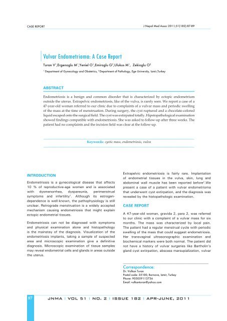
Vulvar Endometrioma: A Case Report
Vulvar Endometrioma: A Case Report
Vulvar Endometrioma: A Case Report
You also want an ePaper? Increase the reach of your titles
YUMPU automatically turns print PDFs into web optimized ePapers that Google loves.
CASE REPORT<br />
INTRODUCTION<br />
Endometriosis is a gynecological disease that affects<br />
10 % of reproductive-age women and is associated<br />
with dysmenorrhea, dyspareunia, perimenstrual<br />
symptoms and infertility 1 . Although its estrogendependence<br />
is well-known, the pathophysiology is still<br />
unclear. Retrograde menstruation is a widely accepted<br />
mechanism causing endometriosis that might explain<br />
ectopic endometrial tissues.<br />
Endometriosis can not be diagnosed with symptoms<br />
and physical examination alone and histopathology<br />
is the mainstay of the diagnosis. Visualization of the<br />
endometriosis implants, taking a sample of suspected<br />
area and microscopic examination give a de� nitive<br />
diagnosis. Microscopic examination of tissue samples<br />
may reveal endometrial cells and glands in areas outside<br />
the uterus.<br />
87<br />
<strong>Vulvar</strong> <strong>Endometrioma</strong>: A <strong>Case</strong> <strong>Report</strong><br />
Turan V 1 ,Ergenoglu M 1 ,Yeniel O 1 ,Emiroglu G 2 ,Ulukus M 1 , Zekioglu O 2<br />
1 Department of Gynecology and Obstetrics, 2 Department of Pathology, Ege University, Izmir,Turkey<br />
ABSTRACT<br />
Extrapelvic endometriosis is fairly rare. Implantation<br />
of endometrial tissues in the vulva, skin, lung and<br />
abdominal wall muscle has been reported before 2 .We<br />
present a case of a patient with vulvar endometrioma<br />
that underwent cyst extirpation, and the diagnosis was<br />
revealed by the histopathologic examination.<br />
CASE REPORT<br />
J Nepal Med Assoc 2011;51(182):87-89<br />
Endometriosis is a benign and common disorder that is characterized by ectopic endometrium<br />
outside the uterus. Extrapelvic endometriosis, like of the vulva, is rarely seen. We report a case of a<br />
47-year-old woman referred to our clinic due to complaints of a vulvar mass and periodic swelling<br />
of the mass at the time of menstruation. During surgery, the cyst ruptured and a chocolate-colored<br />
liquid escaped onto the surgical � eld. The cyst was extirpated totally. Hipstopathological examination<br />
showed � ndings compatible with endometriosis. She was asked to follow-up after three weeks. The<br />
patient had no complaints and the incision � eld was clear at the follow-up.<br />
________________________________________________________________________________________<br />
Keywords: cystic mass, endometriosis, vulva<br />
________________________________________________________________________________________<br />
A 47-year-old woman, gravida 2, para 2, was referred<br />
to our clinic with a complaint of a vulvar mass for six<br />
months. The mass was characterized by local pain.<br />
The patient had a regular menstrual cycle with periodic<br />
swelling of the mass that could suggest endometriosis.<br />
Her transvaginal ultrasonographic examination and<br />
biochemical markers were both normal. The patient did<br />
not have a history of vulvar surgeries like Bartholin’s<br />
gland cyst extirpation, abscess marsupialization, vulvar<br />
__________________________________________________<br />
Correspondence:<br />
Dr. Volkan Turan<br />
Postal code: 35100, Bornova, Izmir, Turkey<br />
Phone: 905059113736<br />
Email: volkanturan@yahoo.com<br />
JNMA I VOL 51 I NO. 2 I ISSUE 182 I APR-JUNE, 2011
iopsy or episiotomy. On physical examination, a � rm<br />
cystic mass, 4 - 5 cm in diameter and covered by<br />
normal skin, was found within the upper-portion of the<br />
right labium majus (Figure 1).<br />
Figure 1. During surgery, the cyst ruptured and a<br />
chocolate-colored liquid escaped onto the surgical<br />
� eld.<br />
During surgery, the cyst ruptured and a chocolate-colored<br />
liquid escaped onto the surgical � eld. Histopathological<br />
� ndings showed endometrial stroma with hemorrhage.<br />
The hemorrhagic areas showed clusters of hemosiderinladen<br />
macrophages (Figure 2).<br />
Figure 2: The histopathologic examination showed<br />
endometrial epithelium (a), the aggregation of<br />
hemosiderin pigments (b) and stroma (c). (Hematoxylin<br />
& Eosin)<br />
Three weeks after the surgery, the patient was called for<br />
a follow-up examination. The patient was asymptomatic<br />
and there was no surgery-related complications.<br />
Turan. et. al Extrapelvic Endometriosis<br />
DISCUSSION<br />
Although endometriosis is one of the mostinvestigated<br />
disorders in gynecology, its etiology and<br />
pathophysiology still remain unclear. An in� ammatory<br />
process has been demonstrated in some studies by<br />
increased concentrations of macrophages and cytokines<br />
– chemokines and their receptors, B cells and T cells 3 .<br />
Because of its regression after menopause, it is believed<br />
to be an estrogen- dependent disease.<br />
The symptomatology of the disease includes chronic<br />
pelvic pain, dyspareunia, dysmenorrhea perimenstrual<br />
symptoms and infertility. Oehmke F. et al. 4 conducted a<br />
research about the impact of endometriosis on the quality<br />
of life. Out of 65 women affected by endometriosis,<br />
they reported dyspareunia in 36 %, dysmenorrhea in<br />
34 % and infertility in 5 % of the patients. CA-125<br />
levels may be high in the presence of endometriosis,<br />
however, it can not be used in monitoring the disease<br />
after treatment.<br />
There are some theories about the spread of<br />
endometriosis. Although retrograde menstrual � ow is<br />
the most accepted theory for extrapelvic endometriosis,<br />
endometriosis including the vulva may be the result of<br />
a lymphatic or hematogenous spread. In a study, vulvar<br />
endometriosis was noted in the Bartholin’s gland and<br />
in a surgical scar that occurred subsequently after<br />
episiotomy, trauma or biopsy 5 . Ulcerations, punctate<br />
foci of endometriosis or cystic mass were established<br />
as the main manifestations in those cases.<br />
In the differential diagnosis of vulvar endometriosis,<br />
mucous cyst, Bartholin’s cyst or abscesses, lipoma and<br />
scene gland cyst must be considered. Furthermore, a<br />
biopsy of the lesion is required for a de� nitive diagnosis.<br />
The treatment must be directed to prevent the<br />
recurrence and provide a good cosmetic result, with<br />
the preferred treatment being complete surgical cyst<br />
extirpation. Although needle aspiration of the lesion is<br />
not a decisive treatment, it can be used as a diagnostic<br />
tool.To the best of our knowledge, � ve cases of clearcell<br />
carcinoma arising from a vulvar endometriosis<br />
have been reported so far 6 . Follow-up after surgery is<br />
warranted for a possible malignant transformation of an<br />
extra-pelvic endometriosis. Recurrence and advanced<br />
age seem to be the main risk factors for the malignancy.<br />
In these cases, a wide surgical excision should be<br />
performed.<br />
In conclusion, the causation of an extrapelvic<br />
endometriosis remains unknown. Although our patient<br />
had not undergone any vulvar surgery that might<br />
have made implantation easier, endometriosis had<br />
occurred. Pruritus or some trauma may have led to this<br />
implantation.<br />
JNMA I VOL 51 I NO. 2 I ISSUE 182 I APR-JUNE, 2011 88
REFERENCES<br />
1. Dechaud H, Dechanet C, Brunet C, Reyftmann L, Hamamah<br />
S, Hedon B. Endometriosis and in vitro fertilization: A<br />
review. Gynecological Endocrinology. 2009;28:1–5.<br />
2. Jubanyik KJ, Comite F. Extrapelvic endometriosis. Obstet<br />
Gynecol Clin North Am. 1997;24:411-40<br />
3. Ulukus M, Ulukus CE, Seval Y, Zheng W, Arici A. Expression<br />
of interleukin-8 receptors in endometriosis. Human<br />
Reproduction. 2005;20(3):794–801.<br />
89<br />
Turan. et. al Extrapelvic Endometriosis<br />
4. Oehmke F, Weyand J, Hackethal A, Konrad L, Omwandho<br />
C, Tinneberg HR. Impact of endometriosis on quality of life:<br />
A pilot study. Gynecological Endocrinology. 2009;25:1–4.<br />
5. Buda A, Ferrari L, Marra C, Passoni P, Perego P, Milani<br />
R. <strong>Vulvar</strong> endometriosis in surgical scar after excision of<br />
the Bartholin gland: report of a case. Arch Gynecol Obstet.<br />
2008;277(3):255-6.<br />
6. Bolis GB, Macciò T. Clear cell adenocarcinoma of the vulva<br />
arising in endometriosis. A case report. Eur J Gynaecol<br />
Oncol. 2000;21(4):416-7.<br />
JNMA I VOL 51 I NO. 2 I ISSUE 182 I APR-JUNE, 2011


