

Aus dem Institut für Tierzucht und Vererbungsforschung - Stiftung ...
Aus dem Institut für Tierzucht und Vererbungsforschung - Stiftung ...
Aus dem Institut für Tierzucht und Vererbungsforschung - Stiftung ...

Create successful ePaper yourself
Turn your PDF publications into a flip-book with our unique Google optimized e-Paper software.
Bibliografische Informationen der Deutschen Bibliothek<br />
Die Deutsche Bibliothek verzeichnet diese Publikation in der Deutschen Nationalbibliografie;<br />
Detaillierte bibliografische Daten sind im Internet über http://dnb.ddb.de abrufbar.<br />
1. Auflage 2007<br />
© 2007 by Verlag: Deutsche Veterinärmedizinische Gesellschaft Service GmbH, Gießen<br />
Printed in Germany<br />
ISBN 978-3-939902-37-9<br />
Verlag: DVG Service GmbH<br />
Frankfurter Straße 89<br />
35392 Gießen<br />
0641/24466<br />
geschaeftsstelle@dvg.net<br />
www.dvg.net
<strong>Aus</strong> <strong>dem</strong> <strong>Institut</strong> <strong>für</strong> <strong>Tierzucht</strong> <strong>und</strong> <strong>Vererbungsforschung</strong><br />
der Tierärztlichen Hochschule Hannover<br />
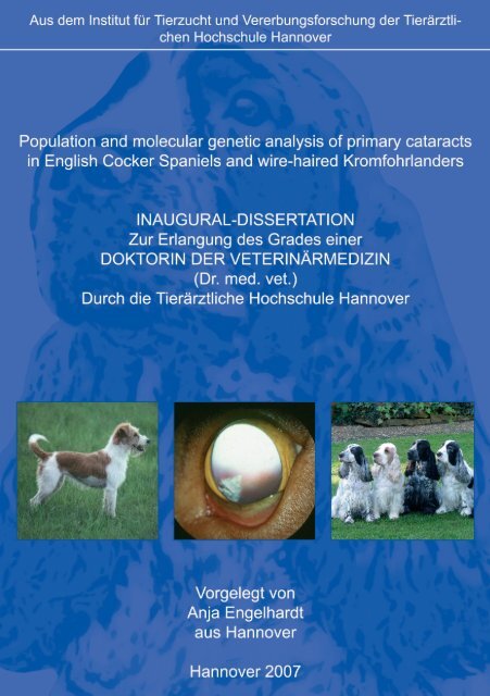
Population and molecular genetic analysis of primary cataracts in<br />
English Cocker Spaniels and wire-haired Kromfohrlanders<br />
INAUGURAL-DISSERTATION<br />
Zur Erlangung des Grades einer<br />
DOKTORIN DER VETERINÄRMEDIZIN<br />
(Dr. med. vet.)<br />
Durch die Tierärztliche Hochschule Hannover<br />
Vorgelegt von<br />
Anja Engelhardt<br />
aus Hannover<br />
Hannover 2007
Wissenschaftliche Betreuung: Prof. Dr. Dr. habil O. Distl<br />
1. Gutachter: Prof. Dr. Dr. habil O. Distl<br />
2. Gutachter: Prof. Dr. M. Boevé<br />
Tag der mündlichen Prüfung: 08.05.2007
Dedicated to my family.
Contents<br />
Chapter 1<br />
Introduction …………………………………………………………………………………... 1<br />
Chapter 2<br />
Primary cataract - A review ………………………………………………………………….. 5<br />
Chapter 3<br />
Analysis of systematic and genetic effects on the prevalence of primary cataract,<br />
persistent pupillary membrane and distichiasis in the two color variants of English<br />
Cocker Spaniels ……………………………………………………………………………... 27<br />
Chapter 4<br />
A retrospective study on the prevalence and formation of primary cataract in<br />
two pedigrees from the German population of English Cocker Spaniel ……………………. 53<br />
Chapter 5<br />
Genetic evaluation of primary cataracts in the German population of single-colored<br />
English Cocker Spaniels ……………………………………………………………………. 71<br />
Chapter 6<br />
Evaluation of canine heat-shock transcription factor 4 (HSF4) as candidate for primary<br />
cataract in English Cocker Spaniels and wire-haired Kromfohrlanders ……………………. 91<br />
Chapter 7<br />
Molecular genetic analysis of primary cataracts in single- and multi-colored<br />
English Cocker Spaniels and wire-haired Kromfohrlanders ………………………………. 101<br />
Chapter 8<br />
General discussion ………………………………………………………………………… 125<br />
Chapter 9<br />
Summary …………………………………………………………………………………... 133<br />
Chapter 10<br />
Erweiterte Zusammenfassung …..…………………………………………………………. 137
Chapter 11<br />
Appendix ……………………………………………………………………………...………. I<br />
Chapter 12<br />
List of publications ……………………………………………………………………. XXVIII<br />
Chapter 13<br />
Acknowledgements ……………………………………………………………………... XXXI
Introduction<br />
CHAPTER 1<br />
Introduction<br />
1
Introduction<br />
Introduction<br />
Eye diseases with established or suspected inheritance are relatively common in domestic<br />
animals, especially in purebred dogs. With increasing knowledge on prevalence and<br />
pathogenesis of eye diseases, breeding guidelines need to be developed for reducing the<br />
prevalences of presumed inherited eye diseases. Therefore, it is of particular importance to<br />
clarify the population and molecular genetic backgro<strong>und</strong> of these conditions.<br />
The English Cocker Spaniel (ECS) is a dog breed with predispositions for many ocular<br />
diseases. Primary cataract, persistent pupillary membrane, progressive retinal atrophy and<br />
distichiasis are counted among the frequently occurring established or presumed inherited eye<br />
diseases in this breed. Primary cataract is defined as any opacity of the lens without<br />
association with other ocular abnormalities and systemic diseases. Persistent pupillary<br />
membrane describes remnants of the embryological vascular network which nourishes the<br />
anterior part of the developing lens and usually regresses in the first four to five weeks after<br />
birth. Extra or supernumerary hairs arising from the free eye lid margin are regarded as<br />
distichiasis. Progressive retinal atrophy is characterized by either primary photoreceptor<br />
dysplasia with an early onset or by primary photoreceptor degeneration with a late onset.<br />
Therefore, primary cataract and progressive retinal atrophy particularly lead to visual<br />
impairment culminating in total blindness. One purpose of this work therefore was to analyze<br />
systematic and genetic influences on the prevalence of these eye diseases in ECS, and to use<br />
the obtained results for developing a selection scheme against primary cataract. The second<br />
purpose of the work was to analyze the molecular genetic backgro<strong>und</strong> of primary cataracts in<br />
ECS. For comparison purposes another dog breed, the wire-haired Kromfohrlander, known to<br />
be commonly affected by primary cataract was considered. Primary cataracts in the<br />
Kromfohrlander occur at a similar age as the early onset form of primary cataract in the ECS.<br />
In human and murine primary cataract many genetic mutations have been described. Because<br />
of the large level of clinical and molecular genetic similarity of this disease in man, mouse<br />
and dog, the mutated human and murine genes seemed to be appropriate candidates for canine<br />
primary cataract.<br />
The contents of the present thesis are presented in single papers as allowed by § 4(4) of the<br />
Rules of Graduation (Promotionsordnung) of the University of Veterinary Medicine<br />
2
Introduction<br />
Hannover. Chapter 2 reviews the literature on primary cataract, while the results of genetic<br />
analyses for the present population of English Cocker Spaniels are presented in chapters 3 to<br />
5. Chapter 6 and 7 comprise the results of the molecular genetic analyses of selected singleand<br />
multi-colored English Cocker Spaniels and wire-haired Kromfohrlaenders. Finally,<br />
results of the present thesis are generally discussed and summarised in chapters 8 to 10.<br />
3
Introduction<br />
4
Review of canine primary cataract<br />
CHAPTER 2<br />
Canine primary cataract - A review<br />
Anja Engelhardt, Ottmar Distl<br />
5
Canine primary cataract – A review<br />
Anja Engelhardt, Ottmar Distl<br />
Review of canine primary cataract<br />
<strong>Institut</strong>e for Animal Breeding and Genetics, University of Veterinary Medicine Hannover,<br />
Fo<strong>und</strong>ation, Bünteweg 17p, D-30559 Hannover, Germany<br />
Abstract<br />
Primary cataract is characterized as a focal or diffuse opacity of the eye lens. It is a very<br />
common eye condition in the dog, and reported prevalences range between 1.8 and 88.0%.<br />
The age of onset of inherited cataracts may be congenital, juvenile or senile. Usually<br />
inheritance is presumed, based on the typical appearance and age in a breed known to be<br />
predisposed to cataracts. In the majority a recessive mode of inheritance is existent, but also<br />
dominant pattern are described. Given the limited success of medical treatment and the<br />
invasiveness of surgical treatment of cataracts, prophylactive measures should be considered<br />
more closely. At this time, many kennel clubs have developed selection programs to reduce<br />
the prevalences of primary cataracts in their breed. In the future, the most successful method<br />
to reduce primary cataract, would be to identify the genetic backgro<strong>und</strong> and the causal<br />
mutation of canine primary cataract in the affected breeds. Genetic tests can then be used to<br />
select breeding animals that do not carry and transmit defect alleles.<br />
Keywords: Canine primary cataract, lens, prevalence, inheritance, genetic backgro<strong>und</strong><br />
6
Review of canine primary cataract<br />
1. Introduction<br />
The eye and its adnexa are prone to a large number of inherited disorders that may impair the<br />
dog’s visual performance. In order to provide accurate advice to breeders, it is essential to<br />
know the biological backgro<strong>und</strong> of relevant eye diseases within a specific breed. One of the<br />
most common and frequently appearing eye diseases and one of the main causes for visual<br />
impairment in dogs, are cataracts.<br />
Cataract is an eye disease which occurs quite frequently in many dog breeds and is<br />
characterized as a focal or diffuse lens opacity. Contrary to secondary cataracts, hereditary<br />
primary cataracts develop independently of any other intraocular disease, of metabolic<br />
diseases like diabetes mellitus or hypocalcaemia, nutritional deficiencies and exogenous<br />
effects such as trauma, radiation, electricity, toxins, medication (Martin et al., 1972; Glaze<br />
and Blanchard, 1982; Gelatt, 1991). Eye diseases which may lead to secondary cataracts<br />
include eye inflammations, progressive retinal atrophy, lens displacement, persistent<br />
hyperplastic primary vitreus, persistent hyperplastic tunica vasculosa lentis or persistent<br />
pupillary membrane. These diseases need to be considered carefully in examinations for<br />
primary cataract. Through the years a variety of medical therapies has been developed in<br />
veterinary medicine for the treatment of cataracts in general, but proved not to provide an<br />
effective medical therapy for established cataracts (Poulos, 1966; Yakely and Filby, 1971;<br />
Brooksby, 1979; Cotlier, 1981; Brainard et al., 1982; MacMillian et al., 1986; West et al.,<br />
1988; Seddon et al., 1991; UK-TIA Study Group, 1992; Gupta et al., 1997; Mares-Perlman et<br />
al., 2000). However, there seems to be no alternative to surgical therapy of established<br />
cataracts by phacoemulsification (Rubin and Gelatt, 1968). Given the limited success of<br />
medical treatment and the invasiveness of surgical treatment of cataracts, prophylactive<br />
measures should be considered more closely. In affected breeds or populations systematic<br />
testing will help to define the metabolic defect present and to identify responsible genetic<br />
defects. Breeding strategies may then be formulated aiming at the reduction of the prevalence<br />
of cataract in the population.<br />
The objective of the present review was to give an overview on the prevalence and genetic<br />
backgro<strong>und</strong> of primary cataract in dogs.<br />
7
Review of canine primary cataract<br />
2. The canine eye lens<br />
The eye lens is an optically dense, flexible structure of the eye, located between the primary<br />
fixed refracting surface of the cornea and the retina. In common with the cornea, the lens has<br />
two principal optical properties: transparency and refractive power.<br />
The lens is high in protein (35 %) and water (65%) and low in minerals. Lens proteins are<br />
divided into two groups: water-soluble and water-insoluble proteins. The water-soluble<br />
crystallins (α, β and γ) are the major structural proteins of the lens and provide transparency<br />
and refraction of the lens by dint of their high concentration. Table 1 gives an overview over<br />
the characteristics of the mammalian crystallins. Lens crystallins are produced within the lens<br />
during formation of the lens fibers and have exceptional longevity. The higher the proportion<br />
of soluble crystallins to water in the lens fibre cytoplasm, the higher the refractive index. The<br />
percentage of soluble protein is important to transparency, too much or too little water and the<br />
cytoplasm becomes turbid. Water-insoluble proteins that are important for maintenance of<br />
lens architecture include the membrane proteins aquaporin, MIP, LIM and the connexins<br />
which provide osmoregulation and help to maintain the intracellular environment (Gruitjers et<br />
al., 1987), the gap junction proteins which form gated channels required for cell-cell<br />
communication, and cytoskeletal proteins such as actin, myosin and vimentin which are<br />
responsible for maintaining cell shape during differentiation and possibly play a role in<br />
accommodation (Kannabiran and Balasubramanian, 2000).<br />
3. Diagnosis and classification of primary cataract<br />
Cataracts refer to a group of lens disorders of varying age of onset, speed and extent of<br />
progression, appearance and etiology. A diagnosis of cataract can only be made by a thorough<br />
eye examination including slit lamp (microscopic) evaluation of the mydriatic eye. Stage of<br />
development, position within the lens and time of development are the most commonly used<br />
categories for classification. The most useful method of classification is the stage of<br />
development, because it is related to the complications of cataract such as lens-induced uveitis<br />
and the prognosis for vision and cataract surgery. There are four typical stages of cataract<br />
development: incipient, immature, mature and hypermature. Visual impairment begins with<br />
bilateral occurring immature cataracts and blindness results when the lens is completey<br />
opaque (mature cataract). In hypermature cataracts some lenses begin to liquefy owing to<br />
8
Review of canine primary cataract<br />
proteolysis, and occasionally some vision may return (lens resorption). The nucleus liquefies<br />
last and may sink to the bottom of the lens, the cortex of which has already liquefied<br />
(morgagnian cataract). Another useful method to supplement the classification of cataracts is<br />
to classify cataracts by description of the anatomic position within the lens. Examples of<br />
anatomic descriptors are anterior capsular, anterior subcapsular, anterior cortical, equatorial,<br />
anterior nuclear, posterior nuclear, posterior cortical, posterior subcapsular, posterior capsular,<br />
polar or sutural. The time of development of the opacity is the third common method of<br />
classification of cataracts. Typical age classifications are congenital, i.e. occurring with up to<br />
eight weeks of age, juvenile or adult, i.e. occurring with up to eight years of age and senile,<br />
i.e. occurring with over eight years of age.<br />
4. Molecular genetic backgro<strong>und</strong> of primary cataracts<br />
Cataracts can result from changes in lens architecture, from disruption of intracellular ordered<br />
arrangement of proteins or from changes in organization of lens fibers due to aberrations in<br />
growth or differentiation. Alternatively, changes in the intracellular environment due to<br />
changes in water or small molecules can result in opacification of the lens due to breakdown<br />
of lens homeostasis (Kannabiran and Balasubramanian, 2000). Development of the cataract<br />
phenotype may be causatively traced to one of the following three main classes of genes: (1)<br />
genes that are active before birth, involved in to embryogenesis and development of the lens<br />
of the eye, e.g. Pax6, (2) genes that are involved in the homeostasis in the lens, therewith<br />
contributing to the maintenance of transparency, e.g. connexins, and (3) genes that produce<br />
gene products, which structurally contribute to transparency, e.g. crystallins (Bhat, 2003).<br />
Initial insights into the genetic causes of cataract came from animal models, mostly mice<br />
(Smith et al., 1997). The first evaluation of large mouse populations for mutations affecting<br />
the eye lens at birth was initiated in 1979, when Kratochvilova and Ehling described for the<br />
first time the systematic screening for murine dominant cataract mutants in the F1 generation<br />
after paternal radiation treatment. Today, a great variety of mouse mutants affecting ocular<br />
development, which arose spontaneously or were recovered after parental treatment by<br />
chemical mutagens or radiation, is available. First, early events will be influenced by genes<br />
coding for transcription factors like Pax6, Pitx3, Maf or Sox, which play a role in various<br />
aspects of lens development, such as crystalline gene expression, cellular elongation and cell<br />
9
Review of canine primary cataract<br />
cycle arrest (Fini et al., 1997; Oliver and Gruss, 1997; Ogino and Yasuda, 2000). However,<br />
only in a few cases cataracts are formed at these early stages of development. More and<br />
diverse phenotypes of cataracts occur, if the lens is maturing and mutations affecting the lens<br />
membranes, e.g. aquaporins (Mip), Lim2 or connexins, or the structural proteins of the<br />
cytosol of the lens fiber cells, the crystallins (Graw, 2004). Mutations in crystalline genes are<br />
resulting in proteins with abnormal structure which can result in lens opacity. Mutations are<br />
predicted to disrupt the tertiary structure of the given crystalline, interfering with solution<br />
with associated crystallins (Hejtmancik, 1998). Thus, maintenance of normal structure as well<br />
as normal amounts of various proteins is essential for lens transparency. A noticeable example<br />
for a mutation in crystallins is the Philly mouse, a strain with dominantly inherited cataract<br />
caused by a mutation in the βB2-crystallin and having an internal deletion of four amino acids<br />
(Kador et al., 1980; Chambers and Russell, 1991). In addition, there are several genetically<br />
engineered mouse models with cataract resulting from abnormalities of development (Lang et<br />
al., 1987; Perez-Castro et al., 1993), immunity (Egwuagu et al., 1994; Geiger et al., 1994),<br />
growth (Mahon et al., 1987; Eva et al., 1991; Griep et al., 1993), cytoskeleton (Capetanaki et<br />
al., 1989; Bloemendal et al., 1997) or membrane transport (Dunia et al., 1996). The mouse as<br />
one of the most important model systems in eye development is currently supplemented by<br />
the rapidely increasing number of mutants in the zebrafish (Glass and Dahm, 2004). Table 2<br />
gives an overview over genes known to be associated with primary cataract and their<br />
localization on human, murine and canine chromosomes.<br />
6. Prevalence and inheritance of primary cataract<br />
Cataract is a very common eye condition in the dog, and many different clinical forms are<br />
exhibited in this species. There is a varied aetiology and many cases are of unknown cause<br />
(Barnett, 1985c).<br />
Primary cataracts have been reported to be inherited in several canine breeds including<br />
Leonbergers (Heinrich et al., 2006), Entlebucher Mountain Dogs (Heitmann et al., 2005;<br />
Davidson and Nelms, 1998; Spiess, 1994), Bichon Frise (Gelatt, 2003, 2005), Tibetan Terriers<br />
(Ketteritzsch et al., 2004), Norwegian Buh<strong>und</strong>s (Bjerkås et al., 1995), Rottweilers (Bjerkås<br />
and Bergsjø, 1991), Chow Chows (Collins et al., 1992), Golden and Labrador Retrievers<br />
(Curtis et al., 1989; Barnett, 1978; Rubin et al., 1974; Gelatt, 1972), German Shepherds<br />
10
Review of canine primary cataract<br />
(Barnett, 1986; von Hippel, 1930), Standard Poodles (Barnett et al., 1985a; Rubin et al.,<br />
1972), Miniature Schnauzers (Barnett, 1985b; Gelatt et al., 1983a), West Highland White<br />
Terriers (Narfström, 1981), Welsh Springer Spaniels (Barnett, 1980), Chesapeake Bay<br />
Retrievers (Gelatt et al., 1979), Boston Terriers (Barnett, 1978), Staffordshire Bull Terriers<br />
(Barnett, 1978), American Cocker Spaniels (Yakely, 1978; Yakely et al., 1971), Cocker<br />
Spaniels (Olesen et al., 1974), Afghan Ho<strong>und</strong>s (Roberts and Helper, 1972), Old English<br />
Sheep Dogs (Koch, 1972) and Beagles (Heywood, 1971).<br />
The genetic mode of inheritance has been proposed for several breeds and includes both<br />
dominant and recessive patterns (Gelatt, 1979; Roberts, 1973). Autosomal recessive<br />
inheritance has been reported for the Boston Terrier, Miniature Schnauzer, Staffordshire Bull<br />
Terrier, Afghan Ho<strong>und</strong>, Standard Poodle, Old English Sheepdog and the Bichon Frise (Rubin<br />
et al., 1969; Rubin and Flowers, 1972; Roberts and Helper, 1972; Barnett, 1978; Koch, 1972;<br />
Gelatt, 2005). Cataracts in the American Cocker Spaniel may be inherited as an autosomal<br />
recessive or polygenic trait (Yakely, 1978). For the English Cocker Spaniel a monogenic<br />
autosomal recessive or a complex mode of inheritance was supposed for primary cataract<br />
(Barnett, 1978, 1980, 1986; Lorimer, 1990; Whitley et al., 1995; Olesen et al., 1974).<br />
Autosomal dominant inheritance has been indicated for the Pointer, German Shepherd Dog,<br />
Labrador Retriever and Golden Retriever (Barnett, 1978; Von Hippel, 1930). An autosomal<br />
dominant inheritance with incomplete penetrance has been indicated for the Beagle and the<br />
Chesapeake Bay Retrievers (Anderson and Schultz, 1958; Gelatt et al., 1979).<br />
In Table 3 reported prevalences and modes of inheritance of primary cataract in several dog<br />
breeds are summarized. The modes of inheritance were determined by inspection of sample<br />
pedigrees.<br />
7. Conclusions<br />
The list of canine breeds exhibiting primary cataract is continuously increasing and, there are<br />
certain breeds in which more than one type of cataract occurs. In response to this increase,<br />
many kennel clubs have developed selection programs to reduce the prevalences of primary<br />
cataracts in their breed. Regular periodical ophthalmological examinations by veterinary<br />
ophthalmologists as well as ban from breeding of affected dogs, in some clubs likewise of<br />
parents and offspring of affected dogs, are the most commonly used terms and conditions. In<br />
11
Review of canine primary cataract<br />
the future, the most successful method to reduce primary cataract, would be to identify the<br />
genetic backgro<strong>und</strong> and the causal mutation, of canine primary cataract in the affected breeds.<br />
Genetic tests can then be used to select breeding animals that do not carry and transmit defect<br />
alleles.<br />
12
Review of canine primary cataract<br />
References<br />
Anderson, A.C., Shultz, F.T., 1958. Inherited (congenital) cataracts in the dog. The American<br />
Journal of Pathology 34, 965-975.<br />
Barnett, K.C., 1972. Types of cataract in the dog. Journal of the American Animal Hospital<br />
Association 8, 2-9.<br />
Barnett, K.C., 1976. Comparative aspects of canine hereditary eye disease. Advances in<br />
Veterinary Science 20, 39-67.<br />
Barnett, K.C., 1978. Hereditary cataract in the dog. Journal of Small Animal Practice 19, 109-<br />
120.<br />
Barnett, K.C., 1980. Hereditary cataract in the Welsh Springer Spaniel. Journal of Small<br />
Animal Practice 21, 621-625.<br />
Barnett, K.C., 1982. Hereditary cataract in the German Shepherd Dog. Proceedings of the<br />
American Society of Veterinary Ophthalmology and International Society of Veterinary<br />
Ophthalmology, Las Vegas, Nevada.<br />
Barnett, K.C., Startup, F.G., 1985a. Hereditary cataract in the Standard Poodle. The<br />
Veterinary Record 117, 15-16.<br />
Barnett, K.C., 1985b. Hereditary cataract in the Miniature Schnauzer. Journal of Small<br />
Animal Practice 26, 635-644.<br />
Barnett, K.C., 1985c. The diagnosis and differential diagnosis of cataract in the dog. Journal<br />
of Small Animal Practice 26, 305-316.<br />
Barnett, K.C., 1986. Hereditary cataract in the German Shepherd Dog. Journal of Small<br />
Animal Practice 27, 387-395.<br />
Barnett, K.C., 1988. Inherited eye diseases in the dog and cat. Journal of Small Animal<br />
Practice 26, 462-475.<br />
Bhat, S.P., 2003. Crystallins, genes and cataract. Progress in Drug Research 60, 205-262.<br />
Bjerkås, E., Bergsjø, T., 1991. Hereditary cataract in the Rottweiler Dog. Veterinary<br />
ophthalmology 1, 7-10.<br />
Bjerkås, E., Haaland, M.B., 1995. Pulverulent nuclear cataract in the Norwegian Buh<strong>und</strong>.<br />
Journal of Small Animal Practice 36, 471-474.<br />
13
Review of canine primary cataract<br />
Bloemendal, H., Raats, J.M., Pieper, F.R., Benedetti, E.L., Dunia, I., 1997. Transgenic mice<br />
carrying chimeric or mutated type III intermediate filament (IF) genes. Cellular and<br />
Molecular Life Sciences 53, 1-12.<br />
Brainard, J., Hanna, C., Petursson, G., 1982. Evaluation of superoxide dismutase (orgotein) in<br />
medical treatment of canine cataract. Archives of Ophthalmology 100, 1832-1834.<br />
Brooksby, L.O., 1979. A practitioner’s experience with selenium-tocopherol in treatment of<br />
cataracts and nuclear sclerosis in the dog. Veterinary Medicine/ Small Animal Clinician<br />
74, 301-301.<br />
Capetanaki, Y., Smith, S., Heath, J.P., 1989. Overexpression of the vimentin gene in<br />
transgenic mice inhibits normal lens cell differentiation. The Journal of Cell Biology 109,<br />
1653-1664.<br />
Chambers, C., Russell, P., 1991. Deletion mutation in an eye lens β-crystallin. Journal of<br />
Biological Chemistry 266, 6742-6746.<br />
Collins, B.K., Collier, L.L., Johnson, G.S., Shibuya, H., Moore, C.P., da Silva Curiel, J.M.A.,<br />
1992. Familial cataracts and concurrent ocular anomalies in Chow Chows. Journal of the<br />
American Veterinary Medical Association 200, 1485-1491.<br />
Cotlier, E., Sharma, Y.R., 1981. Aspirin and senile cataract in rheumatoid arthritis. Lancet 1,<br />
338-339.<br />
Curtis, R., 1982. Primary hereditary cataract in the dog. Veterinary Annual 22, 311-318.<br />
Curtis, R., 1984. Late-onset cataract in the Boston Terrier. Veterinary Record 115, 577-578.<br />
Curtis, R., Barnett, K.C., 1989. A survey of cataracts in Golden and Labrador Retrievers.<br />
Journal of Small Animal Practice 36, 277-286.<br />
Davidson, M.G., Nelms, S.R., 1998. Diseases of the lens and cataract formation. In: Gelatt,<br />
K.N., Veterinary Ophthalmology. Williams & Wilkins, 3 rd edition, 797-826.<br />
Dubielzig, R.R., Swanson, J.F., Wenk, E.J., 1985. Microphthalmia, cataract, lens luxation,<br />
and ciliary body dysplasia in a litter of Springer Spaniels pubs. Transactions of the<br />
Sixteenth Annual Scientific Program of the College of Veterinary Ophthalmologists,<br />
September 27 th to 29 th , San Francisco, California, 96-100.<br />
Dunia, I., Smit, J.J., van der Valk, M.A., Bloemendal, H., Borst, P., Benedetti, E.L., 1996.<br />
Human multidrug resistance 3-P-glycoprotein expression in transgenic mice induces lens<br />
membrane alterations leading to cataract. The Journal of Cell Biology 132, 701-716.<br />
14
Review of canine primary cataract<br />
Egwuagu, C.E., Sztein, J., Chan, C.C., Reid, W., Mahdi, R., Nussenblatt, R.B., Chepelinsky,<br />
A.B., 1994. Ectopic expression of gamma interferon in the eyes of transgenic mice<br />
induces ocular pathology and MHC class II gene expression. Investigative Ophthalmology<br />
and Visual Science 35, 332-341.<br />
Eva, A., Graziani, G., Zannini, M., Merin, L.M., Khillan, J.S., Overbeek, P.A., 1991.<br />
Dominant dysplasia of the lens in transgenic mice expressing the dbl oncogene. The New<br />
Biologist 3, 158-168.<br />
Fini, M.E., Strissel, K.J., West-Mays, J.A., 1997. Perspectives on eye development.<br />
Developmental Genetics 20, 175-185.<br />
Geiger, K., Howes, E., Gallina, M., Huang, X.J., Travis, G.H., Sarvetnick, N., 1994.<br />
Transgenic mice expressing IFN-gamma in the retina develop inflammation of the eye and<br />
photoreceptor loss. Investigative Ophthalmology and Visual Science 35, 2667-2681.<br />
Gelatt, K.N., 1972. Cataracts in the Golden Retriever dog. Veterinary Medicine/ Small<br />
Animal Clinician 67, 1113-1115.<br />
Gelatt, K.N., 1979. Lens and cataract formation in the dog. Compendium on Continuing<br />
Education 1, 75-180.<br />
Gelatt, K.N., Whitley, R.D., Lavach, J.D., Barrie, K.P., Williams, L.W., 1979. Cataracts in<br />
Chesapeake Bay Retrievers. Journal of the American Veterinary Medical Association 175,<br />
1176-1178.<br />
Gelatt, K.N., Samuelson, D.A., Bauer, J.E., Das, N.D., Wolf, E.D., Barrie, K.P., Andresen,<br />
T.L., 1983a. Inheritance of congenital cataracts and microphthalmia in the Miniature<br />
Schnauzer. American Journal of Veterinary Research 44, 1130-1132.<br />
Gelatt, K.N., Samuelson, D.A., Barrie, K.P., Das, N.D., Wolf, E.D., Andresen, T.L., 1983b.<br />
Biometry and clinical characteristics of congenital cataracts and microphthalmia in the<br />
Miniature Schnauzer. Journal of the American Veterinary Medical Association 183, 99-<br />
102.<br />
Gelatt, K.N., 1991. The canine lens. In: Veterinary Ophthalmology. Lea & Febiger,<br />
Philadelphia, pp. 429-460.<br />
Gelatt, K.N., Wallace, M.R., Andrew, S.E., MacKay, E.O., Samuelson, D.A., 2003. Cataracts<br />
in the Bichon Frise. Veterinary Ophthalmology 6, 3-9.<br />
15
Review of canine primary cataract<br />
Gelatt, K.N., MacKay, E.O., 2005. Prevalence of primary breed-related cataracts in the dog in<br />
North America. Veterinary Ophthalmology 8, 101-111.<br />
Glaze, M.B., Blanchard, G.L., 1982. Nutritional cataracts in a Samoyed litter. Journal of the<br />
American Animal Hospital Association 19, 951-954.<br />
Glass, A.S., Dahm, R.S., 2004. The zebrafish as a model organism for eye development.<br />
Ophthalmic Research 36, 4-24.<br />
Graw, J., 2004. Congenital hereditary cataracts. The International Journal of Developmental<br />
Biology 48, 1031-1044.<br />
Griep, A.E., Herber, R., Jeon, S., Lohse, J.K., Dubielzig, R.R., Lambert, P.F., 1993.<br />
Tumorigenicity by human papillomavirus type 16 E6 and E7 in transgenic mice correlates<br />
with alterations in epithelial cell growth and differentiation. Journal of Virology 67, 1373-<br />
1384.<br />
Gruitjers, W.T., Kistler, J., Bullivant, S., Goodenough, D.A., 1987. Immunolocalization of<br />
MP70 in lens fiber 16-17 nm intracellular junctions. Journal of Cell Biology 104, 565-<br />
572.<br />
Gupta, S.K., Joshi, S., Velpandian, T, Varma, S.D., 1997. Protection against cataract by<br />
pyruvate and its ocular kinetics. Annals of Ophthalmology 29, 243-248.<br />
Heinrich, C.L., Lakhani, K.H., Featherstone, H.J., Barnett, K.C., 2006. Cataract in the UK<br />
Leonberger population. Veterinary Ophthalmology 9, 350-356.<br />
Heitmann, M., Hamann, H., Brahm, R., Grußendorf, H., Rosenhagen, C.U., Distl, O., 2005.<br />
Analysis of prevalences of presumed inherited eye diseases in Entlebucher Mountain<br />
Dogs. Veterinary Ophthalmology 8, 145-151.<br />
Hejtmancik, J.F., 1998. The genetics of cataract: Our vision becomes clearer. American<br />
Journal of Human Genetics 62, 520-525.<br />
Heywood, R. 1971. Juvenile cataracts in the Beagle dog. Journal of Small Animal Practice 12,<br />
171-177.<br />
Hirth, R.S., Greenstein, E.T., Peer, R.L., 1974. Anterior capsular opacities (spurious cataracts)<br />
in the Beagle dog. Veterinary Pathology 11, 181-194.<br />
Kador, P.F., Fukui, H.N., Fukushi, S., Jernigan, H.M., Kinoshita, J.H., 1980. Philly mouse: a<br />
new model of hereditary cataract. Experimental Eye Research 30, 59-68.<br />
16
Review of canine primary cataract<br />
Kannabiran, C., Balasubramanian, D., 2000. Molecular genetics of cataract. Indian Journal of<br />
Ophthalmology 48, 5-13.<br />
Ketteritzsch, K., Hamann, H., Brahm, R., Grußendorf, H., Rosenhagen, C.U., Distl, O., 2004.<br />
Genetic analysis of presumed inherited eye diseases in Tibetan Terriers. The Veterinary<br />
Journal 168, 151-159.<br />
Koch, S.A., 1972. Cataracts in interrelated Old English Sheepdogs. Journal of the American<br />
Veterinary Medical Association 160, 299-301.<br />
Kratochvilova, J., Ehling, U.H., 1979. Dominant cataract mutations induced by γ-irradiation<br />
of male mice. Mutation Research 63, 221-223.<br />
Lang, R.A., Metcalf, D., Cuthbertson, R.A., Lyons, I., Stanley, E., Kelso, A., Kannourakis,<br />
G., Williamson, D.J., Klintworth, G.K., Gonda, T.J., et al., 1987. Transgenic mice<br />
expressing a hemopoetic growth factor gene (GM-CSF) develop accumulations of<br />
macrophages, blindness, and a fatal syndrome of tissue damage. Cell 51, 675-686.<br />
Lorimer, D.W., 1990. Cataract in small animals. Pet Focus 2, 55-57.<br />
MacMillian, A., Nelson, D., Munger, R., et al., 1986. A comparison of zinc ascobate versus<br />
saline placebo in the treatment of canine cataracts. Proceedings of the Scientific Meeting<br />
of the American College of Veterinary Ophthalmologists and International Society of<br />
Veterinary Ophthalmology 17, 484.<br />
Mahon, K.A., Chepelinsky, A.B., Khillan, J.S., Overbeek, P.A., Piatigorsky, J., Westphal, H.,<br />
1987. Oncogenesis of the lens in transgenic mice. Science 235, 1622-1628.<br />
Mares-Perlman, J.A., Lyle, B.J., Klein, R., Fisher, A.I., Brady, W.E., VandenLangenberg,<br />
G.M., Trabulsi, J.N., Palta, M., 2000. Vitamin supplement use and incident cataracts in a<br />
population-based study. Archives of Ophthalmology 118, 1556-1563.<br />
Martin, C.L., Christmas, R., Leipold, H.W., 1972. Formation of temporary cataracts in dogs<br />
given a disophenol preparation. Journal of the American Veterinary Medical Association<br />
161, 294-433.<br />
Narfström, K., Dubielzig, R., 1984. Posterior Lenticonus, cataracts and mircophthalmia:<br />
congenital ocular defects in the Cavalier King Charles Spaniel. Journal of Small Animal<br />
Practice 25, 669-677.<br />
Narfström, K., 1981. Cataract in the West Highland White Terrier. Journal of Small Animal<br />
Practice 22, 467-471.<br />
17
Review of canine primary cataract<br />
Olesen, H.P., Jensen, O.A., Norn, M.S., 1974. Congenital hereditary cataract in English<br />
Cocker Spaniel. Journal of Small Animal Practice 15, 741-750.<br />
Oliver, G., Gruss, P., 1997. Current views on eye development. Trends in Neurosciences 20,<br />
415-421.<br />
Ogino, H., Yasuda, K., 2000. Sequential activation of transcription factors in lens induction.<br />
Development, Growth and Differentiation 42, 437-448.<br />
Perez-Castro, A.V., Tran, V.T., Nguyen-Huu, M.C., 1993. Defective lens fiber differentiation<br />
and pancreatic tumorgenesis caused by ectopic expression of the cellular retinoic acid-<br />
binding protein I. Development (Cambridge, England) 119, 363-375.<br />
Poulos, P.Jr., 1966. Selenium-tocopherol treatment of senile lenticular sclerosis in dogs (four<br />
case report). Veterinary Medicine/ Small Animal Clinician 61, 986-988.<br />
Rubin, L.F., Koch, S.A., Huber, R.J., 1969. Hereditary cataracts in Miniature Schnauzers.<br />
Journal of the American Veterinary Medical Association 154, 1456-1458.<br />
Rubin, L.F., 1974. Cataract in Golden Retrievers. Journal of the American Veterinary Medical<br />
Association 165, 457-458.<br />
Roberts, S.R., Helper, L., 1972. Cataracts in the Afghan Ho<strong>und</strong>s. Journal of the American<br />
Veterinary Medical Association 160, 427-432.<br />
Roberts, S.R., 1973. Hereditary cataracts. The Veterinary Clinics of North America 3, 433-<br />
437.<br />
Rubin, L.F., Gelatt, K.N., 1986. Spontaneous resorption of the cataractous lens in dogs.<br />
Journal of the American Veterinary Medical Association 152, 139-152.<br />
Rubin, L.F., Flowers R.D., 1972. Inherited cataract in a family of Standard Poodles. Journal<br />
of the American Veterinary Medical Association 161, 207-208.<br />
Rubin, L.F., Flowers, R.D., 1974. Cataract in Golden Retrievers. Journal of the American<br />
Veterinary Medical Association 165, 457-458.<br />
Seddon, J.M., Christen, W.G., Manson, J.E., Buring, J.E., Sperduto, R.D., Hennekens, C.H.,<br />
1991. Low-dose aspirin and risks of cataracts in a randomized trial of US physicians.<br />
Archives of Ophthalmology 109, 252-255.<br />
Smith, R.S., S<strong>und</strong>berg, J.P., Linder, C.C., 1997. Mouse mutations as models for studying<br />
cataracts. Pathobiology: Journal of Immunopathology, molecular and cellular biology 65,<br />
146-154.<br />
18
Review of canine primary cataract<br />
Spiess, B.M., 1994. Vererbte Augenkrankheiten beim Entlebucher Sennenh<strong>und</strong>. Schweizer<br />
Archiv <strong>für</strong> Augenheilk<strong>und</strong>e 136, 105-110.<br />
Stades, F.C., 1980. Persistent hyperplastic tunica vasculosa lentis and persistent hyperplastic<br />
primary vitreus (PHTVL/ PHPV) in 90 closely related Doberman Pinschers: Clinical<br />
aspects. Journal of the American Animal Hospital Association 16, 739.<br />
Strande, A., Nicolaissen, B., Bjerkås, I., 1988. Persistent pupillary membrane and congenital<br />
cataract in a litter of English Cocker Spaniels. Journal of Small Animal Practice 29, 257-<br />
260.<br />
UK-TIA Study Group, 1992. Does aspirin affect the rate of cataract formation? Cross-<br />
sectional results during a randomised double-blind placebo controlled trial to prevent<br />
serious vascular events. British Journal of Ophthalmology 76, 259-261.<br />
Van der Linde-Sipman, J.S., Stades, F.C., de Wolff-Rouendaal, D., 1985. Persistent<br />
hyperplastic tunica vasculosa lentis and persistent hyperplastic primary vitreus in the<br />
Doberman Pinscher: Pathological aspects. Journal of the American Animal Hospital<br />
Association 19, 791.<br />
Von Hippel, E., 1930. Embryologische Untersuchungen über Vererbung angeborener<br />
Katarakte, über Schichtstar des H<strong>und</strong>es sowie über eine besondere Form von<br />
Kapselkatarakt. Albrecht von Graefe’s Archiv <strong>für</strong> Ophthalmologie 124, 300-324.<br />
Wallace, M.R., MacKay, E.O., Gelatt, K.N., Andrew, S.E., 2005. Inheritance of cataract in<br />
the Bichon Frise. Veterinary Ophthalmology 8, 203-205.<br />
West, S.K., Munoz, B.E., Newland, H.S., Emmett, E.A., Taylor, H.R., 1988. Lack of evidence<br />
for aspirin use and prevention of cataracts. Archives of Ophthalmology 105, 1229-1231.<br />
Whitley, R.D., McLaughlin, S.A., Gilger, B.C., 1995. Update on eye disorders among<br />
purebred dogs. Veterinary Medicine 90, 574-592.<br />
Yakely, W.L., Filby, R.H., 1971. Selenium in the lens of the dog. Journal of the American<br />
Veterinary Medical Association 158, 1564-1571.<br />
Yakely, W.L., Hegreberg, G.A., Padgett, G.A., 1971. Familial cataracts in the American<br />
Cocker Spaniel. Journal of the American Animal Hospital Association 39, 127-135.<br />
Yakely, W.L., 1978. A study of heritability of cataracts in the American Cocker Spaniel.<br />
Journal of the American Animal Hospital Association 172, 814-817.<br />
19
Table 1<br />
Review of canine primary cataract<br />
Type, expression and function of the water-soluble proteins of the lens, the crystallins<br />
Lens protein (type of protein)<br />
Subgroups Place of expression Function in the lens<br />
α-Crystallins (heat shock protein)<br />
αA-crystallin hich leverls in the lens structural component of the lens,<br />
(lens-specific), low levels protective role in maintaining<br />
in the spleen<br />
solubility of intracellular proteins and<br />
promoting resistandce of cells to<br />
stress (chaperone-like function)<br />
αB-crystallin ubiquitously expressed,<br />
high levels in brain,<br />
muscle, lung, thymus,<br />
kidney<br />
structural component of the lens<br />
β-Crystallins (epidermis-specific differentation protein)<br />
acidic β-crstallins<br />
(βA1/A2, βA2 and βA4)<br />
basic β-crystallins<br />
(βB1, βB2 and βB3)<br />
lens-specific structural component of the lens,<br />
protein homodimerization activity<br />
lens-specific, βB2 low structural component of the lens<br />
levels in brain and testis<br />
γ-Crystallins (epidermis-specific differentation protein)<br />
four γ-crystallins<br />
(γA, γB, γC and γD)<br />
three pseudogenes<br />
(γE, γF and γG)<br />
lens-specific structural component of the lens<br />
lens-specific structural component of the lens<br />
20
Review of canine primary cataract<br />
Table 2<br />
Genes associated with primary cataract and their localizations in humans, mice and dogs<br />
Human gene Human<br />
Murine<br />
Canine<br />
symbol chromosome chromosome chromosome<br />
BFSP2 HSA3 MMA9 CFA23<br />
CRYAA HSA21 MMA17 CFA31<br />
CRYAB HSA11 MMA9 CFA5<br />
CYRBA1 HSA17 MMA11 CFA9<br />
CRYBA2 HSA2 MMA1 CFA37<br />
CRYBA4 HSA22 MMA5 CFA26<br />
CRYBB1 HSA22 MMA5 CFA26<br />
CRYBB2 HSA22 MMA5 CFA26<br />
CRYBB3 HSA22 MMA5 CFA26<br />
CRYGA HSA2 MMA1 CFA37<br />
CRYGB HSA2 MMA1 CFA37<br />
CRYGC HSA2 MMA1 CFA37<br />
CRYGD HSA2 MMA1 CFA37<br />
CRYGS HSA3 MMA16 CFA34<br />
EYA1 HSA8 MMA1 CFA29<br />
FOXE3 HSA1 MMA4 CFA15<br />
FTL HSA19 MMA4/ MMA7 CFA1<br />
GCNT2 HSA6 MMA13 CFA35<br />
GJA3 HSA13 MMA14 CFA25<br />
GJA8 HSA1 MMA3 CFA17<br />
HSF4 HSA16 MMA8 CFA5<br />
LIM2 HSA19 MMA7 CFA1<br />
MAF HSA16 MMA8 CFA5<br />
MIP HSA12 MMA10 CFA10<br />
PAX6 HSA11 MMA2 CFA18<br />
PITX3 HSA10 MMA19 CFA28<br />
SIX5 HSA19 MMA7 CFA1<br />
SORD HSA15 MMA2 CFA30<br />
SOX1 HSA13 MMA8 CFA22<br />
SPARC HSA5 MMA12 CFA4<br />
TRNT1 HSA3 MMA6 CFA20<br />
CHX10 HSA14 MMA12 CFA8<br />
21
Table 3<br />
Review of canine primary cataract<br />
Survey of the prevalences, appearance and mode of inheritance of primary cataract in several<br />
dog breeds<br />
22
Table 3 (continued)<br />
Review of canine primary cataract<br />
23
Table 3 (continued)<br />
Review of canine primary cataract<br />
24
Table 3 (continued)<br />
Review of canine primary cataract<br />
25
Table 3 (continued)<br />
Review of canine primary cataract<br />
26
Genetic analysis of primary cataract, persistent pupillary membrane and distichiasis<br />
CHAPTER 3<br />
Analysis of systematic and genetic effects on the prevalence of primary<br />
cataract, persistent pupillary membrane and distichiasis in the two color<br />
variants of English Cocker Spaniels<br />
Anja Engelhardt, Kathrin F. Stock, Henning Hamann, Rolf Brahm, Heinrich Grußendorf,<br />
Carsten U. Rosenhagen, Ottmar Distl<br />
27
Genetic analysis of primary cataract, persistent pupillary membrane and distichiasis<br />
Analysis of systematic and genetic effects on the prevalence of primary<br />
cataract, persistent pupillary membrane and distichiasis in the two color<br />
variants of English Cocker Spaniels<br />
Analyse von systematischen <strong>und</strong> genetischen Effekten auf die Prävalenz von<br />
primärer Katarakt, persistierender Pupillarmembran <strong>und</strong> Distichiasis bei<br />
den beiden Farbvarianten des Englischen Cocker Spaniels<br />
Anja Engelhardt 1 , Kathrin Friederike Stock 1 , Henning Hamann 1 , Rolf Brahm 2 , Heinrich<br />
Grußendorf 2 , Carsten U. Rosenhagen 2 , Ottmar Distl 1*<br />
1<br />
<strong>Institut</strong>e for Animal Breeding and Genetics, University of Veterinary Medicine Hannover,<br />
Fo<strong>und</strong>ation, Bünteweg 17p, 30559 Hannover, Germany<br />
2<br />
Dortm<strong>und</strong>er Kreis – Association for Diagnosis of Inherited Eye Diseases in Animals (DOK),<br />
Dortm<strong>und</strong>, Germany<br />
Running head: Ocular diseases in English Cocker Spaniels<br />
Summary<br />
Genetic parameters were estimated for prevalences of primary cataract (CAT), persistent<br />
pupillary membrane (PPM) and distichiasis (DIST) in 615 single-colored and 617 multicolored<br />
English Cocker Spaniels (ECS) bred in the German kennel club for Spaniels<br />
(Jagdspaniel-Klub e.V.). CAT or CAT diagnosed in dogs up to three and a half years of age<br />
(early-onset cataract, CAT-early) and CAT diagnosed in dogs over three and a half years of<br />
age (late-onset cataract, CAT-late), PPM and DIST were included as binary traits in<br />
multivariate genetic analyses. Heritabilities on the <strong>und</strong>erlying liability scale were 0.15 for<br />
CAT, 0.34 for CAT-early, 0.13 for CAT-late, 0.46 for PPM, and 0.62 for DIST in singlecolored<br />
ECS and 0.06 for CAT, 0.13 for CAT-early, 0.14 for CAT-late, 0.10 for PPM, and<br />
0.61 for DIST in multi-colored ECS. There were indications for a different genetic basis of<br />
28
Genetic analysis of primary cataract, persistent pupillary membrane and distichiasis<br />
CAT-early and CAT-late in single-colored ECS as genetic correlations were close to zero. In<br />
multi-colored ECS, a similar tendency for CAT-early and CAT-late could be observed.<br />
Keywords: primary cataract, persistent pupillary membrane, distichiasis, English Cocker<br />
Spaniel, heritability.<br />
Zusammenfassung<br />
Für die Prävalenzen von primärer Katarakt (CAT), persistierender Pupillarmembran (PPM)<br />
<strong>und</strong> Distichiasis (DIST) wurden genetische Parameters bei 615 einfarbigen <strong>und</strong> 617<br />
mehrfarbigen Englischen Cocker Spanieln (ECS) geschätzt. Alle ophthalmologisch<br />
untersuchten H<strong>und</strong>e wurden im Jagdspaniel-Klub e.V. gezüchtet. Primäre Katarakt oder<br />
primäre Katarakt diagnostiziert bei H<strong>und</strong>en bis zu 3,5 Jahren (früh-manifeste Katarakt, CATearly)<br />
and primäre Katarakt diagnostiziert bei H<strong>und</strong>en mit mehr als 3,5 Jahren (spät-manifeste<br />
Katarakt, CAT-late), PPM and DIST wurden als binäre Merkmale in den multivariaten<br />
genetischen Analysen berücksichtigt. Die Heritabilitäten betrugen nach Transformation in ein<br />
Schwellenwertmodell h 2 = 0,15 <strong>für</strong> die primäre Katarakt, 0,34 <strong>für</strong> die früh-manifeste primäre<br />
Katarakt, 0,13 <strong>für</strong> die spät-manifeste primäre Katarakt, 0,46 <strong>für</strong> PPM <strong>und</strong> 0,62 <strong>für</strong> DIST bei<br />
einfarbigen ECS <strong>und</strong> h 2 = 0,06 <strong>für</strong> die primäre Katarakt, 0,13 <strong>für</strong> die früh-manifeste primäre<br />
Katarakt, 0,14 <strong>für</strong> die spät-manifeste primäre Katarakt, 0,10 <strong>für</strong> PPM <strong>und</strong> 0,61 <strong>für</strong> DIST bei<br />
mehrfarbigen ECS. Da die genetischen Korrelationen bei einfarbigen ECS nahe bei Null<br />
waren, ist davon auszugehen, dass bei dieser Farbvariante der ECS früh-manifeste Katarakt<br />
<strong>und</strong> spät-manifeste Katarakt genetisch unterschiedliche Merkmale darstellen. Ähnliches<br />
konnte bei mehrfarbigen ECS <strong>für</strong> diese beiden sich früh <strong>und</strong> spät im Leben manifestierenden<br />
Kataraktformen beobachtet werden.<br />
Schlüsselwörter: Primäre Katarakt, persistierende Pupillarmembran, Distichiasis, Englischer<br />
Cocker Spaniel, Heritabilität.<br />
Introduction<br />
The English Cocker Spaniel (ECS) is a breed predisposed to several diseases of the eye and<br />
its adnexes including primary cataract (CAT), persistent pupillary membrane (PPM),<br />
distichiasis (DIST) and progressive retinal atrophy (PRA). For these ocular diseases<br />
inheritance was established or supposed (ACVO, 1999; Barnett, 1976; Robinson, 1991; Veith<br />
29
Genetic analysis of primary cataract, persistent pupillary membrane and distichiasis<br />
and Gelatt, 1970; Walde, 1986, 1994; Whitley et al., 1995). CAT is defined as any opacity of<br />
the lens without other associated ocular diseases and systemic abnormalities (ECVO, 1998,<br />
2004). Rubin (1989) described two different types of familial CAT in ECS. The first type, a<br />
cataracta corticalis posterior, occurs as a form with an early age of onset of one and a half to<br />
three years of age and a second form with a later age of onset, usually at eight to nine years of<br />
age. The second type, a cataracta nuclearis fibrillaris, appears in two year-old dogs (Rubin,<br />
1989). By contrast Gelatt and MacKay (2005) distinguished between a congenital form of<br />
CAT and a later appearing form, mostly seen in four to seven year-old dogs. PPM describes<br />
remnants of the embryological vascular network which nourishes the anterior part of the<br />
developing lens and according to the ECVO usually regresses in the first weeks after birth.<br />
There can be: tiny, more or less triangular shaped dots, centrally, on the anterior capsule of<br />
the lens; retrocorneal, opaque brown/white material against the endothelium; strands from<br />
cornea to iris, from iris to iris, from iris to lens, connected to areas of dense white cataract;<br />
and strands connected to a sheet/ “spider web” of tissue in the anterior chamber (ECVO,<br />
1998, 2004). Single or multiple hairs arising from the free eye lid margin (ECVO, 1998,<br />
2004) are regarded as distichiasis. PRA is characterized by either primary photoreceptor<br />
dysplasia with an early onset or by primary photoreceptor degeneration with a late-onset<br />
(ECVO, 1998, 2004).<br />
Concerning the modes of inheritance of the above mentioned eye diseases, monogenic<br />
autosomal recessive inheritance (Barnett, 1978, 1980; Barnett and Startup, 1985; Gelatt,<br />
1983a, 1983b; Whitley et al., 1995) or a mode of inheritance of a more complex nature<br />
(Olesen, 1974; Yakely, 1978) has been assumed for CAT in different dog breeds. In ECS,<br />
complex segregation analyses could show the significant contribution of many gene loci to<br />
CAT, DIST and PPM as well the exclusion of pure monogenic models for these eye diseases<br />
(Zadil, 2004). Monogenic autosomal dominant (Barnett, 1976; Whitley et al., 1995) or<br />
monogenic autosomal recessive inheritance (Wiesner and Willer, 1983) has been assumed for<br />
DIST and monogenic autosomal recessive inheritance (Black, 1972; Barnett, 1976; Chader,<br />
1991; Peiffer and Gelatt, 1991; Rubin, 1989) has been assumed for PRA in the ECS.<br />
According to the ECVO (1998, 2004), PPM is not considered as hereditary in ECS, although<br />
a polygenic inheritance has been supposed (Veith and Gelatt, 1970; Strande et al., 1988) and<br />
confirmed by Zadil (2004).<br />
30
Genetic analysis of primary cataract, persistent pupillary membrane and distichiasis<br />
Ophthalmological examinations have been compulsory for breeding dogs in the German<br />
kennel club for Spaniels (Jagdspaniel-Klub e.V., JSK) since 2001. Since 2004, the JSK<br />
recognizes only examinations for presumed inherited eye diseases (PIED) performed by<br />
veterinarians approved by the German panel of the European Eye Scheme for diagnosis of<br />
inherited eye diseases in animals, the Dortm<strong>und</strong>er Kreis (DOK, www.dok-vet.de). Dogs<br />
affected by CAT, congenital blindness, PRA, ectropion or entropion will be excluded from<br />
breeding in this kennel club. The offspring from dogs affected by PRA, CAT or congenital<br />
blindness are banned from further breeding as well. A certificate supplied by a DOK<br />
approved veterinarian stating that dogs intended to be mated within the next year are free<br />
from the above-mentioned eye diseases is required for breeding allowance.<br />
The objective of this study was to estimate genetic parameters for presumed inherited and<br />
prevalent eye diseases in the ECS bred in the JSK. CAT, PPM and DIST were included in the<br />
genetic analyses using liability models due to the involvement of a significant polygenic<br />
component in the mode of inheritance in ECS. In refined analyses, age at diagnosis of CAT<br />
was used for definition of two distinct traits, i.e. CAT diagnosed in up to three and a half<br />
year-old dogs (CAT-early) and CAT diagnosed in over three and a half year-old dogs (CATlate).<br />
The subdivision of CAT reported here was derived using different cut off ages for<br />
expression of CAT and genetic correlations among the different traits distinguished. The<br />
analysis of primary cataracts defined by age of diagnosis should allow us to distinguish<br />
between a single form of CAT with variable age of onset and genetically different age related<br />
forms of CAT. All analyses were performed separately for single-colored and multi-colored<br />
ECS.<br />
Materials and Methods<br />
The study was based on the results of ophthalmological examinations for PIED of 1232 ECS<br />
which were collected between January 2001 and July 2006 by the DOK. Dogs included in this<br />
analysis were born between 1981 and 2005. All ophthalmological examinations were<br />
performed by DOK-certified ophthalmologists using slit-lamp biomicroscopy and indirect<br />
ophthalmoscopy. These DOK-members were approved for examination of PIED after<br />
successful completion of a two-year training program. Eye examinations were performed<br />
according to the standardized protocol of the European College of Veterinary<br />
31
Genetic analysis of primary cataract, persistent pupillary membrane and distichiasis<br />
Ophthalmologists (ECVO). The diagnoses were recorded on official forms proposed by the<br />
ECVO (1998, 2004). According to the ECVO, all cataracts that occur bilaterally or<br />
unilaterally and affect especially the cortex are defined as hereditary primary cataracts except<br />
for cases with obvious association with trauma, inflammation, metabolic disease or nutritional<br />
deficiencies. Also excepted from the hereditary forms are minor, clearly cirumscript cataracts<br />
located in the suture lines or distinctly in the nucleus and cataracts that are located in or on the<br />
anterior capsule associated with persistent pupillary membrane or in or on the posterior<br />
capsule as ‘scarghosts’ of the tunica vasculosa lentis (ECVO, 1998, 2004).<br />
The distribution of prevalences of CAT indicated that there may be differences in the age of<br />
manifestation of CAT in the German population of ECS. Out of the 100 single-colored ECS<br />
ophthalmologically examined up to the age of one and a half years, CAT was diagnosed in 20<br />
dogs (Table 1). In addition, the cumulative distributions of age at first diagnosis of CAT<br />
(Figure 1) and at first ophthalmological examinations (Figure 2) showed a large proportion of<br />
dogs examined early in life and a large prevalence of early-onset primary cataracts in ECS<br />
analysed here. Almost 50 % of the affected single- and multi-colored ECS were diagnosed as<br />
affected by CAT by the age of three and a half years. A high proportion of animals were<br />
diagnosed as affected by CAT with less than four years of age. Subsequently we performed<br />
preliminary analyses defining early- and late-onset forms of CAT. Here, we used each two<br />
classes, such as ≤1.5 (≤2, ≤3, ≤3.5, ≤4, ≤5) and >1.5 (>2, >3, >3.5, >4, >5) years of onset and<br />
multivariate estimation of genetic parameters. Using the subdivision in ≤3.5 and >3.5 years,<br />
the smallest genetic correlations were estimated among these classifications of CAT and thus,<br />
we used this differentiation of CAT. A further analysis distinguishing three age-related forms<br />
of CAT (≤1.5, >1.5 and ≤3.5 and >3.5) was performed in single-colored ECS to take into<br />
account the high prevalence of CAT in young dogs. Genetic correlations between CAT<br />
diagnosed ≤1.5 years and >1.5 and ≤3.5 years was close to one (0.98 ± 0.09) indicating that a<br />
distinction in these two traits did not seem necessary. Therefore, primary cataracts diagnosed<br />
by 3.5 years of age were jointly considered in the analyses because these primary cataracts<br />
appeared as identical traits expressed at variable age.<br />
We therefore analyzed the prevalences of CAT, CAT-early (CAT diagnosed in dogs of up to<br />
three and a half years of age), CAT-late (CAT diagnosed in dogs over three and a half years<br />
of age), PPM and DIST as separate binary traits. Animals classified as affected by or<br />
32
Genetic analysis of primary cataract, persistent pupillary membrane and distichiasis<br />
suspicious for CAT, PPM or DIST were encoded as 1 for the respective trait, animals<br />
classified as not affected were encoded as 0 for the respective trait. If dogs were up to three<br />
and a half years old when diagnosed as affected by or suspicious for CAT, they were encoded<br />
as 1 for CAT-early, whilst all other dogs included in this study were encoded as 0 for CAT-<br />
early. If dogs were older than three and a half years when diagnosed as affected by or<br />
suspicious for CAT, they were encoded as 1 for CAT-late, whilst all other dogs included in<br />
this study were encoded as 0 for CAT-late.<br />
Pedigree information was provided by the JSK and was available for a total of 43,524 ECS<br />
including the 615 single-colored and 617 multi-colored ECS with ophthalmological records.<br />
Coancestry and inbreeding coefficients were calculated using PEDIG software (Boichard,<br />
2002). Data included information on sire and dam as well as on date of birth, kennel, sex and<br />
color. Single-colored ECS included dogs of the colors blond, buff, brown, golden, red, liver<br />
and black as well as black with tan, brown with tan and sable. Dogs registered as singlecolored<br />
are only permitted to have small white markings at their chest. Multi-colored ECS<br />
included dogs of the colors black and white, brown and white, brown roan, blue roan, liver<br />
and white, liver roan, red and white, red roan, orange and white, orange roan, sable and white<br />
and tricolor.<br />
Details on the distribution of data are given in Table 1. Ophthalmological data were available<br />
for 412 female and 203 male single-colored ECS and for 413 female and 204 male multicolored<br />
ECS. The single-colored ECS descended from 229 sires with on average 2.65 ± 2.54<br />
offspring (range 1 to 14) and 394 dams with on average 1.56 ± 1.11 offspring (range 1 to 10).<br />
The multi-colored ECS descended from 232 sires with on average 2.66 ± 2.83 offspring<br />
(range 1 to 20) and 363 dams with on average 1.70 ± 1.13 offspring (range 1 to 9). The<br />
average litter size was 5.20 ± 2.27 puppies with on average 36% examined dogs per litter in<br />
single-colored ECS and 5.51 ± 2.18 puppies with on average 33% examined dogs per litter in<br />
multi-colored ECS. The mean inbreeding coefficient, calculated on the basis of all available<br />
pedigree information, was 3.13 ± 4.43% with a maximum of 28.60% in the single-colored<br />
ECS, and 3.60 ± 4.17% with a maximum of 27.40% in the multi-colored ECS.<br />
Genetic parameters were estimated using Residual Maximum Likelihood (REML) and VCE<br />
4.2.5 software (Groeneveld, 1998). Because of the low prevalences and the assumed<br />
monogenic inheritance of PRA in single-colored and multi-colored ECS, estimation of genetic<br />
33
Genetic analysis of primary cataract, persistent pupillary membrane and distichiasis<br />
parameters was confined on the traits CAT, CAT-juvenil, CAT-late, PPM and DIST. Pedigree<br />
information over eight generations was considered. The completeness of pedigrees was larger<br />
than 91% in all eight generations for both single- and multi-colored ECS. Because the average<br />
coefficient of coancestry in all animals born after 1999 was 2.1% within the single-colored<br />
and 1.8% within the multi-colored ECS, but only 0.09% between single- and multi-colored<br />
ECS, all analyses were performed separately for single-colored and multi-colored ECS.<br />
All traits were included as dependent dichotomously distributed variates in the multivariate<br />
animal model:<br />
Yijkl = µ + Fi + b*cj + ak + eijkl<br />
where Yijkl is the occurrence of the respective eye disease (0/1 trait) of the k-th animal, µ is<br />
the model constant, Fi represents the trait-specific systematic effects, cj represents the traitspecific<br />
covariate with linear regression coefficient b, ak is the random additive genetic effect<br />
of the animal, and eijkl is the random residual effect.<br />
The number of animals included in the additive relationship matrix was 3931 for the singlecolored<br />
and 3936 for the multi-colored ECS, including 615 single-colored and 617 multicolored<br />
animals with ophthalmological records and 895 single-colored and 857 multi-colored<br />
ECS base animals (fo<strong>und</strong>ers) without records.<br />
Models for the genetic analyses were developed on the basis of the results of single and<br />
multiple analyses of variance using the GENMOD and GLIMMIX procedures of SAS<br />
(Statistical Analysis System), version 9.1.3 (SAS <strong>Institut</strong>e, Cary, NC, USA, 2006). We used a<br />
binomial distribution function and the probit function as link function for the generalized<br />
linear model. Significance level was set to P < 0.05. Analyses of variance were performed<br />
separately for single-colored and multi-colored ECS. The following factors were tested as<br />
fixed effects: sex (female, male), year of birth (≤1996, 1997 to 1998, 1999 to 2000, 2001 to<br />
2002, >2002), month of birth (January to March, April to June, July to September, October to<br />
December), litter size (≤3, 4 to 5, 6 to 7, >7), inbreeding coefficient (≤0.01, >0.01 to 0.04,<br />
>0.04), number of ophthalmological examinations in ECS as indicator for the experience of<br />
the veterinary ophthalmologists (≤5, 6 to 20, >20), percentage of examined animals per<br />
kennel (≤5.0, >5.0 to 10.0, >10.0), percentage of examined animals per litter (≤15.0, >15.0 to<br />
20.0, 20.0 to 30.0, 30.0 to 50.0, >50.0), number of ophthalmological examinations per animal<br />
(singularly ophthalmologically examined, multiply ophthalmologically examined), age at first<br />
34
Genetic analysis of primary cataract, persistent pupillary membrane and distichiasis<br />
diagnosis for dogs affected by CAT or at last ophthalmological examination for dogs<br />
unaffected by CAT (0 to 1.5, 1.5 to 3.5, 3.5 to 5.5, >5.5 years) and age at first diagnosis for<br />
dogs affected by PPM or age at first ophthalmological examination for dogs unaffected by<br />
PPM (≤1.5, 1.5 to 3.5, >3.5 years). Inbreeding coefficient was considered as linear covariate.<br />
The effects of dam, litter, kennel and veterinary ophthalmologist were treated as random<br />
effects. The percentage of variance explained by dam, litter, kennel or veterinary<br />
ophthalmologist was less than 0.22% in the single-colored and less than 0.11% in the multi-<br />
colored ECS. None of the considered random effects had a significant influence on the<br />
prevalence of the PIED analyzed in this study.<br />
The final model for single-colored and multi-colored ECS included age at first diagnosis or at<br />
last ophthalmological examination as fixed effect for CAT, inbreeding coefficient as linear<br />
covariate for CAT, CAT-early and CAT-late, age at first diagnosis or at first<br />
ophthalmological examination as fixed effect for PPM and number of ophthalmological<br />
examinations per dog as fixed effect for DIST.<br />
Heritabilities (h 2 obs) and additive genetic (rg) and residual correlations (re) were calculated<br />
from the estimated additive genetic (σa 2 ) and residual variances (σe 2 ) and covariances (cova,<br />
cove):<br />
h 2 obs = σa 2 / σp 2 = σa 2 / (σa 2 + σe 2 )<br />
rg = cov(a1,a2 ) / σa1σa2<br />
re = cov(e1,e2 ) / σe1σe2<br />
In order to compensate for the <strong>und</strong>erestimation of heritabilities of binary traits in linear model<br />
analyses, the estimated heritabilities and residual correlations were transformed from the<br />
linear model into the threshold model according to Dempster and Lerner (1950) and Vinson et<br />
al. (1976):<br />
h 2 DL = h 2 obs [pi (1- pi )] / zi 2<br />
with h²DL = heritability of trait i on the <strong>und</strong>erlying continuous scale, h²obs = heritability of trait<br />
i on the observed scale, pi = frequency of outcome 1 for trait i, and zi = ordinate of a standard<br />
normal distribution at the threshold point corresponding to a fraction pi of the population<br />
which has the character in question.<br />
reV = reobs {[pi (1- pi )] / zi 2 } 1/2 {[pj (1- pj )] / zj 2 } 1/2<br />
35
Genetic analysis of primary cataract, persistent pupillary membrane and distichiasis<br />
with reV = residual correlation between traits i and j on the <strong>und</strong>erlying continuous scale, reobs =<br />
residual correlation between traits i and j on the observed scale, pj = frequency of outcome 1<br />
for trait j, and zj = ordinate of a standard normal distribution at the threshold point<br />
corresponding to a fraction pj of the population which has the character in question.<br />
Results<br />
On average, each single-colored ECS was examined 1.81 ± 1.04 times and each multi-colored<br />
ECS was examined 1.71 ± 1.03 times in its life by a veterinary expert for ophthalmology.<br />
About 50% of the single-colored ECS and more than 42% of the multi-colored ECS were<br />
examined between two and six times. Mean age at first ophthalmological examination was<br />
2.83 ± 1.76 years in single-colored ECS and 3.00 ± 1.86 years in multi-colored ECS. Age at<br />
first ophthalmological examination varied between one and seven years for most of the<br />
single- and multi-colored examined dogs. In the multiply examined dogs the last registered<br />
examination took place with 3.92 ± 2.12 years of age in the single-colored ECS and 4.00 ±<br />
2.11 years of age in the multi-colored ECS.<br />
Among the single-colored ECS analyzed in this study, the age at first diagnosis was 3.41 ±<br />
1.84 years for CAT, 1.87 ± 0.85 years for CAT-early, 5.08 ± 0.94 years for CAT-late, 2.91 ±<br />
1.65 years for PPM, 3.09 ± 1.88 years for DIST, and 5.29 ± 1.02 years for PRA. Among the<br />
multi-colored ECS, the age at first diagnosis was 4.28 ± 2.32 years for CAT, 2.02 ± 0.82 years<br />
for CAT-early, 5.91 ± 1.52 years for CAT-late, 2.77 ± 1.82 years for PPM, 3.12 ± 1.82 years<br />
for DIST and 4.79 ± 2.15 years for PRA. Cumulative distribution of age at diagnosis of CAT<br />
is given in Figure 1. Table 2 shows the prevalences of the analyzed PIED separately for<br />
single-colored and multi-colored ECS. CAT was diagnosed almost three times as often and<br />
PPM was diagnosed more than four times as often in single-colored as in multi-colored ECS.<br />
Conversely, PRA was diagnosed almost three times as often in multi-colored as in singlecolored<br />
ECS. In Table 3, the prevalences of CAT, PPM and DIST for the offspring of<br />
unaffected and affected sires and dams separately for single- and multi-colored ECS are<br />
shown. Ophthalmological data was available for 71 sires and 149 dams in single-colored ECS<br />
and for 60 sires and 149 dams in multi-colored ECS. For CAT-early in single-colored ECS,<br />
there were seven affected sires with a total of 21 offspring of which three animals were<br />
affected by CAT-early, and five affected dams with a total of eight offspring of which none<br />
36
Genetic analysis of primary cataract, persistent pupillary membrane and distichiasis<br />
was affected by primary cataract. For CAT-late in single-colored ECS, there were ten affected<br />
sires with a total of 57 offspring of which two animals were affected by CAT-late and nine<br />
animals by CAT-early, and 17 affected dams with a total of 35 offspring of which two<br />
animals were affected by CAT-late and two animals by CAT-early. About 8 to 9% of the<br />
single-colored progeny were affected by CAT-early and about 2 to 4% by CAT-late when<br />
parents were unaffected by CAT-early or CAT-late.<br />
In the analyses of variance, age at first diagnosis or at last ophthalmological examination was<br />
significant for CAT in the single-colored ECS (P = 0.04). The prevalence of CAT<br />
significantly increased with the inbreeding coefficient for CAT and CAT-early in singlecolored<br />
ECS (P < 0.01) as well as for CAT-early in multi-colored ECS (P = 0.03). None of<br />
these effects had a significant influence on the prevalence of CAT-late in single-colored ECS<br />
and on the prevalence of CAT, CAT-early and CAT-late in multi-colored ECS. Likewise, a<br />
significant influence of non-genetic effects could not be fo<strong>und</strong> for the prevalence of PPM in<br />
single- and multi-colored ECS. The prevalence of DIST was significantly influenced by the<br />
number of ophthalmological examinations per dog (P < 0.01). As the number and size of<br />
distichia may change over time, dogs with repeated ophthalmological examinations were<br />
more likely to be diagnosed as affected by DIST than singularly examined dogs (Table 4).<br />
Results of the genetic analyses for CAT, PPM and DIST are given in Table 5 and for CATearly,<br />
CAT-late, PPM and DIST in Table 6. Heritability estimates in the linear model ranged<br />
between h 2 = 0.04 and h 2 = 0.39 in single-colored ECS and between h 2 = 0.01 and h 2 = 0.40 in<br />
multi-colored ECS. Heritabilities transformed onto the liability scale were hDL 2 = 0.15 for<br />
CAT, hDL 2 = 0.34 for CAT-early, hDL 2 = 0.13 for CAT-late, hDL 2 = 0.46 for PPM, and hDL 2 =<br />
0.62 for DIST in single-colored ECS. Standard errors of heritabilities ranged between 0.02<br />
and 0.06 before and between 0.07 and 0.13 after transformation. In the single-colored ECS,<br />
moderately to highly negative additive genetic correlations were estimated between CAT and<br />
PPM (rg = -0.37 ± 0.25), between CAT-late and PPM (rg = -0.92 ± 0.13) and between CATearly<br />
and DIST (rg = -0.28 ± 0.19). Residual correlations were in the range of -0.09 to 0.20<br />
(SEre = 0.03 – 0.05) before and -0.30 to 0.35 (SEre = 0.09 – 0.11) after transformation. In the<br />
multi-colored dogs, heritabilities transformed onto the liability scale were hDL 2 = 0.06 for<br />
CAT, hDL 2 = 0.13 for CAT-early, hDL 2 = 0.14 for CAT-late, hDL 2 = 0.10 for PPM, and hDL 2 =<br />
0.61 for DIST. Standard errors of heritabilities ranged between 0.02 and 0.06 before and<br />
37
Genetic analysis of primary cataract, persistent pupillary membrane and distichiasis<br />
between 0.09 and 0.19 after transformation. Additive genetic correlations were in the range of<br />
-0.92 to 0.66 with large (SErg = 0.22 – 0.96) or not estimable standard errors. Residual<br />
correlations were in the range of -0.02 to 0.18 (SEre = 0.03 – 0.05) before and<br />
-0.14 to 0.77 (SEre = 0.10 – 0.20) after transformation.<br />
Discussion<br />
The aim of the study was to estimate genetic parameters for the prevalent eye diseases CAT,<br />
PPM and DIST in single-colored and multi-colored ECS. Estimation of genetic parameters of<br />
PIED had not yet been performed in other ECS populations. According to our results and age<br />
distributions of CAT previously reported in ECS (Rubin, 1989; Gelatt and MacKay, 2005),<br />
we analysed CAT for all dogs independently of their age of manifestation and in addition, for<br />
CAT diagnosed in dogs ≤3.5 (CAT-early) and >3.5 year-old dogs (CAT-late). In both ECS<br />
color variants, CAT was diagnosed almost at equal prevalences for early-onset and late-onset<br />
forms. However, it must be taken into account that the observed distribution of manifestation<br />
of CAT does not necessarily resemble the distribution by age of onset as the first<br />
ophthalmological examination did not take place in the first year of life for all dogs in the<br />
present study. Therefore, the genetic correlations among CAT-early and CAT-late may be<br />
<strong>und</strong>erestimated and possibly the genetic correlations could be also slightly negative.<br />
The prevalences of CAT in single-colored and multi-colored ECS were in agreement with<br />
studies in North America and UK (Yakely, 1978; Rubin, 1989; Williams et al., 2004; Gelatt<br />
and MacKay, 2005). The prevalence of 26.8% for CAT reported by Lehmann et al. (2000) for<br />
<strong>Aus</strong>trian ECS was much higher than all other reports on CAT of ECS. A possible reason for<br />
this high prevalence may be that this study only included dogs which were presented at the<br />
hospital for surgery and ophthalmology of the University of Veterinary Medicine Vienna (VU<br />
Vienna). In addition, it is not clear whether a high proportion of single-colored ECS may have<br />
influenced the prevalence of CAT reported in this previous study.<br />
Heritability of CAT in single colored ECS was similar to the heritabilities estimated for CAT<br />
in the Entlebucher Mountain Dogs (h 2 = 0.15; Heitmann et al., 2005) and the Tibetan Terriers<br />
(h 2 = 0.13; Ketteritzsch et al., 2004). The high heritabilities of CAT-early in single-colored<br />
ECS support the presumed existence of a form of heritable CAT which manifests until three<br />
and a half years of age in the ECS (Rubin, 1989; Gelatt and MacKay, 2005). An additive<br />
38
Genetic analysis of primary cataract, persistent pupillary membrane and distichiasis<br />
genetic correlation of almost zero between CAT-early and CAT-late may indicate that<br />
different genes could be involved in the development of CAT-early and CAT-late in ECS.<br />
Otherwise, a single form of CAT with variable age of onset would have shown a genetic<br />
correlation close to one. Furthermore, offspring of single-colored sires and dams affected by<br />
CAT-early were affected only by CAT-early and not by CAT-late, what confirms the<br />
assumption of two genetically different age-dependent forms of CAT in single-colored ECS.<br />
In multi-colored ECS, the standard errors of heritabilities were higher than the estimates for<br />
CAT and CAT-early. This was due to the small additive genetic variance fo<strong>und</strong> in multicolored<br />
ECS. On the other hand, transformation factors for the threshold model were larger<br />
because of the lower prevalences of cataracts in multi-colored ECS than in single-colored<br />
ECS and this fact also led to larger standard errors in multi-colored ECS.<br />
As an interbreeding of single-and multi-colored ECS was forbidden in the JSK for more than<br />
30 years ago to avoid changes in coat color, genetic drift between these two color variants<br />
may have occurred. Differences between the mean inbreeding coefficients between singleand<br />
multi-colored ECS were very small and so cannot help to explain the differences in the<br />
size of heritabilities fo<strong>und</strong> here.<br />
The reasons for the large differences in heritabilities between single- and multi-colored ECS<br />
may be due to the high percentage of breeding animals recognized as affected by CAT in<br />
single-colored ECS, the large number of affected offspring of affected sires, whereas the<br />
percentage of breeding animals recognized as affected in multi-colored ECS was low and, in<br />
addition, the prevalence of CAT-affected offspring did not differ very much from the<br />
prevalences of unaffected sires and dams.<br />
The prevalence of PPM in single-colored ECS agrees with the reported prevalence for<br />
<strong>Aus</strong>trian ECS (29.5%; Lehmann et al., 2000). For multi-colored ECS a noticeably lower<br />
prevalence was calculated in this study.<br />
Heritability for PPM was markedly higher in single-colored ECS than in Tibetan Terriers (h 2<br />
= 0.17; Ketteritzsch et al., 2004). Heritabilities for PPM in multi-colored ECS PPM showed<br />
large standard errors and due to the small additive genetic variance, standard errors exceeded<br />
the heritability estimates. Polygenic inheritance of PPM has been assumed for the ECS (Veith<br />
and Gelatt, 1970; Strande et al., 1988; Zadil, 2004) and the Basenji (Bistner et al., 1971;<br />
Mason, 1976), but PPM is not recognized as an inherited eye disease for ECS by the ECVO<br />
39
Genetic analysis of primary cataract, persistent pupillary membrane and distichiasis<br />
(1998, 2004) and is therefore not yet considered in the breeding program of the JSK. In the<br />
single-colored ECS, the prevalence of PPM was much higher among the ophthalmologically<br />
examined sires and dams than in the multi-colored ECS. Furthermore, the prevalence of PPM<br />
in the offspring of affected single-colored breeding animals was much higher than the<br />
prevalence of PPM among the offspring of unaffected breeding animals and among all<br />
ophthalmologically examined single-colored ECS. These differences in PPM prevalences<br />
were not observed in the multi-colored ECS. According to our results, PPM should at least for<br />
the single-colored ECS be added to the list of PIED and considered in future breeding<br />
schemes.<br />
DIST showed a high prevalence in single-colored and in multi-colored ECS with more than<br />
half of the examined ECS in this study being affected by this eye disease. Nevertheless, there<br />
are no population genetic analyses available so far, considering DIST as a PIED in the ECS.<br />
The high heritability of DIST in single- and multi-colored ECS, provides an opportunity for<br />
genetic measures against this eye disease. On the basis of inspection of pedigrees, a<br />
monogenic inheritance was presumed for DIST (Barnett, 1976; Whitley et al., 1995). Barnett<br />
(1976) and Whitley et al. (1995) suggested an autosomal dominant mode of inheritance,<br />
whereby Smythe (1958) suggested an autosomal recessive mode of inheritance. As a<br />
presumed dominantly inherited disease, DIST would be easily and effectively antagonized by<br />
exclusion of affected animals from breeding because affected animals are the carriers of the<br />
defect gene. However, given that more than half of the analyzed population of German ECS<br />
was fo<strong>und</strong> to be affected by this disease, strict prohibition of breeding with DIST affected<br />
ECS could hardly be realized without negative side effects on the size of the breeding<br />
population. In single- and multi-colored ECS over 50% of the ophthalmological examined<br />
sires and over 60% of the ophthalmological examined dams were affected by DIST. In the<br />
offspring of affected breeding animals, prevalences of DIST were even higher than the<br />
already high overall prevalence of DIST. However, there was also a moderate prevalence of<br />
DIST in the progeny of matings among unaffected sires and unaffected dams interfering with<br />
the assumption of a completely dominant mode of inheritance. Furthermore, a study of the<br />
mode of inheritance in the German population of ECS could not confirm monogenic<br />
inheritance for DIST (Zadil, 2004). An effective selection program against DIST needs to be<br />
designed, which does not compromise the existence of this breed.<br />
40
Genetic analysis of primary cataract, persistent pupillary membrane and distichiasis<br />
This study showed the relevant role of genetic variance for the development of CAT, PPM<br />
and DIST in single-colored and multi-colored ECS. The large standard errors for CAT, CATearly,<br />
CAT-late and PPM in multi-colored ECS are due to the low frequencies of these eye<br />
diseases and the subsequent large transformation factors onto the liability model. Thus, larger<br />
numbers of ophthalmologically examined multi-colored ECS are necessary to obtain<br />
estimates with smaller standard errors. We fo<strong>und</strong> indications for the existence of two<br />
genetically different forms of CAT with different ages of onset in single-colored ECS and a<br />
late-onset form of CAT in multi-colored ECS. In order to optimize the breeding program of<br />
ECS, all puppies at an age of eight weeks should be examined by a veterinary<br />
ophthalmologist and these examinations should be yearly repeated to specify the different<br />
age-dependent forms of CAT in single- and multi-colored ECS. The advantage of these<br />
ophthalmological examinations in young dogs would be that dogs affected by congenital or<br />
juvenile forms of CAT could be prevented from breeding.<br />
Acknowledgements<br />
This study was financially supported by the veterinary ophthalmologists of the Dortm<strong>und</strong>er<br />
Kreis (DOK), Dortm<strong>und</strong>, Germany, and the Gesellschaft <strong>für</strong> kynologische Forschung e.V.<br />
(GKF), Bonn, Germany. The authors express their gratitude for provision of the pedigree data<br />
to the Jagdspaniel-Klub e.V. (JSK), Munich, Germany, and for provision of the<br />
ophthalmological records to the Dortm<strong>und</strong>er Kreis (DOK), Dortm<strong>und</strong>, Germany.<br />
41
Genetic analysis of primary cataract, persistent pupillary membrane and distichiasis<br />
References<br />
The ACVO - Genetics Committee (1999): Ocular disorders proven or suspected to be<br />
hereditary in dogs. American College of Veterinary Ophthalmologists.<br />
Barnett, K.C. (1976): Comparative aspects of canine hereditary eye diseases. Adv. Vet. Sci.<br />
Comp. Med. 20, 39-67.<br />
Barnett, K.C. (1978): Hereditary cataract in the dog. J. Small Anim. Pract. 19, 109-120.<br />
Barnett, K.C. (1980): Hereditary cataract in the Welsh Springer Spaniel. J. Small Anim.<br />
Pract. 21, 621-625.<br />
Barnett, K.C., F.G. Startup (1985): Hereditary cataract in the Standard Poodle. Vet. Rec.<br />
117, 15-16.<br />
Bistner, S.I., L.F. Rubin, S.R. Roberts (1971): A review of persistent pupillary membranes<br />
in the Basenji dog. J. Am. Anim. Hosp. Ass. 7, 143-157.<br />
Black, L. (1972): Progressive retinal atrophy. A review of the genetics and an appraisal of the<br />
eradication scheme. J. Small Anim. Pract. 13, 295-314.<br />
Boichard, D. (2002): PEDIG: a Fortran package for pedigrees analysis suited for large<br />
populations, Proceedings of the 7 th WCGALP, Montpellier, France, August 19-23, CD-<br />
ROM Communication No. 28-13.<br />
Chader, G.J. (1991): On presentation of the Proctor Award of the Association for Research<br />
in Vision and Ophthalmology to Drs. Waldon Wacker and Robert Nussenblatt. Invest.<br />
Ophthalmol. Vis. Sci. 32, 3115-3117.<br />
Dempster, E.R., I.M. Lerner (1950): Heritability of threshold characters. Genetics 35, 212-<br />
235.<br />
ECVO – European College of Veterinary Ophthalmologists (1998): Presumed inherited<br />
eye diseases. Procedure notes of the ECVO: Proc. 1998 Meeting, 55-63.<br />
ECVO – European College of Veterinary Ophthalmologists (2004): Changes to the ECVO<br />
Procedure Notes and the form for hereditary eye diseases and quiz. International<br />
Veterinary Ophthalmology Meeting–ECVO–ESVO–DOK, Munich, 70-82.<br />
Gelatt, K.N., D.A. Samuelson, J.E. Bauer, N.D. Das, E.D. Wolf, K.P. Barrie, T.L.,<br />
Andresen (1983a): Inheritance of congenital cataracts and microphthalmia in the<br />
Miniature Schnauzer. Am. J. Vet. Res. 44, 1130-1132.<br />
42
Genetic analysis of primary cataract, persistent pupillary membrane and distichiasis<br />
Gelatt, K.N., D.A. Samuelson, K.P. Barrie, N.D. Das, E.D. Wolf, T.L. Andresen (1983b):<br />
Biometry and clinical characteristics of congenital cataracts and microphthalmia in the<br />
Miniature Schnauzer. J. Am. Vet. Med. Ass. 183, 99-102.<br />
Gelatt, K.N., E.O. MacKay (2005): Prevalence of primary breed-related cataracts in the dog<br />
in North America. Vet. Ophthalmol. 8, 101-111.<br />
Groeneveld, E. (1998): VCE4 User’s Guide and Reference Manual Version 1.1 <strong>Institut</strong>e for<br />
Animal Science and Animal Husbandry, Federal Agricultural Research Centre<br />
(B<strong>und</strong>esforschungsanstalt <strong>für</strong> Landwirtschaft, FAL), Mariensee/Neustadt, Germany.<br />
Heitmann, M., H. Hamann, R. Brahm, H. Grußendorf, C.U. Rosenhagen, O. Distl<br />
(2005): Analysis of prevalences of presumed inherited eye diseases in Entlebucher<br />
Mountain Dogs. Vet. Ophthalmol. 8, 145-151.<br />
Jagdspaniel - Klub e.V. (2004): Zucht- <strong>und</strong> Eintragungsbestimmungen (ZEB) In:<br />
Spanielzuchtbuch mit Mitgliederverzeichnis Band 2004. Munich, 281-298.<br />
Ketteritzsch, K., H. Hamann, R. Brahm, H. Grußendorf, C.U. Rosenhagen, O. Distl<br />
(2004): Genetic analysis of presumed inherited eye diseases in Tibetan Terriers. Vet. J.<br />
168, 151-159.<br />
Lehmann, K., I. Stur, I. Walde (2000): Erbliche Augenerkrankungen in der<br />
österreichischen Population des Englischen Cocker Spaniels – eine populationsgenetische<br />
Studie. Wien. Tierärztl. Mschr. 87, 286-292.<br />
Mason, T.A. (1976): Persistent pupillary membrane in the Basenji. <strong>Aus</strong>tr. Vet. J. 52, 343-<br />
344.<br />
Olesen, H.P., O.A. Jensen, M.S. Norn (1974): Congenital hereditary cataract in English<br />
Cocker Spaniel. J. Small Anim. Pract. 15, 741-750.<br />
Peiffer, R.L. Jr., K.N. Gelatt (1991): Progressive retinal atrophy in two atypical breeds of<br />
dogs. Vet. Med. Small Anim. Clin. 70, 1476-1478.<br />
Robinson, R. (1991): Genetic anomalies in dogs. Can. Pract. 16, 29-34.<br />
Rubin, L.F. (1989): Inherited eye diseases in purebred dogs. Williams and Wilkins,<br />
Baltimore, Hong Kong, London, Sydney, 117-118.<br />
SAS <strong>Institut</strong>e (2006): SAS/STAT User´s Guide, Version 9.1.3, Cary, NC, USA.<br />
Smythe, R.H. (1958): Veterinary ophthalmology. Butterworth, London, 187-188.<br />
Strande, A., B. Nicolaissen, I. Bjerkås (1988): Persistent pupillary membrane and<br />
43
Genetic analysis of primary cataract, persistent pupillary membrane and distichiasis<br />
congenital cataract in a litter of English Cocker Spaniels. J. Small Anim. Pract. 29, 257-<br />
260.<br />
Veith, L.A., K.N. Gelatt (1970): Persistent pupillary membrane in the dog. Vet. Med. 65,<br />
1145-1146.<br />
Vinson, W.E., J.M. White, R.H. Kliewer (1976): Overall classification as a selection for<br />
improving categorically scored components of type in Holsteins. J. Dairy Sci. 59, 2104-<br />
2114.<br />
Walde, I. (1986): Erbliche Katarakte <strong>und</strong> F<strong>und</strong>userkrankungen beim H<strong>und</strong> (Diagnose,<br />
Differentialdiagnose, Behandlung <strong>und</strong> Prophylaxe). Österr. Tierärzte Zeitung 39, 15-19.<br />
Walde, I. (1994): Augenkrankheiten. In: Niemand, H.G., Suter, P.F. (Hrsg.). Praktikum der<br />
H<strong>und</strong>eklinik. 8th ed., Parey, Berlin, 311-337.<br />
Whitley, R.D., S.A. McLaughlin, B.C. Gilger (1995): Update on eye disorders among<br />
purebred dogs. Vet. Med. 90, 574-592.<br />
Wiesner, E., S. Willer (1983): Lexikon der Genetik der H<strong>und</strong>ekrankheiten, Karger, Basel.<br />
Williams, D.L., M.F. Heath, C. Wallis (2004): Prevalence of canine cataract: preliminary<br />
results of a cross-sectional study. Vet. Ophthalmol. 7, 29-35.<br />
Yakely, W.L. (1978): A study of heritability of cataracts in the American Cocker Spaniel. J.<br />
Am. Anim. Hosp. Ass. 39, 127-135.<br />
Zadil, S.-J. (2004): Inheritance of eye diseases in English Cocker spaniels. Thesis, University<br />
of Veterinary Medicine Hannover.<br />
44
Genetic analysis of primary cataract, persistent pupillary membrane and distichiasis<br />
Table 1: Survey of single-colored and multi-colored English Cocker Spaniels (n = 1232)<br />
included in this study.<br />
Number of dogs examined for inherited eye<br />
diseases<br />
Single-colored<br />
English Cocker<br />
Spaniels<br />
Multi-colored<br />
English Cocker<br />
Spaniels<br />
615 617<br />
Number of male dogs examined 203 204<br />
Number of female dogs examined 412 413<br />
Number of kennels 173 166<br />
Number of litters 517 506<br />
Average litter size 5.20 ± 2.27 5.51 ± 2.18<br />
Number of examined dogs per litter 1.39 ± 0.71 1.45 ± 0.77<br />
Percentage of examined dogs per litter 36.29 ± 28.20 33.10 ± 24.43<br />
Number of sires 229 232<br />
Number of dams 394 363<br />
Average number of ophthalmological<br />
examinations per dog<br />
Percentage of ophthalmological examinations<br />
per dog:<br />
1.81 ± 1.04 1.71 ± 1.03<br />
1 examination 51.06 57.21<br />
2 examinations 28.13 25.61<br />
3 examinations 11.71 8.75<br />
> 3 examinations 9.11 8.42<br />
Number of ophthalmologically examined dogs and prevalence (%)<br />
by age at first diagnosis of primary cataract<br />
≤ 1.5 years of age 100 (20.0) 94 (5.3)<br />
1.5 to 3.5 years of age 176 (9.7) 145 (4.8)<br />
> 3.5 years of age 348 (15.8) 378 (5.8)<br />
45
Genetic analysis of primary cataract, persistent pupillary membrane and distichiasis<br />
Table 2: Prevalences (%) of primary cataract (CAT), primary cataract diagnosed in dogs of<br />
up to three and a half years of age (CAT-early), primary cataract diagnosed in dogs older than<br />
three and a half years of age (CAT-late), persistent pupillary membrane (PPM), distichiasis<br />
(DIST) and progressive retinal atrophy (PRA) in single-colored and multi-colored English<br />
Cocker Spaniels (ECS).<br />
Single-colored ECS<br />
(n = 615)<br />
Multi-colored ECS<br />
(n = 617)<br />
CAT 14.96 5.51<br />
CAT-early 6.67 2.11<br />
CAT-late 8.29 3.40<br />
PPM 24.55 5.02<br />
DIST 56.26 56.73<br />
PRA 1.30 3.57<br />
46
Genetic analysis of primary cataract, persistent pupillary membrane and distichiasis<br />
Table 3: Prevalences of primary cataract, persistent pupillary membrane and distichiasis in<br />
the offspring of unaffected and affected sires and dams in single- and multi-colored English<br />
Cocker Spaniels (ECS).<br />
Single-colored ECS Multi-colored ECS<br />
n noff<br />
Affected offspring<br />
absolute relative<br />
n noff<br />
Affected offspring<br />
absolute relative<br />
Primary cataract<br />
Unaffected sires 54 212 24 11.32 57 271 13 4.80<br />
dams 127 228 27 11.84 143 268 10 3.73<br />
Affected sires 17 78 14 17.95 3 20 1 5.00<br />
dams 22 43 4 9.30 6 14 1 7.14<br />
Persistent pupillary membrane<br />
Unaffected sires 55 207 46 22.22 57 275 21 7.64<br />
dams 99 178 35 19.66 140 263 14 5.32<br />
Affected sires 16 83 32 38.55 3 16 0 0.00<br />
Distichiasis<br />
dams 50 93 33 35.48 9 19 1 5.26<br />
Unaffected sires 32 116 58 50.00 26 97 41 42.27<br />
dams 55 84 32 38.10 52 90 32 35.56<br />
Affected sires 39 174 104 59.77 34 194 120 61.86<br />
dams 94 187 127 67.91 97 192 123 64.06<br />
n = number of affected breeding animals<br />
noff = number of offspring of affected breeding animals<br />
47
Genetic analysis of primary cataract, persistent pupillary membrane and distichiasis<br />
Table 4: Prevalences (f) and their 95% confidence intervals (95%-CI) of primary cataract<br />
(CAT), and distichiasis (DIST) in single-colored (n = 615) and multi-colored (n = 617)<br />
English Cocker Spaniels (ECS) for significant systematic effects.<br />
Trait/ Effect with levels<br />
regarded<br />
n f (%) 95%-CI<br />
CAT (single-colored ECS) P = 0.04<br />
Age at first diagnosis or at last ophthalmlogical examination (years)<br />
0.0 - 1.5 100 20.12 a 13.10 - 29.02<br />
> 1.5 - 3.5 213 9.44 b 6.06 - 14.04<br />
> 3.5 - 5.5 157 17.71 b 12.35 - 24.33<br />
> 5.5 145 15.5 a,b 10.34 - 22.13<br />
DIST (single-colored ECS) P < 0.01<br />
Number of ophthalmological examinations per dog<br />
Singularly examined 314 47.13 a 41.66 - 52.66<br />
Multiply examined 301 65.78 b 60.28 - 70.97<br />
DIST (multi-colored ECS) P < 0.01<br />
Number of ophthalmological examinations per dog<br />
Singularly examined 353 50.71 a 45.50 - 55.90<br />
Multiply examined 264 64.77 b 58.86 - 70.35<br />
Different superscripts indicate significant differences between effect levels.<br />
48
Genetic analysis of primary cataract, persistent pupillary membrane and distichiasis<br />
Table 5: Heritabilities (transformed estimates, on the diagonal), additive genetic correlations<br />
(above the diagonal) and residual correlations (transformed estimates, below the diagonal)<br />
with their standard errors for primary cataract (CAT), persistent pupillary membrane (PPM)<br />
and distichiasis (DIST) in single- and multi-colored English Cocker Spaniels.<br />
Trait CAT PPM DIST<br />
Single-colored English Cocker Spaniels (n = 615)<br />
CAT 0.150 (0.073) -0.368 (0.247) -0.009 (0.222)<br />
PPM 0.168 (0.086) 0.458 (0.099) 0.143 (0.134)<br />
DIST -0.006 (0.087) 0.351 (0.090) 0.620 (0.089)<br />
Multi-colored English Cocker Spaniels (n = 617)<br />
CAT 0.059 (0.155) 0.287 (n.e.) -0.570 (0.958)<br />
PPM 0.769 (0.138) 0.098 (0.129) 0.149 (0.385)<br />
DIST 0.209 (0.121) 0.154 (0.104) 0.611 (0.090)<br />
n.e. = not estimable<br />
49
Genetic analysis of primary cataract, persistent pupillary membrane and distichiasis<br />
Table 6: Heritabilities (transformed estimates, on the diagonal), additive genetic correlations<br />
(above the diagonal) and residual correlations (transformed estimates, below the diagonal)<br />
with their standard errors for primary cataract diagnosed in up to three and a half year-old<br />
dogs (CAT-early), primary cataract diagnosed in over three and a half year-old dogs (CAT-<br />
late), persistent pupillary membrane (PPM) and distichiasis (DIST) in single- and multi-<br />
colored English Cocker Spaniels<br />
Trait CAT-early CAT-late PPM DIST<br />
Single-colored English Cocker Spaniels (n = 615)<br />
CAT-early 0.335 (0.126) -0.033 (0.275) 0.029 (0.199) -0.283 (0.143)<br />
CAT-late -0.310 (0.108) 0.160 (0.095) -0.719 (0.320) 0.226 (0.239)<br />
PPM 0.103 (0.092) 0.257 (0.091) 0.357 (0.069) 0.176 (0.140)<br />
DIST 0.136 (0.097) -0.102 (0.095) 0.344 (0.076) 0.618 (0.081)<br />
Multi-colored English Cocker Spaniels (n = 617)<br />
CAT-early 0.129 (0.193) -0.333 (0.735) 0.662 (0.793) 0.294 (0.218)<br />
CAT-late -0.143 (0.204) 0.138 (0.127) -0.458 (0.655) -0.922 (0.278)<br />
PPM 0.635 (0.162) 0.725 (0.167) 0.107 (0.107) 0.142 (0.349)<br />
DIST 0.039 (0.125) 0.372 (0.151) 0.157 (0.101) 0.632 (0.086)<br />
50
Genetic analysis of primary cataract, persistent pupillary membrane and distichiasis<br />
Figure 1: Cumulative distribution of age at first diagnosis in single-colored (n = 92) and<br />
multi-colored (n = 34) English Cocker Spaniels (ECS) affected by primary cataract.<br />
Cumulative percentage<br />
100<br />
75<br />
50<br />
25<br />
0<br />
single-colored ECS<br />
multi-colored ECS<br />
0.5 1 1.5 2 2.5 3 3.5 4 4.5 5 5.5 6 6.5 > 7<br />
Age at first diagnosis for primary cataract (years)<br />
51
Genetic analysis of primary cataract, persistent pupillary membrane and distichiasis<br />
Figure 2: Cumulative distribution of age at first ophthalmological examination in single-<br />
colored (n = 615) and multi-colored (n = 617) English Cocker Spaniels (ECS).<br />
Cumulative percentage of dogs<br />
100<br />
90<br />
80<br />
70<br />
60<br />
50<br />
40<br />
30<br />
20<br />
10<br />
0<br />
Single-colored ECS<br />
Multi-colored ECS<br />
≤0.5 1 1.5 2 2.5 3 3.5 4 4.5 5 5.5 6 6.5 7 >7<br />
Age at first ophthalmological examination (years)<br />
52
Retrospective study on prevalence and formation of primary cataracts<br />
CHAPTER 4<br />
A retrospective study on the prevalence and formation of primary cataracts<br />
in two pedigrees of the German population of English Cocker Spaniels<br />
Anja Engelhardt, Kathrin F. Stock, Henning Hamann, Rolf Brahm, Heinrich Grußendorf,<br />
Carsten U. Rosenhagen, Ottmar Distl<br />
53
Retrospective study on prevalence and formation of primary cataracts<br />
A retrospective study on the prevalence and formation of primary cataracts<br />
in two pedigrees from the German population of English Cocker Spaniels<br />
Anja Engelhardt a , Kathrin F. Stock a , Henning Hamann a , Rolf Brahm b , Heinrich Grußendorf b ,<br />
Carsten U. Rosenhagen b , Ottmar Distl a<br />
a<br />
<strong>Institut</strong>e for Animal Breeding and Genetics, University of Veterinary Medicine Hannover,<br />
Fo<strong>und</strong>ation, Bünteweg 17p, 30559 Hannover, Germany<br />
b<br />
Dortm<strong>und</strong>er Kreis – Association for Diagnosis of Inherited Eye Diseases in Animals (DOK),<br />
Hoher Wall 20, 44137 Dortm<strong>und</strong>, Germany<br />
Abstract<br />
Objective Two pedigrees from the German English Cocker Spaniel population are presented<br />
to illustrate the familial occurrence of primary cataract (CAT) in single- and multi-colored<br />
English Cocker Spaniels. The aim was to characterize similarities and differences in the<br />
prevalence and formation of CAT in these separately bred color variants of English Cocker<br />
Spaniels.<br />
Materials The study was based on the veterinary records for presumed inherited eye diseases<br />
of 1232 English Cocker Spaniels which were provided by the German panel of the European<br />
Eye Scheme for diagnosis of inherited eye diseases in animals (DOK, www.dok-vet.de). Data<br />
included information on 615 single-colored and 617 multi-colored English Cocker Spaniels.<br />
Results CAT was diagnosed in 92 (14.96%) of the single-colored and 34 (5.51%) of the multicolored<br />
English Cocker Spaniels. The pedigree of the single-colored English Cocker Spaniels<br />
included 40 ophthalmologically examined dogs with 18 unaffected and 22 affected dogs and<br />
the pedigree of the multi-colored English cocker Spaniels 16 ophthalmologically examined<br />
dogs with eleven unaffected and five affected dogs.<br />
Conclusions In both color variants of the English Cocker Spaniels different forms of primary<br />
cataract with respect to location within the lens occurred among close relatives. Appearance<br />
of cataract was very heterogeneous without obvious sex differences. The sample pedigrees do<br />
not support the assumption of familial segregation of specific forms of primary cataract in<br />
English Cocker Spaniels.<br />
54
Retrospective study on prevalence and formation of primary cataracts<br />
Keywords: primary cataract, single-colored, multi-colored, English Cocker Spaniel<br />
Introduction<br />
Cataract is an eye disease characterized by partial or total opacity of the crystalline lens or its<br />
capsule. It is very common in dogs, and one of the main reasons of blindness in this<br />
species. 1,2,3 Focal cataracts may not affect vision, but severe cataracts cause blindness by<br />
disrupting the ability of the lens to focus light into the retina. 4 Cataracts can be present at birth<br />
(congenital cataracts) or can occur later in life (non-congenital cataracts). Because both forms<br />
occur in the dog, the prevalence of canine cataracts increases with age. 5 Primary cataracts, i.e.<br />
cataracts without any other associated ocular defects and systemic abnormalities, and diabetic<br />
cataracts are the most common types of cataracts in dogs. 1,6<br />
Cocker Spaniels are relatively often affected by primary cataracts (CAT) with reported<br />
cataract prevalences ranging between six and 13%. 2,5,7,8 A monogenic autosomal recessive or<br />
a complex mode of inheritance was supposed for CAT in the English Cocker<br />
Spaniel. 9,10,11,12,13,14<br />
CAT manifests at different age in the English Cocker Spaniel. Congenital cataracts frequently<br />
occur as primary cataracts without associated diseases or in combination with persistent<br />
pupillary membrane or microphthalmia. 14,15,16,17 For non-congenital forms of primary cataract<br />
in English Cocker Spaniels, age of manifestation of four to seven years has been reported. 8<br />
Other investigators distinguished between posterior cortical cataract, manifesting with<br />
between one and a half and three years of age (juvenile form) or between eight and nine years<br />
of age (adult form), and nuclear fibrillar cataract, manifesting at two years of age. 2 Given its<br />
familiar occurrence, CAT was considered to be heritable in the English Cocker Spaniel. 2,15<br />
Since 2001, ophthalmological examinations are compulsory for English Cocker Spaniels bred<br />
in the German kennel club for Spaniels (Jagdspaniel-Klub e.V., JSK). Since 2004, the JSK<br />
recognizes only examinations for presumed inherited eye diseases (PIED) performed by<br />
veterinarians approved by the German panel of the European Eye Scheme for diagnosis of<br />
inherited eye diseases in animals, the Dortm<strong>und</strong>er Kreis (DOK, www.dok-vet.de). 18<br />
In this report, two pedigrees from the German English Cocker Spaniel population are<br />
presented to illustrate the familial occurrence of CAT in single- and multi-colored English<br />
55
Retrospective study on prevalence and formation of primary cataracts<br />
Cocker Spaniels. The aim of this study was to characterize similarities and differences in the<br />
prevalence and formation of CAT in these separately bred color variants of English Cocker<br />
Spaniels.<br />
Materials and methods<br />
The study was based on the veterinary records for presumed inherited eye diseases of 1232<br />
English Cocker Spaniels which were provided by the DOK. Data included information on 615<br />
single-colored and 617 multi-colored English Cocker Spaniels.<br />
The ophthalmological examinations were performed by DOK-certified ophthalmologists<br />
using slit-lamp biomicroscopy and indirect ophthalmoscopy. Members of the DOK were<br />
approved for examination for presumed inherited eye diseases (PIED) after successful<br />
completion of a two-year training program and examination according to the rules of the<br />
European College of Veterinary Ophthalmologists (ECVO). For all dogs the diagnoses were<br />
recorded on official forms based on the standardized eye examination corresponding to the<br />
ECVO. 19,20 ECVO considers all bilateral or unilateral cataracts and especially cortical<br />
cataracts to be hereditary. Exceptions include cases of obvious association with trauma,<br />
inflammation, metabolic disease or nutritional deficiencies, and minor, clearly circumscript<br />
cataracts located in the suture lines or the nucleus (other than specifically described to be<br />
hereditary), in or on the posterior capsule as whitish ‘scar-ghosts’ of the tunica vasculosa<br />
lentis and in or on the anterior capsule associated with persistent pupillary membrane. For this<br />
study all English Cocker Spaniels officially diagnosed as affected by or suspicious for CAT<br />
were classified as affected by CAT. All other English Cocker Spaniels were classified as not<br />
affected by CAT.<br />
Pedigree data of the dogs were provided by the JSK. Coancestry and inbreeding coefficients<br />
were calculated using PEDIG software. 21 Pedigree information over eight generations was<br />
considered. The completeness of pedigrees was larger than 91% in all eight generations for<br />
both single- and multi-colored English Cocker Spaniels. The number of animals included in<br />
the additive relationship matrix was 3931 for the single-colored and 3936 for the multicolored<br />
ECS, including 615 single-colored and 617 multi-colored animals with<br />
ophthalmological records and 895 single-colored and 857 multi-colored base animals<br />
(fo<strong>und</strong>ers) without records. All analyses were performed separately for single-colored and<br />
56
Retrospective study on prevalence and formation of primary cataracts<br />
multi-colored English Cocker Spaniels. Firstly, these two color variants are not allowed to<br />
interbreed. Secondly, a coefficient of coancestry of 0.09% was estimated between the singlecolored<br />
and the multi-colored dogs supporting the adherence to the ban of interbreeding.<br />
Single-colored English Cocker Spaniels had the colors blond, buff, brown, golden, red, liver,<br />
black, black with tan, brown with tan or sable and were permitted to have only small white<br />
markings at their chest. Multi-colored dogs had the colors black and white, brown and white,<br />
brown roan, blue roan, liver and white, liver roan, red and white, red roan, orange and white,<br />
orange roan, sable and white or tricolor. 18<br />
Information on PIED and pedigree information on two ancestral generations were used to<br />
select families with at least two related English Cocker Spaniels affected by CAT. Separate<br />
pedigrees were assembled for single- and multi-colored English Cocker Spaniels. To illustrate<br />
the familial occurrence of CAT in single- and multi-colored English Cocker Spaniels two<br />
pedigrees were chosen, in which the highest number of related English Cocker Spaniels was<br />
available.<br />
The pedigree of the single-colored English Cocker Spaniels was used for simple segregation<br />
analyses to test whether the data were compatible with the respective simple Mendelian<br />
model, a recessive mode of inheritance. The Singles Method is a very straightforward method<br />
of simple segregation analysis. 22 The test involves a statistical comparison of the estimated<br />
segregation frequency p (probability that an offspring is affected by the respective disease)<br />
with the hypothesized value (p0) arising from the particular model of inheritance being tested.<br />
If a recessive mode of inheritance is assumed, and both parents in each of a set of fullsib<br />
families are unaffected, the null hypothesis is that the true value of p is p0 = 0.25.<br />
The most straightforward use of the Singles Methods can be made when the investigator is<br />
certain that either:<br />
(1) all families with affected offspring are included in the data, or<br />
(2) a random sample of families with affected offspring has been included in the data.<br />
If either of these assumptions can be satisfied, then the segregation frequency can be<br />
estimated as<br />
p = (A – A1) / (T – A1)<br />
and its estimated variance is given by<br />
Est. Var. (p) = (T – A) / (T – A1) 3 [A – A1 + 2A2(T – A) / (T – A1)]<br />
57
Retrospective study on prevalence and formation of primary cataracts<br />
where A is the total number of affected offspring in the available data, T is the total number<br />
of all examined offspring in the available data, A1 is the total number of families with just one<br />
affected offspring, and A2 is the total number of families with two affected offspring. Then<br />
the null hypothesis is tested using: Z 2 = (p – p0) 2 / Est. Var. (p). If the calculated value Z 2 is<br />
not significant at α = 0.05, the data are consistent with a simple recessive mode of<br />
inheritance. 23<br />
Relationship of prevalence of primary cataracts with inbreeding coefficients was tested using<br />
χ 2 -tests and differences among coefficients of coancestry were tested using t-tests of the<br />
Statistical Analysis System (SAS), version 9.1.3. 24<br />
Results<br />
CAT was diagnosed in 92 (14.96%) of the single-colored and 34 (5.51%) of the multi-colored<br />
English Cocker Spaniels. More than 70 % of the affected single-colored and almost 86% of<br />
the affected multi-colored English Cocker Spaniels were diagnosed as affected by CAT in the<br />
course of their first ophthalmological examination. Mean inbreeding coefficient in singlecolored<br />
English Cocker Spaniels was 2.90% in the unaffected and 4.54% in the affected dogs<br />
(χ 2 = 9.01, P = 0.003). In multi-colored English Cocker Spaniels, the mean inbreeding<br />
coefficient was 3.60% in the unaffected and 4.55% in the affected dogs (χ 2 = 1.49, P = 0.22).<br />
Figure 1 shows the cumulative distribution of age at diagnosis of CAT in all examined singleand<br />
multi-colored English Cocker Spaniels. Almost 50% of the single- and multi-colored<br />
dogs were diagnosed as affected by primary cataract up to an age of three and a half years. In<br />
the two sample pedigrees, the cumulative distribution of age at diagnosis of CAT was very<br />
similar.<br />
Figure 2 shows the pedigree of a family of 180 single-colored English Cocker Spaniels<br />
segregating for CAT. There were 40 ophthalmologically examined dogs included in this<br />
pedigree with 18 unaffected and 22 affected dogs. The phenotype of the other 140 dogs was<br />
unknown. Mean age at first diagnosis of CAT was 3.27 years. However, 21 of the affected<br />
dogs were diagnosed as affected by CAT in the course of their first ophthalmological<br />
examination. Only one affected dog was diagnosed as unaffected by CAT in the first and<br />
diagnosed as affected by CAT in the second ophthalmological examination with 2.07 years of<br />
age. Mean age at first ophthalmological examination was 3.31 years in all examined dogs,<br />
58
Retrospective study on prevalence and formation of primary cataracts<br />
3.46 years in the unaffected dogs and 3.18 years in the affected dogs. Of the 40 dogs included<br />
with ophthalmological examinations, 60% were examined more than once (up to five times).<br />
In the multiply examined dogs, mean age at the last ophthalmological examination was 4.36<br />
years in all examined dogs, 4.73 years in the unaffected dogs and 4.05 years in the affected<br />
dogs. In the unaffected dogs, age at last ophthalmological examination ranged from 0.92 to<br />
7.75 years of age, and mean age at first diagnosis was exceeded by almost 78% of these dogs.<br />
CAT appeared unilaterally in four and bilaterally in 17 cases. Unilateral CAT was specified as<br />
posterior polar (n = 1), nuclear (n = 1), nuclear with opaque suture lines (n = 1) or anterior<br />
cortical CAT (n = 2). Bilateral CAT was located in the nucleus (n = 7), the nucleus and cortex<br />
(n = 2) and the anterior capsule (n = 1). In one case of bilateral CAT, one eye showed<br />
posterior polar CAT and the other nuclear CAT. Specification of CAT was missing for six<br />
dogs.<br />
The mean inbreeding coefficient was 5.68% in the unaffected dogs and 8.76% in the affected<br />
dogs included in the sample pedigree of single-colored English Cocker Spaniels (χ 2 = 1.52; P<br />
= 0.22). Mating types of parents of affected dogs were affected x unaffected, affected x<br />
unknown, unaffected x unaffected, unaffected x unknown, and unknown x unknown. Simple<br />
segregation analysis revealed a Z 2 of 1.757 which was not significant at α = 0.05.<br />
Accordingly, the observed distribution of CAT in single-colored English Cocker Spaniels was<br />
consistent with a simple recessive mode of inheritance and a segregation frequency of p =<br />
0.667 ± 0.315.<br />
Figure 3 shows a pedigree of a family of 76 multi-colored English Cocker Spaniels<br />
segregating for CAT. There were 16 ophthalmologically examined dogs included in this<br />
pedigree with eleven unaffected and five affected dogs. The phenotype of the other 60 dogs<br />
was unknown. Mean age at first diagnosis of CAT was 2.60 years, but all affected dogs were<br />
diagnosed as affected by CAT in the course of their first ophthalmological examination. Mean<br />
age at first ophthalmological examination was 3.60 years in all examined dogs, 4.03 years in<br />
the unaffected dogs and 2.66 years in the affected dogs. Of the examined dogs included in the<br />
pedigree almost 47% were examined more than once (up to four times). In the multiply<br />
examined dogs, mean age at the last ophthalmological examination was 4.66 years in all<br />
examined dogs, 5.22 years in the unaffected dogs and 3.42 years in the affected dogs. In the<br />
unaffected dogs, age at the last ophthalmological examination ranged from 1.68 to 10.80<br />
59
Retrospective study on prevalence and formation of primary cataracts<br />
years of age, and mean age at first diagnosis was exceeded by almost 82% of these dogs.<br />
Unilateral CAT of the anterior or posterior cortex or unilateral CAT of the suture lines<br />
occurred in three dogs. Bilateral CAT occurred in two of the multi-colored dogs. In one case<br />
of bilateral CAT one eye showed posterior polar subcapsular CAT and the other anterior<br />
capsular CAT. Specification of bilateral CAT was missing for one dog.<br />
The mean inbreeding coefficient was 2.45% in the unaffected dogs and 6.20% in the affected<br />
dogs (χ 2 = 3.06; P = 0.08). Affected dogs had parents with mating types unaffected x<br />
unaffected, unaffected x unknown, and unknown x unknown.<br />
Table 1 shows the coefficients of coancestry for all single- and multi-colored English Cocker<br />
Spaniels with ophthalmological records and for the single-colored and multi-colored English<br />
Cocker Spaniels included in the two sample pedigrees. In all examined single-colored English<br />
Cocker Spaniels the coefficient of coancestry was highest between the affected, intermediate<br />
between affected and unaffected and lowest between the unaffected dogs. Conversely, in all<br />
examined multicolored English Cocker Spaniels the highest coefficient of coancestry was<br />
determined between the unaffected and the lowest between the affected dogs. Coefficients of<br />
coancestry in the English Cocker Spaniels included in the two pedigrees were considerably<br />
higher, but relations between coefficients of coancestry were largely the same. Group means<br />
of coefficient of coancestry were in most cases significantly different in both, single- and<br />
multi-colored English Cocker Spaniels (Table 1).<br />
Table 2 shows the distribution of primary cataracts in single- and multi-colored English<br />
Cocker Spaniels by disease status of their parents, comparing all ophthalmologically<br />
examined dogs and the ophthalmologically examined dogs included in the sample pedigrees.<br />
There were 191 ophthalmologically examined single-colored English Cocker Spaniels with<br />
ophthalmologically examined parents, descending from 54 unaffected and 17 affected sires<br />
and 127 unaffected and 22 affected dams. In multi-colored dogs, there were 202<br />
ophthalmologically examined English Cocker Spaniels with ophthalmologically examined<br />
parents, descending from 57 unaffected and 3 affected sires and 143 unaffected and 6 affected<br />
dams. Unaffected single-colored dogs arose from all possible mating types, whereas in multicolored<br />
dogs affected animals were not used as breeding animals. Affected single-colored<br />
dogs arose from unaffected x unaffected as well as from unaffected x affected mating types,<br />
60
Retrospective study on prevalence and formation of primary cataracts<br />
whereas affected multi-colored dogs only arose from matings among unaffected dogs.<br />
Distributions of affected and unaffected dogs in the sample pedigrees were very similar.<br />
Discussion<br />
The prevalence and formation of primary cataract was described in the German population of<br />
English Cocker Spaniels. Determined prevalences of 14.96% in the single-colored and 5.96%<br />
in the multi-colored English Cocker Spaniels agree with literature and document the<br />
importance of this eye disease in both color variants of English Cocker Spaniel. 2,5,7,8<br />
Canine cataracts that have a genetic basis are often expressed in specific regions of the lens.<br />
Dependent on the breed and form of primary cataract, prevalences may or may not differ<br />
between male and female dogs. For the Cocker Spaniels it has been postulated that they<br />
characteristically involve the cortical portion of the lens, particularly the posterior cortex. The<br />
anterior cortex and equatorial areas were assumed to be less frequently involved. 2 Two<br />
different types of familial primary cataract in English Cocker Spaniels were described. 2 The<br />
first type, a cataracta corticalis posterior, usually appears bilaterally, shows slow progression<br />
and appears as a juvenile or an adult form. The juvenile form affects both sexes equally,<br />
whereas the adult form affects mainly females. The second type, a cataracta nuclearis<br />
fibrillaris, appears in two year-old dogs. Male and female dogs are equally affected.<br />
Respectively, others did not see any difference in cataract prevalences of male and female<br />
dogs. 8 In our study, we did not see a predominating form of primary cataract in either the<br />
single-colored or the multi-colored English Cocker Spaniels. Incompleteness of data should<br />
not be the reason, because specification of the form of primary cataract present in one or both<br />
eyes was missing for only few dogs. In both color variants of the English Cocker Spaniels,<br />
different forms of primary cataract with respect to location within the lens occurred among<br />
close relatives. The sample pedigrees did not support the assumption of familial segregation<br />
of specific forms of primary cataracts in this breed.<br />
In many cases, dogs were diagnosed as affected by primary cataract in the course of their first<br />
ophthalmological examination. Accordingly, the available data do not allow drawing clear<br />
conclusions regarding age of manifestation of primary cataract. In both color variants, the age<br />
of about 80 percent of the dogs categorized as unaffected by primary cataract exceeded the<br />
mean age at positive diagnosis of cataract. The percentage of false negatives should therefore<br />
61
Retrospective study on prevalence and formation of primary cataracts<br />
be rather low in our data. Records of dogs affected by primary cataract before three and a half<br />
years of age may prove the presence of a juvenile form of primary cataract in the English<br />
Cocker Spaniel. Cases of dogs diagnosed as unaffected in their first and as affected in a later<br />
ophthalmological examination with more than three and a half years of age may provide<br />
evidence of an adult form of primary cataract.<br />
Previous reports on sex related differences in the prevalences of different forms of cataract are<br />
contradictory. 2,8 In our data, appearance of cataract was very heterogeneous and obvious sex<br />
differences were not seen.<br />
Inbreeding coefficient was fo<strong>und</strong> to have a significant effect on the prevalence of CAT in<br />
single-colored English Cocker Spaniels. Comparison of coefficients of coancestry within the<br />
two presented pedigrees revealed a considerably higher mean coancestry coefficient among<br />
affecteds than among unaffecteds or between affected and unaffected single-colored English<br />
Cocker Spaniels. Inbreeding decreases genetic diversity and may have resulted in<br />
accumulation of detrimental or defect alleles and increased expression of a recessive disease,<br />
particularly, when inbreeding on affected dogs was performed.<br />
Most genetic studies for evaluation of the mode of inheritance for primary cataract in dogs<br />
have included limited numbers of matings and affected offspring. Cataract that does not<br />
become manifest until adulthood may necessitate several years of observations. However, in<br />
many cases assumptions on the mode of inheritance were based on visual inspection of<br />
available pedigrees. Statistical validation of these assumptions was possible in exceptional<br />
cases only, e. g. the Miniature Schnauzer with inherited autosomal recessive congenital<br />
cataracts and the Bichon Frise with inherited autosomal recessive non-congenital<br />
cataract. 25,26,27 In this study, distribution of information on ophthalmological examinations<br />
allowed simple segregation analyses only in the single-colored, but not in the multi-colored<br />
English Cocker Spaniels. However, the results of this simple segregation analysis do not<br />
preclude other modes of inheritance. So, also other models of genetic transmission may be<br />
possible. The results of the simple segregation analysis as well as the reported presumed<br />
mode of inheritance for the English Cocker Spaniel were supported by the distribution of<br />
primary cataract in single- and multi-colored English Cocker Spaniels by disease status of<br />
their parents. As affected offspring descended from the matings among unaffected dogs in<br />
both the total data set and the sample pedigrees of single- and multi-colored English Cocker<br />
62
Retrospective study on prevalence and formation of primary cataracts<br />
Spaniels, a dominant mode of inheritance seemed implausible in this breed. As our study<br />
showed that a segregation of a monogenic recessive factor appears compatible with the<br />
pedigree of single-colored ECS, families should be collected for molecular genetic analyses in<br />
order to identify the causal mutation of primary cataract in English Cocker Spaniels.<br />
Acknowledgements<br />
This study was financially supported by the Association for Diagnosis of Inherited Eye<br />
Diseases in Animals (DOK), Dortm<strong>und</strong>, Germany, and the Gesellschaft <strong>für</strong> kynologische<br />
Forschung e.V. (GKF), Bonn, Germany. The authors express their gratitude to the<br />
Jagdspaniel-Klub e.V. (JSK), Munich, for providing the pedigree data and to the DOK for<br />
providing the ophthalmological records.<br />
63
References<br />
Retrospective study on prevalence and formation of primary cataracts<br />
1. Davidson MG, Nelms SR. Diseases of the lens and cataract formation In: Gelatt KN, ed.<br />
Veterinary Ophthalmology. 3 rd edition Philadelphia: Lippincott Williams & Wilkins,<br />
1999:797-825.<br />
2. Rubin LF. Inherited eye diseases in purebred dogs. Williams & Wilkins Baltimore 1989;<br />
117.<br />
3. Slatter D. F<strong>und</strong>amentals of Veterinary Ophthalmology, 3 rd edition. W.B. Sa<strong>und</strong>ers,<br />
Philadelphia, 2001; 381-410.<br />
4. Adkins EA, Hendrix DVH. Outcomes of dogs presented for cataract evaluation: a<br />
retrospective study. Journal of the American Animal Hospital Association 2005; 41: 235-<br />
240.<br />
5. Williams DL, Heath MF, Wallis C. Prevalence of canine cataract: preliminary results of a<br />
cross-sectional study. Veterinary Ophthalmology 2004; 7: 29-35.<br />
6. Basher AW, Roberts SM. Ocular manifestation of diabetes mellitus: diabetic cataracts in<br />
dogs. Veterinary Clinic of North American Small Animal Practice 1995; 25: 661-676.<br />
7. Yakely WL. A study of heritability of cataracts in the American Cocker Spaniel. Journal<br />
of the American Animal Hospital Association 1978; 39: 127-135.<br />
8. Gelatt KN, MacKay EO. Prevalence of primary breed-related cataracts in the dog in North<br />
America. Veterinary Ophthalmology 2005; 8, 2: 101-111.<br />
9. Bjerkås E, Haaland MB. Pulverulent nuclear cataract in the Norwegian Buh<strong>und</strong>. Journal<br />
of Small Animal Practice 1995; 36: 471-474.<br />
10. Rubin LF, Flowers RD. Cataract in Golden Retrievers. Journal of the American<br />
Veterinary Medical Association 1974; 165: 457-458.<br />
11. Barnett KC. Hereditary cataract in the Welsh Springer Spaniel. Journal of Small Animal<br />
Practice 1980; 21: 621-625.<br />
12. Lorimer DW. Cataract in small animals. Pet Focus 1990; 2: 55-57.<br />
13. Whitley RD, McLaughlin SA, Gilger BC. Update on eye disorders among purebred dogs.<br />
Veterinary Medicine 1995; 90: 574-592.<br />
14. Olesen HP, Jensen OA, Norn MS. Congenital hereditary cataract in English Cocker<br />
Spaniel. Journal of Small Animal Practice 1974; 15: 741-750.<br />
64
Retrospective study on prevalence and formation of primary cataracts<br />
15. Strande A, Nicolaissen B, Bjerkås I. Persistent pupillary membrane and congenital<br />
cataract in a litter of English Cocker Spaniels. Journal of Small Animal Practice 1988; 29:<br />
257-260.<br />
16. Überreiter O. Membrana pupillaris corneae adhaerens persistens beim H<strong>und</strong>e. Deutsche<br />
Tierärztliche Wochenschrift 1957; 64: 507-509.<br />
17. Barnett KC. The diagnosis and differential diagnosis of cataract in the dog. Journal of<br />
Small Animal Practice 1985b; 26: 305.<br />
18. Jagdspaniel-Klub e.V. Zucht- <strong>und</strong> Eintragungsbestimmungen (ZEB) In: Spanielzuchtbuch<br />
mit Mitgliederverzeichnis 2004; 281-298.<br />
19. ECVO – European College of Veterinary Ophthalmologists. Presumed inherited eye<br />
diseases. Procedure notes of the ECVO: Proceedings of the 1998 meeting 1998; 55-63.<br />
20. ECVO – European College of Veterinary Ophthalmologists. Changes to the ECVO<br />
procedure notes and the form for hereditary eye diseases and quiz. International<br />
Veterinary Ophthalmology Meeting – ECVO – ESVO – DOK, Munich 2004; 70-82.<br />
21. Boichard D. PEDIG: a Fortran package for pedigrees analyses suited for large<br />
populations. Proceedings of the 7 th WCGALP, Montpellier, France 2002, August 19-23,<br />
CD-ROM Communication No. 28-13.<br />
22. Davie AM. The singles method for segregation analysis <strong>und</strong>er incomplete ascertainment.<br />
Annals of Human Genetics 1979; 42: 507-512.<br />
23. Nicholas FW. Introduction to Veterinary genetics. Blackwell Science, 1987; 224-231.<br />
24. SAS <strong>Institut</strong>e. SAS/STAT User´s Guide, Version 9.1.3, Cary, NC, USA, 2006.<br />
25. Rubin LF, Koch SA, Huber RJ. Hereditary cataracts in Miniature Schnauzers. Journal of<br />
the American Veterinary Medical Association 1969; 154: 1456-1458.<br />
26. Gelatt KN, Samuelson DA, Bauer JE, Das ND, Wolf ED, Barrie KP, Andresen TL.<br />
Inheritance of congenital cataracts and microphthalmia in the Miniature Schnauzer.<br />
American Journal of Veterinary Research 1983; 44: 1130-1132.<br />
27. Wallace MR, MacKay EO, Gelatt KN, Andrew SE. Inheritance of cataract in the Bichon<br />
Frise. Veterinary Ophthalmology 2005; 8, 3: 203-205.<br />
65
Retrospective study on prevalence and formation of primary cataracts<br />
Table 1. Coefficients of coancestry and differences among coefficients of coancestry within<br />
affecteds and unaffecteds (on the diagonal), between affecteds and unaffecteds (off diagonals)<br />
in single- and multi-colored English Cocker spaniels (ECS) with respect to primary cataract<br />
for all ophthalmologically examined dogs and dogs included in the sample pedigrees<br />
Sample State of<br />
primary<br />
Single-colored ECS Multi-colored ECS<br />
cataract Affected Unaffected Affected Unaffected<br />
All ophthalmologically<br />
examined dogs<br />
(ns = 615, nm = 617)<br />
Ophthalmologically<br />
examined dogs<br />
included in the sample<br />
pedigrees<br />
(ns = 40, nm = 16)<br />
Affected 0.029 a 0.023 b 0.016 d 0.020 e<br />
Unaffected 0.023 b 0.021 c 0.020 e 0.023 f<br />
Affected 0.094 g 0.085 h 0.037 j 0.080 k<br />
Unaffected 0.085 h 0.066 i 0.080 k 0.057 l<br />
ns = number of single-colored ECS; nm = number of multi-colored ECS.<br />
Differences among coefficients of coancestry in single- and multi-colored English Cocker<br />
Spaniels are given using superscript letters:<br />
a versus b: P
Retrospective study on prevalence and formation of primary cataracts<br />
Table 2. Distribution of progeny affected by primary cataracts in single- and multi-colored<br />
English Cocker Spaniels (ECS) by disease status of their parents, in all dogs examined<br />
ophthalmologically and in dogs examined ophthalmologically included in the sample<br />
pedigrees<br />
Sample<br />
Single-colored ECS Multi-colored ECS<br />
All ophthalmologically examined progeny (ns = 191; nm = 202)<br />
Unaffected<br />
offspring<br />
Affected<br />
offspring<br />
Unaffected sires<br />
(ns = 54; nm = 57)<br />
Affected sires<br />
(ns = 17; nm = 3)<br />
Unaffected sires<br />
(ns = 54; nm = 57)<br />
Affected sires<br />
(ns = 17; nm = 3)<br />
Unaffected<br />
dams<br />
(ns = 127)<br />
Affected<br />
dams<br />
(ns = 22)<br />
Unaffected<br />
dams<br />
(nm = 143)<br />
Affected<br />
dams<br />
(nm = 6)<br />
110 20 172 9<br />
34 9 12 0<br />
10 1 9 0<br />
7 0 0 0<br />
Ophthalmologically examined progeny included in the sample pedigrees (ns = 6; nm = 6)<br />
Unaffected<br />
offspring<br />
Affected<br />
offspring<br />
Unaffected sires<br />
(ns = 6; nm = 5)<br />
Affected sires<br />
(ns = 4; nm = 0)<br />
Unaffected sires<br />
(ns = 6; nm = 5)<br />
Affected sires<br />
(ns = 4; nm = 0)<br />
Unaffected<br />
dams<br />
(ns = 4)<br />
Affected<br />
dams<br />
(ns = 2)<br />
Unaffected<br />
dams<br />
(nm = 5)<br />
Affected<br />
dams<br />
(nm = 2)<br />
2 0 2 1<br />
0 0 0 0<br />
3 0 3 0<br />
1 0 0 0<br />
ns = number of single-colored ECS; nm = number of multi-colored ECS.<br />
67
Retrospective study on prevalence and formation of primary cataracts<br />
Figure 1. Cumulative distribution of age at diagnosis in single-colored (n = 92) and multi-<br />
colored (n = 34) English Cocker Spaniels (ECS) affected by primary cataract<br />
Cumulative percentage<br />
100<br />
75<br />
50<br />
25<br />
0<br />
single-colored ECS<br />
multi-colored ECS<br />
0.5 1 1.5 2 2.5 3 3.5 4 4.5 5 5.5 6 6.5 > 7<br />
Age at diagnosis for primary cataract (years)<br />
68
Retrospective study on prevalence and formation of primary cataracts<br />
Figure 2. Pedigree of single-colored English Cocker Spaniels with primary cataract formation<br />
cataract<br />
= male, female single-colored English Cocker Spaniels unaffected by primary<br />
= male, female single-colored English Cocker Spaniels affected by primary cataract<br />
= male, female single-colored English Cocker Spaniels with unknown phenotype<br />
Below the symbols for male and female, numbers of animals of the same sex and phenotypic<br />
status are given.<br />
69
Retrospective study on prevalence and formation of primary cataracts<br />
Figure 3. Pedigree of multi-colored English Cocker Spaniels with primary cataract formation<br />
cataract<br />
4<br />
2 3 3 4 4 3<br />
3 3 6 2<br />
= male, female multi-colored English Cocker Spaniels unaffected by primary<br />
= male, female multi-colored English Cocker Spaniels affected by primary cataract<br />
= male, female multi-colored English Cocker Spaniels with unknown phenotype<br />
Above the symbols for male or female, numbers of animals of the same sex and phenotypic<br />
status are given.<br />
70<br />
4 3
CHAPTER 5<br />
Genetic evaluation of primary cataracts in the German population of<br />
single-colored English Cocker Spaniels<br />
Anja Engelhardt, Kathrin F. Stock, Ottmar Distl<br />
71
Genetic evaluation of primary cataracts in the German population of<br />
single-colored English Cocker Spaniels<br />
Anja Engelhardt, Kathrin F. Stock, Ottmar Distl.<br />
<strong>Institut</strong>e for Animal Breeding and Genetics, University of Veterinary Medicine<br />
Hannover, Fo<strong>und</strong>ation, Bünteweg 17p, 30559 Hannover, Germany<br />
Summary<br />
The results of ophthalmological examinations of 615 single-colored English Cocker<br />
Spaniels (ECS) were used for prediction of breeding values for primary cataract (CAT)<br />
and two different age-related forms of primary cataract, i.e. CAT-early and CAT-late.<br />
Breeding values (BV) for CAT, CAT-early and CAT-late were predicted using an<br />
animal threshold model and Gibbs sampling. Breeding values were then standardized<br />
onto a mean of 100 and a standard deviation of 20. Relative efficiency in reducing<br />
prevalence of CAT and CAT-early were compared and we could show that selection<br />
based on RBV for CAT-early of both parents lets expect the largest relative reduction of<br />
prevalence of all forms of CATs considered here. A selection scheme based on sires or<br />
dams with RBV for CAT-early of ≤90 would have resulted in a decrease of nearly half<br />
of the population mean of 6.67% in the next generation, while a selection scheme based<br />
on sires or dams with RBV for CAT of ≤90 would have resulted in a decrease of 2.2% or<br />
3.6% of the population mean of 14.96% in the next generation. In contrast to these<br />
results, selection for CAT-late is difficult due to insufficient numbers of<br />
ophthalmologically examined dogs at higher ages. However, at the present situation the<br />
most effective selection strategy to reduce the prevalence of primary cataract in the<br />
German population of single-colored ECS would be to consider all animals affected by<br />
CAT, because differentiation in RBVs for CAT-early and CAT-late is less efficient due<br />
to the unmeaning RBV for CAT-late.<br />
Keywords: prediction of breeding values, selection scheme, primary cataract, English<br />
Cocker Spaniel.<br />
72
Zusammenfassung<br />
Auf der Basis von ophthalmologischen Untersuchungen an 615 einfarbigen Englischen<br />
Cocker Spaniels wurden Zuchtwerte <strong>für</strong> primäre Katarakt über alle Altersstufen (KAT)<br />
<strong>und</strong> getrennt nach früher (CAT-early“, KAT-früh) <strong>und</strong> später („CAT-late“, KAT-spät)<br />
Manifestation im Leben geschätzt. Die Zuchtwerte <strong>für</strong> primäre Katarakt insgesamt, die<br />
früh- <strong>und</strong> spät-manifestierende primäre Kataraktformen wurden mittels eines<br />
Tiermodells unter einem Schwellenwertmodellansatz <strong>und</strong> Gibbs Sampling geschätzt.<br />
Die Zuchtwerte wurden auf einen Mittelwert von 100 <strong>und</strong> eine Standardabweichung von<br />
20 standardisiert. Inwieweit die Prävalenz von KAT <strong>und</strong> KAT-früh mittels Selektion<br />
nach Zuchtwerten vermindert werden kann, wurde vergleichend überprüft. Die relativ<br />
größte Abnahme an primären Katarakten kann bei Selektion mittels Zuchtwerten der<br />
Eltern <strong>für</strong> KAT-früh erreicht werden. Bei einem Selektionsschema, bei <strong>dem</strong> Väter <strong>und</strong><br />
Mütter mit Relativzuchtwerten ≤ 90 <strong>für</strong> die Zucht ausgewählt würden, würde die<br />
Prävalenz der früh-manifesten Katarakte bereits in der nächsten Generation auf die<br />
Hälfte der Populationsprävalenz von 6,67% fallen. Dagegen würde eine<br />
Selektionsgrenze von 90 <strong>und</strong> weniger anhand der Relativzuchtwerte der Väter oder<br />
Mütter <strong>für</strong> primäre Katarakte insgesamt nur zu einer Abnahme um 2,2% bzw. um 3,6%<br />
bei <strong>dem</strong> Populationsmittel von 14,96% in der nächsten Generation führen. Eine<br />
Selektion gegen die späte Form der primären Katarakt ist schwierig, da bisher zu wenig<br />
ophthalmologische Bef<strong>und</strong>e <strong>für</strong> ältere H<strong>und</strong>e vorliegen. Insgesamt gesehen kann die<br />
Anzahl der primären Kataraktfälle am besten dadurch reduziert werden, wenn alle von<br />
primärer Katarakt betroffenen einfarbigen Englischen Cocker Spaniels in der<br />
Zuchtwertschätzung berücksichtigt werden, da die Differenzierung in die frühe <strong>und</strong><br />
späte Form der primären Katarakt bisher noch keine aussagekräftige<br />
Zuchtwertschätzung <strong>für</strong> die später im Leben auftretenden primären Katarakte erlaubt.<br />
Schlüsselwörter: Zuchtwertschätzung, Selektionsschema, primäre Katarakt, Englischer<br />
Cocker Spaniel.<br />
73
Introduction<br />
Primary cataracts (CAT) are a leading cause of visual impairment and blindness in the<br />
dog. They are characterized by an opacity or cloudiness in the lens of the eye and can<br />
adversely affect behavior and performance (GELATT et al., 1983).<br />
For the English Cocker Spaniel (ECS) a monogenic autosomal recessive inheritance as<br />
well a complex mode of inheritance was supposed for primary cataracts (BARNETT,<br />
1978, 1980, 1986; LORIMER, 1990; WHITLEY et al., 1995; OLESEN et al., 1974). In<br />
a previous study, systematic and genetic effects on the prevalence of CAT in the German<br />
population of single-colored ECS were analyzed (ENGELHARDT et al., 2007). The<br />
cumulative distribution of age at first diagnosis of CAT suggested that there may be two<br />
different age-related forms of CAT. Almost 50 % of the affected single-colored ECS<br />
were diagnosed as affected by CAT by the age of three and a half years. Therefore, age<br />
at diagnosis of CAT was used in refined analyses for definition of two distinct traits, i.e.<br />
CAT diagnosed in up to three and a half year-old dogs (early onset cataract, CAT-early)<br />
and CAT diagnosed in over three and a half year-old dogs (late onset cataract, CATlate).<br />
Estimated heritabilities on the liability scale were hDL 2 = 0.15 for CAT, hDL 2 = 0.34<br />
for CAT-early, and hDL 2 = 0.13 for CAT-late (ENGELHARDT et al., 2007).<br />
Since 2004, yearly repeated examinations for presumed inherited eye diseases (PIED)<br />
performed by veterinarians approved by the German panel of the European Eye Scheme<br />
for diagnosis of inherited eye diseases in animals (DOK, www.dok-vet.de) are required<br />
for breeding licenses in the German kennel club for Spaniels (Jagdspaniel-Klub e.V.,<br />
JSK). At present, selection relies on phenotypic records without considering information<br />
from relatives. Dogs and the offspring of dogs affected by primary cataract (CAT) are<br />
banned from breeding in the JSK, whilst parents of affected dogs are allowed to breed<br />
further.<br />
Heritabilities for the prevalence of primary cataracts are high enough to achieve genetic<br />
progress through breeding values. For this reason, the results of ophthalmological<br />
records of single-colored ECS were used for prediction of breeding values for primary<br />
cataract (CAT) and the previously defined age-related forms of primary cataract, i.e.<br />
CAT-early and CAT-late. The main objective of this study was to test the feasibility of<br />
genetic evaluation for primary cataract in the German population of single-colored ECS.<br />
74
Materials and methods<br />
The results of the ophthalmological examinations for PIED of 615 single-colored ECS<br />
were used. All ophthalmological examinations were performed by DOK-certified<br />
ophthalmologists according to the standardized protocol of the European College of<br />
Veterinary Ophthalmologists (ECVO) and recorded on official forms proposed by the<br />
ECVO (1998, 2004).<br />
Breeding values (BVs) were predicted for the prevalence of primary cataract (CAT) in<br />
single-colored ECS for this study in order to evaluate genetic progress in comparison to<br />
selection based on phenotypic records. In addition, BVs predicted for the previously<br />
defined age-related forms of CAT, CAT-early and CAT-late were analyzed whether<br />
selection on age-specific CATs has advantages to the overall classification of CAT.<br />
Animals classified as affected by or suspicious for CAT were encoded as 1 for the<br />
respective trait, animals classified as not affected were encoded as 0 for the respective<br />
trait. If dogs were up to three and a half years old when diagnosed as affected by CAT or<br />
as suspicious, they were encoded as 1 for CAT-early, whilst all other dogs were encoded<br />
as 0 for CAT-early. If dogs were older than three and a half years when diagnosed as<br />
affected by CAT or as suspicious, they were encoded as 1 for CAT-late, whilst all other<br />
dogs were encoded as 0 for CAT-late.<br />
Pedigree data were provided by the JSK and included information on sire and dam and<br />
further ancestors up to eight generations as well as on date of birth, kennel, sex and<br />
color. Single-colored ECS included dogs of the colors blond, buff, brown, golden, red,<br />
liver and black as well as black with tan, brown with tan and sable. Dogs registered as<br />
single-colored are only permitted to have small white markings at their chest (JSK,<br />
2004).<br />
Breeding values were predicted for CAT, CAT-early and CAT-late using Gibbs<br />
sampling with the threshold version of the program MTGSAM (Multiple-Trait Gibbs<br />
Sampler for Animal Models; Van TASSELL and Van VLECK, 1996). Pedigree<br />
information on eight ancestral generations was considered, resulting in a relationship<br />
matrix, which comprised a total of 5242 animals including 895 base animals. In a<br />
previous study, models for the genetic analyses were developed on the basis of the<br />
results of single and multiple analyses of variance using the GENMOD and GLIMMIX<br />
procedures of SAS (Statistical Analysis System), version 9.1.3 (SAS <strong>Institut</strong>e, Cary, NC,<br />
75
USA, 2006). In multivariate analyses with other frequently appearing eye diseases with<br />
presumed inheritance, i.e. persistent pupillary membrane and distichiasis, additive<br />
genetic variances <strong>und</strong>er the threshold model were 0.019 for CAT, 0.021 for CAT-early<br />
and 0.010 for CAT-late and the additive genetic covariance between CAT-early and<br />
CAT-late was -0.001. The residual variances were 0.106 for CAT, 0.041 for CAT-early<br />
and 0.066 for CAT-late and the residual covariance between CAT-early and CAT-late<br />
was -0.016 (ENGELHARDT et al., 2007). Breeding values were predicted with fixed<br />
additive genetic variances and covariances using a univariate animal threshold model for<br />
CAT and a bivariate animal threshold model for CAT-early and CAT-late:<br />
YCAT ijkl = µ + AGEi + b*ICj + ak + eijkl<br />
YCAT-early jkl = µ + b*ICj + ak + ejkl<br />
YCAT-late jkl = µ + b*ICj + ak + ejkl<br />
where Y is the occurrence (0/1 trait) of CAT, CAT-early or CAT-late in the k-th animal,<br />
µ is the model constant, AGEi is the fixed systematic effect of age at first diagnosis or at<br />
last ophthalmological examination, ICj is the covariate inbreeding coefficient with linear<br />
regression coefficient b, ak is the random additive genetic effect of the k-th animal, and<br />
eijkl (ejkl) is the random residual effect.<br />
Breeding values for CAT, CAT-early and CAT-late were standardized to a relative scale<br />
with a mean of 100 and a standard deviation of 20 using the 615 ophthalmologically<br />
examined single-colored ECS as reference population. The larger the relative breeding<br />
value (RBV) for the dog, the more likely will this dog transmit a predisposition for CAT,<br />
CAT-early or CAT-late. Accordingly, dogs with lower RBV are less likely to transmit a<br />
predisposition for CAT, CAT-early or CAT-late.<br />
The distribution of the RBV in the ophthalmologically examined dogs (n = 615), in their<br />
parents, and in sires and dams with at least three ophthalmologically examined offspring<br />
were tested for normality using the Kolmogorov-Smirnov test with the procedure<br />
UNIVARIATE of the Statistical Analysis System (SAS), version 9.1.3 (SAS <strong>Institut</strong>e,<br />
Cary, NC, USA, 2006).<br />
Classes of RBV (≤90, 91-110, >110) were defined by parents of the 615<br />
ophthalmologically examined dogs, and prevalences of CAT, CAT-early and CAT-late<br />
of these progeny were compared between the classes of RBV of parents.<br />
76
Results<br />
The 615 ophthalmologically examined dogs descended from 232 sires and 394 dams.<br />
The average number of ophthalmologically examined offspring was 2.65 ± 2.54 (range<br />
one to 14) for the sires and 1.56 ± 1.11 (range one to ten) for the dams.<br />
Ophthalmological records were available for 71 sires and 147 dams. The proportion of<br />
parents with three or more ophthalmologically examined offspring was 33% in the sires<br />
(n = 77), but only 12% (n = 48) in the dams.<br />
Mean, minimum and maximum RBVs for CAT, CAT-early and CAT-late in<br />
ophthalmologically examined dogs and their parents by disease status are given in Table<br />
1. For a high percentage of the parents there was no information on the phenotype<br />
available. For CAT and CAT-early mean RBV were lower in unaffected than in affected<br />
dogs as opposed to CAT-late with higher means in the unaffected dogs. These<br />
differences were largest for CAT-early. The same trends were obvious for mean RBVs<br />
of sires and dams when they were classified by their disease status. Table 2 shows mean<br />
and ranges of RBVs of the parents by their disease status and prevalences for CAT,<br />
CAT-early and CAT-late in their progeny. Mean RBVs of the parents and prevalence of<br />
CAT increased in progeny when one parent was affected. Compared to the population<br />
mean of 14.96%, prevalence for CAT decreased when both parents were unaffected. In<br />
matings of affected parents, only unaffected progeny were observed for CAT, CATearly<br />
and CAT-late.<br />
Kolmogorov-Smirnov test statistics indicated normal distributions of RBV for CAT in<br />
the ophthalmologically examined dogs and their parents, and for CAT-early and CATlate<br />
in the sires and dams of ophthalmologically examined dogs and in the sires and dams<br />
with at least three ophthalmologically examined dogs, with skewness coefficients ranging<br />
between -0.180 and 0.535. Distributions of RBVs deviated significantly from normality<br />
for CAT-early and CAT-late in the ophthalmologically examined dogs with skewness<br />
coefficients of 0.045 and -0.303 (P = 0.019 and P = 0.046).<br />
Prevalences of CAT, CAT-early and CAT-late in the ophthalmologically examined dogs<br />
by the class of RBV of their parents are given in Tables 3 to 5. Progeny of sires with<br />
RBVs for CAT of 90 or smaller showed a decrease of the prevalence of CAT by 2.17%<br />
in comparison to the population mean of 14.96, while the prevalence of CAT in the<br />
progeny of dams with RBVs of 90 or smaller decreased by 3.59%. Considering progeny<br />
77
with parents having RBVs for CAT of ≤90 for one parent and the other parent not larger<br />
than 110, the prevalence of CAT is reduced by 3.20% in comparison to the population<br />
mean. Mating of parents with RBVs of more than 110 resulted in a considerable increase<br />
of the prevalence for CAT by 7.26% compared to the population mean. Considering the<br />
previously defined age-related forms of primary cataract, i.e. CAT-early and CAT-late,<br />
distributions of prevalences for CAT-late in progeny in dependence of mean RBVs of<br />
the parents were less consistent. Comparing RBVs for CAT-early, a selection scheme<br />
based on sires or dams with RBVs for CAT-early of ≤90 would have resulted in a<br />
decrease by 3.46 or 2.97% in the next generation in comparison to the population mean<br />
of 6.67. Mating constellations of parents with RBV for one parent ≤90 and the other<br />
parent ≤110 for CAT-early would mean a decrease by 3.69% in progeny. Mating parents<br />
with each parental RBV ≥91 would lead to an increase of the prevalence for CAT-early<br />
by 3.77% in the progeny. Comparing RBVs for CAT-late, there was no distinct trend<br />
apparent, which mating constellation would lead to a reduction of the prevalence of<br />
CAT-late. However, progeny of parents with RBVs ≥110 showed an increase of the<br />
prevalence of CAT-late by 1.48% in comparison to the population mean of 8.29. Results<br />
for the sires and dams with three or more investigated offspring were very similar to the<br />
results for all sires and all dams (data not shown). There was no trend visible in the<br />
inbreeding coefficients of the offspring by the RBV class of their parents.<br />
Cumulative percentages of dogs affected by CAT and CAT-early in dependence of the<br />
respective RBVs of their parents are shown in Figures 1 to 3. There were 92 ECS<br />
affected by CAT and 41 ECS affected by CAT-early. Sires, dams or sires and dams with<br />
RBVs from 90 to 120 showed the highest number of affected offspring. While the<br />
number of offspring decreased in dams with RBV ≥131, the number of affected<br />
offspring remained relatively constant in the higher classes of RBV. Comparisons<br />
between RBVs for CAT with values smaller than 80 and higher than 120, showed<br />
differences in the percentage of affected progeny of 70.7% (n = 65) for dams, 55.4% (n<br />
= 51) for sires and 73.9% (n = 68) for mid-parents. The corresponding differences in<br />
prevalences of CAT-early in progeny were 75.6% (n = 31) for dams, 70.7% (n = 29) for<br />
sires and 90.3% (n = 37) for mid-parents.<br />
78
Discussion<br />
Phenotype based selection showed disadvantages compared to selection based on<br />
breeding values with respect to reduction of the prevalence of primary cataracts in the<br />
German population of the single-colored ECS. This may be due to the presumed<br />
complex mode of inheritance of primary cataracts in the ECS (OLESEN et al., 1974).<br />
Carriers of the defect genes without clinical symptoms cannot be identified by the<br />
available diagnostic methods like ophthalmological examinations. Even though the JSK<br />
excluded affected dogs and all of their offspring from further breeding, there was no<br />
noticeable reduction of the prevalence of primary cataract in the single-colored ECS yet.<br />
However, a phenotype based selection program may be more successful if parents of<br />
affected dogs are also excluded from further breeding, but exclusion of whole families<br />
would probably compromise the existence of this breed. The considerable reduction of<br />
the number of available breeding animals may have negative side effects, like an<br />
increase of the inbreeding coefficient and other inherited diseases and/or decrease in the<br />
performance of single-colored ECS. In addition, selection based on phenotypes does not<br />
consider dogs without an ophthalmological examination and, therefore, with unknown<br />
phenotype, whereas breeding values can be predicted for non-examined dogs, when<br />
enough close relatives with records are available. Therefore, selection based on breeding<br />
values can take into account all breeding animals.<br />
Previous population genetic analyses indicated a significant polygenic additive<br />
component of primary cataracts in the single-colored ECS (ENGELHARDT et al.,<br />
2007), providing the opportunity for genetic evaluation. In contrast to the phenotype<br />
based selection, selection based on breeding values considers the information of the<br />
individual, and of all related dogs. For this reason, the results of ophthalmological<br />
records of single-colored ECS were used for prediction of breeding values for CAT and<br />
the previously defined age-related forms of primary cataract, i.e. CAT-early and CATlate,<br />
for all ophthalmologically examined dogs and their relatives. Selection based on<br />
breeding values <strong>dem</strong>onstrated a more powerful alternative to the phenotype based<br />
selection performed by the JSK. Relative efficiency in reducing prevalence of CAT and<br />
CAT-early were compared and we could show that selection based on RBVs for CATearly<br />
of both parents had the largest relative efficiency in discriminating parents<br />
transmitting a high or low susceptibility to CAT-early. In contrast to these encouraging<br />
79
esults, selection for CAT-late cannot be recommended due to insufficient numbers of<br />
ophthalmologically examined dogs at higher ages. Reliability of genetic evaluation<br />
could be significantly improved by a higher proportion of animals being examined at<br />
least twice in their life, i.e. with less than three and a half years of age and more than<br />
three and a half years of age, respectively. When selection on RBVs of parents is<br />
practiced, mating constellations in the following generations have to be planned<br />
thoroughly to avoid an increase of inbreeding and other negative side effects. In the<br />
present data analysis, an increase of inbreeding would not have been expected in the<br />
following generation, however, when the number of breeding animals would be reduced<br />
in the next generation, it has to be expected that inbreeding increases. However, at the<br />
present situation the most effective selection strategy to reduce the prevalence of<br />
primary cataract in the German population of single-colored English Cocker Spaniels<br />
would be to consider the number of animals affected by CAT. Distinguishing RBVs for<br />
CAT-early and CAT-late is less efficient due to the meaningless RBVs for CAT-late.<br />
Acknowledgements<br />
This study was financially supported by the Association for Diagnosis of Inherited Eye<br />
Diseases in Animals (DOK), Dortm<strong>und</strong>, Germany, and the Gesellschaft <strong>für</strong><br />
kynologische Forschung e.V. (GKF), Bonn, Germany. The authors express their<br />
gratitude to the Jagdspaniel-Klub e.V. (JSK), Munich, for providing the pedigree data<br />
and to the DOK for providing the ophthalmological records.<br />
80
References<br />
BARNETT, K.C. (1976): Comparative aspects of canine hereditary eye disease. Adv.<br />
Vet. Sci. Comp. Med. 20, 39-67.<br />
BARNETT, K.C. (1980): Hereditary cataract in the Welsh Springer Spaniel. J. Small<br />
Anim. Pract. 21, 621-625.<br />
BARNETT, K.C. (1986): Hereditary cataract in the German Shepherd Dog. J. Small<br />
Anim. Pract. 27, 387-395.<br />
ECVO – European College of Veterinary Ophthalmologists (1998): Presumed<br />
inherited eye diseases. Procedure notes of the ECVO: Proc. 1998 Meet., 55-63.<br />
ECVO – European College of Veterinary Ophthalmologists (2004): Changes to the<br />
ECVO Procedure Notes and the form for hereditary eye diseases and quiz.<br />
International Veterinary Ophthalmology Meeting – ECVO – ESVO – DOK, Munich,<br />
70-82.<br />
ENGELHARDT, A., STOCK, K.F., HAMANN, H., BRAHM, R., GRUßENDORF,<br />
H., ROSENHAGEN, C.U., DISTL, O. ( 2007): Analyses of systematic and genetic<br />
effects on the prevalence of primary cataract, persistent pupillary membrane and<br />
distichiasis in two color variants of English Cocker Spaniels. Submitted.<br />
GELATT, K.N., SAMUELSON, D.A., BAUER, J.E., DAS, N.D., WOLF, E.D.,<br />
BARRIE, K.P., ANDRESEN, T.L. (1983): Inheritance of congenital cataracts and<br />
microphthalmia in the Miniature Schnauzer. Am. J. Vet. Res. 44, 1130-1132.<br />
Jagdspaniel-Klub e.V. (2004): Zucht- <strong>und</strong> Eintragungsbestimmungen (ZEB) In:<br />
Spanielzuchtbuch mit Mitgliederverzeichnis Band 2004. Munich, 281-298.<br />
LORIMER, D.W. (1990): Cataract in small animals. Pet Focus 2, 55-57.<br />
OLESEN, H.P., JENSEN, O.A., NORN, M.S. (1974): Congenital hereditary cataract<br />
in English Cocker Spaniel. J. Small Anim. Pract. 15, 741-750.<br />
SAS <strong>Institut</strong>e (2006): SAS/STAT User´s Guide, Version 9.1.3, Cary, NC, USA.<br />
VAN TASSELL, C.P., VAN VLECK, L.D. (1996): Multiple-trait Gibbs Sampler for<br />
Animal Models: Flexible programs for Bayesian and Likelihood-based (co)variance<br />
component interference. J. Anim. Sci. 74, 2586-2597.<br />
WHITLEY, R.D., MCLAUGHLIN, S.A. AND GILGER, B.C. (1995): Update on eye<br />
disorders among purebred dogs. Vet. Med. 90, 574-592.<br />
81
Tab. 1: Means (first line) and ranges (second line) of relative breeding values (RBV) for<br />
primary cataract (CAT), primary cataract diagnosed in dogs of up to three and a half<br />
years of age (early onset cataract, CAT-early) and primary cataract diagnosed in dogs of<br />
over three and a half years of age (late onset cataract, CAT-late), in all ophthalmologically<br />
examined single-colored ECS, in their sires and dams by disease status.<br />
Ophthalmologically<br />
examined dogs<br />
Unaffected<br />
Affected<br />
Sires<br />
Unaffected<br />
Affected<br />
Unknown<br />
phenotype<br />
Dams<br />
Unaffected<br />
Affected<br />
Unknown<br />
phenotype<br />
n RBVCAT n RBVCAT-early n RBVCAT-late<br />
615<br />
523<br />
92<br />
232<br />
54<br />
17<br />
161<br />
394<br />
127<br />
22<br />
100.00 ± 20.00<br />
(32.28 - 151.21) 615<br />
99.35 ± 19.77<br />
(32.28 - 151.21) 574<br />
103.72 ± 21.00<br />
(49.14 - 143.49) 47<br />
102.41 ± 24.68<br />
(32.28 - 162.44) 232<br />
97.77 ± 26.41<br />
(32.28 - 146.67) 64<br />
113.05 ± 24.35<br />
(67.74 - 143.14)<br />
7<br />
102.83 ± 23.87<br />
(41.37 - 162.44) 161<br />
100.15 ± 20.83<br />
(36.50 - 150.48) 394<br />
98.04 ± 20.21<br />
(57.45 - 147.42) 144<br />
105.37 ± 21.31<br />
(74.79 - 143.49)<br />
5<br />
100.00 ± 20.00<br />
(45.30 - 163.43) 615<br />
99.45 ± 20.11<br />
(45.30 - 163.34) 564<br />
107.69 ± 16.73<br />
(61.37 - 154.39) 51<br />
96.18 ± 23.93<br />
(34.27 - 186.67) 232<br />
97.23 ± 22.57<br />
(51.70 - 155.26) 61<br />
117.76 ± 16.63<br />
(105.51 - 154.39) 10<br />
94.82 ± 24.36<br />
(34.72 - 186.67) 161<br />
97.89 ± 22.87<br />
(38.17 - 163.53) 394<br />
99.74 ± 22.19<br />
(51.33 - 163.34) 132<br />
113.26 ± 7.51<br />
(102.49 - 120.07) 17<br />
245<br />
100.77 ± 21.07<br />
(36.50 - 150.48)<br />
n = number of dogs examined, sires or dams.<br />
245<br />
96.48 ± 23.32<br />
(38.17 - 163.53) 245<br />
82<br />
100.00 ± 20.00<br />
(23.19 - 164.88)<br />
100.13 ± 19.45<br />
(23.19 - 164.88)<br />
98.57 ± 25.52<br />
(42.08 - 154.58)<br />
100.68 ± 26.13<br />
(3.77 - 186.20)<br />
99.60 ± 22.54<br />
(34.69 - 153.28)<br />
96.34 ± 31.48<br />
(42.08 - 154.58)<br />
101.35 ± 27.16<br />
(3.77 - 186.20)<br />
98.71 ± 22.65<br />
(28.24 - 158.51)<br />
98.37 ± 21.45<br />
(44.29 - 144.67)<br />
93.27 ± 26.92<br />
(48.62 - 142.77)<br />
99.28 ± 23.67<br />
(28.24 - 158.51)
Tab. 2: Means and ranges of relative breeding values (RBV) of parents and prevalences<br />
for primary cataract (CAT), early onset primary cataract (CAT-early) and late onset<br />
primary cataract (CAT-late) of their progeny by disease status of the parents.<br />
Mating<br />
constellations<br />
Sire unaffected x<br />
dam unaffected 120<br />
Sire unaffected x<br />
dam affected 21<br />
Sire affected x<br />
dam unaffected 41<br />
Sire affected x<br />
dam affected 9<br />
noff = number of offspring.<br />
RBVs of the parents (first and second line)<br />
Prevalence (%) in progeny (third line)<br />
noff CAT noff CAT-early noff CAT-late<br />
99.06 ± 16.56<br />
(63.84 - 140.72)<br />
8.33<br />
92.21 ± 21.85<br />
(68.24 - 126.74)<br />
14.76<br />
97.96 ± 15.73<br />
(65.46 - 128.49)<br />
17.07<br />
112.60 ± 13.29<br />
(101.29 - 131.24)<br />
0.00<br />
169<br />
5<br />
17<br />
0<br />
83<br />
101.83 ± 16.59<br />
(63.29 - 140.67)<br />
8.28<br />
101.00 ± 8.11<br />
(91.84 - 114.11)<br />
0.00<br />
111.37 ± 13.75<br />
(90.06 - 135.19)<br />
11.76<br />
-.--<br />
(-.--)<br />
-.--<br />
140<br />
18<br />
26<br />
7<br />
100.45 ± 15.67<br />
(65.17 - 136.88)<br />
1.43<br />
101.66 ± 17.30<br />
(63.29 - 131.30)<br />
0.00<br />
112.37 ± 16.42<br />
(73.70 - 140.67)<br />
0.00<br />
113.37 ± 13.08<br />
(89.49 - 130.30)<br />
0.00
Tab. 3: Prevalences of primary cataract (CAT; first line) and primary early and late onset cataract<br />
(CAT-early, CAT-late; in brackets), number of ophthalmologically examined single-colored<br />
English Cocker Spaniels (second line), and their inbreeding coefficients (third line) in dependence<br />
of the class of relative breeding values (RBV) for CAT of their sires (n = 232) and dams (n = 394).<br />
RBV for CAT<br />
of parents<br />
Sires<br />
≤ 90<br />
(n = 73)<br />
91-110<br />
(n = 66)<br />
> 110<br />
(n = 93)<br />
Total<br />
(n = 232)<br />
≤ 90<br />
(n = 128)<br />
13.25 (3.62, 9.64)<br />
83<br />
0.046<br />
7.55 (1.89, 5.66)<br />
53<br />
0.022<br />
12.12 (4.55, 7.58)<br />
66<br />
0.026<br />
11.37 (3.47, 7.92)<br />
202<br />
0.033<br />
91-110<br />
(n = 134)<br />
12.94 (7.06, 5.88)<br />
85<br />
0.020<br />
24.07 (11.11, 12.96)<br />
54<br />
0.024<br />
14.87 (6.76, 8.11)<br />
74<br />
0.030<br />
16.43 (7.98, 8.45)<br />
213<br />
0.024<br />
84<br />
Dams<br />
> 110<br />
(n = 132)<br />
11.76 (5.88, 5.88)<br />
51<br />
0.022<br />
13.56 (10.17, 3.39)<br />
59<br />
0.019<br />
22.22 (8.89, 13.33)<br />
90<br />
0.057<br />
17.00 (8.50, 8.50)<br />
200<br />
0.037<br />
Total<br />
(n = 394)<br />
12.79 (5.48, 7.31)<br />
219<br />
0.030<br />
15.06 (7.83, 7.83)<br />
166<br />
0.021<br />
16.96 (6.96, 10.00)<br />
230<br />
0.040<br />
14.96 (6.67, 8.29)<br />
615<br />
0.031
Tab. 4: Prevalences of early onset primary cataract (CAT-early; first line) and late onset primary<br />
cataract and primary cataract (CAT-late, CAT; in brackets), number of ophthalmologically<br />
examined single-colored English Cocker Spaniels (second line), and their inbreeding coefficients<br />
(third line) in dependence of the class of relative breeding values (RBV) for CAT-early of their<br />
sires (n = 232) and dams (n = 394).<br />
RBV for CATearly<br />
of parents<br />
Sires<br />
≤ 90<br />
(n = 101)<br />
91-110<br />
(n = 63)<br />
> 110<br />
(n = 68)<br />
Total<br />
(n = 232)<br />
≤ 90<br />
(n = 139)<br />
2.83 (9.43, 12.26)<br />
106<br />
0.034<br />
3.51 (7.02, 10.53)<br />
57<br />
0.020<br />
5.66 (7.55, 13.21)<br />
53<br />
0.017<br />
3.70 (8.33, 12.04)<br />
216<br />
0.026<br />
91-110<br />
(n = 143)<br />
2.74 (10.96, 13.70)<br />
73<br />
0.023<br />
10.91 (5.46, 16.36)<br />
55<br />
0.030<br />
9.68 (8.60, 18.28)<br />
93<br />
0.042<br />
7.69 (8.60, 16.29)<br />
221<br />
0.033<br />
85<br />
Dams<br />
> 110<br />
(n = 112)<br />
5.13 (12.82, 17.95)<br />
39<br />
0.017<br />
12.50 (4.69, 17.19)<br />
64<br />
0.031<br />
8.00 (8.00, 16.00)<br />
75<br />
0.049<br />
8.99 (7.87, 16.85)<br />
178<br />
0.035<br />
Total<br />
(n = 394)<br />
3.21 (10.55, 13.76)<br />
218<br />
0.027<br />
9.09 (5.68, 14.77)<br />
176<br />
0.027<br />
8.15 (8.15, 16.29)<br />
221<br />
0.039<br />
6.67 (8.29, 14.96)<br />
615<br />
0.031
Tab. 5: Prevalences of late onset primary cataract (CAT-late; first line) and early onset primary<br />
cataract and primary cataract (CAT-early, CAT; in brackets), number of ophthalmologically<br />
examined single-colored English Cocker Spaniels (second line), and their inbreeding coefficients<br />
(third line) in dependence of the class of relative breeding values (RBV) for CAT-late of their sires<br />
(n = 232) and dams (n = 394).<br />
RBV for CATlate<br />
of parents<br />
Sires<br />
≤ 90<br />
(n = 71)<br />
91-110<br />
(n = 77)<br />
> 110<br />
(n = 84)<br />
Total<br />
(n = 232)<br />
≤ 90<br />
(n = 135)<br />
14.67 (2.67, 17.33)<br />
75<br />
0.047<br />
5.00 (10.00, 15.00)<br />
60<br />
0.043<br />
12.28 (7.02, 19.30)<br />
57<br />
0.027<br />
10.94 (6.25, 17.19)<br />
192<br />
0.039<br />
91-110<br />
(n = 135)<br />
1.47 (11.77, 13.24)<br />
68<br />
0.032<br />
7.14 (5.71, 12.86)<br />
70<br />
0.033<br />
10.71 (8.33, 19.05)<br />
84<br />
0.026<br />
6.76 (8.56, 15.32)<br />
222<br />
0.030<br />
86<br />
Dams<br />
> 110<br />
(n = 124)<br />
7.69 (3.85, 11.54)<br />
52<br />
0.016<br />
4.48 (7.46, 11.94)<br />
67<br />
0.021<br />
9.77 (3.66, 13.42)<br />
82<br />
0.032<br />
7.46 (4.98, 12.44)<br />
201<br />
0.025<br />
Total<br />
(n = 394)<br />
8.20 (6.15, 14.36)<br />
195<br />
0.033<br />
5.58 (7.61, 13.20)<br />
197<br />
0.032<br />
10.76 (6.28, 17.04)<br />
223<br />
0.029<br />
8.29 (6.67, 14.96)<br />
615<br />
0.031
Fig. 1: Cumulative percentages of progeny affected by primary cataract (CAT; n = 92)<br />
in dependence of the relative breeding values (RBVs) for CAT of their sires (n = 232)<br />
and dams (n = 394) in single-colored English Cocker Spaniels.<br />
Cumulative percentage of affected dogs<br />
100<br />
90<br />
80<br />
70<br />
60<br />
50<br />
40<br />
30<br />
20<br />
10<br />
0<br />
Sires<br />
Dams<br />
≤60 61-70 71-80 81-90 91-101 100-110 111-120 121-130 131-140 >140<br />
Classes of RBVs for CAT<br />
87
Fig. 2: Cumulative percentages of progeny affected by primary cataract diagnosed of up<br />
to three and a half years of age (early onset cataract, CAT-early; n = 41) in dependence<br />
of the relative breeding values (RBVs) for CAT-early of their sires (n = 232) and dams (n<br />
= 394) in single-colored English Cocker Spaniels.<br />
Cumulative percentage of affected dogs<br />
100<br />
90<br />
80<br />
70<br />
60<br />
50<br />
40<br />
30<br />
20<br />
10<br />
0<br />
Sires<br />
Dams<br />
≤60 61-70 71-80 81-90 91-101 100-110 111-120 121-130 131-140 >140<br />
Classes of RBVs for CAT-early<br />
88
Fig. 3: Cumulative percentages of progeny affected by primary cataract (CAT; n = 92)<br />
or early onset primary cataract (CAT-early; n = 41) in dependence of the mean relative<br />
breeding values (RBVs) of their parents in single-colored English Cocker Spaniels.<br />
Cumulative percentage of affected dogs<br />
100<br />
90<br />
80<br />
70<br />
60<br />
50<br />
40<br />
30<br />
20<br />
10<br />
0<br />
RBV for CAT<br />
RBV for CAT-early<br />
≤60 61-70 71-80 81-90 91-101 100-110 111-120 121-130 131-140 >140<br />
Classes of RBVs<br />
89
Evaluation of canine HSF4 with hereditary cataract<br />
CHAPTER 6<br />
Evaluation of canine heat-shock transcription factor 4 (HSF4) as candidate<br />
for primary cataract in English Cocker Spaniels and wire-haired<br />
Kromfohrlanders<br />
Anja Engelhardt, Anne Wöhlke, Ottmar Distl<br />
91
Evaluation of canine HSF4 with hereditary cataract<br />
Evaluation of canine heat-shock transcription factor 4 (HSF4) as candidate<br />
for primary cataracts in English Cocker Spaniels and wire-haired<br />
Kromfohrlanders<br />
A. Engelhardt, A. Wöhlke & O. Distl<br />
<strong>Institut</strong>e for Animal Breeding and Genetics, University of Veterinary Medicine Hannover,<br />
Hannover, Germany<br />
Running head HSF4 as candidate for primary cataracts in dogs<br />
Keywords cataract; dog; heat-shock transcription factor 4; linkage; association<br />
Summary<br />
Canine heat-shock transcription factor 4 (HSF4) was evaluated as candidate for primary<br />
cataracts in English Cocker Spaniels (ECS) and wire-haired Kromfohrlanders (KFL). We<br />
sequenced exon 9 and its flanking regions of the HSF4 gene in ECS and KFL but were not<br />
able to detect a previously reported insertion mutation associated with primary cataracts in<br />
Staffordshire Bull Terriers and Boston Terriers. Another single nucleotide polymorphism<br />
(LOC489766:g3243A>G) in intron 9 was identified and included in linkage and association<br />
tests together with two flanking microsatellites. Linkage and association tests with primary<br />
cataracts in ECS and KFL were not significant. Therefore, we could not verify that the<br />
mutation in exon 9 of canine HSF4 gene is causative for primary cataracts in ECS and wirehaired<br />
KFL.<br />
Introduction<br />
Cataracts are the most frequent causes of blindness in dogs (Davidson and Nelms 1999,<br />
Slatter 2001) and humans (Foster 1999), with the majority of cataracts occurring in elder<br />
individuals. Primary cataracts have been documented in more than a 100 dog breeds and<br />
some breeds may show more than one form. The majority of primary cataracts are expressed<br />
early in life or in the first two to five years of age (Gelatt and MacKay 2005). Monogenic<br />
92
Evaluation of canine HSF4 with hereditary cataract<br />
inheritance is often supposed for primary cataracts, however, complex segregation analyses<br />
revealed more complex modes of inheritance (Ketteritzsch 2002, Zadil 2004).<br />
In humans and mice, several mutations have been identified in the heat-shock transcription<br />
factor 4 (HSF4) with both autosomal dominant and recessive congenital cataracts (Bu et al.<br />
2002, Smaoui et al. 2004, Forshew et al. 2005, Talamas et al. 2006). Mellersh et al. (2006)<br />
tested 20 candidate genes for linkage with primary cataracts on a panel of 90 samples from<br />
four dog breeds and could show nearly complete linkage with microsatellites adjacent to<br />
HSF4 for Staffordshire Bull Terriers. The primary cataract of Staffordshire Bull Terriers is<br />
bilateral, symmetrical in both eyes, located in the posterior capsule of the lens and<br />
progressing to blindness by 2 to 3 years of age (Barnett 1976). Boston Terriers develop a<br />
non-congenital primary cataract which manifests up to 6 years of age. This cataract is located<br />
in the anterior subcapsular part of the lens (Curtis 1984). Mutations in the HSF4 gene were<br />
identified in Staffordshire Bull Terriers, Boston Terriers and <strong>Aus</strong>tralian Shepherds affected<br />
by primary cataract (Mellersh et al. 2006). The mutation identified in these dog breeds alters<br />
the open reading frame of the gene, causes a premature stop codon and is associated with<br />
autosomal recessive primary cataracts in Staffordshire Bull Terriers and Boston Terriers and<br />
possibly with autosomal dominant cataracts in <strong>Aus</strong>tralian Shepherds (Mellersh et al. 2006).<br />
Therefore, a sequence analysis of exon 9 with its flanking intron sequences was performed<br />
and subsequently linkage and association were tested with primary cataracts in English<br />
Cocker Spaniels (ECS) and wire-haired Kromfohrlanders (KFL) using two microsatellites<br />
flanking the canine HSF4 gene and an intragenic single nucleotide polymorphism (SNP) of<br />
this gene. ECS develop primary cataracts early in life, usually with 1.5 to three years of age,<br />
manifesting in the posterior capsule of the lens and progressing to total blindness (Rubin<br />
1989). Furthermore, congenital and late-onset forms of primary cataracts are seen in ECS<br />
(Gelatt and MacKay 2005). KFL exhibit a type of primary cataract similar to those reported<br />
for Fox Terriers (Rubin 1989). This familiar cataract manifests in the posterior subcapsular<br />
area of the lens in the first year of life or with three years of age and is progressing to total by<br />
the age of one year or with four to five years of age.<br />
Materials and methods<br />
Ophthalmological records for primary cataract were supplied by the German panel of the<br />
European Eye Scheme for diagnosis of inherited eye diseases in animals (Dortm<strong>und</strong>er Kreis:<br />
93
Evaluation of canine HSF4 with hereditary cataract<br />
DOK; http://www.dok-vet.de). Pedigree data of ECS was provided by the German kennel<br />
club for Spaniels (Jagdspaniel-Klub e.V., JSK) and pedigree data of KFL by the<br />
Interessengemeinschaft Rauhaarige Kromfohrländer (IGRK). Non-parametric multipoint<br />
linkage analysis was performed using three pedigrees of ECS with 19 affected and 11<br />
unaffected dogs and one pedigree of KFL with seven affected, seven unaffected and one dog<br />
with unknown phenotype. Primary cataracts of ECS and KFL were mainly localized in the<br />
posterior capsule of both eyes. In five of the 22 affected ECS, the primary cataract<br />
manifested in the nucleus of the lens. For the association tests based on microsatellites and an<br />
intragenic SNP in intron 9 of HSF4 we considered 44 ECS, 22 unaffected and 22 affected by<br />
primary cataract, and 40 wire-haired KFL, 27 unaffected and 13 affected by primary cataract.<br />
Unaffected ECS were at least five years of age and unaffected KFL were at least four years<br />
of age at their last ophthalmological examination. Genomic DNA was extracted from EDTAblood<br />
samples using the NucleoSpin Kit 96 Blood Quick Pure Kit (Macherey-Nagel, Düren,<br />
Germany). Microsatellites HSF4_5_85.24 and HSF4_5_85.60 were used for amplification as<br />
described by Mellersh et al. (2006) and marker alleles were determined by gel<br />
electrophoresis on an automated sequencer (LI-COR 4300, Lincoln, NE, USA) using 4%<br />
polyacrylamide denaturing gels (Rotiphorese Gel40, Roth, Karlsruhe, Germany). Alleles<br />
were evaluated by visual examination of two independent persons. In the study by Mellersh<br />
et al. (2006), the microsatellites HSF4_5_85.24 and HSF4_5_85.60 were homozygous in<br />
twelve of the 13 affected Staffordshire Bull Terriers and heterozygous in all 11<br />
(HSF4_5_85.60) or in 10 (HSF4_5_85.24) of the eleven carriers. Three-point linkage<br />
analysis was performed to test for the proportion of alleles identical by descent (IBD) for<br />
affected animals (Kong and Cox, 1997). We used the test statistics Zmean and LOD Score<br />
according to Kong and Cox (1997) from three-point analyses. A 653 bp PCR product<br />
including the complete exon 9 of canine HSF4 was amplified using primer pairs<br />
HSF4_ex9_F: ATGGGACCAGGCTTTCTC and HSF4_Ex9_R:<br />
ACACCGGTGGTTCTTAAACTC and PCR conditions according to Mellersh et al. (2006).<br />
The PCR product was sequenced using a MegaBASE 1000 capillary sequencer (GE<br />
Healthcare, Freiburg, Germany). Sequence analysis using Sequencher, version 4.7<br />
(GeneCodes, Ann Arbor, USA) revealed a SNP in intron 9 of HSF4. Further SNPs could not<br />
be detected for the samples analysed or when these sequences were compared to the dog<br />
genome assembly, Build 2.1. A restriction fragment length polymorphism (RFLP) was then<br />
94
Evaluation of canine HSF4 with hereditary cataract<br />
developed using the restriction enzyme BfaI with recognition site ‘CTAG’. The A>G<br />
mutation of HSF4 in intron 9 changed the BfaI recognition site resulting in two restriction<br />
fragments for the wildtype allele A and one fragment for the mutated allele G. We designated<br />
A as the wildtype allele and G as the mutated allele because we chose the dog genome<br />
assembly, Build 2.1, as reference. Additionally, a constitutive cutting site could be identified<br />
in the PCR product controlling the success of the RFLP test. The PCR product with a size of<br />
653 bp was cut into a 91 bp and 562 bp fragment due to this constitutive cutting site. In<br />
presence of wildtype allele A, the 562 bp fragment was cut into 198 bp and 364 bp<br />
fragments, while in presence of the mutated allele G the 562 bp fragment remained<br />
unchanged (Figure 1). Linkage analysis of the data was done using Merlin version 1.0.1<br />
(Abecasis et al., 2002; Fingerlin et al., 2004) and association was tested using the<br />
CASECONTROL procedure of SAS/Genetics (SAS <strong>Institut</strong>e, Cary, NC, USA, 2006).<br />
Results and discussion<br />
We could not identify the indel in exon 9 (CFA5 g.85286582-85286583insC) which was<br />
associated with hereditary cataracts in Staffordshire Bull Terriers and Boston Terriers as<br />
described by Mellersh et al. (2006). In our analysis, all ECS and wire-haired KFL were<br />
homozygous for the wildtype allele and thus, the HSF4 mutation associated with hereditary<br />
cataracts in Staffordshire Bull Terriers and Boston Terriers may be breed-specific. In<br />
addition, we fo<strong>und</strong> a SNP in intron 9 (LOC489766:g.3243A>G) 75 bp downstream of the<br />
end of exon 9. Both affected and unaffected ECS and KFL showed all three possible<br />
genotypes for this intronic mutation. A heterozygous genotype was fo<strong>und</strong> in ten affected<br />
(three of the ten affected ECS had nuclear cataract) and eight unaffected ECS and five<br />
affected and 16 unaffected KFL. Five affected (two of the five affected ECS had nuclear<br />
cataract) and eleven unaffected ECS and five affected and nine unaffected KFL were<br />
homozygous for the wild type allele A. There were seven affected and three unaffected ECS<br />
and three affected and two unaffected KFL which were homozygous for the mutated allele G.<br />
The RFLP test confirmed the results of sequencing. In order to clarify the involvement of<br />
canine HSF4 in primary cataracts in ECS and KFL, linkage and association analyses were<br />
performed. In the ECS, heterozygosity (HET) of the three markers was between 39 and 61%,<br />
and the polymorphism information content (PIC) between 38 and 57%. Corresponding<br />
95
Evaluation of canine HSF4 with hereditary cataract<br />
values for KFL were between 37 to 70% (HET), and between 55 and 69% (PIC). The error<br />
probabilities of the Zmean and the LOD score as well as of the χ 2 -tests for allele and<br />
genotype distribution between affected and unaffected dogs were not significant (Table 1).<br />
Exclusion of the five ECS affected by nuclear cataracts did not influence the results for<br />
linkage and associations. The maximum achievable Zmean and LOD scores were 5.51 and<br />
1.02 in the ECS and 6.31 and 0.64 in the KFL indicating enough power to achieve significant<br />
error probabilities for linkage. Therefore, we could not verify that HSF4 was associated with<br />
primary cataracts in ECS and wire-haired KFL and also mutations other than previously<br />
reported for Staffordshire Bull Terriers and Boston Terriers are not responsible for primary<br />
cataracts in ECS and KFL. The number of genes implicated in hereditary cataracts in humans<br />
and mice are steadily increasing and many of the mutations are specific for families (Reddy<br />
et al. 2004). Also in dogs genetic heterogeneity between breeds may be assumed due to their<br />
breeding history over h<strong>und</strong>reds of years and their origin in different fo<strong>und</strong>ers/fo<strong>und</strong>er lines<br />
(Leroy et al. 2006, Lüpke and Distl 2005). This applies particularly for ECS and Terrier.<br />
KFL has a relationship to Terrier breeds as a fo<strong>und</strong>er animal was a Fox Terrier. The KFL dog<br />
breed was established in the 1950s in Germany and this breed is officially recognized by the<br />
Fédération Cynologique International since 1955 (FCI no. 192). The fo<strong>und</strong>ers of the KFL<br />
were a French wire-haired pointing dog, the Griffon Vendéen, and a wire-haired Fox Terrier.<br />
So our study showed that the HSF4 mutation associated with primary cataracts was not<br />
shared by this Fox Terrier and the other fo<strong>und</strong>er animals of KFL.<br />
Acknowledgements<br />
The authors express their gratitude to the Jagdspaniel-Klub e.V. (JSK) for providing the<br />
pedigree data and the blood samples of the English Cocker Spaniels, to the<br />
Interessengemeinschaft Rauhaarige Kromfohrländer (IGRK) for providing the pedigree data<br />
and the blood samples of the Kromfohrlanders, to the veterinary ophthalmologists of the<br />
Dortm<strong>und</strong>er Kreis (DOK) for providing the ophthalmological data and to Stefan Neander and<br />
Heike Klippert-Hasberg for technical expert assistance.<br />
96
References<br />
Evaluation of canine HSF4 with hereditary cataract<br />
Abecasis G.R., Cherny S.S., Cookson W.O., Cardon L.R. (2002) Merlin - rapid analysis of<br />
dense genetic maps using sparse gene flow trees. Nat. Genet. 30, 97-101.<br />
Barnett K.C. (1976) Comparative aspects of canine hereditary eye diseases. Adv. Vet. Sci.<br />
Comp. Med. 20, 39-67.<br />
Bu L., Jin Y., Chu R., Ban A., Eiberg H., Andres L., Jiang H., Zheng G., Qian M., Cui B.,<br />
Xia Y., Liu J., Hu L., Zhao G., Hayden M.R., Kong X. (2002) Mutant DNA-binding<br />
domain of HSF4 is associated with autosomal dominant lamellar and Marner cataract.<br />
Nat. Genet. 31, 276-278.<br />
Curtis R. (1984) Late-onset cataract in the Boston Terrier. Vet. Rec. 115, 577-578.<br />
Davidson M.G., Nelms S.R. (1999) Diseases of the lens and cataract formation. In: Gelatt,<br />
K.N., ed. Veterinary Ophthalmology. 3 rd edition, Lippincott Williams & Wilkins,<br />
Philadelphia, 797-825.<br />
Fingerlin T.E., Boehnke M., Abecasis G.R. (2004) Increasing the power and efficiency of<br />
disease marker case-control association studies through use of allele-sharing information.<br />
Am. J. Hum. Genet. 74, 432-443.<br />
Forshew T., Johnson C.A., Khaliq S., Pasha S., Willis C., Abbasi R., Tee L., Smith U.,<br />
Trembath R.C., Mehdi S.Q., Moore A.T., Maher E.R. (2005) Locus heterogeneity in<br />
autosomal recessive congenital cataracts: linkage to 9q and germline HSF4 mutations.<br />
Hum. Genet. 117, 452-459.<br />
Foster A. (1999) Cataract - a global perspective: output, outcome and outlay. Eye 13, 449-<br />
453.<br />
Gelatt K.N., MacKay, E.O. (2005) Prevalence of primary breed-related cataracts in the dog of<br />
North America. Vet. Ophthalmol. 8, 101-111.<br />
Ketteritzsch K. (2002) Study on the inheritance of eye diseases in Tibetan Terrier dogs using<br />
complex segregation analysis. Thesis, University of Veterinary Medicine Hannover.<br />
Kong A., Cox J. (1997) Allele-sharing models: LOD scores and accurate linkage tests. Am. J.<br />
Hum. Genet. 61, 1179-1988.<br />
Leroy G., Rognon X., Varlet A., Joffrin C., Verrier E. (2006) Genetic variability in French<br />
dog breeds assessed by pedigree data. J. Anim. Breed. Genet. 123, 1-9.<br />
Lüpke L., Distl O. (2005) Microsatellite marker analysis of the genetic variability in<br />
Hanoverian Ho<strong>und</strong>s. J. Anim. Breed. Genet. 122, 131-139.<br />
97
Evaluation of canine HSF4 with hereditary cataract<br />
Mellersh C.S., Pettitt L., Forman O.P, Vaudin M., Barnett K.C. (2006) Identification of<br />
mutations in HSF4 in dogs of three different breeds with hereditary cataracts. Vet.<br />
Ophthalmol. 9, 396-378.<br />
Reddy M.A., Francis P.J., Bhattacharya S.S., Moore A.T. (2004) Molecular genetic basis of<br />
inherited cataract and associated phenotypes. Surv. Ophthalmol. 49, 300-315.<br />
Rubin L.F. (1989) Inherited eye diseases in purebred dogs. Williams and Wilkins, Baltimore,<br />
Hongkong, London, Sydney.<br />
SAS <strong>Institut</strong>e (2006) SAS/Genetics, Version 9.1.3, Cary, NC, USA.<br />
Slatter D.H. (2001) F<strong>und</strong>amentals of veterinary ophthalmology. Elsevier Books, Oxford,<br />
381-410.<br />
Smaoui N., Beltaief, O., Benhamed S., M’Rad R., Maazoul F., Ouertani A., Chaabouni H.,<br />
Fielding Hejtmancik J. (2004) A homozygous splice mutation in the HSF4 gene is<br />
associated with an autosomal recessive congenital cataract. Invest. Ophthalmol. Vis. Sci.<br />
45, 2716-2721.<br />
Talamas E., Jackson L., Koerbel M., Jackson T., McElwee J.L., Hawes N.L., Chang B.,<br />
Jablonski M.M, Sidjanin D.J. (2006) Early transposable element insertion in intron 9 of<br />
the HSF4 gene results in autosomal recessive cataracts in lop11 and ldis1 mice.<br />
Genomics 88, 44-51.<br />
Zadil S.-J. (2004) Inheritance of ocular diseases in the English Cocker Spaniel. Thesis,<br />
University of Veterinary Medicine Hannover.<br />
98
Evaluation of canine HSF4 with hereditary cataract<br />
Table 1 Shown are three-point test statistics Zmean and LOD Score, their error probabilities<br />
(pZ, pL), χ 2 -tests for allele and genotype distribution of the case-control analysis, degrees of<br />
freedom (DF) and their corresponding error probabilities (P) for the two microsatellites and<br />
the intronic SNP of HSF4 in English Cocker Spaniels and Kromfohrlanders<br />
HSF4 on CFA5<br />
(85.204-85.209 Mb) Zmean pZ LOD<br />
English Cocker Spaniels<br />
Linkage test Association test<br />
score<br />
pL<br />
χ 2 allele DF<br />
p<br />
allele<br />
χ 2<br />
genotype DF<br />
p<br />
genotype<br />
HSF4_5_85.24 1.01 0.2 0.30 0.12 3.78 4 0.44 11.33 9 0.25<br />
HSF4_5_85.60 0.99 0.2 0.29 0.12 2.53 4 0.64 6.50 6 0.37<br />
LOC489766:g.3243A>G 1.01 0.2 0.30 0.12 2.82 1 0.09 2.96 2 0.23<br />
Wire-haired Kromfohrlanders<br />
HSF4_5_85.24 0.09 0.5 0.02 0.4 5.93 3 0.12 9.83 9 0.36<br />
HSF4_5_85.60 0.09 0.5 0.02 0.4 6.76 4 0.15 11.22 12 0.51<br />
LOC489766:g.3243A>G -0.23 0.6 -0.02 0.6 0.42 1 0.51 1.96 2 0.37<br />
Figure 1 Restriction fragment length polymorphism of LOC489766:g.3243A>G in intron 9<br />
of canine HSF4 in nine Kromfohrlanders and ten English Cocker Spaniels and length<br />
standard in base pairs (M). Genotypes are indicated below the gel.<br />
99
Evaluation of canine HSF4 with hereditary cataract<br />
100
Molecular genetic analysis of primary cataract in English Cocker Spaniels and<br />
Kromfohrlanders<br />
CHAPTER 7<br />
Molecular genetic analyses of primary cataracts in single- and multicolored<br />
English Cocker Spaniels and wire-haired Kromfohrlanders<br />
Anja Engelhardt, Christina Müller, Anne Wöhlke, Ottmar Distl<br />
101
Molecular genetic analysis of primary cataract in English Cocker Spaniels and<br />
Kromfohrlanders<br />
Molecular genetic analysis of primary cataracts in single- and multi-colored<br />
English Cocker Spaniels and wire-haired Kromfohrlanders<br />
A. Engelhardt, C. Müller, A. Wöhlke, O. Distl<br />
<strong>Institut</strong>e for Animal Breeding and Genetics, University of Veterinary Medicine Hannover,<br />
Fo<strong>und</strong>ation, Bünteweg 17p, 30559 Hannover, Germany<br />
Abstract<br />
Purpose<br />
We analyzed 32 candidate genes associated with primary cataracts in human and mice and<br />
tested 45 microsatellites for linkage with primary cataracts in English Cocker Spaniels (ECS)<br />
and wire-haired Kromfohrlanders (KFL), two breeds with a high incidence of capsular and<br />
cortical primary cataracts.<br />
Material and methods<br />
Veterinary records for presumed inherited eye diseases were used to identify pedigrees of<br />
ECS and KFL with multiple dogs affected by primary cataract. For the linkage analysis we<br />
chose two families of single-colored ECS with a total of 17 affected and eight unaffected<br />
dogs, one family of multi-colored ECS with two affected and three unaffected dogs and one<br />
family of wire-haired KFL with seven affected and seven unaffected dogs and one dog with<br />
unknown phenotype. In addition, we split the pedigree of Kromfohrlanders into three<br />
subfamilies. For the association analysis we chose 46 single- and multi-colored ECS, 23<br />
unaffected and 23 affected dogs, and 42 wire-haired KFL, 28 unaffected and 14 affected dogs.<br />
Results<br />
We did not find significant linkage for the analyzed microsatellites in ECS and KFL.<br />
However, considering three KFL subfamilies, significant linkage was fo<strong>und</strong> for PAX6 and<br />
CRYAA. In addition, CRYBB1, CRYBB2, CRYBB3 and CRYBA4 were close to significant<br />
linkage with primary cataracts. To verify the possible role of these candidate genes in<br />
formation of primary cataract, we searched for sequence variations in these genes in the<br />
Kromfohrlander. For CRYAA, CRYBA4, CRYBB1, CRYBB2 and CRYBB3 one to three PCR<br />
products were developed and for PAX6 we developed PCR products for all exons with<br />
102
Molecular genetic analysis of primary cataract in English Cocker Spaniels and<br />
Kromfohrlanders<br />
flanking sequences. In addition, we sequenced the whole cDNA of PAX6 using different<br />
tissues (brain, lens, and kidney) from several dog breeds, whereas there was no expression of<br />
PAX6 in lens and kidney. However, tests for association were significant for CRYAA, MAF<br />
and SORD in ECS and for CRYAA and CRYBA1 in KFL.<br />
Conclusions<br />
The polymorphism reported for CRYAA, CRYBB1, CRYBB2 and CRYBB3 can be excluded as<br />
causative for primary cataract in wire-haired Kromfohrlanders. We did not find sequence<br />
variations in CRYBA4 and PAX6. In both considered breeds, we fo<strong>und</strong> indications for<br />
association of candidate genes with primary cataracts, which should be approved by further<br />
molecular genetic analyses considering larger samples.<br />
Keywords: Primary cataract, English Cocker Spaniel, Kromfohrlander, linkage,<br />
association, gene<br />
Introduction<br />
Primary cataracts are characterized as opacities of the lens of the eye with no other ocular<br />
abnormality present. At the present time, they are recognized in several breeds of dogs and<br />
vary from breed to breed in clinic, age of onset, progression and mode of inheritance.<br />
The English Cocker Spaniel (ECS) is the most popular spaniel breed in Germany and<br />
throughout the world. The first kennel club was fo<strong>und</strong>ed as early as 1873. Since the 20 th<br />
century three different spaniel breeds were distinguished: the Field Spaniel, the Springer<br />
Spaniel and the Cocker Spaniel. There are two variants, defined by the coat color: single- and<br />
multi-colored, whereas single-colored dogs are only allowed to have small white markings at<br />
the chest. Rubin (1989) described two different types of familial primary non-congenital<br />
cataract in ECS. The first type, a cataracta corticalis posterior, occurs as a juvenile form with<br />
an age of onset of one and a half to three years of age and a second form with a later age of<br />
onset, usually at eight to nine years of age. The second type, a cataracta nuclearis fibrillaris,<br />
appears in two year-old dogs. A congenital cataract was observed by Olesen et al. (1974),<br />
Davidson (1988), and Strande et al. (1988) in the ECS. In a retrospective study, Gelatt and<br />
MacKay (2005) distinguished between a congenital form of primary cataract and a later<br />
appearing form, mostly seen in four to seven year-old dogs. In Germany, the prevalence of<br />
103
Molecular genetic analysis of primary cataract in English Cocker Spaniels and<br />
Kromfohrlanders<br />
primary cataract has been estimated to be 14.96% in single-colored and 5.51% in multi-<br />
colored ECS (Engelhardt et al., 2007).<br />
The Kromfohrlander (KFL) dog breed was established in the 1950s in Germany. KFL are<br />
officially recognized by the FCI (Fédération Cynologique International) since 1955 <strong>und</strong>er the<br />
FCI number 192. There are two coat type variants: wire- and smooth-haired. The fo<strong>und</strong>ers of<br />
the KFL were a French wire-haired pointing dog, the Griffon Vendéen, and a wire-haired Fox<br />
terrier. Rubin (1989) described two different types of familiar cataracts in the Fox Terrier<br />
which were distinguished by age of onset. The first type begins in the posterior subcapsular<br />
area of the lens with an age of onset of three years. This form has a steady progression and<br />
appears mature with 4 to 5 years of age. The second type has an even stronger progression<br />
and appears mature by the age of one year. Due to the non-available information on the<br />
presence of primary cataract in the Griffon Vendéen, non-available reports of primary cataract<br />
in the KFL and an analogue age of onset of primary cataract in the KFL, it may be assumed,<br />
that the KFL is affected by a form of primary cataract similar to the reported form in the Fox<br />
Terrier. In Germany, 11% of the KFL ophthalmologically examined by veterinarians<br />
approved by the German panel of the European Eye Scheme for diagnosis of inherited eye<br />
diseases in animals (Dortm<strong>und</strong>er Kreis, DOK, www.dok-vet.de, 2007) are affected by<br />
primary cataract.<br />
Many genetic disorders in humans (Homo sapiens), mice (Mus musculus) and domestic dogs<br />
(Canis familiaris) <strong>dem</strong>onstrate a high level of clinical and molecular genetic similarity. The<br />
objective of the present study is to evaluate candidate genes for primary cataract in single- and<br />
multi-colored ECS and the wire-haired KFL. Therefore, 32 genes were identified as<br />
associated with various forms of primary cataracts in humans and mice. The canine<br />
orthologues of these genes were considered as potential candidate genes for primary cataract<br />
in ECS and wire-haired KFL. Microsatellites adjacent to and flanking each candidate gene<br />
were genotyped for 46 ECS and 42 wire-haired KFL.<br />
Material and methods<br />
Animals<br />
Ophthalmological examinations were performed by veterinary ophthalmologists using slitlamp<br />
biomicroscopy and indirect ophthalmoscopy. These veterinary ophthalmologists were<br />
104
Molecular genetic analysis of primary cataract in English Cocker Spaniels and<br />
Kromfohrlanders<br />
approved by the German panel of the European Eye Scheme for diagnosis of inherited eye<br />
diseases in animals, the Dortm<strong>und</strong>er Kreis (DOK, www.dok-vet.de). Eye examinations were<br />
performed according to the standardized protocol of the European College of Veterinary<br />
Ophthalmologists (ECVO). The diagnoses were recorded on official forms proposed by the<br />
ECVO (1998, 2004). According to the ECVO, all cataracts that occur bilaterally or<br />
unilaterally and affect especially the cortex are defined as hereditary primary cataracts except<br />
for cases with obvious association with trauma, inflammation, metabolic disease or nutritional<br />
deficiencies. Also excepted from the hereditary forms are minor, clearly cirumscript cataracts<br />
located in the suture lines or distinctly in the nucleus and cataracts that are located in or on the<br />
anterior capsule associated with persistent pupillary membrane or in or on the posterior<br />
capsule as ‘scarghosts’ of the tunica vasculosa lentis (ECVO, 1998, 2004). These veterinary<br />
records for presumed inherited eye diseases (PIED) were used to identify pedigrees of ECS<br />
and KFL with multiple dogs affected by primary cataract. For the linkage analysis we chose<br />
two families of single-colored ECS with a total of 17 affected and eight unaffected dogs, one<br />
family of multi-colored ECS with two affected and three unaffected dogs and one family of<br />
wire-haired KFL with seven affected and seven unaffected dogs and one dog with unknown<br />
phenotype (Appendix Figure 1 to 4). In addition, we split the pedigree of Kromfohrlanders<br />
into three subfamilies (Appendix Figure 5 to 7). For the association analysis we chose 46<br />
single- and multi-colored ECS, 23 unaffected and 23 affected dogs, and 42 wire-haired KFL,<br />
28 unaffected and 14 affected dogs (Appendix Table 1 and 2). Most of the affected ECS<br />
included in this analysis had an opacification localized in the posterior capsule and cortex of<br />
the lens (65.2%); the same applied to the affected KFL with 92.9%. In addition, opacifications<br />
were fo<strong>und</strong> in the nucleus (six ECS and one KFL). For two affected ECS no information on<br />
the phenotype was available. The unaffected ECS were at least five and the unaffected KFL at<br />
least four years old at the last ophthalmologically examination. In addition, three unaffected<br />
dogs from other breeds (one Labrador Retriever, two German Shepherd Mixes) were chosen<br />
as reference animals.<br />
105
Molecular genetic analysis of primary cataract in English Cocker Spaniels and<br />
Kromfohrlanders<br />
DNA samples<br />
Blood samples were collected and preserved in EDTA. DNA was isolated using the<br />
NucleoSpin Kit 96 Blood Quick Pure Kit (Macherey-Nagel, Düren, Germany) according to<br />
the manufacturer’s instructions.<br />
Selection of candidate genes and microsatellites<br />
Mellersh et al. (2006) identified 20 candidate genes associated with various inherited cataract<br />
conditions in human or mice by searching the Pubmed database<br />
(http://www.ncbi.nlm.nih.gov/entrez/qzery.fcgi). Hunter et al. (2006) identified 21 candidate<br />
genes for primary cataract by radiation hybrid mapping. These markers were consistent with<br />
each other in part. In addition, we identified four more candidate genes associated with<br />
primary cataract in human and mice (Everett et al., 1994; Mackay et al., 2002; Lapko et al.,<br />
2005; Willoughby et al., 2005; Billingsley, et al., 2006) and selected microsatellites identified<br />
by Jouquand et al. (2000) adjacent to each of these candidate genes using the dog genome<br />
assembly Build 2.1. Information on localization of the described markers was controlled using<br />
the BLAST (Basic Local Alignment Search Tool) function<br />
(http://www.ncbi.nlm.nih.gov/BLAST/). In contrast to Mellersh et al. (2006), the<br />
microsatellites CRYBA1_9_35.59 and CRYBA1_9_36.14 were fo<strong>und</strong> on position 46.31 and<br />
46.86 on CFA9, and PAX6_18_46.18 and PAX6_18_46.45 on position 38.61 and 38.88 on<br />
CFA18 (Canis familiaris Build 2.1), but were still flanking the respective genes.<br />
In this study, in total 32 canine candidate gene for cataract were evaluated using 45<br />
microsatellite markers (Appendix Table 3). All forward primers were fluorescently labeled at<br />
the 5’ end with Infra Red Dye (IRD) 700 or IRD800 for visualization.<br />
PCR amplification and genotyping<br />
PTC 100 TM or PTC 200 TM (MJ Research, Watertown, MA, USA) thermocyclers and two<br />
different standard PCR programs with variable annealing temperature (Ta) were employed for<br />
the PCR amplification of all markers. The PCRs were performed in 11.5 to 12.5 µl volumes<br />
using 1.5 to 2.5 µl DNA, 1.2 µl 10x incubation buffer containing 1.5 mM MgCl2, 0.3 µl<br />
DMSO, 0.2 µl dNTP (100µM each) and 0.2 µl Taq Polymerase (Roche-applied-bioscience,<br />
Mannheim, Germany). The amount of primers was between 5.0 to 15.0 pmol in the PCR<br />
106
Molecular genetic analysis of primary cataract in English Cocker Spaniels and<br />
Kromfohrlanders<br />
reactions. Most of the primer pairs were pooled into PCR multiplex groups of between two<br />
and four markers. The PCR products of all microsatellite markers were pooled according to<br />
their size and labeling prior to electrophoresis.<br />
The marker genotypes were determined by gel electrophoresis on automated sequencers<br />
(LICOR 4200 and LICOR 4300, Lincoln, NE, USA) using 4% and 6% polyacrylamide<br />
denaturing gels (Rotiphorese Gel 40, Roth, Karlsruhe, Germany). Allele sizes were identified<br />
by IRD700- and IRD800-labeled DNA ladders. The genotypes were evaluated by visual<br />
examination of two independent persons.<br />
Analysis of the canine CRYAA, CRYBB1, CRYBB2, CRYBB3 and CRYBA4, and PAX6 in<br />
Kromfohrlanders<br />
We searched the dog-expressed sequence tag (EST) archive<br />
(http://www.ncbi.nlm.nih.gov/genome/seq/CfaBlast.html) for ESTs by cross-species BLAST<br />
searches with corresponding human reference mRNA sequences for CRYAA (NM_000394),<br />
CRYBB1 (NM_001887), CRYBB2 (NM_000496), CRYBB3 (NM_004076), CRYBA4<br />
(NM_001886), and PAX6 (NM_001604 and NM_000280).<br />
We fo<strong>und</strong> a canine EST (DN865039) isolated from dog lens tissue with 86% identity to the<br />
human CRYAA mRNA sequence. A significant match to this canine EST was identified on<br />
canine chromosome 31 (NW_876295.1|Cfa31_WGA74_2) by means of BLASTN searches of<br />
this canine EST against the dog genome assembly, Build 2.1 (Müller, unpublished data).<br />
In addition, we fo<strong>und</strong> canine ESTs (DN868482, DN869042 and DN867875) isolated from<br />
dog lens tissue with 84 to 92% identity to the human CRBB1, CRYBB2, CRYBB3 and<br />
CRYBA4 mRNA sequence. Significant matches to the canine ESTs were identified on canine<br />
chromosome 26 (NM_876282.1|Cfa26_WGA66_2) by means of BLASTN searches of the<br />
canine ESTs against the dog genome assembly (Müller, unpublished data).<br />
For PAX6 we fo<strong>und</strong> seven ESTs (DN865763, DN865764, DN876892, DN876893,<br />
DN904065, DN905327 and DT542219) isolated from dog lens tissue with 91 to 99% identity<br />
to the human mRNA sequence. A significant match to these canine ESTs were identified on<br />
chromosome 18 (NW_876266.1|Cfa18_WGA52_2) by means of BLASTN searches of the<br />
canine ESTs against the dog genome assembly. We verified these ESTs by sequencing the<br />
cDNA of PAX6. For cDNA analysis of PAX6, we chose tissue of brain, lens and kidney as<br />
107
Molecular genetic analysis of primary cataract in English Cocker Spaniels and<br />
Kromfohrlanders<br />
expression of PAX6 in humans, mice, rats and cattle was reported for these tissues. Lens<br />
tissue was collected from three dogs which <strong>und</strong>erwent cataract surgery (Jack Russell Terrier,<br />
Yorkshire Terrier and Welsh Terrier) and one unaffected dog (mixed breed). The cataract<br />
surgery was done using the phacoemulsification method with ultraso<strong>und</strong>. Brain and kidney<br />
tissue was collected from a Tibetan Terrier without clinical signs of primary cataract and<br />
examined due to other reasons. Tissues were preserved using RNA-later solution (Quiagen,<br />
Hilden, Germany) and RNA was extracted using the Nucleospin RNA II-Kit (Macherey-<br />
Nagel, Düren, Germany) and transcribed into cDNA using Super-Script III Reverse<br />
Transcriptase (Invitrogen, Karlsruhe, Germany). PCR products included the whole encoding<br />
region as well as the 5’-UTR (untranslated region) and 3’-UTR of canine PAX6 and could be<br />
amplified from brain tissue. There was no expression of canine PAX6 in the lens or the<br />
kidney. Size of PAX6, the open reading frame and the structure of amino acids (aa) was<br />
determined using the DNASTAR program Editseq version 4.0.3 (DNAstar Inc., Madison, WI,<br />
USA, 1999).<br />
Primer selection, PCR amplification and sequencing<br />
For the identification of genomic mutations within the canine CRYAA, CRYBB1, CRYBB2,<br />
CRYBB3, CRYBA4 and PAX6 of the KFL, PCR primer pairs were generated using the primer3<br />
program (http://frodo.wi.mit.edu/cgi-bin/primer3/primer3_www.cgi; (Table 1). Primer pairs<br />
for cDNA analysis were designed in such a way that the open reading frame of PAX6 was<br />
amplified including the presumed start and stop codons with flanking sequences (Table 2).<br />
PTC 100 TM or PTC 200 TM (MJ Research, Watertown, MA, USA) thermocyclers and three<br />
different standard PCR programs with variable annealing temperature (Ta) were employed for<br />
the PCR amplification of the markers for analysis of genomic DNA. The PCRs were<br />
performed in 22 µl volumes using 2.0 µl DNA, 2.2 µl 10x incubation buffer containing 1.5<br />
mM MgCl2, 2.2 µl 10x PCR Enhancer Solution (Invitrogen, Karlsruhe, Germany), 1.0 µl<br />
dNTP (100µM each) and 0.4 µl Taq Polymerase (Taq Core Kit, MP Biomedicals, Heidelberg,<br />
Germany). The amount of primers was 50.0 pmol in the PCR reactions. The PCRs of the<br />
cDNA were performed in 41 to 42 µl volumes using 2.0 µl cDNA (lens), 1.5 µl cDNA<br />
(kidney) or 1.0 µl cDNA (brain), 4.2 µl 10x incubation buffer containing 1.5 mM MgCl2, 4.2<br />
µl 10x PCR Enhancer Solution (Invitrogen, Karlsruhe, Germany), 1.0 µl dNTP (100µM each)<br />
108
Molecular genetic analysis of primary cataract in English Cocker Spaniels and<br />
Kromfohrlanders<br />
and 0.4 µl Taq Polymerase (Roche Taq, Roche Diagnostics, Mannheim, Germany). The<br />
amount of primers was 40.0 pmol in the PCR reactions. PCR amplification was controlled<br />
using 1% agarose gels (Invitrogen, Karlsruhe, Germany). After purification with the MinElute<br />
96 UT PCR Purification Kit (Qiagen, Hilden, Germany), sequence reactions were performed<br />
in 8 µl volumes using 2 µl purified PCR products, 2.0 µl buffer containing 400 mM Tris and<br />
20 mM MgCl2, and 1.5 µl DYNamic ET Dye Terminator Kit (GE Healthcare, Freiburg,<br />
Germany). The amount of primers was 10 pmol in the sequence reactions. A TProfessional 96<br />
(Biometra, Göttingen, Germany) thermocycler was used for the PCRs. After purification<br />
using Sephadex plates (Sephadex TM G-50 Superfine, GE Healthcare, Freiburg, Germany),<br />
PCR products were directly sequenced using a MegaBACE 1000 capillary sequencer (GE<br />
Healthcare, Freiburg, Germany). Sequence data were analyzed using Sequencher version 4.7<br />
(GeneCodes, Ann Arbor, MI).<br />
Analysis of data<br />
A non-parametric multipoint linkage analysis was performed by estimation of the proportion<br />
of alleles identical by descent (IBD) for affected animals (Kong and Cox, 1997). We used the<br />
test statistics Zmean and LOD Score according to Kong and Cox (1997) from chromosomewide<br />
multipoint analyses. Significant chromosome-wide cosegregation of microsatellite<br />
marker alleles with primary cataract in the examined dogs was assumed for p-values ≤ 0.05.<br />
The analysis of the data was done using MERLIN version 1.0.1 (Abecasis et al. 2002,<br />
Abecasis and Wigginton 2005, Fingerlin et al. 2004). Haplotypes were estimated using the<br />
option “best” of MERLIN 1.0.1. A case-control analysis based on χ 2 tests for genotypes,<br />
alleles, and trends of the most prevalent allele was performed. The CASECONTROL and<br />
ALLELE procedures of SASGenetics (Statistical Analysis System, SAS <strong>Institut</strong>e, Cary, NC,<br />
USA, 2006) were used for association tests, tests for Hardy-Weinberg equilibrium, and the<br />
estimation of allele frequency.<br />
Results and discussion<br />
Multipoint linkage analysis<br />
Multipoint linkage analyses based on 45 microsatellites flanking the 32 candidate genes for<br />
primary cataract did not show significant linkage for ECS. The same applied to the KFL,<br />
109
Molecular genetic analysis of primary cataract in English Cocker Spaniels and<br />
Kromfohrlanders<br />
whereas 16 microsatellites showed no information at all (Appendix Table 4 and 5). Therefore,<br />
we could not test linkage for CHX10, CRYBA4, CRYBB1, CRYBB2, CRYBB3, CRYGS, EYA1,<br />
FTL, MIP, LIM2, SIX5 and TRNT1. Considering three subfamilies of KFL, the two<br />
microsatellites adjacent to PAX6 showed a significant linkage with primary cataract. The<br />
Zmean and LOD score were 2.30 and 0.63 with chromosome-wide error probabilities of 0.011<br />
and 0.040. In addition, one microsatellite close to CRYAA showed a linkage significant with<br />
primary cataract with a Zmean and LOD score of 1.64 and 0.57 and corresponding error<br />
probabilities of 0.05. Linkage could not be precluded for CRYBA4, CRYBB1, CRYBB2 and<br />
CRYBB3. Linkage could not be tested for CHX10 and GRYGS, because all KFL were<br />
homozygous for the analyzed microsatellites. Paired-box gene 6 (PAX6) is one of the central<br />
genes in eye development and is also required for expression of several crystallins (Smith et<br />
al., 1997). Sequence analysis of cDNA approved the sequence of the identified ESTs<br />
(DN865763, DN865764, DN876892, DN876893, DN904065, DN905327 and DT542219) for<br />
PAX6. With a length of 1799 bp, and an open reading frame of 1401 bp, this gene encodes a<br />
protein of 467 aa. In addition, we could show that the canine PAX6 does not contain exon 6 in<br />
contrast to the annotated human PAX6 gene. The canine ESTs and our cDNA sequence<br />
analysis of PAX6 contained only exons 1 to 5 and exons 7 to 14 corresponding to human<br />
PAX6 (Figure 1). In addition, we used genomic DNA to sequence exons 1 to 5, 7, 9 to 13 and<br />
a large part of exon 14. However, exon 8 could not be sequenced on the genomic DNA. Thus,<br />
we determined nearly the whole coding sequence of the canine PAX6 in wire-haired KFL, but<br />
did not find any sequence variations.<br />
The crystallin proteins are the major structural components of the eye lens and are divided<br />
into three classes, α-, β-, and γ-crystallins, which form two protein superfamilies: the αcrystallin<br />
superfamily and the β-/γ-crystallin superfamily (Bhat, 2003; Hejtmancik, 1998). To<br />
verify the results of the linkage analysis in the KFL, one to three PCR products were<br />
developed for CRYAA, CRYBB1, CRYBB2, CRYBB3, and CRYBA4 and searched for sequence<br />
variations. In total, eleven single nucleotide polymorphisms (SNPs) were revealed in CRYAA,<br />
CRYBB1, CRYBB2 and CRYBB3, but none in CRYBA4 (Table 3). All SNP alleles were in<br />
Hardy-Weinberg equilibrium. The highest Z-mean value was 1.06 and the highest LOD score<br />
was 0.43, while the error probabilities ranged from 0.08 to 0.2. There were no significant<br />
results from the case-control χ 2 -tests for the KFL. The χ 2 test statistics for allelic distributions<br />
110
Molecular genetic analysis of primary cataract in English Cocker Spaniels and<br />
Kromfohrlanders<br />
between cases and controls ranged from 0.06 to 3.37 and their error probabilities from 0.07 to<br />
0.81. Similar results were obtained for the distributions of genotypes between cases and<br />
controls (χ 2 from 0.16 to 3.56 with error probabilities from 0.19 to 0.69).<br />
Association analysis<br />
Association analyses based on 45 microsatellites flanking the 32 candidate genes for primary<br />
cataract showed significant association for CRYAA, MAF and SORD in the ECS. In the KFL,<br />
tests for association with primary cataracts showed significance for CRYAA and CRYBA1<br />
(Table 4). Distribution of genotypes between dogs unaffected or affected by primary cataract<br />
are shown in Tables 5 to 10. Tests for association of markers or haplotypes with primary<br />
cataracts are shown in Table 11. We could determine a significant association between<br />
primary cataracts in ECS and markers for CRYAA and MAF. In addition, three haplotypes of<br />
CRYAA, three haplotypes of MAF and one haplotype of SORD were significantly associated<br />
with primary cataracts in ECS. Primary cataracts in KFL were significantly associated with<br />
markers for CRYAA and CRYBA1. We could identify a significant associated haplotype for<br />
each of these genes.<br />
Conclusions<br />
The candidate gene approach to investigate 32 genes, known to be associated with primary<br />
cataract in humans and mice, did not show any linkage in the considered pedigrees of English<br />
Cocker Spaniels and Kromfohrlanders. However, additionally splitting of the KFL pedigree in<br />
three subfamilies showed significant linkage in two genes, PAX6 and CRYAA. Four genes<br />
could not be precluded as linked with primary cataract in this breed. Causative for these<br />
differences in results of linkage may be less informative markers considering the whole<br />
pedigree of KFL. Searching for sequence variation revealed polymorphism for CRYAA,<br />
CRYBB1, CRYBB2 and CRYBB3, which showed no significant linkage and association,<br />
respectively, and can be excluded as causative for primary cataract in wire-haired<br />
Kromfohrlanders. We did not find sequence variations in CRYBA4 and PAX6. In addition,<br />
tests for association of marker and haplotype with primary cataract in ECS and KFL showed<br />
significances in three genes in ECS and two genes in the KFL. Therefore, we fo<strong>und</strong><br />
111
Molecular genetic analysis of primary cataract in English Cocker Spaniels and<br />
Kromfohrlanders<br />
indications for association of candidate genes with primary cataract in both considered breeds.<br />
To verify the involvement of these genes, sequence analyses have to be performed.<br />
Acknowledgements<br />
The authors express their gratitude to the Jagdspaniel-Klub e.V. for providing the pedigree<br />
data and the blood samples of the English Cocker Spaniels, to the Interessengemeinschaft<br />
Rauhaarige Kromfohrländer (IGRK) for providing the pedigree data and the blood samples of<br />
the Kromfohrlanders, to the veterinary ophthalmologists of the Dortm<strong>und</strong>er Kreis (DOK) for<br />
providing the ophthalmological data and to Stefan Neander and Heike Klippert-Hasberg for<br />
technical expert assistance.<br />
112
Molecular genetic analysis of primary cataract in English Cocker Spaniels and<br />
Kromfohrlanders<br />
References<br />
Abecasis, G.R., Cherny, S.S., Cookson, W.O., Cardon, L.R., 2002. Merlin - rapid analysis of<br />
dense genetic maps using sparse gene flow trees. Nature Genetics 30, 97-101.<br />
Bhat, S.P., 2003. Crystallins, genes and cataract. Progress in Drug Research 60, 205-262.<br />
Billingsley, G., Sathiya, S.T., Pateron, A.D., Ogata, K., Wodak, S., Hosseini, S.M.,<br />
Manisastry, S.M., Vijayalakshmi, P., Gopinath, P.M., Graw, J., Heon, E., 2006. CRYBA4,<br />
a novel human cataract gene, is also involved in microphthalmia. American Journal of<br />
Human Genetics 79, 702-709.<br />
Davidson, M.G., Nelms, S.R., 1999. Diseases of the lens and cataract formation. In: Gelatt,<br />
K.N., ed. Veterinary Ophthalmology. 3 rd edition Philadelphia: Lippincott Williams &<br />
Wilkins, 1999, 797-825.<br />
Davidson, M.G., 1988. Congenital cataracts in English Cocker Spaniels. Veterinary records<br />
122, 494.<br />
Engelhardt, A., Stock, K.F., Hamman, H., Brahm, R., Grußendorf, H., Rosenhagen, C.U.,<br />
Distl, O., 2007. Analysis of systematic and genetic effectes on the prevalence of primary<br />
cataract, persistent pupillary membrane and distichiasis in two color variants of English<br />
Cocker Spaniels. Submitted.<br />
Everett, C.A., Glenister, P.H., Taylor, D.M., Lyon, M.F., Kratochvilova-Loester, J., Foster, J.,<br />
1994. Mapping of six dominant cataract genes in the mouse. Genomics 20, 429-434.<br />
Fingerlin, T.E., Boehnke, M., Abecasis, G.R., 2004. Increasing the power and efficiency of<br />
disease marker case-control association studies through use of allele-sharing information.<br />
American Journal of Human Genetics 74, 432-443.<br />
Gelatt, K.N., MacKay, E.O., 2005. Prevalence of primary breed-related cataracts in the dog of<br />
North America. Veterinary Ophthalmology 8, 101-111.<br />
Graw, J., 2004. Congenital hereditary cataracts. The International Journal of Developmental<br />
Biology 48, 1031-1044.<br />
Hejtmancik, J.F., 1998. The genetics of cataract: Our vision becomes clearer. American<br />
Journal of Human Genetics 62, 520-525.<br />
Hunter, L.S., Sidjanin, D.J., Johnson, J.L., Zangerl, B., Galibert, F., Andre, C., Kirkness, E.,<br />
Talamas, E., Acland, G.M., Aguirre, G.D., 2006. Radiation hybrid mapping of cataract<br />
genes in the dog. Molecular Vision 12, 588-596.<br />
113
Molecular genetic analysis of primary cataract in English Cocker Spaniels and<br />
Kromfohrlanders<br />
Jouquand, S., Priat, C., Hitte, C., Lachaume, P., André, C., Galibert, F., 2000. Identification<br />
and characterization of a set of 100 tri- and dinucleotide microsatellites in the canine<br />
genome. Animal Genetics 31, 266-272.<br />
Kong, A., Cox, J., 1997. Allele-sharing models: LOD scores and accurate linkage tests.<br />
American Journal of Human Genetics 61, 1179-1988.<br />
Lapko, V.N., Cerny, R.L., Smith, D.L., Smith, J.B., 2005. Modification of human<br />
betaA1/betaA3-crstallins include S-methylation, glutathiolation, and truncation. Protein<br />
Science 14, 45-54.<br />
Mackay, D.S., Boskovska, C.B., Knopf, H.L., Lampi, K.J., Shiels, A., 2002. A nonsense<br />
mutation in CRYBB1 associated with autosomal dominant cataract linked to human<br />
chromosome 22q. American Journal of Human Genetics 71, 1216-1221.<br />
Mellersh, C.S., Pettitt, L., Forman, O.P., Vaudin, M. and Barnett, K.C., 2006. Identification of<br />
mutations in HSF4 in dogs of three different breeds with hereditary cataracts. Veterinary<br />
Ophthalmology 9, 396-378.<br />
Olesen, H.P., Jensen, O.A., Norn, M.S., 1974. Congenital hereditary cataract in English<br />
Cocker Spaniel. Journal of Small Animal Practice 15, 741-750.<br />
Rubin, L.F., 1989. Inherited eye diseases in purebred dogs. Williams and Wilkins, Baltimore,<br />
Hong Kong, London, Sydney.<br />
SAS <strong>Institut</strong>e, 2006. SAS/STAT User´s Guide, Version 9.1.3, Cary, NC, USA.<br />
Smith, R.S., Sunberg, J.P., Linder C.C., 1997. Mouse mutations as models for studying<br />
cataracts. Pathobiology 65, 146-154.<br />
Strande, A., Nicolaissen, B., Bjerkås, I., 1988. Persistent pupillary membrane and congenital<br />
cataract in a litter of English Cocker Spaniels. Journal of Small Animal Practice 29, 257-<br />
260.<br />
Willoughby, C.E., Shafiq, A., Ferrini, W., Chan, L.L., Billingsley, G., Priston, M., Mok, C.,<br />
Chandna, A., Kaye, S., Heon, E., 2005. CRYBB1 Mutation associated with congenital<br />
cataract and microcornea. Molecular Vision 8, 587-593.<br />
114
Table 1<br />
Molecular genetic analysis of primary cataract in English Cocker Spaniels and<br />
Kromfohrlanders<br />
PCR primer pairs for the amplification of canine CRYAA, CRYBB1, CRYBB2, CRYBB3,<br />
CRYBA4 and PAX6 in Kromfohrlanders<br />
Gene<br />
symbol<br />
Target Primer Sequence (5' - 3')<br />
115<br />
Product<br />
size (bp)<br />
Annealing<br />
temperatur<br />
(°C)<br />
CRYAA Exon 1 CRYAA_Ex1_F CAAATCCCCCTAATGCTTCC<br />
CRYAA_Ex1_R CCTCCAGACGGTCAAGCTG 444 58<br />
Exon3 and 3'- CRYAA_3_UTR_F CACAGCGAGAGAGCCATC<br />
UTR CRYAA_3_UTR_R CACAGGCTGCATTGTCAAG 587 58<br />
CRYBB1 Exon 5 CRYBB1_Ex5_F GTGCTTCGGTTTCACTACCA<br />
CRYBB1_Ex5_R ACACTTCTGCCAATCTTTTAGC 667 58<br />
CRYBB2 Exon 4 CRYBB2_Ex4_F GGTGTGTAGGTGGATTGAGC<br />
CRYBB2_Ex4_R CTTGGGGATTGAGGTTAAGG 487 58<br />
CRYBB3 Exon 1 CRYBB3_Ex1_F TAGGCAGCAAGGCTCTGTAG<br />
CRYBB3_Ex1_R AGGGTCAGCACCATGTCTC 457 58<br />
CRYBA4 Exon 1 CRYBA4_Ex1_F GGCTGAGAGTAAGGGAGACG<br />
CRYBA4_Ex1_R TTCAATGGAGGAGGATTGAG 352 58<br />
Exon 3 CRYBA4_Ex3_F TACAGCAAGGCGGTGAAC<br />
CRYBA4_Ex3_R TCACATGGCTTGAAGAAGTG 564 58<br />
Exon 5 CRYBA4_Ex5_F GTGGCCTGAATTGGAGAAC<br />
CRYBA4_Ex5_R TTTTTGTTTCGTGCTGACTG 600 58<br />
PAX6 Exon 1 PAX6_Ex1_F GGAGAGGGAGCATCCAATC<br />
PAX6_Ex1_R AAGAGTGTGGGTGAGGGAAG 393 59<br />
Exon 2 PAX6_Ex2_F CCACTTCCCTCACCCACAC<br />
PAX6_Ex2_R TTCTCCGGCCCAACTCTC 485 56<br />
Exon 3 PAX6_Ex3_F CATGGGCCTGTGCTTTTG<br />
PAX6_Ex3_R CCCATGTGAACAATGAAGAAG 229 59<br />
Exon 4 PAX6_Ex4_F GTTTGGGGAAAACAGCTTG<br />
PAX6_Ex4_R GGTTTCTCTACCTCGGCTTC 569 58<br />
Exon 5 PAX6_Ex5_F ACCCAATTGCTGAGGTTGG<br />
PAX6_Ex5_R GAGAGGGCGTTGAGAGTGG 360 59<br />
Exon 7 PAX6_Ex7_F TGTGGTTTTCTGTCCACTTCC<br />
PAX6_Ex7_R CTAAGTGGAGCAGGGAGGAG 375 59<br />
Exon 8 PAX6_Ex8_F AGAGTGGGTGTGCTGAGATG<br />
PAX6_Ex8_R GTCCCTCCGCTACACTCTTC 428 56<br />
Exon 9 PAX6_Ex9_F CTTTTATTAGGAGAAGGCTGACG<br />
PAX6_Ex9_R ACTGAAGATGTGGCATTTAGTTTG 378 59<br />
Exon 10, 11 PAX6_Ex10_11_F GGTTGGAGGTGACAGAAATG<br />
and Intron 10 PAX6_Ex10_11_R TGGTGCTGAAACTACTGCTG 541 58<br />
Exon 11, 12 PAX6_Ex11_12_F AACCCCTTCCATCTGATTTC<br />
and Intron 11 PAX6_Ex11_12_R GCATCAGTCAATCCATCAATC 573 58<br />
Exon 13 PAX6_Ex13_F GGCGTCTTCAATGATCAGAC<br />
PAX6_Ex13_R TTGGCTAATAACCCCTCTCC 586 58<br />
Exon 14 PAX6_Ex14_F TTTCGGAAGGGGCTACTC<br />
PAX6_Ex14_R GATCAAGCATCCATCCAGTC 583 58<br />
PAX6_Ex14_F_intern TCCATGCTTGTCTCTCAAAGG 59
Molecular genetic analysis of primary cataract in English Cocker Spaniels and<br />
Kromfohrlanders<br />
Table 2<br />
PCR Primer for amplification of cDNA of the canine PAX6 in Kromfohrlanders<br />
Primer Target Sequence (5' - 3')<br />
116<br />
Product<br />
size<br />
(bp)<br />
Annealing<br />
temperatur<br />
(°C)<br />
PAX6_Ex1_Fa Exon 1 to 6 GCATGTTGCGGAGTGATTAG<br />
PAX6_Ex1_Fb TTGCTGGAGGATGATGACAG<br />
PAX6_Ex3_Fc GGTGCGCAGATGTGTGAG 59<br />
PAX6_Ex6_R TTTCCCAAGCAAAGATGGAC 726 59<br />
PAX6_Ex6_F Exon 6 to 10 GCGACTCCAGAAGTTGTAAGC<br />
PAX6_Ex10_R CTTTCTCGGGCAAACACATC 492 59<br />
PAX6_Ex9_F Exon 9 to 14 AAGGAGGGGGAGAGAATACC<br />
PAX6_Ex14_Ra CTTCCAGGAACTTGAACTGG 664 59<br />
PAX6_Ex13_F Exon 13 to 14 ACGGGCGGAGTTACGATAC<br />
PAX6_Ex14_Rb ACATAAATCTAGTGCATGTTGTTCC 700 59
Molecular genetic analysis of primary cataract in English Cocker Spaniels and<br />
Kromfohrlanders<br />
Table 3<br />
Locations of single nucleotide polymorphisms (SNPs) within canine CRYAA, CRYBB1,<br />
CRYBB2 and CRYBB3 genes in Kromfohrlanders, SNP allele frequencies for the wild-type<br />
allele and distribution of SNP genotypes as percent of analyzed Kromfohrlanders<br />
(homozygous for allele 1, and heterozygous and homozygous for allele 2)<br />
Gene<br />
Location of<br />
polymorphic<br />
site<br />
Nucleotide polymorphism<br />
117<br />
Wildtype<br />
allele<br />
frequencies<br />
Genotype<br />
frequencies<br />
CRYAA Intron 1 LOC487786:g261C>T 0.93 (92/8/0)<br />
Intron 1 LOC487786:g308T>C 0.67 (50/33/17)<br />
3’-UTR LOC487786:g2705G>C 0.92 (84/16/0)<br />
CRYBB1 Intron 5 LOC486333:g10077T>C 0.61 (39/44/17)<br />
Intron 5 LOC486333:g10109T>C 0.51 (26/51/23)<br />
CRYBB2 Intron 5 LOC486326:g21577C>T 0.55 (45/19/36)<br />
Intron 5 LOC486326:g21597G>A 0.62 (41/22/37)<br />
Intron 5 LOC486326:g21644A>G 0.50 (37/26/37)<br />
Intron 5 LOC486326:g21725G>A 0.56 (50/11/39)<br />
CRYBB3 Intron 2 LOC486325:g210T>C 0.64 (49/30/21)<br />
Intron 2 LOC486325:g274C>T 0.64 (49/30/21)
Position<br />
of gene<br />
(Mb)<br />
Molecular genetic analysis of primary cataract in English Cocker Spaniels and<br />
Kromfohrlanders<br />
Table 4<br />
Significant results of χ 2 -tests for allele and genotype distribution of the case-control analysis,<br />
the degrees of freedom (DF) and their corresponding error probabilities (P) in English Cocker<br />
Spaniels and wire-haired Kromfohrlanders<br />
Microsatellite<br />
SNP<br />
HET<br />
(%)<br />
PIC<br />
(%)<br />
118<br />
χ 2<br />
allele DF<br />
p<br />
allele<br />
χ 2 genotype DF<br />
p<br />
genotype<br />
English Cocker Spaniels<br />
CRYAA on CFA31 (39.639-39.642 Mb)<br />
CRYAA_31_39.03 71.7 69.4 4.38 6 0.625 9.61 11 0.566<br />
CRYAA_31_39.65 54.4 58.5 16.57 4 0.002 15.50 8 0.050<br />
MAF on CFA5 (75.046-75.047 Mb)<br />
MAF_5_74.57 65.9 56.8 12.98 5 0.024 13.82 6 0.032<br />
MAF_5_75.09 34.8 41.9 5.37 2 0.068 3.98 3 0.263<br />
SORD on CFA30 (14.459-14.492 Mb)<br />
SORD_30_14.41 48.9 58.8 8.51 3 0.036 9.80 7 0.200<br />
SORD_30_14.58 58.7 65.2 10.92 5 0.053 11.74 10 0.303<br />
Wire-haired Kromfohrlanders<br />
CRYAA on CFA31 (39.639-39.642 Mb)<br />
CRYAA_31_39.03 47.6 48.1 2.47 4 0.649 4.26 7 0.750<br />
CRYAA_31_39.65 20.0 33.2 3.42 1 0.064 2.49 2 0.288<br />
NC_006613.2:g261C>T 11.5 10.3 1.17 1 0.279 1.25 1 0.264<br />
NC_006613.2:g308T>C 36.0 34.8 3.37 1 0.066 2.82 2 0.245<br />
NC_006613.2:g2705G>C 15.6 13.4 0.15 1 0.699 0.16 1 0.686<br />
CRYBA1 on CFA9 (46.978-46.704 Mb)<br />
CRYBA1_9_35.59 23.8 28.0 8.91 1 0.002 8.76 2 0.013<br />
CRYBA1_9_36.14 72.5 56.2 12.23 3 0.007 10.37 4 0.034
Molecular genetic analysis of primary cataract in English Cocker Spaniels and<br />
Kromfohrlanders<br />
Table 5<br />
Distribution of analyzed genotypes of microsatellite CRYAA_31_39.65 (Mellersh et al.,<br />
2006) in 46 English Cocker Spaniels<br />
Trait<br />
Unaffected<br />
325 325 325 327 325 332 325 335 325 336 327 336 332 332 332 336 336 336<br />
by primary<br />
cataract<br />
Affected<br />
23.08 20.00 80.00 0.00 58.33 0.00 80.00 100.00 100.00<br />
by primary<br />
cataract<br />
76.92 80.00 20.00 100.00 41.67 100.00 20.00 0.00 0.00<br />
Total 100.00 100.00 100.00 100.00 100.00 100.00 100.00 100.00 100.00<br />
Table 6<br />
Distribution of analyzed genotypes of microsatellite MAF_5_74.57 (Mellersh et al., 2006) in<br />
44 English Cocker Spaniels<br />
Trait<br />
Unaffected<br />
341 355 343 355 349 355 351 351 351 355 351 359 355 355<br />
by primary<br />
cataract<br />
Affected<br />
0.00 25.00 0.00 42.86 66.67 85.71 12.50<br />
by primary<br />
cataract<br />
80.00 75.00 100.00 57.14 33.33 14.29 87.50<br />
Total 100.00 100.00 100.00 100.00 100.00 100.00 100.00<br />
119
Molecular genetic analysis of primary cataract in English Cocker Spaniels and<br />
Kromfohrlanders<br />
Table 7<br />
Distribution of analyzed genotypes of microsatellite SORD_30_14.41 (Mellersh et al., 2006)<br />
in 45 English Cocker Spaniels<br />
Trait<br />
Unaffected<br />
131 131 131 135 131 137 131 139 135 135 135 137 137 137 139 139<br />
by primary<br />
cataract<br />
Affected<br />
46.15 0.00 57.14 20.00 100.00 75.00 75.00 100.00<br />
by primary<br />
cataract<br />
53.85 100.00 42.86 80.00 0.00 25.00 25.00 0.00<br />
Total 100.00 100.00 100.00 100.00 100.00 100.00 100.00 100.00<br />
Table 8<br />
Distribution of analyzed genotypes of microsatellite CRYAA_31_39.65 (Mellersh et al.,<br />
2006) in 39 wire-haired Kromfohrlanders<br />
Trait 329 329 329 332 332 332<br />
Unaffected by<br />
primary cataract<br />
87.50 62.50 56.52<br />
Affected by<br />
primary cataract<br />
12.50 37.50 43.48<br />
Total 100.00 100.00 100.00<br />
120
Molecular genetic analysis of primary cataract in English Cocker Spaniels and<br />
Kromfohrlanders<br />
Table 9<br />
Distribution of analyzed genotypes of microsatellite CRYBA1_9_35.59 (Mellersh et al.,<br />
2006) in 41 wire-haired Kromfohrlanders<br />
Trait 145 145 145 150 150 150<br />
Unaffected by<br />
primary cataract<br />
75.00 66.67 0.00<br />
Affected by<br />
primary cataract<br />
25.00 33.33 100.00<br />
Total 100.00 100.00 100.00<br />
Table 10<br />
Distribution of analyzed genotypes of microsatellite CRYBA1_9_36.14 (Mellersh et al.,<br />
2006) in 41 wire-haired Kromfohrlanders<br />
Trait 202 202 202 210 208 210 209 210 210 210<br />
Unaffected by<br />
primary cataract<br />
0.00 62.50 76.47 100.00 71.43<br />
Affected by primary<br />
cataract<br />
100.00 37.50 23.53 0.00 28.57<br />
Total 100.00 20.51 43.59 100.00 100.00<br />
121
Molecular genetic analysis of primary cataract in English Cocker Spaniels and<br />
Kromfohrlanders<br />
Table 11<br />
Tests for association of markers or haplotypes with primary cataracts and their error<br />
probabilities (P) in English Cocker Spaniels and wire-haired Kromforhlanders<br />
Gene symbol<br />
Marker-trait<br />
association<br />
Haplotype-trait association<br />
χ 2 P Haplotype Unaffected Affected χ 2 English Cocker Spaniels<br />
P<br />
CRYAA 29.17 0.004 170-327 0.065 0.000 4.71 0.030<br />
180-325 0.335 0.087 10.43 0.001<br />
180-336 0.121 0.283 3.70 0.055<br />
MAF 17.69 0.024 351-307 0.217 0.424 4.58 0.032<br />
355-303 0.477 0.214 7.09 0.008<br />
359-307 0.022 0.152 5.09 0.024<br />
SORD 12.56 0.402 137-202 0.087 0.283 5.84 0.016<br />
Wire-haired Kromfohrlanders<br />
CRYAA 16.21 0.013 180-332-2 0.458 0.692 4.07 0.044<br />
CRYBA1 10.26 0.036 150-202 0.111 0.393 8.91 0.003<br />
122
Molecular genetic analysis of primary cataract in English Cocker Spaniels and<br />
Kromfohrlanders<br />
Figure 1<br />
Protein alignment of canine PAX6 (467 amino acids) derived from cDNA sequence analysis<br />
with the known orthologous sequences NP_001595.2 (human PAX6 variant 1), NP_000271.1<br />
(human PAX6 variant 2) and NP_038655 (murine PAX6)<br />
Residuals identical to the dog are indicated by asterisks.<br />
123
Molecular genetic analysis of primary cataract in English Cocker Spaniels and<br />
Kromfohrlanders<br />
124
General discussion<br />
CHAPTER 8<br />
General discussion<br />
125
General discussion<br />
General discussion<br />
Primary cataracts are a common disease in purebred dogs and a leading cause of blindness<br />
(Helper, 1989; Rubin, 1989; Davidson and Nelms, 1999; Slatter, 2001). At this time about<br />
120 dog breeds are known to be affected by cataracts with presumed or established<br />
inheritance. In discussing of cataract risk in man, Jay (1976) has drawn attention to the<br />
importance of accurate diagnosis, good family history, and the mode of inheritance of eye<br />
disorders. The same issues need to be considered in animals.<br />
A panel of the European Eye Scheme for diagnosis of inherited eye diseases in animals, the<br />
Dortm<strong>und</strong>er Kreis (DOK, www.dok-vet.de) was therefore fo<strong>und</strong>ed in Germany. The<br />
responsibility of the DOK is the standardization of veterinary ophthalmological diagnostics,<br />
encouragement and further ophthalmological education of veterinarians, counseling and<br />
information of kennel clubs concerning the diagnostic possibilities and the implementation of<br />
selection schemes, and finally the advancement of veterinary ophthalmological science and<br />
research. The members of the DOK are approved for examination of PIED after successful<br />
completion of a two-year training program and after passing a theoretical and practical exam<br />
with a national and international examining board. DOK-certified veterinary ophthalmologists<br />
perform the eye examinations according to the standardized protocol of the European College<br />
of Veterinary Ophthalmologists (ECVO) and record the diagnoses on official forms proposed<br />
by the ECVO (1998, 2004). According to the ECVO, all cataracts that occur bilaterally or<br />
unilaterally and affect especially the cortex are defined as hereditary primary cataracts except<br />
for cases with obvious association with trauma, inflammation, metabolic disease or nutritional<br />
deficiencies. Also excepted from the hereditary forms are minor, clearly cirumscript cataracts<br />
located in the suture lines or distinctly in the nucleus and cataracts that are located in or on the<br />
anterior capsule associated with persistent pupillary membrane or in or on the posterior<br />
capsule as ‘scarghosts’ of the tunica vasculosa lentis (ECVO, 1998, 2004).<br />
In this work, the English Cocker Spaniel with two color variants was considered for the<br />
population genetic analyses, whereas samples of English Cocker Spaniels and<br />
Kromfohrlanders were included in the molecular genetic analysis. Regarding to the breeding<br />
and registration rules of the JSK, single- and multi-colored English Cocker Spaniels are not<br />
allowed to interbreed for at least the last 30 years. However, single-colored black English<br />
Cocker Spaniels were specially mentioned in the breeding and registration rules already since<br />
1924 (Jagdspaniel-Klub e.V., 1924). Today, single-colored English Cocker Spaniels include<br />
dogs of the colors blond, buff, brown, golden, red, liver and black as well as black with tan,<br />
126
General discussion<br />
brown with tan and sable. Dogs registered as single-colored are only permitted to have small<br />
white markings at their chest (Fédération Cynologique International, FCI-number 5). Multicolored<br />
ECS include dogs of the colors black and white, brown and white, brown roan, blue<br />
roan, liver and white, liver roan, red and white, red roan, orange and white, orange roan, sable<br />
and white and tricolor. The Kromfohrlanders are also differentiated into two variants, wireand<br />
smooth-haired, with wire-haired Kromfohrlanders having beards and longer facial hair<br />
(FCI-number 192). In the IGRK an interbreeding of these two variants is forbidden. Previous<br />
studies on English Cocker Spaniels had revealed the quantitative importance and relevant<br />
heritability of cataract in these dogs. Breeders of Kromfohrlanders reported problems with<br />
primary cataract and presumed a genetic backgro<strong>und</strong> in this breed. We could establish some<br />
similarities concerning primary cataract in these two breeds.<br />
The Jagdspaniel-Klub e.V. (JSK) is the only German kennel club for Spaniels with obligatory<br />
ophthalmological examinations performed by veterinary ophthalmologists approved by the<br />
DOK. Ophthalmological examinations are obligatory in the Interessengemeinschaft<br />
rauhaarige Kromfohrländer (IGRK) as well, but examination conditions are slightly different.<br />
In the JSK, dogs affected by primary cataract, congenital blindness, progressive retinal<br />
atrophy, ectropion or entropion will be excluded from breeding. The offspring from dogs<br />
affected by progressive retinal atrophy, primary cataract or congenital blindness are banned<br />
from further breeding as well. A certificate supplied by a DOK approved veterinarian stating<br />
that dogs intended to be mated within the next year are free from the above-mentioned eye<br />
diseases is required for breeding allowance. In the IGRK, breeding dogs have to be<br />
ophthalmologically examined once a year by a DOK-member and family dogs every two<br />
years. Dogs affected by primary cataract will be excluded from further breeding.<br />
Despite these selection provisions, we fo<strong>und</strong> prevalence of primary cataract of 14.96% in the<br />
single-colored ECS, 5.51% in the multi-colored ECS and 23.66% in the wire-haired<br />
Kromfohrlanders. The ophthalmological data were linked with the pedigree data provided by<br />
the respective kennel club and were available for 412 female and 203 male single-colored<br />
English Cocker Spaniels, for 413 female and 204 male multi-colored English Cocker Spaniels<br />
and for 49 female and 44 male wire-haired Kromfohrlanders.<br />
Primary cataracts in English Cocker Spaniel manifest at different age. Congenital cataracts<br />
frequently occur as primary cataracts without associated diseases or in combination with<br />
persistent pupillary membrane or microphthalmia (Überreiter, 1957; Olesen et al., 1974;<br />
Barnett, 1985; Strande et al., 1988). For non-congenital forms of primary cataract in English<br />
Cocker Spaniels, age of manifestation of one and a half to nine years of age has been reported<br />
127
General discussion<br />
(Rubin, 1989; Gelatt and MacKay, 2005). A large proportion of the English Cocker Spaniels<br />
analyzed in our study, was examined early in life and showed a high prevalence of early-onset<br />
primary cataracts. Almost 50 % of the affected single- and multi-colored English Cocker<br />
Spaniels were diagnosed as affected by primary cataract by the age of three and a half years.<br />
Therefore, two different age-related forms of primary cataract were analyzed and were fo<strong>und</strong><br />
to be genetically different. Primary cataracts in Kromfohrlanders were diagnosed in dogs of<br />
ten months of age and of up to eight years of age. Most cases of primary cataract were seen in<br />
dogs of one to two years of age, similar to the early-onset form of primary cataract in English<br />
Cocker Spaniels. To verify the age of onset of these early forms of primary cataracts in<br />
English Cocker Spaniels and wire-haired Kromfohrlanders, ophthalmological examinations<br />
should already be performed puppies. In addition, breed specific schemes for the<br />
ophthalmological examination should be developed and performed by DOK-members in<br />
consideration of specific age of onset and occurrence of primary breed related cataracts.<br />
In addition to the similarity in age of onset of primary cataracts, occurrence of this eye disease<br />
was very similar, too. In both dog breeds locations of primary cataracts were most often the<br />
anterior or posterior or the anterior and posterior capsule or cortex, and less often the nucleus.<br />
However, primary cataract in Kromfohrlanders usually showed steady progression, but<br />
progressed slowly in English Cocker Spaniels.<br />
There are no examples, to date, of sex-linked or sex-limited inheritance of primary cataracts<br />
in dogs (Barnett, 1976; Gelatt and MacKay, 2005). However, Rubin (1989) described a<br />
posterior cortical cataract affecting dogs of eight to nine years of age and occurring mostly in<br />
females. In our study more female than male Kromfohrlanders were affected by primary<br />
cataract, but in the English Cocker Spaniels prevalences of primary cataract differed<br />
negligibly between sexes.<br />
Comparing the molecular genetic backgro<strong>und</strong> of primary cataract in the two breeds, the<br />
causative mutation in exon 9 of canine HSF4 described by Mellersh et al (2006) in<br />
Staffordshire Bull Terriers and Boston Terriers was neither fo<strong>und</strong> in ECS nor in KFL. In<br />
addition, none of the 45 microsatellites associated with the analyzed 32 candidate genes<br />
displayed linkage with primary cataract in ECS or KFL. Tests for association revealed similar<br />
results for CRYAA in the two breeds, and we fo<strong>und</strong> additional significant association for MAF<br />
and SORD in the ECS and for CRYBA1 in the KFL.<br />
Regarding to the identified two different age-related forms of primary in English Cocker<br />
Spaniels, it was difficult to analyze the molecular genetic backgro<strong>und</strong> of this disease in<br />
English Cocker Spaniels. Late first ophthalmological examinations impeded an explicit<br />
128
General discussion<br />
classification of the analyzed English Cocker Spaniels by the age of manifestation of primary<br />
cataract, and therefore may have resulted in a mixture of the two different age-related forms<br />
of primary cataract in the analyzed pedigrees. In further molecular genetic analyses, larger<br />
samples of closely related dogs ophthalmologically examined at a young age should be<br />
considered. In order to achieve this aim, blood samples should be collected in the course of<br />
ophthalmological examinations. In addition, lens tissue of dogs which <strong>und</strong>erwent cataract<br />
surgery should be collected and included in the molecular genetic analyses to make larger<br />
highly informative samples available for molecular genetic analyses.<br />
Concerning the similarities and differences of primary cataract in the English Cocker Spaniel<br />
and the wire-haired Kromfohrlander, we can conclude that these two breeds are probably<br />
affected by dissimilar forms of primary cataract. There were indications that different genes<br />
are responsible for the development of primary cataract in ECS and KFL. To verify this<br />
conclusion and to identify the breed specific genetic backgro<strong>und</strong> of primary cataract<br />
presumed involvement of candidate genes in formation of primary cataract should be clarified<br />
by further refined molecular genetic analysis of larger samples.<br />
129
General discussion<br />
References<br />
Barnett, K.C., 1976. Comparative aspects of canine hereditary eye disease. Advances in<br />
Veterinary Science and Comparative Medicine 20, 39-67.<br />
Barnett, K.C., 1985. The diagnosis and differential diagnosis of cataract in the dog. Journal of<br />
Small Animal Practice 26, 305.<br />
Davidson, M.G., Nelms, S.R., 1999. Diseases of the lens and cataract formation. In:<br />
Veterinary Ophthalmology, 3 rd edition, Lippincott/ Williams & Wilkins, Philadelphia,<br />
797-825.<br />
ECVO – European College of Veterinary Ophthalmologists, 1998. Presumed inherited eye<br />
diseases. Procedure notes of the ECVO: Proceedings of the 1998 meeting, 55-63.<br />
ECVO – European College of Veterinary Ophthalmologists, 2004. Changes to the ECVO<br />
Procedure Notes and the form for hereditary eye diseases and quiz. International<br />
Veterinary Ophthalmology Meeting – ECVO – ESVO – DOK, Munich, 70-82.<br />
Gelatt, K.N., MacKay, E.O., 2005. Prevalence of primary breed-related cataracts in dogs in<br />
North America. Veterinary Ophthalmology 8, 101-111.<br />
Helper, L.C., 1989. Magrane’s Canine Ophthalmology, 4 th edition, Lea & Febiger,<br />
Philadelphia.<br />
Jagdspaniel-Klub e.V., 1924. Das Spaniel-Zuchtbuch, Band S, München, 29.<br />
Jay, B., 1976. Cataracts in early childhood: etiology and genetic aspects. Proceedings of the<br />
Royal Society of Medicine 69, 268-269.<br />
Mellersh, C.S., Pettitt, L., Forman, O.P., Vaudin, M. and Barnett, K.C., 2006. Identification of<br />
mutations in HSF4 in dogs of three different breeds with hereditary cataracts. Veterinary<br />
Ophthalmology 9, 396-378.<br />
Olesen, H.P., Jensen, O.A., Norn, M.S., 1974. Congenital hereditary cataract in English<br />
Cocker Spaniel. Journal of Small Animal Practice 15, 741-750.<br />
Rubin, L.F., 1989. Eye diseases in purebred dogs. Wiiliams & Wilkins, Baltimore.<br />
Slatter, D., 2001. F<strong>und</strong>amentals of Veterinary Ophthalmology, 3 rd edition, Sa<strong>und</strong>ers,<br />
Philadelphia.<br />
Strande, A., Nicolaissen, B., Bjerkås, I., 1988. Persistent pupillary membrane and congenital<br />
cataract in a litter of English Cocker Spaniels. Journal of Small Animal Practice 29, 257-<br />
260.<br />
130
General discussion<br />
Überreiter, O., 1957. Membrana pupillaris corneae adhaerens persistens beim H<strong>und</strong>e.<br />
Deutsche Tierärztliche Wochenschrift 64, 507-509.<br />
131
General discussion<br />
132
Summary<br />
CHAPTER 9<br />
Summary<br />
133
Summary<br />
Anja Engelhardt (2007)<br />
Summary<br />
Population and molecular genetic analysis of primary cataracts in English<br />
Cocker Spaniels and wire-haired Kromfohrlanders<br />
The first purpose of the present study was to analyze systematic and genetic influences on the<br />
prevalences of frequently occurring eye diseases in single- and multi-colored English Cocker<br />
Spaniels (ECS), to characterize similarities and differences in the prevalence and formation of<br />
primary cataract (CAT) in these separately bred color variants, and to use the obtained results<br />
for developing a selection scheme against primary cataract. The second purpose of the work<br />
was to analyze the molecular genetic backgro<strong>und</strong> of primary cataracts in ECS. For<br />
comparison purposes another dog breed, the wire-haired Kromfohrlander (KFL), known to be<br />
commonly affected by primary cataract was considered for the molecular genetic analyses.<br />
Ophthalmological records of 615 single-colored and 617 multi-colored English Cocker<br />
Spaniels bred in the German kennel club for Spaniels (Jagdspaniel-Klub e.V.) were analyzed<br />
for systematic and genetic influences on the prevalence of primary cataract (CAT), persistent<br />
pupillary membrane (PPM) and distichiasis (DIST). In refined analyses, age at first diagnosis<br />
of CAT was used to define two different age-related forms of CAT, primary cataract<br />
diagnosed in dogs of up to three and a half years of age (early onset cataract, CAT-early) and<br />
primary cataract diagnosed in dogs of more than three and a half years of age (late onset<br />
cataract, CAT-late). Almost 50 % of the affected single- and multi-colored ECS were<br />
diagnosed as affected by CAT by the age of three and a half years. A high proportion of<br />
animals were diagnosed as affected by CAT with less than four years of age. We therefore<br />
analyzed the prevalences of CAT, CAT-early, CAT-late, PPM and DIST as separate binary<br />
traits. Because the average coefficient of coancestry in all animals born after 1999 was 2.1%<br />
within the single-colored and 1.8% within the multi-colored ECS, but only 0.09% between<br />
single- and multi-colored ECS, all analyses were performed separately for single-colored and<br />
multi-colored ECS. Models for the genetic analyses were developed on the basis of the results<br />
of single and multiple analyses of variance using the GENMOD and GLIMMIX procedures<br />
134
Summary<br />
of SAS (Statistical Analysis System), version 9.1.3 (SAS <strong>Institut</strong>e, Cary, NC, USA, 2006).<br />
Heritabilities on the <strong>und</strong>erlying liability scale were 0.15 for CAT, 0.34 for CAT-early, 0.13<br />
for CAT-late, 0.46 for PPM, and 0.62 for DIST in single-colored ECS, and 0.06 for CAT,<br />
0.13 for CAT-early, 0.14 for CAT-late, 0.10 for PPM, and 0.61 for DIST in multi-colored<br />
ECS. We fo<strong>und</strong> indications for the existence of two genetically different forms of CAT with<br />
different ages of onset in single-colored ECS and a late-onset form of CAT in multi-colored<br />
ECS.<br />
Inspection of sample pedigrees of single- and multi-colored ECS affected by CAT was<br />
performed to characterize similarities and differences in the prevalence and formation of CAT<br />
in these separately bred color variants. The pedigree of the single-colored ECS included 40<br />
ophthalmologically examined dogs with 18 unaffected and 22 affected dogs and the pedigree<br />
of the multi-colored ECS 16 ophthalmologically examined dogs with eleven unaffected and<br />
five affected dogs. In both color variants of the ECS different forms of CAT with respect to<br />
location within the lens occurred among close relatives. Appearance of cataract was very<br />
heterogeneous without obvious sex differences. The sample pedigrees did not support the<br />
assumption of familial segregation of specific forms of CAT in ECS.<br />
The results of ophthalmological examination were used for prediction of breeding values for<br />
CAT and the two different age-related forms of primary cataract, i.e. CAT-early and CATlate,<br />
for the German population of single-colored ECS using an animal threshold model and<br />
Gibbs sampling. Breeding values were then standardized to a mean of 100 and a standard<br />
deviation of 20. Relative efficiencies in reducing prevalence of CAT and CAT-early were<br />
compared and we could show that selection based on RBV for CAT-early of both parents is<br />
expected to result in the largest relative reduction of the prevalence of all forms of CATs<br />
considered here. A selection scheme based on sires or dams with RBV for CAT-early of ≤90<br />
would have decreased the population mean of 6.67% in the next generation by about 50%,<br />
while a selection scheme based on sires or dams with RBV for CAT of ≤90 would have<br />
decreased the population mean of 14.96% in the next generation by 2.2 to 3.6%. In contrast to<br />
these results, selection for CAT-late is difficult due to insufficient numbers of<br />
ophthalmologically examined dogs at older ages, which may be due to the management of<br />
ophthalmologically examination in the JSK. However, in the present situation the most<br />
135
Summary<br />
advantageous selection strategy to reduce the prevalence of primary cataract in the German<br />
population of single-colored ECS would be to select using RBVs for CAT.<br />
For molecular genetic analyses of primary cataracts in ECS and KFL a candidate gene<br />
approach was used to investigate 32 genes, known to be associated with primary cataract in<br />
humans, for their potential linkage and association with primary cataracts in these two breeds.<br />
None of the 45 microsatellites associated with the analyzed 32 candidate genes displayed<br />
linkage with primary cataract in ECS or KFL. Tests for association revealed similar results for<br />
CRYAA in the two breeds, and we fo<strong>und</strong> additional significant association for MAF and SORD<br />
in the ECS and for CRYBA1 in the KFL. In addition, the causative mutation in exon 9 of<br />
canine HSF4 described by Mellersh et al (2006) in Staffordshire Bull Terriers and Boston<br />
Terriers was neither fo<strong>und</strong> in ECS nor in KFL. Indications of association of candidate genes<br />
should be approved by further molecular genetic analyses considering larger samples of<br />
closely related dogs ophthalmologically examined at a young age. In order to achieve these<br />
larger highly informative samples, blood samples should be collected in the course of<br />
ophthalmological examination schemes. In addition, lens tissue of dogs which <strong>und</strong>erwent<br />
cataract surgery should be collected and included in the molecular genetic analyses.<br />
136
Zusammenfassung<br />
CHAPTER 10<br />
Erweiterte Zusammenfassung<br />
137
Erweiterte Zusammenfassung<br />
Anja Engelhardt (2007)<br />
Zusammenfassung<br />
Populations- <strong>und</strong> molekular genetische Analysen primärer Katarakte beim<br />
Englischen Cocker Spaniel <strong>und</strong> rauhaarigen Kromfohrländer<br />
Ziel der vorliegenden Studie war es das Vorkommen <strong>und</strong> die <strong>Aus</strong>prägung der primären<br />
Katarakt in der Deutschen Population des Englischen Cocker Spaniels populationsgenetisch<br />
zu analysieren, eine Selektionsstrategie gegen diese Augenerkrankung zu entwickeln <strong>und</strong> die<br />
genetische Gr<strong>und</strong>lage dieser Erkrankung mittels molekulargenetischer Methoden aufzuklären.<br />
Der Englische Cocker Spaniel (ECS) gehört zu den H<strong>und</strong>erassen, <strong>für</strong> die eine Prädisposition<br />
<strong>für</strong> mehrere erbliche Augenerkrankungen postuliert wird (ACVO, 1999; Gelatt <strong>und</strong> MacKay,<br />
2005; Rubin, 1989; Whitley et al., 1995). Für die Zuchtzulassung sind daher Untersuchungen<br />
auf erbliche Augenerkrankungen vorgeschrieben, die von einem Mitglied des Dortm<strong>und</strong>er<br />
Kreises (Gesellschaft zur Diagnostik erblich bedingter Augenerkrankungen bei Tieren e.V. –<br />
DOK) durchgeführt werden müssen.<br />
Übersicht über die primäre Katarakt des H<strong>und</strong>es<br />
Die primäre Katarakt gilt als eine der am häufigsten vorkommenden erblichen<br />
Augenerkrankungen <strong>und</strong> ist einer der Hauptgründe <strong>für</strong> die Erblindung beim H<strong>und</strong>. Als<br />
Katarakt wird jede Trübung der Linse bezeichnet. Von einer primären Katarakt spricht man,<br />
wenn es sich um ein eigenständiges Krankheitsgeschehen handelt <strong>und</strong> die Katarakt nicht<br />
infolge anderer Augenerkrankungen, Stoffwechselerkrankungen oder Umwelteinwirkungen,<br />
wie Trauma, Entzündung, Gifte oder Medikamente, auftritt. Die Erblichkeit der primären<br />
Katarakt wird bei den prädisponierten H<strong>und</strong>erassen meist aufgr<strong>und</strong> eines typischen<br />
Manifestationsalters <strong>und</strong> Erscheinungsbild dieser Augenerkrankung vermutet. Berücksichtigt<br />
man das Manifestationsalter der primären Katarakt, so werden diese in kongenitale (bis zur<br />
achten Lebenswoche), juvenile oder entwicklungsbedingte (ab der achten Lebenswoche bis zu<br />
einem Alter von acht Jahren) oder senile (ab einem Alter von acht Jahren) Katarakte<br />
138
Zusammenfassung<br />
unterschieden. Des Weiteren werden die verschiedenen Kataraktformen anhand ihrer<br />
Lokalisation in der Linse klassifiziert. Auch das Reifestadium, d.h. das <strong>Aus</strong>maß der<br />
Linsentrübung, kann Aufschluss über die jeweils vorliegende Form der primären Katarakt<br />
geben. Eine primäre Katarakt wurde inzwischen bei 120 H<strong>und</strong>erassen beschrieben, wobei<br />
sowohl autosomal rezessive als auch dominante Erbgänge angenommen werden. Aufgr<strong>und</strong><br />
des Fehlens erfolgreicher konservativer Therapiemöglichkeiten <strong>und</strong> der Invasivität der<br />
chirurgische Therapie, die mit einigen Komplikationen verb<strong>und</strong>en sein kann, haben<br />
Rassezuchtvereine Bekämpfungsmaßnahmen gegen die primäre Katarakt entwickelt, die auf<br />
<strong>dem</strong> Phänotyp dieser Erkrankung basieren. Den größtmöglichen Erfolg würde man allerdings<br />
durch die Entdeckung der kausalen Mutation <strong>und</strong> die Entwicklung molekulargenetischer<br />
Testverfahren, deren Information in Verbindung mit einer Zuchtwertschätzung genutzt<br />
werden könnte, erreichen.<br />
Populationsgenetische Analyse der primären Katarakt, der persistierenden<br />
Pupillarmembran <strong>und</strong> der Distichiasis beim einfarbigen <strong>und</strong> mehrfarbigen Englischen<br />
Cocker Spaniel<br />
Einleitung<br />
Das Ziel dieser Studie bestand darin, genetische Parameter <strong>für</strong> die Prävalenz der gehäuft<br />
vorkommenden Augenerkrankungen primäre Katarakt, persistierende Pupillarmembran<br />
(PPM) <strong>und</strong> Distichiasis bei ein- <strong>und</strong> mehrfarbigen Englischen Cocker Spaniel (ECS) zu<br />
schätzen. Neben der Gesamtanalyse der primären Katarakt wurde zusätzlich zwischen zwei<br />
altersbezogenen Formen unterschieden.<br />
Material <strong>und</strong> Methoden<br />
Das Datenmaterial <strong>für</strong> die populationsgenetische Analyse setzte sich aus den Zuchtbuchdaten<br />
des ECS <strong>und</strong> den Untersuchungsbögen <strong>für</strong> erbliche Augenerkrankungen zusammen. Erstere<br />
wurden vom Jagdspaniel-Klub e.V., letztere vom Dortm<strong>und</strong>er Kreis (DOK) zur Verfügung<br />
gestellt. Aufgr<strong>und</strong> eines Verpaarungsverbotes <strong>und</strong> eines Verwandtschaftskoeffizienten von<br />
0,09 % zwischen ein- <strong>und</strong> mehrfarbigen Tieren wurden die <strong>Aus</strong>wertungen getrennt <strong>für</strong> diese<br />
beiden Farbvarianten durchgeführt. Augenuntersuchungsergebnisse lagen <strong>für</strong> 615 einfarbige<br />
139
Zusammenfassung<br />
<strong>und</strong> 617 mehrfarbige ECS vor. Sowohl bei einfarbigen als auch bei mehrfarbigen ECS wurde<br />
bei etwa 50 % der Tiere bis zu einem Alter von dreieinhalb Jahren eine primäre Katarakt<br />
diagnostiziert. Aufgr<strong>und</strong> dieser Altersverteilung <strong>und</strong> der in der Literatur beschriebenen<br />
unterschiedlichen Manifestationszeitpunkte der primären Katarakt wurde zusätzlich zwischen<br />
bei bis zu dreieinhalb Jahre alten H<strong>und</strong>en diagnostizierter Katarakt (früh auftretender<br />
Katarakt) <strong>und</strong> bei ab dreieinhalb Jahre alten H<strong>und</strong>en diagnostizierter Katarakt (spät<br />
auftretender Katarakt) unterschieden. Die Schätzung genetischer Parameter erfolgte <strong>für</strong> die<br />
Merkmale primäre Katarakt, PPM, Distichiasis sowie früh auftretende Katarakt <strong>und</strong> spät<br />
auftretende Katarakt multivariat im linearen Tiermodell mittels Residual Maximum<br />
Likelihood (REML) <strong>und</strong> VCE 4.2.5 (GROENEVELD, 1998).<br />
Ergebnissse <strong>und</strong> Diskussion<br />
Die in das Schwellenmodell transformierten Heritabilitäten betrugen bei den einfarbigen ECS<br />
0,15 <strong>für</strong> primäre Katarakt, 0,34 <strong>für</strong> früh auftretende Katarakt, 0,13 <strong>für</strong> spät auftretende<br />
Katarakt, 0,46 <strong>für</strong> PPM <strong>und</strong> 0,62 <strong>für</strong> Distichiasis. Bei den mehrfarbigen ECS betrugen die<br />
transformierten Heritabilitäten <strong>für</strong> primäre Katarakt 0,06, <strong>für</strong> früh auftretende Katarakt 0,13,<br />
<strong>für</strong> spät auftretende Katarakt 0,14, <strong>für</strong> PPM 0,10 <strong>und</strong> <strong>für</strong> Distichiasis 0,61. Die hohe<br />
Heritabilität der juvenilen Katarakt beim einfarbigen ECS unterstützte die Annahme einer<br />
erblichen Genese der Katarakt mit einer Manifestation bis zu einem Alter von dreieinhalb<br />
Jahren (RUBIN, 1989; GELATT and MACKAY, 2005). Eine additiv genetische Korrelation<br />
von nahezu Null zwischen juveniler <strong>und</strong> adulter Katarakt könnte da<strong>für</strong> sprechen, dass<br />
unterschiedliche Gene <strong>für</strong> die Entstehung der Katarakt in juvenilen <strong>und</strong> adulten ECS eine<br />
Rolle spielen. Diese Annahme wurde dadurch bestätigt, dass die Nachkommen einfarbiger<br />
von juveniler Katarakt betroffener Rüden <strong>und</strong> Hündinnen ebenfalls nur diese Form der<br />
primären Katarakt aufwiesen. Beim mehrfarbigen ECS waren die ermittelten Standardfehler<br />
höher als die geschätzten genetischen Parameter selbst. Lediglich <strong>für</strong> die Existenz einer<br />
adulten Form der primären Katarakt ergaben sich Hinweise.<br />
Anhand dieser Studie konnte eine relevante Beteiligung genetischer Faktoren bezüglich der<br />
Entstehung von primärer Katarakt, MPP <strong>und</strong> Distichiasis beim einfarbigen ECS <strong>und</strong><br />
Distichiasis beim mehrfarbigen ECS festgestellt werden. Beim einfarbigen ECS sprachen die<br />
Ergebnisse <strong>für</strong> eine unterschiedliche genetische Gr<strong>und</strong>lage der juvenilen <strong>und</strong> der adulten<br />
140
Zusammenfassung<br />
Katarakt. Beim mehrfarbigen ECS kann vom Auftreten einer genetisch bedingten adulten<br />
Form der primären Katarakt ausgegangen werden. Um die unterschiedlichen altersbedingten<br />
Formen der primären Katarakt näher charakterisieren zu können, sollten bereits die Welpen<br />
mit einem Alter von 8 Wochen ophthalmologisch untersucht werden.<br />
Retrospektive über Prävalenz <strong>und</strong> Auftreten der primären Katarakt anhand zweier<br />
Beispielpedigrees der Deutschen Population Englischer Cocker Spaniel<br />
Einleitung<br />
In dieser Studie wurden Pedigrees der Deutschen Population Englischer Cocker Spaniel<br />
(ECS) erstellt, um das familiäre Auftreten der primären Katarakt beim einfarbigen <strong>und</strong><br />
mehrfarbigen ECS aufzuzeigen. Die Zielsetzung der Studie bestand darin, Ähnlichkeiten <strong>und</strong><br />
Unterschiede in der Prävalenz <strong>und</strong> in der Entstehung der primären Katarakt in diesen beiden<br />
getrennt gezüchteten Farbvarianten des ECS zu charakterisieren.<br />
Material <strong>und</strong> Methoden<br />
Das Datenmaterial setzte sich aus den Zuchtbuchdaten des ECS <strong>und</strong> den Untersuchungsbögen<br />
<strong>für</strong> erbliche Augenerkrankungen zusammen. Erstere wurden vom Jagdspaniel-Klub e.V.,<br />
letztere vom Dortm<strong>und</strong>er Kreis zur Verfügung gestellt. Diese Informationen dienten als<br />
Gr<strong>und</strong>lage zur Erstellung der Probandenpedigrees, welche so erstellt wurden, dass möglichst<br />
viele verwandtschaftliche Beziehungen zwischen den ophthalmologisch untersuchten Tieren<br />
eingingen <strong>und</strong> mindestens zwei von primärer Katarakt betroffene ECS pro Familie vorkamen.<br />
<strong>Aus</strong> diesen Pedigrees wurde dann jeweils ein Pedigree der einfarbigen ECS <strong>und</strong> ein Pedigree<br />
der mehrfarbigen ECS ausgewählt, die die höchste Anzahl an von primärer Katarakt<br />
betroffenen Tieren aufwiesen.<br />
Ergebnisse <strong>und</strong> Diskussion<br />
Das erstellte Pedigree der einfarbigen ECS berücksichtigte 40 ophthalmologisch untersuchten<br />
H<strong>und</strong>en (22 von primärer Katarakt betroffene ECS, 18 nicht von primärer Katarakt betroffene<br />
ECS). Für dieses Pedigree konnte eine einfache Segregationsanalyse nach DAVIE (1979)<br />
durchgeführt werden, um zu testen, ob sich das untersuchte Datenmaterial als kompatibel zu<br />
141
Zusammenfassung<br />
einem einfachen rezessiven Mendelschen Vererbungsmodell erweisen würde. Das erstellte<br />
Pedigree der mehrfarbigen ECS berücksichtigte 16 ophthalmologisch untersuchte H<strong>und</strong>en (5<br />
von primärer Katarakt betroffene ECS, 11 nicht von primärer Katarakt betroffene ECS). In<br />
den meisten Fällen wurde die primäre Katarakt beim ECS bereits im Zuge der ersten<br />
ophthalmologischen Untersuchung festgestellt. Demnach konnten anhand des vorliegenden<br />
Datenmaterials keine klaren Schlüsse hinsichtlich des Manifestationsalters gezogen werden.<br />
Bei beiden Farbvarianten wurde das durchschnittliche Diagnosealter <strong>für</strong> primäre Katarakt von<br />
etwa 80 % der als frei eingestuften H<strong>und</strong>e überschritten, womit von einem geringen Anteil an<br />
falsch negativen Untersuchungsergebnissen im vorliegenden Datenmaterial auszugehen ist.<br />
Im Vergleich der Inzucht- <strong>und</strong> Verwandtschaftskoeffizienten zeigte sich ein wesentlich<br />
höherer durchschnittlicher Verwandtschaftskoeffizient zwischen betroffenen ECS als<br />
zwischen nicht-betroffenen <strong>und</strong> betroffenen oder zwischen nicht-betroffenen ECS. Inzucht<br />
führt zu einer sinkenden genetischen Diversität. In diesem Fall resultierte dies wahrscheinlich<br />
in einer Anhäufung von Defekt-Allelen <strong>und</strong> somit in einer vermehrten <strong>Aus</strong>bildung der<br />
entsprechenden Erkrankung. Aufgr<strong>und</strong> dieser Ergebnisse sollte zur Durchführung einer<br />
molekulargenetischen Studie aussagekräftiges Familienmaterial gesammelt werden, um die<br />
kausale Mutation der primären Katarakt beim Englischen Cocker Spaniel zu identifizieren.<br />
Schätzung genetischer Parameter <strong>für</strong> die primäre Katarakt in der deutschen Population<br />
einfarbiger Englischer Cocker Spaniel<br />
Einleitung<br />
In dieser Studie wurden anhand der ophthalmologischen Bef<strong>und</strong>bögen <strong>und</strong> der Pedigreedaten<br />
der einfarbigen ECS des Jagdspaniel-Klub e.V. (JSK) Zuchtwerte <strong>für</strong> primäre Katarakt <strong>und</strong><br />
zwei altersspezifische Formen der primären Katarakt (früh auftretende <strong>und</strong> spät auftretende<br />
Katarakt) geschätzt, mit <strong>dem</strong> Ziel ein Selektionsprogramm gegen primäre Katarakt <strong>für</strong> den<br />
einfarbigen Englischen Cocker Spaniel zu entwickeln. Als einfarbige ECS gelten laut Zucht<strong>und</strong><br />
Eintragungsbestimmungen des JSK die Farben blond, sandfarben, braun, golden, rot,<br />
leberfarben <strong>und</strong> schwarz sowie schwarz mit loh, braun mit loh <strong>und</strong> zobel. Einfarbige ECS<br />
dürfen lediglich kleine weiße Abzeichen an der Brust aufweisen (Jagdspaniel-Klub, 2004).<br />
142
Zusammenfassung<br />
Material <strong>und</strong> Methoden<br />
Die Schätzung der Zuchtwerte <strong>für</strong> primäre Katarakt, primäre Katarakt diagnostiziert bei<br />
H<strong>und</strong>en im Alter von bis zu dreieinhalb Jahren (früh auftretende Katarakt) <strong>und</strong> primäre<br />
Katarakt diagnostiziert bei H<strong>und</strong>en im Alter von über dreieinhalb Jahren (spät auftretende<br />
Katarakt) wurde <strong>für</strong> 615 einfarbige ECS mittels Gibbs sampling des Programmes MTGSAM<br />
durchgeführt (Van Tassell <strong>und</strong> van Vleck, 1996). Eine Beschränkung auf einfarbige ECS<br />
wurde aufgr<strong>und</strong> der Ergebnisse der vorangegangenen Schätzung genetischer Parameter <strong>für</strong> die<br />
primäre Katarakt bei ein- <strong>und</strong> mehrfarbigen ECS durchgeführt.<br />
Die geschätzten Zuchtwerte wurden auf einen Rassedurchschnitt mit einem Mittel von 100<br />
mit einer Standardabweichung von 20 standardisiert. Als Referenzpopulation wurden hier<strong>für</strong><br />
die 615 ophthalmologisch untersuchten einfarbigen Englischen Cocker Spaniel gezählt. Ein<br />
relativer Zuchtwert (RZW) über 100 führt zu einer Erhöhung der Häufigkeit der jeweiligen<br />
Kataraktform, wohingegen ein relativer Zuchtwert unter 100 die Erkrankungshäufigkeit<br />
vermindert.<br />
Ergebnisse <strong>und</strong> Diskussion<br />
Die Häufigkeit <strong>für</strong> primäre Katarakt, früh auftretende primäre Katarakt <strong>und</strong> spät auftretende<br />
primäre Katarakt wurden anhand der RZW-Klassen der Väter <strong>und</strong> Mütter verglichen. Es<br />
zeigte sich, dass die auf den RZW <strong>für</strong> früh auftretende primäre Katarakt basierende Selektion<br />
die größte Reduktion der Häufigkeiten der drei untersuchten Kataraktformen erreichte. Die<br />
Selektion basierend auf Väter- oder Mütter-RZW ≤90 <strong>für</strong> früh auftretende primäre Katarakt<br />
würde die durchschnittliche Häufigkeit dieser Erkrankung von 6,67 % auf nahezu die Hälfte<br />
reduzieren, während die Berücksichtigung von RZW ≤90 <strong>für</strong> primäre Katarakt, die<br />
durchschnittliche Häufigkeit der Population von 14,96% nur um 2,2 bis 3,6 % reduzieren<br />
würde. Dahingegen gestaltet sich die Selektion basierend auf spät auftretender primärer<br />
Katarakt aufgr<strong>und</strong> einer unzureichenden Anzahl an älteren ophthalmologisch untersuchten<br />
Tieren schwierig. Dennoch, würde der größte Selektionserfolg <strong>für</strong> die vorliegende Situation in<br />
der Deutschen Population einfarbiger Englischer Cocker Spaniels unter Berücksichtigung der<br />
Anzahl der von primärer Katarakt betroffenen Tiere erreicht werden, da eine Differenzierung<br />
der RZW aufgr<strong>und</strong> der wenig aussagekräftigen RZW <strong>für</strong> spät auftretende Katarakt in zwei<br />
unterschiedliche Altersformen weniger effizient wäre.<br />
143
Zusammenfassung<br />
Evaluierung des Hitzesckock-Transkriptionsfaktors 4 (HSF4) als Kandidatengen <strong>für</strong> die<br />
primäre Katarakt beim Englischen Cocker Spaniel <strong>und</strong> rauhaarigen Kromfohrländer<br />
Einleitung<br />
Ziel dieser Studie war es, die Bedeutung des Hitzeschock-Transkriptionsfaktors 4 (HSF4) als<br />
Kandidatengen <strong>für</strong> primäre Katarakt beim Englischen Cocker Spaniel (ECS) <strong>und</strong> rauhaarigen<br />
Kromfohrländer (KFL) mittels nicht-parametrischer Kopplungs- <strong>und</strong> Assoziationsanalyse zu<br />
untersuchen. Des Weiteren wurde die Sequenz von Exon 9 des kaninen HSF4 auf die von<br />
Mellersh et al. (2006) beschriebene ursächliche Mutation der primären Katarakt beim<br />
Staffordshire Bull Terrier <strong>und</strong> Boston Terrier hin überprüft.<br />
Material <strong>und</strong> Methoden<br />
Für die Kopplungsanalyse wurden drei Pedigrees mit insgesamt 19 von Katarakt betroffenen<br />
<strong>und</strong> 11 nicht betroffenen ECS <strong>und</strong> ein Pedigree mit sieben betroffenen <strong>und</strong> sieben nicht<br />
betroffenen KFL sowie einem KFL mit unbekanntem Phänotyp ausgewählt. Für die<br />
Assoziationsstudie wurden 22 betroffene <strong>und</strong> 22 nicht betroffene ECS sowie 13 betroffene<br />
<strong>und</strong> 27 nicht betroffene KFL berücksichtigt. Zwei das kanine HSF4 Gen flankierende<br />
Mikrosatelliten (HSF4_5_85.24 <strong>und</strong> HSF4_5_85.60; Mellersh et al., 2006) wurden <strong>für</strong><br />
insgesamt 88 H<strong>und</strong>e genotypisiert. Des Weiteren wurde <strong>für</strong> Exon 9 <strong>und</strong> dessen angrenzende<br />
Intronbereiche eine Mutationsanalyse durchgeführt. Mittels des Restriktionsenzyms BfaI<br />
(Erkennungsschnittstelle: „CTAG“) konnte ein Restriktionsfragmentlängenpolymorphismus<br />
(RFLP) bestimmt werden.<br />
Ergebnisse <strong>und</strong> Diskussion<br />
Die von Mellersh beschriebene Mutation in Exon 9 des caninen HSF4 Gens (CFA5<br />
g85286582-85286583insC) wurde weder beim ECS noch beim KFL gef<strong>und</strong>en. Alle Tiere<br />
waren homozygot <strong>für</strong> das Wildtypallel. In Intron 9 wurde dagegen bei beiden Rassen ein SNP<br />
(single nucleotide polymorphism) gef<strong>und</strong>en (LOC489766:g3243A>G). Sowohl betroffene als<br />
auch nicht betroffene Tiere zeigten alle drei möglichen Genotypen. Der RFLP bestätigte diese<br />
Sequenzierungsergebnisse. Tests auf Kopplung <strong>und</strong> Assoziation zeigten hierbei allerdings<br />
144
Zusammenfassung<br />
keine signifikanten Ergebnisse. Folglich ergaben sich keine hinweise auf eine Beteiligung des<br />
caninen HSF4 bei der Entstehung der primären Katarakt beim ECS <strong>und</strong> KFL.<br />
Molekulargenetische Untersuchung primärer Katarakte beim Englischen Cocker<br />
Spaniel <strong>und</strong> rauhaarigen Kromfohrländer<br />
Einleitung<br />
Ziel dieser Studie war es, Kandidatengene <strong>für</strong> die primäre Katarakt beim Englischen Cocker<br />
Spaniel <strong>und</strong> rauhaarigen Kromfohrländer zu evaluieren. Anhand zweier aktuell<br />
veröffentlichter Studien (MELLERSH et al., 2006; HUNTER et al., 2006) wurden 32 canine<br />
Kandidatengene ausgewählt <strong>und</strong> mittels 45 flankierender Mikrosatelliten auf Kopplung <strong>und</strong><br />
Assoziation getestet.<br />
Material <strong>und</strong> Methoden<br />
Für die Kopplungsanalyse wurden drei Pedigrees mit insgesamt 19 von Katarakt betroffenen<br />
<strong>und</strong> 11 nicht betroffenen ECS <strong>und</strong> ein Pedigree mit sieben betroffenen <strong>und</strong> sieben nicht<br />
betroffenen KFL sowie einem KFL mit unbekanntem Phänotyp ausgewählt. Das Pedigree der<br />
KFL wurde ferner in drei Subfamilien geteilt <strong>und</strong> in dieser Form ebenfalls in die<br />
Kopplungsanalyse miteinbezogen. Für die Assoziationsstudie wurden 23 betroffene <strong>und</strong> 23<br />
nicht betroffene ECS sowie 14 betroffene <strong>und</strong> 28 nicht betroffene KFL berücksichtigt. Die<br />
nicht-parametrische Kopplungsanalyse wurde basierend auf <strong>dem</strong> "identical-by-descent" (IBD)<br />
Verfahren unter Verwendung der Software MERLIN Version 1.0.1 (Abecasis et al. 2002,<br />
Abecasis and Wigginton 2005, Fingerlin et al. 2004) durchgeführt. Dabei wurden die<br />
Markerallele auf Kosegregation mit der phänotypischen <strong>Aus</strong>prägung der primären Katarakt<br />
getestet. Die Daten wurden mit <strong>dem</strong> Softwarepaket SAS (Statistical Analysis System, Version<br />
9.1.3) bearbeitet, um die Marker bezüglich Allelfrequenzen, Heterozygotiegrad <strong>und</strong> PIC<br />
(Polymorphism information content) charakterisieren zu können. Für den Assoziationstest<br />
wurde eine case-control Analyse basierend auf χ 2 -Tests <strong>für</strong> Genotypen, Allele <strong>und</strong> Trend des<br />
häufigsten Allels durchgeführt. Für den Kromfohrländer wurde des Weiteren eine Sequenz<strong>und</strong><br />
Mutationsanalyse <strong>für</strong> die Gene CRYAA, CRYBA4, CRYBB1, CRYBB2, CRYBB3 <strong>und</strong><br />
PAX6 sowie eine cDNA-Analyse des PAX6 Gens durchgeführt, deren Ergebnisse in die<br />
145
Zusammenfassung<br />
Kopplungs- <strong>und</strong> Assoziationsanalysen miteinbezogen wurden. Für die cDNA-Analyse<br />
wurden Gewebeproben von Gehirn, Niere <strong>und</strong> Linse verwendet.<br />
Ergebnisse <strong>und</strong> Diskussion<br />
Mittels der nicht-parametrischen Kopplungsanalyse der 45 Mikrosatelliten konnte keine<br />
signifikante Kopplung der untersuchten Mikrosatelliten mit der primären Katarakt <strong>für</strong> die<br />
beschriebenen Pedigrees des ECS <strong>und</strong> KFL festgestellt werden. Lediglich unter<br />
Berücksichtigung der Subfamilien des KFL ergab sich <strong>für</strong> PAX6 <strong>und</strong> CRYAA eine signifikante<br />
Kopplung mit der Erkrankung. Des Weiteren war die Analyse nahezu signifikant <strong>für</strong><br />
CRYBA4, CRYBB1, CRYBB2 <strong>und</strong> CRYBB3. Mittels der daraufhin folgenden Sequenz- <strong>und</strong><br />
Mutationsanalyse dieser Gene konnten keine SNPs (single nucleotide polymorphisms) <strong>für</strong><br />
PAX6 <strong>und</strong> CRYBA4, allerdings insgesamt elf SNPs <strong>für</strong> CRYAA, CRYBB1, CRYBB2 <strong>und</strong><br />
CRYBB3 gef<strong>und</strong>en werden. Keiner der identifizierten SNP zeigte eine signifikante Kopplung<br />
oder Assoziation mit der primären Katarakt. Die Sequenz von PAX6 wurde zusätzlich auf<br />
cDNA-Ebene analysiert, wobei PAX6 lediglich in Gewebeproben von Groß-, Klein- <strong>und</strong><br />
Stammhirn exprimiert wurde. Hierbei konnte festgestellt werden, dass das PAX6 Gen beim<br />
H<strong>und</strong> im Gegensatz zum Menschen kein Exon 6 enthält, während Exon 1 bis 5 <strong>und</strong> Exon 7<br />
bis 14 übereinstimmend vorhanden waren.<br />
Die Assoziationsanalyse der 45 Mikrosatelliten lieferte signifikante Ergebnisse hinsichtlich<br />
der primären Katarakt beim ECS <strong>und</strong> den Markern <strong>für</strong> CRYAA <strong>und</strong> MAF. Des Weiteren<br />
konnten <strong>für</strong> CRYAA drei signifikant assoziierte Haplotypen identifiziert werden sowie drei<br />
Haplotypen <strong>für</strong> MAF <strong>und</strong> ein Haplotyp <strong>für</strong> SORD. Beim KFL waren ebenfalls ein Marker <strong>für</strong><br />
CRYAA sowie die zwei Marker <strong>für</strong> CRYBA1 signifikant mit der primären Katarakt assoziiert.<br />
Auch <strong>für</strong> die KFL konnte <strong>für</strong> jedes dieser Gene zusätzlich ein signifikant assoziierter<br />
Haplotyp identifiziert werden.<br />
Anhand der durchgeführten Untersuchungen können die gef<strong>und</strong>en SNPs <strong>für</strong> CRYAA,<br />
CRYBB1, CRYBB2 <strong>und</strong> CRYBB3 als ursächlich <strong>für</strong> die primäre Katarakt ausgeschlossen<br />
werden. Es ergaben sich Hinweise auf eine mögliche Beteiligung bestimmter Gene an der<br />
primären Katarakt bei ECS <strong>und</strong> KFL. Um die Bedeutung dieser Gene als ursächlich <strong>für</strong> die<br />
primäre Katarakt bei diesen Rassen zu verifizieren, sind weitergehende molekulargenetische<br />
Untersuchungen durchzuführen.<br />
146
Appendix<br />
CHAPTER 11<br />
Appendix<br />
I
Appendix<br />
Appendix<br />
Table 1<br />
Survey of English Cocker Spaniels affected by primary cataract included in the molecular<br />
genetic analyses<br />
Animal<br />
Age at first diagnosis of<br />
primary cataract (years)<br />
Characterization of cataract form<br />
ECS_1 5.6 bilateral nuclear cataract<br />
ECS_2 2.1 bilateral nuclear cataract<br />
ECS_3 6.4 posterior capsular cataract<br />
ECS_4 1.9 bilateral posterior nuclear cataract<br />
ECS_5 5.8 bilateral posterior capsular cataract<br />
ECS_6 5.9 bilateral posterior capsular cataract<br />
ECS_7 4.6 unilateral posterior capsular cataract<br />
ECS_8 3.6 bilateral nuclear cataract<br />
ECS_9 1.3 no information<br />
ECS_10 1.0 unilateral posterior capsular cataract<br />
ECS_11 2.2 unilateral anterior cortical cataract<br />
ECS_12 1.3 posterior polar cataract<br />
ECS_13 4.2 bilateral posterior nuclear cataract<br />
ECS_14 4.7 unilateral posterior cortical cataract<br />
ECS_15 5.6 bilateral posterior polar cataract<br />
ECS_16 4.3 no information<br />
ECS_17 1.7 bilateral anterior cortical cataract<br />
ECS_18 1.0 bilateral posterior polar cataract<br />
ECS_19 7.1 bilateral nuclear cataract<br />
ECS_20 3.0 unilateral posterior cortical cataract<br />
ECS_21 1.3 no information<br />
ECS_22 8.7 bilateral nuclear cataract<br />
ECS_23 5.8 bilateral nuclear cataract<br />
II
Appendix<br />
Table 2<br />
Survey of Kromfohrlanders affected by primary cataract included in the molecular genetic<br />
analyses<br />
Animal<br />
Age at first diagnosis of<br />
primary cataract (years)<br />
Characterization of cataract form<br />
KFL_1 7.3 bilateral posterior cortical cataract<br />
KFL_2 4.3 unilateral nuclear cataract<br />
KFL_3 6.6 no information<br />
KFL_4 3.3 bilateral cortical cataract<br />
KFL_5 1.2 no information<br />
KFL_6 1.9 anterior capsular cataract<br />
KFL_7 1.9 unilateral anterior capsular cataract<br />
KFL_8 0.4 bilateral cortical cataract<br />
KFL_9 1.6 bilateral anterior cortical cataract<br />
KFL_10 1.4 bilateral cataract<br />
KFL_11 6.3 unilateral cataract<br />
KFL_12 4.5 posterior cortical cataract<br />
KFL_13 1.6 bilateral cortical nuclear cataract<br />
KFL_14 7.7 no information<br />
III
Appendix<br />
Table 3<br />
Survey of 45 microsatellites (Mellersh et al., 2006, M; Hunter et al., 2006, H; Jouquand et al.,<br />
2000; J) associated with 32 canine candidate genes for primary cataract<br />
IV
Table 3 (continued)<br />
Appendix<br />
V
Table 3 (continued)<br />
Appendix<br />
VI
Position<br />
of gene<br />
(Mb)<br />
Appendix<br />
Table 4<br />
Linkage and association analysis of 45 microsatellites (Mellersh et al., 2006; Hunter et al.,<br />
2006; Jouquand, 2000) in English Cocker Spaniels<br />
Microsatellite<br />
HET<br />
(%)<br />
PIC<br />
(%)<br />
Zmean<br />
p Zmean<br />
LOD<br />
score<br />
p LOD<br />
score<br />
χ 2<br />
allele<br />
p<br />
allele<br />
χ 2<br />
genotype<br />
p<br />
genotype<br />
BFSP2 on CFA23 (33.388-33.451 Mb)<br />
BFSP2_23_33.31 65.2 57.8 0.61 0.3 0.17 0.2 2.99 0.560 9.76 0.370<br />
BFSP2_23_33.67 54.6 48.3 0.61 0.3 0.17 0.2 0.61 0.736 3.81 0.432<br />
CRYAA on CFA31 (39.639-39.642 Mb)<br />
CRYAA_31_39.03 71.7 69.4 0.58 0.3 0.16 0.2 4.38 0.625 9.61 0.566<br />
CRYAA_31_39.65 54.4 58.5<br />
CRYAB on CFA5 (24.190-24.194 Mb)<br />
0.56 0.3 0.16 0.2 16.57 0.002 15.50 0.050<br />
CRYAB_5_24.38 54.4 76.9 -0.12 0.5 -0.01 0.6 12.36 0.194 12.89 0.611<br />
CRYBA1 on CFA9 (46.978-46.704 Mb)<br />
CRYBA1_9_35.59 82.2 72.2 -0.55 0.7 -0.05 0.7 8.99 0.343 17.68 0.280<br />
CRYBA1_9_36.14 34.1 34.4 -0.55 0.7 -0.05 0.7 2.52 0.113 4.35 0.113<br />
CRYBA2 on CFA34 (28.564-28.556 Mb)<br />
REN03C19 0.00 0.00 0.56 0.3 0.17 0.2 0.00 - 0.00 -<br />
CRYBA4 on CFA26 (23.302-23.309 Mb)<br />
REN01O23 34.8 26.5 -0.36 0.6 -0.03 0.7 0.28 0.599 2.31 0.315<br />
CRYBB1 on CFA26 (23.298-23.287 Mb)<br />
REN131L06 52.3 68.6 -0.36 0.6 -0.03 0.7 4.70 0.454 10.97 0.532<br />
CRYBB2 on CFA26 (22.315-22.339 Mb)<br />
CRYBB2_26_23.50 60.5 75.4 -0.36 0.6 -0.03 0.7 5.22 0.389 11.52 0.715<br />
CRYBB2_26_23.59 41.3 39.2 -0.36<br />
CRYBB3 on CFA26 (22.419-22.424 Mb)<br />
0.6 -0.03 0.7 3.33 0.344 8.10 0.231<br />
REN01O23 34.8 26.5 -0.36 0.6 -0.03 0.7 0.28 0.599 2.31 0.315<br />
CRYGA on CFA37 (19.486-19.453 Mb)<br />
CRYGA_37_19.28 54.4 45.8 0.83 0.2 0.24 0.15 4.97 0.291 7.02 0.219<br />
CRYGA_37_39.59 63.0 68.9<br />
CRYGB on CFA37 (19.441-19.445 Mb)<br />
0.83 0.2 0.24 0.15 3.46 0.325 6.14 0.726<br />
REN105M20 44.4 48.6 0.83 0.2 0.24 0.15 5.10 0.165 4.32 0.505<br />
CRYGC on CFA37 (19.403-19.407 Mb)<br />
REN105M20 44.4 48.6 0.83 0.2 0.24 0.15 5.10 0.165 4.32 0.505<br />
VII
Position<br />
of gene<br />
(Mb)<br />
Table 4 (continued)<br />
Microsatellite<br />
HET<br />
(%)<br />
PIC<br />
(%)<br />
Zmean<br />
Appendix<br />
p Zmean<br />
LOD<br />
score<br />
p LOD<br />
score<br />
χ 2<br />
allele<br />
p<br />
allele<br />
χ 2<br />
genotype<br />
p<br />
genotype<br />
CRYGD on CFA37 (19.425-19.435 Mb)<br />
REN105M20 44.4 48.6 0.83 0.2 0.24 0.15 5.10 0.165 4.32 0.505<br />
CRYGS on CFA34 (22165-22.172 Mb)<br />
REN266K05 17.4 35.1 -0.59 0.7 -0.05 0.7 3.07 0.080 5.15 0.076<br />
EYA1 on CFA29 (23.178-23.343 Mb)<br />
EYA1_29_23.05 31.8 40.9 0.14 0.4 0.04 0.3 3.66 0.300 3.15 0.790<br />
FOXE3 on CFA15 (16.288-16.300 Mb)<br />
FOXE3_15_16.23 33.3 32.9 -0.02 0.5 -0.00 0.5 2.08 0.556 2.46 0.783<br />
FOXE3_15_16.33 34.1 32.7 -0.02<br />
FTL on CFA1 (110.306-110.307 Mb)<br />
0.5 -0.00 0.5 1.39 0.707 1.46 0.833<br />
FTL_1_109.88 11.9 14.6 0.58 0.3 0.15 0.2 4.54 0.103 3.91 0.271<br />
FTL_1_109.84 39.1 46.2 0.58 0.3 0.15 0.2 5.12 0.363 4.33 0.363<br />
GCNT2 on CFA35 (133.130-133.212 Mb)<br />
GCNT2_35_13.04 43.5 34.6 0.28 0.4 0.07 0.3 4.74 0.192 4.84 0.184<br />
GCNT2_35_13.64 58.7 63.0<br />
GJA3 on CFA25 (20.987-20.988 Mb)<br />
0.31 0.4 0.08 0.3 5.60 0.231 10.24 0.331<br />
GJA3_25_20.87 55.6 39.8 0.07 0.5 0.02 0.4 1.26 0.532 2.98 0.562<br />
GJA3_25_21.70 63.0 56.3 0.08 0.5 0.02 0.4 6.13 0.294 14.31 0.216<br />
GJA8 on CFA17 (61.504-61.505 Mb)<br />
GJA8_17_61.25 34.8 43.3 -0.03 0.5 -0.00 0.5 2.60 0.458 6.90 0.547<br />
HSF4 on CFA5 (85.204-85.209 Mb)<br />
HSF4_5_85.24 60.9 56.9 1.01 0.2 0.30 0.12 3.78 0.437 11.33 0.254<br />
HSF4_5_85.60 39.1 38.3 0.99 0.2 0.29 0.12 2.53 0.639 6.50 0.369<br />
LIM2 on CFA1 (108.490-108.492 Mb)<br />
LIM2_1_107.53 33.3 33.5 0.59 0.3 0.15 0.2 6.77 0.080 7.04 0.217<br />
LIM2_1_108.39 20.5 37.9 0.62 0.3 0.16 0.2 6.98 0.031 8.69 0.122<br />
MAF on CFA5 (75.046-75.047 Mb)<br />
MAF_5_74.57 65.9 56.8 0.96 0.2 0.28 0.13 12.98 0.024 13.82 0.032<br />
MAF_5_75.09 34.8 41.9 0.97 0.2 0.28 0.13 5.37 0.068 3.98 0.263<br />
VIII
Position<br />
of gene<br />
(Mb)<br />
Table 4 (continued)<br />
Microsatellite<br />
HET<br />
(%)<br />
PIC<br />
(%)<br />
Zmean<br />
Appendix<br />
p Zmean<br />
LOD<br />
score<br />
p LOD<br />
score<br />
χ 2<br />
allele<br />
p<br />
allele<br />
χ 2<br />
genotype<br />
p<br />
genotype<br />
MIP on CFA10 (3.684-3.689 Mb)<br />
MIP_10_3.58 86.7 81.6 -0.77 0.8 -0.08 0.7 13.99 0.301 19.32 0.625<br />
MIP_10_3.77 37.0 50.7 -0.75 0.8 -0.08 0.7 2.35 0.503 4.18 0.759<br />
PAX6 on CFA18 (38.665-38.693 Mb)<br />
PAX6_18_46.18 47.6 50.9 -0.23 0.6 -0.02 0.6 4.16 0.385 6.06 0.640<br />
PAX6_18_46.45 58.1 58.8 -0.22 0.6 -0.02 0.6 3.47 0.324 10.52 0.230<br />
PITX3 on CFA28 (17.762-17.763 Mb)<br />
PITX3_28_16.97 30.4 28.2 -0.60<br />
SIX5 on CFA1 (112.727-112.730 Mb)<br />
0.7 -0.06 0.7 1.21 0.546 2.13 0.547<br />
FH2598 75.0 84.6 0.52 0.3 0.13 0.2 16.96 0.201 23.62 0.483<br />
SORD on CFA30 (14.459-14.492 Mb)<br />
SORD_30_14.41 48.9 58.8 0.91 0.2 0.21 0.2 8.51 0.036 9.80 0.200<br />
SORD_30_14.58 58.7 65.2 0.91 0.2 0.21 0.2 10.92 0.053 11.74 0.303<br />
SOX1 on CFA22 (62.689-62.726 Mb)<br />
REN202B14 52.2 53.6 0.21 0.4 0.06 0.3 1.93 0.588 8.66 0.372<br />
SPARC on CFA 4 (60.863-60.886 Mb)<br />
REN195B08 28.3 37.2 0.14 0.4 0.04 0.3 2.16 0.142 2.29 0.318<br />
TRNT1 on CFA20 (17.279-17.298 Mb)<br />
TRNT1_20_16.80 59.1 48.9 -0.42 0.7 -0.04 0.7 4.23 0.517 7.41 0.387<br />
TRNT1_20_17.24 69.6 67.1 -0.42<br />
CHX10 on CFA8 (50.493-50.509 Mb)<br />
0.7 -0.04 0.7 10.31 0.067 9.38 0.497<br />
REN288F11 8.7 8.0 0.54 0.3 0.13 0.2 1.05 0.307 1.10 0.295<br />
IX
Position<br />
of gene<br />
(Mb)<br />
Appendix<br />
Table 5<br />
Linkage and association analysis of 45 microsatellites (Mellersh et al., 2006; Hunter et al.,<br />
2006; Jouquand, 2000) and eleven single nucleotide polymorphisms (SNP) in wire-haired<br />
Kromfohrlanders<br />
Microsatellite<br />
SNP<br />
HET<br />
(%)<br />
PIC<br />
(%)<br />
Zmean<br />
p Zmean<br />
LOD<br />
score<br />
p LOD<br />
score<br />
χ 2<br />
allele<br />
p<br />
allele<br />
χ 2<br />
genotype<br />
p<br />
genotype<br />
BFSP2 on CFA23 (33.388-33.451 Mb)<br />
BFSP2_23_33.31 14.3 12.4 0.16 0.4 0.03 0.3 3.36 0.067 3.64 0.056<br />
BFSP2_23_33.67<br />
CRYAA on CFA31 (39.639-39.642 Mb)<br />
26.2 22.7 0.16 0.4 0.03 0.3 6.61 0.010 6.46 0.040<br />
CRYAA_31_39.03 47.6 48.1 0.41 0.3 0.09 0.3 2.47 0.649 4.26 0.750<br />
CRYAA_31_39.65 20.0 33.2 0.40 0.3 0.08 0.3 3.42 0.064 2.49 0.288<br />
LOC487786:g261C>T 11.5 10.3 0.41 0.3 0.09 0.3 1.17 0.279 1.25 0.264<br />
LOC487786:g308T>C 36.0 34.8 0.41 0.3 0.09 0.3 3.37 0.066 2.82 0.245<br />
LOC487786:g2705G>C<br />
CRYAB on CFA5 (24.190-24.194 Mb)<br />
15.6 13.4 0.41 0.3 0.09 0.3 0.15 0.699 0.16 0.686<br />
CRYAB_5_24.38<br />
CRYBA1 on CFA9 (46.978-46.704 Mb)<br />
2.4 2.3 -0.04 0.5 -0.00 0.5 0.52 0.469 0.53 0.466<br />
CRYBA1_9_35.59 23.8 28.0 -0.48 0.7 -0.03 0.6 8.91 0.002 8.76 0.013<br />
CRYBA1_9_36.14 72.5 56.2 -0.49 0.7 -0.03 0.7 12.23 0.007 10.37 0.034<br />
CRYBA2 on CFA34 (28.564-28.556 Mb)<br />
REN03C19 0.0 0.0 -0.55 0.7 -0.04 0.7 0.00 - 0.00 -<br />
CRYBA4 on CFA26 (23.302-23.309 Mb)<br />
REN01O23 53.7 32.8 - - - - 0.06 0.802 0.49 0.781<br />
CRYBB1 on CFA26 (23.298-23.287 Mb)<br />
REN131L06 52.4 37.3 - - - - 1.23 0.268 1.35 0.510<br />
LOC486333:g10077T>C 51.4 37.5 - - - - 0.06 0.809 3.35 0.187<br />
LOC486333:g10109T>C 44.4 36.2 - - - - 0.46 0.497 0.73 0.693<br />
X
Position<br />
of gene<br />
(Mb)<br />
Table 5 (continued)<br />
Microsatellite<br />
SNP<br />
HET<br />
(%)<br />
PIC<br />
(%)<br />
Appendix<br />
Zmean<br />
p Zmean<br />
LOD<br />
score<br />
p LOD<br />
score<br />
χ 2<br />
allele<br />
p<br />
allele<br />
χ 2<br />
genotype<br />
p<br />
genotype<br />
CRYBB2 on CFA26 (22.315-22.339 Mb)<br />
CRYBB2_26_23.50 53.9 45.0 - - - - 2.16 0.339 4.75 0.313<br />
CRYBB2_26_23.59 60.0 46.6 - - - - 0.71 0.702 3.08 0.545<br />
LOC486326:g21577C>T 19.4 37.3 - - - - 1.56 0.211 1.00 0.607<br />
LOC486326:g21597G>A 21.4 37.5 - - - - 0.60 0.439 1.03 0.598<br />
LOC486326:g21644A>G 25.9 37.5 - - - - 1.42 0.233 0.96 0.617<br />
LOC486326:g21725G>A 11.1 37.2<br />
CRYBB3 on CFA26 (22.419-22.424 Mb)<br />
- - - - 1.17 0.279 1.27 0.531<br />
REN01O23 53.7 32.8 - - - - 0.06 0.802 0.49 0.781<br />
LOC486325:g210T>C 30.6 35.1 - - - - 0.79 0.375 3.56 0.169<br />
LOC486325:g274C>T 30.6 35.1 - - - - 0.79 0.375 3.56 0.169<br />
CRYGA on CFA37 (19.486-19.453 Mb)<br />
CRYGA_37_19.28 25.6 23.5 -0.52 0.7 -0.04 0.7 0.44 0.803 0.91 0.824<br />
CRYGA_37_39.59 50.0 61.4 -0.66 0.7 -0.04 0.7 1.43 0.698 6.44 0.695<br />
CRYGB on CFA37 (19.441-19.445 Mb)<br />
REN105M20 45.2 61.0 -0.66 0.7 -0.04 0.7 1.14 0.767 9.28 0.412<br />
CRYGC on CFA37 (19.403-19.407 Mb)<br />
REN105M20 45.2 61.0 -0.66 0.7 -0.04 0.7 1.14 0.767 9.28 0.412<br />
CRYGD on CFA37 (19.425-19.435 Mb)<br />
REN105M20 45.2 61.0 -0.66 0.7 -0.04 0.7 1.14 0.767 9.28 0.412<br />
CRYGS on CFA34 (22165-22.172 Mb)<br />
REN266K05<br />
EYA1 on CFA29 (23.178-23.343 Mb)<br />
0.0 0.0 - - - - 0.00 - 0.00 -<br />
EYA1_29_23.05 15.0 19.6 - - - - 2.17 0.339 6.52 0.164<br />
FOXE3 on CFA15 (16.288-16.300 Mb)<br />
FOXE3_15_16.23 54.8 37.1 -0.25 0.6 -0.02 0.6 1.23 0.268 2.02 0.365<br />
FOXE3_15_16.33<br />
FTL on CFA1 (110.306-110.307 Mb)<br />
54.8 37.1 -0.25 0.6 -0.02 0.6 1.23 0.268 2.02 0.365<br />
FTL_1_109.88 65.9 57.4 - - - - 0.43 0.808 3.43 0.634<br />
FTL_1_109.84 18.9 30.7 - - - - 0.65 0.723 0.44 0.932<br />
XI
Position<br />
of gene<br />
(Mb)<br />
Table 5 (continued)<br />
Microsatellite<br />
SNP<br />
HET<br />
(%)<br />
PIC<br />
(%)<br />
Appendix<br />
Zmean<br />
p Zmean<br />
LOD<br />
score<br />
p LOD<br />
score<br />
χ 2<br />
allele<br />
p<br />
allele<br />
χ 2<br />
genotype<br />
p<br />
genotype<br />
GCNT2 on CFA35 (133.130-133.212 Mb)<br />
GCNT2_35_13.04 59.5 37.4 0.93 0.2 0.17 0.2 0.09 0.767 6.13 0.047<br />
GCNT2_35_13.64<br />
GJA3 on CFA25 (20.987-20.988 Mb)<br />
40.5 33.6 0.92 0.2 0.17 0.2 1.36 0.507 4.99 0.288<br />
GJA3_25_20.87 66.7 56.8 0.12 0.5 0.03 0.4 6.4 0.969 1.31 0.934<br />
GJA3_25_21.70<br />
GJA8 on CFA17 (61.504-61.505 Mb)<br />
45.2 37.1 0.12 0.5 0.03 0.4 8.8 0.767 0.17 0.920<br />
GJA8_17_61.25<br />
HSF4 on CFA5 (85.204-85.209 Mb)<br />
64.1 47.9 0.57 0.3 0.12 0.2 4.24 0.120 6.47 0.091<br />
HSF4_5_85.24 69.1 64.0 0.09 0.5 0.02 0.4 5.93 0.115 9.83 0.365<br />
HSF4_5_85.60<br />
LIM2 on CFA1 (108.490-108.492 Mb)<br />
69.1 70.2 0.09 0.5 0.02 0.4 6.76 0.149 11.22 0.511<br />
LIM2_1_107.53 63.4 61.5 - - - - 1.26 0.739 1.75 0.972<br />
LIM2_1_108.39<br />
MAF on CFA5 (75.046-75.047 Mb)<br />
57.1 58.1 - - - - 0.20 0.904 2.28 0.809<br />
MAF_5_74.57 31.0 25.0 -0.41 0.7 -0.03 0.6 0.01 0.941 2.74 0.254<br />
MAF_5_75.09<br />
MIP on CFA10 (3.684-3.689 Mb)<br />
57.1 55.2 -0.41 0.7 -0.03 0.6 0.13 0.939 2.93 0.710<br />
MIP_10_3.58 71.4 62.2 - - - - 1.66 0.894 5.95 0.653<br />
MIP_10_3.77<br />
PAX6 on CFA18 (38.665-38.693 Mb)<br />
19.1 29.7 - - - - 1.89 0.170 1.24 0.538<br />
PAX6_18_46.18 51.2 48.2 1.01 0.2 0.19 0.2 2.77 0.596 4.35 0.739<br />
PAX6_18_46.45<br />
PITX3 on CFA28 (17.762-17.763 Mb)<br />
39.0 33.9 1.01 0.2 0.19 0.2 0.30 0.582 0.57 0.753<br />
PITX3_28_16.97<br />
SIX5 on CFA1 (112.727-112.730 Mb)<br />
45.2 45.3 -0.45 0.7 -0.03 0.6 3.54 0.170 3.94 0.268<br />
FH2598<br />
SORD on CFA30 (14.459-14.492 Mb)<br />
73.2 70.8 - - - - 13.85 0.128 16.37 0.498<br />
SORD_30_14.41 0.0 0.0 0.12 0.5 0.03 0.4 0.00 - 0.00 -<br />
SORD_30_14.58 19.1 15.8 0.12 0.5 0.03 0.4 0.04 0.833 0.05 0.824<br />
XII
Position<br />
of gene<br />
(Mb)<br />
Table 5 (continued)<br />
Microsatellite<br />
SNP<br />
HET<br />
(%)<br />
PIC<br />
(%)<br />
Appendix<br />
Zmean<br />
p Zmean<br />
LOD<br />
score<br />
p LOD<br />
score<br />
χ 2<br />
allele<br />
p<br />
allele<br />
χ 2<br />
genotype<br />
p<br />
genotype<br />
SOX1 on CFA22 (62.689-62.726 Mb)<br />
REN202B14 31.0 22.7 0.21 0.4 0.05 0.3 0.99 0.320 1.22 0.269<br />
SPARC on CFA 4 (60.863-60.886 Mb)<br />
REN195B08 40.5 31.9 0.14 0.4 0.03 0.4 0.01 0.940 0.02 0.992<br />
TRNT1 on CFA20 (17.279-17.298 Mb)<br />
TRNT1_20_16.80 0.0 0.0 - - - - 0.00 - 0.00 -<br />
TRNT1_20_17.24<br />
CHX10 on CFA8 (50.493-50.509 Mb)<br />
7.1 6.8 - - - - 2.21 0.332 2.26 0.322<br />
REN288F11 0.0 0.0 - - - - 0.00 - 0.00 -<br />
XIII
Appendix<br />
Legend for pedigrees considered in this study<br />
= male, female dog unaffected by primary cataract<br />
= male, female dog affected by primary cataract<br />
= male, female dog with unknown phenotype<br />
Above the symbols for male and female, numbers of animals of the same sex and phenotypic<br />
status are given.<br />
XIV
Appendix<br />
Figure 1<br />
First pedigree of single-colored English Cocker Spaniels used for linkage analysis including<br />
ten English Cocker Spaniels affected by primary cataract (ECS1 to ECS10)<br />
XV
Appendix<br />
Figure 2<br />
Second pedigree of single-colored English Cocker Spaniels used for linkage analysis<br />
including three English Cocker Spaniels affected by primary cataract (ECS11 to ECS13)<br />
XVI
Appendix<br />
Figure 3<br />
Pedigree of multi-colored English Cocker Spaniels used for linkage analysis including two<br />
English Cocker Spaniels affected by primary cataract (ECS14 and ECS15)<br />
XVII
Appendix<br />
Figure 4<br />
Pedigree of wire-haired Kromfohrlanders used for linkage analysis including seven<br />
Kromfohrlanders affected by primary cataract (KFL1 to KFL6)<br />
XVIII
Appendix<br />
Figure 5<br />
Collective pedigree of wire-haired Kromfohrlanders including the seven Kromfohrlanders<br />
affected by primary cataract used for linkage analysis<br />
XIX
Appendix<br />
Figure 6<br />
Pedigree of subfamily three of wire-haired Kromfohrlanders including five of the seven<br />
Kromfohrlanders affected by primary cataract (KFL1 to KFL5) used for linkage analyses and<br />
an additional Kromfohrlander affected by primary cataract (KFL8)<br />
XX
Appendix<br />
Figure 7<br />
Pedigrees of subfamilies on and two of wire-haired Kromfohrlanders including two<br />
Kromfohrlanders affected by primary cataract (KFL6 and KFL7) used for linkage analyses<br />
and two additional Kromfohrlanders affected by primary cataract (KFL 9 and KFL10)<br />
Subfamily 1<br />
Subfamily 2<br />
XXI
Laboratory paraphernalia<br />
Equipment<br />
Appendix<br />
Thermocycler<br />
PTC-100 Programmable Thermal Controller (MJ Research, Watertown, USA)<br />
PTC-100 Peltier Thermal Cycler (MJ Research, Watertown, USA)<br />
PTC-200 Peltier Thermal Cycler (MJ Research, Watertown, USA)<br />
TProfessional 96 (Biometra, Göttingen, Germany)<br />
Automated sequencers<br />
LI-COR Gene Read IR 4200 DNA Analyzer (LI-COR, Inc., Lincoln, NE, USA)<br />
LI-COR Gene Read IR 4300 DNA Analyzer (LI-COR, Inc., Lincoln, NE, USA)<br />
MegaBACE 1000 capillary sequencer (GE Healthcare, Freiburg, Germany)<br />
Centrifuges<br />
Sigma centrifuge 4-15 (Sigma Laborzentrifugen GmbH, Osterode, Germany)<br />
Desk-centrifuge 5415D (Eppendorf, Hamburg, Germany)<br />
Biofuge stratos (Heraeus, Osterode, Germany)<br />
Agarose gel electrophoresis<br />
Electrophoresis chambers Biometra, Göttingen, Germany<br />
BioRad, München, Germany<br />
Generators 2301 Macrodrive 1 (LKB, Bromma, Sweden)<br />
Power Pac 3,000 (BioRad, München, Germany)<br />
Gel documentation system BioDocAnalyze 312 nm, Göttingen, Germany<br />
Others<br />
Milli-Q® biocel water purification system (Millipore GmbH, Eschborn, Germany)<br />
Incubator VT 5042 (Heraeus, Osterode, Germany)<br />
XXII
Kits<br />
Appendix<br />
DNA purification<br />
MinELUTE 96 UT PCR Purification Kit (24) (Qiagen, Hilden, Germany)<br />
Isolation of DNA<br />
NucleoSpin Kit 96 Blood Quick Pure Kit (Macherey-Nagel, Düren, Germany)<br />
Sequencing<br />
DYEnamic-ET-Terminator Cycle Sequencing Kit (GE Healthcare, Freiburg, Germany)<br />
Size standards<br />
100 bp Ladder (New England Biolabs, Schwalbach Taunus, Germany)<br />
1 kb Ladder (New England Biolabs, Schwalbach Taunus, Germany)<br />
IRDye 700 or 800 (LI-COR, Inc., Lincoln, NE, USA)<br />
Reagents and buffers<br />
APS solution (10 %)<br />
1 g APS<br />
10 ml H2O<br />
Bromophenol blue solution<br />
0.5 g bromophenol blue<br />
10 ml 0.5 M EDTA solution<br />
H2O ad 50 ml<br />
XXIII
dNTP solution<br />
Appendix<br />
100 µl dATP [100 mM]<br />
100 µl dCTP [100 mM]<br />
100 µl dGTP [100 mM]<br />
100 µl dTTP [100 mM]<br />
1600 µl H2O<br />
the concentration of each dNTP in the ready-to-use solution is 5 mM<br />
Gel solution<br />
12.75 ml Urea/TBE solution (Roth, Karlsruhe)<br />
2.25 ml Rotiphorese® Gel 40 (38% acrylamide and 2% bisacrylamide)<br />
95 µl APS solution (10 %)<br />
9.5 µl TEMED<br />
Loading buffer for agarose gels<br />
EDTA, pH 8 100 mM<br />
Ficoll 400 20% (w/v)<br />
Bromophenol blue 0,25% (w/v)<br />
Xylencyanol 0,25% (w/v)<br />
Loading buffer for gel electrophoresis<br />
2 ml bromophenol blue solution<br />
20 ml formamide<br />
TBE-buffer (10x)<br />
108 g Tris [121.14 M]<br />
55 g boric acid [61.83 M]<br />
7.44 g EDTA [372.24 M]<br />
H2O ad 1000 ml<br />
pH 8.0<br />
XXIV
TBE-buffer (1x)<br />
100 ml TBE-buffer (10x)<br />
900 ml H2O<br />
Urea/TBE solution (6 %)<br />
425 g urea [60.06 M]<br />
250 ml H2O<br />
100 ml TBE-buffer (10x)<br />
solubilise in a water bath at 65°C<br />
H2O ad 850 ml<br />
Chemicals<br />
Appendix<br />
Agarose (Invitrogen, Paisley, UK)<br />
Ammonium persulfate (APS) ≥ 98 % (Sigma-Aldrich Chemie GmbH, Steinheim, Germany)<br />
Boric acid ≥ 99.8 %, p.a. (Carl Roth GmbH & Co, Karlsruhe, Germany)<br />
Bromophenol blue (Merck KgaA, Darmstadt, Germany)<br />
dATP, dCTP, dGTP, dTTP > 98 % (Carl Roth GmbH & Co, Karlsruhe, Germany)<br />
DMSO ≥ 99.5 %, p.a. (Carl Roth GmbH & Co, Karlsruhe, Germany)<br />
dNTP-Mix (Qbiogene GmbH, Heidelberg, Germany)<br />
EDTA ≥ 99 %, p.a. (Carl Roth GmbH & Co, Karlsruhe, Germany)<br />
Ethidium bromide (Carl Roth GmbH & Co, Karlsruhe, Germany)<br />
Ethyl alcohol (AppliChem, Darmstadt, Germany)<br />
Formamide ≥ 99.5 %, p.a. (Carl Roth GmbH & Co, Karlsruhe, Germany)<br />
Paraffin (Merck KgaA, Darmstadt, Germany)<br />
Rotiphorese®Gel40 (Carl Roth GmbH & Co, Karlsruhe, Germany)<br />
SephadexTM G-50 Superfine (Amersham Biosciences, Freiburg, Germany)<br />
TEMED 99 %, p.a. (Carl Roth GmbH & Co, Karlsruhe, Germany)<br />
Tris PUFFERAN® ≥ 99.9 %, p.a. (Carl Roth GmbH & Co, Karlsruhe, Germany)<br />
Urea ≥ 99.5 %, p.a. (Carl Roth GmbH & Co, Karlsruhe, Germany)<br />
XXV
Enzymes<br />
Appendix<br />
Taq-DNA-Polymerase 5 U/µl (Qbiogene GmbH, Heidelberg, Germany)<br />
Taq-DNA-Polymerase 5 U/ µl (Qiagen, Hilden, Germany)<br />
TAq-DNA-Polymerase 5 U/ µl (Roche Diagnostics, Mannheim, Germany)<br />
The polymerase was always used in the presence of incubation Mix T.Pol 10x buffer.<br />
The enzyme BfaI (New England Biolabs, Schwalbach Taunus, Germany) was used with the<br />
adequate 10x enzyme buffer.<br />
Consumables<br />
Thermo-fast 96 well plate, skirted (ABgene, Hamburg, Germany)<br />
Combitips® plus (Eppendorf AG, Hamburg, Germany)<br />
Pipette tips 0.1 – 10 µl, 0.1 – 10 µl, 5 – 200 µl (Carl Roth GmbH & Co, Karlsruhe, Germany)<br />
Reaction tubes 1.5 and 2.0 ml (nerbe plus GmbH, Winsen/Luhe, Germany)<br />
Reaction tubes 10 <strong>und</strong> 50 ml (Falcon) (Renner, Darmstadt, Germany)<br />
Software<br />
BLAST, trace archive http://www.ncbi.nlm.nih.gov<br />
EditSeq 4.0.3 (DNAStar Inc., Madison, WI, USA, 1999)<br />
http://www.ensembl.org<br />
MERLIN 1.0.1 package http://www.sph.umich.edu/csg/abecasis/Merlin<br />
Order of primers MWG Biotech-AG, Ebersberg, Germany<br />
(https://ecom.mwgdna.com/register/index.tcl)<br />
biomers.net GmbH, Ulm, Germany (order@biomers.net)<br />
PED5.0 Dr. H. Plendl et al. (2005) <strong>Institut</strong>e for Human Genetics, Kiel, Germany<br />
Primer design http://frodo.wi.mit.edu/cgi-bin/primer3/primer3_ www.cgi<br />
Repeat Masker http://www.repeatmasker.genome. washington.edu/<br />
Sequencher 4.7 GeneCodes, Ann Arbor, MI, USA<br />
SUN Ultra Enterprise 450 Sun microsystems<br />
XXVI
Appendix<br />
XXVII
Publications<br />
CHAPTER 12<br />
List of publications<br />
XXVIII
List of publications<br />
Publications<br />
1. Engelhardt, A., Stock, K.F., Hamann, H., Brahm, R., Grußendorf, H., Rosenhagen,<br />
C.U., Distl, O., 2007. Analysis of systematic and genetic effects on the prevalence of<br />
primary cataract, persistent pupillary membrane and distichiasis in two color variants<br />
of English Cocker Spaniels. Submitted for publication in Berliner <strong>und</strong> Münchener<br />
Tierärztliche Wochenschrift.<br />
2. Engelhardt, A., Stock, K.F., Hamann, H., Brahrm, R., Grußendorf, H., Rosenhagen,<br />
C.U., Distl, O., 2007. A retrospective study on the prevalence and formation of<br />
primary cataract in two pedigrees from the German population of English Cocker<br />
Spaniel. Submitted for publication in Veterinary Ophthalmology.<br />
3. Engelhardt, A., Stock, K.F., Distl, O., 2007. Genetic evaluation of primary cataracts in<br />
the German population of single-colored English Cocker Spaniels. Submitted for<br />
publication in Deutsche Tierärztliche Wochenschrift.<br />
4. Engelhardt, A., Wöhlke, A., Distl, O., 2007. Evaluation of canine heat-shock<br />
transcription factro 4 (HSF4) as candidate for primary cataract in English Cocker<br />
Spaniels and wire-haired Kromfohrlanders. Submitted for publication in Journal of<br />
Animal Breeding and Genetics.<br />
XXIX
Publications<br />
XXX
Acknowledgements<br />
CHAPTER 13<br />
Acknowledgements<br />
XXXI
Acknowledgements<br />
Acknowledgements<br />
My special thanks go to my supervisor Prof. Dr. Dr. habil Ottmar Distl for providing the<br />
interesting topic of my doctoral thesis, and his aca<strong>dem</strong>ic and guidance support of this work.<br />
I wish to express my appreciation to the Gesellschaft zur Förderung Kynologischer Forschung<br />
(GKF) e.V., Germany, and the Dortm<strong>und</strong>er Kreis (DOK), Dortm<strong>und</strong>, Germany, for f<strong>und</strong>ing<br />
this work with a grant.<br />
I would like to thank Dr. Kathrin Stock and Dr. Henning Hamann for their excellent support<br />
in the time of statistical analyses and their helpful suggestions and advices.<br />
I wish to thank Dr. Anne Wöhlke and Christina Müller for their excellent support in time of<br />
laboratory analyses and their helpful suggestions and advices.<br />
I am very thankful to Stefan Neander and Heike Klippert-Hasberg for technical expertise and<br />
assistance and to Jörn Wrede for his support with all technical problems.<br />
Furthermore, I would like to thank all colleagues and friends of the <strong>Institut</strong>e for Animal<br />
Breeding and Genetics of the University of Veterinary Medicine Hannover for their support,<br />
humour and the friendly atmosphere.<br />
Mein besonderer Dank gilt meiner Familie, Chefchen, Muddi <strong>und</strong> Jan. Ohne Ihre stetige<br />
Unterstützung, sei es nun finanzieller oder moralischer Natur, wäre diese Arbeit nie zustande<br />
gekommen. Danke, <strong>für</strong> das jederzeit offene Ohr, die zahlreichen Aufmunterungsversuche <strong>und</strong><br />
<strong>dem</strong> manchmal nötigen „Tritt in den Hintern“. Piep piep piep.<br />
XXXII


![Tmnsudation.] - TiHo Bibliothek elib](https://img.yumpu.com/23369022/1/174x260/tmnsudation-tiho-bibliothek-elib.jpg?quality=85)


