functional anatomy of the spine by avicenna
functional anatomy of the spine by avicenna
functional anatomy of the spine by avicenna

You also want an ePaper? Increase the reach of your titles
YUMPU automatically turns print PDFs into web optimized ePapers that Google loves.
Sait Naderi, M.D.<br />
Department <strong>of</strong> Neurosurgery,<br />
Dokuz Eylül University School <strong>of</strong><br />
Medicine, I˙zmir, Turkey<br />
Feridun Acar, M.D.<br />
Department <strong>of</strong> Neurosurgery,<br />
Dokuz Eylül University School <strong>of</strong><br />
Medicine, I˙zmir, Turkey<br />
Tansu Mertol, M.D.<br />
Department <strong>of</strong> Neurosurgery,<br />
Dokuz Eylül University School <strong>of</strong><br />
Medicine, I˙zmir, Turkey<br />
M. Nuri Arda, M.D.<br />
Department <strong>of</strong> Neurosurgery,<br />
Dokuz Eylül University School <strong>of</strong><br />
Medicine, I˙zmir, Turkey<br />
Reprint requests:<br />
Sait Naderi, M.D., Department <strong>of</strong><br />
Neurosurgery, Dokuz Eylül<br />
University Hospital, Inciraltı,<br />
35340 I˙zmir, Turkey.<br />
Email: snaderi@deu.edu.tr<br />
Received, October 11, 2002.<br />
Accepted, January 29, 2003.<br />
LEGACY<br />
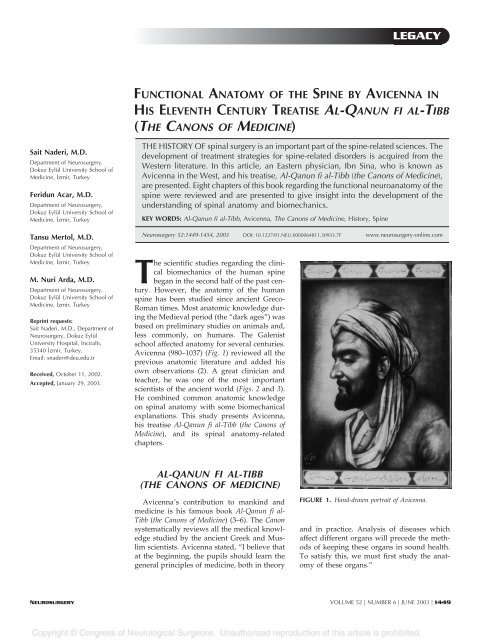
FUNCTIONAL ANATOMY OF THE SPINE BY AVICENNA IN<br />
HIS ELEVENTH CENTURY TREATISE AL-QANUN FI AL-TIBB<br />
(THE CANONS OF MEDICINE)<br />
THE HISTORY OF spinal surgery is an important part <strong>of</strong> <strong>the</strong> <strong>spine</strong>-related sciences. The<br />
development <strong>of</strong> treatment strategies for <strong>spine</strong>-related disorders is acquired from <strong>the</strong><br />
Western literature. In this article, an Eastern physician, Ibn Sina, who is known as<br />
Avicenna in <strong>the</strong> West, and his treatise, Al-Qanun fi al-Tibb (<strong>the</strong> Canons <strong>of</strong> Medicine),<br />
are presented. Eight chapters <strong>of</strong> this book regarding <strong>the</strong> <strong>functional</strong> neuro<strong>anatomy</strong> <strong>of</strong> <strong>the</strong><br />
<strong>spine</strong> were reviewed and are presented to give insight into <strong>the</strong> development <strong>of</strong> <strong>the</strong><br />
understanding <strong>of</strong> spinal <strong>anatomy</strong> and biomechanics.<br />
KEY WORDS: Al-Qanun fi al-Tibb, Avicenna, The Canons <strong>of</strong> Medicine, History, Spine<br />
Neurosurgery 52:1449-1454, 2003 DOI: 10.1227/01.NEU.0000064811.30933.7F www.neurosurgery-online.com<br />
The scientific studies regarding <strong>the</strong> clinical<br />
biomechanics <strong>of</strong> <strong>the</strong> human <strong>spine</strong><br />
began in <strong>the</strong> second half <strong>of</strong> <strong>the</strong> past century.<br />
However, <strong>the</strong> <strong>anatomy</strong> <strong>of</strong> <strong>the</strong> human<br />
<strong>spine</strong> has been studied since ancient Greco-<br />
Roman times. Most anatomic knowledge during<br />
<strong>the</strong> Medieval period (<strong>the</strong> “dark ages”) was<br />
based on preliminary studies on animals and,<br />
less commonly, on humans. The Galenist<br />
school affected <strong>anatomy</strong> for several centuries.<br />
Avicenna (980–1037) (Fig. 1) reviewed all <strong>the</strong><br />
previous anatomic literature and added his<br />
own observations (2). A great clinician and<br />
teacher, he was one <strong>of</strong> <strong>the</strong> most important<br />
scientists <strong>of</strong> <strong>the</strong> ancient world (Figs. 2 and 3).<br />
He combined common anatomic knowledge<br />
on spinal <strong>anatomy</strong> with some biomechanical<br />
explanations. This study presents Avicenna,<br />
his treatise Al-Qanun fi al-Tibb (<strong>the</strong> Canons <strong>of</strong><br />
Medicine), and its spinal <strong>anatomy</strong>-related<br />
chapters.<br />
AL-QANUN FI AL-TIBB<br />
(THE CANONS OF MEDICINE)<br />
Avicenna’s contribution to mankind and<br />
medicine is his famous book Al-Qanun fi al-<br />
Tibb (<strong>the</strong> Canons <strong>of</strong> Medicine) (3–6). The Canon<br />
systematically reviews all <strong>the</strong> medical knowledge<br />
studied <strong>by</strong> <strong>the</strong> ancient Greek and Muslim<br />
scientists. Avicenna stated, “I believe that<br />
at <strong>the</strong> beginning, <strong>the</strong> pupils should learn <strong>the</strong><br />
general principles <strong>of</strong> medicine, both in <strong>the</strong>ory<br />
FIGURE 1. Hand-drawn portrait <strong>of</strong> Avicenna.<br />
and in practice. Analysis <strong>of</strong> diseases which<br />
affect different organs will precede <strong>the</strong> methods<br />
<strong>of</strong> keeping <strong>the</strong>se organs in sound health.<br />
To satisfy this, we must first study <strong>the</strong> <strong>anatomy</strong><br />
<strong>of</strong> <strong>the</strong>se organs.”<br />
NEUROSURGERY VOLUME 52 | NUMBER 6 | JUNE 2003 | 1449
NADERI ET AL.<br />
FIGURE 2. Avicenna giving a lecture to his pupils.<br />
The Canon was translated into Latin <strong>by</strong> Gerard <strong>of</strong> Cremona<br />
in <strong>the</strong> 12th century. Because <strong>of</strong> its systematic approach and its<br />
superiority to Galen’s medical book, <strong>the</strong> Canon became <strong>the</strong><br />
textbook for medical education in <strong>the</strong> European schools for<br />
more than 6 centuries (1). The Canon was regarded as <strong>the</strong><br />
principal medical text until 1650, and it is <strong>the</strong> second most<br />
published book (after <strong>the</strong> Bible) since <strong>the</strong> invention <strong>of</strong> publishing.<br />
The Canon studies drugs, physiology, pathology, and<br />
pharmacology.<br />
Avicenna made several contributions to <strong>anatomy</strong>, and his<br />
Canon contains a number <strong>of</strong> his anatomic findings, many <strong>of</strong><br />
which are accepted today. In <strong>the</strong> Canon <strong>of</strong> Medicine, <strong>the</strong>re are<br />
several chapters <strong>of</strong> <strong>anatomy</strong> in <strong>the</strong> first volume <strong>of</strong> <strong>the</strong> book. In<br />
addition, <strong>the</strong> discussions <strong>of</strong> <strong>anatomy</strong> are scattered throughout<br />
<strong>the</strong> huge encyclopedia, with <strong>the</strong> <strong>anatomy</strong> <strong>of</strong> a particular organ<br />
being discussed in <strong>the</strong> section concerned with diseases particular<br />
to that organ. These anatomic sections <strong>of</strong> <strong>the</strong> Canon were<br />
<strong>of</strong>ten copied out and compiled as separate treatises (Fig. 4).<br />
This study summarizes <strong>the</strong> spinal <strong>anatomy</strong>-related sections <strong>of</strong><br />
this treatise (i.e., Chapters 6 through 13).<br />
FIGURE 3. Depiction <strong>of</strong> three medieval masters <strong>of</strong> medicine side <strong>by</strong> side:<br />
Galen, Avicenna, and Hippocrates.<br />
FIGURE 4. Avicenna studying and taking notes.<br />
CHAPTER 6: FUNCTIONS OF THE SPINE<br />
The <strong>spine</strong> has four functions. First, it serves as a channel for<br />
<strong>the</strong> spinal cord, which is <strong>of</strong> vital importance to animals. Spinal<br />
cord function is explained in <strong>the</strong> spinal cord chapter. In this<br />
chapter, emphasis is placed on <strong>the</strong> brain and nerves. If all <strong>the</strong><br />
nerves exited directly from <strong>the</strong> brain, <strong>the</strong> brain would be<br />
bigger. For example, nerves innervating <strong>the</strong> hands and feet<br />
would travel a longer distance and, thus, would be more<br />
prone to injury; <strong>the</strong>y would also become less able to innervate<br />
<strong>the</strong> big muscles <strong>of</strong> <strong>the</strong> thigh and <strong>the</strong> calf. Therefore, God<br />
created <strong>the</strong> spinal cord below <strong>the</strong> brain. The spinal cord is like<br />
a channel coming out <strong>of</strong> a fountain in <strong>the</strong> way that nerves<br />
emerge from both sides and go down, thus putting <strong>the</strong> organs<br />
1450 | VOLUME 52 | NUMBER 6 | JUNE 2003 www.neurosurgery-online.com
closer to <strong>the</strong> brain. That is why God placed <strong>the</strong> spinal cord into<br />
a hard bony channel called <strong>the</strong> <strong>spine</strong> to protect it from injury.<br />
Second, <strong>the</strong> <strong>spine</strong> protects <strong>the</strong> organs close to itself. That is<br />
why it has bony projections and spikes. Third, <strong>the</strong> <strong>spine</strong> is like<br />
<strong>the</strong> axle <strong>of</strong> a coach. It forms <strong>the</strong> structure <strong>of</strong> <strong>the</strong> bony skeleton.<br />
Finally, in humans, <strong>the</strong> <strong>spine</strong> is a powerful and unchanging<br />
cushion during standing erect and bending to <strong>the</strong> sides. The<br />
back is formed <strong>by</strong> vertebrae allowing <strong>the</strong>se movements. The<br />
joints between <strong>the</strong>se vertebrae are nei<strong>the</strong>r too loose to move<br />
freely, nor too rigid to form a straight block.<br />
CHAPTER SEVEN: VERTEBRAE<br />
The vertebrae are hollow bones to allow <strong>the</strong> spinal cord to<br />
pass through <strong>the</strong>m. Each vertebra has two superior and two<br />
inferior side projections to form joints. There may also be eight<br />
projections. The joint projection <strong>of</strong> one vertebra faces a joint<br />
projection <strong>of</strong> an adjacent vertebra. These projections also protect<br />
<strong>the</strong> vertebra from crashes and injuries. Longitudinally and<br />
transversely, vertebrae are covered with strong fibers. These<br />
are united superiorly and inferiorly throughout <strong>the</strong> length <strong>of</strong><br />
<strong>the</strong> <strong>spine</strong>. A vertebra has transverse processes at both sides<br />
and spinous processes posteriorly so it can protect <strong>the</strong> nerves<br />
and vessels like longitudinal muscles. The transverse process<br />
<strong>of</strong> a thoracic vertebra has smooth, shiny facets forming joints<br />
with tubercles on <strong>the</strong> ribs. Some transverse processes have two<br />
heads, and <strong>the</strong>y seem to have two eminences. However, this is<br />
only seen in cervical vertebrae. In addition to <strong>the</strong> spinal canal,<br />
<strong>the</strong>re are foramina and notches to allow for <strong>the</strong> passage <strong>of</strong><br />
nerves and vessels. Some foramina are in <strong>the</strong> bony vertebrae,<br />
and some are formed on <strong>the</strong> joint surfaces <strong>by</strong> crescents on both<br />
vertebrae. These foramina may be large or small, and <strong>the</strong>y are<br />
found up and down on both sides <strong>of</strong> <strong>the</strong> vertebra. These are<br />
placed laterally, because posteriorly <strong>the</strong>re are no structures to<br />
protect <strong>the</strong> passing structures and anteriorly <strong>the</strong>y are prone to<br />
injury <strong>by</strong> <strong>the</strong> spikes placed anteriorly. The <strong>spine</strong> is empowered<br />
<strong>by</strong> <strong>the</strong> ligaments and tendons. Ligaments facilitate movement<br />
and lubricate <strong>the</strong> muscles. These ligaments also protect<br />
<strong>the</strong> joints. The anterior longitudinal ligament is stronger than<br />
<strong>the</strong> posterior one because anterior movement is needed more<br />
than posterior movement.<br />
Posterior to <strong>the</strong> <strong>spine</strong>, <strong>the</strong>re is a space formed <strong>by</strong> <strong>the</strong> elongation<br />
<strong>of</strong> <strong>the</strong> fibrous tissue. This space has humidity to facilitate<br />
<strong>the</strong> freshness <strong>of</strong> <strong>the</strong> <strong>spine</strong>. The <strong>spine</strong> is formed <strong>by</strong> many<br />
vertebrae so it can move easily. If <strong>the</strong> <strong>spine</strong> was formed <strong>by</strong> a<br />
single bone, it would have been difficult to establish<br />
movement.<br />
CHAPTER EIGHT: CERVICAL SPINE AND<br />
ITS FUNCTION<br />
The neck is designed to form a space for <strong>the</strong> trachea. Cervical<br />
vertebrae are placed on top <strong>of</strong> each o<strong>the</strong>r, and <strong>the</strong> smallest<br />
vertebra occupies <strong>the</strong> top. If <strong>the</strong> object at <strong>the</strong> top has<br />
mobility, it must always be lighter than <strong>the</strong> objects supporting<br />
it. The spinal cord is thicker at <strong>the</strong> top; it gets thinner as it goes<br />
AVICENNA AND SPINAL BIOMECHANICS<br />
down and gives out nerves. The width <strong>of</strong> <strong>the</strong> atlas is less than<br />
<strong>the</strong> o<strong>the</strong>rs, but it has more mobility and density to gain equilibrium.<br />
Cervical vertebrae have short processes instead <strong>of</strong><br />
long processes, which are more prone to injury. Because <strong>of</strong><br />
<strong>the</strong>ir mobility, cervical vertebrae have longer transverse processes.<br />
These vertebrae carry less weight than thoracic vertebrae,<br />
so <strong>the</strong>ir bodies are weaker but <strong>the</strong>ir joints have more<br />
mobility. The weakness <strong>of</strong> <strong>the</strong> spinous processes <strong>of</strong> cervical<br />
vertebrae is balanced <strong>by</strong> <strong>the</strong> organization <strong>of</strong> a network <strong>of</strong><br />
muscles, vessels, and nerves. Superior and inferior facets <strong>of</strong><br />
cervical vertebrae are smaller, and <strong>the</strong>ir ligaments are looser.<br />
The neural foramina are placed between neighboring vertebral<br />
bodies.<br />
Cervical Vertebrae<br />
There are seven cervical vertebrae. Their number, size, and<br />
length are designed in an obvious pattern. Except for <strong>the</strong> C1<br />
vertebra, all <strong>the</strong> cervical vertebrae have spinous, transverse,<br />
superior, and inferior processes. Each transverse process has a<br />
transverse foramen. The atlas and axis have different characteristics.<br />
Movements <strong>of</strong> <strong>the</strong> head to <strong>the</strong> left and right are<br />
facilitated <strong>by</strong> <strong>the</strong> joints between <strong>the</strong> head and <strong>the</strong> first cervical<br />
vertebra. However, anterior and posterior movements <strong>of</strong> <strong>the</strong><br />
head are facilitated <strong>by</strong> <strong>the</strong> joint between <strong>the</strong> head and <strong>the</strong> C2<br />
vertebra. The joint between <strong>the</strong> head and <strong>the</strong> C1 vertebra is<br />
formed <strong>by</strong> <strong>the</strong> superior articular facet <strong>of</strong> <strong>the</strong> C1 and occipital<br />
condyles. If one condyle elevates <strong>the</strong> head, <strong>the</strong> o<strong>the</strong>r bends it.<br />
The second cervical vertebra has no superior articular face;<br />
instead, it has an odontoid process situated in <strong>the</strong> spinal canal.<br />
The spinal canal has a broader anteroposterior diameter to<br />
support <strong>the</strong> odontoid process. The spinal cord is protected <strong>by</strong><br />
strong ligaments and fibrous bands around <strong>the</strong> odontoid process.<br />
The odontoid process enters <strong>the</strong> foramen magnum and<br />
forms <strong>the</strong> joint facilitating anterior and posterior movements<br />
<strong>of</strong> <strong>the</strong> head.<br />
The odontoid process has two functions. First, it is an efficient<br />
protector. Second, it prevents displacement <strong>of</strong> <strong>the</strong> thinner<br />
first cervical vertebra. The C1 vertebra is well protected <strong>by</strong> <strong>the</strong><br />
C2 vertebra. Therefore, it does not need an extra process. The<br />
exit <strong>of</strong> spinal nerves is a different aspect <strong>of</strong> <strong>the</strong> first cervical<br />
vertebra. The nerves do not emerge from <strong>the</strong> intervertebral<br />
foramina; instead, <strong>the</strong>y emerge dorsally on <strong>the</strong> bone. If nerves<br />
emerged laterally, <strong>the</strong>y would be damaged <strong>by</strong> <strong>the</strong> lateral<br />
movements <strong>of</strong> <strong>the</strong> head. Because <strong>of</strong> <strong>the</strong> movement characteristics<br />
<strong>of</strong> <strong>the</strong> first cervical vertebra, <strong>the</strong> nerves cannot exit on its<br />
anterior or posterior aspect; <strong>the</strong> best place for nerve emergence<br />
is <strong>the</strong> posterolateral face. The foramen is smaller, so <strong>the</strong> spinal<br />
nerves are thinner.<br />
On <strong>the</strong> second cervical vertebra, nerves do not emerge on<br />
<strong>the</strong> superior surface because <strong>the</strong>re is a possible risk <strong>of</strong> injury<br />
from <strong>the</strong> movements <strong>of</strong> <strong>the</strong> first cervical vertebra. Also, it is<br />
not possible for <strong>the</strong>se nerves to emerge laterally and posteriorly.<br />
If <strong>the</strong>y did so, <strong>the</strong> first and second cervical spinal nerves<br />
would be so close that <strong>the</strong>y would blend with each o<strong>the</strong>r and<br />
become weaker. The best place for <strong>the</strong> nerves is lateral to <strong>the</strong><br />
joint processes. During <strong>the</strong> anteroposterior and lateral move-<br />
NEUROSURGERY VOLUME 52 | NUMBER 6 | JUNE 2003 | 1451
NADERI ET AL.<br />
ments <strong>of</strong> <strong>the</strong> head, <strong>the</strong> first and second cervical vertebrae act as<br />
a single bone and move toge<strong>the</strong>r.<br />
CHAPTER NINE: THE THORACIC SPINE<br />
AND ITS FUNCTIONS<br />
Costae make joints with thoracic vertebrae and form a cage<br />
around <strong>the</strong> lungs. The first 11 thoracic vertebrae have both<br />
spinal and transverse processes. However, <strong>the</strong> twelfth thoracic<br />
vertebra has no transverse process. The transverse processes<br />
<strong>of</strong> <strong>the</strong> thoracic vertebrae are placed close to <strong>the</strong> vital organs,<br />
and <strong>the</strong>y are <strong>of</strong> varying sizes and shapes. These transverse<br />
processes on <strong>the</strong> superior seven thoracic vertebrae are larger<br />
and thicker to protect <strong>the</strong> heart. The costal joint facets on <strong>the</strong><br />
transverse process <strong>of</strong> <strong>the</strong> ninth thoracic vertebra are shorter<br />
and broader. These facets are <strong>the</strong> same size as <strong>the</strong> costal joint<br />
facets. The spinous processes <strong>of</strong> <strong>the</strong> superior thoracic vertebrae<br />
face inferiorly. The spinous process <strong>of</strong> <strong>the</strong> tenth thoracic<br />
vertebra is straight and thick (it does not face superiorly or<br />
inferiorly). The joint facets <strong>of</strong> <strong>the</strong> tenth thoracic vertebra are<br />
smooth, and <strong>the</strong>re are no tubercles. Below <strong>the</strong> tenth thoracic<br />
vertebra, <strong>the</strong> joint facets are broad and invaginated to form<br />
joints with <strong>the</strong> superior vertebral bodies. Their spinous processes<br />
look superiorly. The twelfth thoracic vertebra has no<br />
transverse process because <strong>the</strong> twelfth rib has no joint projection<br />
to <strong>the</strong> twelfth vertebra. The twelfth rib has strong connections<br />
with muscles and ligaments, and it does not need this<br />
joint with <strong>the</strong> vertebra.<br />
Lumbar vertebrae should be big and strong to carry <strong>the</strong><br />
superior vertebrae. Therefore, <strong>the</strong>se vertebrae have a greater<br />
joint area and more projections. The last thoracic and <strong>the</strong> first<br />
lumbar vertebra are similar. The first lumbar vertebra has<br />
large joint projections to compensate for <strong>the</strong> absence <strong>of</strong> transverse<br />
processes. Starting from <strong>the</strong> cervical <strong>spine</strong>, <strong>the</strong> spinal<br />
canal becomes narrower. The spinal canal is quite narrow in<br />
<strong>the</strong> inferior thoracic and lumbar <strong>spine</strong>. There is not enough<br />
space for <strong>the</strong> spinal cord. The spinal canal in <strong>the</strong> lumbar and<br />
sacral <strong>spine</strong> allows for <strong>the</strong> passage <strong>of</strong> <strong>the</strong> lumbar and sacral<br />
nerves.<br />
CHAPTER TEN: THE LUMBAR SPINE<br />
There are five lumbar vertebrae. They have large spinous<br />
and transverse processes. Joining <strong>the</strong> sacrum, lumbar vertebrae<br />
form <strong>the</strong> foundation <strong>of</strong> <strong>the</strong> <strong>spine</strong> and cushion it. In<br />
addition, <strong>the</strong>y support <strong>the</strong> pubis and allow <strong>the</strong> emerging<br />
nerves to exit to <strong>the</strong> legs.<br />
CHAPTER ELEVEN: THE SACRUM<br />
There are three vertebrae in <strong>the</strong> sacrum. These are rigid and<br />
strong. They have large transverse processes. The foramina for<br />
<strong>the</strong> emerging nerve roots are placed dorsally.<br />
CHAPTER TWELVE: THE COCCYX<br />
The coccyx is formed <strong>by</strong> three vertebrae that have no projections.<br />
The nerves are small like <strong>the</strong> cervical nerves, and <strong>the</strong>y<br />
emerge between <strong>the</strong> vertebral bodies. Only a single nerve<br />
emerges from <strong>the</strong> tip <strong>of</strong> <strong>the</strong> third vertebra <strong>of</strong> <strong>the</strong> coccyx.<br />
CHAPTER THIRTEEN: FINAL<br />
CONSIDERATIONS REGARDING THE<br />
FUNCTION OF THE SPINE<br />
In explaining <strong>the</strong> vertebrae <strong>of</strong> <strong>the</strong> <strong>spine</strong>, its function is also<br />
evaluated. Vertebral bodies <strong>of</strong> <strong>the</strong> <strong>spine</strong> are constructed as a<br />
cylindrical column in <strong>the</strong> best design to act as a single body to<br />
prevent injury. Superior vertebral spinous processes face inferiorly.<br />
Inferior vertebral spinous processes face superiorly.<br />
The spinous process <strong>of</strong> <strong>the</strong> tenth thoracic vertebra is straight<br />
and horizontal, and this central position is not related to<br />
vertebral number; instead, it is related to <strong>the</strong> vertebral length.<br />
During <strong>the</strong> movement <strong>of</strong> <strong>the</strong> <strong>spine</strong> to <strong>the</strong> left and <strong>the</strong> right, it<br />
also turns.<br />
DISCUSSION<br />
The modern era in human <strong>anatomy</strong> started in <strong>the</strong> 16th<br />
century with <strong>the</strong> studies <strong>of</strong> Andreas Vesalius (1514–1564). The<br />
earliest anatomic studies, however, were performed <strong>by</strong> Alcamaeon<br />
<strong>of</strong> Crotona (500 BC) on animals and <strong>by</strong> Herophilus and<br />
Erasistratus <strong>of</strong> Alexandria (300 BC), who made <strong>the</strong> first systematic<br />
anatomic dissections on humans. After 150 BC, human<br />
dissections were prohibited for religious and ethical reasons.<br />
The prohibition was enforced <strong>by</strong> Rome. In <strong>the</strong> 2nd century<br />
AD, Galen made several anatomic dissections on animals<br />
(mostly Barbary apes and pigs). Galenist <strong>anatomy</strong>, with its<br />
correct and incorrect aspects, influenced anatomists for several<br />
centuries.<br />
The anatomic information written <strong>by</strong> Avicenna in <strong>the</strong> Canon<br />
reflects a review <strong>of</strong> <strong>the</strong> treatises written <strong>by</strong> <strong>the</strong> earlier Greco-<br />
Roman authors and Galen and Avicenna’s own observations<br />
and considerations. It is not known, however, how much <strong>of</strong><br />
Avicenna’s anatomic knowledge is original and is based on his<br />
own studies. Because <strong>of</strong> <strong>the</strong> prohibition regarding anatomic<br />
dissections on humans in Islamic cultures, Avicenna’s anatomic<br />
considerations might be based on his clinical<br />
observations.<br />
Although <strong>the</strong>re are several studies on Avicenna and his<br />
Canon, this is <strong>the</strong> first study focusing on <strong>the</strong> spinal <strong>anatomy</strong>related<br />
chapters <strong>of</strong> this treatise. A careful analysis <strong>of</strong> <strong>the</strong>se<br />
eight chapters shows <strong>the</strong> level <strong>of</strong> knowledge regarding <strong>the</strong><br />
biomechanical <strong>anatomy</strong> <strong>of</strong> <strong>the</strong> <strong>spine</strong> in <strong>the</strong> 11th century.<br />
In his treatise, Avicenna tried to find explanations for <strong>the</strong><br />
anatomic features <strong>of</strong> <strong>the</strong> vertebrae and <strong>the</strong> spinal region. He<br />
emphasized that <strong>the</strong> shape and <strong>the</strong> size <strong>of</strong> any given vertebra<br />
is determined <strong>by</strong> its regional function. Therefore, he classified<br />
<strong>the</strong> <strong>spine</strong> into <strong>the</strong> following segments, similar to <strong>the</strong> classification<br />
system used today: cervical, thoracic, lumbar, sacral,<br />
1452 | VOLUME 52 | NUMBER 6 | JUNE 2003 www.neurosurgery-online.com
and coccygeal. Then, he described <strong>the</strong> anatomic features <strong>of</strong> <strong>the</strong><br />
elements <strong>of</strong> <strong>the</strong> vertebrae in each region.<br />
A review <strong>of</strong> <strong>the</strong> Canon reveals some errors in his anatomic<br />
considerations, particularly regarding <strong>the</strong> <strong>anatomy</strong> <strong>of</strong> C2, T12,<br />
and <strong>the</strong> number <strong>of</strong> sacral vertebrae. However, despite <strong>the</strong>se<br />
anatomic errors, Avicenna described <strong>the</strong> biomechanical features<br />
<strong>of</strong> <strong>the</strong> vertebrae and <strong>the</strong> <strong>spine</strong> almost correctly. He<br />
described <strong>the</strong> flexion, extension, and lateral bending aspects <strong>of</strong><br />
<strong>the</strong> motion segments (Chapters 8 and 9), as well as <strong>the</strong> coupling<br />
phenomenon <strong>of</strong> <strong>the</strong> thoracolumbar <strong>spine</strong> (Chapter 13).<br />
The most interesting opinions <strong>of</strong> Avicenna are on <strong>the</strong> biomechanics<br />
<strong>of</strong> <strong>the</strong> craniovertebral junction. Avicenna described<br />
<strong>the</strong> different characteristics <strong>of</strong> <strong>the</strong> atlas and <strong>the</strong> axis. He reported<br />
that “<strong>the</strong> head-atlas motion segment is responsible for<br />
lateral bending, whereas <strong>the</strong> C0 to C2 segment makes anteroposterior<br />
motion possible. One condyle elevates <strong>the</strong> head, <strong>the</strong><br />
o<strong>the</strong>r bends it.” According to Avicenna “<strong>the</strong> odontoid process<br />
has two functions: it protects and it prevents <strong>the</strong> displacement<br />
<strong>of</strong> <strong>the</strong> thinner first cervical vertebra. During <strong>the</strong> anteroposterior<br />
and lateral movements <strong>of</strong> <strong>the</strong> head, C1 and C2 vertebrae<br />
act as a single bone and move toge<strong>the</strong>r.”<br />
The similarities between some parts <strong>of</strong> Avicenna’s Canon<br />
and our current biomechanical knowledge are surprising. In<br />
addition, <strong>the</strong> presented chapters <strong>of</strong> <strong>the</strong> Canon reveal that an<br />
understanding <strong>of</strong> spinal biomechanics was a necessity even<br />
1000 years ago.<br />
REFERENCES<br />
1. Aziz E, Nathan B, McKeever J: Anes<strong>the</strong>tic and analgesic practices in Avicenna’s<br />
Canon <strong>of</strong> Medicine. Am J Chin Med 28:147–151, 2000.<br />
2. Cruse JM: History <strong>of</strong> medicine: The metamorphosis <strong>of</strong> scientific medicine in<br />
<strong>the</strong> ever-present past. Am J Med Sci 318:171–180, 1999.<br />
3. Dunn PM: Avicenna (AD 980–1037) and Arabic perinatal medicine. Criteria<br />
Dis Child 77:75–76, 1997.<br />
4. Kahya E: Ibn-I Sina, El-Kanun Fi‘t Tıbb, 1st book, Atatürk Kültür [in Turkish].<br />
Ankara, Dil ve Tarih Kurumu Press, 1995, p 103.<br />
5. Lyons AS: An Illustrated History <strong>of</strong> Medicine. New York, Abradale Press, 1987.<br />
6. Sharafkandi A: The Canon <strong>of</strong> Medicine <strong>of</strong> Ibn Sina [in Persian]. Tehran, Soroush<br />
Press, 1997.<br />
COMMENTS<br />
The effort expended <strong>by</strong> <strong>the</strong>se authors is immense. The article<br />
gives a clear explanation <strong>of</strong> <strong>the</strong> <strong>anatomy</strong> and biomechanics<br />
<strong>of</strong> <strong>the</strong> <strong>spine</strong>. Four centuries after <strong>the</strong> writing <strong>of</strong> Ibn<br />
Sina (1, 2), I do not think much needs to be added. The<br />
detailed anatomic description, at a time when dissection <strong>of</strong><br />
bodies was not accepted because <strong>of</strong> religious beliefs, suggests<br />
that his knowledge was obtained from <strong>the</strong> large number <strong>of</strong><br />
surgical operations he performed.<br />
I am sure <strong>the</strong> reader will need more details about Ibn Sina,<br />
“<strong>the</strong> Persian physician.” He was born in <strong>the</strong> village <strong>of</strong> Afshama<br />
in Persia, and his family moved to Bukhara when he<br />
was 5 years old. He was a gifted child with an exceptional<br />
memory. He practiced self-education and was a master in<br />
Islamic law and medicine. His interest in medicine appeared<br />
at <strong>the</strong> age <strong>of</strong> 17. He served various rulers during his life as a<br />
AVICENNA AND SPINAL BIOMECHANICS<br />
government <strong>of</strong>ficial and was a physician at <strong>the</strong> age <strong>of</strong> 20. At<br />
<strong>the</strong> same time, he was a free-thinking politician, which involved<br />
him in conflicts with government <strong>of</strong>ficials, forcing him<br />
to move from one place to ano<strong>the</strong>r or to hide for some period<br />
<strong>of</strong> time, and he was even imprisoned.<br />
As a Persian physician, he was a remarkable person in his<br />
time. He completed 45 books on philosophy, medicine, geometry,<br />
astronomy, <strong>the</strong>ology, metaphysics, and poetry. His encyclopedia<br />
<strong>of</strong> philosophy, Ashshifa (The Recovery), encompasses<br />
logic, philosophy, metaphysics, and natural science.<br />
Some <strong>of</strong> his books were translated into Latin, Hebrew, and, in<br />
1953, English.<br />
The Canons <strong>of</strong> Medicine was one <strong>of</strong> <strong>the</strong> earliest Arabic books<br />
to see print. The book was taken as a reference in medicine in<br />
Western universities until <strong>the</strong> 17th century.<br />
His readings in <strong>the</strong> book <strong>of</strong> Aristotle on metaphysics, facilitated<br />
<strong>by</strong> <strong>the</strong> philosopher Al Farabi, allowed him to understand<br />
<strong>the</strong> nature and invention <strong>of</strong> scientific facts. His description<br />
<strong>of</strong> intracranial <strong>anatomy</strong> was based on Galenic studies. He<br />
divided <strong>the</strong> brain into <strong>the</strong> medulla and cortex. He performed<br />
burr hole trephination with <strong>the</strong> patient under opium anes<strong>the</strong>sia,<br />
with numerous students watching him during surgery. He<br />
dealt with all <strong>the</strong> branches <strong>of</strong> medicine, including cancer, and<br />
advised early radical surgery for <strong>the</strong> best results. He also<br />
stressed <strong>the</strong> relation <strong>of</strong> nervous tension and psychological<br />
effects leading to peptic ulcer and irritable colon.<br />
In addition to his clinical examination <strong>of</strong> patients, he always<br />
confirmed <strong>the</strong> diagnosis <strong>by</strong> examining <strong>the</strong> urine, stools, and<br />
pulse. Ibn Sina included love as a disease, “love sickness,”<br />
because it always has physical symptoms, and he explained<br />
how to diagnose it <strong>by</strong> counting <strong>the</strong> pulse during <strong>the</strong> recitation<br />
<strong>of</strong> different names, including that <strong>of</strong> <strong>the</strong> lover, finding that <strong>the</strong><br />
pulse rate rises with tachycardia when <strong>the</strong> name <strong>of</strong> <strong>the</strong> beloved<br />
is mentioned. In his bedside observation, he could differentiate<br />
between pneumonia and acute meningitis and <strong>the</strong><br />
facial palsy caused <strong>by</strong> an intracranial or peripheral lesion. He<br />
was <strong>the</strong> first to diagnose and treat ankylostomiasis; he called<br />
it roundworm.<br />
One <strong>of</strong> his efforts was <strong>the</strong> proper explanation <strong>of</strong> <strong>the</strong> production<br />
<strong>of</strong> a boy or a girl child and that it depends on <strong>the</strong><br />
fa<strong>the</strong>r and not <strong>the</strong> mo<strong>the</strong>r. His methodology in <strong>the</strong> academic<br />
study <strong>of</strong> medicine was to start <strong>by</strong> studying <strong>anatomy</strong> and<br />
physiology. This talented genius and open-minded sheik was<br />
a man <strong>of</strong> excellence in his time, particularly in medicine at a<br />
time when <strong>the</strong>re was no computed tomography or magnetic<br />
resonance imaging.<br />
Sayed El-Gindi<br />
Cairo, Egypt<br />
1. Dorrah A, Ibn Sina: A Physician in Agony and Philosopher in Exile [in Arabic].<br />
Cairo, General Egyptian Book Center, 2002, ed 2, pp 41–59.<br />
2. El Naggar A: History <strong>of</strong> Medicine in <strong>the</strong> Islamic Countries [in Arabic]. Cairo, Dar<br />
el Maaref, 1994, pp 120–145.<br />
NEUROSURGERY VOLUME 52 | NUMBER 6 | JUNE 2003 | 1453
NADERI ET AL.<br />
This is a very welcome brief historical note indicating that<br />
Avicenna devoted a significant degree <strong>of</strong> time and effort to<br />
problems <strong>of</strong> <strong>the</strong> <strong>spine</strong>. It is a delight to read this historical<br />
vignette.<br />
Edward R. Laws, Jr.<br />
Charlottesville, Virginia<br />
The Eastern physician Ibn Sina, known as Avicenna, was a<br />
very important scientist who lived between <strong>the</strong> years AD<br />
980 and 1037. His anatomic works, especially on <strong>spine</strong>, were<br />
written with a surgeon’s consciousness, because he was a<br />
surgeon. The origin <strong>of</strong> his anatomic knowledge is not known,<br />
because anatomic dissection on human bodies was prohibited<br />
during Avicenna’s era. However, his orientation and descriptions<br />
<strong>of</strong> <strong>the</strong> spinal <strong>anatomy</strong> at that period are exceptional.<br />
Yücel Kanpolat<br />
Ankara, Turkey<br />
In <strong>the</strong> present-day literature, <strong>the</strong>re is very little in <strong>the</strong> English<br />
language on <strong>the</strong> original writings <strong>of</strong> Avicenna and his classic<br />
work titled The Canons <strong>of</strong> Medicine. The authors <strong>of</strong> this<br />
article have provided an English translation from <strong>the</strong> original<br />
Arabic <strong>of</strong> <strong>the</strong> various sections dealing with <strong>the</strong> <strong>anatomy</strong> and<br />
function <strong>of</strong> <strong>the</strong> human <strong>spine</strong>. After one reads <strong>the</strong>se various<br />
sections, it becomes quite clear that Avicenna had a most<br />
remarkable grasp <strong>of</strong> <strong>the</strong> <strong>anatomy</strong> <strong>of</strong> <strong>the</strong> <strong>spine</strong> and some <strong>of</strong> its<br />
biomechanical aspects. An intriguing question that arises, and<br />
one not easily answered, is how many <strong>of</strong> <strong>the</strong> original dissections<br />
were performed <strong>by</strong> Avicenna himself, versus what he<br />
adapted from <strong>the</strong> earlier Greco-Roman writers. The authors do<br />
allude to this in <strong>the</strong> text, but it would be wonderful if more<br />
information might be provided with some fur<strong>the</strong>r research.<br />
There has been much discussion in <strong>the</strong> historical literature<br />
about how <strong>the</strong> Koran forbids human dissection, yet I remember<br />
from years <strong>of</strong> reading various sources that <strong>the</strong>re were<br />
exceptions to this edict <strong>by</strong> some <strong>of</strong> <strong>the</strong> early anatomists. This<br />
last thought leads naturally to Avicenna and <strong>the</strong> question <strong>of</strong><br />
whe<strong>the</strong>r he actually performed human dissections. It would<br />
appear that he did.<br />
James T. Goodrich<br />
Bronx, New York<br />
Left, Egas Moniz in 1949 (courtesy <strong>of</strong> Antonio vas Carneiro, M.D., Ph.D., University <strong>of</strong> Lisbon School <strong>of</strong> Medicine, Lisbon, Portugal). Right, <strong>the</strong> “core<br />
operation.” Prefrontal leukotomy was performed <strong>by</strong> Egas Moniz and Almeida Lima. The leukotome was inserted into <strong>the</strong> brain at <strong>the</strong> approximate angles shown;<br />
when <strong>the</strong> leukotome was in place, <strong>the</strong> wire was extended and <strong>the</strong> handle rotated. In <strong>the</strong> first leukotomy, one “core” was cut on each side <strong>of</strong> <strong>the</strong> brain. In subsequent<br />
operations, progressively more “cores” were cut. The right side <strong>of</strong> <strong>the</strong> figure depicts a horizontal slice <strong>of</strong> <strong>the</strong> brain (parallel to <strong>the</strong> top <strong>of</strong> <strong>the</strong> cranium) with Moniz’<br />
estimate (without evidence) <strong>of</strong> <strong>the</strong> extent <strong>of</strong> damage (drawn <strong>by</strong> R. Spencer Phippen, in Valenstein ES: Great and Desperate Cures: The Rise and Decline <strong>of</strong><br />
Psychosurgery and O<strong>the</strong>r Radical Treatments for Mental Illness. New York, Basic Books, Inc., Publishers, 1986).<br />
1454 | VOLUME 52 | NUMBER 6 | JUNE 2003 www.neurosurgery-online.com