X-ray techniques in the investigation of a Gothic sculpture - Nukleonika
X-ray techniques in the investigation of a Gothic sculpture - Nukleonika
X-ray techniques in the investigation of a Gothic sculpture - Nukleonika

You also want an ePaper? Increase the reach of your titles
YUMPU automatically turns print PDFs into web optimized ePapers that Google loves.
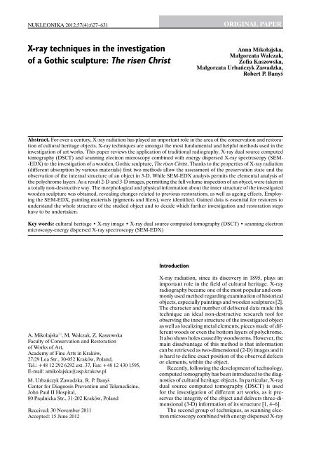
NUKLEONIKA 2012;57(4):627−631<br />
X-<strong>ray</strong> <strong>techniques</strong> <strong>in</strong> <strong>the</strong> <strong>in</strong>vestigation<br />
<strong>of</strong> a <strong>Gothic</strong> <strong>sculpture</strong>: The risen Christ<br />
A. Mikołajska , M. Walczak, Z. Kaszowska<br />
Faculty <strong>of</strong> Conservation and Restoration<br />
<strong>of</strong> Works <strong>of</strong> Art,<br />
Academy <strong>of</strong> F<strong>in</strong>e Arts <strong>in</strong> Kraków,<br />
27/29 Lea Str., 30-052 Kraków, Poland,<br />
Tel.: +48 12 292 6292 ext. 37, Fax: +48 12 430 1595,<br />
E-mail: amikolajska@asp.krakow.pl<br />
M. Urbańczyk Zawadzka, R. P. Banyś<br />
Center for Diagnosis Prevention and Telemedic<strong>in</strong>e,<br />
John Paul II Hospital,<br />
80 Prądnicka Str., 31-202 Kraków, Poland<br />
Introduction<br />
ORIGINAL PAPER<br />
Anna Mikołajska,<br />
Małgorzata Walczak,<br />
Z<strong>of</strong>ia Kaszowska,<br />
Małgorzata Urbańczyk Zawadzka,<br />
Robert P. Banyś<br />
Abstract. For over a century, X-<strong>ray</strong> radiation has played an important role <strong>in</strong> <strong>the</strong> area <strong>of</strong> <strong>the</strong> conservation and restoration<br />
<strong>of</strong> cultural heritage objects. X-<strong>ray</strong> <strong>techniques</strong> are amongst <strong>the</strong> most fundamental and helpful methods used <strong>in</strong> <strong>the</strong><br />
<strong>in</strong>vestigation <strong>of</strong> art works. This paper reviews <strong>the</strong> application <strong>of</strong> traditional radiography, X-<strong>ray</strong> dual source computed<br />
tomography (DSCT) and scann<strong>in</strong>g electron microscopy comb<strong>in</strong>ed with energy dispersed X-<strong>ray</strong> spectroscopy (SEM-<br />
-EDX) to <strong>the</strong> <strong>in</strong>vestigation <strong>of</strong> a wooden, <strong>Gothic</strong> <strong>sculpture</strong>, The risen Christ. Thanks to <strong>the</strong> properties <strong>of</strong> X-<strong>ray</strong> radiation<br />
(different absorption by various materials) first two methods allow <strong>the</strong> assessment <strong>of</strong> <strong>the</strong> preservation state and <strong>the</strong><br />
observation <strong>of</strong> <strong>the</strong> <strong>in</strong>ternal structure <strong>of</strong> an object <strong>in</strong> 3-D. While SEM-EDX analysis permits <strong>the</strong> elemental analysis <strong>of</strong><br />
<strong>the</strong> polychrome layers. As a result 2-D and 3-D images, permitt<strong>in</strong>g <strong>the</strong> full volume <strong>in</strong>spection <strong>of</strong> an object, were taken <strong>in</strong><br />
a totally non-destructive way. The morphological and physical <strong>in</strong>formation about <strong>the</strong> <strong>in</strong>ner structure <strong>of</strong> <strong>the</strong> <strong>in</strong>vestigated<br />
wooden <strong>sculpture</strong> was obta<strong>in</strong>ed, reveal<strong>in</strong>g changes related to previous restorations, as well as age<strong>in</strong>g effects. Employ<strong>in</strong>g<br />
<strong>the</strong> SEM-EDX, pa<strong>in</strong>t<strong>in</strong>g materials (pigments and filers), were identified. Ga<strong>in</strong>ed data is essential for restorers to<br />
understand <strong>the</strong> whole structure <strong>of</strong> <strong>the</strong> studied object and to decide which fur<strong>the</strong>r <strong>in</strong>vestigation and restoration steps<br />
have to be undertaken.<br />
Key words: cultural heritage • X-<strong>ray</strong> image • X-<strong>ray</strong> dual source computed tomography (DSCT) • scann<strong>in</strong>g electron<br />
microscopy-energy dispersed X-<strong>ray</strong> spectroscopy (SEM-EDX)<br />
Received: 30 November 2011<br />
Accepted: 15 June 2012<br />
X-<strong>ray</strong> radiation, s<strong>in</strong>ce its discovery <strong>in</strong> 1895, plays an<br />
important role <strong>in</strong> <strong>the</strong> field <strong>of</strong> cultural heritage. X-<strong>ray</strong><br />
radiography became one <strong>of</strong> <strong>the</strong> most popular and commonly<br />
used method regard<strong>in</strong>g exam<strong>in</strong>ation <strong>of</strong> historical<br />
objects, especially pa<strong>in</strong>t<strong>in</strong>gs and wooden <strong>sculpture</strong>s [2].<br />
The character and number <strong>of</strong> delivered data made this<br />
technique an ideal non-destructive research tool for<br />
observ<strong>in</strong>g <strong>the</strong> <strong>in</strong>ner structure <strong>of</strong> <strong>the</strong> <strong>in</strong>vestigated object<br />
as well as localiz<strong>in</strong>g metal elements, pieces made <strong>of</strong> different<br />
woods or even <strong>the</strong> bottom layers <strong>of</strong> polychrome.<br />
It also shows holes caused by woodworms. However, <strong>the</strong><br />
ma<strong>in</strong> disadvantage <strong>of</strong> this method is that <strong>in</strong>formation<br />
can be retrieved as two-dimensional (2-D) images and it<br />
is hard to def<strong>in</strong>e exact position <strong>of</strong> <strong>the</strong> observed defects<br />
or elements, with<strong>in</strong> <strong>the</strong> object.<br />
Recently, follow<strong>in</strong>g <strong>the</strong> development <strong>of</strong> technology,<br />
computed tomography has been <strong>in</strong>troduced to <strong>the</strong> diagnostics<br />
<strong>of</strong> cultural heritage objects. In particular, X-<strong>ray</strong><br />
dual source computed tomography (DSCT) is used<br />
for <strong>the</strong> <strong>in</strong>vestigation <strong>of</strong> different art works, as it preserves<br />
<strong>the</strong> <strong>in</strong>tegrity <strong>of</strong> <strong>the</strong> object and delivers three-dimensional<br />
(3-D) <strong>in</strong>formation <strong>of</strong> its structure [1, 4–6].<br />
The second group <strong>of</strong> <strong>techniques</strong>, as scann<strong>in</strong>g electron<br />
microscopy comb<strong>in</strong>ed with energy dispersed X-<strong>ray</strong>
628 A. Mikołajska et al.<br />
spectroscopy (SEM-EDX), X-<strong>ray</strong> fluorescence (XRF)<br />
or particle <strong>in</strong>duced X-<strong>ray</strong> emission (PIXE), widely<br />
used <strong>in</strong> <strong>the</strong> research <strong>of</strong> historical objects is based on<br />
<strong>the</strong> registration <strong>of</strong> characteristic radiation [3, 7]. They<br />
permit <strong>the</strong> elemental analysis and so <strong>the</strong> identification<br />
<strong>of</strong> pa<strong>in</strong>t<strong>in</strong>g materials (pigments, filers and foils). In<br />
case <strong>of</strong> SEM-EDX, <strong>the</strong> micro-sized cross sections <strong>of</strong><br />
pa<strong>in</strong>t layers had to be taken due to limited dimensions<br />
<strong>of</strong> vacuum chamber. Whereas it is not necessary to take<br />
any sample us<strong>in</strong>g two o<strong>the</strong>r methods.<br />
In this work we present <strong>the</strong> results <strong>of</strong> <strong>the</strong> analyses<br />
performed by <strong>the</strong> use <strong>of</strong> X-<strong>ray</strong> radiography, DSCT and<br />
SEM-EDX on <strong>the</strong> <strong>Gothic</strong> wooden <strong>sculpture</strong> The Risen<br />
Christ. The object (Fig. 1a) orig<strong>in</strong>ates from a private<br />
collection and it has not been <strong>in</strong>vestigated before. The<br />
orig<strong>in</strong>al polychrome was found <strong>in</strong> ra<strong>the</strong>r bad preservation<br />
state with visible overpa<strong>in</strong>t<strong>in</strong>g layers.<br />
a<br />
Experimental<br />
The X-<strong>ray</strong> radiography and SEM-EDX analyses were<br />
done <strong>in</strong> Poland at <strong>the</strong> Academy <strong>of</strong> F<strong>in</strong>e Arts, Faculty <strong>of</strong><br />
Conservation and Restoration <strong>of</strong> Art Works, Department<br />
<strong>of</strong> Applied Physics (Kraków, Poland). Thanks to<br />
collaboration between <strong>the</strong> Academy <strong>of</strong> F<strong>in</strong>e Arts and<br />
John Paul II Hospital, X-<strong>ray</strong> Dual Source Computed<br />
Tomography was carried out at <strong>the</strong> Center for Diagnosis<br />
Prevention and Telemedic<strong>in</strong>e (Kraków).<br />
The radiographic analysis was carried out us<strong>in</strong>g a<br />
Baltospot BL 100/5 X-<strong>ray</strong> tube with a beryllium w<strong>in</strong>dow,<br />
cooled anode with oil and voltage up to 100 kV. X-<strong>ray</strong><br />
images were taken on a radiographic film, specially cut<br />
for this purpose and packed <strong>in</strong> a special cover to avoid<br />
overexposure <strong>of</strong> <strong>the</strong> film. After each exposure, <strong>the</strong> film<br />
was developed automatically. Three X-<strong>ray</strong> images were<br />
Fig. 1. Comparison <strong>of</strong> photographs <strong>in</strong> visible light (a) and an X-<strong>ray</strong> image (b) <strong>of</strong> <strong>the</strong> <strong>sculpture</strong>.<br />
b
X-<strong>ray</strong> <strong>techniques</strong> <strong>in</strong> <strong>the</strong> <strong>in</strong>vestigation <strong>of</strong> a <strong>Gothic</strong> <strong>sculpture</strong>: The risen Christ<br />
taken, two <strong>of</strong> dimensions <strong>of</strong> 110 × 43 cm and one <strong>of</strong><br />
30 × 40 cm. The first radiographic test was performed<br />
with an X-<strong>ray</strong> tube voltage <strong>of</strong> 60 kV, while <strong>the</strong> exposure<br />
time was 5.5 m<strong>in</strong> at a tube-object distance <strong>of</strong> 180 cm.<br />
The second test was performed with an X-<strong>ray</strong> tube voltage<br />
<strong>of</strong> 80 kV and an exposure time <strong>of</strong> 6 m<strong>in</strong> at <strong>the</strong> same<br />
distance from <strong>the</strong> tube to <strong>the</strong> object. The third test was<br />
performed us<strong>in</strong>g a voltage <strong>of</strong> 70 kV and an exposure<br />
time <strong>of</strong> 1.5 m<strong>in</strong> at a distance <strong>of</strong> 120 cm from <strong>the</strong> tube<br />
to <strong>the</strong> object. The tube current was 5 mA <strong>in</strong> all cases.<br />
Computed tomography scans were performed us<strong>in</strong>g<br />
a dual source CT scanner with a 0.33 s rotation time<br />
(Somatom Def<strong>in</strong>ition, Siemens, Germany, Forcheim).<br />
Scann<strong>in</strong>g was performed us<strong>in</strong>g <strong>the</strong> follow<strong>in</strong>g parameters:<br />
X-<strong>ray</strong> tube potential 120 kV, effective tube current<br />
78 mA, slice collimation 16 × 0.3 mm, slice thickness<br />
0.6 mm. For each image, 246 slices <strong>of</strong> <strong>the</strong> <strong>sculpture</strong><br />
were acquired. Image analysis was performed us<strong>in</strong>g<br />
Leonardo Workstation (Forcheim, Siemens, Germany).<br />
For object assessment, <strong>the</strong> follow<strong>in</strong>g reconstructions<br />
were used: cross-sectional multiplanar reconstructions<br />
(MPRs), maximal <strong>in</strong>tensity projections 5 mm and 3-D<br />
volume rendered.<br />
For chemical analysis, eleven microsamples were<br />
taken from different parts <strong>of</strong> <strong>the</strong> wooden <strong>sculpture</strong> <strong>in</strong><br />
order to prepare cross-sections <strong>of</strong> <strong>the</strong> polychrome layers.<br />
They were embedded <strong>in</strong> epoxy res<strong>in</strong> and polished<br />
manually us<strong>in</strong>g an abrasive paper <strong>of</strong> various gra<strong>in</strong> size<br />
up to 2500. Such a method <strong>of</strong> sample preparation may<br />
cause cross-contam<strong>in</strong>ation with<strong>in</strong> sample layers, however,<br />
<strong>in</strong> <strong>the</strong> present study it has not been observed. First,<br />
colour microphotographs were taken us<strong>in</strong>g an optical<br />
microscope (Carl Zeiss, Neophot) with a coupled<br />
digital camera (Canon EOS 450D). Then, SEM-EDX<br />
analysis was performed. Prior to <strong>the</strong> SEM imag<strong>in</strong>g, all<br />
cross-sections were covered with a th<strong>in</strong> carbon layer<br />
to avoid charg<strong>in</strong>g <strong>of</strong> <strong>the</strong> sample surface. Samples were<br />
<strong>in</strong>troduced to <strong>the</strong> microscope chamber (JOEL 5550).<br />
The imag<strong>in</strong>g was performed with a 20 keV electron<br />
beam. The EDX silicon detector cooled down to <strong>the</strong><br />
liquid nitrogen temperature (IXRF Systems) was used<br />
for analyz<strong>in</strong>g <strong>the</strong> elemental composition <strong>of</strong> <strong>the</strong> samples.<br />
Due to <strong>the</strong> heterogeneity <strong>of</strong> <strong>in</strong>vestigated samples and<br />
<strong>in</strong> order to obta<strong>in</strong> <strong>the</strong> representative results <strong>of</strong> pigment<br />
distribution, <strong>the</strong> EDX analysis was performed <strong>in</strong> two<br />
or three areas <strong>of</strong> each layer. Also <strong>the</strong> s<strong>in</strong>gle pigment<br />
gra<strong>in</strong>s were analyzed.<br />
Results and discussion<br />
X-<strong>ray</strong> images<br />
Figure 1b shows one <strong>of</strong> <strong>the</strong> larger X-<strong>ray</strong> images. It demonstrates<br />
that <strong>the</strong> <strong>sculpture</strong> is made up <strong>of</strong> one piece <strong>of</strong><br />
wood. The range <strong>of</strong> <strong>the</strong> orig<strong>in</strong>al ground can be observed<br />
(first <strong>of</strong> all on <strong>the</strong> Christ’s face and hand). Damage,<br />
cracks, deterioration and areas <strong>of</strong> loss <strong>of</strong> <strong>the</strong> orig<strong>in</strong>al<br />
ground (head and <strong>in</strong>carnates) are also visible. The radiographs<br />
provide evidence that <strong>in</strong> <strong>the</strong> secondary layer<br />
<strong>the</strong>re is a highly absorbent material. The measurements<br />
performed with SEM-EDX (<strong>the</strong>se results are shown<br />
<strong>in</strong> <strong>the</strong> last subsection <strong>of</strong> this section) revealed that this<br />
pigment is lead white. Figure 1b shows all <strong>the</strong> metallic<br />
Fig. 2. Presentation <strong>of</strong> a 3-D volume rendered image.<br />
629<br />
elements, such as nails and wooden tenons added dur<strong>in</strong>g<br />
previous conservations. On <strong>the</strong> chest, <strong>the</strong>re are, not<br />
visible to <strong>the</strong> naked eye, strange contours, similar to<br />
flowers. There are no holes caused by woodworms.<br />
X-<strong>ray</strong> dual source computed tomography (DSCT)<br />
The DSCT <strong>in</strong>vestigation reveals <strong>the</strong> statue’s good<br />
structural condition: <strong>the</strong> wood is healthy – <strong>the</strong>re are no<br />
holes caused by woodworms and tree r<strong>in</strong>gs are clearly<br />
visible (Figs. 2 and 3). The DSCT scans confirm that <strong>the</strong><br />
<strong>sculpture</strong> is made up <strong>of</strong> one piece <strong>of</strong> wood. Each DSCT<br />
Fig. 3. Presentation <strong>of</strong> two DSCT – cross-sectional MPRs: one<br />
with a th<strong>in</strong> contour around <strong>the</strong> <strong>sculpture</strong> <strong>of</strong> lead white (a) and<br />
<strong>the</strong> second with clearly visible metallic elements (b).
630 A. Mikołajska et al.<br />
Fig. 4. The optical microscope image <strong>of</strong> <strong>the</strong> sample taken from <strong>the</strong> blue coat (a). The EDX spectrum <strong>of</strong> layer 5 (b).<br />
image displays a th<strong>in</strong> contour around <strong>the</strong> <strong>sculpture</strong> <strong>of</strong><br />
highly absorbent material (Fig. 3a), which, as we already<br />
know from SEM-EDX analysis, is lead white. It was<br />
also possible to assess <strong>the</strong> depth <strong>of</strong> <strong>the</strong> cracks visible<br />
on <strong>the</strong> surface. The most important element <strong>in</strong> this<br />
<strong>in</strong>vestigation is that it was possible to accurately localize<br />
all <strong>the</strong> metallic elements such as nails and wooden<br />
tenons added dur<strong>in</strong>g previous conservations (Fig. 3b).<br />
On <strong>the</strong> o<strong>the</strong>r hand, creat<strong>in</strong>g three-dimensional images<br />
it is possible to observe, e.g. <strong>the</strong> polychrome layer<br />
(Fig. 2). 3-D images have also a great value for conservatory<br />
documentation.<br />
SEM-EDX analysis<br />
Eleven samples were taken from different parts <strong>of</strong> <strong>the</strong><br />
<strong>sculpture</strong>. All cross-sections were analysed with SEM-<br />
-EDX. The identification <strong>of</strong> <strong>the</strong> m<strong>in</strong>eral pigments present<br />
<strong>in</strong> <strong>the</strong> pa<strong>in</strong>t<strong>in</strong>g layers <strong>of</strong> <strong>the</strong> <strong>in</strong>vestigated <strong>sculpture</strong> was<br />
done by comparison <strong>of</strong> <strong>the</strong> detected elemental composition<br />
with optical microscope images (<strong>the</strong> layers colour)<br />
and <strong>the</strong> basic chemical and conservatory knowledge. The<br />
follow<strong>in</strong>g pigments, both <strong>in</strong> orig<strong>in</strong>al and overpa<strong>in</strong>ted layers,<br />
were identified: <strong>the</strong> lead white (2PbCO3·Pb(OH)2),<br />
orpiment (As2S3) or realgar (As4S4), <strong>the</strong> chrome yellow<br />
(PbCrO4 or PbCrO4·PbO), yellow or red ochres (iron oxides,<br />
clay, silica), azurite (2CuCO3·Cu(OH)2) and smalt<br />
(potassium glass t<strong>in</strong>ted with cobalt oxides). Also <strong>the</strong><br />
presence <strong>of</strong> alum<strong>in</strong>o-silicate compounds was revealed.<br />
The high contents <strong>of</strong> lead <strong>in</strong> some <strong>of</strong> <strong>the</strong> studied layers,<br />
may be related to <strong>the</strong> possible presence <strong>of</strong> lead oxides,<br />
like: m<strong>in</strong>ium (Pb3O4), litharge (PbO) or massicot (PbO).<br />
The filer <strong>of</strong> <strong>the</strong> orig<strong>in</strong>al ground is chalk (CaCO3), however,<br />
<strong>the</strong> presence <strong>of</strong> alum<strong>in</strong>o-silicate and lead compound<br />
(probably <strong>the</strong> lead white) is also observed. In two<br />
samples <strong>the</strong> gold foil, placed on <strong>the</strong> red bole base with<br />
Fe2O3 t<strong>in</strong>ted clay, was detected.<br />
The results obta<strong>in</strong>ed for three, most significant<br />
samples, were chosen to be presented <strong>in</strong> this paper <strong>in</strong><br />
detail. Figures 4–6 show <strong>the</strong> optical microscope images<br />
<strong>of</strong> <strong>the</strong> cross-sections with exemplary EDX spectra (one<br />
spectrum for each sample).<br />
The first sample (Fig. 4a) was taken from <strong>the</strong><br />
blue coat and consists <strong>of</strong> six layers. First four layers<br />
are overpa<strong>in</strong>t<strong>in</strong>gs. Blue, outer layer consists <strong>of</strong> lead<br />
white and probably <strong>of</strong> organic pigment (<strong>in</strong>digo), due to<br />
<strong>the</strong> absence <strong>of</strong> any elements characteristic for blue<br />
m<strong>in</strong>eral pigments. The second layer is created by organic<br />
compounds, conta<strong>in</strong><strong>in</strong>g calcium and lead. The<br />
third layer is similar to <strong>the</strong> first one with <strong>the</strong> white<br />
lead pigment and probably <strong>in</strong>digo. The next layer is<br />
generally transparent but possesses a few blue gra<strong>in</strong>s.<br />
The <strong>in</strong>terpretation <strong>of</strong> <strong>the</strong> results is not unambiguous.<br />
No element responsible for <strong>the</strong> blue colour has been<br />
detected. However, due to <strong>the</strong> high concentrations <strong>of</strong><br />
silicon and <strong>the</strong> presence <strong>of</strong> elements such as potassium<br />
and calcium, smalt is considerated as <strong>the</strong> ma<strong>in</strong> material<br />
<strong>of</strong> <strong>the</strong> layer. It is also known that concentration <strong>of</strong><br />
cobalt <strong>in</strong> degraded smalt is <strong>of</strong>ten below <strong>the</strong> detection<br />
limit <strong>of</strong> <strong>the</strong> common EDX analyzers [8]. This layer also<br />
<strong>in</strong>cludes a lead compound (pigment or siccative). In <strong>the</strong><br />
fifth layer (Fig. 4b) <strong>the</strong> blue pigment-azurite and lead<br />
compound (aga<strong>in</strong> pigment or siccative) were found. The<br />
ground layer reveals chalk and alum<strong>in</strong>o-silicate.<br />
The next sample (Fig. 5a), taken from <strong>the</strong> red coat,<br />
consists <strong>of</strong> n<strong>in</strong>e layers. In <strong>the</strong> first and second layer earth<br />
pigments with lead compounds prevail. In <strong>the</strong> next three<br />
layers lead is <strong>the</strong> dom<strong>in</strong>ant element. The colour <strong>of</strong> layers<br />
3 and 5 suggests that <strong>the</strong>re is probably lead white, while<br />
<strong>in</strong> <strong>the</strong> fourth one, lead orig<strong>in</strong>s probably from siccative.<br />
Layers 6, 7 and 9 are typical chalk ground layers, crossed<br />
by bole (eighth layer), suggest<strong>in</strong>g <strong>the</strong> use <strong>of</strong> glid<strong>in</strong>g<br />
technique. In <strong>the</strong> orig<strong>in</strong>al ground layer besides calcium<br />
<strong>the</strong> addition <strong>of</strong> alum<strong>in</strong>o-silicate and lead compounds<br />
was found (Fig. 5b).<br />
F<strong>in</strong>ally, Figure 6a presents <strong>the</strong> cross-section <strong>of</strong> pa<strong>in</strong>t<br />
layers taken from <strong>the</strong> golden coat edge (trimm<strong>in</strong>g). It<br />
has eight layers. In <strong>the</strong> outer layer lead and calcium was<br />
found. Next, <strong>the</strong> yellow layer conta<strong>in</strong>s a small amount<br />
Fig. 5. The optical microscope image <strong>of</strong> <strong>the</strong> sample taken from <strong>the</strong> red coat (a). The EDX spectrum <strong>of</strong> layer 9 (b).
X-<strong>ray</strong> <strong>techniques</strong> <strong>in</strong> <strong>the</strong> <strong>in</strong>vestigation <strong>of</strong> a <strong>Gothic</strong> <strong>sculpture</strong>: The risen Christ<br />
Fig. 6. The optical microscope image <strong>of</strong> <strong>the</strong> sample taken from <strong>the</strong> golden coat edge (a). The EDX spectrum <strong>of</strong> layer 4 (b).<br />
<strong>of</strong> earth and chrome pigments and it is characterized<br />
by <strong>the</strong> higher concentration <strong>of</strong> elements mentioned<br />
above. In <strong>the</strong> third layer (thick yellow-orange) <strong>the</strong>re<br />
are earth pigments, orpiment or realgar and lead presence<br />
is revealed, too. The high concentration <strong>of</strong> lead<br />
<strong>in</strong> first three layers evidences <strong>the</strong> contribution <strong>of</strong> lead<br />
pigments, however, s<strong>in</strong>ce <strong>the</strong> colour <strong>of</strong> those layers<br />
is not def<strong>in</strong><strong>in</strong>g it is difficult to say exactly which lead<br />
compounds are present. The next, very th<strong>in</strong> layer, it is<br />
gold (Fig. 6b). The spectrum shows also elements present<br />
<strong>in</strong> <strong>the</strong> neighbour layer 5: Al, Ca, Fe, Si and Pb, that<br />
could be assigned to lead and earth pigments (ochers).<br />
The sixth and seventh layers are aga<strong>in</strong> golden foil with<br />
ground bole <strong>of</strong> red colour (earth pigment). The last<br />
layer is <strong>the</strong> orig<strong>in</strong>al ground.<br />
Conclusions<br />
The results presented <strong>in</strong> this study demonstrate <strong>the</strong><br />
application <strong>of</strong> three analytical <strong>techniques</strong>, based on<br />
<strong>the</strong> properties <strong>of</strong> X-<strong>ray</strong> radiation, to <strong>the</strong> area <strong>of</strong> art<br />
conservation. The 2-D and 3-D images <strong>of</strong> <strong>in</strong>vestigated<br />
<strong>sculpture</strong>, toge<strong>the</strong>r with chemical <strong>in</strong>formation obta<strong>in</strong>ed<br />
by SEM-EDX contribute to <strong>the</strong> detailed documentation<br />
<strong>of</strong> <strong>the</strong> object. Ga<strong>in</strong>ed data are <strong>in</strong>terest<strong>in</strong>g and<br />
comb<strong>in</strong>ed with <strong>the</strong> knowledge <strong>of</strong> museum conservators<br />
will be very helpful dur<strong>in</strong>g future conservation <strong>of</strong><br />
<strong>the</strong> <strong>sculpture</strong>. It should be mentioned that <strong>the</strong> data<br />
presented <strong>in</strong> this paper are satisfactory when it comes to<br />
<strong>in</strong>organic pigments. However, for full characterization<br />
<strong>of</strong> <strong>the</strong> object, also o<strong>the</strong>r <strong>techniques</strong>, such as Fourier<br />
transform <strong>in</strong>frared spectroscopy (FTIR) or Raman<br />
spectroscopy suitable for recognition <strong>of</strong> <strong>the</strong> organic<br />
compounds, should be applied.<br />
Fur<strong>the</strong>rmore, <strong>the</strong> presented data confirm <strong>the</strong> importance<br />
<strong>of</strong> multidiscipl<strong>in</strong>ary approach to conservation<br />
science problems. In <strong>the</strong> restoration process it is<br />
essential to <strong>in</strong>tegrate various <strong>techniques</strong> to be able to<br />
understand <strong>the</strong> whole structure and <strong>the</strong> preservation<br />
state <strong>of</strong> <strong>the</strong> object <strong>in</strong>vestigated.<br />
631<br />
Acknowledgment. A. Mikołajska wishes to thank MARR<br />
(Małopolska Agencja Rozwoju Regionalnego) and EU for<br />
<strong>the</strong> contract which helped to carry out <strong>the</strong> results presented<br />
and also wishes to thank <strong>the</strong> John Paul II Hospital authorities<br />
for collaborative partnership. M. Walczak wishes<br />
to thank EU (FP7 Marie Curie Re-<strong>in</strong>tegration Grant,<br />
PERG04-GA-2008-239318) for a contract.<br />
References<br />
1. Bettuzzi M, Brancaccio R, Casali F et al. (2004) Innovative<br />
systems for digital radiography and computed tomography:<br />
applications for cultural heritage diagnostics.<br />
In: Mart<strong>in</strong>i M, Milazzo M, Piacent<strong>in</strong>i M (eds) Physics<br />
methods <strong>in</strong> archaeometry. IOS Press, Amsterdam, pp<br />
461–470<br />
2. Gilardoni A, Ascani Ors<strong>in</strong>i R, Taccani S (1977) X-<strong>ray</strong>s <strong>in</strong><br />
art. Gilardoni S.p.A., Mandello Lario (Como)<br />
3.<br />
Janssens V (2004) X-<strong>ray</strong> based methods <strong>of</strong> analysis.<br />
In: Janssens K, Van Grieken R (eds) Non-destructive<br />
microanalysis <strong>of</strong> cultural heritage materials. Elsevier,<br />
Amsterdam, pp 129–226<br />
4. Morigi MP, Casali F, Berdond<strong>in</strong>i A et al. (2007) X-<strong>ray</strong><br />
3D computed tomography <strong>of</strong> large objects: <strong>in</strong>vestigation<br />
<strong>of</strong> an ancient globe created by V<strong>in</strong>cenzo Coronelli. In:<br />
Fatakis C, Pezzati L, Salimberi R (eds) Optics for arts,<br />
architecture, and archaeology. Proceed<strong>in</strong>gs <strong>of</strong> SPIE,<br />
vol. 6618<br />
5. Morigi MP, Casali F, Bettuzzi M et al. (2007) CT <strong>in</strong>vestigation<br />
<strong>of</strong> two pa<strong>in</strong>t<strong>in</strong>gs on wood tables by Gentile da Fabriano.<br />
Nucl Instrum Methods Phys Res A 580:735–738<br />
6. Morigi MP, Casali F, Bettuzzi M et al. (2010) Applica-<br />
7.<br />
8.<br />
tion <strong>of</strong> X-<strong>ray</strong> computed tomography to cultural heritage<br />
diagnostics. Appl Phys A 100:653–661<br />
Schre<strong>in</strong>er M, Melcher M, Uhlir K (2007) Scann<strong>in</strong>g<br />
electron microscopy and energy dispersive analysis – applications<br />
<strong>in</strong> <strong>the</strong> field <strong>of</strong> cultural heritage. J Anal Bioanal<br />
Chem 387:737–747<br />
Sprig M, Higgitt C, Saunders D (2005) Investigation<br />
<strong>of</strong> pigment-medium <strong>in</strong>teraction processes <strong>in</strong> oil pa<strong>in</strong>t<br />
conta<strong>in</strong><strong>in</strong>g degraded smalt. National Gallery Technical<br />
Bullet<strong>in</strong> 26:56–70