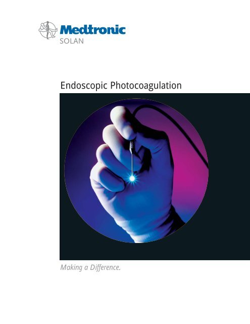
Endoscopic Photocoagulation
Endoscopic Photocoagulation
Endoscopic Photocoagulation

Create successful ePaper yourself
Turn your PDF publications into a flip-book with our unique Google optimized e-Paper software.
<strong>Endoscopic</strong> <strong>Photocoagulation</strong><br />
Making a Difference.
ECP transcends prior<br />
technical limitations and<br />
offers a bright, new solution for<br />
the treatment of glaucoma.<br />
The compact E2 system contains a video<br />
camera, xenon light source, and a semiconductor<br />
diode laser tuned to 810nm<br />
wavelength. This power console features<br />
adjustable laser output, pulse width, light<br />
and aiming beam intensity.<br />
† M Uram, MD, MPH holds multiple patents for<br />
endoscopic technologies, with financial interest in<br />
EndoOptiks, Inc. and its products.<br />
Empowerm<br />
110° panoramic wide field<br />
illumination delivers maximum<br />
intraocular visibility with a<br />
175Watt Xenon light source.<br />
<strong>Endoscopic</strong> Cyclo<strong>Photocoagulation</strong> (ECP) is a surgical<br />
approach to glaucoma management that employs light<br />
endoscopy and visualized laser application. Five<br />
patents combine the modalities of light, image, and<br />
laser in a single, powerful 20Ga instrument. The result<br />
is the selective ablation of pigmented, ciliary epithelium<br />
tissue, with minimal impact to surrounding, nontargeted<br />
anatomy.<br />
Remarkably, all forms of glaucoma can be effectively<br />
treated with this technology. With continuous, direct<br />
imaging of the ciliary processes, controlled laser<br />
energy is delivered; IOP is lowered and aqueous production<br />
is controlled, with superior long-term results.<br />
Light Image Laser<br />
10K pixel resolution allows clear<br />
imaging of ocular tissues from<br />
the anterior or posterior segment.<br />
Precise, 810nm, 2.0Watt diode<br />
laser energy, in pulsed or<br />
continuous mode, is delivered<br />
to targeted intraocular tissues.
ent<br />
The comprehensive armamentarium of endoscopes,<br />
span the need from simple illumination, to laserimaging<br />
fibers - for anterior ECP applications as well as<br />
posterior PRP delivery. DCR probes are also provided<br />
with the higher intensity 12Watt power console.<br />
Optional consoles allow modular flexibility of light,<br />
image, and/or laser power selection.<br />
This powerful new technology has<br />
been embraced by ophthalmologists<br />
across specialties.<br />
System Simplicity<br />
Light<br />
Image<br />
Laser<br />
Sequential organization of system settings and parameters, and backlighted<br />
LCD front panel, makes set up and operation rapid and intuitive.<br />
Light<br />
Cataract<br />
Glaucoma<br />
Pediatric<br />
Laser<br />
Retina<br />
Image<br />
“ECPs greatest worth is its ability to<br />
reduce or eliminate glaucoma<br />
medications and thereby<br />
improve compliance”<br />
Richard J. Mackool, MD<br />
Cataract Surgeon<br />
“...you can use the endoscope to<br />
visualize the angle directly<br />
and treat it.”<br />
Stanley J. Berke, MD, FACS<br />
Glaucoma Specialist<br />
“The advantage of being able<br />
to see each individual ciliary<br />
process, as they are treated,<br />
is tremendous.”<br />
David A. Plager, MD<br />
Pediatric Ophthalmologist<br />
“ECP employs a triple<br />
function micro endoscope to<br />
provide heretofore impossible<br />
intraocular visualization”<br />
Martin Uram, MD, MPH †<br />
Retina Specialist
“In our patients with controlled<br />
glaucoma and cataracts,<br />
combined ECP with<br />
phaco IOL insertion<br />
is my treatment<br />
RJ Mackool, MD of choice.”<br />
This 2-5 minute, reimbursed<br />
technique is ideal for patients<br />
with medically controlled glaucoma.<br />
Intended surgical results lower<br />
intraocular pressure (IOP) longterm,<br />
reduce required patient<br />
medications (minimizing<br />
compliance issues), and<br />
retard progression of the<br />
glaucomatous condition.<br />
“Our goal is to provide the best patient<br />
care in our practice. If I can perform<br />
advanced phaco and reduce my patients’<br />
dependency on glaucoma medications,<br />
I have succeeded to this end.”<br />
Stephen B Wiles, MD<br />
Cataract Surgeon<br />
In a combined phacoemulsification/ECP procedure, an<br />
existing clear corneal or limbal incision is sufficient to<br />
accommodate the 20Ga endoprobe. In a stand-alone ECP<br />
case, the ciliary processes may be approached from a<br />
limbal or a pars plana incision. From this single entry,<br />
anatomy is accurately identified, and between 180° to 200°<br />
of ciliary processes can effectively be reached; a secondary<br />
incision will accommodate a full 270° to 360° treatment.<br />
Versatility &<br />
"All of our procedures are done<br />
through the clear cornea. In the<br />
phakic patient, a sodium hyaluronate<br />
viscoelastic is injected between the iris<br />
and anterior lens surface to broaden<br />
access to the ciliary processes."<br />
DA Plager, MD<br />
Pediatric Ophthalmologist<br />
"I treat some eyes through the pars<br />
plana, with deranged anterior segment<br />
anatomy, where there might<br />
be a lot of synechiae between the iris<br />
and the lens capsule.<br />
RJ Mackool, MD<br />
Direct, continuously monitored visualization,<br />
with aiming beam precision, produces repeatable, titratable<br />
shrinkage of targeted tissues. Most impressive,<br />
beyond the incremental benefits of the ECP procedure,<br />
endoscopy allows the accurate assessment of: zonular<br />
dehiscence, capsular bag integrity, residual cortical<br />
material, intended haptic placement, and anterior<br />
hemorrhage. Clearly, this capability should only enhance<br />
clinical outcomes.<br />
Clear Corneal Entry<br />
Pars Plana Approach<br />
Target Approach<br />
“Anybody who can do phaco can learn ECP in a heartbeat.<br />
You can learn to use the probe to ‘paint’ the ciliary processes<br />
in a continuous motion. Combined with phaco,<br />
ECP takes an extra 2 to 5 minutes.”<br />
RJ Mackool, MD<br />
Clear corneal entry, through the existing<br />
phaco wound, allows treatment of<br />
Phakic, Pseudophakic, or Aphakic eyes.<br />
Laser delivery occurs either over, or<br />
through, the capsular bag.<br />
Pars plana approach may be employed,<br />
combined with Vitreous surgery, for<br />
Pseudophakic or Aphakic patients.
Control<br />
“Unlike transscleral<br />
cylodestructive procedures,<br />
the surgeon can see the ciliary<br />
processes and therefore can titrate<br />
laser delivery properly.”<br />
M Uram, MD, MPH<br />
Visually titratable results are<br />
achieved, from 180 o to 360 o , as<br />
processes blanch and shrink to<br />
desired composition.<br />
Pulsed laser application<br />
delivers selective ciliary process<br />
treatment. Continuous laser mode<br />
allows for rapid ablation, in a<br />
'painting' technique.<br />
“I also use the endoscope during<br />
procedures in which I wish to examine<br />
the ciliary sulcus. Such cases include<br />
those with dislocated or decentered<br />
IOLs, or possible retained lens<br />
fragments and/or abnormalities<br />
of the capsule/zonules.”<br />
RJ Mackool, MD<br />
Haptic in the Bag<br />
180 o -360 o Range Haptic Piercing<br />
Ciliary Body<br />
Laser Treated Processes AC-IOL Piercing Iris<br />
Proper IOL and lens<br />
haptic placement is easily<br />
confirmed using simple<br />
endoscopic technique.<br />
Angle, Iris, Lens<br />
Residual Lens Fragments<br />
Posterior View of Processes<br />
Thickened Zonules in<br />
Pseudoexfoliation<br />
Application/Impact IOL/Haptic Position <strong>Endoscopic</strong> Discovery Retina<br />
Ophthalmic Endoscopy<br />
broadens diagnostic assessment<br />
and surgical treatment<br />
across specialties.<br />
“I do ECP on every one of my cataract patients who is also on glaucoma<br />
medication, because it works so well... through a single incision I can<br />
initially treat approximately 210 o to 240 o of ciliary processes.”<br />
MS McFarland, MD<br />
“Combined with phaco, ECP takes<br />
an extra two minutes. The endoscope<br />
is also a great teaching tool...<br />
providing a virtual tour of the eye.”<br />
Alan B Aker, MD<br />
Cataract Surgeon<br />
Retina at Ora Serrata<br />
Dropped Nucleus<br />
Diabetic Retinal Hemorrhage<br />
Posterior segment views provide<br />
clear imaging of the macula and<br />
optic nerve head, in addition to<br />
post-operative retinal integrity at<br />
the ora serrata.<br />
“Another use of the endoscope is as a<br />
visualization tool in any patient with<br />
a cloudy cornea... to see the status of<br />
the optic nerve and retina.”<br />
SJ Berke, MD, FACS<br />
Glaucoma Specialist
Successful surgical techniques<br />
are measured by compelling<br />
long term, clinical findings.<br />
ECP is supported by more<br />
than five years of data.<br />
“I do a combined procedure on every cataract<br />
patient on glaucoma medication... If I can<br />
eliminate one or two drops... a beta-blocker...<br />
(patients) just feel better. It’s a really nice thing<br />
you can do for your patients.”<br />
Alan B. Aker, MD<br />
“I manage my ECP patients just as I<br />
do a postoperative cataract. I see<br />
them at one day and one week, and<br />
probably 90% of them are cell free<br />
at two weeks.”<br />
MS McFarland, MD<br />
When compared to all other surgical techniques for<br />
glaucoma management, ECP meets and exceeds the<br />
long term goal for reduced IOP and topical medications.<br />
Even in studies that compared 'phaco alone' to 'phaco<br />
with ECP', the combined procedure showed no higher<br />
complication rates, while intended, long-term, positive<br />
results were significantly elevated.<br />
Relative to traditional transscleral “cyclodestructive”<br />
approaches, intraocular ECP has repeatedly earned<br />
marked distinction.<br />
Measured<br />
Without incidence of CME or<br />
major complications, ECP<br />
reduced IOP 38% further than<br />
Phaco surgery alone. In addition,<br />
87% of these patients benefited<br />
from reduced medications, while<br />
more than 60% no longer needed<br />
any pharmaceutical management.<br />
“You can’t do phaco expecting that<br />
pressures will be lowered.”<br />
SJ Berke, MD, FACS<br />
180° ablation resulted in a 15%<br />
decrease in IOP and 68%<br />
reduction in medications, without<br />
visual loss or major complications<br />
Berke, SJ., et al.<br />
J Glaucoma 2000; 9:1.<br />
“The ideal patient for a combined procedure is one who is<br />
controlled, or at least marginally controlled with glaucoma<br />
medications... We should never underestimate how much patients<br />
desire to get off one or more of their glaucoma medications.”<br />
RJ Mackool, MD<br />
ECP mean CONTROL mean<br />
f/u = 25 mo f/u = 44 mo Ps<br />
Mean change in IOP (mmHg) –3.3 –2.4 .48<br />
Decreased Meds (%) 87 9 .01<br />
Same (%) 13 73 .01<br />
Increased Meds (%) 0 18 .001<br />
No meds (%) 61 5 .01<br />
CME (%) 0 0<br />
Major complications (%) 0 0<br />
Mackool: ECP vs. Phaco Alone �<br />
Mean Ablation<br />
25 eyes,<br />
mean f/u 11 months<br />
180°<br />
Mean Decrease IOP 15%<br />
Mean Reduction of Meds 68%<br />
Post-op Visual Loss 0%<br />
Major Complications 0<br />
Berke: ECP Study �<br />
Histology showing selective<br />
ablation of the ciliary epithelium.<br />
� Clinical studies and scientific<br />
data are available upon request.
Without the high failure rates associated with<br />
transscleral techniques, ECP is not reserved for<br />
end-stage glaucoma. Treatments have proven far<br />
superior in accurately isolating ciliary processes,<br />
discretely ablating ciliary epithelium, and achieving<br />
desired results - without missing, or over/under treating<br />
the target tissue.<br />
Further, devastating complication rates are lowest<br />
following ECP, and postoperative patient management<br />
is facilitated relative to traditional glaucoma procedures.<br />
Results<br />
“With ECP, (reduced pressure...) usually happens during the first six to eight<br />
weeks. A seven year follow-up study shows... few patients decompensate after<br />
the second year, as would be expected with filtration procedures.”<br />
M Uram, MD, MPH<br />
2 12 24 36 48 60<br />
Follow-Up (months)<br />
Long Term IOP Control �<br />
OAG OAG with CACG CACG with NVG PED GL Phaco & Uncontrolled Phaco & Controlled<br />
Failed Surgery Failed Surgery Glaucoma Surgery Glaucoma Surgery<br />
TRAB 90% 50% 50% 30% NA < 50% 70% NA<br />
TUBE NA 50-70% 50-70% 50-70% 50-70% < 50% NA NA<br />
TSCPC NA NA 30-50% 30-50% 30-50% < 50% NA NA<br />
ECP 90% 90% 90% 90% 90% > 50% 90% + 90%<br />
Uram: IOP Control - Results per Procedure �<br />
Minor Devastating Visual Endophthalmitis Delayed Operative Intensity of Post<br />
Complications Complications Acuity Loss Failures Time (1-4) Op Care (1-4)<br />
TRAB 10-50% 5-10% 5-10% 1%/yr > 50% 3 4<br />
TUBE 30-50% 30% ~10% 1% ~10-30% 4 3<br />
TSCPC > 50% 30% > 40% NA ~ 40% 2 2<br />
ECP 3-8% < 1% < 1% 0% 0-5% 1 1<br />
Uram: Complications per Procedure �<br />
IOP (mm Hg)<br />
50<br />
40<br />
30<br />
20<br />
10<br />
0<br />
“... if the patient has good visual potential, I use the<br />
endolaser, because I think we risk decreasing their vision<br />
with a transscleral approach.” SJ Berke, MD<br />
NVG<br />
APH/PS<br />
Phakic POAG<br />
Combined<br />
Congenital<br />
“There is no comparison (between ECP and cyclodestructive<br />
techniques). I personally would not do a transscleral laser<br />
or cyclocryo procedure.” RJ Mackool, MD<br />
Remarkably, all forms of<br />
glaucoma respond to ECP with<br />
comparable, sustained results.<br />
“In no way can ECP be<br />
compared with true cyclodestructive<br />
techniques. This distinction exists in<br />
patient selection, patient tolerance,<br />
clinical complications, and long<br />
term results.”<br />
SB Wiles, MD<br />
ECP proves more efficacious in<br />
reducing IOP, over 24 months,<br />
than trabeculectomy, tube implants,<br />
and transscleral treatments.<br />
Relative to these same<br />
procedures, ECP produced the<br />
smallest percentage of minor<br />
complications, visual acuity loss,<br />
or devastating complications.
Endo<br />
Optiks<br />
System<br />
Components<br />
Laser and Endoscopy Systems Light Image Laser<br />
OME2000 E2 Compact Microprobe Laser<br />
and Endoscopy System • • •<br />
OME3000 E3 Compact Microprobe High Power Laser<br />
and Endoscopy System • • •<br />
OME4000 E4 Endoscopy System • •<br />
OME1500 12 Watt Diode Laser •<br />
OME1600 LX - Mini Light Source (Metal Halide) •<br />
Endoscopes, Probes and Fibers Light Image Laser E2 E3 E4 12W<br />
OME200SMAE Ophth. Laser Endoscope - Semi-Disposable (1/ea) • • • • • • •<br />
OME200SMAX Ophth. Laser Endoscope with Custom<br />
Laser Connector - Semi-Disposable (1/ea) • • • •<br />
OME200L Ophth. Endoscope - Light and Camera<br />
Semi-Disposable (1/ea) • • • • •<br />
VPH200 Endophotocoagulation Probes<br />
Disposable, Single Use (5/bx) • • • •<br />
VPH200A Endophotocoagulation Probe<br />
Reusable, Autoclavable (1/ea) • • • •<br />
VP5100 20 Gauge Illumination Probe<br />
Disposable, Single Use (5/bx) • • • •<br />
OME20LA Illuminated Laser Probe, Wide Field<br />
Reusable, Autoclavable (1/ea) • • • • • •<br />
VP200W Wide Field Illumination Probe<br />
Reusable, Autoclavable (1/ea) • • • •<br />
VP5330C Disposable Laser Fiber (5/bx) • • •<br />
VP5330TR Disposable Transcleral Laser Fiber (5/bx) • • • •<br />
Accessories E2 E3 E4 12W<br />
OME300Z Video Adapter Upgrade • • •<br />
MON001 Sony Video Monitor - 13" • • •<br />
CT001 Custom Operating Room System Cart • •<br />
OME100LF Diode Laser Safety Filter for Zeiss Microscope • • •<br />
OME100LFM Diode Laser Safety Filter for Moller Microscope • • •<br />
OME100LFW Diode Laser Safety Filter for Wilde Microscope • • •<br />
LG001 Diode Laser Safety Goggles • • •<br />
VP6200 Sterilization Tray • • • •<br />
FC401655 Flight Case with Wheels & Telescoping Handle • • • •<br />
405681 Flight Case with Wheels • • • •<br />
VP5120 <strong>Endoscopic</strong> Blade • • •<br />
Medtronic Solan<br />
6743 Southpoint Drive North, Jacksonville, Florida 32216-0980 USA<br />
800-535-4646 • 904-296-9600 • Fax 904-279-2630 • www.medtronicsolan.com<br />
All products are manufactured by EndoOptiks, Inc., and exclusively distributed by Medtronic Solan.<br />
Console Compatibilty •<br />
Typical setup of E2 for ECP •<br />
Available Modalities •<br />
� Clinical studies and scientific data are available upon request.<br />
† M Uram, MD, MPH holds multiple patents for <strong>Endoscopic</strong> Technologies, with financial interest in EndoOptiks, Inc. and its products.<br />
980029.0702