Lect3.pdf
Lect3.pdf
Lect3.pdf

You also want an ePaper? Increase the reach of your titles
YUMPU automatically turns print PDFs into web optimized ePapers that Google loves.
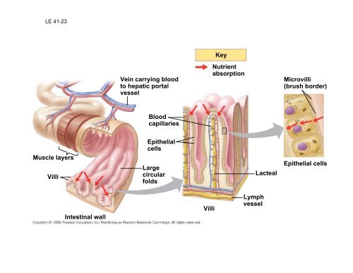
LE 41-23<br />
Muscle layers<br />
Villi<br />
Intestinal wall<br />
Vein carrying blood<br />
to hepatic portal<br />
vessel<br />
Blood<br />
capillaries<br />
Epithelial<br />
cells<br />
Large<br />
circular<br />
folds<br />
Villi<br />
Key<br />
Nutrient<br />
absorption<br />
Lymph<br />
vessel<br />
Lacteal<br />
Microvilli<br />
(brush border)<br />
Epithelial cells
• Each villus contains a network of blood vessels and a<br />
small lymphatic vessel called a lacteal
• Amino acids and sugars pass through the epithelium of the<br />
small intestine and enter the bloodstream (through hepatic<br />
portal vein)<br />
• After glycerol and fatty acids are absorbed by epithelial<br />
cells, they are recombined into fats within these cells<br />
• These fats are mixed with cholesterol and coated with<br />
protein, forming molecules called chylomicrons, which are<br />
transported into lacteals
LE 41-24<br />
Bile salts<br />
Fat droplets<br />
coated with<br />
bile salts<br />
Epithelium<br />
of small<br />
intestine<br />
Epithelium<br />
of lacteal<br />
Fat globule<br />
Micelles made<br />
up of fatty acids,<br />
monoglycerides,<br />
and bile salts<br />
Lacteal
The Large Intestine<br />
• The large intestine, or colon, is connected to<br />
the small intestine<br />
• Its major function is to recover water that<br />
has entered the alimentary canal<br />
• Wastes of the digestive tract, the feces,<br />
become more solid as they move through<br />
the colon<br />
• Feces pass through the rectum and exit via<br />
the anus
• The colon houses strains of the bacterium<br />
Escherichia coli, some of which produce vitamins
Evolutionary adaptations of<br />
vertebrate digestive systems are<br />
often associated with diet<br />
• Digestive systems of vertebrates are<br />
variations on a common plan<br />
• However, there are intriguing<br />
adaptations, often related to diet
Some Dental Adaptations<br />
• Dentition, an animal’s assortment of teeth, is one<br />
example of structural variation reflecting diet<br />
• Mammals have specialized dentition that best<br />
enables them to ingest their usual diet
LE 41-26<br />
Incisors<br />
Canines<br />
Carnivore<br />
Herbivore<br />
Omnivore<br />
Premolars<br />
Molars
Stomach and Intestinal<br />
Adaptations<br />
• Herbivores generally have longer alimentary canals<br />
than carnivores, reflecting the longer time needed to<br />
digest vegetation
LE 41-27<br />
Small<br />
intestine<br />
Stomach<br />
Cecum<br />
Colon<br />
(large<br />
intestine)<br />
Carnivore Herbivore<br />
Small intestine
Symbiotic Adaptations<br />
• Many herbivores have fermentation<br />
chambers, where symbiotic microorganisms<br />
digest cellulose<br />
• The most elaborate adaptations for an<br />
herbivorous diet have evolved in the animals<br />
called ruminants
LE 41-28<br />
Intestine Rumen<br />
Abomasum<br />
Esophagus<br />
Omasum<br />
Reticulum
Gastrovascular Cavities<br />
• Simple animals, such as cnidarians, have a body<br />
wall only two cells thick that encloses a<br />
gastrovascular cavity<br />
• This cavity functions in both digestion and<br />
distribution of substances throughout the body<br />
• Some cnidarians, such as jellies, have elaborate<br />
gastrovascular cavities
LE 42-2<br />
Mouth<br />
Circular<br />
canal<br />
Radial canal<br />
5 cm
Open and Closed Circulatory<br />
Systems<br />
• More complex animals have either open or<br />
closed circulatory systems<br />
• Both systems have three basic components:<br />
– A circulatory fluid (blood or hemolymph)<br />
– A set of tubes (blood vessels)<br />
– A muscular pump (the heart)
• In insects, other arthropods, and most<br />
molluscs blood bathes the organs<br />
directly in an open circulatory system<br />
• There is no distinction between blood<br />
and interstitial fluid, and this general<br />
body fluid is more correctly called<br />
hemolymph
LE 42-3<br />
Anterior<br />
vessel<br />
Lateral<br />
vessel<br />
Ostia<br />
An open circulatory system.<br />
Heart<br />
Hemolymph in sinuses<br />
surrounding organs<br />
Tubular heart<br />
Interstitial<br />
fluid<br />
Dorsal vessel<br />
(main heart)<br />
Auxiliary hearts Ventral vessels<br />
A closed circulatory system.<br />
Heart<br />
Small branch vessels<br />
in each organ
• In a closed circulatory system, blood is<br />
confined to vessels and is distinct from the<br />
interstitial fluid<br />
• Closed systems are more efficient at<br />
transporting circulatory fluids to tissues and<br />
cells
LE 42-4<br />
FISHES<br />
Gill capillaries<br />
Artery<br />
Gill<br />
circulation<br />
Heart:<br />
Ventricle (V)<br />
Atrium (A)<br />
Vein Systemic<br />
circulation<br />
AMPHIBIANS<br />
Lung and skin capillaries<br />
Pulmocutaneous<br />
circuit<br />
V<br />
Right<br />
Systemic<br />
circuit<br />
Left<br />
Systemic capillaries Systemic capillaries<br />
A<br />
A<br />
REPTILES (EXCEPT BIRDS)<br />
Right<br />
systemic<br />
aorta<br />
Lung capillaries<br />
Pulmonary<br />
circuit<br />
Left<br />
A<br />
A systemic<br />
V<br />
V<br />
aorta<br />
Right Left<br />
Systemic capillaries<br />
Systemic circuits include all body tissues except lungs. Note that circulatory systems are depicted<br />
as if the animal is facing you: with the right side of the heart shown at the left and vice-versa.<br />
MAMMALS AND BIRDS<br />
A<br />
Lung capillaries<br />
Pulmonary<br />
circuit<br />
A<br />
V<br />
V<br />
Right Left<br />
Systemic<br />
circuit<br />
Systemic capillaries
LE 42-5<br />
Anterior<br />
vena cava<br />
Pulmonary<br />
artery<br />
Capillaries<br />
of right lung<br />
Pulmonary<br />
vein<br />
Right atrium<br />
Right ventricle<br />
Posterior<br />
vena cava<br />
Aorta<br />
Capillaries of<br />
head and<br />
forelimbs<br />
Pulmonary<br />
artery<br />
Capillaries<br />
of left lung<br />
Pulmonary<br />
vein<br />
Left atrium<br />
Left ventricle<br />
Aorta<br />
Capillaries of<br />
abdominal organs<br />
and hind limbs
LE 42-6<br />
Pulmonary artery<br />
Anterior<br />
vena cava<br />
Right<br />
atrium<br />
Pulmonary<br />
veins<br />
Semilunar<br />
valve<br />
Atrioventricular<br />
valve<br />
Posterior<br />
vena cava<br />
Right<br />
ventricle<br />
Left<br />
ventricle<br />
Aorta<br />
Pulmonary<br />
artery<br />
Left<br />
atrium<br />
Pulmonary<br />
veins<br />
Semilunar<br />
valve<br />
Atrioventricular<br />
valve
• The heart contracts and relaxes in a rhythmic<br />
cycle called the cardiac cycle<br />
• The contraction, or pumping, phase is called<br />
systole<br />
• The relaxation, or filling, phase is called diastole
To adjust blood pressure independently in the<br />
capillaries of the gas-exchange surface and in<br />
the capillaries of the general body circulation, an<br />
organism would need<br />
– A. an open circulatory system.<br />
– B. a hemocoel.<br />
– C. a lymphatic system.<br />
– D. a two-chambered heart.<br />
– E. a four-chambered heart.
LE 42-7<br />
AV valves<br />
open<br />
Atrial and<br />
ventricular<br />
diastole<br />
Semilunar<br />
valves<br />
closed<br />
0.4 sec<br />
0.1 sec<br />
0.3 sec<br />
AV valves<br />
closed<br />
Atrial systole;<br />
ventricular<br />
diastole<br />
Semilunar<br />
valves<br />
open<br />
Ventricular systole;<br />
atrial diastole
• The heart rate, also called the pulse, is<br />
the number of beats per minute<br />
• The cardiac output is the volume of<br />
blood pumped into the systemic<br />
circulation per minute
Maintaining the Heart’s Rhythmic<br />
Beat<br />
• Some cardiac muscle cells are selfexcitable,<br />
meaning they contract without<br />
any signal from the nervous system
• The sinoatrial (SA) node, or pacemaker,<br />
sets the rate and timing at which cardiac<br />
muscle cells contract<br />
• Impulses from the SA node travel to the<br />
atrioventricular (AV) node<br />
• At the AV node, the impulses are delayed<br />
and then travel to the Purkinje fibers that<br />
make the ventricles contract
• Impulses that travel during the cardiac<br />
cycle can be recorded as an<br />
electrocardiogram (ECG or EKG)
LE 42-8<br />
Pacemaker<br />
generates wave of<br />
signals to contract.<br />
SA node<br />
(pacemaker)<br />
ECG<br />
AV<br />
node<br />
Signals are delayed<br />
at AV node.<br />
Signals pass<br />
to heart apex.<br />
Bundle<br />
branches Heart<br />
apex<br />
Purkinje<br />
fibers<br />
Signals spread<br />
throughout<br />
ventricles.
• The pacemaker is influenced by nerves,<br />
hormones, body temperature, and exercise
Concept 42.3: Physical principles<br />
govern blood circulation<br />
• The physical principles that govern movement<br />
of water in plumbing systems also influence<br />
the functioning of animal circulatory systems
Blood Vessel Structure and<br />
Function<br />
• The “infrastructure” of the circulatory<br />
system is its network of blood vessels<br />
• All blood vessels are built of similar<br />
tissues and have three similar layers
LE 42-9<br />
Artery<br />
Endothelium<br />
Smooth<br />
muscle<br />
Connective<br />
tissue<br />
Artery Vein<br />
Endothelium<br />
Capillary<br />
100 µm<br />
Basement<br />
membrane<br />
Endothelium<br />
Smooth<br />
muscle<br />
Connective<br />
tissue<br />
Arteriole Venule<br />
Vein<br />
Valve
• Structural differences in arteries, veins,<br />
and capillaries correlate with functions<br />
• Arteries have thicker walls that<br />
accommodate the high pressure of<br />
blood pumped from the heart
• In the thinner-walled veins, blood flows<br />
back to the heart mainly as a result of<br />
muscle action
LE 42-10<br />
Direction of blood flow<br />
in vein (toward heart)<br />
Valve (open)<br />
Skeletal muscle<br />
Valve (closed)
Blood Flow Velocity<br />
• Physical laws governing movement of fluids<br />
through pipes affect blood flow and blood<br />
pressure<br />
• Velocity of blood flow is slowest in the<br />
capillary beds, as a result of the high<br />
resistance and large total cross-sectional<br />
area