Front Matter (PDF) - Investigative Ophthalmology & Visual Science
Front Matter (PDF) - Investigative Ophthalmology & Visual Science
Front Matter (PDF) - Investigative Ophthalmology & Visual Science
You also want an ePaper? Increase the reach of your titles
YUMPU automatically turns print PDFs into web optimized ePapers that Google loves.
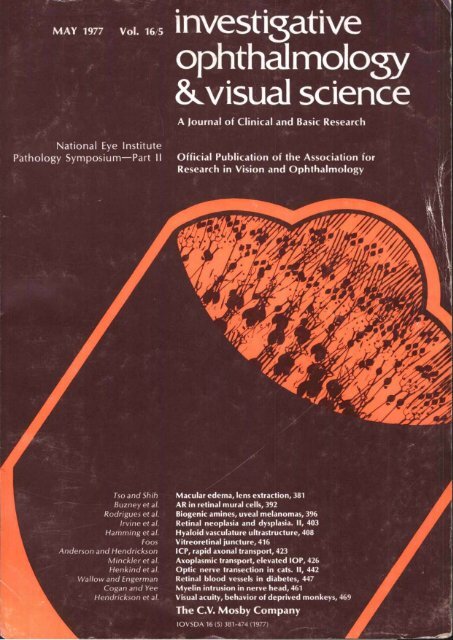
MAY 1977 Vol. 16/5<br />
National Eye Institute<br />
Pathology Symposium—Part II<br />
Tso and Shin<br />
Buzneyet al.<br />
Rodrigues et al.<br />
Irvine et al.<br />
Hamming et al.<br />
Foos<br />
Anderson and Hendrickson<br />
Minckler et al.<br />
Henkind et al.<br />
Wallow and Engerman<br />
Cogan and Yee<br />
Hendrickson et al.<br />
investigative<br />
ophthalmology<br />
&.visual science<br />
A Journal of Clinical and Basic Research<br />
Official Publication of the Association for<br />
Research in Vision and <strong>Ophthalmology</strong><br />
Macular edema, lens extraction, 381<br />
AR in retinal mural cells, 392<br />
Biogenic amines, uveal melanomas, 396<br />
Retinal neoplasia and dysplasia. II, 403<br />
Hyaloid vasculature infrastructure, 408<br />
Vitreoretinal juncture, 416<br />
ICP, rapid axonal transport, 423<br />
Axoplasmic transport, elevated IOP, 426<br />
Optic nerve transection in cats. II, 442<br />
Retinal blood vessels in diabetes, 447<br />
Myelin intrusion in nerve head, 461<br />
<strong>Visual</strong> acuity, behavior of deprived monkeys, 469<br />
The C.V. Mosby Company<br />
IOVSDA 16 (5) 381-474 (1977)
Though it stands only 7.5 cen<br />
meters, Pred ForteJ" (prednisoloi<br />
acetate 1.0%) measures tall amor<br />
ocular steroids.<br />
In studies comparing Pred For1<br />
with dexamethasone phosphate<br />
dexamethasone alcohol and prec<br />
nisolone phosphate, Pred Fort1<br />
emerged the undisputed ruler ir<br />
terms of anti-inflammatory activity<br />
In these studies by Leibowitz<br />
et al. (using rabbit corneas with<br />
the epithelium intact) Pred Forte<br />
exhibited 25% more anti-inflammatory<br />
activity than the closest<br />
steroid, dexamethasone alcohol?<br />
Pred Forte achieved more than<br />
twice the corneal concentrations of<br />
the other steroids tested in the<br />
inflamed rabbit eyes with epithe- - ie<br />
1<br />
lium intact. And further, Pred Forte ; I<br />
achieved significant concentra- -5<br />
-/<br />
tions in the aqueous humor? 5 ' I<br />
Pred Wrte performs both in &<br />
the lab and, more important, 5<br />
on the real firing line: in your<br />
patient's eyes.<br />
Pred Forte, the ocular steroid<br />
with the clinical record. Perhaps<br />
that's why ophthalmologists<br />
believe in Pred Forte and pre-<br />
scribe it most.<br />
When you see severe ocular<br />
inflammation, remember the<br />
"ruler": Pred Forte.<br />
(prednisolone acetate) 1%<br />
the steroid others are<br />
measured against.<br />
I<br />
,<br />
Pred Fortex Iprednis~~lnne acetate) 1% Sterile Ophthalmic<br />
Suspension. INDICATIONS Fnr steroid responsive inflammation<br />
of the palpebral and bulbar conjunctiva, cornea and<br />
anterior segment of the globe. CONTRAINDICATIONS<br />
Acute untreated purulent wular infections. acute superficial<br />
herpes simplex (dendritic kerat~tis). vaccinia, varicella and<br />
most 11ther viral diseases of the cornea and conjunctiva. (~ular<br />
tuberculosis,and fungal diseasesof the eye,and sensitivity to<br />
any mnipments of the formulatron. WARNINGS 1. In th~se<br />
diseases causmg thinning of the cornea, perforation has been<br />
reported with the use of topical steroids. 2. Since PRED<br />
FORTEx contains no antimicrobial, if infection is present<br />
appropriate measures must be taken to a~unteract the orga.<br />
nisms ~nvolved. 3. Acute purulent infections of the eye<br />
may be masked or enhanced by the use of topical steroids.<br />
4. Use II~ sten~~d medication in the presence of stn~mal hews<br />
simplex requlres caution and should be followed by freqwnt<br />
mandatnry slit-lamp microscopy. 5. As fungal infections of<br />
, the a)rnea have been reported coincidentally with longterm<br />
I local stemid applications, fungal invasion may be suspected<br />
$ in any persistent ameal ulcelation where a stemid has been<br />
used. or is in use. 6. Use of topical a~rtjcnsteroids may cause<br />
increased intrawular pressure in certain individuals. This<br />
may result in damage to the optic nerve with defects in the<br />
3 visual fields. It is advisable that the intrawular pressure<br />
be checked frequently. 7. Use in Pregnancy -Safety of<br />
3 intensive or protracted use of topical steroids during<br />
pregnancy has mt been substantiated. PRECAUTIONS<br />
Pmtenor subapsular cataract formation has been rep~nd<br />
after heavy or protracted use of topical ophthalmic cnr.<br />
ticr~steroids. Patients with histories of herpes simplex<br />
keratitis sh~~uld be treated with caution. ADVERSE<br />
REACTIONS Increased intraocular pressure. with optic<br />
nerve damage, defects in the visual fields. A h ptiterior<br />
subcapsularcataract formation, secondary (rular mfections<br />
fnmi fungi or viruses Ilkrated fn~m ocular tissues.<br />
and perfi~rat~on of the globe when used in ctmdit~c~ns<br />
where there is thinnmg of the cornea or dera. Systeniic<br />
side effects niay mur with extenswe use of stero~ds.<br />
DOSAGE AND ADMINISTRATION 1 to 2<br />
drnps instilled into the conjunctival sac two tu four<br />
times daily. During the initial 24 to 48 hours the dosage<br />
may be safely increased t11 2 drops every hour. Care<br />
should be taken not 10 disamtinue therapy prematurely.<br />
REFERENCE NOTES: I. Lcihmitz. H. M, and Kupfcmfan.<br />
A. Bioorw~kbility and Iht~rapcallc. vfjfrrliruncss<br />
of l~~picallv ad~winistrn.d rartioatrn,iils. Trans<br />
An1 Acad Ophthalnlrd Ot~rla~ynpl N (11: 00 iH.@<br />
XX. 1.V7.5. b Ihiil 3. /hid<br />
A&WN Iruine. Caiifarnia
Improve your skills<br />
in the use of fluorescein angiography<br />
and photocoagulation<br />
with this<br />
superbly illustrated atlas<br />
j~+ New 2nd Edition! ^ ^<br />
Stereoscopic Atlas<br />
of<br />
Macular Diseases<br />
For pertinent, practical information on diagnosis and management of<br />
macular diseases, consult this magnificently illustrated new edition. Dr.<br />
Gass correlates appearance with fluorescein angiographic findings and,<br />
when available, histopathology. He makes use of black arid white photographs,<br />
stereo fundus photographs in full color (19 stereo reels—4 new<br />
to this edition); fluorescein angiograms, photomicrographs; and schematic<br />
drawings to elucidate all textual material. Virtually a new book, this edition<br />
has been revised and expanded to provide the most comprehensive<br />
treatise available on macular diseases.<br />
By J. Donald M. Gass, M.D. April, 1977. 412 pages plus FM I-XII,<br />
8'/2" x 11", 951 illustrations plus 121 views in 19 stereo reels in color;<br />
12 views in black and white. (Viewer not included) Price, $65.00.<br />
ORDER BY PHONE: Call (800) 325-4177 ext. 10. In Missouri call collect—(314)<br />
872-8370 ext. 10. 9 am to 5 pm (CDT), Monday through Friday.<br />
MO5BV<br />
TIMES MIRROR<br />
THE C. V MDSBY COMPANY<br />
11830 WESTUNE INDUSTRIAL DRIVE<br />
ST. LOUIS MISSOURI 63141
Now, Iris fluorescein photography. From the developers<br />
of fundus fluorescein photography. From Zeiss.<br />
Another Zeiss First. We were first to bring you an instrument<br />
for fluorescein angiography of the ocular fundus. Now<br />
we have developed a slit lamp accessory for fluorescein angiography<br />
of the iris. Easily attached to the Zeiss Slit Lamps<br />
100/16 or 125/16 or to the Photo Slit Lamp, this special illuminator<br />
with flash tube, ignition unit and power supply lets you<br />
utilize one of the newest diagnostic techniques in ophthalmology.<br />
Use one power supply for two instruments ... If you<br />
already have a Zeiss Slit Lamp or Photo Slit Lamp and a Zeiss<br />
Fundus Camera with Fundus Flash 2 or Power Supply 260, all<br />
you need to do both iris and fundus fluorescein photography are<br />
the illuminator and a switchbox. Simple. Easy to operate. And,<br />
since Zeiss Optics are the world's greatest optics, you are assured<br />
of excellent photographs.<br />
For photography of the anterior segment of the eye.<br />
Not only is iris fluorescein photography valuable for diagnosis<br />
of disorders of the iris, but it has been used to validate a confirmative<br />
diagnosis of diabetes, Iris fluorescein photography is<br />
expected to become one of the most sophisticated diagnostic<br />
techniques in medicine.<br />
Send for more information. Write: Carl Zeiss, Inc.,<br />
444 Fifth Avenue, New York, N. Y. 10018. Or call (212)<br />
730-4400. In Canada: Carl Zeiss Canada Ltd., 45 Valleybrook<br />
Drive, Don Mills, Canada M3B 2S6. Or call (416) 449-4660.<br />
Nationwide service.<br />
BRANCH OFFICES: BOSTON, CHICAGO, COLUMBUS, HOUSTON, LOS ANGELES, SAN FRANCISCO, WASHINGTON, D. C.<br />
Iris Fluorescein Study, photographed by the<br />
Ophthalmic Photo Unit, New York Medical College, New York City.<br />
Fiuorescein angiogram of a diabetic retinopathy,<br />
revealing a gross vasculopathy in micro and macro vessels.<br />
THE GREAT NAME IN OPTICS
prescribe<br />
PE OPHTHdLmiCS<br />
by color coded vials<br />
PINK YELLOW LAVENDER GREEN BLUE<br />
Rx, instructions and dosage are simplified<br />
to increase patient compliance.<br />
Vz % (free base) epinephrine with each<br />
strength of pilocarpine.<br />
15cc vials—dated to assure fresh potent<br />
medication —no refrigeration required.<br />
DCPERSON & COVEY ING<br />
CLENDALE, CALIFORNIA 91201 U.S.A.
3 you see abwe can<br />
rgk conjunctivitis.<br />
I you see below<br />
----- of it.<br />
NW AltlslOtEAm<br />
Formulated with your allergic conjunctivitis*<br />
patient in mind. AlbalonA clears the congestion<br />
with naphazoline HCI; calms the discomforting<br />
itch with antazoline. And AlbalonA is the only<br />
antihistamine-decongestant in an artificial tear<br />
vehicle: Liquifilm? (polyvinyl alcohol 1.4%). Unique<br />
Liquifilm protects against iatrogenic dry eye; in<br />
contrast, aqueous vehicles can cause dry eye.<br />
That's how Albalon-A does it all faster and<br />
more effectively than either active ingredient alone?<br />
*Tha drug has been evaluated as possrbly effectwe for ths mdicatron<br />
See full prescnbrng rnfonnahon<br />
The anti-drying, antihistamine<br />
DKCRPWN A stenle ophthalmic solubon having the following composltlon:<br />
naphazoline HCI . . .0.05%, antazoline phosphate .0.5%.<br />
with: LiqufilmO Ipd/vinyl akohol) 1.4%; benzalkonium chlonde .ON%;<br />
edetate dkodium: odwinvl mlidone: sodium chlonde: sodium ace<br />
tate, anhvdruus: ac&.ac~d and/orsodih hvdroxlde d needed to adjust<br />
the DH MTlON AlbalonA comb~nes the efkcts of Me antihlstamme.<br />
antazoline. and the decongestant naphazoline.<br />
and/or other informabon. FDA has classified me indications as fol-<br />
lows "PoaibK' effeXiw For rdief of ocular initation and/or con-<br />
gestion of for the treatment of allergic. inflammatow. y. inkcticus<br />
ocular conditions Final classification of the lea-thaneffective indi-<br />
cation requires further investigation.<br />
COlYrrUmDKllTlOlYS M/pwsensiWlW to one or more of the comw<br />
nenb of ths weparation WING Do not use in presence of nam angle glaucoma. The prmrahon should be vsed onlv<br />
wth caution In Me wesence of hvcettemlon, cardlac lrreeularities w<br />
hyDerg)ycemla (d~abetes) To prevent mntamlnabng the dm&?r bp and<br />
solution, care should be taken not to touch the evellds or surrounding<br />
area mth the dropper bp of Me bottle Keep bottle Ughtb closed when<br />
not In use Protect from hght ADVERSE REACTIONS The followng<br />
advene reactions mav occur: Puoilla~ dilation. increase in intaocular<br />
preaure. Nstemlc effects due to absorphon 11 e. hypertenuon, cardac<br />
~mulanbes hwfgkem~al DOSAGE One or two d w lnstrlled In each<br />
eve evecy 3 or 4 hwn or less frequentb as required to relieve mptorns<br />
W SUPPUED 15 ml droooerdo olasdc dro~m bdtles On orescnw<br />
tion only.<br />
REFERENCES<br />
Miller. J and \hMf. E H Antazolme phosphate and Wazollne hydrochloride.<br />
wnglv and In combmanon for the mtrnent of alergerpr coryunct~bs<br />
- a contmlled double- blind cl~nlcal ma1 Ann Albg 35 81-85.1975
For uninterrupted control of I.O.R<br />
...never more than one or two instillations<br />
Page 8<br />
Scanning electron microscopy of<br />
primate trabecular meshwork (X300):<br />
Viewed here is Schlemm's canal<br />
along with uveal and corneoscleral<br />
meshwork. (Photo courtesy<br />
Douglas R. Anderson, M.D.]<br />
This area is the site of the prime<br />
pathologic changes which are<br />
responsible for glaucoma and the<br />
focus of most of the medical<br />
procedures for treatment of the disease.
Because PHOSPHOLINE IODIDE is long-acting, it can help provide<br />
uninterrupted control of intraocular pressure in chronic sinnple (open-angle)<br />
glaucoma or glaucoma secondary to aphakia. Just one or, at most,<br />
two instillations of PHOSPHOLINE IODIDE [one at bedtime, and, if necessary,<br />
one in the morning) are generally needed.<br />
Although PHOSPHOLINE IODIDE is longer-acting than other miotics,<br />
it is not more potent. With four concentrations available, it offers a high degree of<br />
dosage flexibility for uninterrupted control of intraocular pressure... used alone<br />
or in combination with other medication.<br />
When starting PHOSPHOLINE IODIDE therapy, 0.03%-the lowest strength -<br />
is the logical choice. If strengths of 0.06%, 0.125%, or 0.25% are required,<br />
the initial use of the 0.03% will be helpful in smoothing the transition.<br />
PHOSPHOLINE IODIDE* EESKSft<br />
(echothiophate iodide for ophthalmic solution)<br />
See next page ol advertisement for prescribing mformatio<br />
Page 9
PHOSPHOLINE IODIDE<br />
®<br />
(echothiophate iodide)<br />
in the management of<br />
chronic simple (open-angle)<br />
glaucoma or glaucoma<br />
secondary to aphakia<br />
BRIEF SUMMARY<br />
(For lull prescribing information, see package circular.)<br />
PHOSPHOLINE IODIDE'<br />
(ECHOTHIOPHATE IODIDE FOR OPHTHALMIC SOLUTION)<br />
PHOSPHOLINE IODIDE is a long-acting cholinesterase inhibitor<br />
for topical use.<br />
Indications: Glaucoma-Chronic open-angle glaucoma. Subacute<br />
or chronic angle-closure glaucoma after iridectomy or<br />
where surgery is refused or contraindicated. Certain non-uveitic<br />
secondary types of glaucoma, especially glaucoma following<br />
cataract surgery.<br />
Accommodative esotropia-Concomitant esotropias with a<br />
significant accommodative component.<br />
Contraindications: 1. Active uveal inflammation.<br />
2. Most cases of angle-closure glaucoma, due to the possibility<br />
of increasing angle block.<br />
3. Hypersensitiyity to the active or inactive ingredients.<br />
Warnings: 1. Use in Pregnancy: Safe use of anticholinesterase<br />
medications during pregnancy has not been established, nor<br />
has the absence of adverse effects on the fetus or on the<br />
respiration of the neonate.<br />
2. Succinylcholine should be administered only with great<br />
caution, if at all, prior to or during general anesthesia to patients<br />
receiving anticholinesterase medication because of possible<br />
respiratory or cardiovascular collapse.<br />
3. Caution should be observed in treating glaucoma with<br />
PHOSPHOLINE IODIDE in patients who are at the same time<br />
undergoing treatment with systemic anticholinesterase medications<br />
for myasthenia gravis, because of possible adverse<br />
additive effects.<br />
Precautions: 1. Gonioscopy is recommended prior to initiation<br />
of therapy.<br />
2. Wh6re;there is a quiescent uveitis or a history of this condition,<br />
anticholinesterase therapy should be avoided or used<br />
cautiously because of the intense and persistent miosis and<br />
ciliary muscle contraction that may occur.<br />
3. While systemic effects are infrequent, proper use of the<br />
drug requires digital compression of the nasolacrimal ducts for<br />
a minute or two following instillation to minimize drainage into<br />
the nasal chamber with its extensive absorption area. The hands<br />
should be washed immediately following instillation.<br />
4. Temporary discontinuance of medication is necessary if<br />
salivation, urinary incontinence, diarrhea, profuse sweating,<br />
muscle weakness, respiratory difficulties, or cardiac irregularities<br />
occur.<br />
5. Patients receiving PHOSPHOLINE IODIDE who are exposed<br />
to carbamate or organophosphate type insecticides and<br />
pesticides (professional gardeners, farmers, workers in plants<br />
manufacturing or formulating such products, etc.) should be<br />
Page 10<br />
warned of the additive systemic effects possible from absorption<br />
of the pesticide through the respiratory tract or skin. During<br />
periods of exposure to such pesticides, the wearing of respiratory<br />
masks, and frequent washing and clothing changes<br />
may be advisable.<br />
6. Anticholinesterase drugs should be used with extreme caution,<br />
if at all, in patients with marked vagotonia, bronchial<br />
asthma, spastic gastrointestinal disturbances, peptic ulcer, pronounced<br />
bradycardia and hypotension, recent myocardial infarction,<br />
epilepsy, parkinsonism, and other disorders that may<br />
respond adversely to vagotonic effects.<br />
7. Anticholinesterase drugs should be employed prior to<br />
ophthalmic surgery only as a considered risk because of the<br />
possible occurrence of hyphema.<br />
8. PHOSPHOLINE IODIDE (echothiophate iodide) should be<br />
used with great caution, if at all, where there is a prior history of<br />
retinal detachment.<br />
Adverse Reactions: 1. Although the relationship, if any, of retinal<br />
detachment to the administration of PHOSPHOLINE IODIDE<br />
has not been established, retinal detachment has been reported<br />
in a few cases during the use of PHOSPHOLINE IODIDE in<br />
adult patients without a previous history of this disorder.<br />
2. Stinging, burning, lacrimation, lid muscle twitching, conjunctival<br />
and ciliary redness, browache, induced myopia with<br />
visual blurring may occur.<br />
3. Activation of latent iritis or uveitis may occur.<br />
4. Iris cysts may form, and if treatment is continued, may<br />
enlarge and obscure vision. This occurrence is more frequent<br />
in children. The cysts usually shrink upon discontinuance of<br />
the medication, reduction in strength of the drops or frequency<br />
of instillation. Rarely, they may rupture or break free into the<br />
aqueous. Regular examinations are advisable when the drug is<br />
being prescribed for the treatment of accommodative esotropia.<br />
5. Prolonged use may cause conjunctival thickening, obstruction<br />
of nasolacrimal canals.<br />
6. Lens opacities occurring in patients under anticholinesterase<br />
therapy have been reported; routine examinations should<br />
accompany prolonged use.<br />
7. Paradoxical increase in intraocular pressure may follow<br />
anticholinesterase instillation. This may be alleviated by prescribing<br />
a sympathomimetic mydriatic such as phenylephrine.<br />
Overdosage: Antidotes are atropine, 2 mg parenterally;<br />
PROTOPAM" CHLORIDE (pralidoxime chloride), 25 mg per kg<br />
intravenously; artificial respiration should be given if necessary.<br />
How Supplied: Four potencies are available. 1.5 mg package<br />
for dispensing 0.03% solution; 3.0 mg package for 0.06% solution;<br />
6.25 mg package for 0.125% solution; 12.5 mg package<br />
for 0.25% solution. Also contains potassium acetate (sodium<br />
hydroxide or acetic acid may have been incorporated to adjust<br />
pH during manufacturing), chlorobutanol (chloral derivative),<br />
mannitol, boric acid and exsiccated sodium phosphate.<br />
7630<br />
The Ophthalmos Division<br />
AYERST LABORATORIES<br />
NewYork, N.Y. 10017
Page 12<br />
, What<br />
you ve come to expect<br />
fromMosby<br />
Eye signs<br />
and symptoms<br />
in brain tumors<br />
references...<br />
A New Book!<br />
SYMPOSIUM ON RETINAL DISEASES<br />
Based on lectures by noted ophthalmologists and<br />
ophthalmic surgeons, this important new book is divided<br />
into three sections: vitreous surgery; diabetic retinopathy;<br />
and macular disease. Case presentations and round table<br />
exchanges, centered on questions from the audience,<br />
provide you with a comprehensive understanding of the<br />
latest advances in this increasingly significant area.<br />
By the New Orleans Academy of <strong>Ophthalmology</strong>; with 9 contributors.<br />
March, 1977. 354 pages plus FM I-XIV, 6%" x 9%", 411 illustrations.<br />
Price, $37.50.<br />
New Volume V!<br />
CURRENT CONCEPTS IN OPHTHALMOLOGY<br />
This authoritative new volume is an essential reading for<br />
every ophthalmologist interested in an up-to-date overview<br />
of the state of the art. It synthesizes recent<br />
developments in all areas of ophthalmology and carefully<br />
explains new diagnostic methods and treatment techniques.<br />
Outstanding discussions, written by prominent<br />
researchers and clinicians, examine: management of corneal<br />
graft complications; "exudative" senile macular<br />
degeneration; laser treatment for glaucoma; herpetic<br />
keratitis; and much more!<br />
Edited by Herbert E. Kaufman, M.D. and Thorn J. Zimmerman,<br />
M.D., Ph.D.; with 17 contributors. September, 1976. 297 pages<br />
plus FM l-XII, 6%" x 9%", 157 illustrations. Price, $34.50.<br />
New Volume V!<br />
ATLAS OF DISEASES OF THE ANTERIOR SEGMENT<br />
OF THE EYE:<br />
The Crystalline Lens<br />
This new volume offers you a precise, scholarly discussion<br />
of the crystalline lens. Each chapter includes a general introduction,<br />
details on various conditions, and a series of<br />
case histories. Clinical black-and-white photographs, as<br />
well as stereoscopic color views especially selected from<br />
Dr. Donaldson's outstanding collection, illustrate each<br />
case history. Two particularly noteworthy chapters discuss:<br />
radiation and toxic cataracts; and iatrogenic and surgical<br />
aspects of cataracts.<br />
By David D. Donaldson, M.D. October, 1976. 212 pages plus FM I-<br />
XIV, 8'/2" x 11", 129 illustrations, 112 stereoscopic views in full<br />
color on 16 View-Master® reels and a View-Master® compact<br />
viewer. Price, $54.50.
authoritative and up-to-date information for<br />
practical management of various ophthalmic<br />
disorders.<br />
New 3rd Edition!<br />
EYE SIGNS AND SYMPTOMS IN BRAIN TUMORS<br />
Thoroughly updated, the new edition of this classic work offers<br />
you definitive information on ocular signs and symptoms in<br />
brain tumors, and precisely explains the correlation of eye signs<br />
with the recognition and localization of tumors. You'll find new<br />
and revised material on: eye motility and its disorders; red-free<br />
funduscopy; electromyography and electro-oculography of the<br />
eye muscles; and computerized axial tomography with the EMIscanner;<br />
along with 35 new illustrations and updated references.<br />
By Alfred. Huber, M.D. and Frederick C. Blodi, M.D. November, 1976.<br />
3rd edition, 424 pages plus FM I-XVI, 6 3 /4" x 9%", 233 illustrations. Price,<br />
$37.50.<br />
CURRENT CONCEPTS OF THE VITREOUS<br />
INCLUDING VITRECTOMY<br />
Presenting the most current information on the vitreous, this<br />
pioneering new book records the results of a two-day seminar<br />
held to disseminate information and share techniques pertinent<br />
to managing vitreous disorders. Innovative surgeons and<br />
researchers offer in-depth examinations of: vitrectomy in<br />
diabetes; preoperative evaluation before surgery; ultrasonic<br />
evaluation; pars plana vitrectomy; and other topics. Stimulating<br />
round table discussion deals with specific questions raised by<br />
seminar participants (i.e., temporal versus nasal entry in vitrectomy,<br />
etc.).<br />
Edited by Kurt A. Gitter, M.D., F.A.C.S.; with 12 contributors. 1976, 290<br />
pages plus FM I-XIV, 6%" x 9%", 271 illustrations. Price, $31.50.<br />
New 4th Edition!<br />
Becker-Shaffer's DIAGNOSIS<br />
AND THERAPY OF THE GLAUCOMAS<br />
This superb new 4th edition provides you with contemporary,<br />
comprehensive information on the pathogenesis, diagnosis, and<br />
management of glaucomas. It includes new material on<br />
Tubinger perimeter; panretinal photocoagulation in the treatment<br />
of diabetic retinopathy; and red-free filter<br />
ophthalmoscopy of nerve fiber bundle defects. The book also<br />
details two new drug delivery systems: soft contact lenses and<br />
membrane controlled drugs. In addition, this revision provides<br />
you with a stereoscopic gonioscopy supplement consisting of<br />
three stereo reels (21 views).<br />
By Allan E. Kolker, M.D. and John Hetherington, Jr., M.D. September,<br />
1976. 4th edition, 526 pages plus FM I-X, 6%" x 9%", 448 illustrations and 8<br />
color plates, and 3 stereoscopic reels. Price, $37.50.<br />
ORDER BY PHONE!<br />
Call (800) 325-4177 ext. 10. In Missouri call collect—<br />
(314) 872-8370 ext. 10. 9 am to 5 pm (CDT), Monday through Friday.<br />
MO5BV<br />
TIMES MIRROR<br />
THE C. V MOSBY COMPANY<br />
1 1830 WESTLINE INDUSTRIAL DRIVE<br />
ST LOUIS. MISSOURI 63141<br />
A70476<br />
Page 13
if ri:j r v<br />
Classified<br />
Advertisements<br />
INVESTIGATIVE OPHTHALMOLOGY &<br />
VISUAL SCIENCE will offer a classified<br />
advertising section in each issue. It will list<br />
opportunities wanted or offered and fellowships,<br />
residencies, or internships wanted or<br />
available. There will be two rates for these<br />
ads, one for members of the Association for<br />
Research in Vision and <strong>Ophthalmology</strong> and<br />
one for other advertisers. Rates are as<br />
follows:<br />
ARVO members<br />
(30 words or less)<br />
Each additional word<br />
Other advertisers<br />
(30 words or less)<br />
Each additional word<br />
1 Time<br />
$ 5.00<br />
.40<br />
$12.00<br />
.40<br />
3 Times<br />
or more<br />
$ 4.00/ad<br />
.35<br />
$10.00/ad<br />
.35<br />
Count all words, including abbreviations.<br />
Initials and numbers count as one word<br />
(Box 000, INVESTIGATIVE OPHTHALMOLOGY<br />
& VISUAL SCIENCE)—counts as 7 words.<br />
Payment must accompany insertion order.<br />
Display ads. Set within a ruled border.<br />
One inch minimum, $25.00 per inch. Forms<br />
close 18th of each month preceding month<br />
of issue.<br />
INVESTIGATIVE OPHTHALMOLOGY<br />
& VISUAL SCIENCE<br />
Journal Advertising Department<br />
The C. V. Mosby Company<br />
11830 Westline Industrial Drive<br />
St. Louis, Missouri 63141<br />
Massachusetts—Crosseyed Domestic (non-<br />
Siamese) Cat. Notice of Availability. The frequency<br />
of Crosseyedness in the ordinary domestic<br />
cat is about 1/400. Over the years we have developed<br />
a strain of such cats, and now offer two<br />
male-female pairs for further research. Acceptance<br />
of these animals is contingent upon an<br />
agreement to continue breeding. For further details<br />
see Vis. Res. 17, 337 (1977) and contact<br />
W. Richards El 0-135, Mass. Inst. of Tech., 79<br />
Amherst St., Cambridge, Ma. 02139. Tel: 617-<br />
253-5776.<br />
Page 14<br />
Editors!<br />
Reviewers!<br />
Authors!<br />
For you ... a new book that<br />
contains virtually everything<br />
you want to know about<br />
THE SCIENTIFIC JOURNAL:<br />
EDITORIAL POLICIES AND<br />
PRACTICES<br />
Guidelines for Editors, Reviewers,<br />
and Authors<br />
In 1665 the first scholarly journal was published<br />
in France. Since that time, there has<br />
been no one source of editorial guidelines for<br />
journal editors. This new book now provides<br />
you with such a source.<br />
YOU DECIDE<br />
YOUR OWN APPROACH!<br />
Presenting the advantages and disadvantages of<br />
each specific decision, eminent editors offer you<br />
various recommendations, discussions, and opinions<br />
concerning the problems you may encounter.<br />
The book is divided into two general sections:<br />
editorial policies, which usually require major decisions;<br />
and editorial practices, which involve<br />
minor decisions, often about format or mechanical<br />
style. You'll find essays exploring: the manuscript<br />
reviewing system: special types of manuscripts<br />
(abstracts, transactions, solicited manuscripts,<br />
book reviews). You'll value chapters<br />
which cover: information for authors: copyright;<br />
errata; references cited; copy-editing; journal<br />
cover; etc.<br />
From "the purpose of scientific journals" to<br />
"binding practices." this new book contains virtually<br />
everything you want to know as editor,<br />
reviewer, or author. See for yourself!<br />
By Lois DeBakey, Ph.D. In collaboration with: Paul F.<br />
Cranefidd, M.D., Ph.D.; Ayodhya P. Gupta, M.D.;<br />
Franz J. Ingelfinger, M.D.; Robert J. Levine, M.D.;<br />
Robert H. Moser, M.D.; J. Roger Porter, Ph.D.; and F.<br />
Peter Woodford, Ph.D. August, 1976. 128 pages,<br />
6" x 9". Price, $9.95.<br />
Please send me on 30-day approval THE SCIENTIFIC<br />
JOURNAL: EDITORIAL POLICIES AND PRACTICES. Price,<br />
$9.95.<br />
D Bill me Q Master-Charge#<br />
• Payment enclosed<br />
Name<br />
Address<br />
City<br />
State -Zip Code<br />
MO5BY<br />
TIMES MIRROR<br />
30-day approval offer<br />
good only in continental<br />
U.S. and Canada.<br />
THE C. V. MOSBY COMPANY<br />
11830 WESTLINE INOUSTRIAL DRIVE<br />
A60513<br />
ST. LOUIS. MISSOURI 63141
Iff<br />
investigative ophthalmology<br />
& visual science<br />
A Journal of Clinical and Basic Research<br />
Official Publication of the Association for Research in Vision and <strong>Ophthalmology</strong><br />
EDITOR<br />
Herbert E. Kaufman, M.D.<br />
Professor of <strong>Ophthalmology</strong> and Pharmacology<br />
College of Medicine, University of Florida<br />
Gainesville, Florida 32610<br />
Manuscripts<br />
Address correspondence related to manuscripts<br />
to the Editor, Herbert E. Kaufman, M.D., Department<br />
of <strong>Ophthalmology</strong>, College of Medicine,<br />
University of Florida, Gainesville, Florida 32601.<br />
Scope and selection. <strong>Investigative</strong> <strong>Ophthalmology</strong><br />
& <strong>Visual</strong> <strong>Science</strong> is intended to convey information<br />
to those interested in all areas of visual research.<br />
We welcome the submission of manuscripts describing<br />
laboratory and clinical investigations ot<br />
the eye and the visual processes. Papers submitted<br />
for publication should be original and should not<br />
be submitted for publication elsewhere. Papers submitted<br />
by non-members of the Association for<br />
Research in Vision and <strong>Ophthalmology</strong> will be<br />
given equal consideration. Papers should be<br />
written in English and contributed solely to<br />
<strong>Investigative</strong> <strong>Ophthalmology</strong> & <strong>Visual</strong> <strong>Science</strong>.<br />
Preference will be given to timely reports, to<br />
manuscripts of 2,000 words or less (approximately<br />
eight double-spaced typewritten pages), and to<br />
reports of broadest general interest.<br />
Statements and opinions expressed in the articles<br />
and communications herein are those of the<br />
author(s) and not necessarily those of the Editors)<br />
or publisher and the Editor(s) and publisher<br />
disclaim any responsibility or liability for<br />
such material. Neither the Editor(s) nor the<br />
publisher guarantee, warrant, or endorse any<br />
product or service advertised in this publication,<br />
nor do they guarantee any claim made by the<br />
manufacturer of such product or service.<br />
Style and organization. Articles should be written<br />
so as to be easily understandable to vision researchers<br />
in many fields. Abstracts should be as<br />
free of jargon and specialized language as possible<br />
and should specifically state the conclusions of the<br />
study. All investigators should specify any direct<br />
or indirect financial interest involved in the outcome<br />
of any paper or study, and all sources of<br />
support.<br />
Submit the original and three (3) copies of<br />
the manuscript and illustrations. Type manuscript<br />
double-spaced on one side of the paper. The<br />
following organization is recommended: 1. Abstract<br />
(250 words or less orienting the problem,<br />
describing the major observations, and stating the<br />
principal conclusion). 2. Introduction and objective<br />
of study (omit extensive reviews of the<br />
literature). 3. Methods and experimental design<br />
Editorial communications<br />
PUBLISHER<br />
The C. V. Mosby Company<br />
11830 Westline Industrial Drive<br />
St. Louis, Missouri 63141, U. S. A.<br />
(brief but compatible with repetition of the work;<br />
refer to published procedures by reference only).<br />
4. Results (describe with minimum of discussion<br />
—use such tables, photographs, and charts as<br />
are necessary to clarify and document the text).<br />
5. Discussion (limit to the data presented, their<br />
significance, and their limitations; avoid unsupported<br />
hypotheses). Avoid unusual abbreviations;<br />
employ standard chemical or nonproprietary pharmaceutical<br />
nomenclature. (See Style Manual for<br />
Biological Journals, ed. 3, 1972, American Institute<br />
of Biological <strong>Science</strong>s, 3900 Wisconsin Ave., N.W.,<br />
Washington, D. C. 20016.)<br />
Key words. A list of 5 to 10 key words should<br />
be provided on a separate sheet. A selection will<br />
be made from these and printed at the head<br />
of the article to facilitate indexing and retrieval<br />
for the medical literature.<br />
References. Restrict the bibliography to pertinent<br />
references. Refer to them in the text by number<br />
only, and list and number them at the end of<br />
the manuscript in the order of their mention,<br />
using style found in the Cumulated Index Medicus<br />
and in the following order: 1. Journal references:<br />
authors, title, journal, volume, page, and<br />
year. 2. Book references: authors, title, edition,<br />
city, year, and publisher. It is the author's responsibility<br />
to verify each reference.<br />
Illustrations. Results may be presented in tables<br />
or figures, but only under exceptional circumstances<br />
should the same data be presented in<br />
both. Illustrations should be numbered consecutively<br />
in Arabic, and marked lightly on the<br />
back with figure number, author's name, and<br />
"top." Type legends on a separate sheet. Provide<br />
unmounted, glossy photographic prints in which<br />
the details are clearly evident, or original illustrations<br />
on good quality paper on which the lining<br />
and lettering are done with India ink. A reasonable<br />
number of halftone illustrations will be reproduced<br />
free of cost to the author, but special<br />
arrangements must be made with the Editor for<br />
color plates, elaborate tables, or extra illustrations.<br />
Reports. Special consideration for rapid review and<br />
prompt publication will be given to Reports. These<br />
should be written in the same format as regular<br />
papers, including an abstract, but should be no<br />
longer than 5 double-spaced, typewritten pages<br />
in length; they may include up to 4 figures or<br />
tables and no more than 10 references.<br />
May 1977 Page 15
When<br />
a Problem... ULTRASONIC<br />
FRAGMENTATION (USF)<br />
Can help!<br />
Vitreous, 12 lens material,<br />
and other ocular structures<br />
can be fragmented and easily<br />
aspirated through an ultrasonically<br />
vibrated needle.<br />
Equipment simplicity,versatile<br />
applications, and small<br />
incisions are just a few reasons<br />
you should know more<br />
about USF.<br />
For more information on our<br />
system and a listing of our<br />
scheduled regional workshops<br />
on Ultrasonic Fragmentation<br />
(USF) please<br />
write or call us toll free,<br />
800-631-1152.<br />
REFERENCES: 1. Girard L.J. et al TR AM ACAD OPHTH<br />
& OTOL, Jan.-Feb. 1974, 2. Girard L.J, et al TR AM ACAD<br />
OPHTH A OTOL, June 1976.<br />
OPHTHALMIC<br />
SYSTEMS<br />
Page 16<br />
OPHTHALMIC SYSTEMS<br />
Division of<br />
SPARTA INSTRUMENT CORPORATION<br />
305 FAIRFIELD AVENUE/FAIRFIELD, NEW JERSEY 07006<br />
Telephone: (Toll free) 800-631-1152
INTRAOCULAR SURG€RY<br />
USING ULTRASONIC FRA3MGMATION (USF)<br />
THREE LOCATIONS<br />
HOUSTON Twelve Oaks Professional Building<br />
Emphasis and Objectives: Advanced course in Ophthalmic Microsurgery,<br />
includes Ultrasonic Fragmentation (USF) of<br />
cataracts, vitreous, iris and associated procedures;<br />
Intraocular Lens Implantation.<br />
Course Director: Louis J. Girard, M.D., F.A.C.S.<br />
Dates: July 21,22,23<br />
Accommodations: Contact Louis J. Girard, M.D. (713) 965-0700<br />
ORLANDO<br />
Emphasis and Objectives:<br />
Course Director:<br />
Dates:<br />
Accommodations:<br />
MARYLAND<br />
Emphasis and Objectives:<br />
Course Directors:<br />
Dates:<br />
Accommodations:<br />
Disney World<br />
Develop the basic skills necessary to perform<br />
cataract extractions and anterior vitrectomy,<br />
using Ultrasonic Fragmentation (USF).<br />
Frank Pollack, M.D.<br />
May 26-27<br />
Contemporary Resort Hotel<br />
Mercy Hospital, Baltimore<br />
Develop the basic skills necessary to perform<br />
cataract extractions and anterior vitrectomy,<br />
using Ultrasonic Fragmentation (USF).<br />
Leeds E. Katzen, M.D.; Jay N. Parran, M.D.;<br />
David L. Schwartzfarb, M.D.<br />
April 23-24<br />
Baltimore Hilton<br />
PROGRAM<br />
Developing micro-surgical skills, using<br />
Ultrasonic Fragmentation (USF) and<br />
operating microscopes. Emphasis will be<br />
on laboratory sessions, accompanied by<br />
faculty lectures and films.<br />
SEMINAR FEE: $650<br />
(Hotel reservations not included)<br />
Ophthalmic Surgery Seminars<br />
155 Clinton Road (P.O. Box 565), Caldwell, New Jersey 07006<br />
PHONES: Toll Free (800) 631-1152 In New Jersey call (201) 575-8227<br />
Page 17
I mgl isidi ngSityvo?<br />
OAur ^<br />
a ne^/ re\tetl ©f elf<br />
Here's hew h 1<br />
SJrfagle a<br />
AM flBS^IpE?se<br />
ptjv©*t*<br />
designed ba's,*<br />
Tilting<br />
The TRC-RII<br />
itHi tiltring facility.<br />
Iterate fundus photography to<br />
ontal Positioning<br />
fture a center of rotation at the<br />
ift F s&allows the operator to easily<br />
rfakphotography. Newlyte"s<br />
stereo photography.<br />
Many©their ntant Benefits<br />
Alfftbur TF^© J Rpl^ifis*(fnodels provide an<br />
automatic fijlrm a]slj|afii©e*system, useful in<br />
f luor=es®eiifi arag'&Fgpaptay Our unique new<br />
Polaroid att&eh v ijij£eRt ('optional)<br />
permits taking ©f tw© side-by-side^<br />
exposures ©RI 'apsir^igle^piece of<br />
Polaroid filim Slarndbrd features<br />
include Spe®t


