Cells, diffusion and osmosis Chapter - Pearson Schools
Cells, diffusion and osmosis Chapter - Pearson Schools
Cells, diffusion and osmosis Chapter - Pearson Schools

You also want an ePaper? Increase the reach of your titles
YUMPU automatically turns print PDFs into web optimized ePapers that Google loves.
<strong>Chapter</strong> 2<br />
<strong>Cells</strong>, diff usion <strong>and</strong> <strong>osmosis</strong><br />
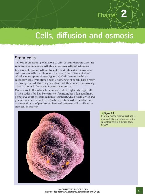
Stem cells<br />
Our bodies are made up of millions of cells, of many different kinds. Yet<br />
each began as just a single cell. How do all these different cells arise?<br />
In a tiny embryo, each cell has the ability to divide <strong>and</strong> form new cells,<br />
<strong>and</strong> these new cells are able to turn into any of the different kinds of<br />
cells that make up your body (Figure 2.1). <strong>Cells</strong> that can do this are<br />
called stem cells. By the time a baby is born, most of its cells have already<br />
become specialised. Once they have done that, they cannot turn into any<br />
other kind of cell. They are not stem cells any more.<br />
Doctors would like to be able to use stem cells to replace damaged cells<br />
in their patients’ bodies. For example, if someone has a damaged heart,<br />
perhaps we could put stem cells into their heart, which would divide <strong>and</strong><br />
produce new heart muscle cells. In theory, this should be possible, but<br />
there are still a lot of problems to be solved before we will be able to use<br />
stem cells in this way.<br />
UNCORRECTED PROOF COPY<br />
Downloaded from www.pearsonIS.com/HeinemannIGCSE<br />
Figure 2.1<br />
In a tiny human embryo, each cell is<br />
able to divide to produce any of the<br />
specialised cells in a human body.<br />
(3000)<br />
17
18<br />
nucleus<br />
cell surface<br />
membrane<br />
cytoplasm<br />
DNA inside<br />
the nucleus<br />
magnification 3500<br />
Figure 2.2<br />
A section through an animal cell. This<br />
is a liver cell.<br />
chloroplast<br />
containing<br />
chlorophyll<br />
cytoplasm<br />
DNA inside<br />
the nucleus<br />
nucleus<br />
large sap<br />
vacuole<br />
Activity 2.1<br />
Looking at animal cells.<br />
Activity 2.2<br />
Looking at plant cells.<br />
cell surface<br />
membrane<br />
cell wall made<br />
of cellulose<br />
magnification 1000<br />
Figure 2.3<br />
A section through a plant cell. This is a<br />
palisade cell from a leaf.<br />
2.1 <strong>Cells</strong><br />
All living organisms are made up of cells. Bacteria have just one cell. Plants<br />
<strong>and</strong> animals are made up of millions of cells. You probably contain around<br />
one million million cells.<br />
<strong>Cells</strong> are very small, so you can only see them using a microscope.<br />
Animal cells<br />
If you are able to do Investigation 2.1, you will be able to see some animal<br />
cells. They will probably look something like the cell shown in Figure 2.2.<br />
All animal cells have cytoplasm <strong>and</strong> a cell surface membrane which<br />
completely surrounds the cell. Most animal cells also have a nucleus. Red<br />
blood cells, however, are unusual <strong>and</strong> do not have a nucleus.<br />
Plant cells<br />
If you are able to do Activity 2.2, you will be able to see some plant cells.<br />
They are often easier to see than animal cells, at least partly because they<br />
are often quite a bit larger.<br />
Figure 2.3 shows the kind of plant cell that you might find in a leaf. You<br />
can see that it has several structures that animal cells don’t have. These are<br />
a cell wall, chloroplasts <strong>and</strong> large vacuole containing cell sap.<br />
The parts of cells<br />
Each of the different parts of a cell has its own particular function (role).<br />
Structures found in both animal <strong>and</strong> plant cells<br />
All cells have cytoplasm. This is a jelly-like substance. It is mostly water<br />
– about 70% in many cells – with proteins <strong>and</strong> other chemicals dissolved<br />
in it. Many different metabolic reactions take place in the cytoplasm.<br />
All cells have a cell surface membrane which completely surrounds the cell<br />
<strong>and</strong> separates it from its environment (surroundings). This membrane<br />
controls what goes in <strong>and</strong> out of the cell. It will let some substances pass<br />
through, but not others, <strong>and</strong> so it is said to be partially permeable. Cell<br />
surface membranes are strong but very flexible.<br />
Most cells have a nucleus. This contains a chemical called DNA. The<br />
DNA is arranged in long threads called chromosomes. You will not have<br />
seen chromosomes in the cells you looked at under the microscope,<br />
<strong>and</strong> they are not shown in Figures 2.2 <strong>and</strong> 2.3. This is because when a<br />
cell is not dividing the chromosomes are very long <strong>and</strong> thin, <strong>and</strong> so are<br />
invisible even with a good school microscope. But when a cell divides, the<br />
chromosomes get shorter <strong>and</strong> fatter, making it much easier to see them.<br />
The DNA in the chromosomes contains coded instructions for how the<br />
cell should behave. It determines which proteins the cell should make,<br />
which in turn determines the characteristics of that cell <strong>and</strong> of the<br />
organism of which it is part.<br />
UNCORRECTED PROOF COPY<br />
Downloaded from www.pearsonIS.com/HeinemannIGCSE
Structures found in plant cells only<br />
All plant cells, but never animal cells, have a cell wall outside their cell<br />
surface membrane. This cell wall is made of cellulose. Unlike the cell<br />
surface membrane, the cell wall does not control what goes through it, <strong>and</strong><br />
so it is said to be fully permeable. The function of the cell wall is to hold<br />
the plant cell in shape <strong>and</strong> to stop it bursting when it takes up a lot of water.<br />
Many plant cells in the leaves have green structures called chloroplasts.<br />
These are found in the cytoplasm. The chloroplasts are green because they<br />
contain a green pigment, chlorophyll. Chlorophyll absorbs energy from<br />
sunlight, <strong>and</strong> helps the chloroplast to use this energy to make sugars. This<br />
is called photosynthesis. Animal cells do not feed by photosynthesis, <strong>and</strong><br />
they do not have chloroplasts.<br />
Plant cells often contain a large, liquid-filled space called a vacuole. The<br />
vacuole is surrounded by a membrane which keeps its contents separate<br />
from the cytoplasm. The liquid inside the vacuole is called cell sap. It is<br />
mostly water, with sugars, amino acids <strong>and</strong> other substances dissolved<br />
in it. It is a storage area for the plant cell. Animal cells often have small<br />
vacuoles, but they are hardly ever as large as the vacuole in a plant cell,<br />
<strong>and</strong> they do not contain cell sap.<br />
QUESTION<br />
2.1 Make a comparison table to summarise the similarities <strong>and</strong><br />
differences between plant <strong>and</strong> animal cells. Draw a table like this:<br />
Structure Is it found in<br />
animal cells?<br />
Is it found in<br />
plant cells?<br />
Comment<br />
You will need six rows, one for each of: cell surface membrane,<br />
cytoplasm, nucleus, cell wall, chloroplasts <strong>and</strong> vacuole. Make the<br />
‘comment’ column wider than the others, so that you have room to<br />
write plenty of information in it.<br />
2.2 How cells are organised<br />
Some specialised cells<br />
Although all animal cells are similar to each other, <strong>and</strong> all plant cells are<br />
similar to each other, they are not all exactly the same. Different cells have<br />
different functions, <strong>and</strong> they often have adaptations that help them to<br />
carry out these functions.<br />
Root hair cells<br />
Root hair cells (Figure 2.4) are specialised plant cells. They are found<br />
on the outside of plant roots, just a little way up from the root tip. Their<br />
functions are to help to anchor the plant in the soil, <strong>and</strong> to absorb water<br />
<strong>and</strong> mineral ions from the soil.<br />
UNCORRECTED PROOF COPY<br />
Downloaded from www.pearsonIS.com/HeinemannIGCSE<br />
nucleus<br />
<strong>Cells</strong>, <strong>diffusion</strong> <strong>and</strong> <strong>osmosis</strong><br />
root hair<br />
cell wall cell surface<br />
membrane<br />
large vacuole<br />
containing cell sap<br />
Figure 2.4<br />
Root hair cells on a plant root.<br />
cytoplasm<br />
19
20<br />
Figure 2.5<br />
Xylem elements in a plant.<br />
magnification 3000<br />
cilia<br />
nucleus<br />
Figure 2.6<br />
Ciliated cells that make up the lining<br />
of the trachea.<br />
Each root hair cell has a long thin part reaching out into the soil. This gives<br />
the cell a much larger surface area than most cells. The large surface area<br />
means that a lot of water <strong>and</strong> mineral ions can get into the cell very quickly.<br />
Xylem cells<br />
Xylem cells are another type of specialised plant cell. They are perhaps the<br />
strangest of all, because they are completely empty <strong>and</strong> dead. They began<br />
as a normal, living plant cell, but then their cell walls gradually filled up<br />
with a substance called lignin. This is strong <strong>and</strong> waterproof. The contents<br />
of the cell gradually died, so that it ended up as a hollow tube with a wall<br />
containing lignin as well as cellulose.<br />
cross-section<br />
xylem vessel<br />
lignin<br />
no cytoplasm<br />
longitudinal section<br />
An individual xylem cell is called a xylem vessel element (Figure 2.5).<br />
Each one has completely lost its end walls. Lots of these cells are arranged<br />
end to end, forming a long, continuous tube that can run all the way from<br />
the roots, up the stem <strong>and</strong> into the leaves of the plants. These tubes are<br />
called xylem vessels. The veins in a leaf contain many xylem vessels.<br />
Xylem vessels have two functions. The first is to transport water <strong>and</strong><br />
mineral ions from the roots to all the other parts of the plant. The second<br />
is to help to support the plant. The lignin in their walls is stiff <strong>and</strong> strong.<br />
You can feel the stiffness of the veins in a plant’s leaves. The wood in trees<br />
is also made of xylem vessels, so wood is mostly lignin.<br />
Ciliated cells<br />
Ciliated cells are specialised animal cells. We have ciliated cells in the lining<br />
of our trachea (windpipe) <strong>and</strong> bronchi (Figure 2.6).<br />
Cilia are tiny extensions of the cell, covered with a cell surface membrane<br />
just like the rest of the cell. Cilia can move, <strong>and</strong> all the cilia on a cell <strong>and</strong> its<br />
neighbours beat together in a rhythmic way so that they look rather like<br />
a field of long grass with the wind sweeping over it. They help to sweep<br />
mucus up the bronchi <strong>and</strong> the trachea towards the back of the throat,<br />
where you swallow it. The mucus traps bacteria <strong>and</strong> dirt particles in the<br />
air that you breathe in, so the cilia are helping to keep this out of your<br />
lungs. You can read more about this on page 000.<br />
Muscle cells<br />
Muscle cells are found in many different animals, including humans. The<br />
cells shown in Figure 2.7 could be found in the biceps muscle in your arm.<br />
UNCORRECTED PROOF COPY<br />
Downloaded from www.pearsonIS.com/HeinemannIGCSE
Like all animal cells, muscle cells have a cell surface membrane, cytoplasm<br />
<strong>and</strong> a nucleus – but each muscle cell has many nuclei rather than just one.<br />
They look striped because they are made up of many str<strong>and</strong>s of protein<br />
arranged in a pattern. The str<strong>and</strong>s of protein can slide between each other,<br />
making the cell much shorter. This is called contraction. A lot of energy is<br />
needed for muscle cells to contract.<br />
Red blood cells<br />
Red blood cells are smaller than almost every other kind of cell in the<br />
human body. They have a cell surface membrane <strong>and</strong> cytoplasm, but<br />
no nucleus (Figure 2.8). The cytoplasm is full of a red protein called<br />
haemoglobin . Haemoglobin carries oxygen from your lungs to all the<br />
other parts of your body.<br />
Being very small enables red blood cells to squeeze through the tiniest of<br />
blood capillaries, taking oxygen as close as possible to each cell that needs<br />
it. Red blood cells are circular <strong>and</strong> have a dent in the middle, which gives<br />
them a large surface area. This speeds up the rate at which oxygen can<br />
move into <strong>and</strong> out of them.<br />
QUESTION<br />
2.2 Figure 2.9 is an annotated diagram of a root hair cell. An annotated<br />
diagram explains something about the functions of what is drawn, as<br />
well as its structure.<br />
nucleus<br />
containing<br />
DNA<br />
large<br />
vacuole<br />
containing<br />
cell sap<br />
cytoplasm<br />
cytoplasm cell surface membrane nucleus<br />
protein str<strong>and</strong>s<br />
Cell wall made of cellulose.<br />
This is fully permeable,<br />
allowing water <strong>and</strong> mineral<br />
ions to move freely into the<br />
cell from the soil.<br />
Cell surface membrane.<br />
This is partially<br />
permeable, allowing<br />
water to move into the<br />
cell by <strong>osmosis</strong>, but<br />
keeping proteins <strong>and</strong><br />
other substances inside<br />
the cell. Mineral ions<br />
pass from the soil into the<br />
cell by active transport.<br />
The long projection of the root hair cell increases its<br />
surface area, which speeds up the rate at which water<br />
<strong>and</strong> mineral ions can move into the cell.<br />
Using Figure 2.9 as a starting point, make annotated diagrams of a<br />
xylem vessel, a muscle cell, a red blood cell <strong>and</strong> a ciliated cell. Your<br />
annotations should explain how the special features of each cell help<br />
it to perform its functions.<br />
UNCORRECTED PROOF COPY<br />
Downloaded from www.pearsonIS.com/HeinemannIGCSE<br />
<strong>Cells</strong>, <strong>diffusion</strong> <strong>and</strong> <strong>osmosis</strong><br />
Figure 2.7<br />
A muscle cell.<br />
cell surface membrane<br />
cytoplasm containing<br />
haemoglobin but no<br />
nucleus<br />
cross-section<br />
Figure 2.8<br />
Red blood cells.<br />
cell surface membrane<br />
cytoplasm containing<br />
haemoglobin<br />
Figure 2.9<br />
An annotated drawing of a root hair<br />
cell.<br />
21
22<br />
Figure 2.10<br />
A plant leaf, an example of an organ.<br />
Tissues, organs <strong>and</strong> organ systems<br />
A tissue is a group of similar cells that all work together to perform the<br />
same function. For example, the layer of cells lining the inside of your<br />
cheeks is a tissue. The layer of cells you peeled from the inside of a piece of<br />
onion, when you looked at plant cells under the microscope, is a tissue. A<br />
group of muscle cells makes up muscle tissue, <strong>and</strong> a group of xylem cells<br />
makes up xylem tissue.<br />
Different kinds of tissues are often arranged together to make an organ.<br />
An organ is a group of different tissues which work together to perform<br />
a particular function. For example, the brain <strong>and</strong> the kidneys are organs,<br />
<strong>and</strong> so is a leaf.<br />
Figure 2.10 shows a leaf, which is an example of a plant organ. Leaves<br />
have many functions, including making sugars by photosynthesis.<br />
This is done by cells inside the leaf, in tissues called the palisade<br />
mesophyll <strong>and</strong> spongy mesophyll. These cells need a supply of water,<br />
which is brought to them in xylem vessels. Some of the sugar that they<br />
make is carried to other parts of the plant inside phloem tubes. The<br />
spongy mesophyll cells are arranged loosely, with air spaces in between<br />
them. Thin layers of cells on the top <strong>and</strong> bottom of the leaf, called<br />
the epidermis, let light through to the mesophyll cells but stop too<br />
much water vapour leaving the leaf, so that it does not dry out. Small<br />
openings in the lower epidermis, called stomata, allow gases to move in<br />
<strong>and</strong> out of the leaf.<br />
spongy mesophyll<br />
palisade mesophyll<br />
lower epidermis<br />
Organs do not work on their own. Most organs work with other organs to<br />
help each other to perform particular functions. These organs form organ<br />
systems. An organ system can be defined as:<br />
a group of organs with related functions, working together to perform<br />
body functions.<br />
For example, the brain <strong>and</strong> the eye are both parts of the nervous system.<br />
The lungs <strong>and</strong> trachea are parts of the gas exchange system. The stomach<br />
<strong>and</strong> intestines are parts of the digestive system.<br />
QUESTION<br />
upper epidermis<br />
vein containing<br />
xylem tissue <strong>and</strong><br />
phloem tissue<br />
2.3 Using the index, look up the nervous system, gas exchange system<br />
<strong>and</strong> digestive system. Make a list of the organs in each of these three<br />
organ systems.<br />
UNCORRECTED PROOF COPY<br />
Downloaded from www.pearsonIS.com/HeinemannIGCSE
2.3 Magnification of drawings <strong>and</strong><br />
micrographs<br />
Many of the things that biologists study are quite small. When we make<br />
drawings of them, we often draw them larger than they really are.<br />
For example, Figure 2.11 is a photograph of a leaf, shown life size.<br />
Figure 2.12 is a diagram of the leaf. The diagram has been drawn larger<br />
than the actual leaf.<br />
We can calculate the magnification of the diagram.<br />
size of drawing<br />
magnification 5 ______________<br />
size of specimen<br />
Measure the length of the real leaf. You should find that it is _____ mm<br />
long. Now measure the same distance on the drawing of the leaf. You<br />
should find that it is _____ mm long.<br />
So the magnification of the drawing is:<br />
_____ mm<br />
_________ 5 3_____<br />
_____ mm<br />
Notice how we write the magnification: 3...<br />
UNCORRECTED PROOF COPY<br />
Downloaded from www.pearsonIS.com/HeinemannIGCSE<br />
<strong>Cells</strong>, <strong>diffusion</strong> <strong>and</strong> <strong>osmosis</strong><br />
Figure 2.11<br />
Scan of a leaf<br />
Figure 2.12<br />
Simple diagram of the leaf shown in<br />
Figure 2.10.<br />
23
start here<br />
24<br />
Activity 2.3<br />
Calculating magnifications<br />
Figure 2.13<br />
A particle in a liquid or a gas moves<br />
around r<strong>and</strong>omly. When it bumps into<br />
another particle, it changes direction.<br />
QUESTION<br />
2.4 A spider is 24 mm long. A drawing of the spider is 88 mm long.<br />
Calculate the magnification of the drawing, showing your working<br />
clearly.<br />
2.4 Movement into <strong>and</strong> out of cells<br />
Diffusion<br />
All substances are made up of tiny particles called atoms. In some<br />
substances, these atoms have lost or gained one or more electrons, to<br />
become ions. In other substances, the atoms are grouped together to form<br />
molecules.<br />
Atoms, ions <strong>and</strong> molecules are always moving. In a solid, each particle<br />
has a fixed position <strong>and</strong> it just vibrates in this position. In a liquid, the<br />
particles move more freely around each other, but stay in fairly close<br />
contact. In a gas, the particles are much further apart <strong>and</strong> move around<br />
freely.<br />
Imagine the lid being taken off a bottle of ammonia solution. Molecules of<br />
ammonia gas move out of the bottle. Each molecule moves r<strong>and</strong>omly – it<br />
is just as likely to go in one direction as another. The ammonia molecules<br />
bump into one another, <strong>and</strong> into other molecules in the air, such as<br />
oxygen <strong>and</strong> nitrogen molecules.<br />
When one molecule hits another, both of them change course. Figure 2.13<br />
shows how one ammonia molecule might move around. Each change in<br />
direction happens when the molecule bumps into another one.<br />
When the lid of the ammonia bottle is first taken off, there are a lot<br />
of ammonia molecules inside the bottle. We say that there is a high<br />
concentration of ammonia inside the bottle. There are probably almost<br />
no ammonia molecules in the air in the room – so here there is a low<br />
concentration of ammonia molecules.<br />
As the ammonia molecules bump r<strong>and</strong>omly around, some of them move<br />
erratically further <strong>and</strong> further away from the bottle. After a while, some<br />
will have moved right into the far corner of the room. The ammonia<br />
molecules have diffused across the room.<br />
It is important to realise that the ammonia molecules do not head<br />
purposefully across the room from the bottle. Each molecule just bumps<br />
r<strong>and</strong>omly around. It is by chance that some of them end up a long<br />
way from the bottle. Some of them might even go back into the bottle.<br />
But, after a while, these r<strong>and</strong>om movements result in their being more<br />
ammonia molecules out in the room, <strong>and</strong> fewer inside the bottle. After a<br />
long time, you would probably end up with ammonia molecules spread<br />
evenly all over the room.<br />
The overall or net result of <strong>diffusion</strong> is that particles spread out<br />
evenly. They tend to spread out from a place where they are in a high<br />
UNCORRECTED PROOF COPY<br />
Downloaded from www.pearsonIS.com/HeinemannIGCSE
concentration <strong>and</strong> into a place where they are in a low concentration. We<br />
say that they spread out down a concentration gradient. Diffusion can<br />
defined as:<br />
the net movement of molecules from a region of their higher<br />
concentration, to a region of their lower concentration down a<br />
concentration gradient, as result of their r<strong>and</strong>om movement<br />
Diffusion <strong>and</strong> living organisms<br />
Diffusion is very important to all living organisms, including humans.<br />
You will meet several examples as you continue your biology course. For<br />
the moment, we’ll look at one example of <strong>diffusion</strong> in animals, <strong>and</strong> one in<br />
plants.<br />
Gas exchange in human lungs<br />
Figure 2.14 shows a tiny part of the human lungs, greatly magnified. In the<br />
lungs, oxygen diffuses from the little air sacs (alveoli) into the blood. It is<br />
carried away inside the red blood cells.<br />
oxygen diuses through<br />
alveolus wall, through<br />
capillary wall, <strong>and</strong> into a<br />
red blood cell<br />
blood capillary<br />
blood<br />
thin wall of<br />
capillary<br />
thin cell of<br />
alvelous wall<br />
red blood cell<br />
oxygen diuses<br />
into alveolus<br />
The lungs are made up of hundreds of thous<strong>and</strong>s of tiny alveoli. Each<br />
alveolus has tiny blood vessels called capillaries wrapped closely around<br />
it. These contain blood, which contains red blood cells.<br />
Oxygen molecules in the air inside the alveoli move r<strong>and</strong>omly around.<br />
Some of them go right through the cells making up the wall of the<br />
alveolus <strong>and</strong> the wall of the blood capillary, right through the blood<br />
plasma, <strong>and</strong> through the cell surface membrane of a red blood cell. Here,<br />
they combine with haemoglobin.<br />
alveolus<br />
UNCORRECTED PROOF COPY<br />
Downloaded from www.pearsonIS.com/HeinemannIGCSE<br />
<strong>Cells</strong>, <strong>diffusion</strong> <strong>and</strong> <strong>osmosis</strong><br />
Activity 2.4<br />
How quickly does ammonia<br />
diffuse?<br />
Figure 2.14<br />
In the lungs oxygen diffuses from an<br />
alveolus into red blood cells.<br />
air<br />
25
26<br />
dilute sugar<br />
solution<br />
Activity 2.3<br />
Can iodine <strong>and</strong> starch molecules<br />
get through Visking tubing?<br />
concentrated<br />
sugar solution<br />
partially<br />
permeable<br />
membrane<br />
water molecules can move through the<br />
holes in the membrane but sugar<br />
molecules can not<br />
sugar<br />
molecule<br />
water<br />
molecule<br />
Figure 2.15<br />
How <strong>osmosis</strong> happens. The net<br />
movement of the water molecules<br />
is from the less concentrated<br />
solution on the left-h<strong>and</strong> side of the<br />
membrane to the more concentrated<br />
solution on the right-h<strong>and</strong> side.<br />
There are usually a lot more oxygen molecules in the air inside the<br />
alveolus than there are in the blood inside the capillary. So, although some<br />
oxygen molecules will travel from the capillary <strong>and</strong> into the alveolus, as a<br />
result of their r<strong>and</strong>om movements, there will be many more travelling in<br />
the opposite direction. The net movement of oxygen is from the alveolus<br />
<strong>and</strong> into the capillary. The oxygen moves down a concentration gradient,<br />
by <strong>diffusion</strong>.<br />
Gas exchange in a plant leaf<br />
In daylight, palisade mesophyll <strong>and</strong> spongy mesophyll cells in a leaf<br />
photosynthesise. They use carbon dioxide <strong>and</strong> water to make sugars.<br />
These sugars may then be turned into starch in the leaf. Where does the<br />
carbon dioxide come from, <strong>and</strong> how does it get to these cells?<br />
Air contains carbon dioxide, though there is not very much of it – only<br />
about 0.04% of the air is carbon dioxide. These carbon dioxide molecules<br />
bump r<strong>and</strong>omly around. Some of them, just by chance, will go through<br />
the stomata on the underside of a leaf, into the air spaces inside the leaf,<br />
into a mesophyll cell <strong>and</strong> into a chloroplast.<br />
Carbon dioxide molecules arriving in a chloroplast can be made into<br />
sugar. This keeps the concentration of carbon dioxide molecules inside<br />
the chloroplasts much lower than in the air outside the leaf. So there<br />
is a concentration gradient from the air into the chloroplast. The net<br />
movement of the carbon dioxide molecules is down this concentration<br />
gradient. They move into the leaf by <strong>diffusion</strong>.<br />
Osmosis<br />
Osmosis is a special kind of <strong>diffusion</strong>, involving water molecules. Figure 2.15<br />
shows how it happens.<br />
This diagram shows a partially permeable membrane separating two sugar<br />
solutions. The membrane is partially permeable because it has tiny holes<br />
in it.<br />
The sugar solutions are mixtures of water molecules <strong>and</strong> sugar molecules.<br />
Sugar molecules are much larger than water molecules. The water<br />
molecules can get through the holes in the membrane, but the sugar<br />
molecules are too big.<br />
The sugar solution on the left is a dilute solution. It does not contain<br />
much sugar. The sugar solution on the right is a concentrated solution,<br />
containing a lot more sugar. There are the same number of water<br />
molecules on both sides of the membrane but there are more ‘free’ water<br />
molecules in the dilute sugar solution.<br />
On both sides of the membrane, the water molecules <strong>and</strong> sugar molecules<br />
bounce around r<strong>and</strong>omly. If a sugar molecule hits the membrane, it just<br />
bounces off. But if a water molecule bounces into a hole in the membrane,<br />
it can shoot through to the other side.<br />
Because there are more ‘free’ water molecules on the left-h<strong>and</strong> side, more<br />
water molecules will go from left to right through the membrane than in<br />
the opposite direction. Although water molecules do go both ways, the net<br />
movement is from left to right.<br />
UNCORRECTED PROOF COPY<br />
Downloaded from www.pearsonIS.com/HeinemannIGCSE
S<br />
Because there are more ‘free’ water molecules on the left-h<strong>and</strong> side we say<br />
that there is a higher water potential on the left-h<strong>and</strong> side of the membrane<br />
than on the right. Concentrated solutions have a lower water potential than<br />
dilute solutions. The net movement of water molecules is down their water<br />
potential gradient, from high water potential to low water potential.<br />
You can probably see that <strong>osmosis</strong> is just like <strong>diffusion</strong>. The only thing<br />
that is different is that we have a membrane in the way, which lets the<br />
water molecules diffuse but stops the sugar molecules from diffusing. This<br />
sort of <strong>diffusion</strong> is called <strong>osmosis</strong>. We can define <strong>osmosis</strong> as:<br />
S<br />
the <strong>diffusion</strong> of water molecules from a region of higher concentration<br />
(dilute solution) to a region of their lower concentration (concentrated<br />
solution), through a partially permeable membrane.<br />
In terms of water potential, we can diffuse <strong>osmosis</strong> as:<br />
the net <strong>diffusion</strong> of water molecules from a region of high water<br />
potential to a region of low water potential, through a partially<br />
permeable membrane.<br />
Osmosis <strong>and</strong> animal cells<br />
Figure 2.16 shows an animal cell in pure water.<br />
The solutions inside <strong>and</strong> outside the cell are separated by the cell surface<br />
membrane. This is a partially permeable membrane. Water molecules pass<br />
easily through it, but larger molecules cannot get through.<br />
Inside the cell, the cytoplasm is a fairly concentrated solution of proteins <strong>and</strong><br />
other substances in water. Outside the cell, there is pure water. So the water<br />
outside the cell has more ‘free’ water molecules than the solution inside the cell.<br />
S<br />
The tap water outside the cell has a higher water potential than the<br />
solution in the cytoplasm insdie thew cell. Therefore, the water molecules<br />
diffuse down the water potential gradient into the cell.<br />
The water therefore diffuses into the cell. The r<strong>and</strong>om movements of the<br />
water molecules result in more of them moving into the cell than move<br />
out of it. The water moves into the cell by <strong>osmosis</strong>.<br />
What will happen to the cell? As more <strong>and</strong> more water enters the cell, the<br />
cell swells up. After a while, it may get so big that is bursts the cell surface<br />
membrane. The contents of the cell escape, <strong>and</strong> the cell dies.<br />
S<br />
QUESTIONS<br />
2.5 Figure 2.17 shows an animal cell in a concentrated sugar solution.<br />
The sugar solution is much more concentrated than the solution<br />
inside the cell.<br />
a) Which has the higher water potential – the sugar solution or the<br />
cytoplasm?<br />
b) In which direction will the net movement of water take place?<br />
c) What do you think the sugar molecules do?<br />
d) Will the cell get bigger or smaller?<br />
UNCORRECTED PROOF COPY<br />
Downloaded from www.pearsonIS.com/HeinemannIGCSE<br />
<strong>Cells</strong>, <strong>diffusion</strong> <strong>and</strong> <strong>osmosis</strong><br />
Activity 2.6<br />
Using Visking tubing to<br />
investigate <strong>osmosis</strong><br />
concentrated solution<br />
of proteins <strong>and</strong> other<br />
substances in the cytoplasm<br />
lower water potential<br />
<strong>diffusion</strong> of water<br />
by <strong>osmosis</strong><br />
Figure 2.16<br />
An animal cell in pure water.<br />
solution of proteins <strong>and</strong> other<br />
sunstances in the cytoplasm<br />
higher water potential<br />
tap water<br />
higher water<br />
potential<br />
cell surface<br />
membrane<br />
a partially<br />
permeable<br />
membrane<br />
very concentrated sugar<br />
solution outside<br />
lower water potential<br />
Figure 2.17<br />
An animal cell in a concentrated sugar<br />
solution.<br />
27
28<br />
solution in cytoplasm<br />
<strong>and</strong> vacuole<br />
lower water potential<br />
<strong>diffusion</strong> of water<br />
by <strong>osmosis</strong> tap water<br />
outside<br />
higher water potential<br />
Figure 2.18<br />
A plant cell in pure water.<br />
cell surface<br />
membrane<br />
cell wall<br />
2.6 A scientist took a sample of human blood, <strong>and</strong> divided it between<br />
three tubes. She then added liquid to each tube, as follows:<br />
• tube A distilled water<br />
• tube B salt solution of the same concentration as the cytoplasm of<br />
the red blood cells<br />
• tube C very concentrated salt solution.<br />
She mixed the contents of each tube very thoroughly. After a few<br />
minutes, she observed the following:<br />
• tube A a clear red solution<br />
• tube B a red cloudy liquid<br />
• tube C a red cloudy liquid.<br />
She then took a few drops from each tube, put them onto microscope<br />
slides <strong>and</strong> looked at them using a microscope.<br />
a) Name the substance that makes blood look red.<br />
b) If you look at blood through a microscope, you see red cells<br />
floating in a colourless liquid called plasma. Why are the blood cells<br />
red?<br />
c) When blood is in a test tube, it looks cloudy because of the cells<br />
floating in the plasma. Suggest why the contents of tubes B <strong>and</strong> C<br />
stayed cloudy, but the contents of tube A went clear.<br />
d) Describe <strong>and</strong> explain what happened to the cells in each tube.<br />
Suggest what the scientist would have seen when she looked at<br />
each sample using a microscope.<br />
Osmosis <strong>and</strong> plant cells<br />
Figure 2.18 shows a plant cell in pure water. As in the animal cell (Figure<br />
2.16), the cell surface membrane, which is partially permeable, separates<br />
the concentrated solution inside the cell from the pure water outside.<br />
Water therefore moves into the cell by <strong>osmosis</strong> as the water molecules<br />
move from the less concentrated solution to the more concentrated one.<br />
S<br />
Here there is a solution with a high water potential outside the cell <strong>and</strong> a<br />
solution with a lower water potential inside the cell. These solutions are<br />
separated by the partially permeable cell surface membrane. Water moves<br />
by <strong>osmosis</strong> into the cell, from the solution with the higher water potential<br />
to the solution with the lower water potential.<br />
However, this time the cell does not burst. This is because the plant cell<br />
has a strong outer covering – its cell wall. As more <strong>and</strong> more water goes<br />
into the cell, the cytoplasm <strong>and</strong> vacuole get bigger <strong>and</strong> bigger, <strong>and</strong> push<br />
outwards on the cell surface membrane <strong>and</strong> the cell wall. But the cell wall<br />
will not give way. It prevents the cell from exp<strong>and</strong>ing enough to break the<br />
cell surface membrane.<br />
A plant cell like this, as full as it can be, is very firm <strong>and</strong> rigid. It is said to<br />
be turgid. The cells of a well-watered plant are all turgid. This helps to<br />
keep the soft parts of a plant, such as its leaves <strong>and</strong> flower petals, firm <strong>and</strong><br />
in shape. Turgidity helps to support the plant.<br />
UNCORRECTED PROOF COPY<br />
Downloaded from www.pearsonIS.com/HeinemannIGCSE
S<br />
QUESTION<br />
2.7 Look back at Figure 2.4, which shows a root hair cell.<br />
Imagine the root hair cell is growing between soil particles. There is a<br />
dilute solution, containing a lot of water, in between the soil particles.<br />
a) Which has the higher water potential – the dilute solution<br />
between the soil particles, or the more concentrated solution<br />
inside the root hair cell?<br />
b) In which direction will the water move by <strong>osmosis</strong>?<br />
c) Where is the partially permeable membrane through which the<br />
water moves?<br />
d) Imagine that someone gives the plant too much fertiliser. The<br />
salts in the fertiliser dissolve in the water in the soil. Now the<br />
concentration of the solution in the soil is much greater than the<br />
concentration inside the root hair cell. What will happen?<br />
Figure 2.19 shows a plant cell in a concentrated solution. The wtaer<br />
molecules diffuse out of the cell. Water moves out of the cell by <strong>osmosis</strong>.<br />
S<br />
cytoplasm <strong>and</strong> cell sap<br />
higher water potential<br />
<strong>diffusion</strong> of<br />
water by<br />
<strong>osmosis</strong><br />
The solution outside the cell has a lower water potential than the solution<br />
inside it. The water molecules move by <strong>osmosis</strong> from the higher water<br />
potential to the lower one, so they move out of the cell.<br />
Just as in an animal cell, this makes the contents of the cell shrink. As the<br />
cell loses more <strong>and</strong> more water, the cytoplasm <strong>and</strong> vacuole get smaller <strong>and</strong><br />
smaller. They stop pushing outwards on the cell wall. Instead of being firm<br />
<strong>and</strong> stiff, the cell becomes soft. It is said to be flaccid.<br />
If water keeps on going out of the cell, <strong>and</strong> the cytoplasm keeps on<br />
shrinking, the cell surface membrane will eventually be pulled away from<br />
the cell wall. When this happens, the cell is said to be plasmolysed.<br />
QUESTION<br />
cell surface<br />
membrane<br />
concentrated sugar<br />
solution<br />
lower water potential<br />
cell wall<br />
cell surface<br />
membrane<br />
<strong>diffusion</strong> of<br />
water by<br />
<strong>osmosis</strong><br />
2.8 Look at Figure 2.19.<br />
a) Is the cell wall of a plant cell partially permeable, or is it fully<br />
permeable? What does this mean?<br />
b) Use your answer to (a) to predict what will be in space X.<br />
UNCORRECTED PROOF COPY<br />
Downloaded from www.pearsonIS.com/HeinemannIGCSE<br />
<strong>Cells</strong>, <strong>diffusion</strong> <strong>and</strong> <strong>osmosis</strong><br />
space X Figure 2.19<br />
A plant cell in a concentrated<br />
solution.<br />
Activity 2.7<br />
Osmosis <strong>and</strong> potato cells<br />
29
30<br />
Table 2.1<br />
Number of plasmolysed cells in<br />
various concentrations of sugar<br />
solution.<br />
EXPERIMENTAL SKILLS AND INVESTIGATIONS<br />
A student decided to test this hypothesis:<br />
<strong>Cells</strong> in onion tissue immersed in a concentrated sugar solution<br />
will become plasmolysed, but if they are immersed in a dilute sugar<br />
solution they will not become plasmolysed.<br />
She took an onion <strong>and</strong> cut 6 equal-sized pieces of epidermis. She<br />
immediately placed each piece into a drop of distilled water or sugar<br />
solution on a microscope slide. Then she lowered a cover slip onto each<br />
drop <strong>and</strong> left the slides for 15 minutes.<br />
She then looked at each slide in turn under the microscope. She focussed<br />
on a small area, <strong>and</strong> counted how many of the cells she could see were<br />
plasmolysed, <strong>and</strong> how many were not plasmolysed. She repeated this<br />
in two more areas on the same slide. She kept counting until she had<br />
counted a total of 100 cells on each slide.<br />
Table 2.1 shows her results.<br />
Concentration<br />
of sugar<br />
solution/%<br />
Number of<br />
plasmolysed<br />
cells out of<br />
100<br />
0 (distilled<br />
water)<br />
a) Display these results as a line graph.<br />
0.5 1.0 1.5 2.0 2.5<br />
0 0 10 75 98 100<br />
b) Explain why no cells were plasmolysed in a 0.5% sugar solution.<br />
c) Explain why all the cells were plasmolysed in a 2.5% sugar solution.<br />
d) Suggest why only some of the cells were plasmolysed in the 1.0%<br />
solution or the 1.5% solution.<br />
e) Do the results support the student’s hypothesis? Explain your answer.<br />
f) State three factors that the student should have kept the same for all<br />
of the tissue samples.<br />
g) Which of the following could have been serious sources of error in the<br />
student’s investigation? Explain each of your answers.<br />
(i) cutting the pieces of epidermis not all exactly the same size<br />
(ii) counting the same cells more than once on the slides<br />
(iii) not leaving all of the pieces for exactly the same time before<br />
counting the plasmolysed cells<br />
h) Suggest two more possible sources of error (other than mistakes made<br />
by the student).<br />
i) Suggest two ways in which the student could make her results more<br />
reliable.<br />
UNCORRECTED PROOF COPY<br />
Downloaded from www.pearsonIS.com/HeinemannIGCSE
S<br />
Active transport<br />
Sometimes, a cell needs to absorb a substance that is in a very low<br />
concentration in its surroundings. For example, a plant needs to take in<br />
nitrate ions through its root hairs from the soil. But the concentration of<br />
nitrate ions dissolved in the water in the soil is usually much less than the<br />
concentration of nitrate ions inside the root hair cell. If left to themselves,<br />
the nitrate ions would diffuse down their concentration gradient, out of<br />
the cell <strong>and</strong> into the soil.<br />
The plant, however, can make the nitrate ions move in the opposite<br />
direction, up their concentration gradient. There are special protein<br />
molecules, called transport proteins, embedded in the cell surface<br />
membranes of the root hair cells. Nitrate ions from the soil bump into<br />
these proteins, <strong>and</strong> the proteins then push the ions through the membrane<br />
<strong>and</strong> into the cell. This needs energy, which the cell provides through<br />
respiration. The process is called active transport. We can defi ne active<br />
transport as:<br />
the movement of ions in or out of a cell through the cell membrane,<br />
from a region of their lower concentration to a region of their higher<br />
concentration, against a concentration gradient, using energy released<br />
by respiration.<br />
Another example of active transport is the uptake of glucose molecules<br />
from the small intestine into the blood. You can read about this on<br />
page 000.<br />
Summary<br />
Now that you have completed this chapter, you should be able to:<br />
• identify <strong>and</strong> describe the structure of plant cells <strong>and</strong> animal cells,<br />
<strong>and</strong> describe the functions of their parts<br />
• describe the differences between plant cells <strong>and</strong> animal cells<br />
• explain the structure <strong>and</strong> function of ciliated cells, root hair cells,<br />
xylem vessels, muscle cells <strong>and</strong> red blood cells<br />
• defi ne the terms tissue, organ <strong>and</strong> organ system, <strong>and</strong> give<br />
examples<br />
• calculate magnifi cation <strong>and</strong> size of biological diagrams<br />
• defi ne the term <strong>diffusion</strong>, <strong>and</strong> explain its importance in <strong>diffusion</strong><br />
of gases <strong>and</strong> solutes <strong>and</strong> give examples in living organisms<br />
• defi ne the term <strong>osmosis</strong> <strong>and</strong> explain its importance to animal <strong>and</strong><br />
plant cells, including the uptake of water by plant roots<br />
S<br />
• defi ne the term <strong>osmosis</strong>, using the term water potential, <strong>and</strong><br />
explain its importance to animal <strong>and</strong> plant cells, including the<br />
uptake of water by plant roots<br />
• defi ne the term active transport <strong>and</strong> discuss its importance to<br />
living cells<br />
UNCORRECTED PROOF COPY<br />
Downloaded from www.pearsonIS.com/HeinemannIGCSE<br />
<strong>Cells</strong>, <strong>diffusion</strong> <strong>and</strong> <strong>osmosis</strong><br />
31