

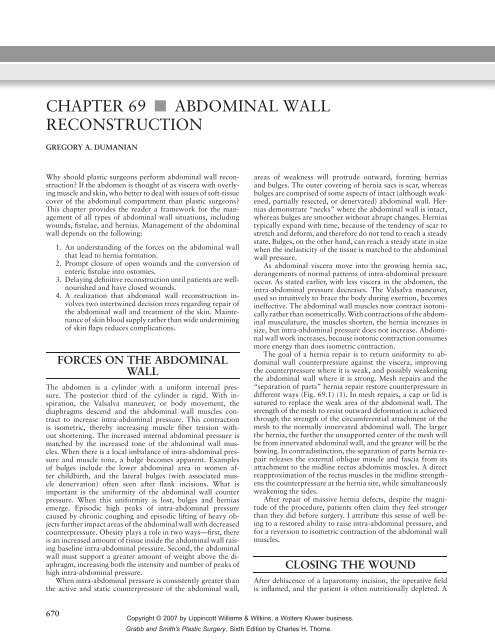
Chapter 69: Abdominal Wall Reconstruction - Gregory Dumanian ...
Chapter 69: Abdominal Wall Reconstruction - Gregory Dumanian ...
Chapter 69: Abdominal Wall Reconstruction - Gregory Dumanian ...

You also want an ePaper? Increase the reach of your titles
YUMPU automatically turns print PDFs into web optimized ePapers that Google loves.
CHAPTER <strong>69</strong> ■ ABDOMINAL WALL<br />
RECONSTRUCTION<br />
GREGORY A. DUMANIAN<br />
Why should plastic surgeons perform abdominal wall reconstruction?<br />
If the abdomen is thought of as viscera with overlying<br />
muscle and skin, who better to deal with issues of soft-tissue<br />
cover of the abdominal compartment than plastic surgeons?<br />
This chapter provides the reader a framework for the management<br />
of all types of abdominal wall situations, including<br />
wounds, fistulae, and hernias. Management of the abdominal<br />
wall depends on the following:<br />
1. An understanding of the forces on the abdominal wall<br />
that lead to hernia formation.<br />
2. Prompt closure of open wounds and the conversion of<br />
enteric fistulae into ostomies.<br />
3. Delaying definitive reconstruction until patients are wellnourished<br />
and have closed wounds.<br />
4. A realization that abdominal wall reconstruction involves<br />
two intertwined decision trees regarding repair of<br />
the abdominal wall and treatment of the skin. Maintenance<br />
of skin blood supply rather than wide undermining<br />
of skin flaps reduces complications.<br />
FORCES ON THE ABDOMINAL<br />
WALL<br />
The abdomen is a cylinder with a uniform internal pressure.<br />
The posterior third of the cylinder is rigid. With inspiration,<br />
the Valsalva maneuver, or body movement, the<br />
diaphragms descend and the abdominal wall muscles contract<br />
to increase intra-abdominal pressure. This contraction<br />
is isometric, thereby increasing muscle fiber tension without<br />
shortening. The increased internal abdominal pressure is<br />
matched by the increased tone of the abdominal wall muscles.<br />
When there is a local imbalance of intra-abdominal pressure<br />
and muscle tone, a bulge becomes apparent. Examples<br />
of bulges include the lower abdominal area in women after<br />
childbirth, and the lateral bulges (with associated muscle<br />
denervation) often seen after flank incisions. What is<br />
important is the uniformity of the abdominal wall counter<br />
pressure. When this uniformity is lost, bulges and hernias<br />
emerge. Episodic high peaks of intra-abdominal pressure<br />
caused by chronic coughing and episodic lifting of heavy objects<br />
further impact areas of the abdominal wall with decreased<br />
counterpressure. Obesity plays a role in two ways—first, there<br />
is an increased amount of tissue inside the abdominal wall raising<br />
baseline intra-abdominal pressure. Second, the abdominal<br />
wall must support a greater amount of weight above the diaphragm,<br />
increasing both the intensity and number of peaks of<br />
high intra-abdominal pressure.<br />
When intra-abdominal pressure is consistently greater than<br />
the active and static counterpressure of the abdominal wall,<br />
670<br />
areas of weakness will protrude outward, forming hernias<br />
and bulges. The outer covering of hernia sacs is scar, whereas<br />
bulges are comprised of some aspects of intact (although weakened,<br />
partially resected, or denervated) abdominal wall. Hernias<br />
demonstrate “necks” where the abdominal wall is intact,<br />
whereas bulges are smoother without abrupt changes. Hernias<br />
typically expand with time, because of the tendency of scar to<br />
stretch and deform, and therefore do not tend to reach a steady<br />
state. Bulges, on the other hand, can reach a steady state in size<br />
when the inelasticity of the tissue is matched to the abdominal<br />
wall pressure.<br />
As abdominal viscera move into the growing hernia sac,<br />
derangements of normal patterns of intra-abdominal pressure<br />
occur. As stated earlier, with less viscera in the abdomen, the<br />
intra-abdominal pressure decreases. The Valsalva maneuver,<br />
used so intuitively to brace the body during exertion, becomes<br />
ineffective. The abdominal wall muscles now contract isotonically<br />
rather than isometrically. With contractions of the abdominal<br />
musculature, the muscles shorten, the hernia increases in<br />
size, but intra-abdominal pressure does not increase. <strong>Abdominal</strong><br />
wall work increases, because isotonic contraction consumes<br />
more energy than does isometric contraction.<br />
The goal of a hernia repair is to return uniformity to abdominal<br />
wall counterpressure against the viscera, improving<br />
the counterpressure where it is weak, and possibly weakening<br />
the abdominal wall where it is strong. Mesh repairs and the<br />
“separation of parts” hernia repair restore counterpressure in<br />
different ways (Fig. <strong>69</strong>.1) (1). In mesh repairs, a cap or lid is<br />
sutured to replace the weak area of the abdominal wall. The<br />
strength of the mesh to resist outward deformation is achieved<br />
through the strength of the circumferential attachment of the<br />
mesh to the normally innervated abdominal wall. The larger<br />
the hernia, the further the unsupported center of the mesh will<br />
be from innervated abdominal wall, and the greater will be the<br />
bowing. In contradistinction, the separation of parts hernia repair<br />
releases the external oblique muscle and fascia from its<br />
attachment to the midline rectus abdominis muscles. A direct<br />
reapproximation of the rectus muscles in the midline strengthens<br />
the counterpressure at the hernia site, while simultaneously<br />
weakening the sides.<br />
After repair of massive hernia defects, despite the magnitude<br />
of the procedure, patients often claim they feel stronger<br />
than they did before surgery. I attribute this sense of well being<br />
to a restored ability to raise intra-abdominal pressure, and<br />
for a reversion to isometric contraction of the abdominal wall<br />
muscles.<br />
CLOSING THE WOUND<br />
After dehiscence of a laparotomy incision, the operative field<br />
is inflamed, and the patient is often nutritionally depleted. A<br />
Copyright © 2007 by Lippincott Williams & Wilkins, a Wolters Kluwer business.<br />
Grabb and Smith's Plastic Surgery, Sixth Edition by Charles H. Thorne.
Mesh Repair<br />
Hernia sac Mesh<br />
Rectus<br />
1. Side strength unchanged<br />
2. Adynamic central zone<br />
3. Circumferential pull by abdominal<br />
wall tightens mesh<br />
good strategy after such a surgical complication, is that the<br />
next procedure “had better work.” Early wound closure in<br />
the simplest manner possible has multiple benefits, including<br />
patient comfort, ease of wound care, and a decreased incidence<br />
of enterocutaneous fistulae (2). When devising a plan to close<br />
the wound, the following questions must be answered:<br />
1. Are the viscera “frozen,” and what are the chances for<br />
an evisceration?<br />
2. Do bowel contents need to be controlled?<br />
3. What are the location, size, and characteristics of the<br />
wound? Should the wound be modified to help achieve<br />
wound closure?<br />
Evisceration<br />
Little is written about evisceration. Patient management depends<br />
on why the abdominal wall lost integrity. Rarely, a<br />
purely technical problem of wound closure leads to a disruption<br />
of the suture line. Patients at exploration who have pristine<br />
wounds with minimal capillary leak, and can simply be reclosed.<br />
More commonly, the patient has an ileus, several days<br />
have elapsed since surgery, and the evisceration is proceeded<br />
by intra-abdominal fluid leaking through the skin suture line.<br />
These are contaminated wounds, and prone to further difficulties.<br />
Therefore, a minimalist approach to prevent a second<br />
evisceration and to control the infected soft tissues should be<br />
employed. As alluded to earlier, abdominal wall reconstructive<br />
surgery can be broken into two parts—the management of the<br />
abdominal wall and the management of the skin. For the abdominal<br />
wall, an absorbable polyglycolic acid mesh is placed<br />
using a running absorbable monofilament suture to prevent a<br />
second evisceration and to keep the viscera in their proper domain.<br />
For the skin, we have temporized the situation with a<br />
vacuum device, or in relatively clean situations, closed the skin<br />
over drains. Eventual skin closure is performed by delayed primary<br />
closure, skin grafts, or by secondary intention. When the<br />
skin gapes widely and several months are expected before closure<br />
by secondary intention, skin grafting provides the simplest<br />
and most reliable closure as is discussed below.<br />
Why not try to reclose the abdomen directly or with the<br />
aid of bioprosthetic mesh? In an infected field with swollen<br />
underlying bowel, the chance for failure is high. It is better<br />
to leave the abdominal muscles where they are and to patch<br />
the central defect. Mass closures with retention sutures can<br />
occasionally be successful, but hernias usually develop, and<br />
the sutures cause necrosis of the skin and medial aspects of the<br />
rectus muscles.<br />
<strong>Chapter</strong> <strong>69</strong>: AbdominaL <strong>Wall</strong> <strong>Reconstruction</strong> 671<br />
FIGURE <strong>69</strong>.1. Diagram of forces on the abdominal<br />
wall after a laparoscopic mesh repair.<br />
(Redrawn from <strong>Dumanian</strong> GA, Denham W.<br />
Comparison of repair techniques for major<br />
incisional hernias. Am J Surg. 2003;185:61,<br />
with permission.)<br />
Open Wounds after <strong>Abdominal</strong> Surgery<br />
Dehiscence of the abdominal closure is not uncommon after<br />
gastrointestinal surgery. Clues for fascial dehiscence include<br />
loose abdominal sutures at the base of the wound, a history of<br />
a “seroma” drained from underneath the skin (intra-abdominal<br />
fluid emerging through the open abdominal wall), and a computed<br />
tomography (CT) scan demonstrating bowel loops located<br />
anterior to the abdominal fascia. Informed consent at this<br />
juncture is important. Patients with open abdominal wounds<br />
after a laparotomy have a fairly high incidence of developing<br />
hernia formation. Patients with a fascial dehiscence essentially<br />
have a hernia already, although it is not yet manifest.<br />
If and when the patient develops a hernia, it is not because<br />
of improper treatment of the open abdominal wound;<br />
rather, it is expected. Another point for informed consent is<br />
that patients with fascial dehiscence are at risk for bowel injury<br />
during debridement. However, waiting and using dressings<br />
to treat the wound also risks bowel injury, because the<br />
intense local inflammation may cause an opening at a bowel<br />
suture line or site of a previous serosal tear. I believe that early<br />
wound closure increases patient comfort, reduces the chances<br />
for bowel injury, and is the first step in abdominal wall reconstruction.<br />
The most reliable method of wound closure is<br />
with skin grafts. The “two-dimensional” healing of skin grafts<br />
is not dependent on the patient’s nutrition, unlike the “threedimensional”<br />
healing required of sutured skin flaps. The timing<br />
from the most recent laparotomy is critical. Wounds that<br />
are grafted 10 to 14 days after the last laparotomy in patients<br />
with demonstrated normal wound healing have a low<br />
risk for evisceration at the time of skin grafting. A week is<br />
added for patients on steroids. The visual clue that the open<br />
abdomen is ready for skin grafting is that individual bowel<br />
loops are no longer discernible amidst the sea of granulation<br />
tissue.<br />
At the time of surgery, the overhanging skin edges are saucerized<br />
to present a flat surface for grafting. Skin bridges are divided.<br />
Blunt dissection with a large periosteal elevator is used<br />
to debride the granulation tissue down to a clean base. So long<br />
as only the bowel surface and not individual bowel loops are<br />
debrided prior to skin grafting, the loops stay matted to the undersurface<br />
of the abdominal wall and to each other. The grafts<br />
are stapled to the edges of the base of the defect, and a moist<br />
dressing applied. Moist dressing changes on the graft itself are<br />
initiated 2 to 3 days after the placement of the graft. Unlike the<br />
base of the wound, the sidewalls take skin graft poorly, probably<br />
as a result of poor vascularity and significant motion on<br />
the sides of the skin flaps.<br />
Copyright © 2007 by Lippincott Williams & Wilkins, a Wolters Kluwer business.<br />
Grabb and Smith's Plastic Surgery, Sixth Edition by Charles H. Thorne.
672 Part VII: Trunk and Lower Extremity<br />
Fistulae<br />
Every tube placed percutaneously into the bowel is a fistula.<br />
The difference between controlled fistulae seen on a general<br />
surgery service and the fistulae in the midst of an open abdominal<br />
wound is lack of overlying soft tissue. When a percutaneous<br />
tube is removed, the overlying integument contracts around the<br />
tract. When a fistula occurs in the center of a wound, there is no<br />
overlying soft tissue to help the fistula to seal. Bowel rest and<br />
octreotide help decrease the flow of succus entericus across the<br />
fistula and aid in wound management. The granulation tissue<br />
surrounding the fistula prevents adherence of an ostomy device<br />
to catch the fluid. The only manner to stop the fistula is to<br />
perform a bowel resection and repair, but the patient is usually<br />
in no condition for an intra-abdominal procedure. The plan,<br />
therefore, is to convert the fistula into an ostomy, allowing for<br />
patient comfort and cleanliness, and to delay definitive surgery.<br />
Skin grafts stick well to the surrounding tissue, and the key maneuver<br />
in the operating room is to temporarily keep the surgical<br />
site dry for the first 24 to 48 hours after graft placement. Typically,<br />
suction is applied to a rubber drain placed into the fistula<br />
to remove succus. Attention to detail is critical to keep this tube<br />
functioning early after surgery. After 48 hours, moist dressings<br />
are begun to the entire grafted area for cleanliness and to aid<br />
epithelialization. After 14 to 21 days, the skin graft is strong<br />
enough to withstand placement of an ostomy bag. Three to 6<br />
months must pass for inflammation to subside and the wound<br />
to soften before definitive reconstruction (3).<br />
Wound Shape and Position<br />
In the infraumbilical area in the obese patient, some wounds<br />
are so deep and with so much fat necrosis that local wound care<br />
does not suffice to achieve closure. In these selected patients,<br />
a panniculectomy encompassing the necrotic tissue is helpful<br />
to change the shape of the wound. Even if part of the wound<br />
is left open on dressings, a transversely oriented wound closes<br />
much more quickly than a vertically oriented wound. Prior to<br />
panniculectomy, a CT scan is obtained to confirm the position<br />
of the bowel to avoid an iatrogenic enterocutaneous fistula.<br />
ABDOMINAL WALL AND<br />
SOFT-TISSUE RECONSTRUCTION<br />
(VENTRAL HERNIA REPAIR)<br />
As mentioned above, successful hernia repair requires a plan<br />
for the abdominal wall and for achieving stable skin coverage.<br />
The timing for abdominal wall reconstruction is also important.<br />
In the ideal case, the patient has a stable, closed wound<br />
with soft, pliable tissues over the hernia sac. An easy rule to<br />
remember is that if the hernia is expanding, it is ready to be<br />
fixed. An expanding hernia implies that bowel adhesions and<br />
scar attaching the bowel to the abdominal wall has significantly<br />
softened and will be straightforward to dissect.<br />
Stable Soft Tissues: Midline <strong>Abdominal</strong><br />
<strong>Wall</strong> Defects<br />
When the skin and subcutaneous tissues are pliable, no wounds<br />
are present, and no gastrointestinal surgery is planned, many<br />
options exist for this hernia repair. For small hernias less than<br />
3 cm across, a direct repair is often performed, although there<br />
is still a surprisingly high recurrence rate (4). For hernias larger<br />
than 3 cm, a laparoscopic mesh hernia repair is ideal. These la-<br />
paroscopic repairs are shown in the literature to have a recurrence<br />
rate in the 3% to 4% range, low incidences of infections,<br />
short hospitalizations, and quick recoveries (5). The hernias<br />
should not have a neck >10 cm to allow for 3 cm of overlap<br />
between the mesh and the posterior aspect of the abdominal<br />
wall, while still having room to place and maneuver the trocars.<br />
Other options for treatment of hernias with stable soft<br />
tissues include open mesh repairs and closure with sliding myofascial<br />
rectus abdominis flaps (modified separation of parts<br />
procedure, as is discussed below).<br />
Conceptually, mesh repairs are lids attached to the top of<br />
an open pot. The quality of the attachment is paramount—<br />
when mesh repairs fail, it is typically because of a lack of a<br />
durable attachment of the mesh to the abdominal wall. Mesh<br />
can be laced on top of the abdominal wall, sewn directly to<br />
the edges of the defect, or used as an underlay. The first two<br />
methods minimize the amount of bowel in contact with the<br />
mesh. Mesh underlays serve to maximize the attachment of the<br />
mesh to the abdominal wall, using the pressure of the viscera to<br />
push the mesh against the abdominal wall. For mesh underlays,<br />
sutures are used to create at least 3 cm of overlap between<br />
abdominal wall and the mesh. Enough sutures are needed to<br />
prevent the herniation of a bowel loop between stitches, but<br />
too many sutures can cause ischemic necrosis of the edge of the<br />
abdominal wall, and in turn lead to a poor mesh attachment.<br />
Numerous nonabsorbable mesh alternatives exist. Selection<br />
of one versus another depends largely on the complication profile<br />
associated with each of the meshes. A brief description of<br />
the mesh choices currently available and the associated complications<br />
for each follows.<br />
Expanded Polytetrafluoroethylene Mesh<br />
Several formulations of this mesh exist (Gore-Tex, W.L. Gore<br />
and Assoc., Flagstaff, AZ). The advantage of this material is the<br />
smooth, nonporous surface of the mesh to prevent bowel adhesions.<br />
The lack of adhesions to the mesh is both its most<br />
favorable characteristic and its major drawback. Placed intraperitoneally<br />
during laparoscopic repairs, it is “tacked” or<br />
“stapled” to the undersurface of the abdominal wall as an underlay<br />
patch, and its smooth surface does, indeed, prevent adhesion<br />
formation. However, this lack of incorporation means<br />
that the mesh is difficult to salvage in the event of an infection.<br />
When infection occurs, antibiotics and drainage are provided<br />
for local wound control for several weeks, allowing a rind of<br />
granulation tissue to occur on the deep side of the mesh. When<br />
the mesh is removed, the granulation tissue is generally strong<br />
enough to prevent an evisceration. The skin can be closed over<br />
the rind (using several drains) to achieve wound closure. The<br />
resultant hernia can be repaired when it begins to expand.<br />
Polypropylene Mesh<br />
Polypropylene mesh is porous, allowing for egress of fluid collections<br />
and ingrowth of fibrous tissue for improved incorporation<br />
into the tissues. Two types of polypropylene mesh are<br />
commonly used to replace full-thickness defects of the abdominal<br />
wall. Marlex mesh (C.R. Bard Corp., Cranston, RI) and<br />
Prolene mesh (U.S. Surgical Corp., Norwalk, CT) are made<br />
of the same material, but differ in how they are weaved. Of<br />
the two, Prolene mesh has a lower complication rate of such<br />
complications as enterocutaneous fistula and the need for removal<br />
after infections (6). Several studies advocate intraperitoneal<br />
placement of Prolene mesh, stating that bowel adhesions<br />
are minimized if the mesh is placed under tension to avoid wrinkles<br />
(7). In those cases when Prolene mesh becomes exposed,<br />
wound contraction of the soft tissues can often cover the exposure.<br />
The strength and good handling characteristics of Marlex<br />
were recently paired with Gore-Tex for a bilaminar mesh. The<br />
Gore-Tex is presented on its deep surface to the bowel to avoid<br />
Copyright © 2007 by Lippincott Williams & Wilkins, a Wolters Kluwer business.<br />
Grabb and Smith's Plastic Surgery, Sixth Edition by Charles H. Thorne.
adhesions and fistulae, whereas the Marlex is on the superficial<br />
side to allow for improved incorporation.<br />
Human Acellular Dermis<br />
Even though the skin of the hernia sac stretches and deforms as<br />
a consequence of underlying abdominal pressure, treated acellular<br />
human dermis (AlloDerm, LifeCell Corporation, Branchburg,<br />
NJ) has shown interesting characteristics when used to<br />
replace full-thickness losses of the abdominal wall (8). In animal<br />
models, it has incorporated well and shown resistance to<br />
infection. Clinically, the mesh has performed well structurally,<br />
but size limitations require that pieces of acellular dermis must<br />
be patched together. Placement intraperitoneally is possible because<br />
of a low rate of visceral adhesions; thus it may be an excellent<br />
adjunct to both direct hernia repairs and to separation<br />
of parts repairs. Hernia recurrence rates using this substance<br />
are being studied.<br />
Porcine Submucosa<br />
Surgisis (Cook Surgical Co., Norwalk, CT), like acellular human<br />
dermis, is a biomaterial touted for properties of incorporation<br />
and replacement by host tissues. As with AlloDerm, the<br />
material is regarded as being more resistant to infection than<br />
prosthetic meshes. Surgisis comes in larger sheets than does<br />
AlloDerm, and has been used laparoscopically in hernia repairs.<br />
No reliable long-term data exists regarding this material.<br />
Fascia Lata<br />
Decades of experience and follow-up exist for use of this autogenous<br />
biomaterial. The long-term hernia rate is 30% with this<br />
graft, although it is used in some of the most difficult and contaminated<br />
cases (8). Sheet grafts up to 22 × 12 cm in size can<br />
be harvested through long incisions along the posterolateral<br />
aspect of the leg. The donor-site complication rate, including<br />
seromas and hematomas, approaches 50%.<br />
Stable Soft Tissues: Lateral <strong>Abdominal</strong><br />
<strong>Wall</strong> Defects<br />
In contrast to midline hernias that tend to be large, lateral<br />
abdominal wall defects tend to be smaller and with good softtissue<br />
cover. The hernia can typically be repaired using mesh,<br />
placed either laparoscopically or by using the open technique.<br />
On occasion, for larger nonmidline hernias where there has<br />
been a mild loss of domain, a contralateral release of the<br />
opposite external oblique (as is described in the next section) is<br />
External oblique<br />
Rectus<br />
Separation of Parts<br />
1. Weakened sides<br />
2. Augmented center<br />
<strong>Chapter</strong> <strong>69</strong>: AbdominaL <strong>Wall</strong> <strong>Reconstruction</strong> 673<br />
performed to give the hernia contents room in the abdominal<br />
cavity.<br />
More troublesome are the lateral bulges that are associated<br />
with some degree of denervation injury to the abdominal musculature.<br />
These bulges often occur after flank incisions for exposure<br />
of the spine and the retroperitoneum. Informed consent<br />
on operative management of these bulges is critical, because<br />
surgery generally improves but does not completely resolve the<br />
bulge. Exposure of the abdominal bulge with wide elevation<br />
of skin flaps, imbrication of the abdominal musculature while<br />
flexing the operating table to take tension off the sutures, and a<br />
large mesh overlay generally improves the bulge by only 50%.<br />
Unstable Soft Tissues and/or Contaminated<br />
Fields: Midline Defects<br />
A large number of possible solutions exist for the repair of<br />
complex abdominal wall defects, as has been delineated by<br />
treatment algorithms published in the literature (9). A simplified<br />
approach to the surgical management of these problems is<br />
presented below. Again, the solution lies in understanding that<br />
abdominal wall reconstruction is the interplay of two competing<br />
problems: how to repair the abdominal wall and how to<br />
achieve cutaneous coverage.<br />
When both skin and abdominal wall are deficient in the midline,<br />
the procedure of choice is abdominal wall reconstruction<br />
using bilateral myofascial rectus abdominis flaps. Referred to as<br />
“components separation” and as the “separation of parts,” the<br />
operation described by Ramirez moves the laterally displaced<br />
skin and rectus muscles toward the midline (10).<br />
The surgical procedure is a radical removal of tissue between<br />
the medial aspects of both rectus abdominis muscles. Thin,<br />
atrophic hernia skin cover, wounds, infected mesh, draining<br />
stitch abscesses, and fistula are removed en bloc, leaving only<br />
unscarred tissue for the eventual closure (11). The releases of<br />
the external oblique muscle and fascia are performed through<br />
bilateral transverse 6-cm incisions located at the inferior border<br />
of the rib cage (Fig. <strong>69</strong>.2). Tissues over the semilunar line<br />
are elevated by blunt dissection. The external oblique muscle<br />
and fascia are then divided under direct vision from above<br />
the rib cage to the level near the inguinal ligament. The inferior<br />
aspect of the release is completed under a small tunnel<br />
that joins the lower aspect of the midline laparotomy incision<br />
with the lateral dissection. The external oblique is then<br />
bluntly dissected off of the internal oblique, allowing the muscles<br />
to slide relative to each other. Performed in this manner,<br />
the skin over the rectus abdominis muscle has a completely preserved<br />
blood supply. After approximation of the fascial edges,<br />
Copyright © 2007 by Lippincott Williams & Wilkins, a Wolters Kluwer business.<br />
Grabb and Smith's Plastic Surgery, Sixth Edition by Charles H. Thorne.<br />
FIGURE <strong>69</strong>.2. Diagram of forces on the abdominal<br />
wall after bilateral releases of the external<br />
oblique muscle and fascia along the semilunar<br />
lines. (Redrawn from <strong>Dumanian</strong> GA, Denham W.<br />
Comparison of repair techniques for major incisional<br />
hernias. Am J Surg. 2003;185:65, with permission.)
674 Part VII: Trunk and Lower Extremity<br />
the midline closure appears identical to a standard laparotomy<br />
incision. As such, without any undermined skin flaps in the<br />
midline, mesh cannot be used in overlay fashion. However, a<br />
mesh underlay can be used to augment the midline closure and<br />
to distribute tension away from the suture line. The operation<br />
done in this manner respects the innervation and vascular<br />
anatomy of the tissues. The significantly improved soft-tissue<br />
vascularity gives the operative team the confidence to perform<br />
simultaneous bowel surgery without an increase in soft-tissue<br />
infections (12).<br />
Rather than focus on the maximal defect size closeable with<br />
releases of the external oblique muscles, an analysis of factors<br />
that make hernias easy or difficult to close is helpful. Significant<br />
weight loss since the last laparotomy, a hernia centered on<br />
the umbilicus, no previous use of retention sutures, and an absence<br />
of previous stomas or lateral incisions all make the hernia<br />
repair more straightforward. Conversely, an upper abdominal<br />
hernia, scarred rectus muscles, stomas, and lateral incisions all<br />
make the repair more difficult. Previous mesh repairs cause<br />
the dissection to be more difficult, but the repair to be easier,<br />
because the mesh typically acts to keep the hernia down to a<br />
smaller size. By CT scan measurement, simple releases of the<br />
external oblique have allowed each of the rectus muscles to be<br />
moved 8 to 9 cm medially. By external measurements, hernias<br />
as large as 40 cm across have been closed successfully without<br />
any additional releases.<br />
Even after external oblique release, there are times when the<br />
rectus muscles cannot be closed in the midline without undue<br />
tension. The technique, however, still brings well-vascularized<br />
skin to the midline, and this good soft-tissue cover allows the<br />
use of prosthetic mesh in clean cases, or a biologic mesh in<br />
contaminated situations. I have combined the release of the<br />
external obliques with a sheet of fascia lata in at least nine<br />
instances with no complications. The mesh is a smaller component<br />
of the repair after the releases, and the forces on the<br />
mesh are decreased because of weakened lateral musculature.<br />
Alternatively, releases of additional components of the abdominal<br />
wall, including either the transversalis fascia or the internal<br />
oblique, can be performed, but this runs the risk of significant<br />
weakness along the semilunar line. Consequently, this maneuver<br />
is to be avoided.<br />
The infraumbilical midline hernia in the obese patient is another<br />
example of how the skin problem and the abdominal<br />
wall problem are approached separately. For these patients, a<br />
panniculectomy addresses the heavy, thick skin while simultaneously<br />
exposing the fascial edges of the hernia. Mesh can be<br />
used to patch the abdominal wall defect, but I prefer the autogenous<br />
closure provided by the separation of parts procedure<br />
(Fig. <strong>69</strong>.3) (13). Tunnels are elevated over the semilunar lines<br />
bluntly, preserving the perforators extending from the rectus<br />
muscle to the upper skin flap. The external oblique muscles are<br />
released, and the rectus muscles can be brought to the midline<br />
in standard fashion. Increased complications, including hernia<br />
recurrence and wound complications, have been encountered<br />
with increasing body mass index.<br />
Unstable Soft Tissues and/or Contaminated<br />
Fields: Lateral Defects<br />
The skin issues are the most important to solve for lateral defects<br />
with poor skin, because mesh can often be used for reconstruction<br />
of the abdominal wall. For smaller skin defects, such<br />
as those that arise from tumor excisions, an assessment is made<br />
of local tissues for closure using the pinch test. Incisions along<br />
the dermatome lines, wide undermining, flexion of the patient<br />
on the operating table, and closure over drains often solves the<br />
skin problem. Prosthetic meshes are selected for clean cases,<br />
FIGURE <strong>69</strong>.3. Separation of parts procedure. Transverse incision located<br />
at the inferior aspect of the rib cage facilitates the exposure of<br />
the semilunar line. Skin vascularity is intact because of preservation of<br />
periumbilical perforators. The external oblique muscle is divided off<br />
of the rectus fascia at the anterior extent of the muscle fibers.<br />
and biologic mesh for contaminated situations. Pedicled flaps<br />
or even free flaps are needed for larger skin replacement situations.<br />
Defects in the infraumbilical area can be treated with<br />
unilateral or even bilateral tensor fascia lata (TFL) flaps. A<br />
delay procedure is helpful to ensure tip viability of the TFL<br />
flap. The TFL flap can also be used for simultaneous structural<br />
support, but inset of the tissue when used as a flap is difficult<br />
in the inguinal area. When used for structural support, tip<br />
viability is a critical issue. Large, supraumbilical, nonmidline<br />
skin deficits are the most problematic situations, with each case<br />
requiring a unique solution for closure. Patients have undergone<br />
large adjacent tissue transfers with skin grafts of the donor<br />
site, pedicled myocutaneous latissimus flaps, and even free flaps<br />
on vein grafts for soft-tissue coverage in this region.<br />
References<br />
1. <strong>Dumanian</strong> GA, Denham W. Comparison of repair techniques for major incisional<br />
hernias. Am J Surg. 2003;185:61.<br />
2. Sukkar SM, <strong>Dumanian</strong> GA, Szczerba SM, et al. Challenging abdominal wall<br />
defects. Am J Surg. 2001;181:115..<br />
3. <strong>Dumanian</strong> GA, Llull R, Ramasastry SS, et al. Postoperative abdominal wall<br />
defects with enterocutaneous fistulae. Am J Surg. 1996;172:332.<br />
4. Luijendijk RW, Hop WCJ, van den Tol MP, et al. A comparison of suture<br />
repair with mesh repair for incisional hernia. N Engl J Med. 2000;343:392.<br />
5. Heniford BT, Park A, Ramshaw BJ, et al. Laparoscopic ventral and incisional<br />
hernia repair in 407 patients. J Am Coll Surg. 2000;190:645.<br />
Copyright © 2007 by Lippincott Williams & Wilkins, a Wolters Kluwer business.<br />
Grabb and Smith's Plastic Surgery, Sixth Edition by Charles H. Thorne.
6. Stone HH, Fabian TC, Turkleson ML, et al. Management of acute fullthickness<br />
losses of the abdominal wall. Ann Surg. 1981;193:612.<br />
7. Mathes SJ, Steinwald PM, Foster RD, et al. Complex abdominal wall reconstruction:<br />
a comparison of flap and mesh closure. Ann Surg. 2000;232:586.<br />
8. Silverman RP, Singh NK, Li EN, et al. Restoring abdominal wall integrity in<br />
contaminated tissue-deficient wounds using autologous fascia grafts. Plast<br />
Reconstr Surg. 2004;113:673.<br />
9. Rohrich RJ, Lowe JB, Hackney FL, et al. An algorithm for abdominal wall<br />
reconstruction. Plast Reconstr Surg. 2000;105:202.<br />
10. Ramirez OM, Ruas E, Dellon AL. “Components separation” method for<br />
<strong>Chapter</strong> <strong>69</strong>: AbdominaL <strong>Wall</strong> <strong>Reconstruction</strong> 675<br />
closure of abdominal wall defects: an anatomic and clinical study. Plast<br />
Reconstr Surg. 1990;86:519.<br />
11. Szczerba SR, <strong>Dumanian</strong> GA. Definitive surgical treatment of infected or exposed<br />
ventral hernia mesh. Ann Surg. 2003;237:437.<br />
12. Saulis AS, <strong>Dumanian</strong> GA. Periumbilical rectus abdominis perforator preservation<br />
significantly reduces superficial wound complications in “separation<br />
of parts” hernia repairs. Plast Reconstr Surg. 2002;109:2275.<br />
13. Reid RR, <strong>Dumanian</strong> GA. Panniculectomy and the separation of parts hernia<br />
repair: a solution for the large infraumbilical hernia in the obese patient.<br />
Plast Reconstr Surg. 2005;116:1006.<br />
Copyright © 2007 by Lippincott Williams & Wilkins, a Wolters Kluwer business.<br />
Grabb and Smith's Plastic Surgery, Sixth Edition by Charles H. Thorne.