Sperm deoxyribonucleic acid fragmentation as a prognostic ...
Sperm deoxyribonucleic acid fragmentation as a prognostic ...
Sperm deoxyribonucleic acid fragmentation as a prognostic ...

Create successful ePaper yourself
Turn your PDF publications into a flip-book with our unique Google optimized e-Paper software.
MALE FACTOR<br />
<strong>Sperm</strong> <strong>deoxyribonucleic</strong> <strong>acid</strong> <strong>fragmentation</strong> <strong>as</strong> a<br />
<strong>prognostic</strong> indicator of <strong>as</strong>sisted reproductive<br />
technology outcome<br />
Mehdi Benchaib, M.D., Ph.D., a,b Jacqueline Lornage, M.D., Ph.D., a,b Claire Mazoyer, M.S., b<br />
Herve Lejeune, M.D., Ph.D., a,b Bruno Salle, M.D., Ph.D., a,b<br />
and Jean François Guerin, M.D., Ph.D. a,b<br />
a b<br />
Département de Médecine de la Reproduction, Hôpital Edouard Herriot, and INSERM U418, Hôpital Debrousse, Lyon,<br />
France<br />
Objective: To examine sperm DNA <strong>fragmentation</strong> in semen used for <strong>as</strong>sisted reproduction procedures to establish<br />
this factor’s <strong>prognostic</strong> role in fertilization rate, embryo development, pregnancy rate, and outcome.<br />
Design: Prospective study.<br />
Setting: Department of Medicine and Biology of Reproduction of the Edouard Herriot Hospital in Lyon, France.<br />
Patient(s): 322 couples, divided into 88 cycles of in vitro fertilization (IVF) or 234 cycles of intracytopl<strong>as</strong>mic<br />
sperm injection (ICSI).<br />
Intervention(s): <strong>Sperm</strong> DNA <strong>fragmentation</strong> w<strong>as</strong> detected in sperm obtained 2 to 5 months before the ART<br />
procedure.<br />
Main Outcome Me<strong>as</strong>ure(s): <strong>Sperm</strong> DNA <strong>fragmentation</strong> w<strong>as</strong> me<strong>as</strong>ured with the terminal deoxynucleotidyl<br />
transfer<strong>as</strong>e-mediated digoxigenin-dUTP nick-end labeling (TUNEL) technique.<br />
Result(s): There w<strong>as</strong> a negative statistical correlation between the rate of <strong>fragmentation</strong> and the semen<br />
characteristics. A statistically significant negative relationship w<strong>as</strong> found for sperm DNA <strong>fragmentation</strong> and<br />
fertilization when ICSI and IVF were compared. With ICSI, a statistically significant negative relationship w<strong>as</strong><br />
found between fertilization rate and percentage of sperm DNA <strong>fragmentation</strong> (DNA <strong>fragmentation</strong> index, or DFI).<br />
The risk of nontransfer due to blocked embryo development incre<strong>as</strong>ed when the DFI exceeded 15% (18.2% for<br />
ICSI vs 4.2% for IVF) with an odds ratio of 5.05. The miscarriage risk incre<strong>as</strong>ed fourfold when the DFI exceeded<br />
15% (37.5% for ICSI vs 8.8% for IVF).<br />
Conclusion(s): <strong>Sperm</strong> DNA <strong>fragmentation</strong> me<strong>as</strong>ured 2 to 5 months before the <strong>as</strong>sisted reproduction procedure<br />
w<strong>as</strong> a <strong>prognostic</strong> indicator of the fertilization, pregnancy, and miscarriage rates and the pregnancy outcome.<br />
(Fertil Steril 2007;87:93–100. ©2007 by American Society for Reproductive Medicine.)<br />
Key Words: <strong>Sperm</strong> DNA <strong>fragmentation</strong>, IVF, ICSI, fertilization, pregnancy rate, pregnancy outcome<br />
The sperm used for <strong>as</strong>sisted reproduction techniques (ART)<br />
often shows signs of deterioration; in other c<strong>as</strong>es, the semen<br />
characteristics seem positive, yet pregnancy proves impossible<br />
to obtain. Ensuring the integrity of spermatozoa DNA<br />
is necessary to obtain a pregnancy in good condition through<br />
ART. Male factors have been shown to have an effect on<br />
embryo development (1). As illustrated by the guidelines of<br />
the World Health Organization for standard semen analysis<br />
(2), count, motility, and morphology have historically been<br />
used <strong>as</strong> indicators of male fertility potential. However, the<br />
values between proven fertile and infertile men have a large<br />
overlap; thus, routine semen analysis is not a reliable indicator<br />
Received October 26, 2005; revised and accepted May 30, 2006.<br />
Reprint requests; Mehdi Benchaib, M.D., Ph.D., Laboratoire de Biologie de la<br />
Reproduction, 8 Avenue Rockefeller, 69373 Lyon Cedex 08, France (FAX:<br />
00 33 4 72 11 69 07; E-mail: mehdi.benchaib@sante.univ-lyon1.fr).<br />
of fertility potential or pregnancy outcome. Elevated levels of<br />
sperm DNA <strong>fragmentation</strong> are now a well-recognized factor<br />
for negative pregnancy outcome (1, 3), and there is incre<strong>as</strong>ing<br />
evidence that sperm nucleus integrity also should be<br />
routinely examined.<br />
<strong>Sperm</strong> genome anomaly is often one of the factors<br />
involved in failure to obtain an embryo and/or pregnancy<br />
(4, 5). <strong>Sperm</strong> DNA <strong>fragmentation</strong> may be caused by<br />
internal factors, such <strong>as</strong> apoptosis or the production of<br />
free radicals by the spermatozoa (6, 7), or external factors<br />
such <strong>as</strong> leucocytes (8). The impact of the DNA <strong>fragmentation</strong><br />
on the fertilization rate (9–11) and pregnancy rate<br />
have been highlighted (1, 12, 13).<br />
Three major methods are generally used to evaluate sperm<br />
DNA integrity: sperm chromatin structure <strong>as</strong>say (SCSA)<br />
(14), the terminal deoxynucleotidyl transfer<strong>as</strong>e-mediated<br />
0015-0282/07/$32.00 Fertility and Sterility Vol. 87, No. 1, January 2007<br />
doi:10.1016/j.fertnstert.2006.05.057 Copyright ©2007 American Society for Reproductive Medicine, Published by Elsevier Inc.<br />
93
digoxigenin-dUTP nick-end labeling (TUNEL) technique<br />
(10), and the comet <strong>as</strong>say method (15).<br />
For the SCSA technique, 30% appears to be the consensus<br />
pathologic threshold (14). For the TUNEL technique, the<br />
pathological threshold is currently unclear, oscillating between<br />
12% (16) and 36.5% (17, 18). Sergerie et al. (19) place<br />
the threshold value for fertility at 20%, a figure that also had<br />
been proposed by Benchaib et al. (12). The Sergerie study<br />
(19), which compared fertile men with infertile men, showed<br />
that fertile men have a lower percentage of spermatozoa with<br />
fragmented DNA than infertile men; however, it did not<br />
answer the question what threshold value is relevant for a<br />
population of infertile men entering an ART protocol—we<br />
expect that the sperm of men who succeed in obtaining a<br />
pregnancy with ART h<strong>as</strong> a different rate of fragmented DNA<br />
than the sperm of those who fail. There are several possible<br />
re<strong>as</strong>ons for the heterogeneity of the threshold value set<br />
among the various studies: different treatment populations<br />
entering the studies (intrauterine insemination, IVF, and/or<br />
ICSI), types of sperm on which the technique is carried out<br />
(spermatozoa obtained after a w<strong>as</strong>hing alone versus after a<br />
selection by gradient), and type of me<strong>as</strong>urement (counting<br />
by an operator versus counting with a flow cytometer). The<br />
range of the pathological threshold of the percentage sperm<br />
DNA <strong>fragmentation</strong> seems to be between 15% and 20%.<br />
The majority of studies carried out on sperm DNA <strong>fragmentation</strong><br />
and ART outcome have examined semen that w<strong>as</strong><br />
used for ART procedures (12, 13, 18). When sperm DNA<br />
<strong>fragmentation</strong> is me<strong>as</strong>ured on the same day <strong>as</strong> the ART<br />
procedure, no improvements are possible in c<strong>as</strong>es of a high<br />
percentage of sperm DNA <strong>fragmentation</strong>. Thus, it would be<br />
interesting to me<strong>as</strong>ure the value of sperm DNA <strong>fragmentation</strong><br />
before ART, in view of forec<strong>as</strong>ting this factor’s role in<br />
the procedure and providing the clinician with an opportunity<br />
to try to decre<strong>as</strong>ing the percentage of sperm DNA<br />
<strong>fragmentation</strong> before the procedure begins.<br />
The stability of sperm DNA <strong>fragmentation</strong> values in comparison<br />
with standard semen parameters (20, 21) allows for<br />
a potential role in predicting ART outcome. The goal of the<br />
present study w<strong>as</strong> to confirm that the percentage of sperm<br />
DNA <strong>fragmentation</strong> (me<strong>as</strong>ured by the TUNEL technique)<br />
could be used <strong>as</strong> part of the pre-ART male <strong>as</strong>sessment and <strong>as</strong><br />
a <strong>prognostic</strong> factor for the success of an ART attempt. In the<br />
period between the me<strong>as</strong>urement of sperm DNA <strong>fragmentation</strong><br />
and the ART procedure’s realization, men could be<br />
exposed to environmental toxicants, run a high fever,<br />
ingest medications that incre<strong>as</strong>e sperm DNA <strong>fragmentation</strong><br />
levels, or encounter a variety of relevant situations,<br />
but this would be the c<strong>as</strong>e for any other semen parameters<br />
me<strong>as</strong>ured before an ART procedure <strong>as</strong> well. Our study<br />
examined the relationship of the percentage of sperm<br />
DNA <strong>fragmentation</strong> (DNA <strong>fragmentation</strong> index, or DFI)<br />
and sperm characteristics, fertilization rate, embryonic<br />
development, pregnancy rate, and outcome.<br />
MATERIALS AND METHODS<br />
Patients<br />
The present prospective study w<strong>as</strong> carried out between January<br />
2002 and March 2005 at the Department of Medicine<br />
and Biology of Reproduction of the Edouard Herriot Hospital<br />
in Lyon, France. The study included 322 couples, divided<br />
into 88 cycles of IVF and 234 of ICSI. Cycles requiring<br />
frozen surgically obtained sperm were excluded; only cycles<br />
carried out with fresh, ejaculated sperm were included in the<br />
study. Written informed consent w<strong>as</strong> obtained from all participants,<br />
and the study w<strong>as</strong> approved by the ethics committee<br />
of Edouard Herriot Hospital.<br />
Standard Semen Analysis<br />
Semen specimen were collected by m<strong>as</strong>turbation after a<br />
period of 48 to 72 hours of sexual abstinence. After liquefaction,<br />
the manual semen analysis w<strong>as</strong> performed according<br />
to World Health Organization (2) criteria.<br />
Ovarian Stimulation<br />
After 3 weeks of desensitizing with gonadotropin-rele<strong>as</strong>ing<br />
hormone (GnRH) analog (Decapeptyl; Ipsen Biotech, Paris,<br />
France), ovarian stimulation w<strong>as</strong> obtained by administering<br />
recombinant follicle-stimulating hormone (FSH) (Gonal-F,<br />
Serono France SA, Boulogne, France; or Puregon, Organon,<br />
Oss, The Netherlands), with monitoring by pl<strong>as</strong>ma estradiol<br />
levels and vaginal ultr<strong>as</strong>ound. When ovarian follicles had<br />
reached a diameter of 18 mm, ovulation w<strong>as</strong> triggered with<br />
10,000 IU of human chorionic gonadotropin (hCG; Organon).<br />
Thirty-six hours after their rele<strong>as</strong>e, oocytes were<br />
retrieved under general anesthesia by vaginal ultr<strong>as</strong>oundguided<br />
puncture.<br />
<strong>Sperm</strong> Preparation for ART<br />
<strong>Sperm</strong> used for the ART procedure w<strong>as</strong> prepared using a<br />
Pure<strong>Sperm</strong> discontinuous density gradient (Nidacon, Gothenburg,<br />
Sweden). The discontinuous density gradient consisted<br />
of three 1-mL layers of Pure<strong>Sperm</strong>: 90%, 70%, and<br />
50%. On the 50% layer, 1 mL of semen w<strong>as</strong> deposited. The<br />
gradient w<strong>as</strong> then centrifuged at 300 g for 20 minutes.<br />
After centrifugation, the 90% layer w<strong>as</strong> collected and<br />
w<strong>as</strong>hed with 5 mL of Ferticult Flushing (FertiPro N.V.,<br />
Beernen, Belgium) at 400 g for 10 minutes. The sperm<br />
pellet w<strong>as</strong> suspended in the IVF medium (Scandinavian IVF,<br />
Gothenburg, Sweden) for IVF, or in a HEPES-buffered<br />
IVF-medium for ICSI.<br />
Embryos<br />
Sixteen to 18 hours after insemination or microinjection, the<br />
oocytes were <strong>as</strong>sessed for the two pronuclei (2PN) stage.<br />
Fertilization w<strong>as</strong> calculated <strong>as</strong> the ratio of the number of<br />
resulting embryos to the number of punctured oocytes. Intrauterine<br />
embryo replacement w<strong>as</strong> performed on day 2 or<br />
day 3 (early replacement) or at the bl<strong>as</strong>tocyst stage (late<br />
94 Benchaib et al. <strong>Sperm</strong> DNA <strong>fragmentation</strong> and pregnancy outcome Vol. 87, No. 1, January 2007
eplacement). In c<strong>as</strong>es of early replacement, supernumerary<br />
embryos were frozen when their morphologic state permitted.<br />
Embryos that were not frozen were cultivated on a<br />
sequential medium until the bl<strong>as</strong>tocyst stage w<strong>as</strong> reached; if<br />
one or more bl<strong>as</strong>tocysts were obtained, these were frozen. In<br />
c<strong>as</strong>es of late replacement, embryos were cultivated in a<br />
sequential medium: P-1 Medium (Irvine Scientific, Santa<br />
Ana, CA) for the first 2 days, and Bl<strong>as</strong>tocyst Medium (Irvine<br />
Scientific) for the l<strong>as</strong>t days of culture. After embryo replacement,<br />
the remaining bl<strong>as</strong>tocysts were frozen. Clinical pregnancy<br />
w<strong>as</strong> <strong>as</strong>sessed by three successive, positive pl<strong>as</strong>ma<br />
-hCG concentrations and by ultr<strong>as</strong>ound detection of fetal<br />
heartbeat after 6 weeks; miscarriage w<strong>as</strong> defined <strong>as</strong> a spontaneous<br />
abortion after an intrauterine pregnancy had been<br />
detected by ultr<strong>as</strong>ound.<br />
<strong>Sperm</strong> Preparation for the Study of DNA Fragmentation<br />
Fragmented DNA w<strong>as</strong> detected in sperm obtained 2 to 5<br />
months before the ART procedure. The spermatozoa were<br />
selected by a discontinuous gradient. The TUNEL technique<br />
w<strong>as</strong> used, <strong>as</strong> described by Benchaib et al. (12). Briefly, the<br />
cells were spread out over slides. After fixation with acetic<br />
<strong>acid</strong>/methanol mixture (3 volumes/1 volume), cells with<br />
fragmented DNA were revealed by TUNEL by using the<br />
Apoptag plus kit (Oncor, Illkirch, France). The cells were<br />
observed under a microscope (Zeiss, Oberkochen, Germany)<br />
equipped with a 100 magnification lens. <strong>Sperm</strong>atozoa with<br />
fragmented DNA had brown-colored nuclei, where<strong>as</strong> the<br />
other cells were blue-gray (countercoloration with Harris’s<br />
hematoxylin). On each slide, approximately 500 cells were<br />
counted, and the percentage of spermatozoa with fragmented<br />
DNA (DFI) w<strong>as</strong> calculated.<br />
Statistical Analysis<br />
The statistical analysis w<strong>as</strong> carried out with SPSS version<br />
12.0 software (SPSS Inc, Chicago, IL). The chi-square test<br />
w<strong>as</strong> used to analyze qualitative parameters. For comparison<br />
of quantitative parameters, Student’s t-test and analysis of<br />
variance (ANOVA) were used. The Spearman correlation<br />
coefficient and odds ratio (OR) were calculated. Logistic<br />
regression w<strong>as</strong> used to calculate the OR, taking into<br />
account several parameters. P .05 w<strong>as</strong> considered statistically<br />
significant.<br />
RESULTS<br />
Description of the Population<br />
The study involved 322 couples who underwent IVF (corresponding<br />
to 322 cycles, one cycle per couple) between<br />
January 2002 and March 2005 for whom sperm <strong>fragmentation</strong><br />
w<strong>as</strong> studied before the ART procedure.<br />
The average age of the men w<strong>as</strong> of 36.2 5.2 years. Very<br />
severe oligospermia (1 million/mL) w<strong>as</strong> present in 6.6% of<br />
the male population, severe oligospermia (5 million/mL)<br />
in 19.2%, and moderate oligospermia (20 million/mL) in<br />
Fertility and Sterility<br />
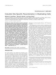
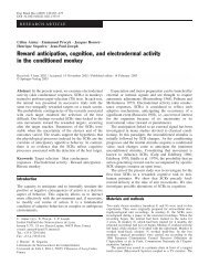
FIGURE 1<br />
Percentage of sperm DNA <strong>fragmentation</strong> (DFI)<br />
distribution in 322 sperm samples.<br />
Frequency (%)<br />
100<br />
80<br />
60<br />
40<br />
20<br />
0<br />
-10 0 10 20 30 40 50 60 70 80<br />
<strong>Sperm</strong> DNA <strong>fragmentation</strong> rate (%)<br />
Benchaib. <strong>Sperm</strong> DNA <strong>fragmentation</strong> and pregnancy outcome. Fertil Steril<br />
2007.<br />
34.0%; 40% did not present with a sperm-count anomaly.<br />
The average mobility w<strong>as</strong> 28.6% 14.03%. The mean of<br />
DFI w<strong>as</strong> 8.6% 8.0% (Figure 1).<br />
The average age of the women w<strong>as</strong> of 33.5 4.2 years.<br />
Tubal deterioration w<strong>as</strong> found in 6.6% of the women, and<br />
dysovulation in 27.6%; 2.2% had endometriosis. A uterine<br />
anomaly w<strong>as</strong> found in 0.6% of the women. From a total of<br />
3812 oocytes, the average number punctured w<strong>as</strong> 11.8<br />
6.1. The average number of embryos per couple w<strong>as</strong><br />
7.5 4.7 (from a total of 2401 embryos).<br />
Microinjection w<strong>as</strong> carried out in 72.7% of c<strong>as</strong>es (234<br />
cycles), early transfer in 61.5%, and late transfer in 32.3%.<br />
Table 1 shows the various characteristics according to the<br />
ART protocol, IVF or ICSI.<br />
The semen characteristics in the ICSI population were<br />
statistically significantly more faded than in the IVF population<br />
(P.001), and the DFI w<strong>as</strong> statistically significantly<br />
lower (6.3% 9.1% vs 9.4% 8.0%, respectively; P.01).<br />
The women undergoing ICSI were statistically significantly<br />
younger than those undergoing IVF (33.2 4.3 years vs<br />
34.4 4.3 years, respectively; P.01). The fertilization<br />
rates were statistically significantly higher with ICSI than in<br />
IVF (74.4% 20.7% vs 68.5% 22.9%, respectively;<br />
P.05).<br />
95
TABLE 1<br />
Parameters according to the ART procedure used (IVF versus ICSI).<br />
Male Factors and Fragmentation<br />
No statistically significant correlation w<strong>as</strong> found between<br />
male age and DFI (r 0.4; not statistically significant<br />
[NS]). Negative weak correlations were found between DFI<br />
and the standard semen analysis me<strong>as</strong>ures. The correlation<br />
coefficients between DFI and semen characteristics were <strong>as</strong><br />
follows: sperm count, r 0.2 (P.01); motility, r 0.2<br />
(P.01); and morphology, r 0.2 (P.01).<br />
Female Factors<br />
No relationship w<strong>as</strong> found between female factors and DFI,<br />
or between female factors and pregnancy. So female factors<br />
should not be considered <strong>as</strong> confounding factors in the<br />
statistical analysis.<br />
Fragmentation and Embryos<br />
No statistically significant correlation w<strong>as</strong> found between<br />
DFI and fertilization rate (r 0.04; NS), regardless of ART<br />
protocol: IVF, r 0.06 (NS) and ICSI, r 0.02 (NS).<br />
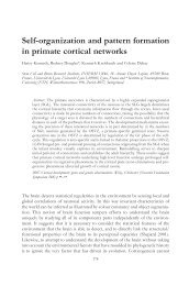
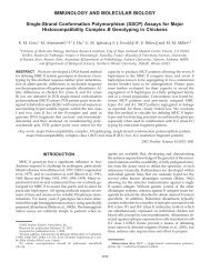
However, when the fertilization rate w<strong>as</strong> transformed into a<br />
qualitative variable and considered according to ART protocol,<br />
with ICSI it became evident that the fertilization rate<br />
decre<strong>as</strong>ed <strong>as</strong> the DFI incre<strong>as</strong>ed (P.05) (Figure 2) in a linear<br />
relationship (P.01). This result w<strong>as</strong> not found with IVF.<br />
Late transfer allowed the evaluation of the impact of DFI on<br />
IVF ICSI P value<br />
Male age (years) 36.5 5.1 36.1 5.2 NS<br />
Progressive motility (%)<br />
Concentration (%)<br />
38.0 10.9 25.0 13.4 .001<br />
1 million/mL 0 9.1 .001<br />
5 million/mL 2.3 25.5 .001<br />
20 million/mL 20.7 39.0 .001<br />
20 million/mL 77.0 26.4 .001<br />
Fragmentation rate (%) 6.3 9.1 9.4 8.0 .01<br />
Female age (years) 34.4 4.0 33.2 4.3 .05<br />
Oocytes 12.2 5.6 11.7 6.3 NS<br />
Embryos 8.3 4.7 7.1 4.6 NS<br />
Fertilization rate (%) 68.5 22.9 74.4 20.7 .05<br />
Embryo replacement (%)<br />
Embryo replacement state<br />
95.5 93.2 NS<br />
Early (%) 56.0 69.3 .05<br />
Late (%) 44.0 30.7 .05<br />
Transferred embryos (number) 2.1 0.8 2.1 0.9 NS<br />
Pregnancy rate (%) 33.3 (28/84) 35.8 (78/218) NS<br />
Early 34.0 (16/47) 35.1 (53/151) NS<br />
Late 32.4 (12/37) 37.3 (25/67) NS<br />
Note: Values are presented <strong>as</strong> mean standard deviation. NS: not statistically significant.<br />
Benchaib. <strong>Sperm</strong> DNA <strong>fragmentation</strong> and pregnancy outcome. Fertil Steril 2007.<br />
embryo development during the first week. With ICSI, the<br />
risk of arrested embryo development incre<strong>as</strong>ed when the DFI<br />
exceeded 15% (18.2% vs 4.2%; P.001). This result, again,<br />
w<strong>as</strong> not found with IVF, where the risk w<strong>as</strong> 5% when the<br />
DFI w<strong>as</strong> less than 15%, and 0 when it w<strong>as</strong> greater than 15%.<br />
Logistic regression for female age, ovarian response, ART<br />
procedure, and DFI showed that only DFI had a statistically<br />
significant influence on arrested embryo development<br />
(P.01), with an OR of 3.8.<br />
Fragmentation and ART Results<br />
Regardless of the ART procedure, no statistically significant<br />
difference in pregnancy rate according to DFI w<strong>as</strong> found in<br />
c<strong>as</strong>es of embryo replacement. The pregnancy rate w<strong>as</strong> 35.7%<br />
when the DFI w<strong>as</strong> less than 15% and 31.8% when it w<strong>as</strong><br />
greater. In IVF, the proportion of patients with a DFI exceeding<br />
15% w<strong>as</strong> too small for statistical testing. In c<strong>as</strong>es of<br />
ICSI, the pregnancy rate w<strong>as</strong> 37.4% when the DFI w<strong>as</strong> less<br />
than 15%, and 27.8% when it w<strong>as</strong> greater, but this difference<br />
w<strong>as</strong> not statistically significant. According to the stage of<br />
embryo replacement (early or late), the DNA <strong>fragmentation</strong><br />
rate did not seem to influence the pregnancy rate. However,<br />
when a pregnancy w<strong>as</strong> obtained with a DFI greater than<br />
15%, the risk of miscarriage w<strong>as</strong> incre<strong>as</strong>ed: 37.5% when the<br />
DFI w<strong>as</strong> greater than 15% versus 8.8% when the DFI w<strong>as</strong><br />
less than 15% (P.05, unilateral test) (Table 2).<br />
96 Benchaib et al. <strong>Sperm</strong> DNA <strong>fragmentation</strong> and pregnancy outcome Vol. 87, No. 1, January 2007
FIGURE 2<br />
Relationship between sperm DNA <strong>fragmentation</strong> and fertilization rate by treatment procedure:<br />
(A) Intracytopl<strong>as</strong>mic sperm injection (ICSI). (B) In vitro fertilization (IVF).<br />
A<br />
<strong>Sperm</strong> DNA <strong>fragmentation</strong> rate (%)<br />
B<br />
<strong>Sperm</strong> DNA Fragmentation (%)<br />
35<br />
30<br />
25<br />
20<br />
15<br />
10<br />
35<br />
30<br />
25<br />
20<br />
15<br />
10<br />
5<br />
0<br />
5<br />
0<br />
Benchaib. <strong>Sperm</strong> DNA <strong>fragmentation</strong> and pregnancy outcome. Fertil Steril 2007.<br />
DISCUSSION<br />
The relationship between sperm DNA <strong>fragmentation</strong> and<br />
male factor infertility is beginning to be recognized, but it<br />
remains a subject of controversy. The male patients in our<br />
study were all young, so it w<strong>as</strong> difficult to highlight the<br />
relationship between DFI and age that w<strong>as</strong> found in other<br />
studies (16, 22). Alterations in semen characteristics were<br />
<strong>as</strong>sociated with an incre<strong>as</strong>e in DFI, which confirmed the<br />
results of other studies (10, 11, 23–26). However, the relationship<br />
between semen characteristics and DFI is weak;<br />
indeed, DNA deterioration would have only a weak impact<br />
on the morphologic and functional characteristics of the<br />
Fertility and Sterility<br />
0 1-25 26-50 51-75 > 75<br />
Fertilization rate (%)<br />
0 1-25 26-50 51-75 > 75<br />
Fertilization rate (%)<br />
ICSI<br />
c-IVF<br />
spermatozoa. <strong>Sperm</strong>atozoa having normal characteristics according<br />
to World Health Organization criteria could have<br />
altered genetic material, <strong>fragmentation</strong> being only one <strong>as</strong>pect<br />
of the DNA deterioration.<br />
The influence of DFI on the fertilization rate varies from<br />
study to study. In our study, in c<strong>as</strong>es of ICSI, the fertilization<br />
rate decre<strong>as</strong>ed when the DFI w<strong>as</strong> elevated. This result confirmed<br />
those of other studies that had used ICSI (11, 27).<br />
However, this influence w<strong>as</strong> not found with IVF, again<br />
confirming the results of other teams (1, 13, 17, 18, 26, 28,<br />
29). The different behavior of fragmented spermatozoa un-<br />
97
TABLE 2<br />
Embryo development according sperm DNA <strong>fragmentation</strong> and procedure.<br />
der ICSI or IVF protocols may have been due to the poor<br />
quality of sperm used in ICSI, with a greater proportion of<br />
spermatozoa with fragmented DNA such that the probability<br />
of using an affected spermatozoon for oocyte injection w<strong>as</strong><br />
greater. In this situation, the oocyte’s DNA repair capacity<br />
would be exceeded (4, 9). Although the ICSI operator tends<br />
to choose a normal, mobile spermatozoon for the injection, a<br />
spermatozoon can be deemed “normal” while still harboring<br />
DNA abnormalities that will result in fertilization failure, <strong>as</strong><br />
testified by the fall in fertilization rates (11, 26).<br />
To optimize the choice of the spermatozoa injected into<br />
the cytopl<strong>as</strong>m of the oocytes, spermatozoa could be selected<br />
under very strong magnification for an intracytopl<strong>as</strong>mic<br />
morphologically selected sperm injection (IMSI) to ensure a<br />
better pregnancy rate (30). However, this technique is both<br />
recent and time consuming, so more information is needed<br />
before it can be proposed systematically in c<strong>as</strong>es of altered<br />
spermatozoa. In IVF, the fertilizing spermatozoon may be<br />
selected by the pellucid zona, which can reject aneuploid<br />
candidates (31).<br />
Controversy remains <strong>as</strong> to the impact of a high DFI on<br />
fertilization, but there is broader agreement about its harmful<br />
effect on embryo development (4, 32–34). The present study<br />
found a relationship between an elevated DFI (over 15%)<br />
and embryo development blocking. When the DFI exceeded<br />
15%, embryo development w<strong>as</strong> blocked in 18% of the cycles;<br />
this type of failure occurred in only 4% of cycles when<br />
the DFI w<strong>as</strong> lower than 15%. Although this result applied<br />
only in c<strong>as</strong>e of ICSI, the differential impact on embryo<br />
development according to the technique used (ICSI or IVF)<br />
had already been reported (1, 12). It h<strong>as</strong> nonetheless been<br />
shown that when the DFI is high, the proportion of embryos<br />
reaching the bl<strong>as</strong>tocyst stage is significantly lower (1, 12,<br />
35). The influence of male inheritance on mouse embryo<br />
development w<strong>as</strong> shown by Ahmadi and Ng (4). <strong>Sperm</strong>ato-<br />
IVF ICSI<br />
<strong>Sperm</strong> DNA<br />
<strong>Sperm</strong> DNA<br />
<strong>fragmentation</strong><br />
P<br />
<strong>fragmentation</strong><br />
15% value 15%<br />
P<br />
value<br />
Fertilization rate (%) 68.3 23.6 70.6 16.1 NS 75.4 20.0 70.3 23.4 NS<br />
Arrested development 5.0% 0 NS 4.2% 18.2% .05<br />
Pregnancy/transfer 31.6 (24/76) 50.0 (4/8) NS 37.4 (68/182) 27.8 (10/36) NS<br />
Pregnancy/transfer/stage<br />
Early (%) 34.9 (15/28) 25.0 (1/4) NS 37.6 (47/125) 23.1 (6/26) NS<br />
Late (%) 27.3 (9/33) 75.0 (3/4) NC 36.8 (21/57) 40.0 (4/10) NS<br />
Miscarriage (%) 9.1 (2/22) 50.0 (2/4) .05 8.6 (5/58) 30.0 (3/10) .05<br />
Note: NS: not statistically significant. NC: not calculated.<br />
Benchaib. <strong>Sperm</strong> DNA <strong>fragmentation</strong> and pregnancy outcome. Fertil Steril 2007.<br />
zoa with DNA damaged by variable amounts of irradiation<br />
before insemination still enabled fertilization, but embryonic<br />
development to the bl<strong>as</strong>tocyst stage w<strong>as</strong> statistically significantly<br />
decre<strong>as</strong>ed.<br />
It is generally thought that the first stages of embryo<br />
development depend on maternal transcripts, and that the<br />
paternal influence only begins at the six to eight cell stage;<br />
embryo transfers are generally performed on day 2 or 3 after<br />
follicle retrieval, which is before the paternal influence<br />
would be felt. However, it h<strong>as</strong> been shown that the paternal<br />
genome can play a role very early in embryo development,<br />
even at the first cell cycle (36), so that the use of a spermatozoon<br />
with a high level of fragmented DNA could cause<br />
fertilization failure. These observations could apply in particular<br />
situations. For example, it h<strong>as</strong> been shown that the<br />
level of fragmented DNA in spermatozoa obtained surgically<br />
is lower than found in those punctured from the epididymal<br />
(37). Thus, in choosing between testicular or epididymal sperm,<br />
especially in the event of obstructive azoospermia, testicular<br />
would be preferred to optimize embryo development.<br />
In the present study, high pregnancy rates tended to be<br />
obtained with low DFI. The correlation w<strong>as</strong> not statistically<br />
significant, but this tendency h<strong>as</strong> been confirmed by other<br />
studies that have shown that an elevated DFI is a deleterious<br />
factor for obtaining and maintaining pregnancies (1, 4, 12,<br />
17, 26, 27, 29). Considering our total data (IVF and ICSI<br />
cycles), in c<strong>as</strong>es of pregnancy where the DFI w<strong>as</strong> greater<br />
than 15%, the risk of miscarriage w<strong>as</strong> multiplied fourfold<br />
(37.5% versus 8.8%). <strong>Sperm</strong> DNA <strong>fragmentation</strong> w<strong>as</strong> a<br />
factor in the poor prognosis for pregnancy, and the risk of<br />
miscarriage w<strong>as</strong> incre<strong>as</strong>ed; similar results have been found<br />
by other investigators (1, 29).<br />
The exact origin of sperm DNA <strong>fragmentation</strong> h<strong>as</strong> yet to<br />
be established. Several causes have been suggested. A defect<br />
98 Benchaib et al. <strong>Sperm</strong> DNA <strong>fragmentation</strong> and pregnancy outcome Vol. 87, No. 1, January 2007
in chromatin replanning and compaction during spermatogenesis<br />
h<strong>as</strong> been proposed (38); however, this mechanism<br />
h<strong>as</strong> been discarded, because it h<strong>as</strong> been shown that the<br />
compaction and the <strong>fragmentation</strong> of sperm DNA are two<br />
independent phenomena (39). Alternatively, the production<br />
of reactive oxygen species (ROS) by immature spermatozoa<br />
or leukocytes may be at the origin of sperm DNA <strong>fragmentation</strong><br />
(40). Events taking place in the epididymal section<br />
(spermatozoon storage section)—in particular reiterative infections<br />
with inflammatory processes—could impact sperm<br />
DNA. This concept is b<strong>as</strong>ed on the fact that ejaculated sperm<br />
can exhibit a high DFI while testicular sperm h<strong>as</strong> a low DFI<br />
(41), indicating that <strong>fragmentation</strong> takes place downstream<br />
of the testicle.<br />
There is no consensus regarding how to approach a high<br />
DFI before an ART procedure or how to “cure” it. However,<br />
high DFI h<strong>as</strong> a negative impact on the ART result, so it<br />
seems re<strong>as</strong>onable to bring the DFI under its pathological<br />
threshold before undertaking an ART procedure. There are<br />
different ways to achieve this. The method of sperm preparation<br />
for an ART cycle influences the DNA <strong>fragmentation</strong>:<br />
selecting sperm using a discontinuous density gradient (42)<br />
or the swim-up technique (25) can decre<strong>as</strong>e the proportion of<br />
spermatozoa with fragmented DNA. In some c<strong>as</strong>es, DFI<br />
incre<strong>as</strong>es due to oxidative stress.<br />
Selection, however, does not solve all the problems, because<br />
some spermatozoa with fragmented DNA can persist<br />
and have an impact on the ART result (12). Although it is<br />
advisable to treat the cause of DNA <strong>fragmentation</strong> upstream<br />
of the ART attempt, the cause of the sperm DNA <strong>fragmentation</strong><br />
is not always clear. <strong>Sperm</strong> DNA <strong>fragmentation</strong> can be<br />
attributed to various pathologic conditions, including cryptorchidism,<br />
cancer, varicocele, fever, age, infection, and<br />
leukocytospermia. Many environmental conditions can also<br />
affect sperm DNA <strong>fragmentation</strong>, such <strong>as</strong> chemotherapy,<br />
radiation, air pollution, smoking, pesticides, chemicals, heat,<br />
and ART preparation protocols. Reactive oxygen species<br />
(ROS) activity may be a major factor in DNA strand breakage.<br />
In the c<strong>as</strong>e of varicocele, curing this pathology could<br />
decre<strong>as</strong>e the DFI (43).<br />
When no cause can be found, a treatment b<strong>as</strong>ed on antioxidants<br />
(vitamins C and E) can be given to decre<strong>as</strong>e sperm<br />
DNA <strong>fragmentation</strong> (44). Though antioxidant therapy does<br />
not help all patients with elevated sperm DNA <strong>fragmentation</strong>,<br />
improvement h<strong>as</strong> been found in 76% of patients in one<br />
study (44). Also, other treatments, such <strong>as</strong> the commercial<br />
dietary supplements ProXeed (Sigma-Tau HealthScience<br />
S.p.A. Gaithersburg, MD) or Fertile One (Co<strong>as</strong>t Reproductive,<br />
Inc., San Diego, CA), could be considered <strong>as</strong> therapy. In<br />
a c<strong>as</strong>e of high DFI in ejaculated sperm, using either testicular<br />
sperm where the DFI will be lessened (41) or ejaculated<br />
donor sperm (1) h<strong>as</strong> been proposed.<br />
The present results indicate a negative correlation between<br />
the DFI and semen characteristics. With ICSI, the risk of<br />
nontransfer due to blocked embryo development incre<strong>as</strong>es<br />
Fertility and Sterility<br />
with a DFI higher than 15%. Moreover, when pregnancy is<br />
obtained with a DFI higher than 15%, the risk of miscarriage<br />
is incre<strong>as</strong>ed. Similar results have been found with SCSA (1,<br />
13, 29, 45) and TUNEL (46). Thus, the me<strong>as</strong>urement of<br />
sperm DNA <strong>fragmentation</strong> h<strong>as</strong> a place in pre-ART <strong>as</strong>sessment<br />
to optimize the results under certain conditions, such <strong>as</strong><br />
repeated ART failure, miscarriage, varicocele, inflammatory<br />
processes, or infection of the genital tract.<br />
REFERENCES<br />
1. Virro MR, Larson-Cook KL, Evenson DP. <strong>Sperm</strong> chromatin structure<br />
<strong>as</strong>say (SCSA) parameters are related to fertilization, bl<strong>as</strong>tocyst development,<br />
and ongoing pregnancy in in vitro fertilization and intracytopl<strong>as</strong>mic<br />
sperm injection cycles. Fertil Steril 2004;81:1289–95.<br />
2. World Health Organization. Laboratory manual for examination of<br />
human semen and sperm-cervical mucus interaction. 4th ed, Cambridge:<br />
Cambridge University Press, 1999.<br />
3. Erenpreiss J, Spano M, Erenpreisa J, Bungum M, Giwercman A. <strong>Sperm</strong><br />
chromatin structure and male fertility: biological and clinical <strong>as</strong>pects.<br />
Asian J Androl 2006;8:11–29.<br />
4. Ahmadi A, Ng SC. Developmental capacity of damaged spermatozoa.<br />
Hum Reprod 1999;14:2279–85.<br />
5. Filatov MV, Semenova EV, Vorob’eva OA, Leont’eva O, Drobchenko<br />
EA. Relationship between abnormal sperm chromatin packing and IVF<br />
results. Mol Hum Reprod 1999;5:825–30.<br />
6. Sakk<strong>as</strong> D, Mariethoz E, Manicardi G, Bizzaro D, Bianchi PG, Bianchi<br />
U. Origin of DNA damage in ejaculated human spermatozoa. Rev<br />
Reprod 1999;4:31–7.<br />
7. Shen HM, Dai J, Chia SE, Lim A, Ong CN. Detection of apoptotic<br />
alterations in sperm in subfertile patients and their correlations with<br />
sperm quality. Hum Reprod 2002;17:1266–73.<br />
8. Zorn B, Virant-klun I, Meden-Vrtovec H. Semen granulocyte el<strong>as</strong>t<strong>as</strong>e:<br />
its relevance for the diagnosis and prognosis of silent genital tract<br />
inflammation. Hum Reprod 2000;15:1978–84.<br />
9. Sakk<strong>as</strong> D, Urner FG, Bianchi PG, Bizarro D, Wagner L, Jaquenoud N,<br />
et al. <strong>Sperm</strong> chromatin anomalies can influence decondensation after<br />
intracytopl<strong>as</strong>mic sperm injection. Hum Reprod 1996;11:837–43.<br />
10. Sun J, Juriscova A, C<strong>as</strong>per RF. Detection of <strong>deoxyribonucleic</strong> <strong>acid</strong><br />
<strong>fragmentation</strong> in human sperm: correlation with fertilization in vitro.<br />
Biol Reprod 1997;56: 602–7.<br />
11. Lopes S, Sun JG, Juriscova A, Meriano J, C<strong>as</strong>per RF. <strong>Sperm</strong> <strong>deoxyribonucleic</strong><br />
<strong>acid</strong> <strong>fragmentation</strong> is incre<strong>as</strong>ed in poor quality semen samples<br />
and correlates with failed fertilization in ICSI. Fertil Steril 1998;<br />
69:528–32.<br />
12. Benchaib M, Braun V, Lornage J, Hadj S, Salle B, Lejeune H, et al.<br />
<strong>Sperm</strong> DNA <strong>fragmentation</strong> decre<strong>as</strong>es the pregnancy rate in an <strong>as</strong>sisted<br />
reproductive technique. Hum Reprod 2003;18:1023–28.<br />
13. Bungum M, Humaidan P, Spano M, Jepson K, Bungum L, Giwerchman<br />
A. The predictive value of sperm chromatin structure <strong>as</strong>say (SCSA)<br />
parameters for the outcome of intrauterine insemination, IVF and ICSI.<br />
Hum Reprod 2004;19:1401–8.<br />
14. Evenson DP, Jost LK, Marshall D, Zinaman MJ, Clegg E, Purvis K, et<br />
al. Utility of the sperm chromatin structure <strong>as</strong>say <strong>as</strong> a diagnostic and<br />
<strong>prognostic</strong> tool in the human fertility clinic. Hum Reprod 1999;14:<br />
1039–49.<br />
15. Hughes CM, Lewis SE, McKelvey-Martin VJ, Thompson W. A comparison<br />
of b<strong>as</strong>eline and induced DNA damage in human spermatozoa<br />
from fertile and infertile men using a modified comet <strong>as</strong>say. Mol Hum<br />
Reprod 1996;2:613–20.<br />
16. Duran EH, Morshedi M, Taylor S, Oehninger S. <strong>Sperm</strong> DNA quality<br />
predicts intrauterine insemination outcome: a prospective cohort study.<br />
Hum Reprod 2002;17:3122–8.<br />
17. Henkel R, Kierspel E, Hajimohammad M, Stalf T, Hoogendijk C,<br />
Mehnert C, et al. DNA <strong>fragmentation</strong> of spermatozoa and <strong>as</strong>sisted<br />
reproduction technology. Reprod Biomed Online 2003;7:477–84.<br />
99
18. Henkel R, Hajimohammad M, Stalf T, Hoogendijk C, Mehnert C,<br />
Menkveld R, et al. Influence of <strong>deoxyribonucleic</strong> <strong>acid</strong> damage on<br />
fertilization and pregnancy. Fertil Steril 2004;81:965–72.<br />
19. Sergerie M, Laforest G, Bujan L, Bissonnette F, Bleau G. <strong>Sperm</strong> DNA<br />
<strong>fragmentation</strong>: threshold value in male fertility. Hum Reprod 2005;20:<br />
3446–51.<br />
20. Evenson DP, Jost LK, Baer RK, Turner TW, Schrader SM. Individuality<br />
of DNA denaturation patterns in human sperm <strong>as</strong> me<strong>as</strong>ured by the<br />
sperm chromatin structure <strong>as</strong>say. Reprod Toxicol 1991;5:115–25.<br />
21. Sergerie M, Laforest G, Boulanger K, Bissonnette F, Bleau G. Longitudinal<br />
study of sperm DNA <strong>fragmentation</strong> <strong>as</strong> me<strong>as</strong>ured by terminal<br />
uridine nick end-labelling <strong>as</strong>say. Hum Reprod 2005;20:1921–7.<br />
22. Aitken RJ, Baker MA, Sawyer D. Oxidative stress in the male germ line<br />
and its role in the aetiology of male infertility and genetic dise<strong>as</strong>e.<br />
Reprod Biomed Online 2003;7:65–70.<br />
23. Gandini L, Lombardo F, Paoli D, Caponecchia L, Familiari G, Verlengia<br />
C, et al. Study of apoptotic DNA <strong>fragmentation</strong> in human<br />
spermatozoa. Hum Reprod 2000;15:830–9.<br />
24. Irvine DS, Twigg JP, Gordon EL, Fulton N, Milne PA, Aitken RJ. DNA<br />
integrity in human spermatozoa: relationships with semen quality.<br />
J Androl 2000;21:33–44.<br />
25. Younglai EV, Holt D, Brown P, Juriscova A, C<strong>as</strong>per RF. <strong>Sperm</strong> swim<br />
up techniques and DNA <strong>fragmentation</strong>. Hum Reprod 2001;16:1950–3.<br />
26. Tomlinson MJ, Moffat O, Manicardi GC, Bizarro D, Afnan M, Sakk<strong>as</strong><br />
D. Interrelationships between seminal parameters and sperm nuclear<br />
DNA damage before and after density gradient centrifugation: implications<br />
for <strong>as</strong>sisted conception. Hum Reprod 2001;16:2160–5.<br />
27. Host E, Linderberg S, Smidt-Jensen S. The role of DNA strand breaks<br />
in human spermatozoa used for IVF and ICSI. Acta Obstet Gynecol<br />
Scand 2000;79:559–63.<br />
28. Saleh RA, Agarwal A, Nelson DR, Nada EA, El-Tonsy MH, Alvarez<br />
JG, et al. Incre<strong>as</strong>ed sperm nuclear DNA damage in normozoospermic<br />
infertile men: a prospective study. Fertil Steril 2002;78:313–8.<br />
29. Check JH, Graziano V, Cohen R, Krotec J, Check ML. Effect of an<br />
abnormal sperm chromatin structural <strong>as</strong>say (SCSA) on pregnancy outcome<br />
following (IVF) with ICSI in previous IVF failures. Arch Androl<br />
2005;51:121–4.<br />
30. Bartoov B, Berkovitz A, Eltes F, Kogosovsky A, Yagoda A, Lederman<br />
H, et al. Pregnancy rates are higher with intracytopl<strong>as</strong>mic morphologically<br />
selected sperm injection than with conventional intracytopl<strong>as</strong>mic<br />
injection. Fertil Steril 2003;80:1413–9.<br />
31. Van Dyk Q, Lanzerdorf S, Kolm P, Hodgen DJ, Manthony MC.<br />
Incidence of aneuploide spermatozoa from subfertile men: selected<br />
with motility versus hemizona bound. Hum Reprod 2000;15:1529–36.<br />
32. Dumoulin JC, Coonen E, Br<strong>as</strong> M, Van Wissen LE, Ignoul-Vanvuchelen<br />
R, Bergers-Jansen JM, et al. Comparison of in vitro development of<br />
embryos originating from either conventional in vitro fertilization or<br />
intracytopl<strong>as</strong>mic sperm injection. Hum Reprod 2000;15:402–9.<br />
33. Tomsu M, Sharma V, Miller D. Embryo quality and IVF treatment<br />
outcomes may correlate with different sperm comet <strong>as</strong>say parameters.<br />
Hum Reprod 2002;17:1856–62.<br />
34. Morris ID, Ilott S, Dixon L, Brison DR. The spectrum of DNA damage<br />
in human sperm <strong>as</strong>sessed by single cell gel electrophoresis (Comet<br />
<strong>as</strong>say) and its relationship to fertilization and embryo development.<br />
Hum Reprod 2002;17:990–8.<br />
35. Seli E, Gardner DK, Schoolcraft WB, Moffat O, Sakk<strong>as</strong> D. Extent of<br />
nuclear DNA damage in ejaculated spermatozoa impacts on bl<strong>as</strong>tocyst<br />
development after in vitro fertilization. Fertil Steril 2004;82:378–83.<br />
36. Tesarik J, Mendoza C, Greco E. Paternal effects acting during the first<br />
cell cycle of human preimplantation development after ICSI. Hum<br />
Reprod 2002;17:184–9.<br />
37. Steele EK, McClure N, Maxwell RJ, Lewis SE. A comparison of DNA<br />
damage in testicular and proximal epididymal spermatozoa in obstructive<br />
azoospermia. Mol Hum Reprod 1999;5:831–5.<br />
38. McPherson S, Longo FJ. Chromatin structure-function alterations during<br />
mammalian spermatogenesis: DNA nicking and repair in elongating<br />
spermatids. Eur J Histochem 1993;30:33–9.<br />
39. Lu Z, Zhang C, Zhai Z. Nucleopl<strong>as</strong>min regulates chromatin condensation<br />
during apoptosis. Proc Natl Acad Sci USA 2005;102:2778–83.<br />
40. Lopes S, Juriscova A, Sun JG, C<strong>as</strong>per RF. Reactive oxygen species:<br />
potential cause for DNA <strong>fragmentation</strong> in human spermatozoa. Hum<br />
Reprod 1998;13:896–900.<br />
41. Greco E, Scarselli F, Iacobelli M, Rienzi L, Ubaldi F, Ferrero S, et al.<br />
Efficient treatment of infertility due to sperm DNA damage by ICSI<br />
with testicular spermatozoa. Hum Reprod 2005;20:226–30.<br />
42. Donelly ET, O’Conell M, McClure N, Lewis SE. Differences in nuclear<br />
DNA <strong>fragmentation</strong> and mitochondrial integrity of semen and prepared<br />
human spermatozoa. Hum Reprod 2000;15:1552–61.<br />
43. Saleh RA, Agarwal A, Sharma RK, Said TM, Sikka SC, Thom<strong>as</strong> AJ Jr.<br />
Evaluation of nuclear DNA damage in spermatozoa from infertile men<br />
with varicocele. Fertil Steril 2003;80:1431–6.<br />
44. Greco E, Iacobelli M, Rienzi L, Ubaldi F, Ferrero S, Tesarik J. Reduction<br />
of the incidence of sperm DNA <strong>fragmentation</strong> by oral antioxidant<br />
treatment. J Androl 2005;26:349–53.<br />
45. Evenson D, Wixon R. Meta-analysis of sperm DNA <strong>fragmentation</strong><br />
using the sperm chromatin structure <strong>as</strong>say. RBM Online 2006;12:466–<br />
472.<br />
46. Carrell DT, Liu L, Peterson CM, Jones KP, Hat<strong>as</strong>aka HH, Erickson L,<br />
Campbell B. <strong>Sperm</strong> DNA <strong>fragmentation</strong> is incre<strong>as</strong>ed in couples with<br />
unexplained recurrent pregnancy loss. Arch Androl 2003;49:49–55.<br />
100 Benchaib et al. <strong>Sperm</strong> DNA <strong>fragmentation</strong> and pregnancy outcome Vol. 87, No. 1, January 2007