Free Radicals, Exercise, and Antioxidants - Setanta College
Free Radicals, Exercise, and Antioxidants - Setanta College
Free Radicals, Exercise, and Antioxidants - Setanta College

Create successful ePaper yourself
Turn your PDF publications into a flip-book with our unique Google optimized e-Paper software.
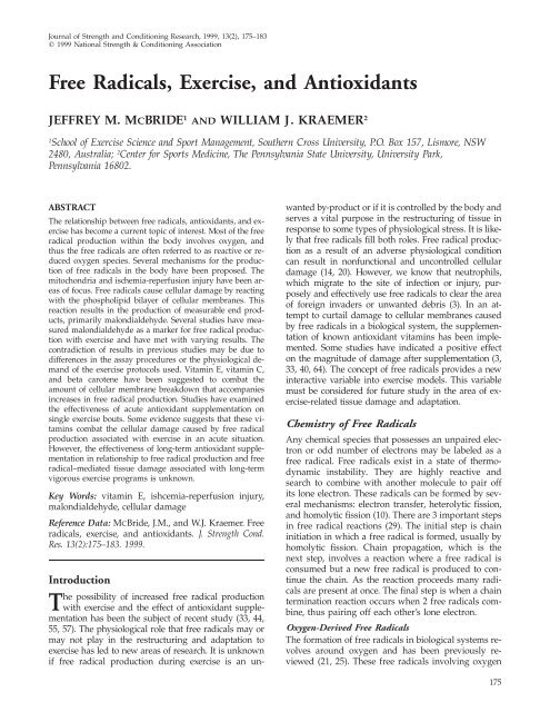
Journal of Strength <strong>and</strong> Conditioning Research, 1999, 13(2), 175–183<br />
1999 National Strength & Conditioning Association<br />
<strong>Free</strong> <strong>Radicals</strong>, <strong>Exercise</strong>, <strong>and</strong> <strong>Antioxidants</strong><br />
JEFFREY M. MCBRIDE 1 AND WILLIAM J. KRAEMER 2<br />
1 School of <strong>Exercise</strong> Science <strong>and</strong> Sport Management, Southern Cross University, P.O. Box 157, Lismore, NSW<br />
2480, Australia; 2 Center for Sports Medicine, The Pennsylvania State University, University Park,<br />
Pennsylvania 16802.<br />
ABSTRACT<br />
The relationship between free radicals, antioxidants, <strong>and</strong> exercise<br />
has become a current topic of interest. Most of the free<br />
radical production within the body involves oxygen, <strong>and</strong><br />
thus the free radicals are often referred to as reactive or reduced<br />
oxygen species. Several mechanisms for the production<br />
of free radicals in the body have been proposed. The<br />
mitochondria <strong>and</strong> ischemia-reperfusion injury have been areas<br />
of focus. <strong>Free</strong> radicals cause cellular damage by reacting<br />
with the phospholipid bilayer of cellular membranes. This<br />
reaction results in the production of measurable end products,<br />
primarily malondialdehyde. Several studies have measured<br />
malondialdehyde as a marker for free radical production<br />
with exercise <strong>and</strong> have met with varying results. The<br />
contradiction of results in previous studies may be due to<br />
differences in the assay procedures or the physiological dem<strong>and</strong><br />
of the exercise protocols used. Vitamin E, vitamin C,<br />
<strong>and</strong> beta carotene have been suggested to combat the<br />
amount of cellular membrane breakdown that accompanies<br />
increases in free radical production. Studies have examined<br />
the effectiveness of acute antioxidant supplementation on<br />
single exercise bouts. Some evidence suggests that these vitamins<br />
combat the cellular damage caused by free radical<br />
production associated with exercise in an acute situation.<br />
However, the effectiveness of long-term antioxidant supplementation<br />
in relationship to free radical production <strong>and</strong> free<br />
radical–mediated tissue damage associated with long-term<br />
vigorous exercise programs is unknown.<br />
Key Words: vitamin E, ishcemia-reperfusion injury,<br />
malondialdehyde, cellular damage<br />
Reference Data: MCBride, J.M., <strong>and</strong> W.J. Kraemer. <strong>Free</strong><br />
radicals, exercise, <strong>and</strong> antioxidants. J. Strength Cond.<br />
Res. 13(2):175–183. 1999.<br />
Introduction<br />
The possibility of increased free radical production<br />
with exercise <strong>and</strong> the effect of antioxidant supplementation<br />
has been the subject of recent study (33, 44,<br />
55, 57). The physiological role that free radicals may or<br />
may not play in the restructuring <strong>and</strong> adaptation to<br />
exercise has led to new areas of research. It is unknown<br />
if free radical production during exercise is an un-<br />
wanted by-product or if it is controlled by the body <strong>and</strong><br />
serves a vital purpose in the restructuring of tissue in<br />
response to some types of physiological stress. It is likely<br />
that free radicals fill both roles. <strong>Free</strong> radical production<br />
as a result of an adverse physiological condition<br />
can result in nonfunctional <strong>and</strong> uncontrolled cellular<br />
damage (14, 20). However, we know that neutrophils,<br />
which migrate to the site of infection or injury, purposely<br />
<strong>and</strong> effectively use free radicals to clear the area<br />
of foreign invaders or unwanted debris (3). In an attempt<br />
to curtail damage to cellular membranes caused<br />
by free radicals in a biological system, the supplementation<br />
of known antioxidant vitamins has been implemented.<br />
Some studies have indicated a positive effect<br />
on the magnitude of damage after supplementation (3,<br />
33, 40, 64). The concept of free radicals provides a new<br />
interactive variable into exercise models. This variable<br />
must be considered for future study in the area of exercise-related<br />
tissue damage <strong>and</strong> adaptation.<br />
Chemistry of <strong>Free</strong> <strong>Radicals</strong><br />
Any chemical species that possesses an unpaired electron<br />
or odd number of electrons may be labeled as a<br />
free radical. <strong>Free</strong> radicals exist in a state of thermodynamic<br />
instability. They are highly reactive <strong>and</strong><br />
search to combine with another molecule to pair off<br />
its lone electron. These radicals can be formed by several<br />
mechanisms: electron transfer, heterolytic fission,<br />
<strong>and</strong> homolytic fission (10). There are 3 important steps<br />
in free radical reactions (29). The initial step is chain<br />
initiation in which a free radical is formed, usually by<br />
homolytic fission. Chain propagation, which is the<br />
next step, involves a reaction where a free radical is<br />
consumed but a new free radical is produced to continue<br />
the chain. As the reaction proceeds many radicals<br />
are present at once. The final step is when a chain<br />
termination reaction occurs when 2 free radicals combine,<br />
thus pairing off each other’s lone electron.<br />
Oxygen-Derived <strong>Free</strong> <strong>Radicals</strong><br />
The formation of free radicals in biological systems revolves<br />
around oxygen <strong>and</strong> has been previously reviewed<br />
(21, 25). These free radicals involving oxygen<br />
175
176 McBride <strong>and</strong> Kraemer<br />
have been named reactive or reduced oxygen species.<br />
These species include singlet oxygen, superoxide anions,<br />
<strong>and</strong> hydrogen peroxide (25). However, perhaps<br />
the most focused on has been the hydroxyl radical.<br />
This free radical is highly reactive <strong>and</strong> is the primary<br />
source of the destructive mechanisms behind free radical<br />
formation in biological systems. The hydroxyl radical<br />
is diffusion controlled, meaning it will not travel<br />
a significant distance in the cell before reacting. It has<br />
a short half-life but can cause an extreme amount of<br />
damage within a small radius of where it is produced<br />
(10, 54). The presence of ferrous iron <strong>and</strong> cuprous copper<br />
in biological systems results in the formation of<br />
the most highly reactive reduced oxygen species. This<br />
can occur by several reactions <strong>and</strong> is a major factor in<br />
oxidative tissue damage (63).<br />
Mechanisms for <strong>Free</strong> Radical Production<br />
Mitochondria. Most free radical generation within the<br />
cell occurs by electron transfer reactions (10). Electrons<br />
leaked from electron transport chains react with molecular<br />
oxygen. This process occurs within the mitochondria<br />
<strong>and</strong> endoplasmic reticulum of the cell. The<br />
steps between the negative end of the respiratory chain<br />
<strong>and</strong> cytochrome c are capable of producing a superoxide<br />
anion via electron transfer (66). This sequence<br />
can also lead to the formation of hydrogen peroxide.<br />
The presence of transition metal ions can lead to the<br />
formation of the highly reactive hydroxyl radical. Production<br />
of these free radicals in mitochondria has been<br />
shown to be a result of inhibition of the respiratory<br />
chain due to hyperoxia <strong>and</strong> calcium overload (66).<br />
Inflammation. Not all free radical formation in biological<br />
systems is accidental. Catalysis caused by some<br />
enzymes is the result of their use of a free radical at<br />
the active site in response to inflammation (10). These<br />
radicals are not truly free because they are targeted<br />
toward a specific reaction. However, the use of such<br />
enzymes may result in leakage of free radicals <strong>and</strong><br />
subsequent uncontrolled tissue damage. There are<br />
many enzymes capable of generating free radicals.<br />
Two enzymes of current interest are nicotinamide adenine<br />
dinucleotide (NADPH) oxidase, which is found<br />
on the cell membrane <strong>and</strong> within phagocytic cells, <strong>and</strong><br />
xanthine oxidase, which is found primarily in the liver<br />
<strong>and</strong> in some endothelial cells (71).<br />
Trauma-mediated agonists that bind to cell membrane<br />
receptors may cause free radical formation via<br />
NADPH oxidase release from phagocytic cells. These<br />
phagocytic cells, primarily neutrophils, are part of an<br />
organism’s immune response to infection or injury.<br />
Neutrophils migrate to the site of injury or infection<br />
by chemotaxis <strong>and</strong> act in 3 functional ways: to phagocytize<br />
foreign or damaged fragments, to release proteolytic<br />
enzymes via degranulation, <strong>and</strong> to release free<br />
radical–forming enzymes that act to break down foreign<br />
or damaged fragments (40). The latter function is<br />
Figure 1. Neutrophil activation can result in free radical<br />
formation via nicotinamide adenine dinucleotide oxidase.<br />
Damage to endothelial cells during ischemia via xanthine<br />
oxidase coupled with histamine enhancement can also result<br />
in free radical production. Modified from Ward et al.<br />
(71).<br />
known as the respiratory or oxidative burst, <strong>and</strong> the<br />
complete sequence of events involved in this function<br />
is still unclear. Xanthine oxidase is thought to be activated<br />
by ischemia. Histamine release by mast cells<br />
has also been suggested to enhance the catalytic ability<br />
of xanthine oxidase. Figure 1 shows how all these<br />
mechanisms may be related to the production of free<br />
radicals <strong>and</strong> subsequent tissue damage (71).<br />
Ischemia Reperfusion. All the previously mentioned<br />
mechanisms for free radical production can be related<br />
back to ischemia-reperfusion injury. Ischemia, from<br />
whatever cause, results in a decrease in oxygen <strong>and</strong><br />
substrate availability. The lack of adenosine triphosphate<br />
(ATP) due to the inability of anaerobic means to<br />
maintain pace with energy dem<strong>and</strong>s results in damaging<br />
effects (46). The breakdown of ATP <strong>and</strong> the activation<br />
of xanthine oxidase from xanthine dehydrogenase<br />
(Figure 2) during ischemia have been related<br />
to production of free radicals <strong>and</strong> tissue damage during<br />
reperfusion.<br />
Ischemia also results in phagocytic cell responses<br />
via NADPH oxidase. The following reaction shows the<br />
formation of the superoxide anion <strong>and</strong> subsequent<br />
damaging free radicals (3).<br />
4O2 4NADPH → 4H <br />
4O2 4NADP<br />
Membrane ionic concentrations may also be disrupted<br />
during ischemia-reperfusion injury (46). Reperfusion<br />
causes an influx of calcium in exchange for<br />
previously influxed sodium due to membrane disruption<br />
(4, 53). This calcium influx could result in the active<br />
sequestering of calcium within the mitochondrial<br />
matrix, which may lead to dysfunction (66). This cal-
Figure 2. Ischemia-reperfusion injury has been shown to<br />
result in tissue damage via xanthine oxidase, which is the<br />
active form of xanthine dehydrogenase. This activation is<br />
the result of ischemic conditions. When reperfusion occurs<br />
the result is free radical formation <strong>and</strong> subsequent tissue<br />
damage. Modified from Ward et al. (71).<br />
cium influx results in an inhibition of the rate of<br />
NADH-supported electron flow, thus blocking the respiratory<br />
chain <strong>and</strong> subsequently stimulating superoxide<br />
<strong>and</strong> hydrogen peroxide production (66). Reperfusion<br />
may be essential for the salvage of reversibly<br />
damaged cells as a result of ischemia. However, the<br />
great increase in oxygen availability or hyperoxia may<br />
result in superoxide anion production from the electron<br />
transport system, resulting in more tissue damage<br />
(66). In one study examining free radical formation,<br />
it was demonstrated that free radical concentrations<br />
increased in rats that underwent 2 hours of ischemic<br />
conditions <strong>and</strong> 1 hour of reperfusion. In<br />
addition, these increases in free radicals were accompanied<br />
by significant increases in muscle water content<br />
<strong>and</strong> serum creatine kinase levels. These increases may<br />
indicate membrane disruption (59). Other studies have<br />
also suggested that ischemia-reperfusion injury may<br />
be mediated by free radical formation (23, 40).<br />
Mechanisms by Which <strong>Free</strong> <strong>Radicals</strong> Cause Tissue<br />
Damage<br />
The reaction of free radicals with cell membranes is<br />
one of the events that leads to tissue damage. Cell<br />
membranes are organizations of phospholipid molecules<br />
into a lipid bilayer consisting of a polar (negatively<br />
charged) phosphate group <strong>and</strong> 2 nonpolar (electrically<br />
neutral) fatty acid tails (43, 60). The inner region<br />
of the lipid bilayer consists of the hydrophobic<br />
polyunsaturated fatty acid tails, which readily cause<br />
free radical reactions (48, 52, 70). This chain reaction,<br />
as mentioned previously, is self-perpetuating. Hydroperoxides<br />
are the products formed as a result of the<br />
free radical reactions that occur with cell membranes<br />
(68). This process by which hydroperoxides are<br />
formed is identified as lipid peroxidation. Lipid peroxidation<br />
is a free radical chain reaction that can be<br />
initiated by the hydroxyl radical or transition metal<br />
<strong>Free</strong> <strong>Radicals</strong> 177<br />
Figure 3. This chain reaction is described as lipid peroxidation.<br />
This process involves the reaction of free radicals<br />
with the polyunsaturated fatty acids within the cell membrane.<br />
Measurable end products consist of aldehydes. The<br />
most abundant of the aldehydes formed is malondialdehyde.<br />
Modified from Cheeseman <strong>and</strong> Slater (10).<br />
Figure 4. The reaction of free radicals results in the disruption<br />
of the structural integrity of the cell membrane resulting<br />
in measurable end products. Modified from Sjodin<br />
et al. (61).<br />
complexes (2, 28, 67). The reactions in Figure 3 are the<br />
primary steps in the formation of hydroperoxides (10).<br />
In the initial reaction LH is the target polyunsaturated<br />
fatty acid <strong>and</strong> R is the ‘‘attacking’’ free radical. Oxidation<br />
of polyunsaturated fatty acids leads to the formation<br />
of a fatty acid radical (L ) <strong>and</strong> adds oxygen to<br />
form a fatty acid peroxyl radical (LOO ). Peroxyl radicals<br />
can oxidize other polyunsaturated fatty acids,<br />
which results in new chain reaction formation. This<br />
process results in the formation of lipid hydroperoxides<br />
(LOOH). These hydroperoxides can break down<br />
into other free radical species or other compounds, the<br />
most significant being aldehydes (18, 49). The decomposition<br />
of hydroperoxides results in formation of aldehydes<br />
of various chain lengths. Malondialdehyde<br />
(MDA), a 3-carbon-chain aldehyde, is one the primary<br />
aldehydes formed (61). Figure 4 shows how free radical<br />
reaction with the membrane results in MDA production.<br />
Measurement of MDA has become the most<br />
commonly used indicator of lipid peroxidation.
178 McBride <strong>and</strong> Kraemer<br />
Measurement of Lipid Peroxidation<br />
The assay used most often for the determination of<br />
products formed from lipid peroxidation is a procedure<br />
that uses thiobarbituric acid (TBA). The general<br />
procedure involves a sample heated with TBA at a low<br />
pH. A pink chromogen (TBA-MDA adduct) is formed<br />
<strong>and</strong> measured at an absorbance of 532 nm or by fluorescence<br />
at 553 nm (6). There have been criticisms of<br />
the TBA test that have been used in many studies looking<br />
at the production of lipid peroxidation products<br />
(27). It has been suggested that substances found in<br />
blood other than MDA also form adducts with TBA.<br />
This may contribute to high estimates of MDA, unless<br />
high-performance liquid chromatography (HPLC) is<br />
used to separate these substances (27). However, it has<br />
also been argued that many of these interfering compounds<br />
are not normal constituents of biological material<br />
(6). Two primary concerns for this assay are not<br />
to overheat the samples <strong>and</strong> to limit the use of metal<br />
catalyst, since both can lead to erroneously high estimates<br />
of serum or plasma MDA concentrations (6).<br />
Studies using an HPLC method typically report much<br />
lower MDA levels compared with spectrophotometric<br />
assays without the use of HPLC. It is unclear at this<br />
time which is the most effective.<br />
<strong>Free</strong> Radical Formation with <strong>Exercise</strong><br />
Aerobic <strong>Exercise</strong>. There are many possible mechanisms<br />
for the production of free radicals during exercise. The<br />
first involves hyperoxic injury that may occur in highly<br />
intense aerobic exercise. During this type of exercise,<br />
oxygen consumption can increase up to 10–20 times<br />
resting levels (1). This may result in a flux of oxygen<br />
into exercising muscles by as much as 100–200 times<br />
above their resting state (35). As mentioned previously,<br />
mitochondria, or more specifically the electron transport<br />
system, is the likely site of free radical generation<br />
during adverse conditions in the body. Mitochondria<br />
subjected to a great influx of oxygen forms superoxide<br />
radicals that may lead to the formation of other more<br />
harmful radicals (72). It has been shown that as intracellular<br />
oxygen concentrations increase the rate of electron<br />
leakage to oxygen increases (66). However, in one<br />
animal model mitochondrial malfunction only occurred<br />
after 40–50 hours of exposure to 100% oxygen<br />
<strong>and</strong> most tissue damage occurred in the lungs (66).<br />
Another situation may involve intensities above<br />
100% of maximal oxygen consumption. <strong>Exercise</strong> at this<br />
intensity may result in a lack of ATP availability along<br />
with an increased rate of adenosine diphosphate production.<br />
This may activate free radical generation via<br />
the aforementioned mechanism involving xanthine oxidase<br />
(61).<br />
The most likely source of free radical production<br />
during aerobic exercise is the situation known as ischemia-reperfusion<br />
injury. During exercise many organs,<br />
such as the liver, kidneys, <strong>and</strong> the splanchnic<br />
region, may experience hypoxia. This hypoxia is due<br />
to the shunting of blood to working muscles (72). After<br />
exercise these organs are reperfused with oxygen after<br />
blood flow is returned to normal. This results in the<br />
formation of free radicals <strong>and</strong> lipid peroxidation products<br />
by the previously mentioned mechanisms seen in<br />
ischemia-reperfusion injury studies (34, 73). It has<br />
been suggested that most of the lipid peroxidation<br />
products that are measured during aerobic activity at<br />
above 100% of maximal oxygen consumption are from<br />
the liver (67). This is due to the finding that blood flow<br />
may be reduced to one-fifth of normal to the liver during<br />
exercise of this type (1).<br />
Several studies have looked at the effect of exercise<br />
above 100% of maximal oxygen consumption on the<br />
formation of free radicals by measuring lipid peroxidation<br />
products. Some studies found no difference in<br />
MDA concentrations before <strong>and</strong> after exercise (56, 64).<br />
Other studies indicated a significant increase with exercise<br />
(32, 39). It is not clear why the studies with aerobic<br />
exercise have contradicted each other. It may be<br />
possible that the physical dem<strong>and</strong> of the exercise protocols<br />
used in the studies that did not show an increase<br />
in free radical production was inadequate. The<br />
studies that did indicate a significant increase in free<br />
radical production used much more vigorous exercise<br />
protocols, such as an 80-km race. A second possibility<br />
for the discrepancies is the assay for the detection of<br />
free radical formation. The types of procedures used<br />
for the TBA test have been controversial. The one<br />
study that used an HPLC method reported that MDA<br />
levels were below the detection limit of 0.1 mol·L 1<br />
in all samples (56). Methods used in other studies not<br />
using HPLC techniques have reported resting levels of<br />
2.26 nmol·ml 1 <strong>and</strong> postexercise values of up to 4.0<br />
nmol·ml 1 (32). It is unclear at this time which method<br />
would be best suited for determination of MDA in<br />
plasma. It has been suggested that resting levels may<br />
be around 0.5 mol·L 1 <strong>and</strong> that assays that strictly<br />
measure MDA content rarely find any in resting human<br />
plasma (27).<br />
Resistance <strong>Exercise</strong>. During resistance exercise ischemia<br />
reperfusion may occur within the active muscles,<br />
possibly even to a higher degree than within other<br />
organs. Muscles undergoing intense concentric <strong>and</strong><br />
eccentric actions, which occur in muscles performing<br />
resistance exercise, may experience brief hypoxic conditions.<br />
Intense muscle actions temporarily decrease<br />
blood flow <strong>and</strong> thus oxygen availability. During muscle<br />
relaxation there is oxygen reperfusion. In addition,<br />
there has also been membrane disruption as indicated<br />
by a leakage of intramuscular enzymes into the blood<br />
such as creatine kinase (64). This could lead to ion fluxes<br />
such as increases in intracellular calcium levels (66).<br />
Calcium levels may also be increased due to fatiguerelated<br />
functional abnormalities in the sarcoplasmic<br />
reticulum. Increased calcium levels have been shown
to be one of the major factors in affecting mitochondrial<br />
function in producing free radicals (66). It is also<br />
likely that the trauma to muscle cells during high-intensity<br />
exercise results in the activation of inflammatory<br />
mediators. These mediators act through phagocytic-<br />
<strong>and</strong> endothelial-mast cell pathways of free radical<br />
generation (70). These possible mechanisms indicate<br />
that resistance exercise may result in free radical<br />
production beyond what has been measured with aerobic<br />
exercise.<br />
From the aforementioned mechanisms, the active<br />
muscle site in resistance training may result in a significant<br />
increase in the production of free radicals either<br />
during or after exercise. Therefore, it is possible<br />
that a resistance exercise protocol will result in measurable<br />
increases in lipid peroxidation. A previously<br />
proposed mechanism of free radical production during<br />
exercise, especially resistance exercise, is an ischemia-reperfusion<br />
environment at the muscle site. A<br />
study looked specifically at this concept using repetitive<br />
static muscle contractions. A knee extension exercise<br />
was used with a 10-second exertion phase <strong>and</strong><br />
a 10-second resting phase protocol at 30% of maximal<br />
voluntary contraction force (55). It was reported that<br />
plasma MDA remained below the detection limit during<br />
all measurement times of the exercise protocol.<br />
This study involved a low-intensity resistance exercise<br />
protocol <strong>and</strong> may not be an effective stimulus for a<br />
significant measurable change in free radical production.<br />
Another study investigated the effects of eccentric<br />
<strong>and</strong> concentric muscle actions on free radical formation<br />
<strong>and</strong> related muscle damage (57). Forearm flexion<br />
<strong>and</strong> knee extension movements were used, <strong>and</strong> repetition<br />
ranges were reported to be 70–80. No significant<br />
changes in plasma thiobarbituric reactive material or<br />
muscle MDA content were reported after both concentric<br />
<strong>and</strong> eccentric muscle action protocols. This protocol<br />
involved a 70–80 repetition range, which in resistance<br />
exercise would also be labeled as low intensity.<br />
To our knowledge, only one study has looked at<br />
high-intensity resistance exercise <strong>and</strong> the effect on free<br />
radical production (44). This study used multiple 10repetition-maximum<br />
sets (10-RM set using resistance<br />
in which only 10 repetitions can be completed) involving<br />
all the body’s major muscle groups <strong>and</strong> found a<br />
significant increase in free radical production. It may<br />
be that a more dem<strong>and</strong>ing resistance training protocol<br />
is necessary to induce significant measurable changes<br />
in markers that indicate free radical reaction with cell<br />
membranes. Possible factors may include the stimulation<br />
of a greater amount of muscle mass at a higher<br />
intensity, resulting in considerably higher levels of<br />
muscle damage.<br />
The current evidence suggests that free radical production<br />
within the body depends on exercise intensity,<br />
whether one is referring to aerobic or resistance exercise.<br />
An exercise protocol must provide a significant<br />
<strong>Free</strong> <strong>Radicals</strong> 179<br />
disruption to the physiological state of the body. This<br />
may include significant ischemia-reperfusion conditions<br />
<strong>and</strong> muscle damage.<br />
<strong>Free</strong> <strong>Radicals</strong> <strong>and</strong> Muscle Damage<br />
Creatine kinase has been used as a marker for muscle<br />
damage in many studies (11, 12, 17, 42, 62). One study<br />
looked at creatine kinase responses <strong>and</strong> serum MDA<br />
content in runners after an 80-km race (32). A positive<br />
correlation was found between MDA <strong>and</strong> creatine kinase.<br />
In a recent investigation a similar pattern of response<br />
between MDA <strong>and</strong> creatine kinase was also reported<br />
in association with resistance exercise (44). It<br />
has been suggested that the membrane disruption that<br />
occurs with high-intensity resistance exercise is, to a<br />
large extent, simply a result of the mechanical loads<br />
placed on the muscle. This would result in a disruption<br />
in the muscle’s structural integrity. However, a<br />
study by Kraemer et al. (36) that compared a group<br />
performing a resistance training protocol using 5-repetition-maximum<br />
sets (5-RM set using resistance in<br />
which only 5 repetitions can be completed) with 1<br />
minute of rest between sets (5/1) <strong>and</strong> a group performing<br />
the same exercises except with 10-RM sets<br />
<strong>and</strong> a 1-minute rest period between sets (10/1) has<br />
provided evidence to the contrary. This study reported<br />
that the 10/1 group had a significantly higher creatine<br />
kinase response. This group was subjected to lighter<br />
loads yet possibly had greater muscle membrane disruption<br />
despite the fact that total work was equivalent.<br />
Studies also have shown that creatine kinase responses<br />
often peak between 2 to 4 days after eccentric exercise<br />
protocols. This may indicate that some mechanism is<br />
continuing to cause damage even after the actual exercise<br />
bout is completed (12, 17, 45). Mechanisms other<br />
than just mechanical forces may be responsible for<br />
muscle membrane disruption after high-intensity exercise.<br />
The number of circulating neutrophils has been<br />
shown to continually increase for several hours after<br />
exercise (9). In addition, malfunctioning mitochondria,<br />
due to intramuscular increases in calcium, could continue<br />
to produce free radicals after exercise ceases.<br />
<strong>Free</strong> radical formation by previously described pathways<br />
may play a role in continuing the amount of<br />
muscle membrane disruption after exercise.<br />
<strong>Antioxidants</strong><br />
An antioxidant has been defined as a substance that,<br />
when present in low concentrations in comparison to<br />
an oxidizable substrate, inhibits or delays the oxidation<br />
of that substrate (28). The human body has several naturally<br />
occurring antioxidants. Enzymes such as catalase<br />
<strong>and</strong> glutathione peroxidase can break down hydrogen<br />
peroxide, thereby stopping subsequent free<br />
radical–generating reactions (25). It has also been<br />
shown that cytochrome oxidase in the electron transport<br />
system acts to counter free radical production<br />
(34).
180 McBride <strong>and</strong> Kraemer<br />
Vitamin E. There are several nonenzymatic antioxidant<br />
substances in the body that can be supplemented<br />
easily. Probably the most focused on <strong>and</strong> important is<br />
vitamin E (tocopherols). It has been shown that this<br />
lipid-soluble vitamin is an effective antioxidant within<br />
the cell membrane (7, 30). The ability of vitamin E to<br />
prevent oxidation of unsaturated fatty acids is believed<br />
to be its primary function in the body (26). The absence<br />
of vitamin E results in the abnormal structure <strong>and</strong> function<br />
of cellular organelles <strong>and</strong> the cell membrane itself<br />
(26). Vitamin E supplementation in humans originally<br />
developed in the search for a treatment of muscle diseases.<br />
In animal models muscular dystrophy is associated<br />
with vitamin E deficiency myopathy (5). The protective<br />
mechanism of vitamin E has been shown in several<br />
animal models that used contractile activity as a<br />
stimulus for muscle damage. Vitamin E status in rats<br />
has been highly correlated with the susceptibility of<br />
that animal to damage from muscle contractions (31).<br />
In addition, studies have shown the protective effect of<br />
oral vitamin E supplementation (51).<br />
As previously mentioned, models looking at the effects<br />
of vitamin E supplementation on muscle damage<br />
have involved muscle contraction. Vitamin E exerts its<br />
major effect by the oxidation of free radicals (65). The<br />
mechanisms of ischemia-reperfusion injury have been<br />
the foundation for which the damaging effects of free<br />
radical formation may be seen. In rats, tissue markers<br />
for free radical generation significantly increase during<br />
ischemia-reperfusion injury. Supplementation of free<br />
radical scavengers significantly decrease this marker<br />
(59). It has been shown that free radicals are mediators<br />
of ischemia-reperfusion injury <strong>and</strong> that antioxidants<br />
such as vitamin E are effective in attenuating this injury<br />
(14, 40). Inflammation that may accompany this<br />
type of injury is directly linked to the formation of free<br />
radicals, which results in tissue injury (71). Ischemia<br />
reperfusion <strong>and</strong> inflammation are 2 conditions that are<br />
related to in vivo environmental situations found at<br />
the site of the muscle during exercise. This may implicate<br />
vitamin E supplementation as an effective<br />
means of reducing exercise-induced muscle damage<br />
due to free radical formation.<br />
Vitamin C. Vitamin C or L-ascorbic acid has also<br />
been implicated as an antioxidant. Vitamin C may be<br />
involved in the regeneration of vitamin E (47). Ascorbic<br />
acid has been shown to be involved in a key pathway<br />
related to the generation of vitamin E (58). A recent<br />
study has shown that ascorbate is an effective antioxidant<br />
in human plasma (19). It is suggested that ascorbate<br />
is a potent reducing agent <strong>and</strong> that it is effective<br />
in the quenching of free radicals. In addition, ascorbate<br />
is vital in the protection of retinoids, carotenoids, tocopherols,<br />
B complex vitamins, <strong>and</strong> lipids (8).<br />
Vitamin A. There has been recent confusion over<br />
how to identify vitamin A because of the varying<br />
forms that exist in nature (8). Vitamin A is a retinol<br />
Table 1. Forms of vitamin E <strong>and</strong> their biological activities.<br />
Vitamer IU·mg 1<br />
RRR-D-alpha-tocopherol<br />
RRR-D-beta-tocopherol<br />
RRR-D-alpha-tocopherol acetate<br />
RRR-D-alpha-tocopherol succinate<br />
1.49<br />
0.75<br />
1.36<br />
1.21<br />
Tocopherol<br />
equivalents<br />
1.00<br />
0.49<br />
1.03<br />
1.03<br />
<strong>and</strong> is related to but different from retinoids <strong>and</strong> carotenoids<br />
(8). Beta carotene, which is commonly mistaken<br />
as a vitamin A equivalent, is actually 2 retinols<br />
with the alcohol groups removed. It is classified as a<br />
carotenoid (8). Beta carotene has been identified as a<br />
possible antioxidant because of its ability to scavenge<br />
singlet oxygen (8, 33). On dem<strong>and</strong> beta carotene can<br />
be broken down into 2 retinol equivalents (RE) if other<br />
sources of vitamin A are not available (8). This mechanism<br />
is how beta carotene has been identified as a<br />
vitamin A precursor. Much less work has been done<br />
with vitamin A compared with vitamin E <strong>and</strong> C as a<br />
protective antioxidant in relation to exercise.<br />
Antioxidant Supplementation <strong>and</strong> <strong>Exercise</strong><br />
Several studies have focused attention on the effects of<br />
dietary antioxidants in relation to exercise (15, 22, 24,<br />
37, 41, 47, 69). The amount of each of these vitamins<br />
to be given has been questioned. The National Research<br />
Council recommendations for vitamins E, A,<br />
<strong>and</strong> C are 8–10 mg·d1 as RRR-D-alpha-tocopherol,<br />
800–1,200 retinol equivalents (RE)·d1 , <strong>and</strong> 30–80<br />
mg·d1 as ascorbic acid, respectively (8). There has<br />
been much confusion as to the appropriate units for<br />
these vitamins <strong>and</strong> how to express them. Vitamin E<br />
should be expressed in milligrams or tocopherol<br />
equivalents (TE). One TE is equivalent to 1 mg of RRRalpha-D-tocopherol<br />
(8). It must be noted that other<br />
forms of vitamin E are not based on a 1-mg to 1-TE<br />
ratio (Table 1). This means that milligram dosages<br />
from different vitamer forms result in different biological<br />
activities. Therefore, if reporting vitamin E dosages<br />
in milligrams the vitamer source must also be<br />
reported. Almost no toxicity or adverse effects have<br />
been reported with oral administration of vitamin E<br />
in any vitamer form in doses up to 1,600 mg·d1 (8).<br />
Vitamin A should be expressed in RE. Precursor<br />
forms have very different ratios in relation to 1 RE (8).<br />
One retinol equivalent is equal to 6 g of beta carotene<br />
as ingested orally as a precursor to vitamin A. However,<br />
1 RE is equal to 12 g of an ingested mixed provitamin<br />
A carotenoid (8). Beta carotene can be reported<br />
in milligrams. Vitamin C can also simply be referred<br />
to in units of milligrams but should be appropriately<br />
referred to as ascorbic acid.<br />
It has been suggested that doses over 1,000% of the<br />
recommended daily allowance (RDA) are not toxic for
all 3 vitamins (38). It is unclear at this time what the<br />
optimal dosage of these vitamins would be or in what<br />
combination to reduce lipid peroxidation during exercise.<br />
Few studies have looked carefully at comparing<br />
the effectiveness of each of these vitamins individually<br />
<strong>and</strong> in different combined ratios. This makes it unclear<br />
as to how antioxidant supplementation should be approached.<br />
One study reported that a mixture of 592 mg of<br />
vitamin E, 1,000 mg of vitamin C, <strong>and</strong> 30 mg of beta<br />
carotene resulted in a decreased level of a lipid peroxidation<br />
marker after exercise (33). A study involving<br />
vitamin E supplementation stated that 300 mg of vitamin<br />
E given for 4 weeks reduced exercise-induced<br />
lipid peroxidation (64). A study involving resistance<br />
exercise has also reported the effectiveness of vitamin<br />
E supplementation in reducing MDA <strong>and</strong> creatine kinase<br />
levels (44). In general, the supplementation has<br />
been implemented for a period of 2–4 weeks before<br />
exercise. These studies give indication that these vitamins<br />
are effective in decreasing lipid peroxidation.<br />
However, specific dosage recommendations cannot be<br />
made.<br />
It must be noted that to compare dosages studies<br />
need to more accurately report the exact form from<br />
which these vitamins were derived. As shown previously,<br />
different precursor forms of vitamins result in<br />
varying ratios of actual vitamin formation in the body.<br />
Many studies have reported vitamin dosages in an improper<br />
form.<br />
Practical Applications<br />
The literature reviewed suggests that high-intensity<br />
exercise can result in the production of free radicals<br />
(32, 39, 44). In addition, it appears that at a minimum<br />
these free radicals can cause significant disruption to<br />
muscle cell membranes (32, 44). This may indicate that,<br />
especially in the early phases of a new <strong>and</strong> unfamiliar<br />
exercise program, antioxidant supplementation is necessary<br />
to combat excessive free radical–mediated tissue<br />
damage. This is further supported by investigations<br />
reporting the effectiveness of antioxidant supplementation<br />
on decreasing free radical–mediated tissue<br />
damage with intense exercise (33, 44, 64). Vitamin E,<br />
vitamin C, <strong>and</strong> beta carotene have been identified as<br />
potent antioxidants, as covered previously. The following<br />
provides some information pertaining to an antioxidant<br />
supplementation program.<br />
Vitamin E<br />
Five times the RDA for vitamin E may be necessary<br />
for prevention of free radical damage (16). Intense exercise<br />
by athletes may result in free radical production<br />
3 times that of sedentary individuals (50). Because of<br />
these findings, it has been stated at the Colgan Institute<br />
that 1,200–2,000 IU (equivalent to 800–1,350 mg<br />
of RRR-D-alpha-tocopherol or 800–1,350 TE) of vita-<br />
<strong>Free</strong> <strong>Radicals</strong> 181<br />
min E have been taken daily by athletes (13). This may<br />
be a necessary dosage to counter free radical formation<br />
during exercise.<br />
Vitamin C<br />
As stated earlier the RDA for vitamin C is 60 mg. It<br />
has been suggested that this is based on an inaccurate<br />
<strong>and</strong> antiquated method for calculating vitamin C requirements<br />
(13). Dosages given to athletes have been<br />
reported to be 2–12 g·d 1 . A consensus on reviews has<br />
shown complete safety with dosages of vitamin C of<br />
1–5 g·d 1 (8). For musculoskeletal healing dosages of<br />
500–1,000 mg 2–4 times daily have been taken in the<br />
form of ascorbic acid (8).<br />
Beta Carotene<br />
The amount of beta carotene that would be necessary<br />
for it to be a significant contributor to antioxidation is<br />
unknown. The RDA for vitamin A is approximately<br />
800–1,200 RE·d 1 . Toxicity has been reported in rare<br />
instances at levels of 25,000 IU, which is approximately<br />
7,500 RE (7,500 g of retinol, 9,000 g of retinyl acetate,<br />
<strong>and</strong> 13,500 g of retinyl palmitate) (8, 23). A safe<br />
dosage would fall somewhere between these 2 values.<br />
However, it is beta carotene <strong>and</strong> not vitamin A that<br />
acts as an antioxidant, but specific values for beta carotene<br />
are not clear. Recommended dosage for vitamin<br />
A in RE·d 1 is 1,000. This would equal 6 mg·d 1 of<br />
beta carotene. Beta carotene has been shown to be safe<br />
at any dose. Adverse effects such as oily diarrhea have<br />
been reported but only at absurdly high levels (8). The<br />
suggested dosage of vitamin A for effective injury repair<br />
assistance is 25,000 IUs or 7,500 REs, which would<br />
be approximately 45 mg·d 1 of beta carotene (8).<br />
Conclusion<br />
It now appears that free radical–mediated tissue damage<br />
may be a new variable in the way tissue remodeling<br />
occurs after intense exercise (44). Thus, future<br />
areas of research must focus on the effect of antioxidant<br />
supplementation <strong>and</strong> determine if it is in fact desirable<br />
to curtail tissue damage after intense exercise.<br />
It is possible that free radical–mediated tissue damage<br />
is a vital <strong>and</strong> necessary component of the tissue remodeling<br />
process. However, it is most likely that the<br />
shock of a new <strong>and</strong> unfamiliar exercise bout leads the<br />
body to overcompensate in the restructuring phase of<br />
muscle tissue following exercise (12, 17, 45). Excessive<br />
free radical production during intense exercise or from<br />
the oxidative burst of neutrophils following exercise<br />
could lead to damage far beyond what was caused by<br />
the mechanical forces of the exercise bout (36). High<br />
doses of anitoxidants before an unfamiliar exercise<br />
bout may combat the body’s natural overcompensation<br />
response due to the fact that free radical production<br />
may be a primary player in this overcompensation<br />
mechanism. Future study will need to assess the via-
182 McBride <strong>and</strong> Kraemer<br />
bility of both short-term <strong>and</strong> long-term antioxidant<br />
supplementation <strong>and</strong> its relation to prolonged involvement<br />
in vigorous exercise programs.<br />
References<br />
1. ASTRAND, P.O., AND K. RODAHL. Textbook of Work Physiology.<br />
New York: McGraw Hill, 1986.<br />
2. AUST, S.B., AND B.A. SVINGEN. The role of iron in enzymatic<br />
lipid peroxidation. In: <strong>Free</strong> <strong>Radicals</strong> in Biology. W.A. Pryor, ed.<br />
New York: Academic Press Inc., 1982. pp. 1–28.<br />
3. BAGGIOLINI, M.,AND M. THELEN. The phagocytes <strong>and</strong> the respiratory<br />
burst. In: Oxidative Stress: Oxidants <strong>and</strong> <strong>Antioxidants</strong>.<br />
H. Sies, ed. San Diego: Academic Press Inc., 1991. pp. 399–420.<br />
4. BERSOHN, M.M., K.D. PHILIPSON, AND J.Y. FUKUSHIMA. Sodiumcalcium<br />
exchange <strong>and</strong> sarcolemmal enzymes in ischemic rabbit<br />
hearts. Am. J. Physiol. 242:C288–295. 1982.<br />
5. BICKNELL, F. Vitamin E in the treatment of muscular dystrophy<br />
<strong>and</strong> nervous disorders. Lancet 1:10–13. 1940.<br />
6. BIRD, R.P., AND H.H. DRAPER. Comparative studies on different<br />
methods of malonaldehyde determination. In: Methods in Enzymology.<br />
L. Packer, ed. New York: Academic Press 1970. pp.<br />
299–305.<br />
7. BJORNEBOE, A., B.E.A. BJORNEBOE, AND C.A. DREVON. Absorption,<br />
transportation, <strong>and</strong> distribution of vitamin E. J. Nutr. 120:<br />
233–242. 1990.<br />
8. BUCCI, L. Nutrition Applied to Injury Rehabilitation <strong>and</strong> Sports<br />
Medicine. Boca Raton: CRC Press Inc., 1995.<br />
9. CANNON, J.G., S.R. ORENCOLE, AND R.A. FIELDING. The acute<br />
phase response in exercise: The interaction of age <strong>and</strong> vitamin<br />
E on neutrophils <strong>and</strong> muscle enzyme release. Am. J. Physiol. 259:<br />
R1214–1219. 1990.<br />
10. CHEESEMAN, K.H., AND T.F. SLATER. An introduction to free<br />
radical biochemistry. Br. Med. Bull. 49:481–493. 1993.<br />
11. CLARKSON, P.M., W.C. BYRNES, K.M. MCCORMICK, L.P. TUR-<br />
COTTE, AND J.S. WHITE. Muscle soreness <strong>and</strong> serum creatine<br />
kinase activity following isometric, eccentric <strong>and</strong> concentric exercise.<br />
Int. J. Sports Med. 7:152–155. 1986.<br />
12. CLARKSON, P.M., AND I. TREMBLAY. <strong>Exercise</strong>-induced muscle<br />
damage, repair <strong>and</strong> adaptation in humans. J. Appl. Physiol. 65:<br />
1–6. 1988.<br />
13. COLGAN, M.Optimum Sports Nutrition. New York: Advanced<br />
Research Press, 1993.<br />
14. DAS, D.K., AND N. MAULIK. Antioxidant effectiveness in ischemia-reperfusion<br />
tissue injury. Methods Enzymol. 233:601–<br />
610. 1994.<br />
15. DILL, D.B., AND D.L. COSTILL. Calculation of percentage<br />
changes in volumes of blood, plasma, <strong>and</strong> red cells in dehydration.<br />
J. Appl. Physiol. 37:247–248. 1974.<br />
16. DIPLOCK, A. Dietary supplementation with antioxidants: Is<br />
there a case for exceeding the recommended dietary allowances?<br />
<strong>Free</strong> Radic. Biol. Med. 3:199–201. 1987.<br />
17. EBBELING, C.B., AND P.M. CLARKSON. Muscle adaptation prior<br />
to recovery following eccentric exercise. Eur. J. Appl. Physiol. 60:<br />
26–31. 1990.<br />
18. ESTERBAUR H.,J.LANG, S.ZADRAVEC, AND T.F. SLATER. Detection<br />
of malonaldehyde by HPLC. Methods Enzymol. 105:319–<br />
328. 1984<br />
19. FREI, B., L. ENGLAND, AND B.N. AMES. Ascorbate is an outst<strong>and</strong>ing<br />
antioxidant in human blood plasma. Proc. Natl. Acad.<br />
Sci.USA.86:6377–6381. 1989.<br />
20. GALEOTTI, T., S. BORRELLO, AND L. MASOTTI. Oxy-radical<br />
sources, scavenger systems <strong>and</strong> membrane damage in cancer<br />
cells. In: Oxygen <strong>Radicals</strong>: Systemic Events <strong>and</strong> Disease Processes.<br />
D.K. Das <strong>and</strong> W.B. Essman, ed. Basel: Thur AG Offsetdruck,<br />
1990. pp. 129–142.<br />
21. GILLE, G.,AND K. SIGLER. Oxidative stress <strong>and</strong> living cells. Folia<br />
Microbiologica 40:131–152. 1995.<br />
22. GOHIL, K., L. PACKER, G.A. BROOKS, AND S.E. TERBLANCHE. Vitamin<br />
E deficiency <strong>and</strong> vitamin C supplements: <strong>Exercise</strong> <strong>and</strong><br />
mitochondrial oxidation. J. Appl. Physiol. 60:1986–1991. 1986.<br />
23. GOLDFARB, A.H. <strong>Antioxidants</strong>: The role of supplementation to<br />
prevent exercise-induced oxidative stress. Med. Sci. Sports Exerc.<br />
25:232–236. 1993.<br />
24. GOLDFARB, A.H., M.K. TODD, B.T. BOYER, H.M. ALESSIO, AND<br />
R.G. CUTLER. Effect of vitamin E on lipid peroxidation at 80%<br />
VO2 max. Med. Sci. Sports Exerc. 21:S16. 1989.<br />
25. GUTTERIDGE, J.M. Biological origin of free radicals <strong>and</strong> mechanisms<br />
of antioxidant protection. Chem. Biol. Interact. 91:133–<br />
140. 1994.<br />
26. GUYTON, A.C. Textbook of Medical Physiology. Philadelphia: W.B.<br />
Saunders Company, 1993.<br />
27. HALLIWELL, B.,AND S. CHIRICO. Lipid peroxidation: Its mechanism,<br />
measurement, <strong>and</strong> significance. Am. J. Clin. Nutr.<br />
57(Suppl.):715S- 725S. 1993.<br />
28. HALLIWELL, B.,AND J.M.C. GUTTERIDGE. Oxygen toxicity, oxygen<br />
radicals, transition metals <strong>and</strong> disease. J. Biochem. 219:1–14.<br />
1984.<br />
29. HART, H.Organic Chemistry: A Short Course. Boston: Houghton<br />
Mifflin Company, 1991. pp. 62–64.<br />
30. HORTON, A.A., AND S. FAIRHURST. Lipid peroxidation <strong>and</strong><br />
mechanisms of toxicity. Crit. Rev. Toxicol. 18:27–79. 1987.<br />
31. JACKSON, M.J., D.A. JONES, AND R.H.T. EDWARDS. Vitamin E<br />
<strong>and</strong> skeletal muscle. In: Ciba Foundation Symposium Series. London:<br />
Pitman Press, 1983. pp. 224–239.<br />
32. KANTER, M.M., G.R. LESUMES, L.A. KAMINSKY, J.L. HAM-SAE-<br />
GER, AND N.C. NEQUIN. Serum creatine kinase <strong>and</strong> lactate dehydrogenase<br />
changes following an eighty kilometer race. Eur.<br />
J. Appl. Physiol. 57:60–63. 1988.<br />
33. KANTER, M.M., L.A. NOLTE, AND J.O. HOLLOSZY. Effects of an<br />
antioxidant vitamin mixture on lipid peroxidation at rest <strong>and</strong><br />
postexercise. J. Appl. Physiol. 74:965–969. 1993.<br />
34. KELLOG, E.W., AND I. FRIDOVICH. Superoxide, hydrogen peroxide,<br />
<strong>and</strong> singlet oxygen in lipid peroxidation by xanthine oxidase<br />
system. J. Biol. Chem. 250:8812–8817. 1975.<br />
35. KEUL, J., E. DOLL, AND D. KEPPLER. Metabolism of skeletal muscle.<br />
Eur. J. Appl. Physiol. 301:198–213. 1968.<br />
36. KRAEMER, W.J., J.D. DZIADOS, L.J. MARCHITELLI, S.E. GORDON,<br />
E.H. HARMAN, R.MELLO, S.J. FLECK, P.N.FRYKMAN, AND N.T.<br />
TRIPLETT. Effects of different heavy-resistance exercise protocols<br />
on plasma beta-endorphin concentrations. J. Appl. Physiol.<br />
74:450–459. 1993.<br />
37. LAWRENCE, J.D., R.C. BOWER, W.P.RIEHL, AND J.L. SMITH. Effects<br />
of alpha tocopherol acetate on the swimming endurance<br />
of trained swimmers. Am. J. Clin. Chem. 28:205–208. 1975.<br />
38. LINK, J. Should primary care physicians be prescribing antioxidants?<br />
Med. Bull. 8(2):1–6. 1994.<br />
39. LOVLIN, R.,W.COTTLE, I.PYKE, M.KAVANAGH, AND A.N. BEL-<br />
CASTRO. Are indices of free radical damage related to exercise<br />
intensity? Eur. J. Appl. Physiol. 56:313–316. 1987.<br />
40. LUCCHESI, B.R. Complement, neutrophils <strong>and</strong> free radicals: Mediators<br />
of reperfusion injury. Arzneimittelforschung 44:420–432.<br />
1994.<br />
41. MACHLIN, L.J., AND A. BENDICH. <strong>Free</strong> radical tissue damage:<br />
Protective role of antioxidant nutrients. FASEB J. 1:441–445.<br />
1976.<br />
42. MANFREDI, T.B., R.A. FEILDING, K.P. O’REILLY, C.N. MEREDITH,<br />
H.Y. LEE, AND W.J. EVANS. Plasma creatine kinase activity <strong>and</strong><br />
exercise-induced muscle damage in older men. Med. Sci. Sports<br />
Exerc. 23:1028–1034. 1991.<br />
43. MCARDLE, W.D., F.I. KATCH, AND V.L. KATCH. <strong>Exercise</strong> Physiology:<br />
Energy, Nutrition, <strong>and</strong> Human Performance. Philadelphia: Lea<br />
<strong>and</strong> Febiger, 1991.<br />
44. MCBRIDE, J.M., W.J. KRAEMER, T.TRIPLETT-MCBRIDE, AND W.
SEBASTIANELLI. The effect of resistance exercise on free radical<br />
production. Med. Sci. Sports Exerc. 30(1):67–72. 1998.<br />
45. NOSAKA, K., P.M. CLARKSON, M.E. MCGUIGGIN, AND J.M.<br />
BYRNE. Time course of muscle adaptation after high force eccentric<br />
exercise. Eur. J. Appl. Physiol. 63:70–76. 1991.<br />
46. OMAR, B.,J.MCCORD, AND J. DOWNEY. Ischaemia-reperfusion.<br />
In: Oxidative Stress: Oxidants <strong>and</strong> <strong>Antioxidants</strong>. H. Sies, ed. San<br />
Diego: Academic Press Inc., 1991. pp. 493–528.<br />
47. PACKER, J.E.,T.F.SLATER, AND R.L. WILSON. Direct observation<br />
of a free radical interaction between vitamin E <strong>and</strong> vitamin C.<br />
Nature 278:737–738. 1979.<br />
48. POLI, G., E. ALBANO, E.POTTO, F.BIASI, AND R. CARINI. Lipid<br />
peroxidation <strong>and</strong> tissue damage. In: Medical, Biochemical <strong>and</strong><br />
Chemical Aspects of <strong>Free</strong> <strong>Radicals</strong>. O. Hayashi, ed. Amsterdam:<br />
Elsevier Science Publishing Company, 1989. pp. 931–936.<br />
49. PORTER, N.A. Autoxidation of polyunsaturated fatty acids: Initiation,<br />
propagation <strong>and</strong> product distribution. In: Membrane Lipid<br />
Oxidation. C. Vigo-Pelfrey, ed. Boca Raton: CRC Press Inc.,<br />
1990. pp. 33–62.<br />
50. PYKE, S., H. LEW, AND A. QUINTANILHA. Severe depletion in<br />
liver glutathione during physical exercise. Biochem. Biophys. Res.<br />
Commun. 139:926–931. 1986.<br />
51. PYNE, D.B. Regulation of neutrophil function during exercise.<br />
Sports Med. 17:245–258. 1994.<br />
52. RAO, P.S., M.V. COHEN, AND H.S. MUELLER. Production of free<br />
radicals <strong>and</strong> lipid peroxides in early experimental myocardial<br />
ischemia. Mol. Cell. Cardiol. 15:713–716. 1983.<br />
53. RENLUND, D.G., G. GERSTENBLITH, E.G. LAKATTA, W.E. JACO-<br />
BUSD, C.H. KALLMAN, AND M.L. WEISFELDT. Perfusate sodium<br />
during ischemia modifies postischemic functional <strong>and</strong> metabolic<br />
recovery in the rabbit heart. J. Mol. Cell. Cardiol. 16(9):795–<br />
801. 1984.<br />
54. RILEY, P.A. <strong>Free</strong> radicals in biology: Oxidative stress <strong>and</strong> the<br />
effects of ionizing radiation. Int. J. Radiat. Biol. 65:27–33. 1994.<br />
55. SAHLIN, K., S. CIZINSKY, M.WARHOLM, AND J. HOBERG. Repetitive<br />
static muscle contractions in humans: A trigger of metabolic<br />
<strong>and</strong> oxidative stress. Eur. J. Appl. Physiol. 64:228–236.<br />
1992.<br />
56. SAHLIN, K., K. EKBERG, AND S. CIZINSKY. Changes in plasma<br />
hypoxanthine <strong>and</strong> free radical markers during exercise in man.<br />
Acta Physiol. Sc<strong>and</strong>. 142:275–281. 1991.<br />
57. SAXTON, J.M., A.E. DONNELLY, AND H.P. ROPER. Indices of free<br />
radical-mediated damage following maximum voluntary eccentric<br />
<strong>and</strong> concentric muscular work. Eur. J. Appl. Physiol. 68:<br />
189–193. 1994.<br />
58. SCHNEIDER, W., AND H. STAUDINGER. Reduced nicotinamindeadenine<br />
dinucleatide-dependent reduction of semidehydroascorbic<br />
acid. Biochim. Biophys. Acta 96:157–162. 1965.<br />
<strong>Free</strong> <strong>Radicals</strong> 183<br />
59. SEYAMA, A. The role of oxygen-derived free radicals <strong>and</strong> the<br />
effect of free radical scavengers on skeletal muscle <strong>and</strong> ischemia/reperfusion<br />
injury. Surg. Today 23:1060–1067. 1993.<br />
60. SHERWOOD, L.Fundamentals of Physiology: A Human Perspective.<br />
St. Paul: West Publishing Company, 1991.<br />
61. SJODIN, B., Y.W. WESTING, AND F.S. APPLE. Biochemical mechanisms<br />
for oxygen free radical formation during exercise. Sports<br />
Med. 10:236–254. 1990.<br />
62. STAUBER, W.T., P.M. CLARKSON, V.K. FRITZ, AND W.J. EVANS.<br />
Extracellular matrix disruption <strong>and</strong> pain after eccentric muscle<br />
action. J. Appl. Physiol. 69:868–874. 1990.<br />
63. STOHS, S.J., AND D. BAGCHI. Oxidative mechanisms in the toxicity<br />
of metal ions. <strong>Free</strong> Radic. Biol. Med. 18:321–336. 1995.<br />
64. SUMIDA, S., K. TANAKA, H.KITAO, AND F. NAKADOMO. <strong>Exercise</strong>-induced<br />
lipid peroxidation <strong>and</strong> leakage of enzymes before<br />
<strong>and</strong> after vitamin E supplementation. Int. J. Biochem. 21:835–<br />
838. 1989.<br />
65. TAPPEL, A.L., AND C.J. DILLARD. In vivo lipid peroxidation:<br />
Measurement via exhaled pentane <strong>and</strong> protection by vitamin<br />
E. Fed. Proc. 40:174. 1981.<br />
66. TURRENS, J.F., AND J.M. MCGORD. <strong>Free</strong> radical production by<br />
the mitochondrion. In: <strong>Free</strong> <strong>Radicals</strong>, Lipoproteins <strong>and</strong> Membrane<br />
Lipids. A. Crastes de Paulet, L. Douste-Blazy, <strong>and</strong> R. Paoletti,<br />
eds. New York: Plenum Press, 1990. pp. 65–72.<br />
67. URSINI, F., M. MAIORINO, P.HOCHSTEIN, AND L. ERNSTER. Microsomal<br />
lipid peroxidation: Mechanism of initiation. <strong>Free</strong> Radic.<br />
Biol. Med. 6:31–36. 1989.<br />
68. URSINI, F., M. MAIORINO, AND A. SEVANIAN. Membrane hydroperoxides.<br />
In: Oxidative Stress: Oxidants <strong>and</strong> <strong>Antioxidants</strong>. H.<br />
Sies, ed. San Diego: Academic Press Inc., 1991. pp. 319–336.<br />
69. VIGUIE, C.A., L. PACKER, AND B.A. BROOKS. Antioxidant supplementation<br />
affects indices of muscle trauma <strong>and</strong> oxidant<br />
stress in human blood during exercise. Med. Sci. Sports Exerc.<br />
21:S16. 1989.<br />
70. WADE, C.R., P.G. JACKSON, J.HIGHTON, AND A.M.V. RIJ. Lipid<br />
peroxidation <strong>and</strong> malondialdehyde in the synovial fluid <strong>and</strong><br />
plasma of patients with rheumatoid arthritis. Clin. Chim. Acta<br />
164:245–250. 1987.<br />
71. WARD, P.A.,G.O.TILL, AND K.J. JOHNSON. Oxygen-derived free<br />
radicals <strong>and</strong> inflammation. In: Sports-Induced Inflammation. W.<br />
Ledbetter, J. Buckwalter, <strong>and</strong> S. Gordon, eds. Park Ridge:<br />
American Academy of Orthopedic Surgeons, 1990. pp. 315–<br />
324.<br />
72. WITT, E.H., A.Z. REZNICK, C.A. VIGUIE, P.STARKE-REED, AND<br />
L. PACKER. <strong>Exercise</strong>, oxidative damage <strong>and</strong> effects of antioxidant<br />
manipulation. J. Nutr. 122(Suppl. 3):766–773. 1992.<br />
73. WOLBARSHT, M.L., AND I. FRIDOVICH. Hyperoxia during reperfusion<br />
is a factor in reperfusion injury. <strong>Free</strong> Radic. Biol. Med. 6:<br />
61–62. 1989.