Author's personal copy
Author's personal copy
Author's personal copy

You also want an ePaper? Increase the reach of your titles
YUMPU automatically turns print PDFs into web optimized ePapers that Google loves.
protein separation, the microchannels were primarily filled with<br />
a 500 M F108 solution (10 mM phosphate buffer pH 8.6). After<br />
an incubation of 20 h the channels were thoroughly washed. For<br />
all injections, 10 mM phosphate buffer (pH = 8.1) was used.<br />
For an increased reservoir volume a 5 mm thick PMMA<br />
block with corresponding access holes was put on top of the<br />
microfluidic chip. Voltage was applied via four platinum electrodes<br />
dipped into the reservoirs using power suppliers from<br />
FUG (model HCN 14-12500 and HCN 7E-12500, Germany).<br />
Instrumental control and data acquisition were performed by<br />
software programmed in LabView (National Instruments, USA).<br />
For investigation of the optimal pinhole diameter a 250 nM Trp<br />
solution was injected into a single microchannel. A voltage of<br />
1500 V was applied to the 20 mm long channel. The fluorescence<br />
signal was recorded by pumping the Trp solution electrokinetically<br />
along the detection window. For all other experiments the<br />
pinched injection was used.<br />
Single experiments with individual Sf9 cells were performed<br />
as recently published [9,25]. Briefly, a cell is trapped, navigated<br />
and steered along the microdevice by optical tweezers. One or<br />
four cells are electrically lysed by a short pulse of high electric<br />
field strength (50 ms at 1900 V/cm) and thus injected into the<br />
separation channel. Separation of cell compounds is performed<br />
in separation buffer (100 mM Tris, 100 mM CHES, 4% Pullulan,<br />
pH 8.4) at a field strength of 240 V/cm for one cell and at<br />
360 V/cm in the case of four cells at a distance of 2.5 mm from<br />
the cross injector and detected by UV-LIF.<br />
3. Results and discussion<br />
3.1. Optimisation with microchannel coating<br />
On the basis of recently developed coatings with triblock<br />
copolymers on PDMS surfaces [28] we extend our studies and<br />
investigate the performance of electrophoretic separations with<br />
such coatings. Our data revealed good surface coverage and<br />
stable electroosmotic flow over a week for the triblock copolymer<br />
F108. More precisely, the benefit of F108 coatings on the<br />
PDMS/quartz surfaces is investigated here under electrophoretic<br />
separation conditions.<br />
Electropherograms of the fluorescent amino acid Trp were<br />
performed at a 10 M concentration with an electric field<br />
strength of 440 V/cm. The migration time extends to 28.6 s in<br />
the case of F108 coating compared to 16.4 s in non-coated channels.<br />
This is in agreement with our previous study [28], which<br />
indicated a reduction to 50% of the electroosmotic mobility in<br />
coated PDMS/quartz microfluidic channels.<br />
Furthermore, we investigated the separation of a model protein<br />
sample with native UV-LIF detection of lysozyme C and<br />
W. Hellmich et al. / J. Chromatogr. A 1130 (2006) 195–200 197<br />
<strong>Author's</strong> <strong>personal</strong> <strong>copy</strong><br />
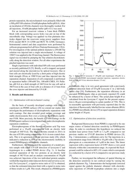
trypsinogen in a coated PDMS device (Fig. 1). A baseline<br />
resolved separation of the two proteins at an electric field<br />
strength of 463 V/cm and an analyte concentration of 50 M<br />
was achieved. Injections of the two individual protein samples<br />
verified the migration order (data not shown) and served as a control.<br />
For lysocyme C, a signal-to-noise ratio of 123 is obtained,<br />
so that the estimated detection limit with a signal-to-noise ratio<br />
of three results in 1 M. This result, obtained in a PDMS/quartz<br />
Fig. 1. Separation of lysozyme C (50 M) and trypsinogen (50 M) in a<br />
20 m × 20 m PDMS microchannel (pinched injection; separation electric<br />
field strength 463 V/cm, detection distance 3.5 cm).<br />
microfluidic chip, is in very good agreement with a previously<br />
reported detection limit of 0.9 M lysocyme C in a full-body<br />
quartz chip [26]. Furthermore, the separation efficiency in an<br />
uncoated PDMS/quartz chip as previously reported [9] could<br />
be enhanced by a factor of three. The actual plate height H as<br />
calculated from the lysozyme C signal for the present separation<br />
is 46 m (corresponding to a plate number of 730). This is<br />
in reasonable agreement with previously reported data for the<br />
injection of fluorescently labelled bovine serum albumin (BSA)<br />
in a PDMS microfluidic device [29] at equal separation distance<br />
(plate height of 30 m).<br />
3.2. Optimisation of detection sensitivity<br />
Background fluorescence from PDMS is expected to be one<br />
major source of limited detection sensitivity in PDMS/quartz<br />
chips. In order to corroborate this hypothesis we reduced the<br />
incident laser power from 5 mW to 1.1 W compared to our<br />
previous studies on UV-LIF detection in PDMS devices [9].<br />
Fig. 2 demonstrates a calibration curve for Trp injections in a<br />
concentration range from 100 nM to 10 M with a Trp electropherogram<br />
at a concentration of 1 M in the inset. The linear<br />
regression with a regression factor of 0.997 shows a very good<br />
linearity within this concentration range. As expected the background<br />
could be reduced to 605 ± 81 counts which corresponds<br />
to a reduction by a factor of 6.4 compared to our previous study<br />
[9].<br />
Further, we investigated the influence of spatial filtering on<br />
the detection sensitivity in our confocal setup by varying the<br />
pinhole diameter. Fig. 3 demonstrates the background corrected<br />
signal against the pinhole diameter obtained with electrokinetic<br />
injections of 250 nM tryptophan solutions into a single