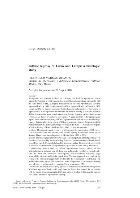
248 Diffuse leprosy of Lucio and Latapi - LEPRA Health in Action
248 Diffuse leprosy of Lucio and Latapi - LEPRA Health in Action
248 Diffuse leprosy of Lucio and Latapi - LEPRA Health in Action

Create successful ePaper yourself
Turn your PDF publications into a flip-book with our unique Google optimized e-Paper software.
Lepr Rev (2007) 78, <strong>248</strong>–260<br />
<strong>Diffuse</strong> <strong>leprosy</strong> <strong>of</strong> <strong>Lucio</strong> <strong>and</strong> Latapí: a histologic<br />
study<br />
FRANCISCO VARGAS-OCAMPO<br />
Instituto de Diagnóstico y Referencia Epidemiológicos (InDRE).<br />
México, D.F., México<br />
Accepted for publication 28 August 2007<br />
Summary<br />
Background <strong>and</strong> purpose Ladislao de la Pascua described the spotted or lazar<strong>in</strong>e<br />
<strong>leprosy</strong> for first time <strong>in</strong> 1844. Later on, <strong>Lucio</strong> <strong>and</strong> Alvarado studied <strong>and</strong> published it with<br />
the same names <strong>in</strong> 1852. Latapí re-discovered it <strong>in</strong> 1938 <strong>and</strong> reported it as ‘Spotted’<br />
<strong>leprosy</strong> <strong>of</strong> <strong>Lucio</strong> <strong>in</strong> 1948. Frenken named it diffuse <strong>leprosy</strong> <strong>of</strong> <strong>Lucio</strong> <strong>and</strong> Latapí <strong>in</strong> 1963.<br />
Latapí <strong>and</strong> Chévez-Zamora expla<strong>in</strong>ed that the fundamental condition <strong>of</strong> this variety <strong>of</strong><br />
<strong>leprosy</strong> was a diffuse generalised cutaneous <strong>in</strong>filtration, nam<strong>in</strong>g it pure <strong>and</strong> primitive<br />
diffuse lepromatosis, upon which necrotis<strong>in</strong>g lesions develop, call<strong>in</strong>g these lesions<br />
Fenómeno de <strong>Lucio</strong> or erythema necrotisans. A great number <strong>of</strong> histopathological<br />
reports have addressed the study <strong>of</strong> <strong>Lucio</strong>’s phenomenon, <strong>and</strong> few about the histologic<br />
changes that take place <strong>in</strong> the course <strong>of</strong> diffuse lepromatous <strong>leprosy</strong>. The purpose <strong>of</strong> this<br />
work is to report the histologic f<strong>in</strong>d<strong>in</strong>gs observed <strong>in</strong> the study <strong>of</strong> l70 cutaneous biopsies<br />
<strong>of</strong> diffuse <strong>leprosy</strong> <strong>of</strong> <strong>Lucio</strong> <strong>and</strong> Latapí <strong>and</strong> 30 <strong>of</strong> <strong>Lucio</strong>’s phenomenon.<br />
Methods This is a retrospective study, which <strong>in</strong>cluded the exam<strong>in</strong>ation <strong>of</strong> 200 biopsy<br />
sk<strong>in</strong> specimens from 199 patients with diffuse <strong>leprosy</strong> at different course <strong>of</strong> the<br />
disease. These cases were diagnosed <strong>in</strong> Mexico from 1970 to 2004.<br />
Results The histologic exam<strong>in</strong>ation revealed a vascular pattern affect<strong>in</strong>g all cutaneous<br />
vessels, characterised by five outst<strong>and</strong><strong>in</strong>g features: a) colonisation <strong>of</strong> endothelial cells<br />
by acid-fast bacilli, b) endothelial proliferation <strong>and</strong> marked thicken<strong>in</strong>g <strong>of</strong> vessel walls<br />
to the po<strong>in</strong>t <strong>of</strong> obliteration, c) angiogenesis, d) vascular ectasia, <strong>and</strong> e) thrombosis.<br />
Necrotis<strong>in</strong>g lesions seen <strong>in</strong> diffuse lepromatous <strong>leprosy</strong> displayed two<br />
histopathological patterns: one <strong>of</strong> them, non-<strong>in</strong>flammatory occlusive vasculopathy<br />
<strong>and</strong>, the other one, occlusive vasculopathy, leukocytoclastic vasculitis, large<br />
neutrophilic <strong>in</strong>filtrate <strong>and</strong> lobular panniculitis. The first appeared as a result <strong>of</strong> the<br />
course <strong>of</strong> the occlusive vasculopathy produced by the colonisation <strong>of</strong> endothelial cells<br />
by Mycobacterium leprae. The second, as a result <strong>of</strong> a previous occlusive vasculopathy<br />
plus a <strong>leprosy</strong> reaction which is considered here as variant <strong>of</strong> ENL.<br />
Conclusions Endothelial cell <strong>in</strong>jury appears to be the ma<strong>in</strong> event <strong>in</strong> the pathogenesis<br />
<strong>of</strong> diffuse <strong>leprosy</strong> <strong>of</strong> <strong>Lucio</strong> <strong>and</strong> Latapí. Once M. leprae has entered the endothelial<br />
cell, the micro-organism damages the blood vessels, lead<strong>in</strong>g to the specific changes<br />
seen <strong>in</strong> this variety <strong>of</strong> lepromatous <strong>leprosy</strong>.<br />
Correspondence to: F. Vargas-Ocampo, Laboratorio de dermatopatología, InDRE Carpio 470, México, D.F.,<br />
11340. México (e-mail: vargasocampo@correo.unam.mx)<br />
0305-7518//064053+13 $1.00 q Lepra <strong>248</strong>
Introduction<br />
In 1844, Ladislao de la Pascua, when Director <strong>of</strong> the ‘Hospital de los Lazar<strong>in</strong>os’ <strong>in</strong> Mexico<br />
City, described three cl<strong>in</strong>ical forms <strong>of</strong> <strong>leprosy</strong>, tuberculous (nodular), anaesthetic <strong>and</strong> a third<br />
characterised by the production <strong>of</strong> pa<strong>in</strong>ful red spots that went on to ulcerate. These patients<br />
were known <strong>in</strong> Mexico as ‘Lazar<strong>in</strong>os’. 1 Later on, Rafael <strong>Lucio</strong>, <strong>in</strong> collaboration with Ignacio<br />
Alvarado, published <strong>in</strong> 1852 a paper entitled ‘Opúsculo de la Enfermedad de San Lázaro o<br />
Elefanciasis de los Griegos’, 2 <strong>in</strong> which <strong>Lucio</strong> described his observations, made dur<strong>in</strong>g 8 years<br />
at the same ‘Hospital de los Lazar<strong>in</strong>os’ <strong>in</strong> Mexico City, <strong>of</strong> which he was Director after de la<br />
Pascua. He also dist<strong>in</strong>guished three cl<strong>in</strong>ical forms <strong>of</strong> <strong>leprosy</strong>: tuberculous (nodular),<br />
anaesthetic, <strong>and</strong> spotted (manchada), <strong>and</strong> paid special attention to the spotty form,<br />
characterised by the presence <strong>of</strong> red <strong>and</strong> pa<strong>in</strong>ful spots undergo<strong>in</strong>g necrosis, <strong>and</strong> which was<br />
strik<strong>in</strong>g because the sk<strong>in</strong> underwent characteristic changes accord<strong>in</strong>g to the stage <strong>of</strong> the<br />
condition. This l<strong>and</strong>mark paper by <strong>Lucio</strong> <strong>and</strong> Alvarado was gradually forgotten <strong>and</strong> this<br />
variety <strong>of</strong> <strong>leprosy</strong> was completely unknown among leprologists outside Mexico until Latapí<br />
<strong>and</strong> Chevez-Zamora 3 brought it to attention at the 5th International Congress <strong>of</strong> Leprosy held<br />
<strong>in</strong> Havana <strong>in</strong> 1948. That the authors expla<strong>in</strong>ed that the underly<strong>in</strong>g condition <strong>of</strong> this cl<strong>in</strong>ical<br />
form, unnoticed by <strong>Lucio</strong> <strong>and</strong> Alvarado, was generalised diffuse cutaneous <strong>in</strong>filtration, which<br />
Latapí <strong>and</strong> Chevez-Zamora named ‘pure <strong>and</strong> primitive diffuse lepromatosis’, upon which<br />
secondary cutaneous outbreaks developed. These outbreaks were regarded as a form <strong>of</strong> lepra<br />
reaction produced by multiple, acute, necrotis<strong>in</strong>g vasculitis for which Latapí proposed the<br />
name <strong>of</strong> ‘Fenómeno de <strong>Lucio</strong> or erythema necrotisans’. Before Latapí <strong>and</strong> Chevez-Zamora’s<br />
report, Mart<strong>in</strong>ez Baez had studied spotted lesions <strong>of</strong> five patients <strong>in</strong> 1941, 1,3 – 6,20 not<strong>in</strong>g the<br />
basic lepromatous structure, acute vasculitis with nuclear dust, <strong>and</strong> thicken<strong>in</strong>g <strong>and</strong> occlusion<br />
<strong>of</strong> the larger vessels produc<strong>in</strong>g necrosis. He described for the first time the histological<br />
changes that occurred <strong>in</strong> what Latapí would later name <strong>Lucio</strong>’s phenomenon.<br />
About 1943, Latapí dist<strong>in</strong>guished the pure <strong>and</strong> primitive diffuse <strong>leprosy</strong> that always<br />
beg<strong>in</strong>s as diffuse <strong>leprosy</strong>, <strong>and</strong> the secondary diffuse <strong>leprosy</strong>, which arises from an<br />
<strong>in</strong>determ<strong>in</strong>ate form; both display the same cl<strong>in</strong>ical aspect <strong>and</strong> the same histologic changes<br />
dur<strong>in</strong>g their course. 5,7 In 1963, Frenken <strong>in</strong>cluded both under the name <strong>of</strong> diffuse <strong>leprosy</strong> <strong>of</strong><br />
<strong>Lucio</strong> <strong>and</strong> Latapí, hav<strong>in</strong>g as fundamental characteristic, that the generalised <strong>in</strong>filtration never<br />
develops nodules. 1,5,7<br />
<strong>Diffuse</strong> <strong>leprosy</strong> <strong>of</strong> <strong>Lucio</strong> <strong>and</strong> Latapí is characterised by a diffuse cutaneous <strong>in</strong>filtration,<br />
without nodules, succulent or atrophic accord<strong>in</strong>g to the stage <strong>of</strong> progression. Dysesthesia,<br />
anhidrosis, alopecia, destructive rh<strong>in</strong>itis <strong>and</strong> telangiectasia, <strong>and</strong> a special variant <strong>of</strong> lepra<br />
reaction named <strong>Lucio</strong>’s phenomenon or erythema necrotisans (erythema necroticans).<br />
Histologically it has been reported that the f<strong>in</strong>d<strong>in</strong>g <strong>of</strong> heavy endothelial parasitisation by<br />
Mycobacterium leprae is a peculiar feature to diffuse <strong>leprosy</strong>.<br />
<strong>Diffuse</strong> <strong>leprosy</strong> <strong>of</strong> <strong>Lucio</strong> <strong>and</strong> Latapí 249<br />
4,5,8 – 12<br />
<strong>Lucio</strong>’s phenomenon is cl<strong>in</strong>ically characterised by crops <strong>of</strong> w<strong>in</strong>e-red irregular spots<br />
(manchas) with burn<strong>in</strong>g sensation, sharply del<strong>in</strong>eated, <strong>and</strong> capricious centre, which turn<br />
purpuric <strong>and</strong> become necrotic leav<strong>in</strong>g atrophic stellar scars. Histopathologically <strong>Lucio</strong>’s<br />
phenomenon has been reported to have two types <strong>of</strong> patterns. One <strong>of</strong> them <strong>in</strong>volves<br />
leukocytoclastic vasculitis as the underly<strong>in</strong>g pathologic change, 5,13,14 <strong>and</strong> the other,<br />
endothelial cell proliferation, thrombosis, a mild mononuclear cell <strong>in</strong>filtrate <strong>and</strong> ischemic<br />
necrosis. 15,16 The first pattern is thought to be due to an immune complex disease caused by<br />
leprae or sk<strong>in</strong> antigens. 17 In the second pattern, vascular damage is thought to be due to direct<br />
<strong>in</strong>vasion <strong>of</strong> M. leprae. 15,16
250<br />
<strong>Diffuse</strong> <strong>leprosy</strong> is considered to be the most anergic <strong>of</strong> the all-immunological spectrum <strong>of</strong><br />
<strong>leprosy</strong>. 18 In a study on leprom<strong>in</strong> responsiveness, Leiker reported that not all lepromatous<br />
patients are completely anergic to leprom<strong>in</strong>, as a weak response to leprom<strong>in</strong> had been<br />
observed <strong>in</strong> many such cases <strong>and</strong> it seemed that the diffuse lepromatous variety was the only<br />
truly anergic type <strong>of</strong> <strong>leprosy</strong>. 19 <strong>Lucio</strong> <strong>and</strong> other authors believed that this variety <strong>of</strong> the<br />
disease was exclusive to Mexico. Yet it is found not only <strong>in</strong> Mexico, but also <strong>in</strong> other<br />
countries. 8,13,21 – 24 The purpose <strong>of</strong> this study is to describe histological changes seen <strong>in</strong> 170<br />
cutaneous biopsies <strong>of</strong> diffuse <strong>leprosy</strong> <strong>of</strong> <strong>Lucio</strong> <strong>and</strong> Latapí <strong>and</strong> 30 <strong>of</strong> <strong>Lucio</strong>’s phenomenon.<br />
Materials <strong>and</strong> methods<br />
This is a retrospective study, which <strong>in</strong>cluded the exam<strong>in</strong>ation <strong>of</strong> 200 biopsy specimens <strong>of</strong><br />
diffuse <strong>leprosy</strong> <strong>of</strong> <strong>Lucio</strong> <strong>and</strong> Latapí obta<strong>in</strong>ed from 199 patients. In one patient two biopsy<br />
specimens were taken, one was from <strong>in</strong>filtrated sk<strong>in</strong> <strong>and</strong> other from a necrotic sk<strong>in</strong> lesion. The<br />
cases were diagnosed between 1970 <strong>and</strong> 2004 at the Laboratory <strong>of</strong> Dermatopathology <strong>of</strong> the<br />
Institute for Epidemiologic Diagnosis <strong>and</strong> Reference (InDRE) <strong>in</strong> Mexico City. Twenty-six<br />
cases were from the archive <strong>of</strong> the InDRE; 174, were <strong>in</strong>cluded <strong>in</strong> a previous report by the<br />
National Leprosy Control Programme. 25 Biopsies selected for this study were those that were<br />
taken from patients who had two or more physical signs <strong>of</strong> diffuse lepromatous <strong>leprosy</strong><br />
without nodules (DLL). The cl<strong>in</strong>ical data were obta<strong>in</strong>ed from the histology request forms.<br />
Specimens were dispatched <strong>in</strong> glass or plastic vials conta<strong>in</strong><strong>in</strong>g 10% formal<strong>in</strong> solution from<br />
different prov<strong>in</strong>ces <strong>of</strong> the country to the InDRE <strong>in</strong> Mexico City where all histological<br />
process<strong>in</strong>g was carried out. Specimens were processed rout<strong>in</strong>ely <strong>and</strong> sta<strong>in</strong>ed with<br />
haematoxyl<strong>in</strong> <strong>and</strong> eos<strong>in</strong>; the first 125 specimens with Fite-Faraco sta<strong>in</strong>, <strong>and</strong> the other 75<br />
specimens with a modified Fite-Faraco sta<strong>in</strong> us<strong>in</strong>g carbol-fuchs<strong>in</strong> solution to sta<strong>in</strong> acid-fast<br />
bacilli, Weigert’s iron haematoxyl<strong>in</strong> to sta<strong>in</strong> nuclei, <strong>and</strong> metanil yellow solution to sta<strong>in</strong> other<br />
tissue elements. 26 The <strong>leprosy</strong> programme uses the Madrid classification, 27 which considers<br />
two polar types: lepromatous <strong>and</strong> tuberculoid; <strong>and</strong> two groups: <strong>in</strong>determ<strong>in</strong>ate <strong>and</strong><br />
dimorphous. This classification does not <strong>in</strong>clude the variety diffuse lepromatous <strong>leprosy</strong>.<br />
Results<br />
F. Vargas-Ocampo<br />
The series was composed <strong>of</strong> 128 males <strong>and</strong> 71 females, with a male to female ratio 1·8:1; the<br />
ages ranged from 12 to 88 years, with a median age <strong>of</strong> 37 years <strong>and</strong> an average <strong>of</strong> 28.8 years.<br />
The elapsed time from the appearance <strong>of</strong> the first sign or symptom referable to <strong>leprosy</strong> ranged<br />
from 1 week to 12 years, with a median <strong>of</strong> 3 years <strong>and</strong> an average <strong>of</strong> 4.2 years. Out <strong>of</strong> the 200<br />
cases, 181 (90·5%) were from Pacific Coast states <strong>of</strong> Mexico <strong>and</strong> 19 (9·5%) from other states<br />
<strong>of</strong> Mexico.<br />
The biopsy specimens were obta<strong>in</strong>ed at different times <strong>in</strong> the course <strong>of</strong> the illness, <strong>and</strong><br />
<strong>in</strong>cluded those that showed apparently normal sk<strong>in</strong> <strong>and</strong> those with necrotis<strong>in</strong>g lesions; 170<br />
were from patients with diffuse <strong>leprosy</strong> without necrotis<strong>in</strong>g lesions, <strong>and</strong> 30 were from patients<br />
with diffuse <strong>leprosy</strong> with <strong>Lucio</strong>’s phenomenon. All patients had physical signs <strong>of</strong> DLL),<br />
ma<strong>in</strong>ly diffuse <strong>in</strong>filtration <strong>of</strong> the sk<strong>in</strong>, impairment <strong>of</strong> sensation (numbness <strong>of</strong> the dorsal aspects<br />
<strong>of</strong> the extremities, hypaesthesia, or aesthesia), alopecia <strong>of</strong> eyebrows <strong>and</strong> eyelashes, anhidrosis,<br />
<strong>and</strong> destructive rh<strong>in</strong>itis. All the biopsy specimens were obta<strong>in</strong>ed from sk<strong>in</strong> with sensory
impairment associated with another or other physical signs <strong>of</strong> DLL: 32 from apparently normal<br />
sk<strong>in</strong>, 82 from sk<strong>in</strong> with diffuse <strong>in</strong>filtration, 13 from atrophic sk<strong>in</strong>, 9 from hypochromic macules,<br />
7 from erythematous macules, <strong>and</strong> 30 from sk<strong>in</strong> with <strong>Lucio</strong>’s phenomenon, data on 27 cases<br />
were miss<strong>in</strong>g. Biopsy sites were: 141 from the limbs, 33 from the ear lobe, 12 from the trunk,<br />
data on 14 cases were miss<strong>in</strong>g. As the cl<strong>in</strong>ical diagnosis made by physicians <strong>of</strong> the control<br />
programme did not <strong>in</strong>clude diffuse <strong>leprosy</strong>, 86 <strong>of</strong> the request forms for histological study only<br />
had the cl<strong>in</strong>ical diagnosis ‘lepromatous’. Nonetheless, 62 had the cl<strong>in</strong>ical diagnosis <strong>of</strong> DLL <strong>and</strong><br />
8 the cl<strong>in</strong>ical diagnosis <strong>of</strong> pure <strong>and</strong> primitive diffuse lepromatosis (PPDL), because, these<br />
specific diagnoses were made by physicians with several years work<strong>in</strong>g <strong>in</strong> the programme.<br />
There were other cl<strong>in</strong>ical diagnoses: 7 <strong>of</strong> <strong>in</strong>determ<strong>in</strong>ate <strong>leprosy</strong>, 4 <strong>of</strong> dimorphous <strong>leprosy</strong> 1 <strong>of</strong><br />
tuberculoid <strong>leprosy</strong>, 30 <strong>of</strong> <strong>Lucio</strong> phenomenon, <strong>and</strong> 2 were without cl<strong>in</strong>ical diagnosis. The<br />
physical signs <strong>of</strong> <strong>leprosy</strong> reported by medical staff <strong>in</strong> the request forms for histological exam,<br />
apart from the lesions <strong>of</strong> <strong>Lucio</strong>’s phenomenon, correspond closely with DLL, thus, none <strong>of</strong> the<br />
199 patients had nodular lesions. The 199 (100%) presented impairment <strong>of</strong> sensation<br />
(hypaesthesia or anaesthesia or numbness <strong>of</strong> the extremities), 132 diffuse cutaneous <strong>in</strong>filtration<br />
with absence <strong>of</strong> nodules, 36 apparently normal sk<strong>in</strong>, 15 atrophic sk<strong>in</strong>, 9 hypochromic macules,<br />
7 erythematous macules, 87 anhidrosis, 126 alopecia <strong>of</strong> eyebrows, <strong>and</strong> 63 destructive rh<strong>in</strong>itis.<br />
The histopathological features were divided <strong>in</strong>to five stages: (1) early, (2) bacillary<br />
dissem<strong>in</strong>ation, (3) well-developed, (4) necrosis by vascular occlusion <strong>and</strong> (5) necrosis by<br />
vascular occlusion plus <strong>leprosy</strong> reaction. The changes observed dur<strong>in</strong>g these different<br />
histologic stages are described below.<br />
STAGE 1. EARLY<br />
Fifty-three biopsies comprised 33 males <strong>and</strong> 20 females (M:F ratio 1·7:1), age range 15 to 88<br />
years, (median 43 years <strong>and</strong> mean 43). The delay from the appearance <strong>of</strong> the first sign referable<br />
to <strong>leprosy</strong> was 2 months to 17 years (median 1 year, mean 1.4 years). The sk<strong>in</strong> where the<br />
specimens were taken was: diffuse <strong>in</strong>filtration 36, apparently normal with dysesthesia 3,<br />
atrophic 2, hypocromic macule 1, erythematous macule 2, no <strong>in</strong>formation 9. Biopsy sites were:<br />
<strong>in</strong>ferior extremity 18, upper extremity 19, thorax 4, <strong>and</strong> ear lobe 9, no <strong>in</strong>formation 3.<br />
Histology<br />
Blood vessels looked dilated with a thick wall l<strong>in</strong>ed with normal or plump proliferative<br />
endothelial cells. Slight <strong>and</strong> apparently <strong>in</strong>significant foci <strong>of</strong> <strong>in</strong>filtration were present <strong>in</strong> the<br />
dermis, arranged around dilated blood vessels. Very scarce bacilli could be observed <strong>in</strong> the<br />
endothelial cells <strong>in</strong> few <strong>of</strong> the large vessels <strong>and</strong> <strong>in</strong> some nerve trunks <strong>of</strong> the deep dermis.<br />
STAGE 2. BACILLARY DISSEMINATION<br />
<strong>Diffuse</strong> <strong>leprosy</strong> <strong>of</strong> <strong>Lucio</strong> <strong>and</strong> Latapí 251<br />
Sixty-two biopsies comprised 32 males <strong>and</strong> 30 females (M:F ratio 1·1:1; the age range 15<br />
years to 85 years, (median 39 years). The delay from the appearance <strong>of</strong> the first sign referable<br />
to <strong>leprosy</strong> ranged from 1 month to 22 years (median 3 years, mean 3.4 years). The sk<strong>in</strong> where<br />
the specimens were taken were: diffuse <strong>in</strong>filtration 34, dysesthesia, anhidrosis <strong>and</strong> alopecia 5,<br />
atrophic 5, hypochromic macule 7, erythematous macule 3, no <strong>in</strong>formation 8. The biopsy<br />
sites were: <strong>in</strong>ferior extremity 20, upper extremity 22, thorax 2, abdomen 1, ear lobe 13, no<br />
<strong>in</strong>formation 4.
252<br />
F. Vargas-Ocampo<br />
Histology<br />
Dissem<strong>in</strong>ation <strong>of</strong> acid-fast-bacilli (AFB) extended progressively from the deep plexus to all<br />
cutaneous blood vessels, first, <strong>in</strong>to the blood vessels <strong>of</strong> the subcutis <strong>and</strong> reticular dermis; then<br />
to vessels <strong>of</strong> the superficial plexus; <strong>and</strong> f<strong>in</strong>ally to capillaries <strong>of</strong> the papillary dermis. The<br />
vessels showed acid-fast bacilli <strong>in</strong>side swollen endothelial cells, thrombosis, passive<br />
congestion <strong>and</strong> ectasia. The lumen <strong>of</strong> the large vessels was reduced <strong>in</strong> size by <strong>in</strong>timal<br />
proliferative thicken<strong>in</strong>g. Small <strong>in</strong>filtrates <strong>of</strong> mononuclear cells were arranged around blood<br />
vessels <strong>of</strong> the dermis <strong>and</strong> subcutaneous fat tissue.<br />
STAGE 3. WELL-DEVELOPED<br />
Fifty-five biopsies comprised 34 males <strong>and</strong> 21 females, M:F ratio 1·6:1; age range 12 years to<br />
77 years, (median 28 years, mean 34·6. The delay from the appearance <strong>of</strong> the first sign<br />
referable to <strong>leprosy</strong> ranged from 4 months to 20 years (median 2 years, mean 3.4 years). The<br />
sk<strong>in</strong> where the specimens were taken were: diffuse <strong>in</strong>filtration 29, dysesthesia, anhidrosis <strong>and</strong><br />
alopecia) 7, atrophic sk<strong>in</strong> with dysesthesia, anhidrosis <strong>and</strong> alopecia 6, hypocromic macule 1,<br />
erythematous macule 2, no <strong>in</strong>formation 10. Biopsy sites were: <strong>in</strong>ferior extremity 26, upper<br />
extremity 9, thorax 5, ear lobe 7, no <strong>in</strong>formation 8.<br />
Histology<br />
Well-developed lesions were characterised by vascular ectasia, focal passive congestion,<br />
thrombi <strong>and</strong> an <strong>in</strong>crease <strong>in</strong> capillaries. The blood vessels <strong>of</strong> the dermis <strong>and</strong> the subcutis<br />
showed a swollen <strong>and</strong> prom<strong>in</strong>ent endothelium, thick wall <strong>and</strong> narrowed or obliterated lumen<br />
(Figure 1A).<br />
The <strong>in</strong>filtrate, which was composed <strong>of</strong> lymphocytes <strong>and</strong> macrophages, was slight, <strong>in</strong> the<br />
dermis cuff<strong>in</strong>g the blood vessels (Figure 1A), <strong>and</strong> <strong>in</strong> the subcutis <strong>in</strong>vad<strong>in</strong>g the subcutaneous<br />
fat. Variable numbers <strong>of</strong> Virchow cells were diffusely distributed around blood vessels <strong>in</strong> the<br />
dermis <strong>and</strong> subcutaneous fat. Acid-fast sta<strong>in</strong><strong>in</strong>g showed bacilli <strong>in</strong> the perivascular<br />
<strong>in</strong>flammatory <strong>in</strong>filtrate (Figure 1B) <strong>and</strong>, <strong>in</strong> the most cases with<strong>in</strong> one or more <strong>of</strong> the<br />
endothelial cells <strong>of</strong> the cutaneous blood vessels (Figures 1C–1E).<br />
STAGE 4. ISCHAEMIC NECROSIS DUE TO NON-INFLAMMATORY VASCULAR<br />
OCCLUSION<br />
Eight biopsies comprised 6 males <strong>and</strong> 2 females, M:F ratio 3:1; age range 29 years to 67<br />
years, (median 38 years mean 3.1 years). The delay from the appearance <strong>of</strong> the first sign<br />
referable to <strong>leprosy</strong> ranged from 6 days to 14 years (median 2 years, mean 3.1 years). All had<br />
necrotic lesions <strong>of</strong> the sk<strong>in</strong>, from which the specimens were taken. Biopsy sites were: <strong>in</strong>ferior<br />
extremity 2, upper extremity 3, <strong>and</strong> abdomen 1, no <strong>in</strong>formation 2.<br />
Histology<br />
The ma<strong>in</strong> feature <strong>in</strong> this fourth stage was a severe damage <strong>of</strong> blood vessels. The presence <strong>of</strong><br />
M. leprae <strong>in</strong> endothelial cells seemed responsible for this change. Histological appearance<br />
was characterised by necrosis <strong>of</strong> epidermis <strong>and</strong> superficial dermis, angiogenesis, ectasia,
<strong>Diffuse</strong> <strong>leprosy</strong> <strong>of</strong> <strong>Lucio</strong> <strong>and</strong> Latapí 253<br />
Figure 1. Each section is from a different third stage case. 1A) shows a large <strong>and</strong> thick subcutaneous blood vessel,<br />
<strong>and</strong> <strong>in</strong> the dermis, th<strong>in</strong> perivascular <strong>in</strong>flammatory cuffs with a streak-like appearance. 1B) A b<strong>and</strong> <strong>of</strong> <strong>in</strong>flammatory<br />
<strong>in</strong>filtrate is composed <strong>of</strong> mononuclear cells, without foam cells, conta<strong>in</strong><strong>in</strong>g numerous bacilli. Two capillaries <strong>in</strong><br />
transverse section with bacilli (ca) can be seen. 1C–1E) shows high power views <strong>of</strong> blood vessels with numerous<br />
bacilli <strong>in</strong> endothelial cells <strong>and</strong> absence <strong>of</strong> <strong>in</strong>filtrated <strong>in</strong>flammatory, (1C) <strong>in</strong> superficial dermis, 1D) <strong>in</strong> mid dermis, <strong>and</strong><br />
1E) <strong>in</strong> deep dermis (1A. H&E; £ 35, 1B. Fite-Faraco; X400, !C-1E. Fite-Faraco; £ 1000).<br />
passive venous congestion <strong>and</strong> vascular occlusion caused by lum<strong>in</strong>al thrombi <strong>and</strong>/or by<br />
thicken<strong>in</strong>g <strong>of</strong> the vessel wall. Angiogenesis, ectasia, passive venous congestion, <strong>and</strong> lum<strong>in</strong>al<br />
thrombi were most evident <strong>in</strong> superficial dermis (Figures 2B, 2C, 3F) <strong>and</strong> <strong>in</strong> middle dermis<br />
(Figure 2A).<br />
Thicken<strong>in</strong>g <strong>of</strong> vessel wall with reduction <strong>of</strong> the lumen <strong>and</strong> sometimes with obliteration<br />
was seen <strong>in</strong> vessels <strong>of</strong> the dermis (Figures 3B-3E) <strong>and</strong> <strong>of</strong> the subcutis (Figure 3A). The<br />
<strong>in</strong>flammatory <strong>in</strong>filtrate <strong>and</strong> the presence <strong>of</strong> bacilli were similar to that <strong>of</strong> the previous third<br />
stage.<br />
STAGE 5. ISCHAEMIC NECROSIS DUE TO INFLAMMATORY VASCULAR OCCLUSION<br />
PLUS LEPROSY REACTION<br />
Twenty-two biopsies comprised 9 males <strong>and</strong> 13 females, M:F ratio <strong>of</strong> 1:1·4; age range 12<br />
years to 75 years, (median 39 years, mean 7.8 years). Delay from the appearance <strong>of</strong> the first<br />
sign referable to <strong>leprosy</strong> ranged from 3 months to 28 years (median 3 years, mean 7.8 years).<br />
All had necrotic lesions <strong>of</strong> the sk<strong>in</strong>, from which the specimens were taken. The part <strong>of</strong> the<br />
body where the biopsies specimens were taken was: <strong>in</strong>ferior extremity 9, upper extremity 11,<br />
no <strong>in</strong>formation 2.
254<br />
F. Vargas-Ocampo<br />
Figure 2. 2A) third stage lesion, show<strong>in</strong>g a blood vessel distended by thrombus. 2B) this is a transition lesion between<br />
third <strong>and</strong> fourth stages. The epidermis appears stretched <strong>and</strong> th<strong>in</strong>ned with pyknotic malpighian cells. In the<br />
dermis, beneath <strong>of</strong> epidermis, thrombi (T) occlude the lum<strong>in</strong>a <strong>of</strong> some superficial capillaries; there is no<br />
<strong>in</strong>flammatory <strong>in</strong>filtrate. 2C) from a lesion <strong>in</strong> fourth stage. This superficial lesion shows loss <strong>of</strong> epidermis, thrombosis<br />
<strong>and</strong> congestion <strong>of</strong> capillaries papillae Note the absence <strong>of</strong> <strong>in</strong>flammatory cells. This lesion heals “restitutio ad<br />
<strong>in</strong>tegrum”. 2D) from a case <strong>in</strong> fourth stage, exhibits loss <strong>of</strong> tissue that <strong>in</strong>cludes the epidermis <strong>and</strong> some superficial<br />
dermis, <strong>and</strong> a superficial vessel occluded by an amorphous homogeneous <strong>and</strong> acidophilic substance. (2A. H&E;<br />
£ 100, 2B–2D. H&E; £ 250).<br />
Histology<br />
This fifth stage showed changes superimposed on the pre-exist<strong>in</strong>g occlusive vascular damage.<br />
They <strong>in</strong>cluded necrosis <strong>of</strong> epidermis (Figure 4B) <strong>and</strong> superficial dermis, hyal<strong>in</strong>isation <strong>and</strong>/or<br />
deposits <strong>of</strong> fibr<strong>in</strong> <strong>in</strong> small blood vessel walls <strong>of</strong> the dermis <strong>and</strong> the subcutaneous fat tissue.<br />
(Figures 4A–4E).<br />
The cellular <strong>in</strong>filtrate around these vessels consisted <strong>of</strong> neutrophils, nuclear dust, a few<br />
eos<strong>in</strong>ophils, lymphocytes, <strong>and</strong> <strong>in</strong> rare cases red blood cells. Large vessels <strong>in</strong> the deeper<br />
reticular dermis <strong>and</strong> <strong>in</strong> the subcutis showed pronounced thicken<strong>in</strong>g <strong>and</strong> <strong>in</strong>timal proliferation<br />
caus<strong>in</strong>g reduction <strong>in</strong> vascular lumen <strong>and</strong> sometimes complete occlusion; their wall was<br />
<strong>in</strong>filtrated by neutrophils <strong>and</strong> lymphocytes (Figure 4E). In addition, a dense predom<strong>in</strong>antly<br />
neutrophil <strong>in</strong>filtrate was seen <strong>in</strong>volv<strong>in</strong>g the deeper dermis <strong>and</strong> the lobular portion <strong>of</strong> the
subcutaneous fat tissue as a lobular panniculitis. In one case there was a granulomatous<br />
reaction with<strong>in</strong> <strong>and</strong> around a large blood vessel <strong>of</strong> the subcutis.<br />
CHANGES FOUND IN OTHER STRUCTURES OF THE SKIN<br />
AFB were found not only <strong>in</strong> blood vessels, but also <strong>in</strong> lymph vessels, nerves, hair follicles,<br />
erector pili muscles, sebaceous gl<strong>and</strong>s, sweat gl<strong>and</strong>s, <strong>and</strong> <strong>in</strong> a few cases <strong>in</strong> the different layers<br />
<strong>of</strong> the epidermis (Table 1).<br />
Usually, bacilli were more abundant <strong>in</strong> sections <strong>of</strong> the biopsy specimens taken from the<br />
ear lobe. Nerves <strong>and</strong> epidermal appendages showed different degrees <strong>of</strong> <strong>in</strong>vasion,<br />
degeneration <strong>and</strong> destruction by the <strong>in</strong>flammatory <strong>in</strong>filtrate, these changes be<strong>in</strong>g more<br />
noticeable <strong>in</strong> the ischaemic tissue necrosis stages.<br />
Discussion<br />
<strong>Diffuse</strong> <strong>leprosy</strong> <strong>of</strong> <strong>Lucio</strong> <strong>and</strong> Latapí 255<br />
Figure 3. 3A) from a biopsy <strong>of</strong> a third stage lesion, show<strong>in</strong>g a large ve<strong>in</strong> <strong>in</strong> the subcutis with very thick wall, lumen<br />
almost occluded <strong>and</strong> bacilli with<strong>in</strong> endothelial cells. There is no <strong>in</strong>flammatory <strong>in</strong>filtrate. (3B–3D) three illustrations<br />
from different third stage cases. They show vessels <strong>in</strong> the reticular dermis with prom<strong>in</strong>ent endothelial proliferation<br />
<strong>and</strong> numerous bacilli with<strong>in</strong> a small (L <strong>in</strong> 3B) or obliterated vascular lumen. 3E) from fourth stage biopsy, shows a<br />
blood vessel occluded by extensive <strong>in</strong>timal proliferation <strong>and</strong> dermal necrosis. 3F) another example from a fourth<br />
stage lesion, exhibits superficial necrosis with loss <strong>of</strong> epidermis, obliterative changes <strong>of</strong> two superficial blood vessels<br />
with homogenization <strong>and</strong> hyal<strong>in</strong>ization <strong>of</strong> their walls. (3A. Fite-Faraco sta<strong>in</strong>; £ 250, 3B–3D. Fite-Faraco sta<strong>in</strong>; £<br />
1000, 3E. H&E; £ 250, 3F. H&E; £ 400).<br />
Histopathologically, five stages <strong>of</strong> DLL were considered <strong>in</strong> this study, each <strong>of</strong> them hav<strong>in</strong>g<br />
different features. When the cl<strong>in</strong>ical data are correlated with the histological changes, the<br />
three first stages, early, dissem<strong>in</strong>ated, <strong>and</strong> well-developed, correspond to the phase <strong>in</strong> which<br />
patients have diffuse <strong>in</strong>filtration <strong>of</strong> entire sk<strong>in</strong> The fourth <strong>and</strong> fifth stages <strong>of</strong> ischaemic<br />
necrosis correspond to <strong>Lucio</strong>’s phenomenon or erythema necroticans. Endothelial cells seem<br />
to be the ma<strong>in</strong> targets <strong>of</strong> the organism, <strong>and</strong> follow<strong>in</strong>g <strong>in</strong>vasion <strong>of</strong> M. leprae vascular damage
256<br />
F. Vargas-Ocampo<br />
Figure 4. All sections are from a fifth stage case. 4A) shows necrosis <strong>of</strong> the epidermis, leukocytoclastic vasculitis <strong>in</strong><br />
small blood vessels <strong>of</strong> the dermis <strong>and</strong> subcutis. In the lower left is an almost occluded blood vessel <strong>in</strong>filtrated by<br />
<strong>in</strong>flammatory cells. 4B) necrosis <strong>of</strong> the epidermis <strong>and</strong> leukocytoclastic vasculitis. 4C) leukocytclastic vasculitis <strong>in</strong> the<br />
upper dermis. 4D) This illustration shows two small vessels, <strong>in</strong> the lower dermis, sta<strong>in</strong>ed acidophillicaly (v) <strong>and</strong><br />
surrounded by <strong>in</strong>flammatory <strong>in</strong>filtrate composed largely by neutrophils mixed with mononuclear cells. 4E)<br />
leukocytoclastic vasculitis <strong>in</strong> the subcutis. 4F) the subcutaneous blood vessel seen <strong>in</strong> 4A, shows endothelial<br />
proliferation <strong>and</strong> heavy <strong>in</strong>filtration by neutrophils, lymphocytes <strong>and</strong> histiocytes. (4A. H&E; £ 35, 4B. H&E; £ 160,<br />
4C. H&E; £ 400, 4D. H&E; £ 160, 4E. H&E; £ 400, 4F. H&E; £ 250).
Table 1. Presence <strong>of</strong> Mycobacterium leprae <strong>in</strong> different structures <strong>of</strong> the sk<strong>in</strong> <strong>in</strong> the 200 cases <strong>of</strong> this study<br />
Structure<br />
Cases with the structure<br />
present <strong>in</strong> sections Percentage<br />
<strong>Diffuse</strong> <strong>leprosy</strong> <strong>of</strong> <strong>Lucio</strong> <strong>and</strong> Latapí 257<br />
Cases with M. leprae<br />
<strong>in</strong> the structure Percentage<br />
Epidermis: 200 100 3 1·5<br />
Basal cells 200 100 3 1·5<br />
Melanocytes 200 100 2 1<br />
Sp<strong>in</strong>ous cells 200 100 2 1<br />
Granulous cells 200 100 1 0·5<br />
Corneocytes 200 100 1 0·5<br />
Hair follicles 102 51 47 46<br />
Sebaceous gl<strong>and</strong>s 81 40,5 6 7·4<br />
Pilli-erector muscles 101 50,5 51 50·5<br />
Sweat gl<strong>and</strong>s 155 77,5 12 7·7<br />
Nerves 43 21,5 40 93<br />
Lymp vessels 23 11·5 15 65·22<br />
Blood vessels 200 100 100 100<br />
occurs, suggest<strong>in</strong>g that the colonisation <strong>of</strong> the endothelial cells by acid-fast bacilli could be<br />
responsible for this damage. At the early stage, the changes consist <strong>of</strong> dilated vessels,<br />
angiogenesis, <strong>and</strong> congestion. Endothelial bacillation varies considerably with the<br />
progression <strong>of</strong> lesions. In the beg<strong>in</strong>n<strong>in</strong>g, there are very few bacilli <strong>in</strong> the endothelial cells<br />
<strong>of</strong> large blood vessels. As time goes by, organisms <strong>in</strong>crease with<strong>in</strong> the endothelial cells. Later,<br />
bacilli spread progressively from the deep plexus to mid <strong>and</strong> small-sized vessels <strong>of</strong> the dermis<br />
<strong>and</strong> subcutis to give rise to a well-developed histological lesion <strong>in</strong> which most <strong>of</strong> the sections<br />
show all blood vessels hav<strong>in</strong>g a bacillated endothelium, a f<strong>in</strong>d<strong>in</strong>g already reported by<br />
Mart<strong>in</strong>ez-Baez, who said: “: ::every time one sees the picture <strong>of</strong> a blood-vessel, one can<br />
observe that the mentioned bacilli are present <strong>in</strong> one or more to the endothelial cells”. 4<br />
The presence <strong>and</strong> constitution <strong>of</strong> the <strong>in</strong>filtrate were variable <strong>and</strong> did not seem to be related<br />
to the age <strong>of</strong> the lesion. Biopsies <strong>of</strong> some patients at a well-developed histological stage <strong>and</strong><br />
with a long cl<strong>in</strong>ical evolution up to 10 years showed only a slight perivascular mononuclear<br />
<strong>in</strong>filtrate, while other cases with well-developed histological lesions but with a short cl<strong>in</strong>ical<br />
history showed a dense mononuclear <strong>in</strong>filtrate. Relatively few Virchow-cells were seen<br />
compared with the great number <strong>of</strong> bacilli.<br />
The fourth stage <strong>of</strong> ischaemic necrosis due to non-<strong>in</strong>flammatory vascular occlusion is<br />
characterised histologically by the presence <strong>of</strong> necrotic cutaneous lesions due to vascular<br />
occlusion by endothelial proliferation or thrombosis, <strong>and</strong> cl<strong>in</strong>ically it co<strong>in</strong>cides with the<br />
appearance <strong>of</strong> discrete purple-coloured pa<strong>in</strong>ful spots, which lead to ulceration <strong>of</strong> the sk<strong>in</strong><br />
without systemic symptoms. It should be stressed that the fourth stage <strong>of</strong> ischaemic necrosis<br />
due to non-<strong>in</strong>flammatory vascular occlusion seems to be caused by vascular <strong>in</strong>jury <strong>in</strong>duced by<br />
<strong>in</strong>vasion <strong>of</strong> the endothelial cells by Mycobacterium leprae 15,16 <strong>and</strong> that tissue necrosis is due<br />
to vascular occlusion by proliferative endothelium or thrombosis. At this stage systemic<br />
symptoms do not develop <strong>and</strong> these are the cases that do not respond to thalidomide. This<br />
pattern, the damage <strong>of</strong> which is seen with or without sparse <strong>in</strong>flammatory cells, seems to<br />
correspond to the one that Rea used the term vasculosis. 28<br />
The fifth stage <strong>of</strong> ischaemic necrosis appears as an acute reactional state that may<br />
supervene <strong>in</strong> such patients <strong>and</strong> aggravates the exist<strong>in</strong>g vascular damage. It is manifested<br />
microscopically by necrosis <strong>of</strong> the superficial dermis <strong>and</strong> epidermis, leukocytoclastic
258<br />
vasculitis <strong>in</strong> small blood vessel <strong>of</strong> the dermis <strong>and</strong> <strong>of</strong> the subcutis, <strong>and</strong> lobular panniculitis.<br />
Mid-sized <strong>and</strong> large blood vessels show <strong>in</strong>timal proliferative thicken<strong>in</strong>g, a narrow vascular<br />
lumen, <strong>and</strong> walls <strong>in</strong>filtrated by neutrophils <strong>and</strong> lymphocytes.<br />
The fifth stage <strong>of</strong> ischaemic necrosis is considered here as a severe variant <strong>of</strong> ENL. 29<br />
When this happens the cl<strong>in</strong>ical disease is exacerbated; the number <strong>of</strong> spots <strong>in</strong>creases suddenly<br />
<strong>in</strong> several parts <strong>of</strong> the body, the pre-exist<strong>in</strong>g necrotic cutaneous lesions become aggravated<br />
<strong>and</strong> systemic symptoms appear. It is suggested that ENL <strong>and</strong> <strong>Lucio</strong>’s phenomenon may result<br />
from similar immunological mechanisms, 10 both show immune-vasculitis result<strong>in</strong>g from<br />
deposits <strong>of</strong> complement-fix<strong>in</strong>g immune complexes <strong>in</strong> small blood vessel walls, 17,29 <strong>and</strong> both<br />
are may respond to thalidomide. In patients with either ENL or <strong>Lucio</strong>’s Phenomenon,<br />
deposits <strong>of</strong> IgG <strong>and</strong> C3, as well as circulat<strong>in</strong>g immune complexes have been found <strong>in</strong> the wall<br />
<strong>of</strong> small blood vessels. 17 It is known that immune complexes activate the complement system<br />
<strong>and</strong> become chemotatic for neutrophils, which <strong>in</strong>gest the immune complexes 30 <strong>and</strong> are<br />
subsequently destroyed <strong>in</strong> the form <strong>of</strong> nuclear fragmentation. Activation <strong>of</strong> the clott<strong>in</strong>g<br />
system results <strong>in</strong> the conversion <strong>of</strong> fibr<strong>in</strong>ogen to fibr<strong>in</strong>. The histological picture shows<br />
neutrophils, nuclear dust, <strong>and</strong> fibr<strong>in</strong> <strong>in</strong> the wall <strong>of</strong> small blood vessels, features that establish<br />
the diagnosis <strong>of</strong> leukocytoclastic vasculitis (LCV). 31 Cl<strong>in</strong>ically different manifestations are<br />
described to LCV, <strong>in</strong>clud<strong>in</strong>g macules, papules, nodules, vesicles, bullae, <strong>and</strong> ulcers. 32<br />
Some immunological, histological <strong>and</strong> cl<strong>in</strong>ical aspects dist<strong>in</strong>guish <strong>Lucio</strong>’s phenomenon<br />
from ENL. Immunologically, <strong>Lucio</strong>’s phenomenon occurs <strong>in</strong> patients considered the most<br />
anergic <strong>of</strong> the all-immunological spectrum <strong>of</strong> <strong>leprosy</strong>. Histologically, <strong>Lucio</strong>’s phenomenon<br />
starts with heavy colonisation <strong>of</strong> the endothelial cells by acid-fast bacilli. It develops <strong>in</strong> a<br />
tissue that already has severe vascular damage, <strong>and</strong> it is always associated with sk<strong>in</strong> necrosis.<br />
Cl<strong>in</strong>ically, <strong>Lucio</strong>’s phenomenon is an erythema necorcitans, characterised by red <strong>and</strong> pa<strong>in</strong>ful<br />
spots (manchas) that progress to necrosis <strong>and</strong> ulceration leav<strong>in</strong>g atrophic stellar scars,<br />
occurr<strong>in</strong>g <strong>in</strong> a sk<strong>in</strong> with diffuse generalised <strong>in</strong>filtration <strong>and</strong> complete absence <strong>of</strong> nodules, <strong>and</strong><br />
appear<strong>in</strong>g <strong>in</strong> untreated patients. In this study, one case <strong>of</strong> granulomatous vasculitis <strong>in</strong> the<br />
subcutis was only observed <strong>in</strong> patients <strong>in</strong> this stage; a change that could contribute to<br />
anoxia. 12<br />
Another noteworthy fact, <strong>in</strong> stages four <strong>and</strong> five, was the f<strong>in</strong>d<strong>in</strong>g <strong>of</strong> vascular occlusion by<br />
thrombi, with no signs <strong>of</strong> perivascular <strong>in</strong>flammation. The endothelium has on its surface a<br />
number <strong>of</strong> anticoagulant <strong>and</strong> procoagulation properties which, when perturbed, may lead to<br />
<strong>in</strong>travascular coagulation. 33 Therefore the activation <strong>of</strong> the endothelial cell by organisms can<br />
trigger a cascade <strong>of</strong> events lead<strong>in</strong>g to the conversion <strong>of</strong> fibr<strong>in</strong>ogen to fibr<strong>in</strong> <strong>in</strong>travascular <strong>and</strong><br />
thrombosis that occlude arterioles <strong>and</strong> capillaries. 34 – 36 <strong>Lucio</strong> <strong>and</strong> Alvarado remarked <strong>in</strong> their<br />
work that <strong>in</strong> these patients the level <strong>of</strong> fibr<strong>in</strong> was always elevated, <strong>and</strong> the patients bleed less<br />
dur<strong>in</strong>g a surgical amputation. 2<br />
The nasal mucosa is <strong>in</strong>volved early <strong>in</strong> the course <strong>of</strong> <strong>Lucio</strong>’s <strong>leprosy</strong> 2 <strong>and</strong> at least 95% <strong>of</strong><br />
all patients with lepromatous <strong>leprosy</strong> have nasal <strong>in</strong>volvement too. 37 Leprosy bacilli may<br />
spread though the bloodstream from the nose to another area <strong>of</strong> the body, among them the<br />
sk<strong>in</strong>.<br />
38 – 41<br />
F. Vargas-Ocampo<br />
The results <strong>of</strong> this study support the hypothesis that <strong>in</strong> <strong>Lucio</strong>’s <strong>leprosy</strong> there are <strong>leprosy</strong><br />
bacilli <strong>in</strong> endothelial cells throughout the vasculature that promote the liberation <strong>of</strong><br />
substances by the endothelium that <strong>in</strong>duce vascular reactivity as manifested by distended<br />
vessels, morphological changes <strong>of</strong> the endothelial cells, areas <strong>of</strong> passive venous congestion<br />
<strong>and</strong> foci <strong>of</strong> <strong>in</strong>travascular clott<strong>in</strong>g, which proceed to dissem<strong>in</strong>ated <strong>in</strong>travascular coagulation<br />
(DIC). DIC is one <strong>of</strong> the causes <strong>of</strong> death among these patients. 14,21,22
It is suggested here that <strong>in</strong> all cases <strong>of</strong> lepromatous <strong>leprosy</strong>, be they nodular or diffuse, the<br />
first lesion is the same: the colonisation <strong>of</strong> the endothelium by bacilli <strong>and</strong> that the course <strong>of</strong><br />
the disease be determ<strong>in</strong>ed by the resistance <strong>of</strong> each patient to the micro-organism.<br />
Suitable treatment can reverse the obliterative changes. Sk<strong>in</strong> biopsies from treated<br />
patients show vessels cleared <strong>of</strong> organisms. Some solid bacilli, however, can rema<strong>in</strong> <strong>in</strong> the<br />
media <strong>of</strong> great blood vessels, <strong>and</strong> persist <strong>in</strong> spite <strong>of</strong> treatment. In vessels <strong>of</strong> treated patients<br />
thickness <strong>of</strong> the walls dim<strong>in</strong>ishes gradually <strong>and</strong> the lumen becomes wide <strong>and</strong> clean, the<br />
disease does not progress <strong>and</strong> necrosis does not occur. Treatment with dapsone <strong>and</strong><br />
rifampic<strong>in</strong> 3,6,15,16,42,43 is associated with the prompt cessation <strong>of</strong> new acute lesions. 3,6,15,43<br />
Consequently, the restoration <strong>of</strong> a normal endothelial cell state would be an important<br />
objective <strong>in</strong> the treatment <strong>of</strong> a patient with diffuse <strong>leprosy</strong> <strong>of</strong> <strong>Lucio</strong> <strong>and</strong> Latapí. The two<br />
important criteria for this diagnosis are: 1) diffuse non-nodular <strong>in</strong>filtration, <strong>and</strong> 2) heavy<br />
vascular endothelial parasitisation by M. leprae. 12<br />
Conclusions<br />
Histopathological f<strong>in</strong>d<strong>in</strong>gs suggest that diffuse <strong>leprosy</strong> <strong>of</strong> <strong>Lucio</strong> <strong>and</strong> Latapí is a vascular<br />
disorder produced by the <strong>in</strong>vasion <strong>of</strong> vascular endothelial cells by Mycobacterium leprae. It<br />
would seem that once the bacilli have entered the endothelial cell, they <strong>in</strong>duce endothelial cell<br />
activation that leads to specific morphologic <strong>and</strong> functional changes, among which are<br />
vascular ectasia, proliferation <strong>and</strong> swell<strong>in</strong>g <strong>of</strong> the endothelium with thickness <strong>of</strong> the vessel<br />
walls, angiogenesis, <strong>and</strong> activation <strong>of</strong> clott<strong>in</strong>g which gives rise to thrombosis. These changes<br />
characterise this variety <strong>of</strong> lepromatous <strong>leprosy</strong>. 3 – 5,7 – 16,18,20 – 25,28,41,43 The necrotis<strong>in</strong>g<br />
lesions <strong>of</strong> <strong>Lucio</strong>’s phenomenon are due first, to occlusive vasculopathy, without systemic<br />
symptoms, 16,17 <strong>and</strong> secondly, to a lepra reaction that aggravates vascular damage <strong>and</strong> causes<br />
systemic signs <strong>and</strong> symptoms.<br />
References<br />
1 Rodríguez O, Lepra de <strong>Lucio</strong> I. Historia y concepto. Dermatología Rev. Mex, 1978; 22: 117–140.<br />
2 <strong>Lucio</strong> R, Alvarado I. “Opúsculo sobre el mal de San Lázaro o elefancíasis de los Griegos”. In: González-UrueñaJ<br />
(ed). La Lepra en México El Ateneo, Buenos Aires, Argent<strong>in</strong>a 1941, pp. 199–231.<br />
3 Latapí F, Chévez-Zamora A. The “spotted” <strong>leprosy</strong> <strong>of</strong> <strong>Lucio</strong>: an <strong>in</strong>troduction to its cl<strong>in</strong>ical <strong>and</strong> histological study.<br />
Int J Lepr, 1948; 16: 421–430.<br />
4 Martínez-Báez M. Nota prelim<strong>in</strong>ar sobre la histopatología de las manifestaciones cutáneas de la “forma de <strong>Lucio</strong>”<br />
de la lepra. Rev Inst Salub Enf Trop, 1941; 2: 245–256.<br />
5 Frenken JH. <strong>Diffuse</strong> <strong>leprosy</strong> <strong>of</strong> <strong>Lucio</strong> <strong>and</strong> Latapí. De Wit, Inc., Oranjest. Aruba, Netherl<strong>and</strong>s Antilles 1963.<br />
6 Novales J, Lepra de <strong>Lucio</strong>, III. Aspectos histopatológicos. Dermatología Rev Mex, 1978; 22: 164–174.<br />
7 Saul A, Novales J. La lèpre de <strong>Lucio</strong>-Latapí et le phénomène de <strong>Lucio</strong>. Acta Leprol, 1983; 1: 115–132.<br />
8 Obermayer ME, Bonar SC, Rosenquist R. <strong>Diffuse</strong> lepra. J Inv Dermatol, 1949; 12: 243–<strong>248</strong>.<br />
9 Derbes VJ, Samuels M, Williams OP et al. <strong>Diffuse</strong> <strong>leprosy</strong>. Arch Dermatol, 1960; 81: 88–102.<br />
10 Rea TH, Ridley DS. <strong>Lucio</strong>’s phenomenon: a comparative histological study. Int J Lepr, 1979; 47: 161–166.<br />
11 Donner RS, Shively JA. The “<strong>Lucio</strong> phenomenon” <strong>in</strong> diffuse <strong>leprosy</strong>. Ann Intern Med, 1967; 67: 831–836.<br />
12 Rea TH, Jerskey RS. Cl<strong>in</strong>ical <strong>and</strong> histologic variations among thirty patients with <strong>Lucio</strong>’s phenomenon <strong>and</strong> pure<br />
<strong>and</strong> primitive diffuse lepromatosis (Latapí’s lepromatosis). Int J Lepr, 2005; 73: 169–188.<br />
13 Moschella SL. The lepra reaction with necrotiz<strong>in</strong>g sk<strong>in</strong> lesions: a report <strong>of</strong> six cases. Arch Derm, 1967; 95:<br />
565–575.<br />
14 Souza CS, Rosel<strong>in</strong>o AM, Figueiredo F et al. <strong>Lucio</strong>’s phenomenon: cl<strong>in</strong>ical <strong>and</strong> therapeutic aspects. Int J Lepr,<br />
2000; 68: 417–425.<br />
15 Rea TH, Levan NE. <strong>Lucio</strong>’s phenomenon <strong>and</strong> diffuse nonnodular lepromatous <strong>leprosy</strong>. Arch Dermatol, 1978; 114:<br />
1023–1028.<br />
<strong>Diffuse</strong> <strong>leprosy</strong> <strong>of</strong> <strong>Lucio</strong> <strong>and</strong> Latapí 259
260<br />
F. Vargas-Ocampo<br />
16 Pursley TV, Jacobson RR, Apisarnthanarax P. <strong>Lucio</strong>’s phenomenon. Arch Dermatol, 1980; 116: 201–204.<br />
17 Quismorio FP Jr, Rea T, Ch<strong>and</strong>or S et al. <strong>Lucio</strong>’s phenomenon an immune complex deposition syndrome <strong>in</strong><br />
lepromatous <strong>leprosy</strong>. Cl<strong>in</strong> Immunol Immunopathol, 1978; 9: 184–193.<br />
18 Rea TH, Ridley DS. <strong>Lucio</strong>’s phenomenon: a comparative histological study. Int J Lepr, 1979; 47: 161–166.<br />
19 Leiker DL. Studies on the leprom<strong>in</strong> test. IV. Influence <strong>of</strong> <strong>leprosy</strong> on the reactions to leprom<strong>in</strong>, tubercul<strong>in</strong> <strong>and</strong> the<br />
“875 bacillus suspension”. Int J Lepr, 1961; 39: 496–501.<br />
20 Furtado TA. The <strong>Lucio</strong>-Alvarado form <strong>of</strong> <strong>leprosy</strong>: a case observed <strong>in</strong> Brazil. Int J Lepr, 1959; 27: 110–115.<br />
21 Ang P, Tay YK, Ng SK et al. Fatal <strong>Lucio</strong>’s phenomenon <strong>in</strong> 2 patients with previously undiagnosed <strong>leprosy</strong>. J. Am<br />
Acad Dermatol, 2003; 48: 958–961.<br />
22 Golchai J, Zargari O, Maboudi A. Leprosy with extensive unusual ulcerations <strong>and</strong> cachexia. Is it the first case <strong>of</strong><br />
<strong>Lucio</strong>’s phenomenon from Iran?. Int J Lepr, 2004; 72: 56–59.<br />
23 Arnold HL Jr, Sloan NR. <strong>Lucio</strong>’s spotted <strong>leprosy</strong> (diffuse lepromatous <strong>leprosy</strong> <strong>of</strong> Mexico): report <strong>of</strong> a case <strong>in</strong><br />
Hawaii. Int J Lepr, 1951; 19: 23–27.<br />
24 Romero A, Ibarra AB, Fallas M. Cl<strong>in</strong>ical study <strong>of</strong> lepromatous <strong>leprosy</strong> <strong>in</strong> Costa Rica. Int J Lepr, 1949; 17: 27–33.<br />
25 Vargas-Ocampo F. Analysis <strong>of</strong> 6000 sk<strong>in</strong> biopsies <strong>of</strong> the National Leprosy Control Program <strong>in</strong> Mexico. Int J Lepr,<br />
2004; 72: 427–436.<br />
26 Vargas-Ocampo F. La biopsia. Manual de procedimientos de laboratorio de lepra InDRE, México, DF 2000,<br />
pp. 46–57.<br />
27 Memoria del VI Congreso Internacional de Leprología.:Gráficas González, Madrid 1954, pp. 75–80.<br />
28 Rea TH. An overview <strong>of</strong> reactional states <strong>in</strong> Hansen’s disease (HD). The Star, 1989; 16: 1–5.<br />
29 Jolliffe DS. Leprosy reactional states <strong>and</strong> their treatment. Br J Dermatol, 1977; 97: 345–352.<br />
30 Cochrane CG, Weigle WO, Dixon FJ. The role <strong>of</strong> polymorphonuclear leukocytes <strong>in</strong> the <strong>in</strong>itiation <strong>and</strong> cessation <strong>of</strong><br />
Arthus vasculitis. J Exp Med, 1959; 110: 481–494.<br />
31 Ackerman AB Vasculitis. In: Ackerman AB (ed). Histologic diagnosis <strong>of</strong> <strong>in</strong>flammatory sk<strong>in</strong> diseases Lea &<br />
Febiger, Philadelphia 1978, pp. 332–368.<br />
32 Ackerman AB, Kerl H, Sanchez J et al. Allergic (leucytoclastic vasculitis). In: Ackerman AB, Kerl H, Sanchez J<br />
(eds). A cl<strong>in</strong>ical atlas <strong>of</strong> 101 common sk<strong>in</strong> diseases with histopathologic correlation Ardor Scribendi Ltd, New<br />
York 2000, pp. 28–34.<br />
33 Cotran RS. New roles for the endothelium <strong>in</strong> <strong>in</strong>flammation <strong>and</strong> immunity. Am J Pathol, 1987; 129: 407–413.<br />
34 Bick RL. Dissem<strong>in</strong>ated <strong>in</strong>travascular coagulation: pathophysiological mechanisms <strong>and</strong> manifestations. Sem<strong>in</strong>.<br />
Thromb. Hemostas, 1998; 24: 3–18.<br />
35 Flute PT. Intravascular coagulation. Postgrad Med J, 1972; 48: 346–350.<br />
36 Dugué C, Perraut R, You<strong>in</strong>ou P et al. Effects <strong>of</strong> anti-endothelial cell antibodies <strong>in</strong> <strong>leprosy</strong> <strong>and</strong> malaria. Infect<br />
Immun., 2004; 72: 301–309.<br />
37 Barton RPE. A cl<strong>in</strong>ical study <strong>of</strong> the nose <strong>in</strong> lepromatous <strong>leprosy</strong>. Lepr Rev, 1974; 45: 135–144.<br />
38 Ridley DS. The pathogenesis <strong>of</strong> the early sk<strong>in</strong> lesions <strong>in</strong> <strong>leprosy</strong>. J Pathol, 1973; 111: 191–206.<br />
39 Sreevatsa S, Seungupta U, Ramu G et al. Evaluation <strong>of</strong> bacteramia <strong>in</strong> <strong>leprosy</strong> patients. Lepr Ind, 1978; 50:<br />
381–387.<br />
40 Suneetha S, Arunthathi S, Job A et al. Histological studies <strong>in</strong> primary neuritic <strong>leprosy</strong>: changes <strong>in</strong> the nasal<br />
mucosa. Lepr Rev, 1998; 69: 358–366.<br />
41 Scollard DM, McCormick G, Allen JL. Localization <strong>of</strong> Mycobacterium leprae to endothelial cells <strong>of</strong> ep<strong>in</strong>eurial<br />
<strong>and</strong> per<strong>in</strong>eurial blood vessels <strong>and</strong> lymphatics. Am J Pathol, 1999; 154: 1611–1620.<br />
42 Escalona E. La lepra en la actualidad. El Médico, 1954; 4: 26–28.<br />
43 Rea TH. <strong>Lucio</strong>’s phenomenon: an overview. Lepr Rev, 1979; 50: 107–112.