a systematic approach to full-mouth reconstruction of the severely ...
a systematic approach to full-mouth reconstruction of the severely ...
a systematic approach to full-mouth reconstruction of the severely ...

You also want an ePaper? Increase the reach of your titles
YUMPU automatically turns print PDFs into web optimized ePapers that Google loves.
CONTINUING EDUCATION 3<br />
A SYSTEMATIC APPROACH TO<br />
FULL-MOUTH RECONSTRUCTION OF THE<br />
SEVERELY WORN DENTITION<br />
Jay Lerner, DDS*<br />
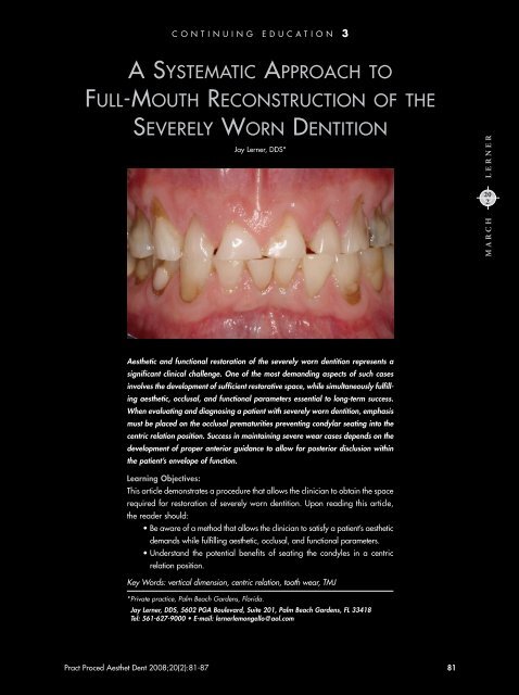
Aes<strong>the</strong>tic and functional res<strong>to</strong>ration <strong>of</strong> <strong>the</strong> <strong>severely</strong> worn dentition represents a<br />
significant clinical challenge. One <strong>of</strong> <strong>the</strong> most demanding aspects <strong>of</strong> such cases<br />
involves <strong>the</strong> development <strong>of</strong> sufficient res<strong>to</strong>rative space, while simultaneously fulfilling<br />
aes<strong>the</strong>tic, occlusal, and functional parameters essential <strong>to</strong> long-term success.<br />
When evaluating and diagnosing a patient with <strong>severely</strong> worn dentition, emphasis<br />
must be placed on <strong>the</strong> occlusal prematurities preventing condylar seating in<strong>to</strong> <strong>the</strong><br />
centric relation position. Success in maintaining severe wear cases depends on <strong>the</strong><br />
development <strong>of</strong> proper anterior guidance <strong>to</strong> allow for posterior disclusion within<br />
<strong>the</strong> patient’s envelope <strong>of</strong> function.<br />
Learning Objectives:<br />
This article demonstrates a procedure that allows <strong>the</strong> clinician <strong>to</strong> obtain <strong>the</strong> space<br />
required for res<strong>to</strong>ration <strong>of</strong> <strong>severely</strong> worn dentition. Upon reading this article,<br />
<strong>the</strong> reader should:<br />
Be aware <strong>of</strong> a method that allows <strong>the</strong> clinician <strong>to</strong> satisfy a patient’s aes<strong>the</strong>tic<br />
demands while fulfilling aes<strong>the</strong>tic, occlusal, and functional parameters.<br />
Understand <strong>the</strong> potential benefits <strong>of</strong> seating <strong>the</strong> condyles in a centric<br />
relation position.<br />
Key Words: vertical dimension, centric relation, <strong>to</strong>oth wear, TMJ<br />
*Private practice, Palm Beach Gardens, Florida.<br />
Jay Lerner, DDS, 5602 PGA Boulevard, Suite 201, Palm Beach Gardens, FL 33418<br />
Tel: 561-627-9000 E-mail: lernerlemongello@aol.com<br />
Pract Proced Aes<strong>the</strong>t Dent 2008;20(2):81-87 81<br />
LERNER<br />
20<br />
2<br />
MARCH
Practical Procedures & AESTHETIC DENTISTRY<br />
Res<strong>to</strong>ration <strong>of</strong> <strong>the</strong> <strong>severely</strong> worn dentition is one <strong>of</strong> <strong>the</strong><br />
most challenging procedures in dentistry. In order <strong>to</strong><br />
success<strong>full</strong>y res<strong>to</strong>re and maintain <strong>the</strong> teeth, one must gain<br />
insight in<strong>to</strong> how <strong>the</strong> teeth arrived at this state <strong>of</strong> destruction.<br />
Tooth wear can result from abrasion, attrition, and<br />
erosion. 1-5 Research has shown that <strong>the</strong>se wear mechanisms<br />
rarely act alone and <strong>the</strong>re is nearly always a combination<br />
<strong>of</strong> <strong>the</strong> processes. 1-5 Evaluation and diagnosis<br />
should account for <strong>the</strong> patient’s diet, his<strong>to</strong>ry <strong>of</strong> eating<br />
and/or gastric disorders, along with <strong>the</strong> present state <strong>of</strong><br />
<strong>the</strong> occlusion. Emphasis must be placed on <strong>the</strong> evaluation<br />
<strong>of</strong> occlusal prematurities preventing condylar seating<br />
in<strong>to</strong> <strong>the</strong> centric relation position. 6 Behavioral fac<strong>to</strong>rs that<br />
may contribute <strong>to</strong> parafunctional habits and/or nocturnal<br />
bruxism are also important <strong>to</strong> understand and manage<br />
in order <strong>to</strong> success<strong>full</strong>y res<strong>to</strong>re and maintain a healthier<br />
dentition. 7 Once a complete understanding <strong>of</strong> <strong>the</strong> etiology<br />
<strong>of</strong> <strong>the</strong> dentition’s present state is appreciated, a<br />
treatment plan can be formulated, taking in<strong>to</strong> account <strong>the</strong><br />
number <strong>of</strong> teeth <strong>to</strong> be treated, condylar position, space<br />
availability, <strong>the</strong> vertical dimension <strong>of</strong> occlusion (VDO),<br />
and <strong>the</strong> choice <strong>of</strong> res<strong>to</strong>rative material. 8<br />
While all occlusions wear <strong>to</strong> some degree over <strong>the</strong><br />
lifetime <strong>of</strong> <strong>the</strong> patient, normal physiological wear usually<br />
does not require correction. 6 Severe or excessive wear<br />
refers <strong>to</strong> <strong>to</strong>oth destruction that requires res<strong>to</strong>rative intervention.<br />
Severe attritional wear can result from occlusal<br />
prematurities that prevent functional or parafunctional<br />
movements <strong>of</strong> <strong>the</strong> jaw. This wear can be seen at <strong>the</strong><br />
site <strong>of</strong> <strong>the</strong> prematurity or on <strong>the</strong> anterior teeth as a result<br />
<strong>of</strong> <strong>the</strong> “hit and slide” forward. 6 Res<strong>to</strong>ration <strong>of</strong> <strong>the</strong> worn<br />
anterior teeth <strong>the</strong>n becomes a challenge as space availability<br />
for pros<strong>the</strong>tics becomes limited. If leng<strong>the</strong>ning<br />
<strong>the</strong> teeth is a goal in order <strong>to</strong> achieve a more aes<strong>the</strong>tic<br />
smile, <strong>the</strong>n <strong>the</strong> question <strong>of</strong> <strong>the</strong> need <strong>to</strong> alter VDO subsequently<br />
arises.<br />
There is some debate among pr<strong>of</strong>essionals as <strong>to</strong><br />
what constitutes <strong>the</strong> need <strong>to</strong> open VDO in <strong>the</strong> res<strong>to</strong>ration<br />
<strong>of</strong> anterior teeth. 9 In most cases, clinicians look <strong>to</strong><br />
alter vertical dimension for one or all <strong>of</strong> <strong>the</strong> following<br />
reasons: <strong>to</strong> gain space for <strong>the</strong> res<strong>to</strong>ration <strong>of</strong> <strong>the</strong> teeth; <strong>to</strong><br />
improve aes<strong>the</strong>tics; or <strong>to</strong> correct occlusal relationships.<br />
Understanding what determines <strong>the</strong> VDO and what <strong>the</strong><br />
effects <strong>of</strong> altering it have on <strong>the</strong> temporomandibular joint<br />
(TMJ), muscle comfort, bite force, speech, and longterm<br />
occlusal stability are prerequisites <strong>to</strong> res<strong>to</strong>ring <strong>the</strong><br />
worn dentition. Spear clearly outlines <strong>the</strong> principles <strong>of</strong><br />
VDO and concludes that patients can function at many<br />
acceptable vertical dimensions, provided <strong>the</strong> condyles<br />
are functioning from centric relation and <strong>the</strong> joint complex<br />
is healthy. He states that “vertical is a highly adaptable<br />
position, and <strong>the</strong>re is no single correct vertical<br />
dimension.” He fur<strong>the</strong>r concludes that <strong>the</strong> best vertical<br />
82 Vol. 20, No. 2<br />
B<br />
Figure 1. Posterior prematurities can cause <strong>the</strong><br />
mandible <strong>to</strong> close in a position forward <strong>of</strong> centric<br />
relation.<br />
B<br />
Figure 2. When <strong>the</strong> condyles are seated in centric<br />
relation, posterior teeth act as a fulcrum <strong>to</strong><br />
prevent contact with <strong>the</strong> anterior teeth.<br />
Figure 3. Preoperative view <strong>of</strong> a patient who<br />
presented with <strong>severely</strong> worn dentition.<br />
Figure 4. Bonded res<strong>to</strong>rations were present on<br />
<strong>the</strong> lingual aspect <strong>of</strong> <strong>the</strong> maxillary anterior teeth,<br />
originally placed <strong>to</strong> res<strong>to</strong>re a combination <strong>of</strong><br />
attrition and erosion.<br />
A<br />
A
Figure 5. A centric relation bite record was performed<br />
with <strong>the</strong> use <strong>of</strong> a leaf gauge.<br />
Figure 6. Mounted study casts revealed <strong>the</strong> second<br />
molars <strong>to</strong> be in premature contact when <strong>the</strong><br />
condyles were seated in centric relation.<br />
Figure 7. An intraoral composite mockup was<br />
performed <strong>to</strong> establish <strong>the</strong> ideal length for <strong>the</strong><br />
central incisors.<br />
Figure 8. The <strong>full</strong>-<strong>mouth</strong> diagnostic waxup <strong>to</strong>ok<br />
in<strong>to</strong> account that <strong>the</strong> second molars would be<br />
removed and aes<strong>the</strong>tic crown-leng<strong>the</strong>ning procedures<br />
performed.<br />
dimension is <strong>the</strong> one that satisfies <strong>the</strong> patient’s aes<strong>the</strong>tic<br />
desires and <strong>the</strong> practitioner’s functional goals with <strong>the</strong><br />
most conservative <strong>approach</strong>. 9<br />
Vertical dimension is developed by <strong>the</strong> balance <strong>of</strong><br />
ramus growth and <strong>to</strong>oth eruption 9 and is affected by<br />
<strong>the</strong> repetitive contracted length <strong>of</strong> <strong>the</strong> eleva<strong>to</strong>r muscles<br />
during growth and development. It is, <strong>the</strong>refore, generally<br />
measured by a point on <strong>the</strong> maxilla and a point on<br />
<strong>the</strong> mandible at <strong>the</strong> area <strong>of</strong> first molars. Often, due <strong>to</strong><br />
posterior prematurities <strong>the</strong> muscles <strong>of</strong> mastication are in<br />
a state <strong>of</strong> imbalance and will close <strong>the</strong> mandible in a<br />
position that is not in alignment with centric relation due<br />
<strong>to</strong> accommodation <strong>of</strong> <strong>the</strong> teeth. 10 This position is usually<br />
forward <strong>of</strong> centric relation (Figure 1).<br />
Clinical examination <strong>of</strong> this condition will reveal<br />
anterior <strong>to</strong>oth wear with minimal posterior wear. When<br />
<strong>the</strong> condyles are seated in <strong>the</strong> centric relation position<br />
and <strong>the</strong> teeth come <strong>to</strong>ge<strong>the</strong>r, <strong>the</strong> posterior teeth act as<br />
a fulcrum that prevents <strong>the</strong> anterior teeth from <strong>to</strong>uching<br />
(Figure 2). This anterior separation may provide enough<br />
space for <strong>the</strong> clinician <strong>to</strong> res<strong>to</strong>re <strong>the</strong> aes<strong>the</strong>tic requirements<br />
<strong>of</strong> <strong>to</strong>oth length while maintaining a position that<br />
allows res<strong>to</strong>ration <strong>of</strong> maximum intercuspation in conjunction<br />
with centric relation. 10<br />
When starting from a centric relation position,<br />
opening <strong>of</strong> <strong>the</strong> anterior teeth by 3 mm will yield a posterior<br />
separation <strong>of</strong> approximately 1 mm and stretch<br />
<strong>the</strong> masseter muscle length approximately 1 mm. If <strong>the</strong><br />
condyles are not in centric relation and are subsequently<br />
seated <strong>to</strong> a more superior position, every millimeter <strong>of</strong><br />
vertical seating will reduce <strong>the</strong> masseter muscle length<br />
by 1 mm, 9 <strong>the</strong>reby eliminating <strong>the</strong> need for a true opening<br />
<strong>of</strong> vertical dimension. The following case presentation<br />
demonstrates a means <strong>to</strong> obtain <strong>the</strong> space required<br />
for <strong>the</strong> res<strong>to</strong>ration <strong>of</strong> <strong>severely</strong> worn dentition without<br />
altering <strong>the</strong> VDO.<br />
Case Presentation<br />
A 55-year-old male patient presented with <strong>the</strong> chief complaint<br />
<strong>of</strong> anterior <strong>to</strong>oth wear and requested aes<strong>the</strong>tic<br />
enhancement (Figure 3). Clinical examination revealed<br />
<strong>severely</strong> worn anterior teeth and premolars in addition<br />
<strong>to</strong> bonded res<strong>to</strong>rations on <strong>the</strong> lingual aspects <strong>of</strong><br />
<strong>the</strong> maxillary anterior teeth <strong>to</strong> res<strong>to</strong>re what appeared <strong>to</strong> be<br />
an erosive process. Advanced abrasion and or erosion<br />
were present on many buccal surfaces <strong>of</strong> <strong>the</strong> canines and<br />
premolar teeth (Figure 4). The patient related a his<strong>to</strong>ry<br />
that included clenching, grinding, and, as a young man,<br />
gastric regurgitation. His periodontal status included<br />
areas <strong>of</strong> posterior pocketing with advanced bone loss<br />
in <strong>the</strong> second molar regions. The gingiva also exhibited<br />
areas <strong>of</strong> clefting in <strong>the</strong> anterior regions.<br />
P PAD 83<br />
Lerner
Practical Procedures & AESTHETIC DENTISTRY<br />
In order <strong>to</strong> properly diagnose <strong>the</strong> case, a comprehensive<br />
examination was conducted, inclusive <strong>of</strong> a<br />
<strong>full</strong>-<strong>mouth</strong> radiographic series, caries detection, and periodontal<br />
probing. Evaluation <strong>of</strong> <strong>the</strong> TMJs was unremarkable,<br />
with normal jaw opening and range <strong>of</strong> motion.<br />
No joint sounds, signs or symp<strong>to</strong>ms <strong>of</strong> instability were<br />
evident. Joint loading in centric relation was performed<br />
utilizing bimanual manipulation and a leaf gauge. 11,12<br />
Both methods resulted in no reported tension or tenderness<br />
and revealed first point <strong>of</strong> contacts on <strong>the</strong> second<br />
molars, with a forward slide in<strong>to</strong> <strong>the</strong> maximum intercuspation<br />
position.<br />
Impressions for study casts were <strong>the</strong>n made, along<br />
with a centric relation occlusal record utilizing <strong>the</strong> leaf<br />
gauge and a facebow transfer (Figure 5). 11-13 Following<br />
<strong>the</strong> mounting <strong>of</strong> <strong>the</strong> study casts, it became apparent that<br />
by seating <strong>the</strong> condyles in a centric relation position, <strong>the</strong><br />
second molars were in premature contact and <strong>the</strong>re was<br />
sufficient space gained <strong>to</strong> res<strong>to</strong>re <strong>the</strong> anterior teeth <strong>to</strong> <strong>the</strong><br />
proper aes<strong>the</strong>tic length (Figure 6).<br />
Treatment Planning<br />
Following periodontal consultation, it was determined<br />
that all <strong>of</strong> <strong>the</strong> second molars would be extracted due<br />
<strong>to</strong> advanced bone loss. Osseous surgery would follow<br />
in all four posterior quadrants, as would aes<strong>the</strong>tic<br />
crown leng<strong>the</strong>ning in <strong>the</strong> anterior region. Due <strong>to</strong> <strong>the</strong><br />
advanced wear <strong>of</strong> <strong>the</strong> remaining teeth, <strong>the</strong> treatment<br />
plan involved <strong>full</strong>-coverage res<strong>to</strong>rations on all teeth.<br />
The presence <strong>of</strong> sclerotic dentin and <strong>the</strong> possibility<br />
<strong>of</strong> continued clenching and/or bruxism established<br />
<strong>the</strong> need for cemented, as opposed <strong>to</strong> adhesive, res<strong>to</strong>rations.<br />
14,15 For long-term predictability, <strong>the</strong> author<br />
selected porcelain-fused-<strong>to</strong>-metal (PFM) res<strong>to</strong>rations.<br />
Zirconia crowns would also have represented an<br />
acceptable choice.<br />
Once <strong>the</strong> treatment plan was accepted, an intraoral<br />
composite mockup was performed and pho<strong>to</strong>graphed <strong>to</strong><br />
establish an ideal length for <strong>the</strong> central incisors from an<br />
aes<strong>the</strong>tic standpoint (Figure 7). These images and <strong>the</strong><br />
measured length <strong>of</strong> <strong>the</strong> maxillary central incisors were<br />
<strong>the</strong>n communicated <strong>to</strong> <strong>the</strong> labora<strong>to</strong>ry technician <strong>to</strong> aid in<br />
<strong>the</strong> fabrication <strong>of</strong> a <strong>full</strong>-<strong>mouth</strong> diagnostic waxup, which<br />
would be completed with <strong>the</strong> understanding that <strong>the</strong> second<br />
molars were <strong>to</strong> be removed and that aes<strong>the</strong>tic crown<br />
leng<strong>the</strong>ning procedures would be performed <strong>to</strong> raise <strong>the</strong><br />
gingival tissues in <strong>the</strong> anterior region (Figure 8). Prior <strong>to</strong><br />
waxing <strong>the</strong> case, <strong>the</strong> ceramist fabricated a centric relation<br />
anterior index that would maintain <strong>the</strong> centric relation<br />
position at <strong>the</strong> desired VDO during <strong>the</strong> preparation<br />
phase (Figure 9). This index can be made from hard<br />
labora<strong>to</strong>ry putty or GC pattern resin.<br />
84 Vol. 20, No. 2<br />
Figure 9. The ceramist fabricated a centric relation<br />
anterior index that held <strong>the</strong> centric relation<br />
position at <strong>the</strong> desired vertical dimension.<br />
Figure 10. Measurement <strong>of</strong> <strong>the</strong> anterior space<br />
from marginal tissue <strong>of</strong> teeth #9(21) through<br />
#24(31) (14.64 mm) in maximum intercuspation.<br />
Figure 11. By seating <strong>the</strong> condyles, a gain <strong>of</strong><br />
2.38 mm in anterior space was achieved without<br />
appreciably stretching <strong>the</strong> eleva<strong>to</strong>r muscles.<br />
Figure 12. Preparation <strong>of</strong> <strong>the</strong> maxillary posterior<br />
teeth was performed using <strong>the</strong> index, and posterior<br />
bites were taken.
Figure 13. Anterior bite record taken with<br />
posterior bite records in place, maintaining <strong>the</strong><br />
desired centric relation and VDO position.<br />
Figure 14. The provisional res<strong>to</strong>rations were<br />
placed with petroleum jelly, in order <strong>to</strong> facilitate<br />
simple removal <strong>the</strong> following day.<br />
Figure 15. The mandibular posterior teeth were<br />
prepared with <strong>the</strong> anterior bite records from day<br />
1 in place <strong>to</strong> hold centric relation and VDO.<br />
Figure 16. The mandibular anterior teeth were<br />
prepared and a new anterior bite record was<br />
taken utilizing <strong>the</strong> new posterior bite records <strong>to</strong><br />
maintain centric relation and VDO.<br />
Tooth Preparation<br />
Following a two-month period <strong>of</strong> periodontal healing and<br />
maturation, <strong>the</strong> patient was scheduled for appointments<br />
on two consecutive days <strong>to</strong> prepare first <strong>the</strong> maxillary,<br />
<strong>the</strong>n <strong>the</strong> mandibular arches. On <strong>the</strong> first day, <strong>the</strong> author<br />
utilized <strong>the</strong> centric relation index and measured from<br />
<strong>the</strong> marginal tissue <strong>of</strong> teeth #9(21) through #24(31)—<br />
gaining 2.38 mm <strong>of</strong> anterior space by simply having <strong>the</strong><br />
condyles seated in centric relation. This anterior opening<br />
was accomplished without appreciably stretching<br />
<strong>the</strong> eleva<strong>to</strong>r muscles (Figures 10 and 11). Preparation<br />
<strong>of</strong> <strong>the</strong> maxillary right and left posterior teeth was <strong>the</strong>n<br />
performed using <strong>the</strong> index <strong>to</strong> confirm clearance. With<br />
<strong>the</strong> index in place, posterior bites were taken utilizing a<br />
rigid bite-registration material (ie, Futar-D, Roydent Dental<br />
Products, Johnson City, TN) (Figure 12).<br />
The index was <strong>the</strong>n removed, and <strong>the</strong> anterior<br />
teeth were prepared utilizing <strong>the</strong> posterior bite records<br />
<strong>to</strong> verify clearance. Following completion <strong>of</strong> <strong>the</strong> anterior<br />
preparations, an anterior bite was obtained with <strong>the</strong><br />
posterior bite records in place. By <strong>systematic</strong>ally recording<br />
<strong>the</strong> posterior bite with <strong>the</strong> centric index in place and<br />
<strong>the</strong>n <strong>the</strong> anterior bite with <strong>the</strong> posterior bites in place,<br />
<strong>the</strong> centric relation and vertical dimension position were<br />
maintained (Figure 13).<br />
A <strong>full</strong>-arch polye<strong>the</strong>r impression (Perma-s<strong>of</strong>t, Garant-L,<br />
3M ESPE, St. Paul, MN) was <strong>the</strong>n taken, followed by <strong>the</strong><br />
fabrication <strong>of</strong> provisional res<strong>to</strong>rations (Luxatemp, Zenith/<br />
DMG, Englewood, NJ) created in three sections: two<br />
posterior sections from molar <strong>to</strong> first premolar, and an<br />
anterior section from canine <strong>to</strong> canine. Since <strong>the</strong> maxillary<br />
arch was prepared on <strong>the</strong> first day, occlusion was<br />
adjusted against <strong>the</strong> provisionals through equilibration<br />
<strong>of</strong> <strong>the</strong> mandibular teeth (Figure 14).<br />
During <strong>the</strong> second visit, <strong>the</strong> maxillary provisional<br />
res<strong>to</strong>rations were removed and <strong>the</strong> anterior bite record<br />
from day one was inserted <strong>to</strong> hold <strong>the</strong> centric relation<br />
and vertical dimension while <strong>the</strong> mandibular posterior<br />
teeth were prepared. Following bilateral preparation <strong>of</strong><br />
mandibular posterior teeth, bite records were taken with<br />
<strong>the</strong> anterior bite record in place (Figure 15). The mandibular<br />
anterior teeth were <strong>the</strong>n prepared utilizing <strong>the</strong><br />
posterior bite records <strong>to</strong> check clearance, and a new<br />
anterior bite record was taken (Figure 16).<br />
A polye<strong>the</strong>r final impression was <strong>the</strong>n made, and<br />
mandibular provisional res<strong>to</strong>rations were fabricated from<br />
<strong>the</strong> index <strong>of</strong> <strong>the</strong> diagnostic waxup. As with <strong>the</strong> maxillary<br />
provisional res<strong>to</strong>rations, <strong>the</strong> mandibular provisionals<br />
were fabricated in three sections (Figure 17). The<br />
provisional res<strong>to</strong>rations were subsequently equilibrated<br />
<strong>to</strong> establish maximum intercuspation in centric relation<br />
along with canine guidance and anterior coupling in<br />
protrusive guidance (Figure 18).<br />
P PAD 85<br />
Lerner
Practical Procedures & AESTHETIC DENTISTRY<br />
Once <strong>the</strong> provisional res<strong>to</strong>rations were equilibrated<br />
and <strong>the</strong> aes<strong>the</strong>tics and phonetics were deemed satisfac<strong>to</strong>ry,<br />
an occlusal bite record was taken <strong>of</strong> <strong>the</strong> maxillary and<br />
mandibular provisional res<strong>to</strong>rations. The maxillary posterior<br />
sections were removed and, with <strong>the</strong> anterior section still in<br />
place, posterior bite records were taken. The anterior section<br />
was <strong>the</strong>n removed and, with <strong>the</strong> posterior bite records<br />
in place, an anterior bite record was taken.<br />
Impressions <strong>of</strong> <strong>the</strong> provisional res<strong>to</strong>rations were made,<br />
and a facebow recording was taken <strong>of</strong> <strong>the</strong> maxillary provisionals.<br />
Utilizing <strong>the</strong> facebow, <strong>the</strong> maxillary provisional<br />
model was mounted on <strong>the</strong> articula<strong>to</strong>r; <strong>the</strong> mandibular<br />
model was <strong>the</strong>n mounted using <strong>the</strong> occlusal bite record <strong>of</strong><br />
<strong>the</strong> provisionals against each o<strong>the</strong>r. The ceramist was thus<br />
able <strong>to</strong> fabricate a cus<strong>to</strong>m incisal guide table (Figure 19).<br />
A cus<strong>to</strong>m incisal guide table, as described by Dawson,<br />
allows <strong>the</strong> ceramist <strong>to</strong> reproduce <strong>the</strong> anterior guidance<br />
established in <strong>the</strong> <strong>mouth</strong> with <strong>the</strong> provisional res<strong>to</strong>rations. 6<br />
The protrusive path and lateral excursions were recorded<br />
in pattern resin on a flat guide table by movement <strong>of</strong> <strong>the</strong><br />
articula<strong>to</strong>r pin in <strong>the</strong> unset resin. 6<br />
Once <strong>the</strong> incisal guide table was fabricated, cross<br />
mounting began. The maxillary preparation model was<br />
mounted against <strong>the</strong> mandibular provisional res<strong>to</strong>rations<br />
utilizing <strong>the</strong> third set <strong>of</strong> bite records. The mandibular preparation<br />
model was next mounted against <strong>the</strong> maxillary preparation<br />
model with <strong>the</strong> first set <strong>of</strong> bite records (Figure 20).<br />
Along with digital pho<strong>to</strong>graphs <strong>of</strong> <strong>the</strong> preparations<br />
and provisional res<strong>to</strong>rations, <strong>the</strong> ceramist had all <strong>the</strong><br />
information necessary <strong>to</strong> fabricate <strong>the</strong> definitive res<strong>to</strong>rations.<br />
A putty index was made from <strong>the</strong> provisional<br />
model <strong>to</strong> confirm <strong>the</strong> exact length and shape for <strong>the</strong><br />
final res<strong>to</strong>rations, while <strong>the</strong> cus<strong>to</strong>m guide table provided<br />
information on <strong>the</strong> shape <strong>of</strong> <strong>the</strong> lingual aspects and <strong>the</strong><br />
path taken for <strong>the</strong> canine and protrusive guidance.<br />
Definitive Res<strong>to</strong>rations<br />
Following a three-week period, <strong>the</strong> provisional res<strong>to</strong>rations<br />
were removed, <strong>the</strong> case was tried in, and <strong>the</strong>n<br />
evaluated for aes<strong>the</strong>tics, occlusion, and phonetics. Since<br />
<strong>the</strong> ceramist followed <strong>the</strong> guidelines <strong>of</strong> <strong>the</strong> provisional<br />
res<strong>to</strong>rations, minimal adjustments were necessary at this<br />
stage (Figures 21 through 23). Final equilibration <strong>of</strong> <strong>the</strong><br />
case was accomplished with a leaf gauge and a computerized<br />
occlusal analysis system (ie, T-Scan III, Tekscan,<br />
Bos<strong>to</strong>n, MA) (Figure 24). 16-17<br />
Conclusion<br />
Severe wear cases present many challenges <strong>to</strong> <strong>the</strong><br />
res<strong>to</strong>rative dentist, including gaining <strong>the</strong> space <strong>to</strong> create<br />
res<strong>to</strong>rations <strong>to</strong> satisfy <strong>the</strong> patient's aes<strong>the</strong>tic desires, while<br />
86 Vol. 20, No. 2<br />
Figure 17. Final provisional res<strong>to</strong>rations fabricated<br />
in three sections.<br />
Figure 18. Occlusal equilibration in centric relation<br />
establishing canine and protrusive guidance.<br />
Figure 19. Fabrication <strong>of</strong> a cus<strong>to</strong>m incisal<br />
guide table.<br />
Figure 20. The mandibular preparation model<br />
was <strong>the</strong>n mounted against <strong>the</strong> maxillary preparation<br />
model with <strong>the</strong> first set <strong>of</strong> bite records.
Figure 21. The provisionals were removed, and<br />
<strong>the</strong> definitive crowns were tried in and evaluated<br />
for aes<strong>the</strong>tics, occlusion, and phonetics.<br />
Figure 22. View <strong>of</strong> <strong>the</strong> completed maxillary<br />
res<strong>to</strong>rations. Guidelines established through<br />
provisionalization ensured minimal adjustments<br />
were needed.<br />
Figure 23. Occlusal view <strong>of</strong> <strong>the</strong> aes<strong>the</strong>tic final<br />
res<strong>to</strong>rations seated on <strong>the</strong> mandibular arch.<br />
Figure 24. Final equilibration <strong>of</strong> <strong>the</strong> case was<br />
accomplished with a computerized occlusal<br />
analysis (T-Scan III, Tekscan, Bos<strong>to</strong>n, MA).<br />
also fulfilling occlusal and functional parameters that are<br />
essential for long-term success. The case presented has<br />
demonstrated that <strong>the</strong> required space may be obtained<br />
by seating <strong>the</strong> condyles in centric relation position. The<br />
maintenance <strong>of</strong> severe wear cases can be ensured by <strong>the</strong><br />
development <strong>of</strong> proper anterior guidance that allows for<br />
posterior disclusion within <strong>the</strong> patient’s envelope <strong>of</strong> function.<br />
Taking this guidance in<strong>to</strong> account during provisionalization<br />
ensures minimal adjustments in <strong>the</strong> definitive res<strong>to</strong>rations<br />
and a greater long-term predictability <strong>of</strong> <strong>the</strong> case.<br />
Acknowledgement<br />
The author mentions his gratitude <strong>to</strong> Dr. Robert Holt<br />
for his expertise in managing <strong>the</strong> periodontal aspect<br />
<strong>of</strong> this case and Mr. Jason Kim, Oral Design, New<br />
York, NY for <strong>the</strong> labora<strong>to</strong>ry fabrication <strong>of</strong> <strong>the</strong> res<strong>to</strong>rations<br />
depicted. Figures 1 and 2 appear courtesy <strong>of</strong><br />
Dr. Michael Sesemann. The author declares no financial<br />
interest in any product referenced herein.<br />
References<br />
1. Addy M, Shellis RP. Interaction between attrition, abrasion and<br />
erosion in <strong>to</strong>oth wear. Monogr Oral Sci 2006;20:17-31.<br />
2. Beyth N, Sharon E, Lipovetsky M, Smidt A. Wear and different<br />
res<strong>to</strong>rative materials—A review. Refuat Hapeh Vehasshinayim<br />
2006;24(3):6-14.<br />
3. Grippo JO, Simring M, Schreiner S. Attrition, abrasion, corrosion<br />
and abfraction revisited: A new perspective on <strong>to</strong>oth<br />
surface lesions. J Am Dent Assoc 2004;135(8):1109-1118.<br />
4. Verrett RG. Analyzing <strong>the</strong> etiology <strong>of</strong> an extremely worn dentition.<br />
J Prosthodont 2001;10(4):224-233.<br />
5. Li<strong>to</strong>njua LA, Andreana S, Bush PJ, Cohen RE. Tooth wear: Attrition,<br />
erosion, and abrasion. Quint Int 2003;34(6):435-446.<br />
6. Dawson PE. Functional Occlusion: From TMJ <strong>to</strong> Smile Design.<br />
St. Louis, MO: Mosby; 2006:432-433.<br />
7. Neff P. Trauma from occlusion. Res<strong>to</strong>rative concerns. Dent Clin<br />
North Am 1995;39(2):335-354.<br />
8. Dahl BL, Carlsson GE, Ekfelt A. Occlusal wear <strong>of</strong> teeth and<br />
res<strong>to</strong>rative materials. A review <strong>of</strong> classifications, etiology, mechanism<br />
<strong>of</strong> wear, and some aspects <strong>of</strong> res<strong>to</strong>rative procedures. Acta<br />
Odon<strong>to</strong>l Scand 1993;51(5):299-311.<br />
9. Spear FM. Approaches <strong>to</strong> vertical dimension. Adv Es<strong>the</strong>t<br />
Interdiscip Dent 2006;2(3):2-12.<br />
10. Sesemann MR. Enhancing facial appearance with aes<strong>the</strong>tic<br />
dentistry, centric relation, and proper occlusal management.<br />
Pract Proced Aes<strong>the</strong>t Dent 2005;17(9):615-620.<br />
11. Long JH Jr. Location <strong>of</strong> <strong>the</strong> terminal hinge axis by intraoral means.<br />
J Pros<strong>the</strong>t Dent 1970;23(1):11-24.<br />
12. Mckee JR. Comparing condylar positions achieved through<br />
bimanual manipulation <strong>to</strong> condylar positions achieved through<br />
mastica<strong>to</strong>ry muscle contraction against an anterior deprogrammer:<br />
A pilot study. J Pros<strong>the</strong>t Dent 2005;94(4):389-393.<br />
13. Fenlon Mr, Woelfel JB. Condylar position recorded using leaf<br />
gauges and specific closure forces. Int J Prosthodont 1993;<br />
6(4):402-408.<br />
14. Tay FR, Pashley DH. Resin bonding <strong>to</strong> cervical sclerotic dentin:<br />
A review. J Dent 2004;32(3):173-196.<br />
15. Kwong SM, Tay FR, Yip HK, et al. An ultrastructural study <strong>of</strong><br />
<strong>the</strong> application <strong>of</strong> dentine adhesive <strong>to</strong> acid-conditioned sclerotic<br />
dentine. J Dent 2000;28(7):515-528.<br />
16. Kerstein RB, Wilkerson DW. Locating <strong>the</strong> centric relation prematurity<br />
with a computerized occlusal analysis system. Compend<br />
Contin Educ Dent 200;22(6):525-528.<br />
17. Kerstein RB. Disocclusion time-reduction <strong>the</strong>rapy with immediate<br />
complete anterior guidance development <strong>to</strong> treat<br />
chronic my<strong>of</strong>ascial pain-dysfunction syndrome. Quint Int<br />
1992;23(11):735-747.<br />
P PAD 87<br />
Lerner
Practical Procedures & AESTHETIC DENTISTRY<br />
CONTINUING EDUCATION<br />
(CE) EXERCISE NO. 3<br />
1. Which <strong>of</strong> <strong>the</strong> following fac<strong>to</strong>rs contribute <strong>to</strong> <strong>the</strong><br />
development <strong>of</strong> <strong>to</strong>oth wear?<br />
a. Abrasion.<br />
b. Attrition.<br />
c. Erosion.<br />
d. All <strong>of</strong> <strong>the</strong> above.<br />
2. Preoperative evaluation is critical <strong>to</strong> res<strong>to</strong>rative success,<br />
and should include:<br />
a. The patient’s diet, his<strong>to</strong>ry <strong>of</strong> eating, and/or<br />
gastric disorders.<br />
b. The patient’s present state <strong>of</strong> occlusion.<br />
c. Behavioral fac<strong>to</strong>rs that may contribute <strong>to</strong><br />
parafunctional habits.<br />
d. All <strong>of</strong> <strong>the</strong> above.<br />
3. How is vertical dimension developed?<br />
a. By balancing ramus growth and <strong>to</strong>oth eruption.<br />
b. By allowing <strong>the</strong> teeth <strong>to</strong> grow in a state <strong>of</strong> imbalance.<br />
c. By allowing <strong>the</strong> mandible <strong>to</strong> close in a state that is not<br />
in alignment with centric relation.<br />
d. None <strong>of</strong> <strong>the</strong> above.<br />
4. Which <strong>of</strong> <strong>the</strong> following reasons justify <strong>the</strong> clinician’s<br />
alteration <strong>of</strong> vertical dimension?<br />
a. To gain space for <strong>to</strong>oth res<strong>to</strong>ration.<br />
b. To improve aes<strong>the</strong>tics.<br />
c. To correct occlusal relationships.<br />
d. All <strong>of</strong> <strong>the</strong> above.<br />
5. According <strong>to</strong> this article, patients can only function at<br />
only one standardized, acceptable vertical dimension.<br />
Vertical is not an adaptable position, and must be maintained<br />
at <strong>the</strong> same degree for all cases.<br />
a. Both statements are true.<br />
b. Both statements are false.<br />
c. The first statement is true, <strong>the</strong> second statement is false.<br />
d. The first statement is false, <strong>the</strong> second statement is true.<br />
88 Vol. 20, No. 2<br />
CE 3<br />
CONTINUING EDUCATION<br />
To submit your CE Exercise answers, please use <strong>the</strong> answer sheet found within <strong>the</strong> CE Edi<strong>to</strong>rial Section <strong>of</strong> this issue and complete as follows:<br />
1) Identify <strong>the</strong> article; 2) Place an X in <strong>the</strong> appropriate box for each question <strong>of</strong> each exercise; 3) Clip answer sheet from <strong>the</strong> page and mail<br />
it <strong>to</strong> <strong>the</strong> CE Department at Montage Media Corporation. For fur<strong>the</strong>r instructions, please refer <strong>to</strong> <strong>the</strong> CE Edi<strong>to</strong>rial Section.<br />
The 10 multiple-choice questions for this Continuing Education (CE) exercise are based on <strong>the</strong> article “A <strong>systematic</strong> <strong>approach</strong> <strong>to</strong> <strong>full</strong>-<strong>mouth</strong><br />
<strong>reconstruction</strong> <strong>of</strong> <strong>the</strong> <strong>severely</strong> worn dentition,” by Jay Lerner, DDS. This article is on Pages 81-87.<br />
6. Severe attritional wear occurs as a result <strong>of</strong>:<br />
a. Lifetime degradation.<br />
b. Res<strong>to</strong>rative intervention.<br />
c. Occlusal prematurities preventing functional and parafunctional<br />
movement <strong>of</strong> <strong>the</strong> jaw.<br />
d. None <strong>of</strong> <strong>the</strong> above.<br />
7. When a patient presents with sever anterior wear <strong>the</strong><br />
clinician should:<br />
a. Au<strong>to</strong>matically open <strong>the</strong> vertical dimension.<br />
b. Res<strong>to</strong>re <strong>the</strong> dentition <strong>to</strong> <strong>the</strong> existing occlusal scheme.<br />
c. Look <strong>to</strong> remove any posterior prematurities <strong>to</strong> centric<br />
relation before res<strong>to</strong>ring dentition.<br />
d. None <strong>of</strong> <strong>the</strong> above.<br />
8. When <strong>the</strong> condyles are seated in <strong>the</strong> centric relation<br />
position and <strong>the</strong> teeth come <strong>to</strong>ge<strong>the</strong>r:<br />
a. Prematurities on posterior teeth act as a fulcrum that<br />
prevent <strong>the</strong> anterior teeth from <strong>to</strong>uching.<br />
b. The anterior teeth will limit adequate canine guidance<br />
for compromised function.<br />
c. Insufficient space will be provided for <strong>the</strong> clinician <strong>to</strong><br />
res<strong>to</strong>re <strong>the</strong> aes<strong>the</strong>tic requirements <strong>of</strong> <strong>the</strong> <strong>to</strong>oth length.<br />
d. Res<strong>to</strong>ration <strong>of</strong> maximum intercuspation in conjunction<br />
with centric relation is prevented.<br />
9. Why did <strong>the</strong> clinician maintain <strong>the</strong> placement <strong>of</strong> <strong>the</strong><br />
posterior bite records while obtaining <strong>the</strong> anterior bite<br />
registration throughout <strong>the</strong> <strong>to</strong>oth preparation sequence?<br />
a. To maintain <strong>the</strong> centric relation.<br />
b. To maintain <strong>the</strong> vertical dimension position.<br />
c. Both a and b are correct.<br />
d. Nei<strong>the</strong>r a nor b are correct.<br />
10. How was <strong>the</strong> length <strong>of</strong> <strong>the</strong> central incisors determined <strong>to</strong><br />
ensure optimal aes<strong>the</strong>tics?<br />
a. Using a composite mockup.<br />
b. Using clinical pho<strong>to</strong>graphy.<br />
c. Using a <strong>full</strong>-<strong>mouth</strong> diagnostic waxup.<br />
d. All <strong>of</strong> <strong>the</strong> above.