![Download the brochure [PDF, 594 KB] - Canon Europe](https://img.yumpu.com/17197970/1/500x640/download-the-brochure-pdf-594-kb-canon-europe.jpg)
Download the brochure [PDF, 594 KB] - Canon Europe
Download the brochure [PDF, 594 KB] - Canon Europe
Download the brochure [PDF, 594 KB] - Canon Europe

Create successful ePaper yourself
Turn your PDF publications into a flip-book with our unique Google optimized e-Paper software.
you can<br />
Redefining<br />
true versatility.<br />
CX-1 DIGITAL RETINAL CAMERA<br />
MYDRIATIC & NON-MYDRIATIC
More reliable diagnoses with FAF<br />
The diagnosis of retinal disorders with fundus auto<br />
fluorescence (FAF) is becoming more and more<br />
popular. This is because it is very gentle, being<br />
non-invasive, and gives quick and easy information<br />
on <strong>the</strong> health of <strong>the</strong> retinal pigment epi<strong>the</strong>lium<br />
(RPE). FAF imaging has shown to be very useful<br />
for <strong>the</strong> early detection of Age-related Macular<br />
Degeneration (AMD), one of <strong>the</strong> leading causes of<br />
visual impairment. Recent studies indicate that FAF<br />
imaging can also aid in <strong>the</strong> diagnosis of a variety<br />
of o<strong>the</strong>r diseases and even detection of intraocular<br />
tumors. When using FAF in conjunction with o<strong>the</strong>r<br />
diagnostic equipment, eye care profesionals can<br />
more confidently diagnose ocular disease.<br />
Redefine your horizons.<br />
The best of both worlds.<br />
The multifaceted CX-1 has integrated mydriatic<br />
and non-mydriatic imaging capabilities,<br />
for true versatility in retinal imaging, in one<br />
simple digital system.<br />
CX-1: All in one, one for all. The versatile CX-1 can quite simply be<br />
characterized as <strong>the</strong> hero in your clinic, with <strong>the</strong> motto “all in one,<br />
one for all”.<br />
The CX-1 is an “All in one” solution: it is a Mydriatic camera with FAF<br />
mode that has all <strong>the</strong> advantages of Non-Mydriatic functionality.<br />
This provides exceptional versatility and enables diagnosis, screening<br />
and monitoring of all major eye diseases.<br />
The CX-1 is also a “One for all” instrument. It is an ideal camera that<br />
captures high-quality retinal images of even <strong>the</strong> most challenging<br />
patients such as those with small pupils or high photosensitivity.<br />
In addition, from simple screening to complex diagnosis, any user<br />
can operate <strong>the</strong> device with ease.<br />
The CX-1 compact digital camera combines <strong>the</strong> best of both worlds.
Redefining true versatility. Mydriatic Retinal<br />
Camera with full Non-Mydriatic functionalities<br />
and FAF photography.<br />
<strong>Canon</strong> EOS<br />
Camera Technology<br />
Stereo Mode Non-Mydriatic Mydriatic<br />
FAF
THE MULTIFACETED CX-1<br />
Extensive photography modes;<br />
in an attractive body and under all<br />
conditions.<br />
Integrated EOS technology<br />
In addition to being optimized for ophthalmic imaging,<br />
<strong>the</strong> dedicated EOS camera combines several<br />
functions in one:<br />
• IRed observation<br />
• 3.0 inch vari-angle LCD screen for optimized<br />
viewing angles<br />
• 18 MegaPixel CMOS sensor<br />
• Intelligent monitor assistance; automatic<br />
magnification during focusing and displaying<br />
of stereo guide marks<br />
Mydriatic Retinal Camera with full Non-Mydriatic functionality, equipped with FAF<br />
photography as a standard feature.<br />
Extensive photography modes Colour, Red Free, Cobalt, FLUO and Fundus Auto Fluorescence,<br />
for wide diagnostic applications. All photography modes can be used ei<strong>the</strong>r in Mydriatic or<br />
Non-Mydriatic mode.<br />
Extremely versatile One camera for diagnosis of all major ocular diseases: Glaucoma, Diabetic<br />
Retinopathy, AMD, etc.<br />
Use of <strong>Canon</strong> EOS Camera Technology The CX-1 is equipped with a unique 18 MegaPixel<br />
digital camera unit that has been created for unsurpassed ophthalmic imaging.<br />
Ideal for non dilatable and sensitive patients The Non-Mydriatic mode is essential for non<br />
dilatable patients such as glaucoma suspects or children. Photosensitive patients will benefit<br />
from <strong>the</strong> non-invasive IRed observation light.<br />
High quality FAF images Sophisticated optical FAF filters for highest contrast and best image<br />
quality. Dedicated camera with optimized image processing for FAF and automatic image<br />
enhancement for cataract eyes.<br />
Ease of use: Compact design for maximum patient interaction, vari-angle LCD screen for<br />
optimized viewing angles. Easy switching between capturing modes. Easy changing between<br />
Mydriatic and Non-Mydriatic mode.<br />
Easy panning and tilting for working around central obstructions (cataracts, vitreous<br />
hemorrhages) and imaging <strong>the</strong> peripheral retina.<br />
Small pupil mode In Non-Mydriatic mode a pupil size of only ø 3.8 mm is required.<br />
2x zoom <strong>Canon</strong>’s high precision optics and <strong>the</strong> high resolution allow for 2x digital zoom of <strong>the</strong><br />
original wide angle image.<br />
Stereo photography was never as easy Clear stereo guide marks to create an image pair in<br />
just two simple steps.
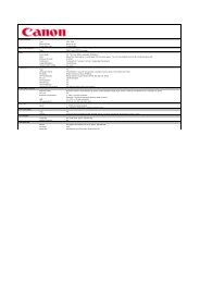
Archive<br />
RICS<br />
Dimensions<br />
Weight<br />
Angle of view<br />
Minimum pupil size<br />
Magnification<br />
Photography modes<br />
Working distance<br />
Mounted camera<br />
Fixation target<br />
Configuration<br />
Practice<br />
management<br />
systems<br />
Image<br />
capture<br />
External<br />
media<br />
Control Station<br />
RICS<br />
Network<br />
Print-out<br />
RICS Retinal Imaging Control Software<br />
Display SQL<br />
Database<br />
Archive<br />
Photography modes<br />
Observation<br />
light<br />
Observation Angle<br />
of view<br />
Colour Red<br />
Free<br />
PACS / HIS / RIS<br />
Export JPEG<br />
or DICOM file<br />
Full DICOM<br />
Print<br />
Study logs<br />
Cobalt FLUO FAF<br />
Non-Myd IR light LCD screen 45 ° + + + + * +<br />
Mydriatic Visible<br />
light<br />
Through<br />
viewfinder<br />
Specifications<br />
320 W x 531 D x 577 H mm<br />
26 kg<br />
Myd: 50 degrees, Non-Myd: 45 degrees<br />
Myd: ø 5.1 mm (SP mode ø 4.3 mm)<br />
Non-Myd: ø 4.3 mm (SP mode ø 3.8 SP)<br />
X2 (digital)<br />
Colour / FA / Red Free / Cobalt and FAF<br />
35 mm<br />
Dedicated digital EOS (18 MegaPixel)<br />
External<br />
Internal LED dot matrix for Non-Myd mode<br />
Internal fixation target for Myd mode<br />
(optional)<br />
<strong>Canon</strong> is constantly thinking ahead<br />
when it comes to software design,<br />
and understands <strong>the</strong> importance of<br />
networkability and ease of integration.<br />
This has resulted in <strong>the</strong> development<br />
of new solutions that are designed<br />
to be flexible to suit <strong>the</strong> needs of <strong>the</strong><br />
user and <strong>the</strong>ir image management<br />
systems.<br />
The <strong>Canon</strong> Retinal Imaging Control<br />
Software allows <strong>the</strong> CX-1 to be<br />
used as a stand-alone system. But<br />
it can also be easily integrated with<br />
an existing clinic network or DICOMcompliant<br />
network system.<br />
In <strong>the</strong> latest version of <strong>Canon</strong>’s<br />
extensive Retinal Image Control<br />
Software. Image capturing, processing,<br />
archiving, referencing and <strong>the</strong> export<br />
of data have been made much easier.<br />
Features include:<br />
• Full screen mode<br />
• Loupe function<br />
• Stereo view screen<br />
• Image comments function<br />
• White mask printing<br />
• Study comparison<br />
• RGB channel view<br />
• Cup / Disc ratio<br />
• Full DICOM compliance<br />
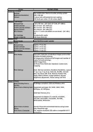
50 ° + + + + + * For fluorescein photography in Non-Mydriatic<br />
mode mydriasis will be required<br />
Patient’s diopter<br />
compensation<br />
Focus adjustment<br />
Working distance<br />
adjustment<br />
Panning and tilting<br />
range<br />
Light sources<br />
Optional accessories<br />
–31 D ~ –7 D, –10 D ~ +15 D (standard),<br />
+11 D ~ +33 D<br />
Split lines<br />
Reflection dots<br />
30 degrees to <strong>the</strong> left and right<br />
15 degrees up, 10 degrees down<br />
Xenon tube for photography<br />
Halogenlamp for observation (Myd)<br />
IRED LED for observation (Non-Myd)<br />
Stereo Unit SU-1, Internal eye fixation (CX-IF)<br />
Chin rest paper (500 sheets)
<strong>Canon</strong> has been defining <strong>the</strong> future with innovative<br />
solutions for more than 70 years. In all that time we’ve<br />
constantly strived to improve medical diagnostics<br />
in healthcare. Perhaps that’s what made us a leading<br />
global provider of eye care solutions.<br />
<strong>Canon</strong> Eco <strong>Canon</strong> Quality <strong>Canon</strong> Flexibility<br />
Our actions are based on<br />
honesty and sustainability.<br />
Choose <strong>the</strong> eye care system of <strong>the</strong> future and let our local,<br />
authorized <strong>Canon</strong> dealer advise you:<br />
Safety and quality are an<br />
integral component of our<br />
actions.<br />
CX-1<br />
English-NL Edition 2097V720<br />
© <strong>Canon</strong> Europa N.V. 2012<br />
Everything we do has to have<br />
a superior customer advantage.<br />
<strong>Canon</strong> Europa N.V.<br />
Medical Systems Division<br />
Bovenkerkerweg 59 – 61<br />
1185 XB Amstelveen<br />
The Ne<strong>the</strong>rlands<br />
Phone: +31(0)20-5 45-85 45<br />
Fax: +31(0)20-5 45-82 20<br />
www.canon-europe.com/medical

![PIXMA MG2250 Specification Sheet [PDF, 40 KB] - Canon Europe](https://img.yumpu.com/10835417/1/184x260/pixma-mg2250-specification-sheet-pdf-40-kb-canon-europe.jpg?quality=85)