

State of the Art - Cleveland Clinic
State of the Art - Cleveland Clinic
State of the Art - Cleveland Clinic

Create successful ePaper yourself
Turn your PDF publications into a flip-book with our unique Google optimized e-Paper software.
<strong>State</strong> <strong>of</strong> <strong>the</strong> <strong>Art</strong><br />
Lung Cancer—Where Are We Today?<br />
Current Advances in Staging and Nonsurgical Treatment<br />
Stephen G. Spiro and Joanna C. Porter<br />
Department <strong>of</strong> Respiratory Medicine, University College, London Hospitals National Health Service Trust, London, United Kingdom<br />
CONTENTS<br />
times more lung cancers than a chest X-ray, with more than 70%<br />
Abstract<br />
Epidemiology<br />
Screening<br />
Chest X-ray and Sputum Cytology<br />
Spiral Computed Tomography<br />
Biological Screening Tools<br />
Staging Tests: an Update<br />
Who Sees <strong>the</strong> Patient?<br />
Computerized Tomography <strong>of</strong> <strong>the</strong> Chest<br />
Magnetic Resonance Imaging<br />
Positron Emission Tomography<br />
<strong>of</strong> tumors being Stage I. The incidence <strong>of</strong> benign nodules is high,<br />
making interpretation difficult. Randomized controlled trials are<br />
required to determine whe<strong>the</strong>r spiral CT detects lung cancer early<br />
enough to improve mortality. Preoperative staging has relied on<br />
CT scans, but positron emission tomography scanning has greater<br />
sensitivity, specificity, and accuracy than CT and is recommended as<br />
<strong>the</strong> final confirmatory investigation when <strong>the</strong> CT shows resectable<br />
disease. In locally advanced non–small cell lung cancer, <strong>the</strong>re is a<br />
small advantage for <strong>the</strong> addition <strong>of</strong> chemo<strong>the</strong>rapy to radio<strong>the</strong>rapy,<br />
but no advantage for postoperative radio<strong>the</strong>rapy. Chemo<strong>the</strong>rapy<br />
gives no benefit when given as neoadjuvant or adjuvant treatment<br />
around surgery. In advanced disease, newer cytotoxic agents confer<br />
Endoscopic Biopsy Techniques a small survival advantage over older combinations, but <strong>the</strong> advan-<br />
The Search for Extrathoracic Metastasis tage in median survival over best supportive care remains a few<br />
Refining <strong>of</strong> <strong>the</strong> Staging Classification in an Attempt months with modest improvements in quality <strong>of</strong> life. Survival with<br />
to Increase Resectability small cell lung cancer has shown little increase over <strong>the</strong> last 15<br />
Advances in Radio<strong>the</strong>rapy in Non–Small Cell Lung Cancer years despite multiple attempts to manipulate <strong>the</strong> timing, dose<br />
Radical Radio<strong>the</strong>rapy for Stage I and II Disease intensity <strong>of</strong> chemo<strong>the</strong>rapy, and <strong>the</strong> potential <strong>of</strong> radio<strong>the</strong>rapy. Novel<br />
Postoperative Radio<strong>the</strong>rapy<br />
Radical Radio<strong>the</strong>rapy for Stage IIIA and IIIB Disease<br />
<strong>the</strong>rapies are urgently needed for all cell types <strong>of</strong> lung cancer.<br />
Palliative Radio<strong>the</strong>rapy<br />
Keywords: chemo<strong>the</strong>rapy; lung cancer; radio<strong>the</strong>rapy; screening;<br />
Interventional Bronchoscopy and Brachy<strong>the</strong>rapy<br />
staging<br />
Chemo<strong>the</strong>rapy for Non–Small Cell Lung Cancer<br />
Neoadjuvant Chemo<strong>the</strong>rapy<br />
Adjuvant Chemo<strong>the</strong>rapy and Surgery<br />
Chemo<strong>the</strong>rapy and Radio<strong>the</strong>rapy in Locally Advanced<br />
Disease<br />
Chemo<strong>the</strong>rapy in Advanced Disease<br />
Lung cancer is one <strong>of</strong> <strong>the</strong> most important diseases in respiratory<br />
medicine. Worldwide, it is <strong>the</strong> commonest cancer in men, virtually<br />
<strong>the</strong> commonest in women, and has a greater total incidence<br />
than that <strong>of</strong> colorectal, cervical, and breast cancer combined. In<br />
2001, lung cancer will have caused more than 1 million deaths<br />
Newer Chemo<strong>the</strong>rapy Combinations<br />
worldwide and this global incidence is rising at 0.5% per annum.<br />
Small Cell Lung Cancer<br />
The etiology <strong>of</strong> <strong>the</strong> great majority <strong>of</strong> lung cancers has been<br />
Chemo<strong>the</strong>rapy<br />
known for nearly 50 years (1), but we have failed to make<br />
Treatment <strong>of</strong> Limited Disease serious inroads into <strong>the</strong> powerbase <strong>of</strong> <strong>the</strong> tobacco industry. In<br />
Extensive Disease <strong>the</strong> Western world, although <strong>the</strong> incidence <strong>of</strong> lung cancer in men<br />
Elderly Patients has fallen over <strong>the</strong> last 20 years, a similar decline among female<br />
Prophylactic Cranial Irradiation smokers is not yet evident, and adolescents are smoking in in-<br />
Detection <strong>of</strong> Early Lung Cancer and Photodynamic Therapy<br />
The Future<br />
Conclusion<br />
creasing numbers. Global cigarette sales are rising steadily with<br />
<strong>the</strong> ruthless pursuit <strong>of</strong> new conquests by <strong>the</strong> tobacco industry<br />
in Asia, China, and South America, some <strong>of</strong> <strong>the</strong> poorest countries<br />
in <strong>the</strong> world that cannot afford <strong>the</strong> cost <strong>of</strong> tobacco-related dis-<br />
Lung cancer remains <strong>the</strong> commonest cause <strong>of</strong> cancer death in both eases. In China in 1998, one in four smokers died from tobaccomen<br />
and women in <strong>the</strong> developed world, although mortality rates related causes, and 0.6 million deaths in 1990 were tobacco<br />
for men are dropping. Spiral computed tomography (CT) <strong>of</strong> <strong>the</strong> related, a figure that rose to 0.8 million in 2000 (2).<br />
chest in middle-aged, smoking subjects may identify two to four Lung cancer is a disease for which <strong>the</strong>re is no established<br />
screening, which presents late in its course, and has a median<br />
survival <strong>of</strong> 6–12 months from <strong>the</strong> time <strong>of</strong> diagnosis with an<br />
overall 5-year survival <strong>of</strong> 5–10%; and yet <strong>the</strong> major cause <strong>of</strong><br />
(Received in original form February 1, 2002; accepted in final form July 31, 2002) this disease is clearly understood. Communities and countries<br />
Correspondence and requests for reprints should be addressed to S. G. Spiro,<br />
M.D., F.R.C.P., Department <strong>of</strong> Thoracic Medicine, The Middlesex Hospital, Morti-<br />
mer Street, London W1N 8AA, UK. E-mail: stephen.spiro@uclh.org<br />
Am J Respir Crit Care Med Vol 166. pp 1166–1196, 2002<br />
DOI: 10.1164/rccm.200202-070SO<br />
Internet address: www.atsjournals.org<br />
addressing a smoking ban would probably achieve far more<br />
in <strong>the</strong> long term than we currently are with all our available<br />
treatments.<br />
Although surgery <strong>of</strong>fers <strong>the</strong> best chance <strong>of</strong> cure in lung cancer,<br />
particularly in <strong>the</strong> case <strong>of</strong> non–small cell lung cancer, only a small
<strong>State</strong> <strong>of</strong> <strong>the</strong> <strong>Art</strong> 1167<br />
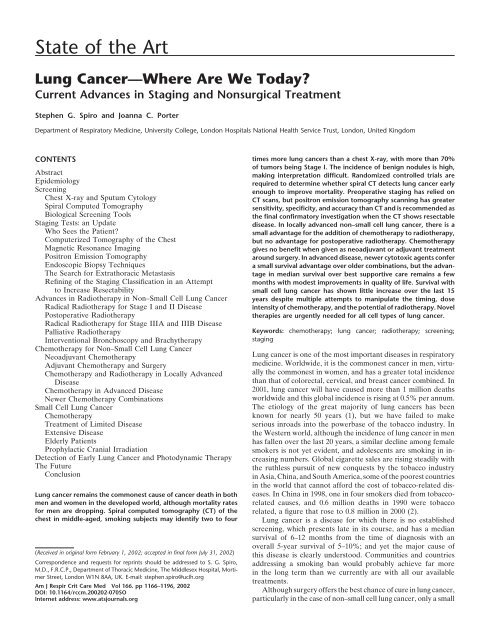
TABLE 1. AGE-ADJUSTED LUNG CANCER MORTALITY RATES PER 100,000 POPULATION FOR<br />
SELECTED COUNTRIES, IN MALES AND FEMALES, 1992–1995<br />
Males Females<br />
Country Lung Cancer Mortality Rate Country Lung Cancer Mortality Rate<br />
Hungary 84.0 United <strong>State</strong>s 26.3<br />
Poland 71.4 Denmark 24.9<br />
The Ne<strong>the</strong>rlands 64.8 United Kingdom 20.9<br />
Italy 56.2 Hungary 17.9<br />
United <strong>State</strong>s 55.3 China 15.8<br />
United Kingdom 51.8 The Ne<strong>the</strong>rlands 12.6<br />
Greece 49.8 Japan 8.3<br />
Germany 47.3 Italy 7.9<br />
France 47.0 Greece 6.9<br />
China 37.3 Mexico 5.8<br />
Japan 31.0<br />
Mexico 16.1<br />
Adapted from Schottenfeld D. Etiology and epidemiology <strong>of</strong> lung cancer. In: Pass HI, Mitchell JB, Johnson DH, Turrisi AT, and<br />
Minna JD, editors. Lung cancer. Baltimore, MD: Lippincott-Williams & Wilkins; 2000, p. 369.<br />
proportion <strong>of</strong> patients are ever suitable for curative resection and (9). However, <strong>the</strong>re is mounting evidence that at least in <strong>the</strong><br />
<strong>the</strong> majority must rely on nonsurgical and adjuvant <strong>the</strong>rapies. developed world death rates from lung cancer may have peaked.<br />
This review focuses on <strong>the</strong> role <strong>of</strong> chemo<strong>the</strong>rapy and radio<strong>the</strong>r- Between 1973 and 1994, <strong>the</strong> incidence <strong>of</strong> lung cancer in <strong>the</strong><br />
apy in both small cell lung cancer (SCLC) and non–small cell United <strong>State</strong>s for those over 65 years <strong>of</strong> age increased by 220%<br />
lung cancer (NSCLC). In addition, <strong>the</strong> potential role <strong>of</strong> screening in women, but fell by 18% in men (10). For those under 65 years<br />
for lung cancer and advances in staging tests are discussed. <strong>of</strong> age, <strong>the</strong> incidence <strong>of</strong> lung cancer increased by 58% in women<br />
and fell by 16% in men over <strong>the</strong> same period (10), and for those<br />
EPIDEMIOLOGY<br />
younger than 45 years old, age-adjusted incidence and mortality<br />
At <strong>the</strong> end <strong>of</strong> <strong>the</strong> twentieth century, lung cancer had become<br />
one <strong>of</strong> <strong>the</strong> world’s leading causes <strong>of</strong> preventable deaths. By 1950,<br />
case-control epidemiologic studies showed that cigarettes were<br />
strongly associated with <strong>the</strong> risk <strong>of</strong> lung cancer (3, 4). In 1962,<br />
<strong>the</strong> Royal College <strong>of</strong> Physicians in London intervened in a public<br />
health matter for <strong>the</strong> first time since 1725 and published a compelling<br />
document supporting <strong>the</strong> evidence that smoking caused<br />
lung cancer (5).<br />
Worldwide it is estimated that 47–52% <strong>of</strong> men and 10–12%<br />
<strong>of</strong> women smoke. Compared with women, men started smoking<br />
younger, smoked more and for a longer duration, inhaled more<br />
deeply, and bought cigarettes with a higher tar content (6, 7).<br />
Women took up smoking in <strong>the</strong> United <strong>State</strong>s and Western<br />
rates from lung cancer fell in both sexes (more so for men) with<br />
a projection that, in <strong>the</strong> United <strong>State</strong>s, overall incidence <strong>of</strong> and<br />
mortality from <strong>the</strong> disease may begin a decline for both sexes<br />
at <strong>the</strong> beginning <strong>of</strong> <strong>the</strong> new millennium (11).<br />
Although lung cancer incidence has fallen in <strong>the</strong> United<br />
<strong>State</strong>s, it remains <strong>the</strong> leading cause <strong>of</strong> cancer deaths worldwide,<br />
with a global incidence that continues to rise. There is also<br />
concern in <strong>the</strong> United <strong>State</strong>s that <strong>the</strong> incidence <strong>of</strong> <strong>the</strong> disease<br />
may start to increase again as a result <strong>of</strong> increasing tobacco<br />
consumption (12). In addition, shifts have occurred in <strong>the</strong> inci-<br />
dence rates <strong>of</strong> <strong>the</strong> different histologic subtypes <strong>of</strong> lung cancer,<br />
with adenocarcinoma surpassing squamous cell tumors as <strong>the</strong><br />
most frequent type in both white and black Americans.<br />
Europe during <strong>the</strong> second World War. Recent case-control studies<br />
have shown female smokers to have a higher relative risk <strong>of</strong><br />
lung cancer than males, after adjusting for age and average daily<br />
SCREENING<br />
Chest X-Ray and Sputum Cytology<br />
consumption. There has been much interest in <strong>the</strong> idea <strong>of</strong> screening to detect<br />
The incidence <strong>of</strong> lung cancer shows marked geographical presymptomatic lung cancers, when presumably <strong>the</strong>y would be<br />
variation, and is most common in developed countries and less at an earlier and more curable stage <strong>of</strong> <strong>the</strong>ir growth. In <strong>the</strong> 1970s<br />
so in developing countries, for example, those <strong>of</strong> Africa and <strong>the</strong>re was a major effort, using chest X-ray and sputum cytology,<br />
South America (8). The low rates in <strong>the</strong>se countries will inevita- to assess <strong>the</strong> prevalence <strong>of</strong> tumors and demonstrate that early<br />
bly rise to match those <strong>of</strong> <strong>the</strong> developed world. Within countries, detection would enhance survival and ultimately decrease morlung<br />
cancer incidence among men considerably exceeds that <strong>of</strong> tality. The Mayo Lung Project (13), <strong>the</strong> Czechoslovakian Screenwomen,<br />
but <strong>the</strong> highest rates occur in <strong>the</strong> same regions for both ing Study (14), and similar trials at Johns Hopkins Hospital (15)<br />
sexes (Table 1). Only 5–10% <strong>of</strong> all lung cancers are diagnosed and Memorial Sloan-Kettering Hospital (16) all enrolled male<br />
in patients under <strong>the</strong> age <strong>of</strong> 50 years, with adenocarcinoma and smokers and variously compared annual or more frequent chest<br />
a positive family history being common in <strong>the</strong>se cases. X-rays with or without additional sputum cytologic evaluation<br />
The mortality rates for lung cancer closely parallel <strong>the</strong> inci- against a control group who had an initial or annual chest X-ray<br />
dence rates because <strong>of</strong> poor survival. Age-adjusted mortality only. All <strong>the</strong>se studies identified more tumors in <strong>the</strong> screened<br />
rates increase exponentially until <strong>the</strong> age <strong>of</strong> 80 years in men and than control groups. The tumors were smaller, <strong>of</strong> a lower (more<br />
70 years in women and <strong>the</strong>n decline. In <strong>the</strong> United <strong>State</strong>s, lung favorable) stage, and <strong>the</strong> resection rate and 5-year survival rates<br />
cancer accounts for 28% <strong>of</strong> all cancer deaths each year. Whereas were better. However, overall mortality was not improved. Al-<br />
it was responsible for 3% <strong>of</strong> all female cancer deaths in 1950, though all <strong>the</strong>se were randomized controlled trials, only <strong>the</strong><br />
it accounted for 24% in 1995. The age-adjusted lung cancer Mayo Lung Project had a true control group that was unscreened.<br />
death rate passed that <strong>of</strong> breast cancer among white women in However, this study lacked power from <strong>the</strong> outset, with less than<br />
<strong>the</strong> United <strong>State</strong>s in 1986, and among black women in 1990<br />
20% power to detect a 10% benefit in lung cancer mortality and
1168 AMERICAN JOURNAL OF RESPIRATORY AND CRITICAL CARE MEDICINE VOL 166 2002<br />
a 55% power to detect a 20% benefit. Moreover, <strong>the</strong>re was underwent fluorophotography and low-dose helical CT, and<br />
fur<strong>the</strong>r contamination as 55% <strong>of</strong> <strong>the</strong> control group had a chest each was matched with two control subjects from <strong>the</strong> same<br />
X-ray in <strong>the</strong> previous year and 73% had a chest X-ray during population who underwent fluorophotography only. Smokers<br />
<strong>the</strong> last 2 years. Compliance was also a significant problem. from both groups underwent a 72-hour sputum collection for<br />
Interestingly, <strong>the</strong> screening seemed ra<strong>the</strong>r ineffectual, as <strong>the</strong> cytology. Each CT was read by four radiologists. Of <strong>the</strong> 3,967<br />
incidence <strong>of</strong> new tumors provided 206 new cancers, <strong>of</strong> which only participants, 19 (0.48%) were diagnosed with histologically<br />
45 (22%) were resectable, compared with 60% <strong>of</strong> <strong>the</strong> prevalence confirmed lung cancer. In only one was <strong>the</strong> tumor detected<br />
tumors at baseline. One <strong>of</strong> <strong>the</strong> problems with <strong>the</strong>se studies may by fluorophotography. Eight had abnormalities on a convenhave<br />
been <strong>the</strong> choice <strong>of</strong> mortality from lung cancer as <strong>the</strong> end tional chest radiograph. High-resolution CT missed one cen-<br />
point. It has been argued that all-cause mortality (17) or survival tral tumor that was detected by sputum cytology. Of <strong>the</strong> 19<br />
from lung cancer may be less biased end points (18). Indeed, cancers, 16 were Stage I and 3 were Stage IV; 12 <strong>of</strong> <strong>the</strong> 19<br />
Strauss has performed an important and impressive reanalysis were peripheral adenocarcinomas. Despite <strong>the</strong> relatively large<br />
<strong>of</strong> <strong>the</strong> Mayo Lung Cohort data. This analysis has shown that number screened <strong>the</strong> pick-up rate was relatively low, and<br />
although <strong>the</strong> incidence <strong>of</strong> lung cancer was higher in <strong>the</strong> screened <strong>the</strong>re was no difference in pick-up rates between smokers<br />
group, <strong>the</strong> survival <strong>of</strong> patients with lung cancer was also much and nonsmokers. Also, to find 16 resectable cancers, 223 partihigher<br />
in this group when compared with <strong>the</strong> control group; and cipants were examined fur<strong>the</strong>r by chest radiography and high-<br />
this increased survival was directly related to tumor resection resolution CT, and some by transbronchial biopsy; 204<br />
and <strong>the</strong>refore not due to overdiagnosis <strong>of</strong> “pseudo-disease” (18). showed nothing wrong. This was, however, a lower risk popu-<br />
Strauss argues that <strong>the</strong> randomization procedure for <strong>the</strong> Mayo lation.<br />
Lung Project was suboptimal, because <strong>of</strong> unidentified confound- 3. The study by Henschke and coworkers (24) chose a higher risk<br />
ing variables (18). So that although we understand a certain population. The Early Lung Cancer Action Project screened<br />
amount about <strong>the</strong> etiology <strong>of</strong> lung cancer, we still cannot accu- 1,000 symptom-free volunteers who were 60 years <strong>of</strong> age or<br />
rately distinguish those 16% <strong>of</strong> male and 9% <strong>of</strong> female life-long older, had a smoking history <strong>of</strong> at least 10 pack-years (in<br />
smokers who will develop <strong>the</strong> disease from <strong>the</strong>ir fellow smokers fact, a median <strong>of</strong> 45 pack-years), and were deemed fit for<br />
who will not. Strauss finally lays to rest <strong>the</strong> years <strong>of</strong> debate thoracotomy and a life expectancy <strong>of</strong> at least 5 years. Each<br />
around <strong>the</strong> Mayo Lung Project and explains <strong>the</strong> findings without participant underwent chest radiography and low-dose helical<br />
having to resort to <strong>the</strong> counterintuitive concept <strong>of</strong> overdiagnosis; CT. There were specific recommendations for <strong>the</strong> interpretascreening<br />
is worth doing because more resectable cases are tion and fur<strong>the</strong>r investigation <strong>of</strong> noncalcified pulmonary nod-<br />
picked up and more patients are cured (18). Ano<strong>the</strong>r problem ules. At <strong>the</strong> initial screening, CT identified 233 individuals<br />
highlighted from <strong>the</strong> Mayo study is that <strong>of</strong> identifying early<br />
tumors on <strong>the</strong> chest X-ray, as 90% <strong>of</strong> peripheral and 75% <strong>of</strong><br />
perihilar tumors were visible in retrospect on previous films (19).<br />
Quekel and coworkers more recently also reported a 19% miss<br />
rate <strong>of</strong> peripheral tumors, with an average size <strong>of</strong> 16 mm (20).<br />
with noncalcified pulmonary nodules, compared with 68 seen<br />
on <strong>the</strong> chest X-ray. All 233 <strong>the</strong>n underwent high-resolution<br />
CT scans and biopsy samples from 28 subjects confirmed<br />
malignancy in 27, <strong>of</strong> which 18 were adenocarcinomas; <strong>the</strong>re<br />
were no small cell tumors. Stage I tumors made up 23 <strong>of</strong> <strong>the</strong><br />
27 discovered, <strong>of</strong> which only 4 were visible on chest radiogra-<br />
Spiral Computed Tomography<br />
phy and 26 were resectable. The prevalence <strong>of</strong> lung cancer<br />
The current interest in screening is due to <strong>the</strong> advent <strong>of</strong> low-<br />
dose spiral computed tomography (CT), using short scanning<br />
times <strong>of</strong> 12 to 15 seconds, with <strong>the</strong> potential for mass application.<br />
To date, several prevalence studies have reported <strong>the</strong>ir early<br />
findings. They have, however, all examined different numbers<br />
<strong>of</strong> volunteers <strong>of</strong> different ages, both sexes, mostly smokers, but<br />
with a wide range <strong>of</strong> smoking histories.<br />
found by CT was 2.7%, four times that <strong>of</strong> chest radiography,<br />
and <strong>the</strong> highest <strong>of</strong> all <strong>the</strong> prevalence studies reported to date;<br />
a reflection on <strong>the</strong> choice <strong>of</strong> population screened.<br />
4. The University <strong>of</strong> Münster (Münster, Germany) has to date<br />
screened by annual CT 817 subjects who are over 40 years<br />
<strong>of</strong> age with a smoking history <strong>of</strong> at least 20 pack-years. In 11<br />
subjects 12 cases <strong>of</strong> lung cancer (1.3%) were found, <strong>of</strong> which<br />
only 7 (58.3%) were Stage I. Three lesions that were investi-<br />
1. Kaneko and coworkers (21) studied 1,369 participants, over gated invasively were found to be benign (25).<br />
50 years <strong>of</strong> age and with a smoking history <strong>of</strong> more than 20 5. Ano<strong>the</strong>r study from <strong>the</strong> Mayo <strong>Clinic</strong> enrolled 1,520 individupack-years.<br />
Eighty-two percent were male. They underwent als, aged 50 years or older, with a smoking history <strong>of</strong> 20<br />
6 monthly scans and at <strong>the</strong> initial scan 15 lung cancers (0.43%) pack-years or more (26). All subjects agreed to undergo a<br />
were identified, <strong>of</strong> which 14 were Stage I, with a mean tumor prevalence CT scan and three annual incidence scans. The<br />
diameter <strong>of</strong> 16 mm. However, a total <strong>of</strong> 11.5% <strong>of</strong> CT scans initial prevalence screen identified 22 cases <strong>of</strong> lung cancer, and<br />
were positive, requiring fur<strong>the</strong>r assessment. The study has <strong>the</strong> first incidence screen <strong>of</strong> 1,464 <strong>of</strong> <strong>the</strong> original population<br />
been updated to assess <strong>the</strong> incidence <strong>of</strong> lung cancers on <strong>the</strong> discovered an additional 3 tumors. Cell types were as follows:<br />
six monthly follow-up scans (22). A total <strong>of</strong> 1,180 participants squamous, 6; adenocarcinoma, 15; large cell, 1; and small cell,<br />
were given two or more examinations, for a total <strong>of</strong> 7,891 3. Twenty-two patients underwent curative resection and 7<br />
scans. Of <strong>the</strong>se, 721 (9.1%) were positive by helical CT, three benign nodules were resected. There were 13 postsurgical<br />
times <strong>the</strong> rate <strong>of</strong> chest X-ray, and 22 (0.28%) <strong>of</strong> <strong>the</strong>se were Stage IA patients (60%). A cause <strong>of</strong> concern was <strong>the</strong> high<br />
found to have lung cancer with 18 (82%) being Stage IA. rate <strong>of</strong> detection <strong>of</strong> noncalcified benign nodules: 2,244 among<br />
The lung cancer detection rate was lower for <strong>the</strong> additional 1,000 participants. A total <strong>of</strong> 2,053 were present in <strong>the</strong> prevascreening<br />
rounds compared with <strong>the</strong> initial screen and <strong>the</strong> lence scan. On <strong>the</strong> first annual incidence scan, 195 had re-<br />
5-year survival rate was 64.9% compared with 76.2% for <strong>the</strong> solved, 36 had been removed (more than 1 nodule was reinitial<br />
prevalence cancers. moved in some patients), 86 had grown, and 79 had become<br />
2. Sone and coworkers (23) screened a low-risk Japanese popu- smaller; 1,657 were stable. Thus, about 98% <strong>of</strong> nodules are<br />
lation <strong>of</strong> unselected volunteers including smokers and non- false-positive findings, which means, assuming <strong>the</strong> 13% inci-<br />
smokers aged 40 to 74 years who had already undergone dence rate in this study, almost all subjects could have at least<br />
annual chest fluorophotography and sputum cytology as part one false-positive screening after a few years, with consider-<br />
<strong>of</strong> a national screening program. A total <strong>of</strong> 3,967 people<br />
able implications for resources and patient management.
<strong>State</strong> <strong>of</strong> <strong>the</strong> <strong>Art</strong> 1169<br />
These studies are all hypo<strong>the</strong>sis generating, but it is too early screening is not possible. Once hundreds <strong>of</strong> scans are generated<br />
to know whe<strong>the</strong>r detecting tumors that are in general smaller by screening, radiographers will have to be trained to report<br />
than when discovered on a chest radiograph will decrease mortal- <strong>the</strong>m and show only abnormal scans to radiologists for practical<br />
ity. All <strong>the</strong>se studies have <strong>the</strong> in-built problems <strong>of</strong> lead time time reasons. Inevitably, fur<strong>the</strong>r high-resolution CT scans will<br />
bias, length time bias, and overdiagnosis bias (27). Only large have to be performed on subjects with abnormalities and many<br />
randomized controlled trials (RCTs) with a follow-up <strong>of</strong> 10 years will <strong>the</strong>n require biopsies. Many will also need regular follow-<br />
or more and rigorous use <strong>of</strong> all-cause mortality as an end point up high-resolution scans for several years. The costs and logistics<br />
(17) will answer this fundamental question. In addition to <strong>the</strong> and possible long-term effects <strong>of</strong> <strong>the</strong> investigative irradiation<br />
prevalence data now available, <strong>the</strong> incidence data from <strong>the</strong> cur- are considerable.<br />
rent uncontrolled studies will give valuable information as to There is <strong>the</strong>refore no sensible alternative to embarking on<br />
how many <strong>of</strong> <strong>the</strong> smaller nodules (less than 10 mm in diameter) carefully constructed RCTs in defined populations <strong>of</strong> sufficient<br />
were, in fact, tumors and not identified as such during <strong>the</strong> initial numbers, members <strong>of</strong> which are followed for long enough to<br />
CT screen. Although identifying nodules 10 mm in size or smaller provide a clear answer about <strong>the</strong> potential <strong>of</strong> CT screening.<br />
gives a yield <strong>of</strong> cancers smaller in size than those discovered by Additional problems will occur if <strong>the</strong> technology <strong>of</strong> imaging<br />
conventional chest X-rays, <strong>the</strong>se tumors will have undergone 25 moves ahead so fast that improvements will have to be incorpoto<br />
30 volume doublings and will have a considerable propensity rated into <strong>the</strong>se prospective studies. For example, three-dimen-<br />
to form metastases (28). Fur<strong>the</strong>rmore, <strong>the</strong>re are data to suggest sional volumetric analysis <strong>of</strong> a nodule is already available and<br />
that <strong>the</strong> relationship between tumor size, survival, and stage at is more sensitive for showing size change than simple CT (33).<br />
presentation is not clear cut. One study <strong>of</strong> 510 patients found Finally, will any control population accept an annual chest X-ray,<br />
no statistical relationship between tumors <strong>of</strong> less than 3 cm and or perhaps no screening chest X-ray, while being deprived <strong>of</strong> a<br />
survival; patients with 3-cm masses had <strong>the</strong> same outcome as chance for CT screening?<br />
those with 1-cm nodules (29). In a related study <strong>of</strong> 620 patients<br />
<strong>the</strong>re was no relationship between size <strong>of</strong> <strong>the</strong> primary tumor Biological Screening Tools<br />
and stage at presentation. Patients with a 1-cm tumor had a Biological screening tools are still in development and remain<br />
similar stage distribution as those with 2- to 3-cm masses (30). <strong>the</strong> subject <strong>of</strong> research. One surface marker for early detection<br />
Thus <strong>the</strong> biological behavior <strong>of</strong> tumors is variable and a funda- <strong>of</strong> lung cancer is <strong>the</strong> heterogeneous nuclear ribonucleoprotein<br />
mental part <strong>of</strong> <strong>the</strong> issue <strong>of</strong> long-term outcome. A2/B1, which is upregulated on premalignant bronchial epi<strong>the</strong>-<br />
There is growing pressure to include low-dose helical CT in lial cells. In reassessing sputum archived from <strong>the</strong> Johns Hopkins<br />
<strong>the</strong> armamentarium directed at finding lung cancer for good screening study, overexpression <strong>of</strong> A2/B1 was a more sensitive<br />
emotive (31) but not yet evidence-based reasons. Much work marker <strong>of</strong> early preinvasive malignancy than normal cytologic<br />
still needs to be done. The larger prevalence studies have been screening. Features <strong>of</strong> malignancy were identifiable 1 year before<br />
performed in countries where peripheral adenocarcinomas are <strong>the</strong> conventional cytologic examination showed abnormalities,<br />
commoner—<strong>the</strong> United <strong>State</strong>s and Japan. This appears not to and before <strong>the</strong> tumor became visible by chest radiography (34,<br />
be <strong>the</strong> case in Europe and care must be taken when advocating<br />
a technique such as this more widely. The choice <strong>of</strong> population<br />
to screen will have a major effect on <strong>the</strong> prevalence <strong>of</strong> tumors<br />
found, as already clearly demonstrated in <strong>the</strong> data accumulated<br />
so far. Age, sex, smoking history, and <strong>the</strong> presence <strong>of</strong> airway<br />
obstruction are <strong>the</strong> major risk factors for <strong>the</strong> development <strong>of</strong><br />
lung cancer.<br />
The issue <strong>of</strong> false-positive scans will need to be addressed.<br />
In <strong>the</strong> Japanese and Lung Cancer Action Project studies <strong>the</strong>re<br />
were large numbers <strong>of</strong> subjects with noncalcified pulmonary<br />
nodules: 233 <strong>of</strong> <strong>the</strong> 1,000 in <strong>the</strong> Lung Cancer Action Project<br />
(24) and 66% in <strong>the</strong> Mayo study (26). The anxiety generated,<br />
<strong>the</strong> potential for overinvestigation, and <strong>the</strong> radiologic exposure<br />
35). Similar encouraging results have been shown in prospective<br />
trials <strong>of</strong> Chinese tin miners (36), North American patients with<br />
lung cancer who had undergone resection <strong>of</strong> <strong>the</strong>ir primary tumor<br />
but were at high risk <strong>of</strong> recurrent disease (35, 37), and UK<br />
patients under investigation for lung cancer (38).<br />
Mao and coworkers (39) looked at early chromosomal and<br />
genetic alterations in lung epi<strong>the</strong>lial cells and found that point<br />
mutations in <strong>the</strong> p53 and K-ras genes in sputum samples preceded<br />
<strong>the</strong> clinical diagnosis <strong>of</strong> lung cancer in one case by more<br />
than 1 year. O<strong>the</strong>r groups have identified areas <strong>of</strong> genomic insta-<br />
bility that cause microsatellite alterations that can act as clonal<br />
markers <strong>of</strong> early malignant disease (40).<br />
<strong>the</strong>se individuals receive suggest a need for fur<strong>the</strong>r thought.<br />
It is also worth noting that in clinical practice, most lung<br />
STAGING TESTS: AN UPDATE<br />
cancers occur centrally and are diagnosed by bronchoscopy. It is beyond <strong>the</strong> remit <strong>of</strong> a single review to comprehensively<br />
These tumors hardly feature in <strong>the</strong> CT screening studies; only summarize <strong>the</strong> current lung cancer staging literature, but newer<br />
two central tumors were discovered in <strong>the</strong> Lung Cancer Action techniques are becoming available and <strong>the</strong>se, toge<strong>the</strong>r with basic<br />
Project screen (24) and one in <strong>the</strong> study by Sone and coworkers, assessment <strong>of</strong> <strong>the</strong> patient, are discussed.<br />
and that tumor was found by cytology, not CT (23). It would<br />
appear that central lung cancers are too aggressive to remain<br />
Who Sees <strong>the</strong> Patient?<br />
occult and produce symptoms leading to diagnosis before or As <strong>the</strong> average age <strong>of</strong> presentation for lung cancer is increasing,<br />
between screening tests because <strong>of</strong> <strong>the</strong>ir situation in major air- this may affect who <strong>the</strong> patient is referred to (e.g., a care <strong>of</strong> <strong>the</strong><br />
ways. They behave in an entirely different way than intrapulmo- elderly physician) and how aggressive <strong>the</strong> treatment is. Brown<br />
nary “nodule” or peripheral cancer. and coworkers (41) assessed 563 cases <strong>of</strong> lung cancer diagnosed<br />
What <strong>of</strong> <strong>the</strong> cost in terms <strong>of</strong> machine time, scan interpretation, in a 30-month period around Sou<strong>the</strong>nd, England. Two hundred<br />
and resultant action? Attempts have been made to analyze <strong>the</strong> and forty (43%) were over 70 years <strong>of</strong> age. The incidence <strong>of</strong><br />
cost-effectiveness <strong>of</strong> screening for lung cancer, but such models lung cancer in <strong>the</strong> general population was 69 per 100,000, but<br />
make enormous assumptions and are probably premature (32). in men over 75 years <strong>of</strong> age <strong>the</strong> incidence was 751 per 100,000.<br />
A proper screening program will require dedicated CT scanners, For all patients, <strong>the</strong> active treatment rate was 49% (surgery,<br />
which may need to be mobile. In many countries <strong>the</strong>re is already radio<strong>the</strong>rapy, chemo<strong>the</strong>rapy), but for patients not reviewed by<br />
an unacceptable waiting time for staging CTs in patients known a chest physician (n 86) it was only 21%. There were large<br />
or suspected to have lung cancer, and <strong>the</strong> additional burden <strong>of</strong> differences in initial treatment between age groups. For patients
1170 AMERICAN JOURNAL OF RESPIRATORY AND CRITICAL CARE MEDICINE VOL 166 2002<br />
with NSCLC reviewed by a chest physician, surgery was per- Despite advances in CT scanning technology, <strong>the</strong>re remain<br />
formed in 18% <strong>of</strong> those under 65 years <strong>of</strong> age, in 12% <strong>of</strong> those important limitations for its use in staging, with preoperative<br />
in <strong>the</strong> 65- to 74-year age group, and in 2% in those over 75 years predictions differing from operative staging in 35–45% <strong>of</strong> cases,<br />
<strong>of</strong> age. For patients with SCLC reviewed by a chest physician, with patients being both over- and understaged (48, 49). CT<br />
79% <strong>of</strong> those under 65, 64% <strong>of</strong> those in <strong>the</strong> 64- to 75-year age staging remains unsatisfactory for detecting hilar (N1) and medi-<br />
group, and 41% <strong>of</strong> patients over 75 received chemo<strong>the</strong>rapy. If astinal (N2 and N3) lymph node metastases, and for chest wall<br />
no histologic diagnosis was made, 37% <strong>of</strong> patients under 75 and involvement (T3) or mediastinal invasion (T4), in which sensitiv-<br />
only 5.4% <strong>of</strong> those over 75 received any treatment. Patients not ity and specificity can be less than 65% (50–53). These are critical<br />
reviewed by a chest physician were less likely to obtain a histo- areas that may make <strong>the</strong> difference between surgical and nonsurlogic<br />
diagnosis.<br />
gical management decisions. One development has been single-<br />
A similar review <strong>of</strong> <strong>the</strong> referral and treatment practice in a photon emission CT in which technetium-99m-labeled tetr<strong>of</strong>os-<br />
city in Yorkshire, England also found that almost half <strong>of</strong> patients min is taken up by lung cancers. In one study <strong>of</strong> 34 patients with<br />
with newly diagnosed lung cancer were not sent to a respiratory lung cancer, CT when combined with single-photon emission<br />
physician, and <strong>the</strong> treatment rates for surgery, radio<strong>the</strong>rapy, and CT gave a sensitivity <strong>of</strong> 94.7% and a specificity <strong>of</strong> 93.3% for <strong>the</strong><br />
chemo<strong>the</strong>rapy for those patients were approximately half <strong>the</strong> detection <strong>of</strong> mediastinal metastases; <strong>the</strong>se levels <strong>of</strong> sensitivity<br />
rates for patients seen by a respiratory physician (42). Both and specificity were greater than those achieved with ei<strong>the</strong>r<br />
studies reinforced <strong>the</strong> UK National Cancer Plan to identify a technique alone (54).<br />
respiratory physician with an interest in lung cancer in every Dynamic expiratory CT scanning can be used to assess chest<br />
hospital to organize <strong>the</strong> care for patients with newly diagnosed wall and mediastinal fixation by showing decreased mobility <strong>of</strong><br />
lung cancer. It is probable that in an aging population referral <strong>the</strong> fixed tumor (55). Ultrasound may also be useful for chest<br />
patterns in o<strong>the</strong>r countries will be similar to those in <strong>the</strong> UK, wall assessment. In a series <strong>of</strong> 120 patients with contiguity be-<br />
with no exclusive referral pattern to a respiratory physician. tween <strong>the</strong> tumor and <strong>the</strong> chest wall at CT but no definitive<br />
Computerized Tomography <strong>of</strong> <strong>the</strong> Chest<br />
invasion (as diagnosed by bony erosion), 19 patients were judged<br />
to have invasive tumor on ultrasound with a sensitivity and<br />
Computed tomography <strong>of</strong> <strong>the</strong> chest is important both in <strong>the</strong> specificity <strong>of</strong> 100 and 98%, respectively, when compared with<br />
diagnosis and staging <strong>of</strong> lung cancer. As a diagnostic tool it is operative findings (56). In <strong>the</strong> 1990s, many studies compared<br />
a valuable adjunct to bronchoscopy. The yield at bronchoscopy CT findings with <strong>the</strong> gold standard <strong>of</strong> mediastinoscopy or sur-<br />
is higher if CT shows a bronchus extending to <strong>the</strong> tumor (60 gery. They showed that, regardless <strong>of</strong> <strong>the</strong> threshold size <strong>of</strong> lymph<br />
versus 25%) (43, 44). The probability that a lesion considered node chosen, CT findings in isolation could not be taken as clear<br />
accessible to bronchoscopy on a chest X-ray can actually be evidence <strong>of</strong> malignant nodal involvement and about 20% <strong>of</strong> all<br />
diagnosed in this way is not easy to ascertain (45). A UK nodes deemed malignant on CT criteria will be benign. Size<br />
multicenter prospective study <strong>of</strong> 1,660 consecutive cases investi- alone cannot be an exclusion criterion and <strong>the</strong> clinician needs<br />
gated by fiberoptic bronchoscopy because <strong>of</strong> a prior likelihood to prove by biopsy or resection that a node is indeed malignant.<br />
<strong>of</strong> lung cancer found definite evidence <strong>of</strong> tumor in only 57% CT, however, continues to play an important and necessary part<br />
(46). In a fur<strong>the</strong>r 20%, appearances were suspicious; thus, in in <strong>the</strong> evaluation <strong>of</strong> patients with lung cancer, and its use is<br />
20% <strong>of</strong> <strong>the</strong>se tests, <strong>the</strong> investigation was unhelpful. Only 15% supported by <strong>the</strong> most recent American Thoracic Society/Euro<strong>of</strong><br />
<strong>the</strong>se patients had a prior CT and whe<strong>the</strong>r this was <strong>of</strong> use to pean Respiratory Society statement on pretreatment evaluation<br />
<strong>the</strong> bronchoscopist is not known.<br />
in NSCLC, in which CT is recommended for evaluation <strong>of</strong> medi-<br />
Ano<strong>the</strong>r study (47) suggests <strong>the</strong>re are advantages if CT pre- astinal nodes in all patients with suspected NSCLC (57).<br />
cedes bronchoscopy and <strong>the</strong> information from CT is used by <strong>the</strong><br />
bronchoscopist. Costs were not greater, as <strong>the</strong> number <strong>of</strong> inva- Magnetic Resonance Imaging<br />
sive tests was reduced. Of 171 patients suspected <strong>of</strong> having endo- One <strong>of</strong> <strong>the</strong> most important questions when staging lung cancer<br />
bronchial cancer, 90 had a CT performed and reviewed before is whe<strong>the</strong>r <strong>the</strong> tumor is resectable. Tumor-induced proliferation<br />
bronchoscopy. Six needed no fur<strong>the</strong>r investigation because <strong>the</strong> <strong>of</strong> connective tissue adjacent to <strong>the</strong> tumor may be interpreted<br />
CT was ei<strong>the</strong>r normal, or consistent with benign disease or with as malignant on CT scan and <strong>the</strong> tumor consequently overstaged<br />
widespread metastatic disease. Of <strong>the</strong> remainder, fiberoptic even with <strong>the</strong> new multislice scanners. In <strong>the</strong>se situations, mag-<br />
bronchoscopy was diagnostic in 50 <strong>of</strong> 68 (73%) compared with netic resonance imaging (MRI) has advantages over CT because<br />
44 <strong>of</strong> 81 patients (58%) who had a bronchoscopy first. Overall, <strong>of</strong> its multiplanar imaging and <strong>the</strong> large differences in intensity<br />
a positive diagnosis was made after a single invasive test in 76% between tumor and s<strong>of</strong>t tissue. MRI is superior to CT scanning<br />
<strong>of</strong> <strong>the</strong> group having a CT first, and in 54% <strong>of</strong> <strong>the</strong> group that in delineating <strong>the</strong> mediastinal fat plane, which makes it a power-<br />
underwent bronchoscopy first. Only 7 <strong>of</strong> <strong>the</strong> CT-first group ful tool for assessing mediastinal invasion (58). O<strong>the</strong>r areas in<br />
needed more than one invasive investigation, compared with 15 which MRI plays a role are in assessing invasion <strong>of</strong> <strong>the</strong> root <strong>of</strong><br />
patients (18%) <strong>of</strong> <strong>the</strong> fiberoptic bronchoscopy-first group. The <strong>the</strong> neck, chest wall, vertebral bodies, and diaphragm (51, 59–62).<br />
additional cost <strong>of</strong> a spiral CT in each patient was <strong>of</strong>fset by MRI has no advantage over CT in <strong>the</strong> evaluation <strong>of</strong> enlarged<br />
<strong>the</strong> need for fewer invasive tests, even though <strong>the</strong>y were more lymph nodes except in patients with renal disease, for whom<br />
expensive. Because <strong>the</strong> majority <strong>of</strong> patients with lung cancer contrast is contraindicated (58, 63).<br />
have a CT during <strong>the</strong>ir workup, it may be best done before<br />
fiberoptic bronchoscopy.<br />
Positron Emission Tomography<br />
The spiral CT, using a special staging technique, is <strong>the</strong> main- Because <strong>of</strong> <strong>the</strong> limitations <strong>of</strong> CT and MRI, <strong>the</strong> search for better<br />
stay <strong>of</strong> staging in lung cancer. This involves an automated bolus noninvasive techniques to identify malignant disease has intensiinjection<br />
<strong>of</strong> contrast 20–30 seconds before <strong>the</strong> scanning is initi- fied. Currently, 2-[ 18F]fluoro-2-deoxy-d-glucose-based PET scanated.<br />
This time interval allows optimal enhancement <strong>of</strong> <strong>the</strong> medi- ning (hereafter referred to as PET) is <strong>the</strong> most promising. PET<br />
astinal blood vessels. A maximum slice thickness <strong>of</strong> 5 mm is can detect malignancy in focal pulmonary lesions <strong>of</strong> greater than<br />
used to prevent errors from partial volume effects. The new 1 cm with a sensitivity <strong>of</strong> about 97% and a specificity <strong>of</strong> 78%<br />
multislice CT systems allow <strong>the</strong> whole thorax to be scanned with (64). False-positive findings in <strong>the</strong> lung are seen in granuloma-<br />
3-mm slices during a single breath hold.<br />
tous disease and rheumatoid disease, with false negatives in
<strong>State</strong> <strong>of</strong> <strong>the</strong> <strong>Art</strong> 1171<br />
carcinoid, alveolar cell carcinoma, and lesions <strong>of</strong> less than 1 cm participating hospitals were randomized to conventional workup<br />
(65–68).<br />
(CWU) or CWU plus PET. The primary outcome was <strong>the</strong> ability<br />
As well as having a role in <strong>the</strong> evaluation <strong>of</strong> parenchymal <strong>of</strong> PET to minimize futile thoracotomies. Eighteen patients in<br />
nodules, PET is also valuable for evaluation <strong>of</strong> <strong>the</strong> mediastinum. <strong>the</strong> CWU group and 32 in <strong>the</strong> CWU plus PET group did not<br />
However, image resolution <strong>of</strong> <strong>the</strong> current PET scanners is only have a thoracotomy. In <strong>the</strong> former group, 41% had a futile<br />
4–8 mm and requires complementary CT. The precise anatomical operation, as opposed to only 21% in <strong>the</strong> PET group (p 0.003).<br />
information from CT adds to <strong>the</strong> metabolic map <strong>of</strong> PET and Importantly, <strong>the</strong>re was no decrease in justified surgery due to<br />
helps distinguish, for example, N1 from N2 disease and central PET. Assessment <strong>of</strong> resectability by CT and PET was discordant<br />
tumors from enlarged lymph nodes. A total <strong>of</strong> 29 published in one-third <strong>of</strong> <strong>the</strong> cases, and PET was correct in two-thirds.<br />
studies that examined <strong>the</strong> suitability <strong>of</strong> PET for <strong>the</strong> staging <strong>of</strong> PET was superior to CT in identifying <strong>the</strong> best mediastinoscopy<br />
NSCLC were reviewed by Laking and Price (69). A meta-analy- site and in 10 cases only PET suggested <strong>the</strong> positive biopsy site.<br />
sis confirmed that PET is significantly more accurate than CT Overall one futile thoracotomy was avoided for every five PET<br />
for detection <strong>of</strong> nodal mediastinal metastases, with a sensitivity scans (75).<br />
and specificity <strong>of</strong> 79 and 91%, respectively, for PET versus 60 Who should have a PET scan? Despite <strong>the</strong> expense <strong>of</strong> PET<br />
and 77%, respectively, for CT (70). The usefulness <strong>of</strong> <strong>the</strong> extra scanning and its limited availability, cost–benefit analyses <strong>of</strong><br />
information gained from PET is itself dependent on <strong>the</strong> initial published data, in both <strong>the</strong> United <strong>State</strong>s (76) and Europe (71),<br />
CT scan, so that PET has a sensitivity and specificity <strong>of</strong> 74 have shown that it is cost-effective to carry out total body PET<br />
and 96%, respectively, for detecting metastasis in normal-sized in patients with a negative mediastinal CT and an apparently<br />
mediastinal lymph nodes compared with 95 and 76%, respec- resectable tumor as <strong>the</strong> cost is balanced by a better selection <strong>of</strong><br />
tively, when <strong>the</strong>se lymph nodes are enlarged (71). It is important patients for surgery. Patients with a positive mediastinal CT and<br />
to remember this when drawing up clinical protocols or consider- no clinical suggestion <strong>of</strong> metastatic disease should go straight<br />
ing individual patients. False-positive mediastinal nodal scans to mediastinoscopy. However, <strong>the</strong>se recommendations remain<br />
occur in sarcoid and tuberculosis and o<strong>the</strong>r infections. impractical until <strong>the</strong>re is better access to PET scanners and<br />
Is PET sensitive and specific enough to replace mediastino- radiologists to interpret <strong>the</strong>m. Ideally, because <strong>of</strong> <strong>the</strong> high negascopy<br />
and lymph node sampling before thoracotomy and prevent tive predictive value, PET scanning should be performed in all<br />
futile operations without denying surgery to appropriate candi- those with no evidence <strong>of</strong> metastatic disease on CT who are<br />
dates? Several studies have addressed this question. In one study considered for surgery; and, failing this, definitely in those preop-<br />
<strong>of</strong> 100 patients, PET accurately staged NSCLC in 83% <strong>of</strong> cases erative patients with suspicious N2/N3 disease on CT scan.<br />
compared with 65% by conventional imaging (thoracic CT, bone<br />
scintigraphy, and brain CT or MRI). PET identified 9 patients Endoscopic Biopsy Techniques<br />
with metastases that were missed on conventional imaging Transbronchial lymph node sampling, directed by PET and CT,<br />
whereas 10 patients thought to have metastases were shown not performed via a flexible bronchoscope is less invasive than medito<br />
by PET. PET was more sensitive than conventional imaging astinoscopy and may save time and money in skilled hands.<br />
for bone, and adrenal metastases, but is inappropriate for <strong>the</strong> However, <strong>the</strong> sensitivity is variable (50–89% that <strong>of</strong> mediastinosdetection<br />
<strong>of</strong> brain metastases because <strong>of</strong> <strong>the</strong> high glucose uptake copy), although this may increase with endobronchial ultrasound<br />
<strong>of</strong> <strong>the</strong> normal brain. The negative predictive value <strong>of</strong> PET for (EBUS) or CT guidance (77–79). EBUS has been used mainly<br />
N2 disease was 96%, similar to that <strong>of</strong> mediastinoscopy, sug- to estimate <strong>the</strong> depth <strong>of</strong> tracheobronchial invasion, but <strong>the</strong>re<br />
gesting that patients with negative mediastinal PET could go was preliminary evidence showing that this technique might be<br />
straight to surgical resection <strong>of</strong> <strong>the</strong> primary tumor (72). In a useful in <strong>the</strong> assessment <strong>of</strong> mediastinal and hilar metastases (80).<br />
comparison <strong>of</strong> PET with CT against <strong>the</strong> gold standard <strong>of</strong> medias- In a more recent study, 37 patients with lung cancer underwent<br />
tinal lymph node dissection in 102 patients with resectable NSLC EBUS and CT scanning. EBUS was much better than CT for<br />
(73), results were complicated by <strong>the</strong> high sensitivity (75%) and detecting abnormal hilar nodes <strong>of</strong> less than 1 cm, for resolving<br />
low specificity (66%) <strong>of</strong> CT scanning for detection <strong>of</strong> mediastinal individual nodes from node masses, and for assessing invasion<br />
metastases, but only PET results (91% sensitive and 86% spe- <strong>of</strong> <strong>the</strong> pulmonary artery. It appears that EBUS and CT may<br />
cific) correlated with <strong>the</strong> histopathology <strong>of</strong> <strong>the</strong> mediastinal lymph complement each o<strong>the</strong>r in <strong>the</strong> assessment <strong>of</strong> hilar and subcarinal<br />
nodes. PET altered <strong>the</strong> stage determined by conventional im- (Level 7) lymphadenopathy. However, with only 16 patients with<br />
aging in 62 patients (42 were upstaged and 20 were downstaged). positive node involvement diagnosed surgically it is difficult to<br />
However, PET was still wrong in 13 cases (conventional imaging draw any statistical conclusion from <strong>the</strong> study (81).<br />
was wrong in 32) and surgical staging was required for a definitive Ano<strong>the</strong>r technique that is becoming increasingly important<br />
result (73). This emphasizes that no one with a positive PET in <strong>the</strong> sampling <strong>of</strong> mediastinal, but not hilar, lymph nodes is<br />
scan should be denied surgery without positive histology (71). transesophageal lymph node sampling under endoscopic ultra-<br />
PET was actually <strong>of</strong> greatest value in 11 patients in whom distant sound guidance (EUS) (82). This has <strong>the</strong> added advantage <strong>of</strong><br />
metastases were found. However, in nine patients PET was avoided contamination <strong>of</strong> lymph node samples with malignant<br />
falsely positive for distant metastases. In ano<strong>the</strong>r study, treat- cells from <strong>the</strong> bronchial tree.<br />
ment plans based on conventional staging were compared with EUS is a technique that has been in use for more than 10<br />
those based on incorporation <strong>of</strong> PET. PET changed management years. It makes use <strong>of</strong> a modified endoscope with an ultrasound<br />
for 40 <strong>of</strong> 153 patients (34 patients had <strong>the</strong>ir treatment changed transducer at <strong>the</strong> tip and gives excellent views <strong>of</strong> <strong>the</strong> structures<br />
from curative to palliative and 6 patients had <strong>the</strong>ir treatment that lie adjacent to <strong>the</strong> gut lumen. EUS from <strong>the</strong> esophagus<br />
changed from palliative to curative) and gave more accurate gives access to <strong>the</strong> subcarinal (Level 7), aortopulmonary (Level<br />
prognosis <strong>of</strong> individual patients (74). 5), and posterior (Levels 8 and 9) mediastinum and is able to<br />
More recently, an attempt has been made by a group in The resolve nodes as small as 3 mm. However, <strong>the</strong> views <strong>of</strong> <strong>the</strong><br />
Ne<strong>the</strong>rlands to see whe<strong>the</strong>r patients actually benefit if PET is paratracheal and anterior mediastinal areas are limited by distor-<br />
incorporated into <strong>the</strong> workup, and to address this question treattion caused by tracheal air. By using curved echo-endoscopes it<br />
ment plans based on conventional staging were compared with is possible to perform fine needle aspiration (EUS-FNA) <strong>of</strong><br />
those based on incorporation <strong>of</strong> PET. In this PLUS (PET in abnormal subcarinal and aortopulmonary window nodes with<br />
Lung Cancer Staging) study 188 patients with NSCLC from 9 negligible risk <strong>of</strong> infection or bleeding (83, 84). This had a sensi-
1172 AMERICAN JOURNAL OF RESPIRATORY AND CRITICAL CARE MEDICINE VOL 166 2002<br />
tivity <strong>of</strong> 96% for malignancy in lymph nodes when bronchoscopy is dependent on <strong>the</strong> extent <strong>of</strong> intrathoracic involvement, that is,<br />
had been unhelpful (85, 86). Silvestri and coworkers looked at <strong>the</strong> worse <strong>the</strong> primary tumor and nodal involvement, <strong>the</strong> greater<br />
27 patients with known or suspected lung cancer who underwent <strong>the</strong> likelihood <strong>of</strong> metastatic disease, whereas <strong>the</strong> incidence <strong>of</strong><br />
CT scan and EUS-FNA. They showed that EUS-FNA improved silent metastases in Stage I disease is low (1%) (92, 93). Several<br />
<strong>the</strong> sensitivity <strong>of</strong> CT scanning and granted access to lymph nodes studies, including two meta-analyses <strong>of</strong> <strong>the</strong> literature, have found<br />
not reached at mediastinoscopy (79). Wallace and coworkers distant metastases in only 2.5–5% <strong>of</strong> patients with potentially<br />
studied 121 patients with lung cancer, using EUS-FNA <strong>of</strong> abnor- operable NSCLC despite normal clinical examination (94–97).<br />
mal nodes. Of <strong>the</strong>se, 97 had enlarged mediastinal lymph nodes The metastases most commonly affected brain, bone, liver, and<br />
and EUS-FNA confirmed malignancy in 75 (77%). In addition, adrenal glands in that order (95, 98). How best to identify <strong>the</strong>se<br />
10 <strong>of</strong> 24 (42%) patients with normal mediastinal lymph nodes patients preoperatively and prevent a needless thoracotomy is<br />
on CT had Stage III or IV disease on EUS-FNA (84), suggesting not clear. The literature is divided, with some studies showing<br />
that it might be an even more powerful staging tool than medias- that screening all patients for extrathoracic metastases before<br />
tinoscopy (87). thoracotomy is cost-effective (99) and o<strong>the</strong>rs finding that this<br />
Only one study has directly compared mediastinoscopy (up- was not <strong>the</strong> case (92). It is now standard to include <strong>the</strong> adrenals<br />
per and anterior mediastinum) with EUS-FNA (subcarinal and and liver as part <strong>of</strong> a staging CT <strong>of</strong> <strong>the</strong> chest and upper abdomen<br />
posterior mediastinal lesions) and, although <strong>the</strong>re were only a (100).<br />
small number <strong>of</strong> patients, <strong>the</strong> suggestion is that <strong>the</strong> two tech- Adrenals. The majority <strong>of</strong> unilateral adrenal masses in pa-<br />
niques may prove complementary, with different lymph node tients with lung cancer are benign but are difficult to distinguish<br />
stations targeted by <strong>the</strong> two techniques (88). More recently, from adrenal metastases (101, 102). A negative PET scan or<br />
Larsen and coworkers have looked at <strong>the</strong> effect <strong>of</strong> EUS-FNA MRI <strong>of</strong> <strong>the</strong> adrenals will exclude metastatic disease, but both<br />
on <strong>the</strong> management <strong>of</strong> 84 patients with mediastinal masses adja- tests have a high rate <strong>of</strong> false positives (102, 103). For this reason<br />
cent to <strong>the</strong> esophagus. Diagnosis was confirmed by thoracotomy, FNA should be performed on any suspicious adrenal masses<br />
mediastinoscopy, or follow-up over at least 1 year. In 29 patients (i.e., those <strong>of</strong> more than 2 cm or more or that are positive for<br />
with known lung cancer who underwent mediastinal staging, PET or MRI) if this is <strong>the</strong> only obstacle to possible resection.<br />
EUS-FNA has a specificity <strong>of</strong> 100%, a sensitivity <strong>of</strong> 90%, a Brain. Metastases to <strong>the</strong> brain are more frequent when <strong>the</strong><br />
negative predictive value <strong>of</strong> 82%, and a positive predictive value primary tumor is greater than 3 cm (104) and more frequent for<br />
<strong>of</strong> 100%. Similar figures applied for 50 patients with mediastinal adenocarcinoma than for squamous cell carcinoma (94). Routine<br />
masses but no obvious lung primary. The results from EUS- brain imaging in <strong>the</strong> absence <strong>of</strong> symptoms or clinical signs is not<br />
FNA provided a definite diagnosis and obviated <strong>the</strong> need for recommended as <strong>the</strong> pick-up <strong>of</strong> occult cerebral metastases is<br />
28 mediastinoscopies and 18 thoracotomies. There were no com- less than 3% (57), with a false-positive rate in one study <strong>of</strong> 11%<br />
plications from <strong>the</strong> procedure (89). (105). In a study <strong>of</strong> 114 patients staged by CT <strong>of</strong> <strong>the</strong> brain,<br />
So, what is <strong>the</strong> role <strong>of</strong> EUS-FNA in patient management? thorax, and abdomen occult disease <strong>of</strong> <strong>the</strong> brain and abdomen<br />
Harewood and coworkers have used models based on <strong>the</strong> medi- was found in 15 patients, but in all but 3 cases (2 isolated abdomi-<br />
cal literature to look at cost minimization in <strong>the</strong> accurate staging nal metastases and 1 isolated brain metastasis) <strong>the</strong> CT scan <strong>of</strong><br />
<strong>of</strong> patients with NSCLC and enlarged (greater than 1 cm) subca- <strong>the</strong> thorax was sufficiently abnormal to demonstrate that <strong>the</strong><br />
rinal lymph nodes on CT scan. The lowest cost workup was by tumor was unresectable or to have prompted mediastinoscopy<br />
initial EUS-FNA provided that <strong>the</strong> probability <strong>of</strong> subcarinal before thoracotomy (96). Colice and coworkers performed a<br />
lymph nodes metastases was greater than 24%, assuming a sensi- cost analysis and concluded that, at current costs and given<br />
tivity for EUS-FNA higher than 76% (90), in keeping with o<strong>the</strong>r current available treatments, head CT should be reserved for<br />
studies (91). EUS-FNA may prove as valuable as or more so those patients with abnormal neurologic symptoms and signs<br />
than mediastinoscopy, and ideally is <strong>the</strong> investigation <strong>of</strong> choice (106). High-dose gadolinium contrast-enhanced MRI (which is<br />
for diagnostic evaluation <strong>of</strong> CT-suspicious lymph nodes at Levels more sensitive than routine CT scanning for detecting brain<br />
5, 7, 8, and 9. However, because <strong>of</strong> <strong>the</strong> requirement for expensive metastases [107]) picked up occult cerebral metastases in 6 <strong>of</strong><br />
equipment and a skilled endoscopist, EUS-FNA is available only 29 patients (17%) with lung tumors greater than 3 cm on CT<br />
in a small number <strong>of</strong> institutions. In addition, <strong>the</strong> role <strong>of</strong> EUS- scanning (104). There were no false-positive brain MRIs and<br />
FNA in <strong>the</strong> evaluation <strong>of</strong> patients with apparently resectable no patient who had a negative MRI presented with cerebral<br />
lung cancers and normal mediastinal CT scans is unknown, but metastases in <strong>the</strong> 12 months <strong>of</strong> follow-up. The preoperative<br />
<strong>the</strong>re is some evidence that it might identify some <strong>of</strong> <strong>the</strong> 10% detection <strong>of</strong> cerebral metastases altered treatment and follow-<br />
<strong>of</strong> patients with N2/N3 disease who are not picked up by CT up for all patients, although surgery was not reconsidered for<br />
scan or mediastinoscopy. There may be some advantages over any patient in this study. This does suggest that in patients with<br />
PET scanning, which has a false-positive rate <strong>of</strong> up to 13%, primary tumors <strong>of</strong> greater than 3 cm, especially if <strong>the</strong>se are<br />
although <strong>the</strong> possibility <strong>of</strong> overstaging with EUS-FNA has not adenocarcinomas or large cell, <strong>the</strong>re may be an indication for<br />
really been addressed. MRI <strong>of</strong> <strong>the</strong> head as part <strong>of</strong> <strong>the</strong> staging procedure.<br />
The Search for Extrathoracic Metastasis<br />
More recently, a multicenter, prospective randomized trial<br />
<strong>of</strong> 634 patients by <strong>the</strong> Canadian Oncology Group was designed<br />
Current evidence suggests that, having established resectability to finally answer <strong>the</strong> question concerning whe<strong>the</strong>r to search for<br />
<strong>of</strong> a primary lung tumor by <strong>the</strong> staging procedures described occult metastases in <strong>the</strong> asymptomatic patient with a resectable<br />
above, <strong>the</strong> clinician should search for metastatic disease only if lung tumor and no clinical suggestion <strong>of</strong> extrathoracic spread<br />
<strong>the</strong>re is an indication to do so. The preferred scans for picking (99). Although thoracotomy without recurrence occurred less<br />
up metastatic disease, in addition to <strong>the</strong> CT scan <strong>of</strong> <strong>the</strong> chest, <strong>of</strong>ten in patients who underwent full investigation (bone scintig-<br />
are a CT or MRI with contrast <strong>of</strong> <strong>the</strong> brain and a technetium raphy and CT <strong>of</strong> <strong>the</strong> head, thorax, and abdomen) as opposed<br />
bone scan. The use <strong>of</strong> whole body PET scans for extrathoracic to limited investigation (CT <strong>of</strong> <strong>the</strong> thorax with mediastinoscopy<br />
staging is still evolving, but current studies suggest it can identify and o<strong>the</strong>r investigations as clinically indicated), <strong>the</strong> survival re-<br />
noncerebral metastatic disease not detected by standard techsults were similar (99). In <strong>the</strong> meantime, we agree with <strong>the</strong><br />
niques in up to 20% <strong>of</strong> patients. recommendations <strong>of</strong> Silvestri, that, before attempted resection,<br />
The presence <strong>of</strong> extrathoracic metastatic disease in NSCLC<br />
all patients should have a comprehensive clinical examination
<strong>State</strong> <strong>of</strong> <strong>the</strong> <strong>Art</strong> 1173<br />
TABLE 2. COMPARISON OF 1986 AND 1997 STAGE GROUPING OF TNM SUBSETS<br />
International System for International System for<br />
Staging Lung Cancer, 1986 TNM Subset* Staging Lung Cancer, 1997<br />
0 Carcinoma in situ 0<br />
I T1N0M0 IA<br />
T2N0M0 IB<br />
II T1N1M0 IIA<br />
T2N1M0 IIB<br />
T3N0M0<br />
T3N1M0<br />
IIIA T1N2M0 IIIA<br />
T2N2M0<br />
T3N2M0<br />
T4N0M0<br />
T4N1M0<br />
IIIB T4N2M0 IIIB<br />
T1N3M0<br />
T2N3M0<br />
T3N3M0<br />
T4N3M0<br />
IV Any T, any N, M1 IV<br />
Definition <strong>of</strong> abbreviations: M metastasis; N node; T tumor.<br />
* Staging is not relevant for occult carcinoma, designated TxN0M0.<br />
and even <strong>the</strong> subtlest <strong>of</strong> abnormalities should be investigated heterogeneous, with only a small minority considered resectable.<br />
(108). Asymptomatic patients with Stage I disease should not In particular, <strong>the</strong>re are differing prognoses for those patients<br />
be investigated fur<strong>the</strong>r, but a routine search for metastases is with preoperatively diagnosed N2 disease (5-year survival <strong>of</strong> 9%<br />
recommended in any patient with known or suspected N2 disease for both pathologic [111] and clinically [112] diagnosed disease)<br />
(108). as compared with “unforeseen” N2 disease (5-year survival <strong>of</strong><br />
24–34% [111, 112]).<br />
Refining <strong>of</strong> <strong>the</strong> Staging Classification in an Attempt<br />
to Increase Resectability<br />
Stages IIIB and IV, which are rarely considered resectable,<br />
are unchanged apart from <strong>the</strong> clarification <strong>of</strong> satellite lesions.<br />
NSCLC is staged on <strong>the</strong> basis <strong>of</strong> <strong>the</strong> International System for Those satellite lesions in <strong>the</strong> same lobe are now T4 (Stage IIIB),<br />
Staging Lung Cancer. This system, in use since 1986, was modi- and those that are ipsilateral and in a different lobe or contralatfied<br />
in 1997 into 18 possible subsets that are grouped into 8 eral are now M1 (Stage IV). Stage IV includes any patient with<br />
stages (including a Stage 0) that more accurately group patients distant metastases; however, <strong>the</strong> demarcation for supraclavicular<br />
with similar prognosis and treatment options (109). At <strong>the</strong> same (N3; Stage IIIB) versus cervical nodal metastases (M1; Stage<br />
time anatomic landmarks for 14 hilar, intrapulmonary, and medi- IV) remains imprecise and if <strong>the</strong>re is any doubt <strong>the</strong> patient<br />
astinal lymph node stations were designated for consistent lymph should be assigned <strong>the</strong> better prognostic stage. Tracheobronchial<br />
node mapping (110). In particular, it was hoped that a reclassifi- lymph nodes are designated as intrapulmonary hilar lymph nodes<br />
cation would emphasize <strong>the</strong> suitability <strong>of</strong> surgery for certain (N1) instead <strong>of</strong> mediastinal (N2). (However, if a lymph node can<br />
patients, remove some <strong>of</strong> <strong>the</strong> regional differences in treatments be sampled at mediastinoscopy without creating a pneumothorax<br />
<strong>of</strong>fered, and ultimately lead to increased patient survival. <strong>the</strong>n it should be designated N2.)<br />
What were <strong>the</strong> 1997 modifications and have <strong>the</strong>y made a An extremely detailed single-institution staging analysis was<br />
difference? The modifications included a regrouping <strong>of</strong> TNM made <strong>of</strong> 3,043 patients with primary carcinoma <strong>of</strong> <strong>the</strong> lung who<br />
(T, tumor; N, node; M, metastasis) subsets in Stages I, II, and underwent thoracotomy between 1961 and 1995. The aim <strong>of</strong> <strong>the</strong><br />
IIIA (Table 2), some minor changes to <strong>the</strong> TNM classification study was to see how <strong>the</strong> new staging system stood up in practice<br />
to clarify satellite lesions, and recommendations for <strong>the</strong> classifi- (113). Patients were assigned a clinical stage (cStage) and a<br />
cation <strong>of</strong> mediastinal, hilar, and intrapulmonary lymph nodes, pathologic stage (with at least 100 patients in each stage) and<br />
combining <strong>the</strong> features described by <strong>the</strong> American Joint Com- were followed up for a mean <strong>of</strong> 116 months. All patients who<br />
mittee on Cancer and <strong>the</strong> American Thoracic Society.<br />
underwent complete resections were staged by meticulous medi-<br />
Stage I has been divided into IA and IB to reflect <strong>the</strong> different astinal dissection, ra<strong>the</strong>r than by mediastinal lymph node sam-<br />
5-year survivals <strong>of</strong> 67 and 57% for pathologic stage (pStage) I pling. When survival curves were plotted for each pathologic<br />
and pStage II, respectively (109), and as an impetus to focus stage against time, <strong>the</strong>re were significant differences between<br />
attention on <strong>the</strong> need to improve survival <strong>of</strong> patients with Stage <strong>the</strong> survival curves <strong>of</strong> all pathologic stages except for an overlap<br />
IB disease, among whom <strong>the</strong> 5-year survival is 46–57%. Stage between pStage IB and pStage IIA. There was a much smaller<br />
II includes T1 (IIA) and T2 (IIB) tumors with spread to <strong>the</strong> difference between <strong>the</strong> survival curves for each clinical stage,<br />
peribronchial, lobar, and hilar lymph nodes (N1), and like Stage emphasizing <strong>the</strong> superiority <strong>of</strong> accurate pathologic staging. The<br />
I patients <strong>the</strong>se patients should be considered for surgery. study was presented as an endorsement <strong>of</strong> <strong>the</strong> current staging<br />
T3N0M0 was moved from IIIA to IIB to reflect <strong>the</strong> better prog- with some recommendations: although patients designated as<br />
nosis compared with o<strong>the</strong>r Stage III disease presentations, and T3N0M0 had a good prognosis, those with tumors invading <strong>the</strong><br />
its similar prognosis to that <strong>of</strong> T2N1M0. Stage IIIA now includes chest wall, superior sulcus, diaphragm, and ribs had a poorer<br />
mainly patients with N2 disease. This group remains extremely<br />
prognosis and should be reclassified as T4; patients with separate
1174 AMERICAN JOURNAL OF RESPIRATORY AND CRITICAL CARE MEDICINE VOL 166 2002<br />
tumor nodules in a different lobe had a better prognosis than probable dose–response effect for radical radio<strong>the</strong>rapy up to<br />
o<strong>the</strong>r patients with M1 disease and should be reclassified. doses toward 70 grays (Gy), and standard doses can provide<br />
However, Naruke and coworkers do not comment on <strong>the</strong> excellent palliation for some symptoms in patients with advanced<br />
heterogeneity <strong>of</strong> Stage IIIA, which includes patients with N1 disease. Various questions have been raised: can concurrent<br />
disease (5-year survival <strong>of</strong> 41.8%) and those with both bulky and chemo<strong>the</strong>rapy act synergistically with radio<strong>the</strong>rapy? Does <strong>the</strong><br />
“unforeseen” N2M0 disease (overall 5-year survival <strong>of</strong> 19.9%) manner <strong>of</strong> dose administration matter (e.g., conventional daily<br />
(113). The survival rates <strong>of</strong> patients with T1N2 and T3N1 lung doses versus accelerated regimens <strong>of</strong> more than one dose a day)?<br />
cancers are probably higher than those <strong>of</strong> patients with o<strong>the</strong>r When part <strong>of</strong> a multimodal treatment, should radio<strong>the</strong>rapy be<br />
subsets <strong>of</strong> Stage IIIA, but <strong>the</strong>y are only considered as part <strong>of</strong> given concurrent with chemo<strong>the</strong>rapy or sequentially? Most <strong>of</strong><br />
<strong>the</strong> larger groupings T1–2N2M0 and T3N1–2M0 and <strong>the</strong>ir better <strong>the</strong>se questions surround <strong>the</strong> use <strong>of</strong> radio<strong>the</strong>rapy in locally adprognosis<br />
is masked (113). In two o<strong>the</strong>r prospective studies <strong>of</strong><br />
586 patients (114) and 2,361 patients (115), <strong>the</strong>re was reasonable<br />
correlation between stage groupings and 5-year prognosis but<br />
again no significant difference in survival between Stage IB and<br />
vanced inoperable Stage IIIA or Stage IIIB disease, but it also<br />
has been evaluated as an alternative to surgery and after success-<br />
ful resection (postoperative radio<strong>the</strong>rapy).<br />
IIA patients. There was again significant heterogeneity among<br />
Radical Radio<strong>the</strong>rapy for Stage I and II Disease<br />
those assigned to Stage IIIA, with 5-year survival spanning from<br />
25 to 35% in patients with T3N1 disease (similar to Stage IIB<br />
survival <strong>of</strong> 27–33%) to 6–7% in those patients with T3N2 disease<br />
(114, 115), leading to <strong>the</strong> suggestion that T3N1M0 be moved to<br />
Stage IIB to reflect <strong>the</strong> better prognosis <strong>of</strong> this group (115).<br />
Ano<strong>the</strong>r point from <strong>the</strong> study by Naruke and coworkers (113)<br />
is that we are still failing some patients who have <strong>the</strong> best chance<br />
<strong>of</strong> a surgical cure: 21% <strong>of</strong> pStage IA and 29.2% <strong>of</strong> cStage IA<br />
do not live for 5 years. This suggests that anatomical staging is<br />
ei<strong>the</strong>r too inaccurate or is not <strong>the</strong> whole answer and raises a<br />
question concerning whe<strong>the</strong>r some patients have particularly<br />
aggressive tumors that could be identified preoperatively. One<br />
study <strong>of</strong> 1,020 cases <strong>of</strong> pStage IA and IB lung cancer showed<br />
more prognostic significance if pStage I is divided into 4 groups<br />
depending on tumor size (0–2, 2.1–4, 4.1–6, and 6.1–8 cm), but<br />
even <strong>the</strong> group that does best has only a 63% 5-year survival<br />
(116). This has raised interest in prospective and retrospective<br />
studies to look at molecular genetic markers, growth factors,<br />
receptors, and host and tumor factors relating to cell proliferation<br />
and angiogenesis. For example, D’Amico and colleagues<br />
looked at a series <strong>of</strong> 408 patients who underwent resection for<br />
pStage I NSCLC and found a higher association <strong>of</strong> recurrence<br />
and death with four molecular markers: p53, Factor VIII, Erbb2,<br />
and CD44 (hazard ratio [HR], 1.4–1.68) (117). Molecular<br />
There are patients who, although technically operable, ei<strong>the</strong>r<br />
refuse surgery or are not medically fit. Such patients can be<br />
considered for radical radio<strong>the</strong>rapy with curative intent. However,<br />
<strong>the</strong>re has been only one RCT, conducted between 1954<br />
and 1958 by <strong>the</strong> UK Medical Research Council (MRC), to assess<br />
<strong>the</strong> value <strong>of</strong> radical radio<strong>the</strong>rapy as an alternative to resection<br />
(118). Fifty-eight patients were randomized to resection or radi-<br />
cal radio<strong>the</strong>rapy; survival at 4 years was 23% for surgery and<br />
7% for radiation. This was not a significant difference but became<br />
so when <strong>the</strong> analysis included only those with squamous cell<br />
cancer. Since <strong>the</strong>n, a large number <strong>of</strong> nonrandomized studies,<br />
using a range <strong>of</strong> radiation doses, have reported <strong>the</strong>ir survival<br />
figures (119–125). All <strong>the</strong>se studies suffer <strong>the</strong> disadvantage <strong>of</strong><br />
having only clinical staging data; if staged surgically (at thoracot-<br />
omy), <strong>the</strong>re would be significant upstaging <strong>of</strong> patients as clinically<br />
occult N2/N3 disease was discovered. Also, <strong>the</strong>se studies vary<br />
in patient selection and staging methods, for example, CT versus<br />
chest X-ray, but also with <strong>the</strong> extent <strong>of</strong> comorbid disease present.<br />
Using a complete response (CR) as a prerequisite for long-<br />
term survival or cure, <strong>the</strong> CR rates ranged from 38 to 46%<br />
(120–122), but declined with increasing initial tumor size. The<br />
best results occur in tumors less than 4 cm in diameter, for which<br />
CR rates range from 48 to 52% and local relapse rates are lower,<br />
staging remains a research tool but undoubtedly will play a than for larger tumors with CR rates <strong>of</strong> up to 20% and higher<br />
greater part in lung cancer management over <strong>the</strong> next 10–20 local relapse rates (123, 126, 127). Overall, 5-year survival varies<br />
years, informing our treatment decisions.<br />
among <strong>the</strong>se studies from 6% (118) to 32% (120). Whereas<br />
Although much progress has been made, fur<strong>the</strong>r refinement tumor size and radiation dose appear to be prognostically impor-<br />
<strong>of</strong> <strong>the</strong> classification <strong>of</strong> lung cancer is inevitable, and this will<br />
tant variables, <strong>the</strong>re seems to be no effect <strong>of</strong> age or histologic<br />
require <strong>the</strong> meticulous standardized collection <strong>of</strong> staging and cell type on survival. It is possible that modern treatment with<br />
survival data from individual patients. In particular, <strong>the</strong> new CT planning and conformal treatment, in which <strong>the</strong> shape <strong>of</strong><br />
guidelines will have to incorporate some <strong>of</strong> <strong>the</strong> new methods <strong>of</strong> <strong>the</strong> radiation beam is molded to <strong>the</strong> tumor, may produce better<br />
investigation now available. Probably <strong>the</strong> greatest advance has<br />
been <strong>the</strong> development <strong>of</strong> metabolic staging using PET. This<br />
results, but <strong>the</strong>re are as yet no data.<br />
provides a move away from <strong>the</strong> strictly anatomical imaging used Postoperative Radio<strong>the</strong>rapy<br />
to stage lung cancer and promises to refine <strong>the</strong> selection <strong>of</strong><br />
patients for resection; a promise that remains to be definitively<br />
proven. Ano<strong>the</strong>r advance may be <strong>the</strong> molecular staging <strong>of</strong> lung<br />
cancer in an attempt to classify lung cancers not just by cell type,<br />
but on <strong>the</strong> basis <strong>of</strong> genetic make-up and biochemical behavior<br />
to account for <strong>the</strong> differences in metastatic potential and sensitiv-<br />
ity to chemoradiation that different tumors show. How will we<br />
measure our success? Good staging should result in better treat-<br />
ment choices and ultimately increased survival, with better qual-<br />
ity <strong>of</strong> life, and a reduction in futile thoracotomies, noncurative<br />
operations, and ineffective chemoradio<strong>the</strong>rapy.<br />
Postoperative radio<strong>the</strong>rapy (PORT) had been standard treat-<br />
ment after surgical resection <strong>of</strong> N2 disease. Its ability in moderate<br />
doses <strong>of</strong> 40–55 Gy to eradicate microscopic residual disease and<br />
reduce local recurrent rates is well established (128, 129). What<br />
has remained controversial is whe<strong>the</strong>r this improved local control<br />
leads to better overall survival. A meta-analysis <strong>of</strong> patients<br />
with any resectable stage tumor randomized to no fur<strong>the</strong>r treatment<br />
after surgery or PORT was published in 1998 (130). The<br />
analysis <strong>of</strong> nine RCTs found a significant adverse effect <strong>of</strong> PORT<br />
on survival with an HR <strong>of</strong> 1.21 or a 20% relative increase in <strong>the</strong><br />
risk <strong>of</strong> death. Whilst this has not been disputed for Stage I<br />
ADVANCES IN RADIOTHERAPY IN NON–SMALL CELL<br />
LUNG CANCER<br />
and II disease, <strong>the</strong> negative impact <strong>of</strong> radio<strong>the</strong>rapy tended to<br />
disappear when moving from Stage I to Stage III and from N0<br />
to N2 disease. The possible benefits <strong>of</strong> radio<strong>the</strong>rapy may have<br />
In general, <strong>the</strong>re have been few advances in radio<strong>the</strong>rapy to been lost due to (by today’s standards) poor radiation technique<br />
improve <strong>the</strong> survival <strong>of</strong> patients with lung cancer. There is a in many <strong>of</strong> <strong>the</strong>se older trials conducted between 1965 and 1995.
<strong>State</strong> <strong>of</strong> <strong>the</strong> <strong>Art</strong> 1175<br />
The question, <strong>the</strong>refore, as to whe<strong>the</strong>r PORT has a role in Stage provide radiographer expertise at weekends, CHARTWEL<br />
IIIA disease after resection is still not completely certain. (CHART with WeekEnd Leave) is being examined with and<br />
A study by Keller and coworkers (131) enrolled 488 patients without adjuvant chemo<strong>the</strong>rapy. American groups utilizing ver-<br />
who underwent resection <strong>of</strong> Stage II or Stage IIIA disease, and sions <strong>of</strong> CHART have also eliminated weekend treatments, but<br />
were <strong>the</strong>n randomized to PORT alone (50.4 Gy) or PORT with deliver three fractions <strong>of</strong> irradiation per day as HART (hyperfour<br />
cycles <strong>of</strong> cisplatin and etoposide concurrent with 50.4 Gy. fractionated accelerated radiation <strong>the</strong>rapy). An Eastern Cooper-<br />
The median survival for patients undergoing PORT alone was ative Oncology Group (ECOG) feasibility study using this regi-<br />
39 months, and <strong>the</strong> median survival for those receiving chemomen obtained a median survival <strong>of</strong> 13 months with acceptable<br />
<strong>the</strong>rapy and radio<strong>the</strong>rapy was 38 months, that is, <strong>the</strong>re was no toxicity (143).<br />
additional advantage for chemo<strong>the</strong>rapy.<br />
Palliative Radio<strong>the</strong>rapy<br />
Radical Radio<strong>the</strong>rapy for Stage IIIA and IIIB Disease Radio<strong>the</strong>rapy can be effective at relieving local symptoms <strong>of</strong><br />
Stage III NSCLC comprises a large and heterogeneous group lung cancer. Quality <strong>of</strong> life data from <strong>the</strong> British MRC random<strong>of</strong><br />
patients, probably 30% <strong>of</strong> all new cases. In general, <strong>the</strong> 5- ized trials <strong>of</strong> 1 and 2 fractions <strong>of</strong> treatment versus more conven-<br />
year survival with treatment does not exceed 5% and without tional treatment consisting <strong>of</strong> 10 or 13 fractions have shown<br />
mediastinal staging, it is <strong>of</strong>ten not possible to distinguish those improvement in local symptoms, including chest pain, cough,<br />
with Stage IIIA disease and those with Stage IIIB disease. How- and breathlessness, in more than 50% <strong>of</strong> cases, with 90% <strong>of</strong><br />
ever, regarding outcome, this appears to make no or little differ- those having hemoptysis being controlled (144–146). Although<br />
ence (132) and <strong>the</strong> two groups can be combined. Most studies palliative irradiation is <strong>of</strong> widespread use, <strong>the</strong>re have been only<br />
reporting treatment in RCTs for this group <strong>of</strong> locally advanced a few RCTs addressing <strong>the</strong> question <strong>of</strong> dose, palliative effect,<br />
patients contain a mixture <strong>of</strong> patients with Stage IIIA or IIIB and survival (144–149). These showed that shorter schedules<br />
disease. using one or two fractions <strong>of</strong> radio<strong>the</strong>rapy are just as effective<br />
It is beyond <strong>the</strong> remit <strong>of</strong> this review to discuss <strong>the</strong> basic at obtaining relief <strong>of</strong> local symptoms without detriment to surprinciples<br />
<strong>of</strong> irradiation, fractionation, and field size. However, vival time or an increase in toxicity relative to higher dose, short<br />
prognosis after radical radio<strong>the</strong>rapy depends on initial tumor courses. The MRC studies (1991, 1992, and 1996) also included<br />
size and nodal status, although most studies report an overall careful assessment <strong>of</strong> quality <strong>of</strong> life with daily diary cards, con-<br />
3-year survival <strong>of</strong> 2–20% and a median survival <strong>of</strong> 8–12 months firming good durability <strong>of</strong> palliation and minimal toxicity.<br />
(133–136). Better survival is reported for higher doses, for exam- The most recent MRC palliation study (146) assessed patients<br />
ple, <strong>the</strong> 3-year survival was 6% with 40 Gy, 10% after 50 Gy, with good performance status and compared 2 fractions, each<br />
and 15% after 60 Gy given in daily fractions <strong>of</strong> 2 Gy. The higher separated by 1 week (total, 17 Gy), with 39 Gy given in 13<br />
dose regimens also provided better, more durable local control fractions. The study confirmed good palliation with a two-frac-<br />
(137). Dose intensity can also be increased by three-dimensional tion regimen, but a small survival advantage for <strong>the</strong> higher dose<br />
conformal radio<strong>the</strong>rapy, which restricts <strong>the</strong> dose to <strong>the</strong> tumor regimen (3% at 2 years). This was not confirmed by an RTOG<br />
while protecting normal tissues (138) and is <strong>the</strong> result <strong>of</strong> better study, which showed no survival difference between 30 Gy in<br />
imaging technology using CT and MRI (139). These techniques 10 fractions, 40 Gy in 20 fractions, and 40 Gy in 10 fractions<br />
continue to be evaluated. (147).<br />
In addition to dose, studies have addressed <strong>the</strong> question <strong>of</strong><br />
hyperfractionation (more than one dose per day). Hyperfraction- Interventional Bronchoscopy and Brachy<strong>the</strong>rapy<br />
ation regimens use two or three fractions per day <strong>of</strong> 1–1.2 Gy Smaller doses <strong>of</strong> radio<strong>the</strong>rapy can be used if delivered directly<br />
separated by at least a 6-hour interval, while keeping <strong>the</strong> same to <strong>the</strong> airway (endobronchial brachy<strong>the</strong>rapy) and are particularly<br />
total dose as <strong>the</strong> classic once-daily fraction regimens. This ap- useful in those patients who have received close to <strong>the</strong> maximum<br />
proach was originally investigated by <strong>the</strong> Radiation Therapy safe dose <strong>of</strong> external beam radio<strong>the</strong>rapy, and in those with tumor<br />
Oncology Group (RTOG). After a large Phase II trial to select localized to within or close to <strong>the</strong> airway lumen. Radical radio<strong>the</strong><br />
optimal radiation dose, <strong>the</strong> RTOG performed a three-armed <strong>the</strong>rapy can also be delivered in this way. Endobronchial brachytrial:<br />
a daily schedule to 60 Gy, 69.6 Gy with two daily fractions <strong>the</strong>rapy has been used in one form or ano<strong>the</strong>r for at least 80<br />
each <strong>of</strong> 1.2 Gy, and a third arm <strong>of</strong> induction chemo<strong>the</strong>rapy years with radium needles and cobalt pearls used commonly in<br />
consisting <strong>of</strong> two cycles <strong>of</strong> cisplatin and vinblastine followed by <strong>the</strong> 1960s and 1970s to destroy local tumor in <strong>the</strong> upper airways.<br />
60 Gy in daily fractions for 6 weeks (140, 141). The 3-year Iridium has now become <strong>the</strong> standard mode <strong>of</strong> delivery <strong>of</strong> irradisurvival<br />
rates were 6% after 60 Gy, 15% in <strong>the</strong> induction chemo- ation via a ca<strong>the</strong>ter placed in <strong>the</strong> airway through a flexible bron<strong>the</strong>rapy<br />
arm, and 13% after hyperfractionation. The results were choscope under radiographic control. Iridium provides smallnot<br />
statistically significantly better for induction chemoradiation volume irradiation with a steep decrease in radiation isodoses<br />
compared with radiation alone. within a few millimeters <strong>of</strong> <strong>the</strong> source axis. The target dose<br />
The CHART regimen (continuous hyperfractionated acceler- depends on <strong>the</strong> intent, with 10–15 Gy in 10 mm for palliation,<br />
ated radio<strong>the</strong>rapy) was developed in <strong>the</strong> UK. Treatment is given and 20–25 Gy if cure is intended for a localized small lesion.<br />
three times a day as 1.5-Gy fractions for 12 days including week- The response to brachy<strong>the</strong>rapy is slow, over 10 to 20 days, and<br />
ends to a total <strong>of</strong> 54 Gy and was compared with conventional appears to be safe in doses <strong>of</strong> 5 Gy over two to four sessions,<br />
daily radio<strong>the</strong>rapy to <strong>the</strong> same total dose (142). Overall, in 563 even if radical radio<strong>the</strong>rapy has been given earlier. In fact, <strong>the</strong><br />
randomized patients, CHART demonstrated a 9% improvement commonest setting for brachy<strong>the</strong>rapy is for local relapse after<br />
in survival at 2 years (20 to 29%) and this was even more marked previous radical radio<strong>the</strong>rapy. Few controlled clinical data exist<br />
for squamous cell tumors (82% <strong>of</strong> all cases), for which <strong>the</strong>re for trials <strong>of</strong> brachy<strong>the</strong>rapy; for example, <strong>the</strong>re is no standard<br />
was a 34% reduction in <strong>the</strong> relative risk <strong>of</strong> death and an absolute indication for treatment or for evaluating its response, no stanimprovement<br />
in survival at 2 years from 19 to 33%. Although dards for fractionation or dose calculation, and no recommended<br />
severe dysphagia occurred more commonly in <strong>the</strong> CHART staging process to define <strong>the</strong> effective extent <strong>of</strong> <strong>the</strong> treatment.<br />
group, it was transient and manageable. CHART has provided Most studies report <strong>the</strong>ir results retrospectively.<br />
a significant step forward in <strong>the</strong> methodology <strong>of</strong> administering Brachy<strong>the</strong>rapy seems to be used after endobronchial tumor<br />
radio<strong>the</strong>rapy, but for logistic reasons, mainly <strong>the</strong> inability to<br />
clearance, but few studies have evaluated its benefit. One pro
1176 AMERICAN JOURNAL OF RESPIRATORY AND CRITICAL CARE MEDICINE VOL 166 2002<br />
spective RCT <strong>of</strong> neodymium:yttrium–aluminum–garnet (Nd: for example, N2 nodes found on CT, T4 primary tumors, or N3<br />
YAG) laser alone versus laser plus brachy<strong>the</strong>rapy reported an disease. These patients would not normally be considered for<br />
additional 7-month symptom-free interval with both treatments surgery and are treated by radical radio<strong>the</strong>rapy or chemo<strong>the</strong>rand<br />
a reduced need for fur<strong>the</strong>r endoscopic interventions (150). apy–radio<strong>the</strong>rapy. However, if chemo<strong>the</strong>rapy were a really effec-<br />
As a palliative tool, brachy<strong>the</strong>rapy seems impressive with he- tive treatment, could surgery follow chemo<strong>the</strong>rapy and be more<br />
moptysis being controlled after one treatment in 70% <strong>of</strong> suffer- effective than radical radio<strong>the</strong>rapy? The answer to <strong>the</strong> first quesers,<br />
pneumonia in 57% and cough and dyspnea in 30%. The tion is “possibly,” and <strong>the</strong> answer to <strong>the</strong> second is not at all<br />
main complication is massive hemoptysis which can occur in up clear.<br />
to 9% <strong>of</strong> cases in larger series (151, 152). There are only five published RCTs <strong>of</strong> neoadjuvant chemo<strong>the</strong>rapy<br />
versus surgery alone; three were negative (153–155) and<br />
CHEMOTHERAPY FOR NON–SMALL CELL<br />
two were positive (156, 157) (Table 3). A potential confounder<br />
LUNG CANCER<br />
in all <strong>the</strong>se studies is <strong>the</strong> inconsistent addition <strong>of</strong> pre- and/or<br />
As surgical resection or radical radio<strong>the</strong>rapy may cure only 10%<br />
<strong>of</strong> all patients with NSCLC, 90% will present with or develop<br />
advanced disease and ultimately die from <strong>the</strong>ir tumor. Although<br />
chemo<strong>the</strong>rapy may be a logical approach, <strong>the</strong>re is virtually no<br />
evidence that it can cure NSCLC. There is, however, increasing<br />
evidence that it can palliate and prolong life in some patients,<br />
but only for a few months. The monetary cost for this extension<br />
<strong>of</strong> life is high and increases with <strong>the</strong> development <strong>of</strong> newer,<br />
more expensive drugs, which may have some advantages in ease<br />
<strong>of</strong> administration and toxicity but only small gains in terms <strong>of</strong><br />
prolonging median survival, with more patients alive at 1 year.<br />
A newer regimen, such as paclitaxel in advanced NSCLC, costs<br />
12 times that <strong>of</strong> mitomycin, ifosfamide, and cisplatin (MIC) in<br />
<strong>the</strong> UK. It saves on inpatient administration time and may, in<br />
some studies, result in a greater number <strong>of</strong> patients being alive<br />
at 1 year. The cost effectiveness <strong>of</strong> <strong>the</strong>se regimens is <strong>the</strong> subject<br />
<strong>of</strong> debate in <strong>the</strong> UK at present. The o<strong>the</strong>r costs <strong>of</strong> chemo<strong>the</strong>rapy,<br />
that is, its toxicity and its potential detriment to quality <strong>of</strong> life,<br />
are even more important questions, <strong>the</strong> answers to which are<br />
only now beginning to emerge.<br />
Chemo<strong>the</strong>rapy has been evaluated as neoadjuvant and adjuvant<br />
treatment around surgery, neoadjuvant and adjuvant around<br />
radio<strong>the</strong>rapy, and as primary treatment for advanced inoperable<br />
disease.<br />
postoperative radio<strong>the</strong>rapy for some patients, usually if residual<br />
disease remained after resection.<br />
Much <strong>of</strong> <strong>the</strong> data for <strong>the</strong> possible benefit <strong>of</strong> neoadjuvant<br />
chemo<strong>the</strong>rapy is from Phase II trials, <strong>of</strong>ten with small numbers,<br />
and some trials use chemo<strong>the</strong>rapy alone and o<strong>the</strong>rs use both<br />
chemo<strong>the</strong>rapy and radio<strong>the</strong>rapy preoperatively. The chemo<strong>the</strong>r-<br />
apy regimens vary, and <strong>the</strong> administration <strong>of</strong> radio<strong>the</strong>rapy also<br />
varies: preoperatively, intraoperatively, postoperatively, or not<br />
at all. Some studies accepted clinical staging and o<strong>the</strong>rs docu-<br />
mented only pathologic nodal staging.<br />
Of <strong>the</strong> Phase II studies giving chemo<strong>the</strong>rapy followed by<br />
surgery (158–162), four used mitomycin, vinblastine, and cisplatin<br />
(MVC); one used cisplatin and 5-fluorouracil, and one<br />
used vinblastine and cisplatin. Some <strong>of</strong> <strong>the</strong>se studies also gave<br />
irradiation, usually postoperatively, but details are scanty. Re-<br />
sponse rates to chemo<strong>the</strong>rapy varied from 51 to 78% and resec-<br />
tion rates were high: 51–68%. The overall perioperative mortal-<br />
ity ranged from 0 to 17% and median survival ranged from 12<br />
to 20 months. About 25% <strong>of</strong> all resected patients had a subse-<br />
quent local relapse. Of interest, in a UK study (162), 45% <strong>of</strong><br />
potential patients refused to enter such a study design, refusing<br />
chemo<strong>the</strong>rapy. No clinical or pathologic complete responses<br />
were obtained in those given neoadjuvant treatment.<br />
A retrospective review <strong>of</strong> one institution’s experience <strong>of</strong> <strong>the</strong><br />
perioperative mortality for patients undergoing neoadjuvant<br />
Neoadjuvant Chemo<strong>the</strong>rapy<br />
chemo<strong>the</strong>rapy and surgery (n 76) compared with surgery<br />
alone (n 259 patients) found no differences for mortality or<br />
A place for chemo<strong>the</strong>rapy before surgery has been controversial morbidity based on clinical stage, postoperative stage, or extent<br />
for <strong>the</strong> last 10 years. There are two issues: first, what is <strong>the</strong> <strong>of</strong> resection between <strong>the</strong>se two groups <strong>of</strong> patients, which is reasrole<br />
for neoadjuvant chemo<strong>the</strong>rapy in conventionally resectable suring (163).<br />
patients, that is, those with Stage I or II disease (T1, T2, or T3, There are o<strong>the</strong>r Phase II studies in which neoadjuvant radio-<br />
N0; T1, T2, or T3, N1) and also limited Stage IIIA disease, <strong>the</strong>rapy was given with chemo<strong>the</strong>rapy (164–167). In essence, <strong>the</strong><br />
that is, unforeseen N2 disease with normal nodes at CT but resection rates after <strong>the</strong> combination treatment were a little<br />
microscopic N2 disease at mediastinoscopy? The second issue higher than for chemo<strong>the</strong>rapy alone (52–76%), but <strong>the</strong> median<br />
is whe<strong>the</strong>r chemo<strong>the</strong>rapy can “debulk” more advanced disease, survival rates (13–22 months) were no better.<br />
TABLE 3. PHASE III RANDOMIZED STUDIES OF SURGERY WITH OR WITHOUT INDUCTION THERAPY IN RESECTABLE<br />
NON–SMALL CELL LUNG CANCER<br />
2- to 3-yr Survival<br />
(%)<br />
Study (Ref.) Stage Chemo<strong>the</strong>rapy Radio<strong>the</strong>rapy n No CTX CTX p Value<br />
Pass and coworkers (153) IIIA, N2 by biopsy EC pre- and post-op Post-op in no-CTX arm 28 21 46 NS<br />
Yoneda and coworkers (154) <strong>Clinic</strong>al IIIA and IIIB VdC pre-op Concurrent with CTX 83 40 37 NS<br />
Roth and coworkers (156) II–IIIB, node biopsy not required CyEC pre- and post-op Post-op if residual disease 60 15 56 0.05<br />
Rosell and coworkers (157) IIIA (N2), node biopsy not required MIC pre-op Post-op both arms 60 0 30 0.05<br />
Depierre and coworkers (155) <strong>Clinic</strong>al T2N0; Stage II, IIIA MIC pre-op, post-op Post-op if pT3 or pN2 355 41* 52* NS<br />
if objective response both arms<br />
Definition <strong>of</strong> abbreviations: C cisplatin; CTX chemo<strong>the</strong>rapy; Cy cyclophosphamide; E etoposide; I ifosfamide; M mitomycin; NS not significant; pN2 <br />
pathological N2; pre-op preoperative; post-op postoperative; pT3 pathological T3; V vinorelbine; Vd vinblastine.<br />
Adapted from Albain KS, Pass HI. Induction <strong>the</strong>rapy for locally advanced NSCLC. In: Pass HI, Mitchell JB, Johnson DH, Turrisi AT, Minna JD, editors. Lung cancer.<br />
Baltimore, MD: Lippincott-Williams & Wilkins; 2000.<br />
* Three-year survival.
<strong>State</strong> <strong>of</strong> <strong>the</strong> <strong>Art</strong> 1177<br />
The role <strong>of</strong> induction <strong>the</strong>rapy has been addressed only in Most <strong>of</strong> <strong>the</strong> patients in <strong>the</strong> earlier studies, particularly <strong>the</strong> Phase<br />
RCTs for early-stage or minimal N2 disease (negative CT <strong>of</strong> II studies, would have been fitter, more motivated, <strong>of</strong>ten<br />
<strong>the</strong> mediastinum with ei<strong>the</strong>r no or minimal N2 involvement at younger, with minimal comorbidity compared with <strong>the</strong> average<br />
mediastinoscopy). These patients would be eligible for surgery patient currently undergoing routine resection for lung cancer.<br />
alone, and have less bulky disease than those in <strong>the</strong> Phase II studies We may never have a clear picture <strong>of</strong> <strong>the</strong> real value <strong>of</strong> neoadju-<br />
described above. The five studies are summarized in Table 3. The vant treatment, or for which cell type or disease stage it may<br />
two positive studies by Roth and coworkers (156, 168) and Rosell have a clear-cut benefit compared with surgery alone.<br />
and coworkers (157, 169) closed early because <strong>of</strong> disparity in<br />
survival between <strong>the</strong> two arms, a statistical hazard when applying Adjuvant Chemo<strong>the</strong>rapy and Surgery<br />
early closure rules to small studies. However, <strong>the</strong> median survival The place <strong>of</strong> chemo<strong>the</strong>rapy after surgery is likely to emerge<br />
differences have become smaller at 5 years (0 versus 17% in <strong>the</strong> clearly within <strong>the</strong> next 2 to 3 years. In 1995, <strong>the</strong> NSCLC Collabostudy<br />
by Rosell and coworkers, and 15 versus 36% for Roth and rative Group meta-analysis (170) reported 14 trials (4,357 pacoworkers),<br />
although still clearly favorable for <strong>the</strong> combined tients) in which patients were randomized to receive or not<br />
modality treatment. These studies have been criticized because receive chemo<strong>the</strong>rapy after surgery. Five trials used long-term<br />
<strong>the</strong> surgery-alone arms have fared badly, particularly in <strong>the</strong> study alkylating agents and showed a significant disadvantage for <strong>the</strong><br />
by Rosell and coworkers, in which <strong>the</strong>re was a higher rate <strong>of</strong> addition <strong>of</strong> chemo<strong>the</strong>rapy (HR, 1.15; confidence interval [CI],<br />
tumor K-ras mutation and DNA aneuploidy, which are indica- 1.04–1.27). Eight studies incorporated cisplatin, at a relatively<br />
tors <strong>of</strong> poor prognosis, than in <strong>the</strong> chemo<strong>the</strong>rapy arm. low dose (40–80 mg/m2 ). The HR for <strong>the</strong> eight cisplatin-con-<br />
The much larger French study just published (155) included taining regimens was 0.87 (CI, 0.74–1.02) and <strong>the</strong> absolute benefit<br />
355 patients with clinical Stage I (except T1N0), II, and IIIA for chemo<strong>the</strong>rapy was 3% at 2 years and 5% at 5 years. The metadisease<br />
randomized to surgery or two courses <strong>of</strong> MIC followed by analysis did not derive a significant advantage for <strong>the</strong> addition <strong>of</strong><br />
surgery with an option for two fur<strong>the</strong>r courses postoperatively. chemo<strong>the</strong>rapy after surgery (p 0.08) and, <strong>the</strong>refore, several<br />
Patients with T3 and N2 disease received postoperative irradia- studies are now in progress, or have recently completed, that<br />
tion. A pathologic CR was seen in 11% <strong>of</strong> patients receiving will be expected to answer this question, ei<strong>the</strong>r as a single study<br />
neoadjuvant chemo<strong>the</strong>rapy, but <strong>the</strong> survival data for <strong>the</strong> entire or, more probably, as a new meta-analysis. These studies include<br />
study were not significantly different at 3 years (p 0.15; <strong>the</strong> International Adjuvant Lung Cancer Trial (IALT), which<br />
Table 3). There was no difference in <strong>the</strong> postoperative mortality randomizes completely resected patients to three or four cycles<br />
rates: 6.7% in <strong>the</strong> chemo<strong>the</strong>rapy arm and 4.5% in <strong>the</strong> surgery- <strong>of</strong> cisplatin-based chemo<strong>the</strong>rapy after surgery, or to no fur<strong>the</strong>r<br />
only arm. The median survival was 37 months with neoadjuvant treatment; <strong>the</strong> Adjuvant Lung Project, Italy, which also randomchemo<strong>the</strong>rapy<br />
and 26 months with surgery alone. There was a izes completely resected patients to cisplatin, mitomycin, and<br />
significant survival advantage with neoadjuvant chemo<strong>the</strong>rapy vindesine for three cycles or to control. The National Cancer<br />
for patients with N0 and N1 disease, but not for those with N2 Institute <strong>of</strong> Canada is randomizing patients with completely redisease.<br />
It is suggestive, <strong>the</strong>refore, that <strong>the</strong>re may be an added sected Stage IB and II disease to observation or treatment with<br />
advantage for chemo<strong>the</strong>rapy for survival with Stage I and II cisplatin and vinorelbine. The North American Cancer and Leu-<br />
NSCLC. However, <strong>the</strong>re was no evidence <strong>of</strong> added benefit with kemia Group B is using carboplatin plus paclitaxel versus control<br />
chemo<strong>the</strong>rapy for Stage IIIA disease. in completely resected Stage IB NSCLC. The Big Lung Trial in<br />
Thus, valuable information is emerging to define <strong>the</strong> role <strong>of</strong> <strong>the</strong> UK will contribute about 400 patients randomized to control<br />
neoadjuvant chemo<strong>the</strong>rapy in patients with resectable disease. or to one <strong>of</strong> four cisplatin-containing regimens after surgery.<br />
This population <strong>of</strong> NSCLC patients includes those most likely Toge<strong>the</strong>r, <strong>the</strong>se studies will accrue in excess <strong>of</strong> 5,000 patients<br />
to respond to chemo<strong>the</strong>rapy (good performance status, small- and should clarify whe<strong>the</strong>r <strong>the</strong> precise role <strong>of</strong> adjuvant chemovolume<br />
disease, and normal biochemical values) and, clearly, a <strong>the</strong>rapy is after surgery in NSCLC.<br />
small percentage improvement in overall survival data as a result<br />
<strong>of</strong> adding chemo<strong>the</strong>rapy to surgery would affect a large number Chemo<strong>the</strong>rapy and Radio<strong>the</strong>rapy in Locally Advanced Disease<br />
<strong>of</strong> patients with this common disease and would be an important With <strong>the</strong> lack <strong>of</strong> any clear advantage for adjuvant chemo<strong>the</strong>rapy<br />
step forward. or PORT in resected N2 lung cancer, <strong>the</strong> possible benefits <strong>of</strong><br />
There are o<strong>the</strong>r studies in progress to assess this question. combining chemo<strong>the</strong>rapy with radio<strong>the</strong>rapy after resection have<br />
The UK MRC LU22 study is a pragmatic study <strong>of</strong> randomization been studied. The logic is that radio<strong>the</strong>rapy decreases local rates<br />
to neoadjuvant chemo<strong>the</strong>rapy, which is open to any patients <strong>of</strong> recurrence and chemo<strong>the</strong>rapy may both add to this and treat<br />
deemed operable. However, patient refusal was high in a pilot distant occult disease. There have been four randomized confeasibility<br />
study (162) and some surgeons are reluctant to enter trolled trials <strong>of</strong> surgery plus adjuvant chemo<strong>the</strong>rapy–irradiation<br />
patients because <strong>of</strong> <strong>the</strong> risks <strong>of</strong> tumor progression during chemo- versus surgery and radiation alone (131, 171–173).<br />
<strong>the</strong>rapy and belief that <strong>the</strong>re are risks <strong>of</strong> increased perioperative All <strong>the</strong>se studies failed to show an advantage in overall surmorbidity,<br />
making recruitment slow. vival, with <strong>the</strong> most recent failing to demonstrate any improve-<br />
The o<strong>the</strong>r question, concerning whe<strong>the</strong>r neoadjuvant treat- ment in disease-free survival or overall survival with <strong>the</strong> addition<br />
ment is <strong>of</strong> value in downstaging locally advanced inoperable <strong>of</strong> chemo<strong>the</strong>rapy to radio<strong>the</strong>rapy (131).<br />
disease, appears more difficult to resolve. RCTs are in progress The median survival <strong>of</strong> patients with locally advanced, inoperin<br />
which patients with bulky N2 disease, after induction chemo- able NSCLC after radical radio<strong>the</strong>rapy is about 12 months. Both<br />
<strong>the</strong>rapy or chemo<strong>the</strong>rapy–radio<strong>the</strong>rapy, are randomized to sur- local and distant recurrence rates are high, explaining <strong>the</strong> logic<br />
gery or completion radio<strong>the</strong>rapy or radical radio<strong>the</strong>rapy. These <strong>of</strong> adding chemo<strong>the</strong>rapy to radio<strong>the</strong>rapy to treat not only <strong>the</strong><br />
studies hope to recruit about 500 patients each and are prog- primary tumor but distant micrometastatic disease as well. There<br />
ressing slowly. have been many studies addressing this issue (174–180), with<br />
Although neoadjuvant chemo<strong>the</strong>rapy appears to be widely ei<strong>the</strong>r no benefit or a small benefit for <strong>the</strong> combined treatment.<br />
practiced outside clinical trials in some countries, <strong>the</strong> data for The meta-analysis by <strong>the</strong> NSCLC Collaborative Group <strong>of</strong> indiimproved<br />
survival are more tenuous than for almost any o<strong>the</strong>r vidual data from all 22 RCTs in which patients were randomized<br />
treatment policy in <strong>the</strong> management <strong>of</strong> lung cancer, and <strong>the</strong>se to radiation alone or chemo<strong>the</strong>rapy and radio<strong>the</strong>rapy (170) in-<br />
studies appear to have particular difficulty in recruiting patients.<br />
cluded 3,033 patients. There were two groups: older studies using
1178 AMERICAN JOURNAL OF RESPIRATORY AND CRITICAL CARE MEDICINE VOL 166 2002<br />
long-term alkylating agents and those using modern cisplatin- Chemo<strong>the</strong>rapy in Advanced Disease<br />
based chemo<strong>the</strong>rapy (1,780 patients). The HR for those with<br />
alkylating agents was 1.02 (CI, 0.86–1.20), indicating no benefit.<br />
However, for cisplatin-based regimens, <strong>the</strong> HR was 0.87 (CI,<br />
0.79–0.96), giving an estimated 4% benefit at 2 years for <strong>the</strong><br />
multitreatment regimens. However, <strong>the</strong> long-term effects on cure<br />
rates are less obvious. The final results <strong>of</strong> <strong>the</strong> RTOG three-<br />
armed study reported above (140, 141) have been published<br />
(181). This now shows that <strong>the</strong> arm randomized to two courses<br />
<strong>of</strong> cisplatin and vinblastine followed by 60 Gy <strong>of</strong> daily radio<strong>the</strong>rapy<br />
at 2 Gy per day had a statistically longer median survival<br />
(MS; 13.7 months) and 5-year survival (8%) than <strong>the</strong> arm receiving<br />
69.6 Gy at 1.2 Gy given twice daily (12-month MS, and a<br />
6% 5-year survival) or <strong>the</strong> arm receiving 60 Gy alone, given at<br />
2 Gy per day (11.4-month MS and 5% 5-year survival). These<br />
differences are small, but evidence suggests <strong>the</strong> administration <strong>of</strong><br />
chemo<strong>the</strong>rapy before radio<strong>the</strong>rapy does confer a small survival<br />
advantage. However, different studies have differing radiation<br />
doses, field sizes, fractionation details, and staging tests, making<br />
it difficult to compare <strong>the</strong> data.<br />
Concurrent chemo<strong>the</strong>rapy: radiation. Ano<strong>the</strong>r question con-<br />
cerns <strong>the</strong> timing <strong>of</strong> radiation in relation to chemo<strong>the</strong>rapy. The<br />
studies described above all gave chemo<strong>the</strong>rapy before irradia-<br />
tion (i.e., sequential chemoirradiation). A European Organisation<br />
for Research and Treatment <strong>of</strong> Cancer (EORTC) three-<br />
arm study compared split-course radio<strong>the</strong>rapy concurrent with<br />
Approximately 60% <strong>of</strong> patients <strong>of</strong> NSCLC present with Stage<br />
IIIB or IV (i.e., advanced) disease. They have a median survival<br />
<strong>of</strong> 4 to 6 months untreated and 10 to 15% will remain alive at<br />
1 year (170). Early studies <strong>of</strong> single-agent chemo<strong>the</strong>rapy and<br />
combinations <strong>of</strong> predominantly alkylating agents showed little<br />
benefit, but meta-analyses reported in <strong>the</strong> mid-1990s suggested<br />
a small but definite benefit with cisplatin-containing regimens<br />
compared with best supportive care (BSC) alone. The most<br />
comprehensive <strong>of</strong> <strong>the</strong>se analyses was by <strong>the</strong> NSCLC Collabora-<br />
tive Group (170), which showed, in a total <strong>of</strong> 778 patients in<br />
RCTs between BSC or cisplatin-containing chemo<strong>the</strong>rapy, an<br />
improvement in median survival from 17 weeks with BSC to 24<br />
weeks with chemo<strong>the</strong>rapy and a 10% improvement in survival<br />
at 1 year. These advantages are clearly modest, as <strong>the</strong> gain <strong>of</strong><br />
7 weeks’ median survival is less than <strong>the</strong> duration <strong>of</strong> <strong>the</strong> courses<br />
<strong>of</strong> chemo<strong>the</strong>rapy administered, and patients are exposed to <strong>the</strong><br />
potentially toxic effects <strong>of</strong> <strong>the</strong>rapy. A major criticism by <strong>the</strong><br />
authors <strong>of</strong> <strong>the</strong> meta-analysis was that most <strong>of</strong> <strong>the</strong> RCTs con-<br />
tained little or no quality <strong>of</strong> life data. Clearly, if a disease cannot<br />
be cured by a particular treatment, <strong>the</strong> potential benefits in terms<br />
<strong>of</strong> quality <strong>of</strong> life have to outweigh direct toxic effects and also<br />
be reasonably cost-effective.<br />
Since <strong>the</strong> meta-analysis <strong>of</strong> 1995, fur<strong>the</strong>r studies <strong>of</strong> chemo<strong>the</strong>rapy<br />
versus BSC have been published. Cullen and coworkers<br />
compared four courses <strong>of</strong> mitomycin (6 mg/m2 ), ifosfamide (3 g/m2 daily or weekly cisplatin versus radio<strong>the</strong>rapy alone (136). There<br />
),<br />
and cisplatin (50 mg/m2 was no advantage for <strong>the</strong> weekly chemo<strong>the</strong>rapy plus radio<strong>the</strong>r-<br />
apy arm. Furuse and coworkers (182) compared chemo<strong>the</strong>rapy<br />
(mitomycin, vindesine, and cisplatin) given concurrently with<br />
radio<strong>the</strong>rapy versus chemo<strong>the</strong>rapy followed by radio<strong>the</strong>rapy (56<br />
Gy). The 320 patients were randomized and <strong>the</strong> median survival<br />
was significantly better for concurrent radio<strong>the</strong>rapy and chemo-<br />
<strong>the</strong>rapy (MS, 16.5 months) compared with chemo<strong>the</strong>rapy before<br />
radio<strong>the</strong>rapy (MS, 13.3 months). The 3- and 5-year survival rates<br />
) (MIC) with BSC in 351 patients and<br />
showed a median survival <strong>of</strong> 6.7 months with chemo<strong>the</strong>rapy<br />
versus 4.8 months with BSC, with 25 and 17% alive, respectively,<br />
at 1 year (178). Quality <strong>of</strong> life was assessed in a subgroup <strong>of</strong><br />
109 patients (67 from <strong>the</strong> MIC arm and 42 from <strong>the</strong> BSC arm)<br />
and showed an overall improvement in symptom scores between<br />
<strong>the</strong> two time points measured at 0 and 6 weeks.<br />
Ano<strong>the</strong>r large study (184) compared BSC with ifosfamide<br />
(3 g/m2 ), epirubicin (60 mg/m2 ), and cisplatin (60 mg/m2 were, respectively, 22 and 16% for concurrent <strong>the</strong>rapy and 15 and<br />
) (IEC)<br />
or mitomycin (8 mg/m2 ), cisplatin (100 mg/m2 9% for sequential <strong>the</strong>rapy. Myelosuppression was significantly<br />
), and vinblastine<br />
(4 mg/m2 greater for <strong>the</strong> concurrently treated patients.<br />
More studies are needed and <strong>the</strong> newer chemo<strong>the</strong>rapy agents<br />
are being assessed alone or in combination with a platinum drug,<br />
for example, induction carboplatin with paclitaxel followed by<br />
weekly doses with concurrent radiation in a Phase II trial. This<br />
yielded a good response rate <strong>of</strong> 55% in 38 patients and acceptable<br />
toxicity (183).<br />
However, large Phase III trials will be required to define <strong>the</strong><br />
role and timing for <strong>the</strong>se agents in <strong>the</strong> treatment <strong>of</strong> unresectable<br />
Stage IIIA (N2) disease.<br />
Locally advanced Stage IIIB disease. Stage IIIB includes patients<br />
with T4, any N, N0 and any T, and N3M0 disease. Surgery<br />
may be indicated only for selected T4N0M0 subjects. Patients<br />
with unresectable disease and good performance status may be<br />
) on Days 1 and 15 (MVC). Patients received two to<br />
six courses <strong>of</strong> chemo<strong>the</strong>rapy, provided <strong>the</strong>y did not develop<br />
disease progression. Two hundred and eighty-seven patients<br />
were randomized and <strong>the</strong> median survivals were 4.1 months for<br />
BSC, 5.9 months for IEC, and 8.1 months for MVC with 1-year<br />
survivals <strong>of</strong> 13, 30, and 39%, respectively. Quality <strong>of</strong> life was<br />
assessed by Karn<strong>of</strong>sky Performance Scale, and modified Func-<br />
tional Living Index—Cancer (T-FLIC) and modified Quality <strong>of</strong><br />
Life—Index (T-QLI) whereas Cullen and coworkers used <strong>the</strong><br />
EORTC QLC-LC13 questionnaire and derived a total score.<br />
Thongprasert and coworkers assessed quality <strong>of</strong> life at entry, at<br />
2 months, and 2 months after stopping chemo<strong>the</strong>rapy or at 6<br />
months after entering <strong>the</strong> BSC arm and showed improvement<br />
in quality <strong>of</strong> life at all three interviews in <strong>the</strong> chemo<strong>the</strong>rapy arm<br />
eligible for radical radio<strong>the</strong>rapy or for chemoirradiation trials only (184).<br />
and strategies.<br />
Anderson and coworkers (185) compared single-agent gemci-<br />
Most <strong>of</strong> <strong>the</strong> adjuvant trials <strong>of</strong> radio<strong>the</strong>rapy with or without tabine with BSC and found no survival difference, but better<br />
chemo<strong>the</strong>rapy have included both Stage IIIA and IIIB patients. quality <strong>of</strong> life on <strong>the</strong> active arm.<br />
Results for <strong>the</strong> patients with Stage IIIB disease have not been A study <strong>of</strong> vinorelbine versus BSC in elderly patients (186)<br />
analyzed separately. As <strong>the</strong> data from some Phase III trials and randomized 161 patients aged over 70 years and achieved a<br />
<strong>the</strong> NSCLC Collaborative Group meta-analysis (170) showed a response rate <strong>of</strong> 19.7% and a better median survival <strong>of</strong> 28 weeks<br />
small advantage in survival for adjuvant chemo<strong>the</strong>rapy, this is versus 21 weeks with BSC. One-year survivals were 32 and 14%,<br />
recommended for patients with Stage IIIB disease without pleu- respectively.<br />
ral effusions, performance status 0 or 1, and no weight loss. Finally, ano<strong>the</strong>r single-agent study compared doxetaxel and<br />
What evidence <strong>the</strong>re is suggests that <strong>the</strong>se fitter patients with BSC with BSC alone in 270 patients under <strong>the</strong> age <strong>of</strong> 75 years.<br />
The randomization was to doxetaxel (100 mg/m2 advanced disease can be incorporated into <strong>the</strong> same strategies<br />
) every 3 weeks<br />
for treating bulky unresectable Stage IIIA cancers, although and <strong>the</strong> response rate was 20%, with a median survival <strong>of</strong> 26<br />
<strong>the</strong>re is no separate analysis <strong>of</strong> outcomes <strong>of</strong> <strong>the</strong>se two disease weeks for <strong>the</strong> treatment arm and 25 weeks for BSC (p 0.026),<br />
stage groups. that is, significant but clinically small. However, 1- and 2-year
<strong>State</strong> <strong>of</strong> <strong>the</strong> <strong>Art</strong> 1179<br />
survivals were better on <strong>the</strong> active arm: 25 versus 16% and 12 mance status (PS) ECOG 0–2, as poor performance status corre-<br />
versus 0%, respectively (187).<br />
lates with early death and a risk <strong>of</strong> severe toxicity, and <strong>the</strong>se<br />
These studies, briefly detailed above, do show an undoubtedly patients do badly (196). In a study <strong>of</strong> four more modern chemo-<br />
better median survival and 1-year survivorship for active treat<strong>the</strong>rapy regimens for advanced NSCLC, an interim analysis<br />
ment and, where available, an improvement in some measures showed that patients with PS 2 fared so badly that entry was<br />
<strong>of</strong> quality <strong>of</strong> life. The latter is still difficult to quantify and remains limited to patients with PS 0 and 1 (197).<br />
“s<strong>of</strong>t” data. Most studies have taken patient subsets for quality<br />
<strong>of</strong> life analysis, used different measures with no agreed criteria Newer Chemo<strong>the</strong>rapy Combinations<br />
for reporting, and, because <strong>of</strong> <strong>the</strong> attrition <strong>of</strong> <strong>the</strong> disease itself, At <strong>the</strong> time <strong>of</strong> <strong>the</strong> NSCLC meta-analysis (1995), <strong>the</strong> median<br />
<strong>the</strong> numbers returning questionnaires fall by 30–50% by 6 to 12 survival <strong>of</strong> patients with advanced disease treated by chemo<strong>the</strong>rweeks,<br />
making <strong>the</strong> data more difficult to interpret. Fur<strong>the</strong>rmore, apy was 23 to 35 weeks (5.75–8.75 months), with about 30% <strong>of</strong><br />
<strong>the</strong>re is no agreement as to <strong>the</strong> standardization <strong>of</strong> <strong>the</strong> best time patients alive at 1 year compared with BSC (11–26 weeks, or<br />
points to collect data during a study and it also makes a differ- 2.75–6.5 months) and less than 20% alive at 1 year.<br />
ence, depending on who is assessing patient symptoms (188). During <strong>the</strong> last 5 years, several new drugs have been evaluated<br />
Most studies show that, if qualify <strong>of</strong> life is to improve, it does including paclitaxel, doxetaxel, gemcitabine, vinorelbine, and<br />
so in most patients after two courses <strong>of</strong> chemo<strong>the</strong>rapy (189) and tirapazimine. These drugs are almost always given with a platin,<br />
worsens after prolongation <strong>of</strong> treatment. In a comparison <strong>of</strong> ei<strong>the</strong>r cisplatin or carboplatin, <strong>the</strong> latter seeming as effective as<br />
three courses with six courses <strong>of</strong> MVC, Smith and coworkers cisplatin and, although more myelotoxic, is less oto- and nephro-<br />
(190) showed significant increases in fatigue and adverse trends toxic and needs no intravenous hydration.<br />
in chemo<strong>the</strong>rapy-related side effects in those patients who con- The place <strong>of</strong> <strong>the</strong>se agents has been reviewed extensively in<br />
tinued beyond three courses <strong>of</strong> chemo<strong>the</strong>rapy. textbooks and review articles. However, during <strong>the</strong> last 18<br />
Fur<strong>the</strong>rmore, chemo<strong>the</strong>rapy is not, in general, a popular months, several large Phase III studies have been published and<br />
treatment. In our own study <strong>of</strong> chemo<strong>the</strong>rapy and BSC versus are summarized in Table 4.<br />
BSC alone (<strong>the</strong> Big Lung Trial; our unpublished data), <strong>the</strong> com- All studies summarized in Table 4 are Phase III studies includmonest<br />
reason given by patients refusing to enter <strong>the</strong> study was ing large numbers <strong>of</strong> patients, looking in <strong>the</strong> main at <strong>the</strong> effect<br />
<strong>the</strong> desire not to receive chemo<strong>the</strong>rapy (33% <strong>of</strong> those giving a <strong>of</strong> <strong>the</strong> new agents, usually in combination with a platin drug.<br />
reason for not entering) compared with 4% who did not enter Cardenal and coworkers (198) assessed <strong>the</strong> effects <strong>of</strong> gemcibecause<br />
<strong>the</strong>y wanted chemo<strong>the</strong>rapy (191). Ano<strong>the</strong>r enquiry into tabine with cisplatin compared with a standard regimen <strong>of</strong> cispatient<br />
choice was made by Silvestri and coworkers (192): <strong>the</strong>y platin and etoposide (CE). Gemcitabine has consistently shown<br />
asked patients who had already received six courses <strong>of</strong> chemo- activity as a single agent in Phase II studies (199, 200). The<br />
<strong>the</strong>rapy whe<strong>the</strong>r <strong>the</strong>y would have refused chemo<strong>the</strong>rapy if <strong>the</strong>y study had a higher proportion <strong>of</strong> Stage IIIB patients than o<strong>the</strong>rs<br />
had known <strong>the</strong> treatment <strong>of</strong>fered only a 3-month prolongation published and, <strong>of</strong> <strong>the</strong> intended six courses <strong>of</strong> chemo<strong>the</strong>rapy,<br />
<strong>of</strong> survival; 68% said <strong>the</strong>y would have refused chemo<strong>the</strong>rapy, only 43% <strong>of</strong> <strong>the</strong> gemcitabine–cisplatin group and 26% <strong>of</strong> <strong>the</strong><br />
but <strong>the</strong>y would have chosen it if it would definitely improve cisplatin–etoposide group received all six courses. The response<br />
<strong>the</strong>ir quality <strong>of</strong> life. rate was 41% for GC and 22% for CE. This study, like all<br />
It is certainly <strong>the</strong> authors’ belief that, although chemo<strong>the</strong>rapy modern studies, did attempt to measure quality <strong>of</strong> life. The<br />
does confer a survival advantage to some patients with NSCLC, authors used <strong>the</strong> EORTC QLC-LC13 questionnaire and showed<br />
this effect is in <strong>the</strong> order <strong>of</strong> 3 months, with poorly reproducible no differences between baseline values between <strong>the</strong> arms or<br />
quality <strong>of</strong> life data. The London Lung Cancer Group has just during treatment in functional parameters (physical role, cognicompleted<br />
<strong>the</strong> Big Lung Trial, a multicenter UK pragmatic study tive, emotional, and social) or global quality <strong>of</strong> life. Both groups<br />
including 1,400 patients who are eligible for surgery, radical recorded a significant improvement in pain, insomnia, cough,<br />
radio<strong>the</strong>rapy or have advanced disease, and are randomized to hemoptysis, chest pain, and shoulder pain, but no improvement<br />
receive or not receive three courses <strong>of</strong> cisplatin-based chemo- in dyspnea or fatigue. Despite GC producing higher responses,<br />
<strong>the</strong>rapy in addition to <strong>the</strong>ir primary treatment. In <strong>the</strong> case <strong>of</strong> <strong>the</strong>re was no survival advantage, despite a longer time to disease<br />
those with advanced disease, <strong>the</strong> randomization is to chemo<strong>the</strong>r- progression on this arm. Toxicity differences were only with<br />
apy and BSC versus BSC alone. At closure, more than 700 nausea and vomiting for GC and alopecia with CE. It is disappatients<br />
with advanced disease have been randomized to this pointing that <strong>the</strong> response rate with chemo<strong>the</strong>rapy does not<br />
part <strong>of</strong> <strong>the</strong> study and 280 are in a quality <strong>of</strong> life study, using seem to be a reliable surrogate marker <strong>of</strong> success and longer<br />
daily diary cards during <strong>the</strong> chemo<strong>the</strong>rapy period or for 9 weeks survival.<br />
during BSC, and <strong>the</strong> EORTC QLC-LC13 questionnaire is com- Ano<strong>the</strong>r study looked at gemcitabine and cisplatin versus a<br />
pleted every 3 weeks for 6 months. It is hoped that this large reasonably high dose <strong>of</strong> cisplatin alone (201). This is one <strong>of</strong><br />
study will clarify some <strong>of</strong> <strong>the</strong> issues regarding quality <strong>of</strong> life and three more recent studies that have confirmed that cisplatin<br />
survival in NSCLC. combinations are better than cisplatin alone. A comparison <strong>of</strong><br />
Of course, not all patients with advanced disease will benefit cisplatin with and without vinorelbine in 432 patients with adfrom<br />
chemo<strong>the</strong>rapy. Disease stage and performance status are vanced NSCLC found a better response (26 versus 12%) and<br />
<strong>the</strong> most important prognostic factors at presentation. Those median survival (35 versus 26 weeks) with <strong>the</strong> combination,<br />
patients most likely to respond to chemo<strong>the</strong>rapy and tolerate although it caused more myelotoxicity (202). A combination<br />
side effects well are those with a good performance status, female <strong>of</strong> cisplatin with and without tirapazimine in 446 patients with<br />
sex, a single metastatic site, normal calcium and serum lactate advanced disease also found significant benefits with a combined<br />
dehydrogenase, hemoglobin at more than 11 g/dl, and <strong>the</strong> use treatment for response rate (28 versus 14%) and median survival<br />
<strong>of</strong> cisplatin chemo<strong>the</strong>rapy (193). Of <strong>the</strong>se, performance status (35 versus 28 weeks), but again with more adverse events<br />
is <strong>the</strong> most important factor, and <strong>of</strong> o<strong>the</strong>r variables analyzed, a (Table 4) (203). These data confirm earlier meta-analyses showpoor<br />
prognosis is conferred if <strong>the</strong>re are subcutaneous metastases, ing combination chemo<strong>the</strong>rapy to be a little better than single-<br />
bone marrow infiltration, thrombocytosis, and non-large cell his- agent chemo<strong>the</strong>rapy. There is also no evidence <strong>of</strong> a dose–<br />
tology (194, 195). Most <strong>of</strong> <strong>the</strong> published trials <strong>of</strong> chemo<strong>the</strong>rapy response effect with cisplatin and similar median survivals and<br />
for Stage IIIB and Stage IV disease have been confined to perfor- 1-year survivals were obtained by Sandler and coworkers using
1180 AMERICAN JOURNAL OF RESPIRATORY AND CRITICAL CARE MEDICINE VOL 166 2002<br />
TABLE 4. RANDOMIZED CONTROLLED TRIALS OF CHEMOTHERAPY REGIMENS AND THEIR EFFECTS IN ADVANCED<br />
NON–SMALL CELL LUNG CANCER<br />
Length <strong>of</strong> Cycle Mean Survival Survival at 1 yr<br />
Study (Ref.) Chemo<strong>the</strong>rapy (d) Courses n Stage III/IV (mo) (%) p Value<br />
Cardenal and coworkers (198) G, 1,250 mg/m2 , d 1–8<br />
C, 100 mg/m<br />
21 43% 6 69 33/36 8.7 32 NS<br />
2 ,d1<br />
versus<br />
C, 100 mg/m2 , d 1<br />
E, 100 mg/m<br />
21 26% 6 66 34/32 7.2 26<br />
2 , d 1–3<br />
Sandler and coworkers (201) G, 1,000 mg/m2 , d 1, 8, 15 28 6 mean DI 260 84/174 9.1 39 0.004<br />
C, 100 mg/m2 ,d1 G 769 mg/m2 C 97 mg/m2 versus<br />
C, 100 mg/m2 , d 1 28 6 mean DI<br />
C 98 mg/m<br />
262 88/184 7.6 28<br />
2<br />
von Pawel and coworkers (203) T, 390 mg/m2 C, 75 mg/m<br />
21 8 maximum 219 39/179 9.5 34 0.008<br />
2<br />
versus<br />
C, 75 mg/m2 21 219 35/183 7 23<br />
Bonomi and coworkers (205) P, 135 mg/m2 C, 75 mg/m<br />
21 Not stated 190 40/150 9.5 37 0.048<br />
2<br />
versus<br />
P, 250 mg/m2 C, 75 mg/m<br />
21 At least 2 191 35/156 10 40<br />
2<br />
versus<br />
E, 100 mg/m2 , d 1–3<br />
C, 75 mg/m<br />
21 193 28/165 7.6 32<br />
2<br />
Comella and coworkers (207) C, 50 mg/m2 G, 1,000 mg/m<br />
21 Up to 5 60 26/34 11.75 45 0.006<br />
2<br />
VNR, 25 mg/m2 ,d1,8<br />
versus<br />
C, 100 mg/m2 G, 1,000 mg/m<br />
28 60 26/34 10 46<br />
2 ,d1,8<br />
versus<br />
C, 120 mg/m2 VNR, 30 mg/m<br />
28 60 24/36 9 34<br />
2 /wk<br />
Sculier and coworkers (208) M, 6 mg/m2 21 All got 3, <strong>the</strong>n 147 4/143 6.5 24 NS<br />
I, 3 g/m2 to best response or<br />
C, 50 mg/m2 versus<br />
progression or toxicity<br />
M, 6 mg/m2 I, 3 g/m<br />
21 150 4/146 7.5 23<br />
2<br />
C, 60 mg/m2 Carbo, 200 mg/m2 Continued on next page.<br />
cisplatin at 100 mg/m2 (201), as in <strong>the</strong> study by von Pawel and taxol doses, but each taxol arm was significantly better than <strong>the</strong><br />
coworkers using cisplatin at 75 mg/m CE arm. Quality <strong>of</strong> life was assessed with <strong>the</strong> FACT-L instru-<br />
2 (203).<br />
However, <strong>the</strong> most recent study assessed cisplatin alone (100 ment immediately before chemo<strong>the</strong>rapy and at 6, 12, and 24<br />
mg/m weeks. Although <strong>the</strong>re was no difference in scores between <strong>the</strong><br />
2 ), this time versus paclitaxel with cisplatin (Table 4) (204)<br />
in a similar population <strong>of</strong> mainly Stage IV disease patients, and three arms, all deteriorated over <strong>the</strong> 6 months <strong>of</strong> study. Only<br />
found equivalent median survivals and 1-year survivals using <strong>the</strong> 11–20% <strong>of</strong> patients recorded improved quality <strong>of</strong> life during<br />
single agent, although once again <strong>the</strong> combination arm had a chemo<strong>the</strong>rapy. Although this study was positive for paclitaxel–<br />
longer period before disease progression and similar quality <strong>of</strong> cisplatin, a large study <strong>of</strong> 1,207 patients has been published that<br />
life data. compares cisplatin–paclitaxel, cisplatin–gemcitabine, cisplatin–<br />
The role <strong>of</strong> paclitaxel and best dosage was explored in a large doxetaxel, and carboplatin–paclitaxel, obtaining response rates<br />
study by Bonomi and coworkers (205). Earlier Phase II studies <strong>of</strong> 21, 21, 17, and 16%, respectively, and median survivals <strong>of</strong> 7.8,<br />
using paclitaxel at 250 mg/m 8.1, 7.4, and 8.1 months, respectively, in patients with advanced<br />
2 over 24 hours every 3 weeks resulted<br />
in a 21% response rate, and a 40% survival at 1 year. disease, showing no particular advantage for any <strong>of</strong> <strong>the</strong>se regi-<br />
They chose to test <strong>the</strong> effect on survival <strong>of</strong> paclitaxel combined mens (197) (Table 4). These responses were all PRs and many<br />
with cisplatin compared with a standard regimen <strong>of</strong> etoposide fewer than reported in Phase II studies. The median ages were<br />
and cisplatin at 75 mg/m 62–64 years in <strong>the</strong> 4 groups. Although <strong>the</strong> gemcitabine/cisplatin<br />
2 . They chose CE as it had produced<br />
<strong>the</strong> highest 1-year survival rate (25%) in earlier Phase III studies arm was associated with a significantly longer time to disease<br />
(206). There was a higher proportion <strong>of</strong> Stage IV patients in <strong>the</strong> progression, it was also more likely to cause serious toxicity.<br />
study (Table 4) and it showed no difference in effect for <strong>the</strong> two The study by Comella and coworkers (207) added vinorelbine
<strong>State</strong> <strong>of</strong> <strong>the</strong> <strong>Art</strong> 1181<br />
TABLE 4. CONTINUED<br />
Length <strong>of</strong> Cycle Mean Survival Survival at 1 yr<br />
Study (Ref.) Chemo<strong>the</strong>rapy (d) Courses n Stage III/IV (mo) (%) p Value<br />
Gatzemeier and coworkers (204) C, 100 mg/m2 versus<br />
21 All got 3, <strong>the</strong>n up to 6 207 64/143 8.6 36 NS<br />
P, 175 mg/m2 C, 80 mg/m<br />
21 207 64/143 8.1 30<br />
2<br />
Smith and coworkers (190) M, 8 mg/m2 V, 6 mg/m<br />
21 3 155 69/86 6 22 NS<br />
2<br />
C, 50 mg/m2 versus<br />
M, 8 mg/m2 V, 6 mg/m<br />
21 6 153 72/81 7 25<br />
2<br />
C, 50 mg/m2 Kelly and coworkers (210) VNR, 25 mg/m2 /wk<br />
C, 100 mg/m<br />
28 6–10 202 22/180 8 36 NS<br />
2<br />
versus<br />
P, 225 mg/m2 Carbo, AUC 6<br />
21 206 24/182 8 38<br />
Schiller and coworkers (197) C, 75 mg/m2 21 Assess every 2 288 11/89 7.8 31 NS<br />
P, 135 mg/m2 over 24 h<br />
versus<br />
courses<br />
C, 100 mg/m2 G, 1,000 mg/m<br />
28 Maximum not stated 288 14/86 8.1 36<br />
2 ,d1,8,21<br />
versus<br />
C, 75 mg/m2 D, 75 mg/m<br />
21 289 14/86 7.4 31<br />
2<br />
versus<br />
Carbo, AUC 6<br />
P, 225 mg/m<br />
21 290 14/86 8.1 34<br />
2 over 3 h<br />
Definition <strong>of</strong> abbreviations: AUC area under curve; C cisplatin; Carbo carboplatin; D doxetaxel; DI dose intensity; E etoposide; G gemcitabine; I <br />
ifosfamide; M mitomycin; NS not significant; P paclitaxel; T tirapazamine; V vinblastine; VNR vinorelbine.<br />
to cisplatin and gemcitabine and compared this with cisplatin and Although almost all combinations <strong>of</strong> chemo<strong>the</strong>rapy contain<br />
gemcitabine alone and with cisplatin plus vinorelbine in a smaller a platin, Georgoulias and coworkers (211) compared docetaxel<br />
study <strong>of</strong> 60 patients per arm with only 55% <strong>of</strong> subjects having with cisplatin (80 mg/m2 ) and with gemcitabine (1,100 mg/m2 ,<br />
Days 1 and 8). The docetaxel dose was 100 mg/m2 Stage IV disease. A stratified Cox analysis predicted a signifi-<br />
, and 441<br />
cant reduction in HR for death with <strong>the</strong> cisplatin–gemcitabine– patients with Stage IIIB disease (36%) and Stage IV disease<br />
vinorelbine arm. Quality <strong>of</strong> life was measured as just a 10-item (64%) entered <strong>the</strong> study. There was no difference in outcome.<br />
Lung Cancer Symptom Scale, but only toxicity data are detailed Median survivals were similar, 10 months for docetaxel–cisplatin<br />
in <strong>the</strong> publication, with a trend to a high incidence for Grade 3 and 9.5 months for docetaxel–gemcitabine, with response rates<br />
or 4 toxicity in <strong>the</strong> paclitaxel–vinorelbine arm. <strong>of</strong> 32 and 30%, respectively.<br />
The study by Sculier and coworkers (208) examines a stan- What do all <strong>the</strong>se studies show? Perhaps that <strong>the</strong> newer agents<br />
dard European regimen, MIC, and <strong>the</strong> efficacy <strong>of</strong> a higher platin are a little more active and produce marginally better median<br />
dose, adding carboplatin to cisplatin. There was no benefit, <strong>the</strong> and 1-year survivals. The standard regimens <strong>of</strong> <strong>the</strong> early 1990s,<br />
study showing results comparable to those <strong>of</strong> o<strong>the</strong>r studies <strong>of</strong> that is, MVC, MIC, and CE, produced median survivals for<br />
MIC in patients with Stage IV disease (178).<br />
patients with mainly Stage IV disease <strong>of</strong> 6–8 months, with 25–<br />
The study by Smith and coworkers (190) looked at short- 30% <strong>of</strong> patients alive at 1 year; <strong>the</strong> newer agents, in combination<br />
course chemo<strong>the</strong>rapy consisting <strong>of</strong> MVC (ano<strong>the</strong>r regimen com- mostly with platins, have extended median survival to 7–10<br />
monly applied in <strong>the</strong> UK) administered to patients with Stage months with up to 40% <strong>of</strong> patients alive at 1 year. There seems<br />
IIIB and IV disease, with slightly greater numbers presenting no dose–response relationship to better survival with any <strong>of</strong><br />
with Stage IV disease. They found no benefit for six courses <strong>the</strong>se drugs, and higher doses cause more and worse toxicity.<br />
versus three, with a deterioration in quality <strong>of</strong> life with longer There is no clear advantage <strong>of</strong> a platin combined with paclitaxel,<br />
treatment due mainly to an increase in chemo<strong>the</strong>rapy-related gemcitabine, or vinorelbine. However, an oral form <strong>of</strong> vinorelside<br />
effects. Similarly, Socinski and coworkers, in a study <strong>of</strong> bine will soon be available and is certain to be thoroughly evalu-<br />
Stage IIB/IV NSCLC, showed no benefit <strong>of</strong> continuous paclitaxel ated. In general, <strong>the</strong> larger <strong>the</strong> study, <strong>the</strong> more <strong>the</strong> median<br />
and carboplatin until progression, compared with treatment lim- survival remains at about 8 months with 30% <strong>of</strong> patients alive<br />
ited to four cycles <strong>of</strong> <strong>the</strong> same agents (209). at 1 year.<br />
The SWOG trial (210) followed an earlier study that con- Much <strong>of</strong> <strong>the</strong> quality <strong>of</strong> life data presented in <strong>the</strong>se large<br />
firmed <strong>the</strong> efficacy <strong>of</strong> vinorelbine and cisplatin in advanced dis- studies is rudimentary and little effort has been made to deterease<br />
(202) and compared vinorelbine and cisplatin with paclimine what quality <strong>of</strong> life means to patients; we just score predomtaxel<br />
and carboplatin in predominantly patients with Stage IV inantly physical symptoms. Finally, cost matters. The newer<br />
disease. The median survival was 8 months in both arms and agents are considerably more expensive than MIC and MVC<br />
similar at 1 year (Table 4), but <strong>the</strong> vinorelbine arm was signifi- regimens, but this is to some extent <strong>of</strong>fset by day case administra-<br />
cantly more toxic.<br />
tion. However, increasing <strong>the</strong> use <strong>of</strong> chemo<strong>the</strong>rapy in advanced
1182 AMERICAN JOURNAL OF RESPIRATORY AND CRITICAL CARE MEDICINE VOL 166 2002<br />
TABLE 5. ACTIVE SINGLE AGENTS IN SMALL CELL<br />
LUNG CANCER<br />
a fur<strong>the</strong>r randomization to receive, or not receive, salvage chemo<strong>the</strong>rapy<br />
with different drugs at relapse. This showed a small<br />
Established Agents<br />
Cyclophosphamide<br />
Ifosfamide<br />
Methotrexate<br />
Adriamycin (doxorubicin)<br />
Vincristine<br />
Newer Agents<br />
Teniposide<br />
Paclitaxel<br />
Docetaxel<br />
Vinorelbine<br />
Germcitabine<br />
survival disadvantage for those receiving only four cycles and<br />
no salvage treatment, but no advantage to additional <strong>the</strong>rapy<br />
after eight cycles. Those receiving only four courses with a good<br />
response to treatment had a shorter disease-free interval (214).<br />
The UK Medical Research Council study (215) showed no difference<br />
in survival between 6 and 12 courses, although relapse<br />
Vindesine Irinotecan occurred earlier with shorter treatment, but <strong>the</strong>re was greater<br />
Etoposide toxicity and poorer quality <strong>of</strong> life measures with <strong>the</strong> prolonged<br />
Cisplatin<br />
Carboplatin<br />
treatment In our study, quality <strong>of</strong> life improved temporarily,<br />
particularly when chemo<strong>the</strong>rapy was stopped after four courses<br />
(214). Most centers now treat SCLC with six courses <strong>of</strong> combination<br />
chemo<strong>the</strong>rapy, as fewer treatments seem to reduce <strong>the</strong> disease-free<br />
interval after chemo<strong>the</strong>rapy, thus less than six cycles<br />
NSCLC (which seems <strong>the</strong> current trend) will have a significant disadvantages those patients who have <strong>the</strong> best responses.<br />
cost effect. Great care is needed to consider who should receive The topic <strong>of</strong> maintenance <strong>the</strong>rapy has been carefully examchemo<strong>the</strong>rapy,<br />
clearly those with good performance status only, ined and several RCTs confirm that four to six courses <strong>of</strong> treat-<br />
yet <strong>the</strong>re remains <strong>the</strong> dilemma <strong>of</strong> giving four to six courses <strong>of</strong> ment is as effective as prolonged chemo<strong>the</strong>rapy (216, 217).<br />
chemo<strong>the</strong>rapy to relatively fit cancer sufferers and turning <strong>the</strong>m Long-term results after chemo<strong>the</strong>rapy remain disappointing.<br />
into patients having chemo<strong>the</strong>rapy for a mean prolongation <strong>of</strong> In a nation-wide UK study <strong>of</strong> all patients entered into clinical<br />
life <strong>of</strong> just about 4 months.<br />
trials between 1978 and 1986, only 5.6% <strong>of</strong> patients were alive<br />
at 2 years (limited disease, 8.5% and extensive disease, 2.2%)<br />
SMALL CELL LUNG CANCER<br />
(218). Of 1,714 Danish patients treated by chemo<strong>the</strong>rapy between<br />
1973 and 1987, 3.5 and 1.8% were alive and disease free<br />
In general, this cell type comprises 20% <strong>of</strong> all lung cancers. It<br />
is almost invariably disseminated or at least so locally advanced<br />
at <strong>the</strong> time <strong>of</strong> diagnosis as to be virtually always unresectable.<br />
It has <strong>the</strong> property <strong>of</strong> being extremely sensitive to cytotoxic<br />
chemo<strong>the</strong>rapy. However, what appeared to be an exciting area <strong>of</strong><br />
development and research in <strong>the</strong> mid-1970s, when chemo<strong>the</strong>rapy<br />
was first applied to this tumor, has failed to make <strong>the</strong> progress<br />
that seemed probable 20 years ago. Thousands <strong>of</strong> studies looking<br />
at every variety <strong>of</strong> cytotoxic agent in varying dose schedules<br />
and combinations have shown that most regimens have similar<br />
response rates provided that at least two active drugs are given<br />
for at least four cycles. Advances have been made toward minimizing<br />
<strong>the</strong> toxicity <strong>of</strong> treatment and adding to <strong>the</strong> convenience<br />
<strong>of</strong> schedules, so that <strong>the</strong>y have become predominantly outpatient<br />
based. Attempts to alternate different regimens, to overcome<br />
potential cross-resistance, or to try more intensive regimens at<br />
<strong>the</strong> start or <strong>the</strong> end <strong>of</strong> <strong>the</strong> program have had little impact.<br />
at 5 and 10 years, respectively (219). Age at diagnosis had no<br />
influence on likelihood <strong>of</strong> survival, and only 184 received medias-<br />
tinal radio<strong>the</strong>rapy. Deaths from SCLC continue until 7 years,<br />
at which time 3% <strong>of</strong> patients are alive and deaths from SCLC<br />
cease, although deaths from o<strong>the</strong>r smoking-related causes con-<br />
tinue, most notably NSCLC (218).<br />
Criteria have been identified for patients who have a realistic<br />
chance <strong>of</strong> living 2 years and those who are likely to die quickly<br />
(220–223). Apart from disease extent, performance status, serum<br />
alkaline phosphatase, plasma albumin, and sodium concentra-<br />
tions carry independent prognostic information. Serum lactate<br />
dehydrogenase can be substituted for alkaline phosphatase.<br />
Taken toge<strong>the</strong>r, <strong>the</strong>se simple serum analyses and performance<br />
status give more prognostic information than disease extent de-<br />
fined by more detailed and expensive imaging tests. The value<br />
<strong>of</strong> prognostic factors is that <strong>the</strong>y identify patients with a chance<br />
<strong>of</strong> cure, but also patients with limited disease at risk <strong>of</strong> early<br />
Chemo<strong>the</strong>rapy<br />
death and patients with extensive disease with a chance <strong>of</strong> living<br />
18 months with chemo<strong>the</strong>rapy; and <strong>the</strong>y help to facilitate com-<br />
Small cell lung cancer (SCLC) is sensitive to several chemo<strong>the</strong>ra- parisons between trials.<br />
peutic agents, and most if given as single agents will elicit at Although <strong>the</strong> toxicity from chemo<strong>the</strong>rapy is well understood<br />
least a partial response (50% or greater reduction in tumor size) and to a considerable extent predictable, <strong>the</strong> side effects can be<br />
in more than 30% <strong>of</strong> previously untreated patients. Several new a major problem and a dose-limiting factor, particularly in an<br />
agents have shown similar activity (Table 5). A plethora <strong>of</strong> stud- increasingly elderly population <strong>of</strong> patients. Toxic deaths remain<br />
ies in <strong>the</strong> 1970s showed combinations <strong>of</strong> agents to be superior generally few but <strong>the</strong>y increase as attempts are made to intensify<br />
to single agents both in terms <strong>of</strong> <strong>the</strong> response rates and <strong>the</strong> chemo<strong>the</strong>rapy to improve response rates and median survival.<br />
duration <strong>of</strong> <strong>the</strong> response and prolongation <strong>of</strong> survival (212, 213). Some patients have a particularly high risk <strong>of</strong> death after <strong>the</strong><br />
Several combination regimens have shown acceptable and fairly first chemo<strong>the</strong>rapy cycle (224). These are patients with hepatosimilar<br />
activity, producing an objective response rate <strong>of</strong> 80 to megaly, low plasma albumin, high alkaline phosphatase, and<br />
90%, with complete response (no tumor detectable on restaging poor performance status. Mortality was due to neutropenic sepsis<br />
tests) in up to 50% <strong>of</strong> patients, depending on <strong>the</strong> stage <strong>of</strong> presen- and occurred in 10% <strong>of</strong> 616 patients in one study (224). It was<br />
tation. Patients presenting with limited stage disease (disease subsequently eliminated by recognizing <strong>the</strong>se risk factors and<br />
confined to <strong>the</strong> hemithorax and including <strong>the</strong> ipsilateral supracla- administering antibiotics routinely around <strong>the</strong> neutrophil nadir<br />
vicular fossa) do better than those with extensive stage disease. period. The mortality appeared to be related to etoposide-con-<br />
Median survival averages up to 20 months for limited disease taining chemo<strong>the</strong>rapy and occurred 7 to 15 days after administraand<br />
up to 7 to 10 months for extensive disease after treatment tion. Similar findings were also noted by <strong>the</strong> MRC when using<br />
compared with 3 months and 6 weeks for untreated limited a regimen containing etoposide (215).<br />
disease and extensive disease, respectively. Overall, no single combination regimen has been found to<br />
The optimal duration <strong>of</strong> combination chemo<strong>the</strong>rapy is probably be ideal or clearly superior. During <strong>the</strong> 1970s and 1980s, cyclosix<br />
to eight cycles (214). One study from our group randomized phosphamide-based regimens were used commonly, usually with<br />
patients to receive four or eight courses <strong>of</strong> chemo<strong>the</strong>rapy with<br />
Adriamycin (doxorubicin) and vincristine (CAV) (225). More
<strong>State</strong> <strong>of</strong> <strong>the</strong> <strong>Art</strong> 1183<br />
recently, induction regimens have moved to include etoposide (GM-CSF) or no support. The addition <strong>of</strong> GM-CSF did not<br />
ei<strong>the</strong>r as a substitution for one <strong>of</strong> <strong>the</strong> components <strong>of</strong> CAV, or reduce <strong>the</strong> incidence <strong>of</strong> myelosuppression or have any effect on<br />
more usually with cisplatin. A common <strong>the</strong>rapy is CAV alternat- survival (239). However, <strong>the</strong> improved rate <strong>of</strong> recovery with<br />
ing with platinum and etoposide (PE) or PE on its own. Direct <strong>the</strong>se factors does allow for a potential increase in dose intensity,<br />
comparisons between CAV alone and PE have failed to show that is, doses in milligrams per meter squared per week.<br />
a useful difference (226, 227). However, a meta-analysis <strong>of</strong> ran- Ano<strong>the</strong>r study <strong>of</strong> extensive stage patients gave higher doses<br />
domized controlled trials comparing all regimens containing or <strong>of</strong> cyclophosphamide, epirubicin, etoposide, and cisplatin with<br />
not containing cisplatin (228) found that although <strong>the</strong>re was no GM-CSF, versus <strong>the</strong> same drugs in lower doses without GM-<br />
difference in toxicity, patients treated with a cisplatin-containing CSF, and failed to show a survival advantage (240). Thatcher<br />
regimen had a significant reduction in <strong>the</strong> risk <strong>of</strong> death at 6 and and coworkers (241) randomized 403 patients to receive ei<strong>the</strong>r<br />
12 months. This translated into a survival benefit <strong>of</strong> 2.6 and 4.4% six cycles <strong>of</strong> doxorubicin, cyclophosphamide, and etoposide ev-<br />
at 6 and 12 months, respectively.<br />
ery 3 weeks, or <strong>the</strong> same regimen plus G-CSF every 2 weeks.<br />
Carboplatin when combined with etoposide appears as effec- The latter group achieved a dose intensification <strong>of</strong> 34%. The<br />
tive as cisplatin and etoposide, with less toxicity (apart from complete response rate was higher in <strong>the</strong> G-CSF group (40 versus<br />
increased myelosuppression) (229). The Hellenic Oncology 28%) and <strong>the</strong> survival was better; 47 versus 39% at 12 months<br />
Group also conducted a Phase III RCT <strong>of</strong> carboplatin–etoposide and 13 and 8% at 2 years. Quality <strong>of</strong> life measures were similar<br />
and cisplatin–etoposide in both limited and extensive stage pa- in <strong>the</strong> two groups, but <strong>the</strong> G-CSF patients had more thrombocy-<br />
tients. The median survivals <strong>of</strong> about 12 months were similar in topenia and more blood transfusions.<br />
both arms (230). Weekly chemo<strong>the</strong>rapy. The concept <strong>of</strong> increasing intensity by<br />
The choice <strong>of</strong> regimen outside a clinical trial depends on <strong>the</strong> more frequent administration <strong>of</strong> chemo<strong>the</strong>rapy has resulted in<br />
patient’s potential tolerance <strong>of</strong> <strong>the</strong> chemo<strong>the</strong>rapy. For example, trials comparing conventional 3-weekly with weekly regimens.<br />
cisplatin may be contraindicated because <strong>of</strong> inadequate renal Our study included 438 patients with limited disease and good<br />
function, or significant pre-existing peripheral neuropathy. Heart prognosis, extensive disease who were randomized to receive 12<br />
disease may make <strong>the</strong> hydration necessary for platinum regimens courses <strong>of</strong> weekly ifosfamide and doxorubicin alternating with<br />
difficult. Here carboplatin would be an alternative. Similarly, cisplatin and etoposide versus 6 cycles <strong>of</strong> CAV alternating with<br />
Adriamycin is contraindicated in <strong>the</strong> presence <strong>of</strong> heart disease etoposide and cisplatin every 3 weeks (241A). There was no surand<br />
should be avoided after radio<strong>the</strong>rapy to <strong>the</strong> chest because vival benefit for <strong>the</strong> intensively treated group. Similar results<br />
it enhances postradiation pneumonitis. Vinca alkaloids should were described by o<strong>the</strong>r groups comparing weekly with conven-<br />
also be avoided if <strong>the</strong>re is a pre-existing neuropathy. tional <strong>the</strong>rapy (242–244). However, one <strong>of</strong> <strong>the</strong> difficulties with<br />
Dose intensification. In tumor models one <strong>of</strong> <strong>the</strong> simplest weekly chemo<strong>the</strong>rapy was in achieving administration <strong>of</strong> <strong>the</strong><br />
ways to overcome drug resistance is dose intensification. In <strong>the</strong> intended dose, which in our group’s study was only 71% <strong>of</strong><br />
1970s Cohen and coworkers conducted a series <strong>of</strong> trials with intended in <strong>the</strong> weekly group.<br />
increasing doses <strong>of</strong> cyclophosphamide and lomustine with stan- Late intensification chemo<strong>the</strong>rapy. There are <strong>the</strong>oretical ad-<br />
dard doses <strong>of</strong> methotrexate (231). They observed a higher revantages for late intensification, as initially patients are ill and<br />
sponse rate and prolonged survival in <strong>the</strong> high-dose arm. Also, symptomatic as a consequence <strong>of</strong> <strong>the</strong> extent <strong>of</strong> <strong>the</strong>ir disease.<br />
longer term survival was seen only in <strong>the</strong> high-dose group. By Patients achieving a complete response with induction chemo-<br />
today’s standards <strong>the</strong> doses used would appear modest, but <strong>the</strong>y <strong>the</strong>rapy might be good candidates for high-dose consolidation<br />
introduced <strong>the</strong> concept <strong>of</strong> high-dose chemo<strong>the</strong>rapy for this dis- treatment. However, <strong>the</strong> results <strong>of</strong> this approach contain few<br />
ease. comparative data as <strong>the</strong> cases treated tend to be selected from<br />
Our group was among <strong>the</strong> first to attempt high-dose induction <strong>the</strong> responders and <strong>the</strong> attrition rate from toxicity is high, with<br />
chemo<strong>the</strong>rapy with autologous bone marrow support. Cyclo- only a small number <strong>of</strong> patients achieving <strong>the</strong> intended treatment<br />
phosphamide (200 mg/kg) was given to good performance pa- (245, 246). No useful survival advantage has been reported from<br />
tients with limited disease, but although this and three o<strong>the</strong>r late intensification studies.<br />
uncontrolled pilot studies achieved responses rates <strong>of</strong> 90%, <strong>the</strong> Single-agent chemo<strong>the</strong>rapy. Preliminary assessment <strong>of</strong> oral<br />
survival was no better than among similar patients treated on etoposide given as a single agent gave response rates and survival<br />
study by conventional cyclical chemo<strong>the</strong>rapy (232). Our results times similar to combination regimens. These data were also<br />
were similar to those <strong>of</strong> o<strong>the</strong>rs (233, 234) and on <strong>the</strong> basis <strong>of</strong> obtained in elderly groups and in those considered too ill for<br />
<strong>the</strong>se data, this procedure does not appear to be justified. standard combination chemo<strong>the</strong>rapy (247, 248). However, three<br />
O<strong>the</strong>r groups have compared conventional dose treatment randomized trials <strong>of</strong> oral etoposide versus conventional 3-weekly<br />
with a more intensive regimen (235–238). Overall, patients with intravenous chemo<strong>the</strong>rapy have reported worse survival with<br />
extensive disease fared badly, with a greater incidence <strong>of</strong> Grade etoposide. In <strong>the</strong> first study patients with extensive disease were<br />
randomized to etoposide (130 mg/m2 IV toxicities, in particular neutropenia, and <strong>the</strong> approach has<br />
per day) and cisplatin (25<br />
become confined to those with limited disease. However, <strong>the</strong> mg/m2 per day) on Days 1–3, versus oral etoposide (50 mg/m2 per day) for 21 days plus cisplatin (33 mg/m2 impact on survival was modest and similar to <strong>the</strong> conventional<br />
on Days 1–3). There<br />
arm in each <strong>of</strong> <strong>the</strong> studies except that <strong>of</strong> Arriagada and cowork- were no differences in response rates or median survival, but<br />
ers (238). They performed a randomized study <strong>of</strong> patients with hematologic toxicity was greater among patients receiving oral<br />
limited disease, using conventional dose cisplatin, cyclophospha- etoposide (249).<br />
mide, etoposide, and doxorubicin alternating with thoracic radio- The UK MRC compared single-agent oral etoposide at 50 mg<br />
<strong>the</strong>rapy, versus <strong>the</strong> same regimen except that <strong>the</strong> doses <strong>of</strong> cyclo- twice daily for 10 days every 3 weeks with conventional 3-weekly<br />
phosphamide and cisplatin were 20% higher for just <strong>the</strong> first intravenous combination chemo<strong>the</strong>rapy, in 339 patients with<br />
treatment cycle. The 2-year survival was significantly higher in poor performance status (250). The conventional treatment<br />
<strong>the</strong> dose-intensive group (43 versus 26%) (238). Ano<strong>the</strong>r study group had a better response rate and median survival, again<br />
giving vincristine, ifosfamide, carboplatin, and etoposide every showing inferior results for <strong>the</strong> oral etoposide group. Our study<br />
3 or 4 weeks to patients with limited disease showed no survival <strong>of</strong> a 5-day oral etoposide regimen compared with conventional<br />
advantage for ei<strong>the</strong>r group. Both groups were also randomized three-drug treatment was stopped after 155 patients were en-<br />
to receive granulocyte-macrophage colony-stimulating factor<br />
tered because an interim analysis advised closure <strong>of</strong> <strong>the</strong> study
1184 AMERICAN JOURNAL OF RESPIRATORY AND CRITICAL CARE MEDICINE VOL 166 2002<br />
as <strong>the</strong> 1-year survival, progression-free intervals and quality <strong>of</strong> and 12%, respectively, were similar, and <strong>the</strong> alternating sequence<br />
life data were inferior with etoposide (251). These data con- less toxic (257). Ano<strong>the</strong>r study <strong>of</strong> similar design was also negative<br />
firmed <strong>the</strong> value <strong>of</strong> randomized studies, as oral etoposide was (258).<br />
being commonly used to palliate elderly or ill patients and threat- Concurrent administration <strong>of</strong> chemo<strong>the</strong>rapy and radio<strong>the</strong>rened<br />
to become <strong>the</strong> standard treatment <strong>of</strong>f study for patients apy has been evaluated to explore synergism between <strong>the</strong>rapies,<br />
with extensive disease. There is <strong>the</strong>refore no justification in using although choice <strong>of</strong> drugs is an important factor because <strong>of</strong> poten-<br />
oral etoposide as a single agent in patients fit to receive chemo- tial additional toxicity. Cisplatin and etoposide seem to be <strong>the</strong><br />
<strong>the</strong>rapy.<br />
choice among agents because <strong>of</strong> <strong>the</strong> absence <strong>of</strong> cardiac, esopha-<br />
geal, or pulmonary toxicity. Studies have shown that this ap-<br />
Treatment <strong>of</strong> Limited Disease proach has reported improved survival rates <strong>of</strong> 40% at 2 years<br />
Limited disease SCLC is treated by combination chemo<strong>the</strong>rapy (259).<br />
and chest irradiation. There were many trials performed in <strong>the</strong> Timing. There are five trials that have addressed <strong>the</strong> question<br />
1980s <strong>of</strong> chemo<strong>the</strong>rapy alone versus chemo<strong>the</strong>rapy with <strong>the</strong> addi- <strong>of</strong> timing <strong>of</strong> radio<strong>the</strong>rapy (260–264). The three-arm Cancer and<br />
tion <strong>of</strong> radio<strong>the</strong>rapy. The trials were <strong>of</strong> various designs and Leukemia Group B (CALGB) study included 399 patients with<br />
differed in patient selection as well as in size <strong>of</strong> <strong>the</strong> radio<strong>the</strong>rapy limited disease randomized to chemo<strong>the</strong>rapy alone, or 50 Gy <strong>of</strong><br />
field, dose administered, and <strong>the</strong> timing <strong>of</strong> <strong>the</strong> treatment in thoracic radiation and whole brain irradiation with <strong>the</strong> first<br />
relation to <strong>the</strong> chemo<strong>the</strong>rapy. Some studies included patients course <strong>of</strong> chemo<strong>the</strong>rapy, or to thoracic and brain irradiation at<br />
with extensive disease, o<strong>the</strong>rs included those with limited disease Week 9 with <strong>the</strong> fourth course (260). There was no difference<br />
only but with bulky intrathoracic disease before chemo<strong>the</strong>rapy, between <strong>the</strong> two multimodality arms. In <strong>the</strong> study by Work and<br />
and some had pleural effusions or ipsilateral supraclavicular coworkers 199 patients were given chest irradiation at <strong>the</strong> start<br />
lymphadenopathy. There was also debate as to whe<strong>the</strong>r <strong>the</strong> <strong>of</strong> chemo<strong>the</strong>rapy or at Week 18. Radio<strong>the</strong>rapy was with 40–45<br />
chosen radio<strong>the</strong>rapy field should be that covering <strong>the</strong> initial Gy and <strong>the</strong> two arms had similar survivals (262). A study by <strong>the</strong><br />
disease extent before chemo<strong>the</strong>rapy, or that after <strong>the</strong> response National Cancer Institute <strong>of</strong> Canada (261) treated 309 patients<br />
to chemo<strong>the</strong>rapy. Most studies chose <strong>the</strong> latter, and treated with 6 cycles <strong>of</strong> chemo<strong>the</strong>rapy comprising alternating courses<br />
<strong>the</strong> area <strong>of</strong> residual disease and included <strong>the</strong> mediastinum, and <strong>of</strong> CAV and PE. Patients received 40 Gy <strong>of</strong> radio<strong>the</strong>rapy to<br />
radio<strong>the</strong>rapy was usually given after three to six courses <strong>of</strong> che- <strong>the</strong> primary site concurrent with <strong>the</strong> second or sixth course <strong>of</strong><br />
mo<strong>the</strong>rapy, but in some was concurrent with <strong>the</strong> first course <strong>of</strong> chemo<strong>the</strong>rapy. Dose intensities in <strong>the</strong> two arms were similar, as<br />
chemo<strong>the</strong>rapy.<br />
were <strong>the</strong> complete response rates. However, <strong>the</strong>re was a signifi-<br />
Two meta-analyses in 1992 ended a considerable debate as cant survival advantage for <strong>the</strong> early radio<strong>the</strong>rapy arm, with a<br />
to <strong>the</strong> potential value <strong>of</strong> chest irradiation (252, 253). Warde and median survival <strong>of</strong> 21.2 months for <strong>the</strong> early arm and 16 months<br />
Payne went through all <strong>the</strong> published papers and Pignon and for <strong>the</strong> late arm. The 5-year survival was 22 and 13%, respeccoworkers<br />
reviewed all <strong>the</strong> raw data. The outcomes were similar tively.<br />
and both proved a small but significant advantage in survival A Yugoslavian study assessed both <strong>the</strong> timing and <strong>the</strong> fracfor<br />
receiving radio<strong>the</strong>rapy, with an advantage <strong>of</strong> 5.4% at 3 years tionation <strong>of</strong> radio<strong>the</strong>rapy. All patients received accelerated hy-<br />
after commencing treatment. In <strong>the</strong> analysis by Pignon and co- perfractionated <strong>the</strong>rapy twice daily to 54 Gy, with concurrent<br />
workers, 14.3% <strong>of</strong> 1,111 patients who received <strong>the</strong> combined daily carboplatin and etoposide, and also four cycles <strong>of</strong> <strong>the</strong>se<br />
treatment survived 3 years compared with 8.9% <strong>of</strong> 992 patients two drugs. Early irradiation was between Weeks 1 and 4, and<br />
who received chemo<strong>the</strong>rapy alone (252).<br />
<strong>the</strong> o<strong>the</strong>r group received radiation and concurrent chemo<strong>the</strong>rapy<br />
More recently, <strong>the</strong> 3-year survival rates have climbed to be- between Weeks 6 and 9, that is, 6 weeks later. The median<br />
tween 20 and 30% for chemoirradiation. There are several facsurvival was 34 months for <strong>the</strong> early arm and 26 months for <strong>the</strong><br />
tors that may have contributed to this. CT scanning has been late arm, with 5-year survival <strong>of</strong> 30 and 15%, respectively (263).<br />
increasingly incorporated into optimizing <strong>the</strong> radiation field. Finally, a Japanese study also randomized patients to initial<br />
Since <strong>the</strong> early 1980s cisplatin and etoposide have been increas- concurrent versus sequential irradiation. Again, initial radiation<br />
ingly replacing <strong>the</strong> older cyclophosphamide and Adriamycin regwas significantly superior, with median survivals <strong>of</strong> 31 and 21<br />
imens with much less irradiation toxicity, as platins and etoposide months and projected 5-year survival <strong>of</strong> 30 and 15%, respectively<br />
have shown a lesser tendency to increase toxicity in association (264).<br />
with radiation. The advantage that radio<strong>the</strong>rapy confers is better This remains an interesting and potentially important ap-<br />
local disease control; for example, in one large review, <strong>the</strong> local proach. The negative trials have been criticized as including<br />
relapse rates were 33% after radio<strong>the</strong>rapy alone, 28% after poorer prognosis patients and patient selection plays a critical<br />
chemo<strong>the</strong>rapy followed by radio<strong>the</strong>rapy, and 82% after chemo- role in <strong>the</strong> outcome <strong>of</strong> studies such as <strong>the</strong>se. Our group has just<br />
<strong>the</strong>rapy alone (254).<br />
completed repeating precisely <strong>the</strong> National Cancer Institute <strong>of</strong><br />
Integral to <strong>the</strong> improvements in <strong>the</strong> survival <strong>of</strong> patients under- Canada study, to accruing 320 patients in an attempt to confirm<br />
going multimodal <strong>the</strong>rapy are <strong>the</strong> possible benefits <strong>of</strong> research <strong>the</strong> earlier study and to evaluate a combined data set <strong>of</strong> 700<br />
into <strong>the</strong> sequencing <strong>of</strong> radio<strong>the</strong>rapy with chemo<strong>the</strong>rapy, <strong>the</strong> patients.<br />
timing <strong>of</strong> treatment, and <strong>the</strong> fractionation regimens.<br />
Dose and fractionation. The dose response for SCLC is poorly<br />
The sequence. In studies in which irradiation followed <strong>the</strong> defined. Although it is a sensitive tumor, local recurrence is<br />
completion <strong>of</strong> chemo<strong>the</strong>rapy <strong>the</strong>re was no survival benefit, even common. It seems that <strong>the</strong> higher <strong>the</strong> radiation dose <strong>the</strong> better<br />
if <strong>the</strong> irradiated field was reduced to that <strong>of</strong> <strong>the</strong> postchemo<strong>the</strong>r- <strong>the</strong> local control: for example, local control rates at 2.5 years in<br />
apy tumor volume. The 2-year survival was less than 20% for one review <strong>of</strong> 16% after 30 Gy, 51% after 40 Gy, and 63% after<br />
ei<strong>the</strong>r arm (255–257).<br />
50 Gy (265).<br />
Alternating <strong>the</strong>rapies with 1 or 2 weeks in between allows The effect <strong>of</strong> change in fractionation was evaluated by Turrisi<br />
recovery and minimizes toxicity. This approach was tested in a and coworkers (266) in a randomized study in which patients<br />
randomized study in which radio<strong>the</strong>rapy after five courses <strong>of</strong> with limited disease were treated initially with PE and 45 Gy in<br />
chemo<strong>the</strong>rapy was compared with <strong>the</strong> irradiation being given 5 weeks with a daily fraction <strong>of</strong> 1.8 or 45 Gy in 3 weeks with<br />
concurrently in four short courses between each <strong>of</strong> <strong>the</strong> second two fractions <strong>of</strong> 1.5 Gy each day. Toxicity was greater in <strong>the</strong><br />
to fifth courses <strong>of</strong> chemo<strong>the</strong>rapy. The 3-year survival rates <strong>of</strong> 15<br />
arm receiving two fractions a day, but <strong>the</strong> survival at 3 and 5
<strong>State</strong> <strong>of</strong> <strong>the</strong> <strong>Art</strong> 1185<br />
years was 27 and 20%, respectively, for <strong>the</strong> daily treatment and more pragmatic approach needs to be taken as <strong>the</strong> elderly have<br />
31 and 28% for <strong>the</strong> twice-daily regimen. Local control was also a greater incidence <strong>of</strong> comorbidities including heart disease,<br />
better maintained by <strong>the</strong> two-treatment schedule. The study by cerebrovascular disease, renal problems, ambulatory difficulties,<br />
Jeremic and coworkers reported above, which examined <strong>the</strong> and diabetes and are <strong>of</strong>ten <strong>the</strong> surviving partner and may live<br />
timing <strong>of</strong> radiation, included a hyperfractionated regimen in alone, <strong>of</strong>ten with minimal support, making intensive chemo<strong>the</strong>rboth<br />
arms but showed a promising overall response to hyperfrac- apy or radio<strong>the</strong>rapy inappropriate. There are as yet no guidelines<br />
tionated treatment. Local failure after hyperfractionation re- as to <strong>the</strong> best treatment for <strong>the</strong> elderly. They are disadvantaged,<br />
mains high and studies are examining <strong>the</strong> potential <strong>of</strong> doses in as studies assessing <strong>the</strong> diagnosis rates in new patients with lung<br />
<strong>the</strong> region <strong>of</strong> 60 Gy (263). cancer show that <strong>the</strong> diagnosis is made less <strong>of</strong>ten in those over<br />
Radiation improves <strong>the</strong> effects <strong>of</strong> chemo<strong>the</strong>rapy in limited 70 years <strong>of</strong> age, <strong>the</strong>y are not always seen by a respiratory specialdisease<br />
but <strong>the</strong> major influence on <strong>the</strong> results obtained will be ist, and a smaller percentage are referred for surgery, radio<strong>the</strong>rpatient<br />
selection, and more studies will be required to obtain a apy, and chemo<strong>the</strong>rapy (41). Fur<strong>the</strong>rmore, <strong>the</strong> elderly are approcritical<br />
mass <strong>of</strong> data. Patients in pioneering novel studies tend priately staged in fewer numbers than expected and are<br />
to be fitter and younger than <strong>the</strong> average sufferer <strong>of</strong> <strong>the</strong> disease. underrepresented in units where treatment is given (see above).<br />
Extensive Disease<br />
Dajczman, in a review <strong>of</strong> treatment given in Quebec, found that<br />
suboptimal treatment was given to 26% <strong>of</strong> those over 70 years<br />
The outlook for patients with extensive disease is far worse than <strong>of</strong> age, compared with 9% <strong>of</strong> patients aged 60–69 years <strong>of</strong> age<br />
that for patients with limited disease. In a review <strong>of</strong> 20 Phase and 5% <strong>of</strong> those under 60 years <strong>of</strong> age (273). Chemo<strong>the</strong>rapy<br />
III trials <strong>of</strong> chemo<strong>the</strong>rapy in extensive disease <strong>the</strong>re was an plus radio<strong>the</strong>rapy was given to 43, 65, and 69% <strong>of</strong> patients with<br />
improvement in survival data when comparing trials between limited disease in <strong>the</strong> three age groups, respectively. Response<br />
1972–1981 and 1982–1990. Median survival rose from 7.0 to 8.9<br />
months (267). Treatment with cisplatin and <strong>the</strong> year <strong>the</strong> study<br />
started were significantly correlated with better survival. Ano<strong>the</strong>r<br />
review (268) looked at regimens <strong>of</strong> different intensity<br />
and concluded that patients had better survival chances with<br />
aggressive regimens than with a minimal approach. However,<br />
<strong>the</strong>re is a balance to be struck between toxicity and survival, as<br />
patients with extensive disease are unlikely to live for many<br />
months even with chemo<strong>the</strong>rapy and “aggression” must be reviewed<br />
in this light, that is, <strong>the</strong>re must be a toxicity-to-benefit<br />
ratio.<br />
A Phase II study <strong>of</strong> irinotecan plus cisplatin yielded a CR <strong>of</strong><br />
29% and an overall response rate <strong>of</strong> 86% with a median survival<br />
<strong>of</strong> 13.2 months in patients with extensive disease SCLC (269).<br />
This has led to an RCT <strong>of</strong> irinotecan and cisplatin versus etoposide<br />
and cisplatin in patients with extensive disease SCLC. The<br />
study was halted early because <strong>of</strong> a significant survival advantage<br />
for <strong>the</strong> patients randomized to irinotecan plus cisplatin (median<br />
survival <strong>of</strong> 12.8 versus 9.4 months) (270). The 2-year survival<br />
was 19.5 and 5.2%, respectively. The etoposide–cisplatin arm<br />
was more myelotoxic (65 versus 25% <strong>of</strong> Grade 4 neutropenia),<br />
and survival data reflected <strong>the</strong> treatment decisions, with CR or<br />
PR seen in 25, 49, and 41% <strong>of</strong> <strong>the</strong> three age groups and median<br />
survivals <strong>of</strong> 6, 9, and 8.5 months, respectively.<br />
The palliative approach <strong>of</strong> single-agent chemo<strong>the</strong>rapy as a<br />
more gentle form <strong>of</strong> treatment was given to patients with poor<br />
prognosis SCLC and trials included elderly patients. As discussed<br />
above, comparisons <strong>of</strong> single-agent <strong>the</strong>rapy, in particular oral<br />
etoposide, when compared with conventional multiagent chemo-<br />
<strong>the</strong>rapy produced inferior survival and quality <strong>of</strong> life data, caus-<br />
ing <strong>the</strong> studies to be stopped early (250, 251).<br />
The optimal management <strong>of</strong> <strong>the</strong> elderly with SCLC is an<br />
important issue as 40% <strong>of</strong> those who present with <strong>the</strong> disease<br />
are over 70 years old. Studies that investigated this area suggest<br />
that a reasonably high initial dose <strong>of</strong> chemo<strong>the</strong>rapy is important<br />
and that <strong>the</strong> elderly tolerate radio<strong>the</strong>rapy well (274–276). In-<br />
deed, elderly patients with good performance status and normal<br />
organ function do as well with optimal chemo<strong>the</strong>rapy doses<br />
as <strong>the</strong>ir younger counterparts (277). Greater support will be<br />
required, and <strong>the</strong>re may be more enforced treatment delays, but<br />
without reducing outcome.<br />
but <strong>the</strong> irinotecan–cisplatin arm had a higher incidence <strong>of</strong> severe<br />
or life-threatening diarrhea (5.3 versus 0% <strong>of</strong> Grade 4 toxicity).<br />
Approximately, 70% <strong>of</strong> patients in both groups received <strong>the</strong><br />
intended four courses <strong>of</strong> treatment, and irinotecan plus cisplatin<br />
appears to be a more effective treatment for patients with exten-<br />
sive disease.<br />
Although cisplatin and etoposide remain <strong>the</strong> commonest<br />
choice for combination chemo<strong>the</strong>rapy for patients with extensive<br />
disease, <strong>the</strong> issue <strong>of</strong> adding a third drug has been examined.<br />
Adding ifosfamide to CE versus CE alone in 171 patients with<br />
extensive disease caused an increase in 2-year survival from 5<br />
to 13%, but with increased toxicity (271). Ano<strong>the</strong>r study <strong>of</strong><br />
cisplatin, etoposide, and paclitaxel compared with CE was<br />
stopped early because <strong>of</strong> a high toxic death rate in <strong>the</strong> 3-day<br />
Prophylactic Cranial Irradiation<br />
Patients who achieve a CR have a cumulative risk <strong>of</strong> 50% <strong>of</strong><br />
developing a brain metastasis, <strong>of</strong>ten as <strong>the</strong> first site <strong>of</strong> relapse.<br />
Survival after a brain relapse is short with high morbidity and<br />
much <strong>of</strong> <strong>the</strong> remaining life in care (278). The rate <strong>of</strong> cerebral<br />
relapse is halved if prophylactic cranial irradiation (PCI) is given<br />
after a CR to initial treatment (279, 280), but it has not been<br />
certain whe<strong>the</strong>r this also prolongs survival. A meta-analysis an-<br />
swered this question (281) by looking at 987 patients from seven<br />
RCTs. The cumulative incidence <strong>of</strong> brain metastases 3 years<br />
after randomization was half in <strong>the</strong> patients receiving PCI (33<br />
versus 59%). The risk <strong>of</strong> death was reduced by 16% in <strong>the</strong> PCI<br />
group and <strong>the</strong> survival benefit persisted at 6 years. It is <strong>the</strong>refore<br />
arm, with no overall survival benefit (272).<br />
recommended that patients achieving a CR undergo PCI, al-<br />
though <strong>the</strong>re is still debate over <strong>the</strong> optimal dose (282), and this<br />
Elderly Patients is <strong>the</strong> subject <strong>of</strong> a current European study randomizing to a high<br />
Elderly patients are generally considered those aged 70 years dose <strong>of</strong> 40 Gy or a moderate dose <strong>of</strong> 25 Gy.<br />
or greater. Until <strong>the</strong> early 1990s trials had an upper age limit <strong>of</strong> One <strong>of</strong> <strong>the</strong> main concerns was <strong>of</strong> late neurotoxicity in <strong>the</strong><br />
70 years, and little was known about how this important group early trials <strong>of</strong> PCI, with deterioration <strong>of</strong> quality <strong>of</strong> life. More<br />
<strong>of</strong> patients reacted to treatment. Ei<strong>the</strong>r chemo<strong>the</strong>rapy suitable recent trials have assessed cognitive ability before and after PCI<br />
for younger patients was given or, more commonly, treatment and failed to show deterioration from pretreatment baseline up<br />
was <strong>of</strong> minimal intensity despite accumulating knowledge that to 2 years after treatment (279, 283), including careful investigaminimizing<br />
<strong>the</strong> intensity <strong>of</strong> chemo<strong>the</strong>rapy resulted in fewer re- tion up to 2 years after treatment with CT and MRI and neurosponses<br />
and also responses <strong>of</strong> shorter duration. Never<strong>the</strong>less, a physiologic testing with matched control subjects (284).
1186 AMERICAN JOURNAL OF RESPIRATORY AND CRITICAL CARE MEDICINE VOL 166 2002<br />
DETECTION OF EARLY LUNG CANCER AND<br />
reflect molecular genetic abnormalities that are beyond <strong>the</strong><br />
PHOTODYNAMIC THERAPY<br />
threshold <strong>of</strong> <strong>the</strong> microscopic abilities <strong>of</strong> <strong>the</strong> pathologist. One<br />
If progress is to be made in <strong>the</strong> early detection and treatment<br />
<strong>of</strong> lung cancer <strong>the</strong>n we need to detect lesions before <strong>the</strong>y become<br />
invasive. The World Health Organization has published <strong>the</strong> third<br />
edition <strong>of</strong> International Histological Classification <strong>of</strong> Tumors<br />
(285) (reviewed in [286]) and lists three main forms <strong>of</strong> preinva-<br />
sive lesion in <strong>the</strong> lung: (1) squamous dysplasia/carcinoma in situ<br />
(SD/CIS), (2) atypical adenomatous hyperplasia (AAH), and<br />
(3) diffuse idiopathic pulmonary neuroendocrine cell hyperplasia<br />
(DIPNECH). SD/CIS is graded into four stages (mild, moder-<br />
ate, severe and CIS). Little is known about <strong>the</strong> progression<br />
<strong>of</strong> <strong>the</strong>se lesions, but it is generally thought that squamous cell<br />
carcinomas have <strong>the</strong>ir origin in SD/CIS, and <strong>the</strong>re is reasonable<br />
morphologic evidence that AAH may progresses through low<br />
to high grade and <strong>the</strong>n to bronchoalveolar cell carcinoma (a<br />
noninvasive lesion) and finally peripheral adenocarcinoma (286,<br />
287). DIPNECH is rare and associated with <strong>the</strong> development<br />
<strong>of</strong> multiple carcinoid tumors. A knowledge <strong>of</strong> <strong>the</strong>se preinvasive<br />
lesions and how <strong>the</strong>y might evolve is essential in interpreting<br />
<strong>the</strong> results <strong>of</strong> studies that aim to detect and treat <strong>the</strong>m early.<br />
Attention has focused on detection <strong>of</strong> early central squamous<br />
cell lesions (SD/CIS); it is less clear how peripheral lesions such<br />
as AAH might be detected.<br />
The development <strong>of</strong> fluorescence bronchoscopy is considered<br />
to be one <strong>of</strong> <strong>the</strong> most important new initiatives in <strong>the</strong> detection<br />
<strong>of</strong> early squamous cell lung cancer. Although traditional white<br />
light bronchoscopy has a yield <strong>of</strong> greater than 90% for picking<br />
up macroscopic lesions (288), it is less good at picking up SD/<br />
CIS. It has been recognized for some time that dysplastic and<br />
malignant cells exposed to light <strong>of</strong> a specific frequency will emit<br />
light <strong>of</strong> a wavelength different from that <strong>of</strong> normal tissue. Fluorescence<br />
bronchoscopy takes advantage <strong>of</strong> this difference and<br />
uses a blue light for illumination. Under this illumination premalignant<br />
and malignant tissues give <strong>of</strong>f slightly weaker red fluorescence<br />
but much weaker green fluorescence than normal tissues,<br />
which can be recognized by an experienced operator (289).<br />
SD/CIS and early invasive lesions detected by fluorescence bronchoscopy<br />
are thought to be <strong>the</strong> earliest manifestation <strong>of</strong> lung<br />
criticism <strong>of</strong> <strong>the</strong> study by Lam and coworkers is that LIFE was<br />
always preceded by WLB, which might have biased <strong>the</strong> results.<br />
More recently, Hirsch and coworkers performed ano<strong>the</strong>r ran-<br />
domized study <strong>of</strong> WLB versus LIFE to detect early lung lesions<br />
in 55 patients at high risk <strong>of</strong> lung cancer and found that <strong>the</strong><br />
results were similar regardless <strong>of</strong> <strong>the</strong> order <strong>of</strong> <strong>the</strong> procedures<br />
or <strong>the</strong> order <strong>of</strong> <strong>the</strong> bronchoscopists (295). WLB and LIFE were<br />
performed on all patients and 391 biopsy specimens were taken.<br />
Moderate or severe dysplasia was seen in biopsies from 32 patients<br />
and LIFE was significantly more sensitive than WLB for<br />
detecting this (68.8 versus 21.9%, respectively). However, LIFE<br />
alone would have missed dysplasia in 10 <strong>of</strong> <strong>the</strong> 32 patients. O<strong>the</strong>r<br />
studies have found no improvement in detecting preinvasive<br />
lesions using LIFE in conjunction with WLB despite screening<br />
a similar at-risk population (296, 297). Surprisingly, one <strong>of</strong> <strong>the</strong><br />
studies found no moderate/severe dysplasia <strong>of</strong> CIS in any <strong>of</strong> <strong>the</strong><br />
study patients (296). This variation in <strong>the</strong> reported detection<br />
rates <strong>of</strong> WLB and LIFE is probably related to differences in <strong>the</strong><br />
patient population, <strong>the</strong> number <strong>of</strong> patients in <strong>the</strong> study, <strong>the</strong> skill<br />
<strong>of</strong> <strong>the</strong> bronchoscopists, and differences in pathology interpretation<br />
<strong>of</strong> dysplasia and CIS. It is hoped that <strong>the</strong> World Health<br />
Organization classification <strong>of</strong> preinvasive lung cancer (285) to-<br />
ge<strong>the</strong>r with <strong>the</strong> development <strong>of</strong> objective quantitative image<br />
cytometry will fur<strong>the</strong>r improve diagnostic accuracy and minimize<br />
interobserver variation. Although <strong>the</strong> cumulative evidence looks<br />
favorable (298), it is too early for fluorescence bronchoscopy to<br />
be considered outside its role as a research tool in <strong>the</strong> early<br />
detection <strong>of</strong> lung cancer until <strong>the</strong>se differences have been resolved.<br />
A new problem has been posed by <strong>the</strong>se initiatives. What is<br />
<strong>the</strong> appropriate way to manage <strong>the</strong>se intraepi<strong>the</strong>lial lesions when<br />
<strong>the</strong>y have been identified? It is widely assumed that metaplasia,<br />
dysplasia, and CIS are premalignant although histologic docu-<br />
mentation <strong>of</strong> progression to invasive disease has proved difficult.<br />
LIFE technology has made it much easier to follow up such<br />
lesions and two large studies have published <strong>the</strong>ir findings. Venmans<br />
and coworkers followed up three patients with severe<br />
dysplasia and six patients with CIS. Of <strong>the</strong>se, five patients (incancer<br />
and it is hoped that <strong>the</strong>ir detection and treatment will cluding four who had received endobronchial <strong>the</strong>rapy) proimprove<br />
prognosis in a subsection <strong>of</strong> high-risk patients. False- gressed to invasive carcinoma. The four patients who did not<br />
positive abnormal fluorescence can occur in patients with suction progress all had CIS; two resolved spontaneously and two had<br />
trauma, bronchial asthma, severe mucous gland hyperplasia, or received endobronchial <strong>the</strong>rapy (294). Bota and coworkers monacute<br />
purulent bronchitis. Several systems are available for fluo- itored 104 high-risk patients over a 2-year period with surveilrescence<br />
bronchoscopy, <strong>of</strong> which <strong>the</strong> best known are <strong>the</strong> light- lance LIFE and all high-grade (severe dysplasia or CIS) lesions<br />
induced fluorescence endoscopy (LIFE) device (Xillix Technolo- were treated. They monitored 59 high-grade lesions over 2 years<br />
gies, Vancouver, BC, Canada [290]) <strong>the</strong> D-Light/AF system and treated 35 lesions that showed persistent severe dysplasia<br />
(Karl Storz, Tuttlingen, Germany [291]), and <strong>the</strong> SAFE-1000 or CIS. At <strong>the</strong> end <strong>of</strong> <strong>the</strong> 2-year period, 10 <strong>of</strong> 27 severe dysplastic<br />
(Pentax, Tokyo, Japan [292]). lesions (37%) had persisted or progressed and 28 <strong>of</strong> 32 CIS<br />
Most <strong>of</strong> <strong>the</strong> large multicenter clinical trials comparing conven- lesions (87.5%) persisted. In addition, 11 <strong>of</strong> 27 severe dysplastic<br />
tional white light bronchoscopy (WLB) followed by fluorescence lesions (41%) and 3 <strong>of</strong> 33 CIS lesions (9%) were normal. The<br />
examination have used <strong>the</strong> LIFE device, currently <strong>the</strong> only one only lesion that became invasive over this time was an untreated<br />
approved by <strong>the</strong> U.S. Food and Drug Administration. In a study metaplastic lesion in a patient with ano<strong>the</strong>r CIS lesion at baseline<br />
by Lam and coworkers <strong>of</strong> 173 subjects with known or suspected (299). Unfortunately, nei<strong>the</strong>r <strong>of</strong> <strong>the</strong>se studies can answer <strong>the</strong><br />
lung cancer, all patients underwent WLB followed by LIFE and question <strong>of</strong> what happens if high-grade lesions are left untreated<br />
<strong>the</strong> results <strong>of</strong> biopsies were compared. The relative sensitivity to give a better understanding <strong>of</strong> <strong>the</strong> natural history <strong>of</strong> this<br />
<strong>of</strong> WLB plus LIFE versus WLB alone was 6.3 for intraepi<strong>the</strong>lial disease.<br />
neoplastic (moderate/severe dysplasia and carcinoma in situ) Many <strong>of</strong> <strong>the</strong> patients who take part in screening studies for<br />
lesions and 2.71 when invasive carcinomas were also included. <strong>the</strong> early detection <strong>of</strong> lung cancer are chosen because <strong>of</strong> <strong>the</strong>ir<br />
However, only 95 <strong>of</strong> 285 biopsies taken from areas abnormal high risk. In particular, many <strong>of</strong> <strong>the</strong>m are current or ex-smokers<br />
by LIFE contained abnormal histology, giving a high false-posi- who have undergone successful resection <strong>of</strong> a previous lung<br />
tive rate <strong>of</strong> 66% (293). The significance <strong>of</strong> suspicious findings cancer. These patients have a 10% risk <strong>of</strong> developing a second<br />
with LIFE and negative histology is unknown, but two cases have lung tumor, and if <strong>the</strong>y do it may be impractical to surgically<br />
been shown to progress to dysplasia and carcinoma, respectively remove more lung tissue. One nonsurgical method <strong>of</strong> treating<br />
(294). This suggests that abnormalities detected with LIFE might<br />
early lung cancers is by endobronchial photodynamic <strong>the</strong>rapy
<strong>State</strong> <strong>of</strong> <strong>the</strong> <strong>Art</strong> 1187<br />
(PDT) under local anes<strong>the</strong>sia and sedation. PDT is approved tumoral microvessel density (IMD) has also been variably re-<br />
by <strong>the</strong> U.S. Food and Drug Administration for <strong>the</strong> endobronchial lated to a poorer prognosis (308). Levels <strong>of</strong> cellular expression <strong>of</strong><br />
treatment <strong>of</strong> microinvasive NSCLC and for palliation in patients VEGF (309–314) and its receptor (VEGFR), <strong>the</strong> EGF receptors<br />
with obstructing tumors. A mixture <strong>of</strong> different porphyrin-based (EGFR/HER-1/c-Erb-B1 [315–318] and HER-2/c-Erb-B2 [315–<br />
oligomers (such as Phot<strong>of</strong>rin [porfimer sodium]) is injected intra- 318]), and MMP-9 (317) have all been found to be increased in<br />
venously, with care not to extravasate. The drug is cleared in certain lung cancers, although how this relates to prognosis is<br />
72 hours, but is retained for up to 30 days in tumors, skin, liver, still contentious (310–316, 319, 320).<br />
and spleen. After 48 hours light with a wavelength <strong>of</strong> 630 nm At present many candidate molecules have been developed<br />
is shone from a laser onto <strong>the</strong> tumor and <strong>the</strong> resulting phototoxic to inhibit angiogenic pathways in <strong>the</strong> hope <strong>of</strong> making an impact<br />
reaction destroys tumor to a depth <strong>of</strong> 5 to 10 mm. The light is in cancer treatment, and this review considers those molecules<br />
delivered though a cylindrical diffuser fiber that is passed through that have reached Phase III trials.<br />
<strong>the</strong> working channel <strong>of</strong> <strong>the</strong> flexible bronchoscope and <strong>the</strong> tip is The growth factors and <strong>the</strong>ir receptors are judged to have<br />
<strong>the</strong>n embedded into <strong>the</strong> lesion. The bronchoscopy should be enormous potential as novel <strong>the</strong>rapeutic targets. A Phase II trial<br />
repeated at 48 hours to clear debris and secretions and prevent <strong>of</strong> a recombinant humanized anti-VEGF antibody (rhuMAbcompromise<br />
<strong>of</strong> <strong>the</strong> airway (a particular problem when treating VEGF, Bevacizumab; Genentech, San Francisco, CA) in combi-<br />
tracheal lesions, and those requiring high energy levels). Ano<strong>the</strong>r nation with paclitaxel and carboplatin in NSCLC was sufficiently<br />
complication <strong>of</strong> PDT is skin photosensitivity. Patients are kept encouraging that a large Phase III trial involving metastatic<br />
in special hospital rooms and are given advice before discharge NSCLC is underway. Even more hope has been invested in <strong>the</strong><br />
such as to avoid even normal daylight for 4–6 weeks after <strong>the</strong> EGFR-blocking agents. The most extensively studied <strong>of</strong> <strong>the</strong>se<br />
injection. Care should be taken when using pulse oximeters, agents are (1) monoclonal antibodies against <strong>the</strong> extracellular<br />
which have caused severe finger-tip burns for monitoring pa- domain <strong>of</strong> <strong>the</strong> receptor, including IMC-C225 (Erbitux; ImClone<br />
tients during <strong>the</strong> procedure. PDT has been used to treat early Systems, Somerville, NJ) directed against <strong>the</strong> EGFR and trastulung<br />
cancers (less than 10 mm in diameter) with a cure rate <strong>of</strong> zumab (Herceptin; Genentech) directed against HER-2/c-Erbmore<br />
than 75% (300–302). There is some early evidence that B2, and (2) inhibitors <strong>of</strong> <strong>the</strong> tyrosine kinase region <strong>of</strong> <strong>the</strong> recep-<br />
EBUS can be used to select for tumors in <strong>the</strong> large airways that tors such as ZD 1839 (Iressa; AstraZeneca, Wilmington, DE)<br />
are sufficiently localized (i.e., have not extended beyond <strong>the</strong> and OSI-774 (Tarceva; OSI Pharmaceuticals, Melville, NY). So<br />
airway cartilage) to be treated by PDT with curative intent, as far, only ZD 1839 and OSI-774 have progressed to Phase III<br />
an alternative to surgery (303). In addition, McCaughan and studies in NSCLC. There are currently two multicenter Phase<br />
coworkers treated 16 patients with Stage I NSCLC who were III trials <strong>of</strong> chemo<strong>the</strong>rapy (carboplatin plus paclitaxel in one<br />
not candidates for surgery. The patients had a 93% 5-year sur- study, and cisplatin plus gemcitabine in <strong>the</strong> o<strong>the</strong>r) alone or in<br />
vival (304). PDT has also been assessed for use as a palliative combination with ZD 1839 in newly diagnosed patients with<br />
treatment and has been shown to perform as well as o<strong>the</strong>r modal- advanced Stage III/IV NSCLC. Both trials completed enrolment<br />
ities, in particular <strong>the</strong> Nd:YAG laser, in relieving endobronchial <strong>of</strong> chemo<strong>the</strong>rapy-naive patients in late 2001. Two similar Phase<br />
obstruction by NSCLC (304, 305). However, care must be taken III studies are in early stages, and plan to compare chemo<strong>the</strong>rapy<br />
as <strong>the</strong> time lag between treatment and tissue necrosis means (carboplatin plus paclitaxel in one study, and cisplatin plus gemthat<br />
PDT is not suitable for emergency relief <strong>of</strong> obstruction, citabine in <strong>the</strong> o<strong>the</strong>r) alone or with OSI-774, again in chemo<strong>the</strong>r-<br />
and in addition, obstruction may worsen because <strong>of</strong> <strong>the</strong> intense apy-naive patients with advanced stage NSCLC. The primary<br />
inflammatory response at 24–72 hours posttreatment, so that end point for all four trials is survival.<br />
bronchoscopy and resuscitation equipment must be available. To date, several potent syn<strong>the</strong>tic inhibitors <strong>of</strong> MMPs<br />
THE FUTURE<br />
(MMPIs) have been produced and tested in patients. Many <strong>of</strong><br />
<strong>the</strong>se made it to Phase III trials in advanced lung cancer but,<br />
disappointingly, most <strong>of</strong> <strong>the</strong>se trials were halted following a<br />
The disappointing prognosis for patients with lung cancer has poorer outcome in <strong>the</strong> treated group. It is not clear why <strong>the</strong><br />
prompted nihilism on <strong>the</strong> one hand and determination to im- results were so poor, but it has been suggested that MMP inhibiprove<br />
outcomes on <strong>the</strong> o<strong>the</strong>r. This has led to a search for new tion is needed at <strong>the</strong> time <strong>of</strong> angiogenesis and not once <strong>the</strong><br />
agents to complement <strong>the</strong> antitumor effects <strong>of</strong> chemo<strong>the</strong>rapeutic tumor microvasculature has been established. So that although<br />
drugs. Several observations <strong>of</strong> lung tumor biology have influ- a Phase III trial <strong>of</strong> one such agent, prinomastat (AG3340; Pfizer,<br />
enced <strong>the</strong> selection <strong>of</strong> candidate drugs, many <strong>of</strong> which have New York, NY) in advanced (Stage IV) NSCLC was halted<br />
been designed to affect specific cellular pathways implicated in in August 2001, patients with earlier (Stage IIIB) disease are<br />
oncogenesis. Angiogenesis is thought to play an important role currently being recruited into a Phase II trial comparing standard<br />
in tumor growth and metastasis. Successful blood vessel forma- chemo<strong>the</strong>rapy with and without <strong>the</strong> MMPI. A Phase III trial <strong>of</strong><br />
tion lies in a balance between proangiogenic factors, such as ano<strong>the</strong>r MMPI, BMS-275291 (Bristol-Meyers Squibb, Princeton,<br />
<strong>the</strong> growth factors vascular endo<strong>the</strong>lial growth factor (VEGF), NJ), in combination with paclitaxel and carboplatin, is currently<br />
platelet-derived growth factor (PDGF), transforming growth open for patients with advanced NSCLC. Neovastat (AE-941;<br />
factor (TGF), and epidermal growth factor (EGF) acting through Aeterna, Quebec, PQ, Canada), a naturally occurring MMPI<br />
<strong>the</strong>ir receptor tyrosine kinases; <strong>the</strong> degradation and remodeling extracted from shark cartilage extract, significantly improved<br />
<strong>of</strong> <strong>the</strong> extra cellular matrix by matrix metalloproteinases (MMPs) survival in patients with inoperable Stage III and IV NSCLC<br />
and <strong>the</strong>ir inhibitors, <strong>the</strong> tissue inhibitors <strong>of</strong> matrix metalloprotei- and recruitment has begun for a Phase III trial in inoperable<br />
nases (TIMPs); and <strong>the</strong> naturally circulating antiangiogenic mol- Stage III NSCLC in combination with platinum-based chemo-<br />
ecules angiostatin (306) and endostatin (307). Some observations <strong>the</strong>rapy (cisplatin and vinorelbine or carboplatin and paclitaxel)<br />
in lung cancer itself have reinforced <strong>the</strong> idea that inhibiting and radio<strong>the</strong>rapy. Ano<strong>the</strong>r inhibitor <strong>of</strong> angiogenesis is carboxya-<br />
angiogenesis might prove fruitful. For example, in a study <strong>of</strong> mido-triazole (CAI); how it works is not entirely clear, although<br />
143 patients with fully resected primary NSCLCs, <strong>the</strong> median it is known to inhibit calcium-mediated signal transduction. A<br />
survival <strong>of</strong> patients with angiostatin-negative/VEGF-positive tu- randomized Phase III study <strong>of</strong> oral CAI in patients with admors<br />
was significantly less than those with angiostatin-positive/ vanced NSCLC, who have received chemo<strong>the</strong>rapy, is recruiting<br />
VEGF-negative tumors, 52 versus 184 weeks, respectively. Intra-<br />
patients and aims to assess <strong>the</strong> safety <strong>of</strong> CAI and to collect data
1188 AMERICAN JOURNAL OF RESPIRATORY AND CRITICAL CARE MEDICINE VOL 166 2002<br />
on quality <strong>of</strong> life and time to progression. Thalidomide has been plethora <strong>of</strong> biological growth-modifying agents, ei<strong>the</strong>r with or<br />
shown in preclinical models to be antiangiogenic, and although in place <strong>of</strong> chemo<strong>the</strong>rapy.<br />
<strong>the</strong> exact mechanism is not understood it is thought to be involve No review such as this should close without a plea for tougher<br />
effects on tumor necrosis factor- and VEGF, among o<strong>the</strong>rs<br />
(321, 322). A randomized trial <strong>of</strong> paclitaxel–carboplatin and<br />
legislation worldwide against tobacco.<br />
radiation with or without thalidomide is open for patients with References<br />
Stage IIIB NSCLC in <strong>the</strong> United <strong>State</strong>s.<br />
Ano<strong>the</strong>r potential area <strong>of</strong> interest is that <strong>of</strong> apoptosis or<br />
1. Doll R, Peto R. Mortality in relation to smoking: 20 years’ observations<br />
on male British doctors. BMJ 1976;2:1525–1536.<br />
programmed cell death and both apoptosis-protective molecules 2. Liu BQ, Peto R, Chen ZM, Boreham J, Wu YP, Li JY, Campbell<br />
such as Bcl-2, and apoptosis-stimulating molecules such as Bax,<br />
TC, Chen JS. Emerging tobacco hazards in China. 1. Retrospective<br />
are being pursued as targets for inhibition or activation, respectively.<br />
Genasense (formerly known as G-3139; Genta, Berkeley<br />
Heights, NJ), is an antisense oligonucleotide specific for Bcl-2;<br />
it is administered as an intravenous infusion and is in Phase<br />
proportional mortality study <strong>of</strong> one million deaths. BMJ 1998;317:<br />
1411–1422.<br />
3. Doll R, Hill AB. Smoking and carcinoma <strong>of</strong> <strong>the</strong> lung. BMJ 1950;2:739–<br />
748.<br />
4. Levin ML, Goldstein H, Gerhardt PR. Cancer and tobacco smoking: a<br />
III trials for malignant melanoma and earlier phase studies in<br />
preliminary report. JAMA 1950;143:336–338.<br />
NSCLC.<br />
5. Royal College <strong>of</strong> Physicians <strong>of</strong> London. Smoking and health: summary<br />
Ano<strong>the</strong>r target is protein kinase C (PKC), and some encour-<br />
aging results have been seen with Isis 3521 (Isis Pharmaceuticals,<br />
Carlsbad, CA), an antisense PKC inhibitor that binds to PKC-<br />
RNA and prevents transcription. In Phase II studies, patients<br />
<strong>of</strong> a report <strong>of</strong> <strong>the</strong> Royal College <strong>of</strong> Physicians <strong>of</strong> London on smoking<br />
in relation to cancer <strong>of</strong> <strong>the</strong> lung and o<strong>the</strong>r diseases. London: Royal<br />
College <strong>of</strong> Physicians <strong>of</strong> London; 1962.<br />
6. Risch HA, Howe GR, Jain M, Burch JD, Holowaty EJ, Miller AB. Are<br />
female smokers at higher risk for lung cancer than male smokers? A<br />
with Stage IIIB or IV NSCLC were treated with carboplatin–<br />
case-control analysis by histologic type. Am J Epidemiol 1993;138:<br />
paclitaxel alone or with Isis 3521. Those that received Isis 3521<br />
281–293.<br />
had a median survival <strong>of</strong> 19 months compared with 8 months<br />
for those not receiving <strong>the</strong> drug. A Phase III study <strong>of</strong> similar<br />
design is underway.<br />
O<strong>the</strong>r strategies have also been developed, such as vaccines<br />
7. Zang EA, Wynder EL. Differences in lung cancer risk between men<br />
and women: examination <strong>of</strong> <strong>the</strong> evidence. J Natl Cancer Inst 1996;88:<br />
183–192.<br />
8. Gilliland FD, Samet JM. Lung cancer. Cancer Surv 1994;20:175–195.<br />
9. American Cancer Society. Cancer facts and figures. Atlanta, GA: Amer-<br />
directed against tumor-specific gangliosides; one such anti-idi-<br />
ican Cancer Society; 1999.<br />
otypic monoclonal antibody against <strong>the</strong> GD3 ganglioside is<br />
BEC-2 (Mitumomab; ImClone Systems) (323). An international<br />
randomized Phase III trial is being conducted to evaluate BEC-2<br />
plus BCG as adjuvant <strong>the</strong>rapy after chemo<strong>the</strong>rapy and irradiation<br />
in limited SCLC. In North America, a bivalent ganglioside<br />
10. Ries LAG, Kosary CL, Hankey BF. SEER cancer statistics review<br />
1973–1994: tables and graphs. NIH Publication No. 97-2789. Be<strong>the</strong>sda,<br />
MD: National Cancer Institute; 1997.<br />
11. Devesa SS, Blot WJ, Fraumeni JF Jr. Declining lung cancer rates among<br />
young men and women in <strong>the</strong> United <strong>State</strong>s: a cohort analysis. J Natl<br />
Cancer Inst 1989;81:1568–1571.<br />
vaccine, MGV (Bristol-Myers Squibb), is under study at <strong>the</strong> 12. Wingo PA, Ries LA, Giovino GA, Miller DS, Rosenberg HM, Shopland<br />
Phase II level. If results are promising, a Phase III trial will be<br />
DR, Thun MJ, Edwards BK. Annual report to <strong>the</strong> nation on <strong>the</strong><br />
undertaken. Ano<strong>the</strong>r attempt at immunomodulation involves<br />
<strong>the</strong> use <strong>of</strong> Mycobacterium vaccae (SRL172) given as a monthly<br />
intradermal injection in newly diagnosed patients with inoperable<br />
NSCLC and meso<strong>the</strong>lioma. Results from a Phase II trial<br />
status <strong>of</strong> cancer, 1973–1996, with a special section on lung cancer and<br />
tobacco smoking. J Natl Cancer Inst 1999;91:675–690.<br />
13. Fontana RS, Sanderson DR, Taylor WF, Woolner LB, Miller WE,<br />
Muhm JR, Uhlenhopp MA. Early lung cancer detection: results <strong>of</strong><br />
<strong>the</strong> initial (prevalence) radiologic and cytologic screening in <strong>the</strong> Mayo<br />
showed a tendency toward a better response in patients who<br />
<strong>Clinic</strong> Study. Am Rev Respir Dis 1984;130:561–565.<br />
received SLR172 compared with those who received chemo<strong>the</strong>r- 14. Kubik A, Parkin DM, Khlat M, Erban J, Polak J, Adamec M. Lack <strong>of</strong><br />
apy alone (324). A Phase III study is now in progress.<br />
benefit from semi-annual screening for cancer <strong>of</strong> <strong>the</strong> lung: follow-up<br />
report <strong>of</strong> a randomized controlled trial <strong>of</strong> population <strong>of</strong> high-risk<br />
CONCLUSION<br />
males in Czechoslovakia. Int J Cancer 1990;45:26–33.<br />
15. Frost JK, Ball WC, Levin ML, Tockman MS, Baker RR, Carter D,<br />
Progress in lung cancer remains painfully slow, despite a sus-<br />
tained interest in both basic biological research and clinical trials.<br />
Better understanding <strong>of</strong> genetic predispositions to con-<br />
Eggleston JC, Erozan YS, Gupta PK, Khouri NF. Early lung cancer<br />
detection: results <strong>of</strong> <strong>the</strong> initial (prevalence) radiologic and cytologic<br />
screening in <strong>the</strong> Johns Hopkins Study. Am Rev Respir Dis 1984;130:<br />
549–554.<br />
tracting <strong>the</strong> disease may identify <strong>the</strong> smoker at greatest risk. 16. Melamed MR, Flehinger BJ, Zaman MB, Heelan RT, Perchick WA,<br />
Screening looks promising, but expensive and needs to be as-<br />
Martini N. Screening for early lung cancer: results <strong>of</strong> <strong>the</strong> Memorial<br />
sessed carefully and patiently. Although improved detection <strong>of</strong><br />
occult disease at staging for possible surgery looks likely to be<br />
greatly advanced with PET scanning, it is a “negative” step as<br />
it will prevent ra<strong>the</strong>r than increase <strong>the</strong> resection rate.<br />
Sloan-Kettering Study in New York. Chest 1984;86:44–53.<br />
17. Black WC, Haggstrom DA, Welch HG. All-cause mortality in random-<br />
ized trials <strong>of</strong> cancer screening. J Natl Cancer Inst 2002;94:167–173.<br />
18. Strauss GM. The Mayo Lung Cohort: a regression analysis focusing on<br />
lung cancer incidence and mortality. J Clin Oncol 2002;20:1973–1983.<br />
In <strong>the</strong> field <strong>of</strong> radio<strong>the</strong>rapy, hyperfractionation techniques 19. Muhm JR, Miller WE, Fontana RS, Sanderson DR, Uhlenhopp MA.<br />
hold promise for those with localized but unresectable disease,<br />
Lung cancer detected during a screening program using four month<br />
and chemo<strong>the</strong>rapy–irradiation for this group and those with<br />
more advanced disease appears a modest step forward.<br />
Although chemo<strong>the</strong>rapy in SCLC is not notably advancing,<br />
its role in NSCLC is becoming better defined. Neoadjuvant chechest<br />
radiographs. Radiology 1983;148:609–615.<br />
20. Quekel GBA, Kessels AGH, Goei R, van Engelshoven JMA. Miss rate<br />
<strong>of</strong> lung cancer on <strong>the</strong> chest radiograph in clinical practice. Chest<br />
1999;115:720–724.<br />
21. Kaneko M, Eguchi K, Ohmatsu H, Kakinuma R, Naruke T, Suemasu<br />
mo<strong>the</strong>rapy may find a place before resection for Stage I and II<br />
K, Moriyama N. Peripheral lung cancer: screening and detection with<br />
disease. Its place for <strong>the</strong> huge number <strong>of</strong> sufferers with advanced<br />
low-dose spiral CT versus radiography. Radiology 1996;201:798–802.<br />
disease is limited to those with better performance status, and<br />
provides a small extension to survival with quality <strong>of</strong> life issues<br />
still under debate.<br />
The next decade will be one <strong>of</strong> screening trials for early disease,<br />
22. Sobue T, Moriyama N, Kaneko M, Kusumoto M, Kobayashi T, Tsuchiya<br />
R, Kakinuma R, Ohmatsu H, Nagai K, Nishiyama H, et al. Screening<br />
for lung cancer with low-dose helical computed tomography: anti-<br />
lung cancer association project. J Clin Oncol 2002;20:911–920.<br />
23. Sone S, Takashima S, Li F, Yang Z, Honda T, Maruyama Y, Hasegawa<br />
how to manage carcinoma in situ, and <strong>the</strong> assessment <strong>of</strong> <strong>the</strong><br />
M, Yamanada T, Kubo K, Hanamura K, et al. Mass screening for
<strong>State</strong> <strong>of</strong> <strong>the</strong> <strong>Art</strong> 1189<br />
lung cancer with mobile spiral computed tomography scanner. Lancet Coulden R. Role <strong>of</strong> computed tomographic scanning <strong>of</strong> <strong>the</strong> thorax<br />
1998;351:1242–1245. prior to bronchoscopy in <strong>the</strong> investigation <strong>of</strong> suspected lung cancer.<br />
24. Henschke CI, McCauley DI, Yankelevitz DF, Naidich DP, McGuiness Thorax 2000;55:359–363.<br />
G, Miettinen OS, Libby DM, Pasmantier MW, Koizumi J, Altorki 48. Lewis JW Jr, Pearlberg JL, Beute GH, Alpern M, Kvale PA, Gross<br />
NK, et al. Early lung cancer detection project: overall design and BH, Magilligan DJ Jr. Can computed tomography <strong>of</strong> <strong>the</strong> chest stage<br />
baseline screening. Lancet 1999;354:99–105. lung cancer? Yes and no. Ann Thorac Surg 1990;49:591–595.<br />
25. Diederich S, Wormanns D, Semik M, Thomas M, Lenzen H, Roos N, 49. Gdeedo A, Van Schil P, Corthouts B, Van Mieghem F, Van Meerbeeck<br />
Heindel W. Screening for early lung cancer with low-dose spiral CT: J, Van Marck E. Comparison <strong>of</strong> imaging TNM [(i)TNM] and pathoprevalence<br />
in 817 asymptomatic smokers. Radiology 2002;222:773– logical TNM [pTNM] in staging <strong>of</strong> bronchogenic carcinoma. Eur J<br />
781. Cardiothorac Surg 1997;12:224–227.<br />
26. Swensen SJ, Jett JR, Sloan JA, Midthun DE, Hartman TE, Sykes AM, 50. Quint LE, Francis IR. Radiologic staging <strong>of</strong> lung cancer. J Thorac<br />
Aughenbaugh GL, Zink FE, Hillman SL, Noetzel GR, et al. Screening Imaging 1999;14:235–246.<br />
for lung cancer with low-dose spiral computed tomography. Am J 51. Webb WR, Gatsonis C, Zerhouni EA, Heelan RT, Glazer GM, Francis<br />
Respir Crit Care Med 2002;165:508–513. IR, McNeil BJ. CT and MR imaging in staging non–small cell bron-<br />
27. Patz EF Jr, Goodman PC, Bepler G. Screening for lung cancer. N Engl chogenic carcinoma: report <strong>of</strong> <strong>the</strong> Radiologic Diagnostic Oncology<br />
JMed2000;343:1627–1633. Group. Radiology 1991;178:705–713.<br />
28. Geddes DM. The natural history <strong>of</strong> lung cancer: a review based on rates<br />
<strong>of</strong> tumour growth. Br J Dis Chest 1979;73:1–17.<br />
29. Patz EF Jr, Rossi S, Harpole DH Jr, Herndon JE, Goodman PC. Correlation<br />
<strong>of</strong> tumor size and survival in patients with Stage IA non–small<br />
cell lung cancer. Chest 2000;117:1568–1571.<br />
30. Heyneman LE, Herndon JE, Goodman PC, Patz EF Jr. Stage distribution<br />
in patients with a small ( or 3 cm) primary nonsmall cell<br />
lung carcinoma: implication for lung carcinoma screening. Cancer 2001;<br />
92:3051–3055.<br />
31. Jett JR. Spiral computed tomography screening for lung cancer is ready<br />
for prime time. Am J Respir Crit Care Med 2001;163:812.<br />
32. Chirikos TN, Hazelton T, Tockman M, Clark R. Screening for lung<br />
cancer with CT: a preliminary cost-effectiveness analysis. Chest 2002;<br />
121:1507–1514.<br />
33. Yankelevitz DF, Reeves AP, Kostis WJ, Zhao B, Henschke CI. Small<br />
pulmonary nodules: volumetrically determined growth rates based<br />
on CT evaluation. Radiology 2000;217:251–256.<br />
34. Tockman MS, Gupta PK, Myers JD, Frost JK, Baylin SB, Gold EB,<br />
Chase AM, Wilkinson PH, Mulshine JL. Sensitive and specific mono-<br />
clonal antibody recognition <strong>of</strong> human lung cancer antigen on preserved<br />
sputum cells: a new approach to early lung cancer detection.<br />
J Clin Oncol 1988;6:1685–1693.<br />
35. Tockman MS, Erozan YS, Gupta P, Piantadosi S, Mulshine JL, Ruckdeschel<br />
JC. The early detection <strong>of</strong> second primary lung cancers by<br />
sputum immunostaining. Lung Cancer Early Detection Group Inves-<br />
tigators. Chest 1994;106:385S–390S.<br />
36. Qiao Y-L, Tockman MS, Li L, Erozan YS, Yao SX, Barrett MJ, Zhou<br />
WH, Giffen CA, Luo XC, Taylor PR. A case-cohort study <strong>of</strong> an early<br />
biomarker <strong>of</strong> lung cancer in a screening cohort <strong>of</strong> Yunnan tin miners<br />
in China. Cancer Epidemiol Biomarkers Prev 1997;6:893–900.<br />
37. Tockman MS, Mulshine JL, Piantadosi S, Erozan YS, Gupta PK, Ruckdeschel<br />
JC, Taylor PR, Zhukov T, Zhou W-H, Qiao Y-L, et al. Pro-<br />
spective detection <strong>of</strong> preclinical lung cancer: results from two studies<br />
<strong>of</strong> heterogeneous nuclear ribonucleoprotein A2/B1 over-expression.<br />
Clin Cancer Res 1997;3:2237–2246.<br />
38. Fielding P, Turnbull L, Prime W, Walshaw M, Field JK. Heterogenous<br />
nuclear ribonucleoprotein A2/B1 upregulation in bronchial lavage<br />
specimens: a clinical marker <strong>of</strong> early lung cancer detection. Clin<br />
Cancer Res 1999;5:4048–4052.<br />
39. Mao L, Hruban RH, Boyle JO, Tockman M, Sidransky D. Detection<br />
<strong>of</strong> oncogene mutations in sputum precedes diagnosis <strong>of</strong> lung cancer.<br />
Cancer Res 1994;54:1634–1637.<br />
40. Mulshine JL, Henschke CI. Prospects for lung-cancer screening. Lancet<br />
2000;355:592–593.<br />
41. Brown JS, Eraut D, Trask C, Davison AG. Age and <strong>the</strong> treatment <strong>of</strong><br />
lung cancer. Thorax 1996;51:564–568.<br />
52. Dales RE, Stark RM, Raman S. Computed tomography to stage lung<br />
cancer: approaching a controversy using meta-analysis. Am Rev Respir<br />
Dis 1990;141:1096–1101.<br />
53. McLoud TC, Bourgouin PM, Greenberg RW, Kosiuk JP, Templeton<br />
PA, Shepard JA, Moore EH, Wain JC, Mathisen DJ, Grillo HC.<br />
Bronchogenic carcinoma: analysis <strong>of</strong> staging in <strong>the</strong> mediastinum with<br />
CT by correlative lymph node mapping and sampling. Radiology<br />
1992;182:319–323.<br />
54. Shiun SC, Sun SS, Hsu NY, Kao CH, Lin CC, Lee CC. Detecting<br />
mediastinal lymph node metastases in non–small cell lung cancer<br />
using a combination <strong>of</strong> technetium-99m tetr<strong>of</strong>osmin chest single photon<br />
emission computed tomography and chest computed tomography.<br />
Cancer Invest 2002;20:311–317.<br />
55. Murata K, Takahashi M, Mori M, Shimoyama K, Mishina A, Fujino S,<br />
Itoh H, Morita R. Chest wall and mediastinal invasion by lung cancer:<br />
evaluation with multisection expiratory dynamic CT. Radiology<br />
1994;191:251–255.<br />
56. Suzuki N, Saitoh T, Kitamura S. Tumor invasion <strong>of</strong> <strong>the</strong> chest wall in<br />
lung cancer: diagnosis with US. Radiology 1993;187:39–42.<br />
57. American Thoracic Society, European Respiratory Society.Pretreat-<br />
ment evaluation <strong>of</strong> non–small cell lung cancer. Am J Respir Crit Care<br />
Med 1997;156:320–332.<br />
58. Manfredi R, Pirronti T, Bonomo L, Marano P. Accuracy <strong>of</strong> computed<br />
tomography and magnetic resonance imaging in staging bronchogenic<br />
carcinoma. MAGMA 1996;4:257–262.<br />
59. Hollings N, Shaw P. Diagnostic imaging <strong>of</strong> lung cancer. In: Lung cancer.<br />
Spiro SG, editor. Huddersfield, UK: The Charlesworth Group; 2001,<br />
p. 120–150.<br />
60. Shiotani S, Sugimura K, Sugihara M, Kawamitsu H, Yamauchi M, Yos-<br />
hida M, Kushima T, Kinoshita T, Endoh H, Nakayama H, et al.<br />
Diagnosis <strong>of</strong> chest wall invasion by lung cancer: useful criteria for<br />
exclusion <strong>of</strong> <strong>the</strong> possibility <strong>of</strong> chest wall invasion with MR imaging.<br />
Radiat Med 2000;18:283–290.<br />
61. McLoud TC, Swenson SJ. Lung carcinoma. Clin Chest Med 1999;20:697–<br />
713 [review, vii].<br />
62. Heelan RT, Demas BE, Caravelli JF, Martini N, Bains MS, McCormack<br />
PM, Burt M, Panicek DM, Mitzner A. Superior sulcus tumors: CT<br />
and MR imaging. Radiology 1989;170:637–641.<br />
63. Thompson BH, Stanford W. MR imaging <strong>of</strong> pulmonary and mediastinal<br />
malignancies. Magn Reson Imaging Clin N Am 2000;8:729–739.<br />
64. Gould MK, Maclean CC, Kuschner WG, Rydzak CE, Owens DK. Accuracy<br />
<strong>of</strong> positron emission tomography for diagnosis <strong>of</strong> pulmonary<br />
nodules and mass lesions: a meta-analysis. JAMA 2001;285:914–924.<br />
65. Scott WJ, Schwabe JL, Gupta NC, Dewan NA, Reeb SD, Sugimoto JT.<br />
Positron emission tomography <strong>of</strong> lung tumors and mediastinal lymph<br />
nodes using [ 18 42. Muers MF, Haward RA. Management <strong>of</strong> lung cancer. Thorax 1996;51:<br />
557–560.<br />
F]fluorodeoxyglucose. Members <strong>of</strong> <strong>the</strong> PET-Lung Tumor<br />
Study Group. Ann Thorac Surg 1994;58:698–703.<br />
43. Naidich DP, Sussman R, Kutcher WL, Aranda CP, Garay SM, Ettenger 66. Erasmus JJ, McAdams HP, Patz EF Jr. Non–small cell lung cancer:<br />
NA. Solitary pulmonary nodules: CT-bronchoscopic correlation.<br />
FDG-PET imaging. J Thorac Imaging 1999;14:247–256.<br />
Chest 1988;93:595–598.<br />
67. Erasmus JJ, McAdams HP, Patz EF Jr, Coleman RE, Ahuja V, Good-<br />
44. Gaeta M, Pandolfo I, Volta S, Russi EG, Bartiromo G, Girone G, La<br />
man PC. Evaluation <strong>of</strong> primary pulmonary carcinoid tumors using<br />
Spada F, Barone M, Casablanca G, Minutoli A. Bronchus sign on<br />
FDG PET. AJR Am J Roentgenol 1998;170:1369–1373.<br />
CT in peripheral carcinoma <strong>of</strong> <strong>the</strong> lung: value in predicting results 68. Higashi K, Ueda Y, Seki H, Yuasa K, Oguchi M, Noguchi T, Taniguchi<br />
<strong>of</strong> transbronchial biopsy. AJR Am J Roentgenol 1991;157:1181–1185.<br />
M, Tonami H, Okimura T, Yamamoto I. Fluorine-18-FDG PET im-<br />
45. Muers MF, Robertson RJ. Diagnosis <strong>of</strong> lung cancer: FOB before CT<br />
aging is negative in bronchioloalveolar lung carcinoma. J Nucl Med<br />
or CT before FOB? Thorax 2000;55:350–351.<br />
1998;39:1016–1020.<br />
46. <strong>Clinic</strong>al Effectiveness and Evaluation Unit <strong>of</strong> <strong>the</strong> Royal College <strong>of</strong> 69. Laking G, Price P. 18-Fluorodeoxyglucose positron emission tomogra-<br />
Physicians <strong>of</strong> London. Lung cancer audit. London: Royal College <strong>of</strong><br />
phy (FDG-PET) and <strong>the</strong> staging <strong>of</strong> early lung cancer. Thorax 2001;<br />
Physicians <strong>of</strong> London; 1999.<br />
56:ii38–ii44.<br />
47. Laroche C, Fairbairn I, Moss H, Pepke-Zaba J, Sharples L, Flower C, 70. Dwamena BA, Sonnad SS, Angobaldo JO, Wahl RL. Metastases from
1190 AMERICAN JOURNAL OF RESPIRATORY AND CRITICAL CARE MEDICINE VOL 166 2002<br />
non–small cell lung cancer: mediastinal staging in <strong>the</strong> 1990s—meta- 90. Harewood GC, Wiersema MJ, Edell ES, Liebow M. Cost-minimization<br />
analytic comparison <strong>of</strong> PET and CT. Radiology 1999;213:530–536.<br />
analysis <strong>of</strong> alternative diagnostic approaches in a modeled patient<br />
71. Dietlein M, Weber K, Gandjour A, Moka D, Theissen P, Lauterbach with non–small cell lung cancer and subcarinal lymphadenopathy.<br />
KW, Schicha H. Cost-effectiveness <strong>of</strong> FDG-PET for <strong>the</strong> management<br />
Mayo Clin Proc 2002;77:155–164.<br />
<strong>of</strong> potentially operable non–small cell lung cancer: priority for a 91. Aabakken L, Silvestri GA, Hawes R, Reed CE, Marsi V, H<strong>of</strong>fman B.<br />
PET-based strategy after nodal-negative CT results. Eur J Nucl Med<br />
Cost-efficacy <strong>of</strong> endoscopic ultrasonography with fine-needle aspira-<br />
2000;27:1598–1609. tion vs. mediastinotomy in patients with lung cancer and suspected<br />
72. Marom EM, McAdams HP, Erasmus JJ, Goodman PC, Culhane DK,<br />
mediastinal adenopathy. Endoscopy 1999;31:707–711.<br />
Coleman RE, Herndon JE, Patz EF Jr. Staging non–small cell lung 92. Tanaka K, Kubota K, Kodama T, Nagai K, Nishiwaki Y. Extrathoracic<br />
cancer with whole-body PET. Radiology 1999;212:803–809.<br />
staging is not necessary for non–small cell lung cancer with clinical<br />
73. Pieterman RM, van Putten JW, Meuzelaar JJ, Mooyaart EL, Vaalburg Stage T1-2 N0. Ann Thorac Surg 1999;68:1039–1042.<br />
W, Koeter GH, Fidler V, Pruim J, Groen HJ. Preoperative staging 93. Kormas P, Bradshaw JR, Jeyasingham K. Preoperative computed to<strong>of</strong><br />
non–small cell lung cancer with positron-emission tomography. N mography <strong>of</strong> <strong>the</strong> brain in non–small cell bronchogenic carcinoma.<br />
Engl J Med 2000;343:254–261.<br />
Thorax 1992;47:106–108.<br />
74. Hicks RJ, Kalff V, MacManus MP, Ware RE, Hogg A, McKenzie 94. Silvestri GA, Littenberg B, Colice GL. The clinical evaluation for de-<br />
AF, Mat<strong>the</strong>ws JP, Ball DL. tecting metastatic lung cancer: a meta-analysis. Am J Respir Crit Care<br />
18F-FDG PET provides high-impact and<br />
powerful prognostic stratification in staging newly diagnosed non– Med 1995;152:225–230.<br />
small cell lung cancer. J Nucl Med 2001;42:1596–1604.<br />
95. Sider L, Horejs D. Frequency <strong>of</strong> extrathoracic metastases from broncho-<br />
75. van Tinteren H, Hoekstra OS, Smit EF, van den Bergh JH, Schreurs genic carcinoma in patients with normal-sized hilar and mediastinal<br />
AJ, Stallaert RA, van Velthoven PC, Comans EF, Diepenhorst FW,<br />
lymph nodes on CT. AJR Am J Roentgenol 1988;151:893–895.<br />
Verboom P, et al. Effectiveness <strong>of</strong> positron emission tomography in 96. Grant D, Edwards D, Goldstraw P. Computed tomography <strong>of</strong> <strong>the</strong> brain,<br />
<strong>the</strong> preoperative assessment <strong>of</strong> patients with suspected non–small<br />
chest, and abdomen in <strong>the</strong> preoperative assessment <strong>of</strong> non–small cell<br />
cell lung cancer: <strong>the</strong> PLUS Multicentre Randomised Trial. Lancet lung cancer. Thorax 1988;43:883–886.<br />
2002;359:1388–1393.<br />
97. Hillers TK, Sauve MD, Guyatt GH. Analysis <strong>of</strong> published studies on<br />
76. Scott WJ, Shepherd J, Gambhir SS. Cost-effectiveness <strong>of</strong> FDG-PET for <strong>the</strong> detection <strong>of</strong> extrathoracic metastases in patients presumed to<br />
staging non–small cell lung cancer: a decision analysis. Ann Thorac<br />
have operable non–small cell lung cancer. Thorax 1994;49:14–19.<br />
Surg 1998;66:1876–1883 [discussion 1883–1885]. 98. Quint LE, Tummala S, Brisson LJ, Francis IR, Krupnick AS, Kazerooni<br />
77. Bilaceroglu S, Cagiotariotaciota U, Gunel O, Bayol U, Perim K. Com-<br />
EA, Iannettoni MD, Whyte RI, Orringer MB. Distribution <strong>of</strong> distant<br />
parison <strong>of</strong> rigid and flexible transbronchial needle aspiration in <strong>the</strong> metastases from newly diagnosed non–small cell lung cancer. Ann<br />
staging <strong>of</strong> bronchogenic carcinoma. Respiration 1998;65:441–449.<br />
Thorac Surg 1996;62:246–250.<br />
78. Schenk DA, Chambers SL, Derdak S, Komadina KH, Pickard JS, Strollo 99. Canadian Lung Oncology Group. Investigating extrathoracic metastatic<br />
PJ, Lewis RE, Patefield AJ, Henderson JH, Tomski SM, et al. Com-<br />
disease in patients with apparently operable lung cancer. Ann Thorac<br />
parison <strong>of</strong> <strong>the</strong> Wang 19-gauge and 22-gauge needles in <strong>the</strong> mediastinal Surg 2001;71:425–433 [discussion 433–434].<br />
staging <strong>of</strong> lung cancer. Am Rev Respir Dis 1993;147:1251–1258. 100. Remer EM, Obuchowski N, Ellis JD, Rice TW, Adelstein DJ, Baker<br />
79. Silvestri GA, H<strong>of</strong>fman BJ, Bhutani MS, Hawes RH, Coppage L, Sand- ME. Adrenal mass evaluation in patients with lung carcinoma: a cost-<br />
ers-Cliette A, Reed CE. Endoscopic ultrasound with fine-needle aspieffectiveness<br />
analysis. AJR Am J Roentgenol 2000;174:1033–1039.<br />
ration in <strong>the</strong> diagnosis and staging <strong>of</strong> lung cancer. Ann Thorac Surg 101. Oliver TW Jr, Bernardino ME, Miller JI, Mansour K, Greene D, Davis<br />
1996;61:1441–1445.<br />
WA. Isolated adrenal masses in non–small cell bronchogenic carci-<br />
80. Goldberg BB, Steiner RM, Liu JB, Merton DA, <strong>Art</strong>icolo G, Cohn JR, noma. Radiology 1984;153:217–218.<br />
Gottlieb J, McComb BL, Spirn PW. US-assisted bronchoscopy with 102. Burt M, Heelan RT, Coit D, McCormack PM, Bains MS, Martini N,<br />
use <strong>of</strong> miniature transducer-containing ca<strong>the</strong>ters. Radiology 1994;190: Rusch V, Ginsberg RJ. Prospective evaluation <strong>of</strong> unilateral adrenal<br />
233–237.<br />
masses in patients with operable non–small cell lung cancer: impact<br />
81. Okamoto H, Watanabe K, Nagatomo A, Kunikane H, Aono H, Yama- <strong>of</strong> magnetic resonance imaging. J Thorac Cardiovasc Surg 1994;107:<br />
gata T, Kase M. Endobronchial ultrasonography for mediastinal and<br />
584–588.<br />
hilar lymph node metastases <strong>of</strong> lung cancer. Chest 2002;121:1498– 103. Erasmus JJ, Patz EF Jr, McAdams HP, Murray JG, Herndon J, Coleman<br />
1506.<br />
RE, Goodman PC. Evaluation <strong>of</strong> adrenal masses in patients with<br />
82. Roberts SA. Obtaining tissue from <strong>the</strong> mediastinum: endoscopic ultra- bronchogenic carcinoma using 18F-fluorodeoxyglucose positron emis-<br />
sound guided transoesophageal biopsy. Thorax 2000;55:983–985.<br />
sion tomography. AJR Am J Roentgenol 1997;168:1357–1360.<br />
83. Gress FG, Savides TJ, Sandler A, Kesler K, Conces D, Cummings O, 104. Earnest FT, Ryu JH, Miller GM, Luetmer PH, Forstrom LA, Burnett<br />
Mathur P, Ikenberry S, Bilderback S, Hawes R. Endoscopic ultraso-<br />
OL, Rowland CM, Swensen SJ, Midthun DE. Suspected non–small<br />
nography, fine-needle aspiration biopsy guided by endoscopic ultraso- cell lung cancer: incidence <strong>of</strong> occult brain and skeletal metastases<br />
nography, and computed tomography in <strong>the</strong> preoperative staging <strong>of</strong><br />
and effectiveness <strong>of</strong> imaging for detection: pilot study. Radiology<br />
non–small cell lung cancer: a comparison study. Ann Intern Med 1997; 1999;211:137–145.<br />
127:604–612.<br />
105. Patchell RA, Tibbs PA, Walsh JW, Dempsey RJ, Maruyama Y, Kryscio<br />
84. Wallace MB, Silvestri GA, Sahai AV, Hawes RH, H<strong>of</strong>fman BJ, Durkal- RJ, Markesbery WR, Macdonald JS, Young B. A randomized trial<br />
ski V, Hennesey WS, Reed CE. Endoscopic ultrasound-guided fine<br />
<strong>of</strong> surgery in <strong>the</strong> treatment <strong>of</strong> single metastases to <strong>the</strong> brain. N Engl<br />
needle aspiration for staging patients with carcinoma <strong>of</strong> <strong>the</strong> lung. JMed1990;322:494–500.<br />
Ann Thorac Surg 2001;72:1861–1867.<br />
106. Colice GL, Birkmeyer JD, Black WC, Littenberg B, Silvestri G. Cost-<br />
85. Fritscher-Ravens A, Soehendra N, Schirrow L, Sriram PV, Meyer A, effectiveness <strong>of</strong> head CT in patients with lung cancer without clinical<br />
Hauber HP, Pforte A. Role <strong>of</strong> transesophageal endosonography-<br />
evidence <strong>of</strong> metastases. Chest 1995;108:1264–1271.<br />
guided fine-needle aspiration in <strong>the</strong> diagnosis <strong>of</strong> lung cancer. Chest 107. Sze G, Milano E, Johnson C, Heier L. Detection <strong>of</strong> brain metastases:<br />
2000;117:339–345.<br />
comparison <strong>of</strong> contrast-enhanced MR with unenhanced MR and en-<br />
86. Wiersema MJ, Vazquez-Sequeiros E, Wiersema LM. Evaluation <strong>of</strong> me- hanced CT. AJNR Am J Neuroradiol 1990;11:785–791.<br />
diastinal lymphadenopathy with endoscopic US-guided fine-needle 108. Silvestri GA. Invited commentary. Ann Thorac Surg 2001;71:433–434.<br />
aspiration biopsy. Radiology 2001;219:252–257. 109. Mountain CF. Revisions in <strong>the</strong> International System for Staging Lung<br />
87. Daly BD, Mueller JD, Faling LJ, Diehl JT, Bank<strong>of</strong>f MS, Karp DD,<br />
Cancer. Chest 1997;111:1710–1717.<br />
Rand WM. N2 lung cancer: outcome in patients with false-negative 110. Mountain CF, Dresler CM. Regional lymph node classification for lung<br />
computed tomographic scans <strong>of</strong> <strong>the</strong> chest. J Thorac Cardiovasc Surg<br />
cancer staging. Chest 1997;111:1718–1723.<br />
1993;105:904–910. 111. Pearson FG, DeLarue NC, Ilves R, Todd TR, Cooper JD. Significance<br />
88. Serna DL, Aryan HE, Chang KJ, Brenner M, Tran LM, Chen JC. An<br />
<strong>of</strong> positive superior mediastinal nodes identified at mediastinoscopy<br />
early comparison between endoscopic ultrasound-guided fine-needle in patients with resectable cancer <strong>of</strong> <strong>the</strong> lung. J Thorac Cardiovasc<br />
aspiration and mediastinoscopy for diagnosis <strong>of</strong> mediastinal malig-<br />
Surg 1982;83:1–11.<br />
nancy. Am Surg 1998;64:1014–1018. 112. Martini N, Flehinger BJ, Nagasaki F, Hart B. Prognostic significance<br />
89. Larsen SS, Krasnik M, Vilmann P, Jacobsen GK, Pedersen JH,<br />
<strong>of</strong> N1 disease in carcinoma <strong>of</strong> <strong>the</strong> lung. J Thorac Cardiovasc Surg<br />
Faurschou P, Folke K. Endoscopic ultrasound guided biopsy <strong>of</strong> medi- 1983;86:646–653.<br />
astinal lesions has a major impact on patient management. Thorax 113. Naruke T, Tsuchiya R, Kondo H, Asamura H. Prognosis and survival<br />
2002;57:98–103. after resection for bronchogenic carcinoma based on <strong>the</strong> 1997 TNM-
<strong>State</strong> <strong>of</strong> <strong>the</strong> <strong>Art</strong> 1191<br />
staging classification: <strong>the</strong> Japanese experience. Ann Thorac Surg or standard fraction radio<strong>the</strong>rapy with or without concurrent car-<br />
2001;71:1759–1764.<br />
boplatin in inoperable non–small cell lung cancer: final report <strong>of</strong> an<br />
114. Jassem J, Skokowski J, Dziadziuszko R, Jassem E, Szymanowska A, Australian multi-centre trial. Radio<strong>the</strong>r Oncol 1999;52:129–136.<br />
Rzyman W, Roszkiewicz A. Results <strong>of</strong> surgical treatment <strong>of</strong> non– 135. Arriagada R, Le Chevalier T, Quoix E, Ruffie P, de Cremoux H, Douilsmall<br />
cell lung cancer: validation <strong>of</strong> <strong>the</strong> new postoperative pathologic lard JY, Tarayre M, Pignon JP, Laplanche A. ASTRO (American<br />
TNM classification. J Thorac Cardiovasc Surg 2000;119:1141–1146.<br />
Society for Therapeutic Radiology and Oncology) plenary: effect <strong>of</strong><br />
115. van Rens MT, de la Riviere AB, Elbers HR, van Den Bosch JM. chemo<strong>the</strong>rapy on locally advanced non–small cell lung carcinoma:<br />
Prognostic assessment <strong>of</strong> 2,361 patients who underwent pulmonary<br />
a randomized study <strong>of</strong> 353 patients. GETCB (Groupe d’Etude et<br />
resection for non–small cell lung cancer, Stage I, II, and IIIA. Chest Traitement des Cancers Bronchiques), FNCLCC (Federation Natio-<br />
2000;117:374–379.<br />
nale des Centres de Lutte contre le Cancer) and <strong>the</strong> CEBI trialists.<br />
116. Lopez-Encuentra A, Duque-Medina JL, Rami-Porta R, De La Camara Int J Radiat Oncol Biol Phys 1991;20:1183–1190.<br />
AG, Ferrando P. Staging in lung cancer: is 3 cm a prognostic threshold 136. Schaake-Koning C, van den Bogaert W, Dalesio O, Festen J, Hoogenin<br />
pathologic Stage I non–small cell lung cancer? A multicenter study hout J, van Houtte P, Kirkpatrick A, Koolen M, Maat B, Nijs A, et<br />
<strong>of</strong> 1,020 patients. Chest 2002;121:1515–1520.<br />
al. Effects <strong>of</strong> concomitant cisplatin and radio<strong>the</strong>rapy on inoperable<br />
117. D’Amico TA, Massey M, Herndon JE II, Moore MB, Harpole DH Jr. non–small cell lung cancer. NEnglJMed1992;326:524–530.<br />
A biologic risk model for Stage I lung cancer: immunohistochemical 137. Perez CA, Bauer M, Edelstein S, Gillespie BW, Birch R. Impact <strong>of</strong><br />
analysis <strong>of</strong> 408 patients with <strong>the</strong> use <strong>of</strong> ten molecular markers. J tumor control on survival in carcinoma <strong>of</strong> <strong>the</strong> lung treated with irradi-<br />
Thorac Cardiovasc Surg 1999;117:736–743.<br />
ation. Int J Radiat Oncol Biol Phys 1986;12:539–547.<br />
118. Morrison R, Deeley TJ, Cleland WP. The treatment <strong>of</strong> carcinoma <strong>of</strong> 138. Hayman JA, Martel MK, Ten Haken RK, Normolle DP, Todd RF III,<br />
<strong>the</strong> bronchus: a clinical trial to compare surgery and supervoltage<br />
Littles JF, Sullivan MA, Possert PW, Turrisi AT, Lichter AS. Dose<br />
radio<strong>the</strong>rapy. Lancet 1963;1:683–684. escalation in non–small cell cell lung cancer using three-dimensional<br />
119. Coy P, Kennelly GM. The role <strong>of</strong> curative radio<strong>the</strong>rapy in <strong>the</strong> treatment<br />
conformal radiation <strong>the</strong>rapy: update <strong>of</strong> a Phase I trial. J Clin Oncol<br />
<strong>of</strong> lung cancer. Cancer 1980;45:698–702. 2001;19:127–136.<br />
120. Zhang HX, Yin WB, Zhang LJ, Yang ZY, Zhang ZX, Wang M, Chen 139. Giraud P, Grahek D, Montravers F, Carette MF, Deniaud-Alexandre<br />
DF, Gu XZ. Curative radio<strong>the</strong>rapy <strong>of</strong> early operable non–small cell E, Julia F, Rosenwald JC, Cosset JM, Talbot JN, Housset M, et al.<br />
CT and 18 lung cancer. Radio<strong>the</strong>r Oncol 1989;14:89–94.<br />
F-deoxyglucose (FDG) image fusion for optimization <strong>of</strong><br />
121. Hayakawa K, Mitsuhashi N, Nakajima N, Saito Y, Mitomo O, Nakayama conformal radio<strong>the</strong>rapy <strong>of</strong> lung cancers. Int J Radiat Oncol Biol Phys<br />
Y. Radiation <strong>the</strong>rapy for Stage I–III epidermoid carcinoma <strong>of</strong> <strong>the</strong><br />
2001;49:1249–1257.<br />
lung. Lung Cancer 1992;8:213–224. 140. Sause WT, Scott C, Taylor S, Johnson D, Livingston R, Komaki R,<br />
122. Rosenthal SA, Curran WJ Jr, Herbert SH, Hughes EN, Sandler HM,<br />
Emami B, Curran WJ, Byhardt RW, Turrisi AT, et al. Radiation<br />
Stafford PM, McKenna WG. <strong>Clinic</strong>al Stage II non–small cell lung Therapy Oncology Group (RTOG) 88-08 and Eastern Cooperative<br />
cancer treated with radiation <strong>the</strong>rapy alone: <strong>the</strong> significance <strong>of</strong> clini-<br />
Oncology Group (ECOG) 4588: preliminary results <strong>of</strong> a Phase III<br />
cally staged ipsilateral hilar adenopathy (N1 disease). Cancer 1992; trial in regionally advanced, unresectable non–small cell cell lung<br />
70:2410–2417.<br />
cancer. J Natl Cancer Inst 1995;87:198–205.<br />
123. Morita K, Fuwa N, Suzuki Y, Nishio M, Sakai K, Tamaki Y, Niibe H, 141. Cox JD, Azarnia N, Byhardt RW, Shin KH, Emami B, Pajak TF. A<br />
Chujo M, Wada S, Sugawara T, et al. Radical radio<strong>the</strong>rapy for medi-<br />
randomized Phase I/II trial <strong>of</strong> hyperfractionated radiation <strong>the</strong>rapy<br />
cally inoperable non–small cell lung cancer in clinical Stage I: a retro- with total doses <strong>of</strong> 60.0 Gy to 79.2 Gy: possible survival benefit with<br />
spective analysis <strong>of</strong> 149 patients. Radio<strong>the</strong>r Oncol 1997;42:31–36.<br />
greater than or equal to 69.6 Gy in favorable patients with Radiation<br />
124. Dosoretz DE, Katin MJ, Blitzer PH, Rubenstein JH, Salenius S, Rashid Therapy Oncology Group Stage III non–small cell cell lung carci-<br />
M, Dosani RA, Mestas G, Siegel AD, Chadha TT, et al. Radiation<br />
noma: report <strong>of</strong> Radiation Therapy Oncology Group 83-11. J Clin<br />
<strong>the</strong>rapy in <strong>the</strong> management <strong>of</strong> medically inoperable carcinoma <strong>of</strong> <strong>the</strong> Oncol 1990;8:1543–1555.<br />
lung: results and implications for future treatment strategies. Int J 142. Saunders M, Dische S, Barrett A, Harvey A, Gibson D, Parmar M.<br />
Radiat Oncol Biol Phys 1992;24:3–9. Continuous hyperfractionated accelerated radio<strong>the</strong>rapy (CHART)<br />
125. Sandler HM, Curran WJ Jr, Turrisi AT III. The influence <strong>of</strong> tumor size<br />
versus conventional radio<strong>the</strong>rapy in non–small cell cell lung cancer:<br />
and pre-treatment staging on outcome following radiation <strong>the</strong>rapy a randomised multicentre trial. CHART Steering Committee. Lancet<br />
alone for Stage I non–small cell lung cancer. Int J Radiat Oncol Biol<br />
1997;350:161–165.<br />
Phys 1990;19:9–13. 143. Mehta MP, Tannehill SP, Adak S, Martin L, Petereit DG, Wagner H,<br />
126. Krol AD, Aussems P, Noordijk EM, Hermans J, Leer JW. Local irradia-<br />
Fowler JF, Johnson D. Phase II trial <strong>of</strong> hyperfractionated accelerated<br />
tion alone for peripheral Stage I lung cancer: could we omit <strong>the</strong> radiation <strong>the</strong>rapy for nonresectable non–small cell lung cancer: results<br />
elective regional nodal irradiation? Int J Radiat Oncol Biol Phys<br />
<strong>of</strong> Eastern Cooperative Oncology Group 4593. J Clin Oncol 1998;<br />
1996;34:297–302. 16:3518–3523.<br />
127. Slotman BJ, Antonisse IE, Njo KH. Limited field irradiation in early 144. Medical Research Council Lung Cancer Working Party. Inoperable<br />
stage (T1-2N0) non–small cell lung cancer. Radio<strong>the</strong>r Oncol 1996;41: non–small cell lung cancer (NSCLC): a Medical Research Council<br />
41–44.<br />
randomised trial <strong>of</strong> palliative radio<strong>the</strong>rapy with two fractions or ten<br />
128. Choi NC, Grillo HC, Gardiello M, Scannell JG, Wilkins EW Jr. Basis fractions. Report to <strong>the</strong> Medical Research Council by its Lung Cancer<br />
for new strategies in postoperative radio<strong>the</strong>rapy <strong>of</strong> bronchogenic<br />
Working Party. Br J Cancer 1991;63:265–270.<br />
carcinoma. Int J Radiat Oncol Biol Phys 1980;6:31–35. 145. Medical Research Council Lung Cancer Working Party. A Medical<br />
129. Kirsh MM, Sloan H. Mediastinal metastases in bronchogenic carcinoma:<br />
Research Council (MRC) randomised trial <strong>of</strong> palliative radio<strong>the</strong>rapy<br />
influence <strong>of</strong> postoperative irradiation, cell type, and location. Ann with two fractions or a single fraction in patients with inoperable<br />
Thorac Surg 1982;33:459–463.<br />
non–small cell lung cancer (NSCLC) and poor performance status.<br />
130. PORT Meta-analysis Trialists Group. Postoperative radio<strong>the</strong>rapy in Br J Cancer 1992;65:934–941.<br />
non–small cell cell lung cancer: systematic review and meta-analysis 146. Macbeth FR, Bolger JJ, Hopwood P, Bleehen NM, Cartmell J, Girling<br />
<strong>of</strong> individual patient data from nine randomised controlled trials. DJ, Machin D, Stephens RJ, Bailey AJ. Randomized trial <strong>of</strong> palliative<br />
Lancet 1998;352:257–263.<br />
two-fraction versus more intensive 13-fraction radio<strong>the</strong>rapy for pa-<br />
131. Keller SM, Adak S, Wagner H, Herskovic A, Komaki R, Brooks BJ, tients with inoperable non–small cell lung cancer and good perfor-<br />
Perry MC, Livingston RB, Johnson DH. A randomized trial <strong>of</strong> postmance<br />
status Medical Research Council Lung Cancer Working Party.<br />
operative adjuvant <strong>the</strong>rapy in patients with completely resected Stage Clin Oncol 1996;8:167–175.<br />
II or IIIA non–small cell lung cancer. Eastern Cooperative Oncology 147. Simpson JR, Francis ME, Perez-Tamayo R, Marks RD, Rao DV. Pallia-<br />
Group. NEnglJMed2000;343:1217–1222. tive radio<strong>the</strong>rapy for inoperable carcinoma <strong>of</strong> <strong>the</strong> lung: final report<br />
132. Curran WJ Jr, Stafford PM. Lack <strong>of</strong> apparent difference in outcome<br />
<strong>of</strong> a RTOG multi-institutional trial. Int J Radiat Oncol Biol Phys<br />
between clinically staged IIIA and IIIB non–small cell lung cancer 1985;11:751–758.<br />
treated with radiation <strong>the</strong>rapy. J Clin Oncol 1990;8:409–415.<br />
148. Teo P, Tai TH, Choy D, Tsui KH. A randomized study on palliative<br />
133. Sause WT. What have we accomplished in <strong>the</strong> management <strong>of</strong> patients radiation <strong>the</strong>rapy for inoperable non–small cell carcinoma <strong>of</strong> <strong>the</strong> lung.<br />
with small cell anaplastic carcinoma <strong>of</strong> <strong>the</strong> lung? Int J Radiat Oncol<br />
Int J Radiat Oncol Biol Phys 1988;14:867–871.<br />
Biol Phys 1995;31:1019–1020. 149. Rees GJ, Devrell CE, Barley VL, Newman HF. Palliative radio<strong>the</strong>rapy<br />
134. Ball D, Bishop J, Smith J, O’Brien P, Davis S, Ryan G, Olver I, Toner<br />
for lung cancer: two versus five fractions. Clin Oncol 1997;9:90–95.<br />
G, Walker Q, Joseph D. A randomised Phase III study <strong>of</strong> accelerated 150. Chella A, Ambrogi MC, Ribechini A, Mussi A, Fabrini MG, Silvano
1192 AMERICAN JOURNAL OF RESPIRATORY AND CRITICAL CARE MEDICINE VOL 166 2002<br />
G, Cionini L, Angeletti CA. Combined Nd-YAG laser/HDR brachy- Putnam JB Jr, Lee JS, Dhingra H, De Caro L, Chasen M, et al. Long<strong>the</strong>rapy<br />
versus Nd-YAG laser only in malignant central airway term follow-up <strong>of</strong> patients enrolled in a randomized trial comparing<br />
involvement: a prospective randomized study. Lung Cancer 2000;27: perioperative chemo<strong>the</strong>rapy and surgery with surgery alone in resect-<br />
169–175. able Stage IIIA non–small cell lung cancer. Lung Cancer 1998;21:1–6.<br />
151. Speiser BL, Spratling L. Remote afterloading brachy<strong>the</strong>rapy for <strong>the</strong> 169. Rosell R, Gomez-Codina J, Camps C, Javier Sanchez J, Maestre J,<br />
local control <strong>of</strong> endobronchial carcinoma. Int J Radiat Oncol Biol Padilla J, Canto A, Abad A, Roig J. Preresectional chemo<strong>the</strong>rapy<br />
Phys 1993;25:579–587. in Stage IIIA non–small cell lung cancer: a 7-year assessment <strong>of</strong> a<br />
152. Gollins SW, Ryder WD, Burt PA, Barber PV, Stout R. Massive haemo- randomized controlled trial. Lung Cancer 1999;26:7–14.<br />
ptysis death and o<strong>the</strong>r morbidity associated with high dose rate intra- 170. Non–small Cell Lung Cancer Collaborative Group. Chemo<strong>the</strong>rapy in<br />
luminal radio<strong>the</strong>rapy for carcinoma <strong>of</strong> <strong>the</strong> bronchus. Radio<strong>the</strong>r Oncol non–small cell lung cancer: a meta-analysis using updated data on<br />
1996;39:105–116. individual patients from 52 randomised clinical trials. BMJ 1995;311:<br />
153. Pass HI, Pogrebniak HW, Steinberg SM, Mulshine J, Minna J. Random- 899–909.<br />
ized trial <strong>of</strong> neoadjuvant <strong>the</strong>rapy for lung cancer: interim analysis. 171. Lung Cancer Study Group. The benefit <strong>of</strong> adjuvant treatment for re-<br />
Ann Thorac Surg 1992;53:992–998. sected locally advanced non–small cell lung cancer. J Clin Oncol<br />
154. Yoneda S, Hibino S, Gotoh I, Sakai H, Noguchi Y, Yamamoto M. A 1988;6:9–17.<br />
comparative trial on induction chemoradio<strong>the</strong>rapy followed by sur- 172. Pisters KM, Kris MG, Gralla RJ, Hilaris B, McCormack PM, Bains MS,<br />
gery (CRS) or immediate surgery (IS) for Stage III non–small cell<br />
lung cancer (NSCLC). Proc Am Soc Clin Oncol 1995;14:A1128.<br />
155. Depierre A, Milleron B, Moro-Sibilot D, Chevret S, Quoix E, Lebeau<br />
B, Braun D, Breton JL, Lemarie E, Gouva S, et al. Preoperative<br />
chemo<strong>the</strong>rapy followed by surgery compared with primary surgery<br />
in resectable Stage I (except T1N0), II, and IIIa non–small cell cell<br />
lung cancer. J Clin Oncol 2002;20:247–253.<br />
156. Roth JA, Fossella F, Komaki R, Ryan MB, Putnam JB Jr, Lee JS,<br />
Dhingra H, De Caro L, Chasen M, McGavran M, et al. A randomized<br />
trial comparing perioperative chemo<strong>the</strong>rapy and surgery with surgery<br />
alone in resectable Stage IIIA non–small cell cell lung cancer. J Natl<br />
Cancer Inst 1994;86:673–680.<br />
157. Rosell R, Gomez-Codina J, Camps C, Maestre J, Padille J, Canto A,<br />
Mate JL, Li S, Roig J, Olazabal A, et al. A randomized trial comparing<br />
preoperative chemo<strong>the</strong>rapy plus surgery with surgery alone in pa-<br />
tients with non–small cell cell lung cancer. N Engl J Med 1994;<br />
330:153–158.<br />
158. Martini N, Kris MG, Flehinger BJ, Gralla RJ, Bains MS, Burt ME,<br />
Heelan R, McCormack PM, Pisters KM, Rigas JR, et al. Preoperative<br />
chemo<strong>the</strong>rapy for Stage IIIa (N2) lung cancer: <strong>the</strong> Sloan-Kettering<br />
experience with 136 patients. Ann Thorac Surg 1993;55:1365–1373.<br />
159. Burkes RL, Sheperd FA, Ginsberg RJ. Induction chemo<strong>the</strong>rapy with<br />
MVP in patients with Stage III(T1-3, N2M0) unresectable non–small<br />
lung cancer: <strong>the</strong> Toronto experiences. Proc Am Soc Clin Oncol 1994;<br />
13:327.<br />
160. Elias AD, Skarin AT, Leong T, Mentzer S, Strauss G, Lynch T,<br />
Shulman L, Jacobs C, Abner A, Baldini EH, et al. Neoadjuvant<br />
<strong>the</strong>rapy for surgically staged IIIA N2 non–small cell lung cancer<br />
(NSCLC). Lung Cancer 1997;17:147–161.<br />
161. Sugarbaker DJ, Herndon J, Kohman LJ, Krasna MJ, Green MR. Results<br />
<strong>of</strong> cancer and leukemia group B protocol 8935. A multiinstitutional<br />
Phase II trimodality trial for Stage IIIA (N2) non–small cell lung<br />
cancer. Cancer and Leukemia Group B Thoracic Surgery Group. J<br />
Thorac Cardiovasc Surg 1995;109:473–483.<br />
162. de Boer RH, Smith IE, Pastorino U, O’Brien ME, Ramage F, Ashley<br />
S, Goldstraw P. Pre-operative chemo<strong>the</strong>rapy in early stage resectable<br />
non–small cell cell lung cancer: a randomized feasibility study justifying<br />
a multicentre Phase III trial. Br J Cancer 1999;79:1514–1518.<br />
163. Siegenthaler MP, Pisters KM, Merriman KW, Roth JA, Swisher SG,<br />
Walsh GL, Vaporciyan AA, Smy<strong>the</strong> WR, Putnam JB Jr. Preoperative<br />
chemo<strong>the</strong>rapy for lung cancer does not increase surgical morbidity.<br />
Ann Thorac Surg 2001;71:1105–1111.<br />
164. Albain KS, Rusch VW, Crowley JJ, Rice TW, Turrisi AT III, Weick<br />
JK, Lonchyna VA, Presant CA, McKenna RJ, Gandara DR, et al.<br />
Concurrent cisplatin/etoposide plus chest radio<strong>the</strong>rapy followed by<br />
surgery for Stages IIIA (N2) and IIIB non–small cell cell lung cancer:<br />
mature results <strong>of</strong> Southwest Oncology Group Phase II Study 8805.<br />
J Clin Oncol 1995;13:1880–1892.<br />
Martini N. Randomized trial comparing postoperative chemo<strong>the</strong>rapy<br />
with vindesine and cisplatin plus thoracic irradiation with irradiation<br />
alone in Stage III (N2) non–small cell lung cancer. J Surg Oncol<br />
1994;56:236–241.<br />
173. Dautzenberg B, Chastang C, Arriagada R, Le Chevalier T, Belpomme<br />
D, Hurdebourcq M, Lebeau B, Fabre C, Charvolin P, Guerin RA.<br />
Adjuvant radio<strong>the</strong>rapy versus combined sequential chemo<strong>the</strong>rapy<br />
followed by radio<strong>the</strong>rapy in <strong>the</strong> treatment <strong>of</strong> resected nonsmall cell<br />
lung carcinoma: a randomized trial <strong>of</strong> 267 patients. GETCB (Groupe<br />
d’Etude et de Traitement des Cancers Bronchiques). Cancer 1995;<br />
76:779–786.<br />
174. Le Chevalier T, Arriagada R, Quoix E, Ruffie P, Martin M, Tarayre<br />
M, Lacombe-Terrier MJ, Douillard JY, Laplanche A. Radio<strong>the</strong>rapy<br />
alone versus combined chemo<strong>the</strong>rapy and radio<strong>the</strong>rapy in nonresectable<br />
non–small cell lung cancer: first analysis <strong>of</strong> a randomized trial<br />
in 353 patients. J Natl Cancer Inst 1991;83:417–423.<br />
175. Morton RF, Jett JR, McGinnis WL, Earle JD, Therneau TM, Krook<br />
JE, Elliott TE, Mailliard JA, Nelimark RA, Maksymiuk AW, et al.<br />
Thoracic radiation <strong>the</strong>rapy alone compared with combined chemora-<br />
dio<strong>the</strong>rapy for locally unresectable non–small cell lung cancer: a ran-<br />
domized, Phase III trial. Ann Intern Med 1991;115:681–686.<br />
176. Mattson K, Holsti LR, Holsti P, Jakobsson M, Kajanti M, Liippo K,<br />
Mantyla M, Niitamo-Korhonen S, Nikkanen V, Nordman E, et al.<br />
Inoperable non–small cell lung cancer: radiation with or without<br />
chemo<strong>the</strong>rapy. Eur J Cancer Clin Oncol 1988;24:477–482.<br />
177. Dillman RO, Herndon J, Seagren SL, Eaton WL Jr, Green MR. Improved<br />
survival in Stage III non–small cell lung cancer: seven-year<br />
follow-up <strong>of</strong> Cancer and Leukemia Group B (CALGB) 8433 trial. J<br />
Natl Cancer Inst 1996;88:1210–1215.<br />
178. Cullen MH, Billingham LJ, Woodr<strong>of</strong>fe CM, Chetiyawardana AD,<br />
Gower NH, Joshi R, Ferry DR, Rudd RM, Spiro SG, Cook JE, et<br />
al. Mitomycin, ifosfamide, and cisplatin in unresectable non–small<br />
cell lung cancer: effects on survival and quality <strong>of</strong> life. J Clin Oncol<br />
1999;17:3188–3194.<br />
179. Trovo MG, Minatel E, Veronesi A, Roncadin M, De Paoli A, Franchin<br />
G, Magri DM, Tirelli U, Carbone A, Grigoletto E. Combined radio<strong>the</strong>rapy<br />
and chemo<strong>the</strong>rapy versus radio<strong>the</strong>rapy alone in locally ad-<br />
vanced epidermoid bronchogenic carcinoma: a randomized study.<br />
Cancer 1990;65:400–404.<br />
180. White DR, Powell BL, Craig JB, Stuart RK, Schnell FM, Goldklang<br />
GA, Atkins JN, Jackson DV, Richards F, Muss HB, et al. A Phase<br />
II trial <strong>of</strong> high-dose cytarabine and cisplatin in previously untreated<br />
non–small cell carcinoma <strong>of</strong> <strong>the</strong> lung: a Piedmont Oncology Association<br />
Study. Cancer 1990;65:1700–1703.<br />
181. Sause W, Kolesar P, Taylor SI, Johnson D, Livingston R, Komaki R,<br />
Emami B, Curran W Jr, Byhardt R, Dar AR, et al. Final results <strong>of</strong><br />
Phase III trial in regionally advanced unresectable non–small cell lung<br />
165. Weiden PL, Piantadosi S. Preoperative chemo<strong>the</strong>rapy (cisplatin and<br />
cancer. Radiation Therapy Oncology Group, Eastern Cooperative<br />
fluorouracil) and radiation <strong>the</strong>rapy in Stage III non–small cell cell<br />
Oncology Group, and Southwest Oncology Group. Chest 2000;117:<br />
lung cancer: a Phase II study <strong>of</strong> <strong>the</strong> Lung Cancer Study Group. J<br />
358–364.<br />
Natl Cancer Inst 1991;83:266–273.<br />
182. Furuse K, Fukuoka M, Kawahara M, Nishikawa H, Takada Y, Kudoh<br />
166. Faber LP, Kittle CF, Warren WH, Bonomi PD, Taylor SGT, Reddy S,<br />
S, Katagami N, Ariyoshi Y. Phase III study <strong>of</strong> concurrent versus<br />
Lee MS. Preoperative chemo<strong>the</strong>rapy and irradiation for Stage III<br />
sequential thoracic radio<strong>the</strong>rapy in combination with mitomycin, vinnon–small<br />
cell lung cancer. Ann Thorac Surg 1989;47:669–675.<br />
desine, and cisplatin in unresectable Stage III non–small cell lung<br />
167. Thomas M, Rube C, Semik M, von Eiff M, Freitag L, Macha HN,<br />
cancer. J Clin Oncol 1999;17:2692–2699.<br />
Wagner W, Klinke F, Scheld HH, Willich N, et al. Impact <strong>of</strong> preopera- 183. Langer CJ. Concurrent chemoradiation using paclitaxel and carboplatin<br />
tive bimodality induction including twice-daily radiation on tumor<br />
in locally advanced non–small cell lung cancer. Semin Radiat Oncol<br />
regression and survival in Stage III non–small cell lung cancer. J Clin<br />
1999;9:108–116.<br />
Oncol 1999;17:1185.<br />
184. Thongprasert S, Sanguanmitra P, Juthapan W, Clinch J. Relationship<br />
168. Roth JA, Atkinson EN, Fossella F, Komaki R, Bernadette Ryan M, between quality <strong>of</strong> life and clinical outcomes in advanced non–small
<strong>State</strong> <strong>of</strong> <strong>the</strong> <strong>Art</strong> 1193<br />
cell lung cancer: best supportive care (BSC) versus BSC plus chemo- Clark P, Talbot D, Rey A, Butler TW, Hirsh V, et al. Tirapazamine<br />
<strong>the</strong>rapy. Lung Cancer 1999;24:17–24. plus cisplatin versus cisplatin in advanced non–small cell lung cancer.<br />
185. Anderson H, Hopwood P, Stephens RJ, Thatcher N, Cottier B, Nichol- International CATAPULT (cisplatin and tirapazamine in subjects<br />
son M, Milroy R, Maughan TS, Falk SJ, Bond MG, et al. Gemcitabine with advanced previously untreated non–small cell lung tumors cell)<br />
plus best supportive care (BSC) vs BSC in inoperable non–small cell I Study Group. J Clin Oncol 2000;18:1351–1359.<br />
lung cancer: a randomized trial with quality <strong>of</strong> life as <strong>the</strong> primary 204. Gatzemeier U, von Pawel J, Gottfried M, ten Velde GP, Mattson K,<br />
outcome. UK NSCLC Gemcitabine Group. Br J Cancer 2000;83:447– DeMarinis F, Harper P, Salvati F, Robinet G, Lucenti A, et al. Phase<br />
453. III comparative study <strong>of</strong> high-dose cisplatin versus a combination <strong>of</strong><br />
186. Elderly Lung Cancer Vinorelbine Italian Study Group. Effects <strong>of</strong> vinore- paclitaxel and cisplatin in patients with advanced non–small cell lung<br />
lbine on quality <strong>of</strong> life and survival <strong>of</strong> elderly patients with advanced cancer. J Clin Oncol 2000;18:3390–3399.<br />
non–small cell lung cancer. J Natl Cancer Inst 1999;91:66–72. 205. Bonomi P, Kim K, Fairclough D, Cella D, Kugler J, Rowinsky E,<br />
187. Roszkowski K, Pluzanska A, Krzakowski M, Smith AP, Saigi E, Aasebo Jiroutek M, Johnson D. Comparison <strong>of</strong> survival and quality <strong>of</strong> life<br />
U, Parisi A, Pham Tran N, Olivares R, Berille J. A multicenter, in advanced non–small cell lung cancer patients treated with two dose<br />
randomized, Phase III study <strong>of</strong> docetaxel plus best supportive care levels <strong>of</strong> paclitaxel combined with cisplatin versus etoposide with<br />
versus best supportive care in chemo<strong>the</strong>rapy-naive patients with met- cisplatin: results <strong>of</strong> an Eastern Cooperative Oncology Group trial. J<br />
astatic or non-resectable localized non–small cell lung cancer Clin Oncol 2000;18:623–631.<br />
(NSCLC). Lung Cancer 2000;27:145–157.<br />
188. Hopwood P. Quality <strong>of</strong> life assessment in chemo<strong>the</strong>rapy trials for non–<br />
small cell lung cancer: are <strong>the</strong>ory and practice significantly different?<br />
Semin Oncol 1996;23:60–64.<br />
189. Ellis PA, Smith IE, Hardy JR, Nicolson MC, Talbot DC, Ashley SE,<br />
Priest K. Symptom relief with MVP (mitomycin C, vinblastine and<br />
cisplatin) chemo<strong>the</strong>rapy in advanced non–small cell lung cancer. Br<br />
J Cancer 1995;71:366–370.<br />
190. Smith IE, O’Brien ME, Talbot DC, Nicolson MC, Mansi JL, Hickish<br />
TF, Norton A, Ashley S. Duration <strong>of</strong> chemo<strong>the</strong>rapy in advanced<br />
non–small cell lung cancer: a randomized trial <strong>of</strong> three versus six<br />
courses <strong>of</strong> mitomycin, vinblastine, and cisplatin. J Clin Oncol 2001;<br />
19:1336–1343.<br />
191. Spiro SG, Gower NH, Evans MT, Facchini FM, Rudd RM. Recruitment<br />
<strong>of</strong> patients with lung cancer into a randomised clinical trial: experience<br />
at two centres. On behalf <strong>of</strong> <strong>the</strong> Big Lung Trial Steering Committee.<br />
Thorax 2000;55:463–465.<br />
192. Silvestri G, Pritchard R, Welch HG. Preferences for chemo<strong>the</strong>rapy in<br />
patients with advanced non–small cell lung cancer: descriptive study<br />
based on scripted interviews. BMJ 1998;317:771–775.<br />
193. Albain KS, Crowley JJ, LeBlanc M, Livingston RB. Survival determi-<br />
nants in extensive-stage non–small cell lung cancer: <strong>the</strong> Southwest<br />
Oncology Group experience. J Clin Oncol 1991;9:1618–1626.<br />
194. Borges M, Sculier JP, Paesmans M, Richez M, Bureau G, Dabouis G,<br />
Lecomte J, Michel J, Van Cutsem O, Schmerber J, et al. Prognostic<br />
factors for response to chemo<strong>the</strong>rapy containing platinum derivatives<br />
in patients with unresectable non–small cell lung cancer (NSCLC).<br />
Lung Cancer 1996;16:21–33.<br />
195. Takigawa N, Segawa Y, Okahara M, Maeda Y, Takata I, Kataoka M,<br />
Fujii M. Prognostic factors for patients with advanced non–small cell<br />
lung cancer: univariate and multivariate analyses including recursive<br />
partitioning and amalgamation. Lung Cancer 1996;15:67–77.<br />
196. Ruckdeschel JC, Finkelstein DM, Ettinger DS, Creech RH, Mason BA,<br />
Joss RA, Vogl S. A randomized trial <strong>of</strong> <strong>the</strong> four most active regimens<br />
for metastatic non–small cell lung cancer. J Clin Oncol 1986;4:14–22.<br />
197. Schiller JH, Harrington D, Belani CP, Langer C, Sandler A, Krook J,<br />
Zhu J, Johnson DH. Comparison <strong>of</strong> four chemo<strong>the</strong>rapy regimens for<br />
advanced non–small cell lung cancer. N Engl J Med 2002;346:92–98.<br />
198. Cardenal F, Lopez-Cabrerizo MP, Anton A, Alberola V, Massuti B,<br />
Carrato A, Barneto I, Lomas M, Garcia M, Lianes P, et al. Random-<br />
ized Phase III study <strong>of</strong> gemcitabine–cisplatin versus etoposide–<br />
cisplatin in <strong>the</strong> treatment <strong>of</strong> locally advanced or metastatic non–small<br />
cell lung cancer. J Clin Oncol 1999;17:12–18.<br />
199. Gatzemeier U, Shepherd FA, Le Chevalier T, Weynants P, Cottier B,<br />
Groen HJ, Rosso R, Mattson K, Cortes-Funes H, Tonato M, et al.<br />
Activity <strong>of</strong> gemcitabine in patients with non–small cell lung cancer: a<br />
multicentre, extended Phase II study. Eur J Cancer 1996;32A:243–248.<br />
200. Anderson H, Lund B, Bach F, Thatcher N, Walling J, Hansen HH.<br />
206. Finkelstein DM, Ettinger DS, Ruckdeschel JC. Long-term survivors<br />
in metastatic non–small cell lung cancer: an Eastern Cooperative<br />
Oncology Group Study. J Clin Oncol 1986;4:702–709.<br />
207. Comella P, Frasci G, Panza N, Manzione L, De Cataldis G, Ci<strong>of</strong>fi R,<br />
Maiorino L, Micillo E, Lorusso V, Di Rienzo G, et al. Randomized<br />
trial comparing cisplatin, gemcitabine, and vinorelbine with ei<strong>the</strong>r<br />
cisplatin and gemcitabine or cisplatin and vinorelbine in advanced<br />
non–small cell lung cancer: interim analysis <strong>of</strong> a Phase III trial <strong>of</strong> <strong>the</strong><br />
Sou<strong>the</strong>rn Italy Cooperative Oncology Group. J Clin Oncol 2000;18:<br />
1451–1457.<br />
208. Sculier JP, Lafitte JJ, Paesmans M, Thiriaux J, Alexopoulos CG, Bau-<br />
mohl J, Schmerber J, Koumakis G, Florin MC, Zacharias C, et al.<br />
Phase III randomized trial comparing moderate-dose cisplatin to combined<br />
cisplatin and carboplatin in addition to mitomycin and ifosfam-<br />
ide in patients with Stage IV non–small cell lung cancer. Br J Cancer<br />
2000;83:1128–1135.<br />
209. Socinski MA, Schell MJ, Peterman A, Bakri K, Yates S, Gitten R,<br />
Unger P, Lee J, Lee JH, Tynan M, et al. Phase III trial comparing a<br />
defined duration <strong>of</strong> <strong>the</strong>rapy versus continuous <strong>the</strong>rapy followed by<br />
second-line <strong>the</strong>rapy in advanced-Stage IIIB/IV non–small cell lung<br />
cancer. J Clin Oncol 2002;20:1335–1343.<br />
210. Kelly K, Crowley J, Bunn PA Jr, Presant CA, Grevstad PK, Moinpour<br />
CM, Ramsey SD, Wozniak AJ, Weiss GR, Moore DF, et al. Randomized<br />
Phase III trial <strong>of</strong> paclitaxel plus carboplatin versus vinorelbine<br />
plus cisplatin in <strong>the</strong> treatment <strong>of</strong> patients with advanced non–small<br />
cell lung cancer: a Southwest Oncology Group trial. J Clin Oncol<br />
2001;19:3210–3218.<br />
211. Georgoulias V, Papadakis E, Alexopoulos A, Tsiafaki X, Rapti A,<br />
Veslemes M, Palamidas P, Vlachonikolis I. Platinum-based and nonplatinum-based<br />
chemo<strong>the</strong>rapy in advanced non–small cell lung can-<br />
cer: a randomised multicentre trial. Lancet 2001;357:1478–1484.<br />
212. Bleehen NM, Fayers PM, Girling DJ, Stephens RJ. Survival, adverse<br />
reactions, and quality <strong>of</strong> life during combination chemo<strong>the</strong>rapy compared<br />
with selective palliative treatment for small cell lung cancer.<br />
Respir Med 1989;83:51–58.<br />
213. Lowenbraun S, Bartolucci A, Smalley RV, Lynn M, Krauss S, Durant<br />
JR. The superiority <strong>of</strong> combination chemo<strong>the</strong>rapy over single agent<br />
chemo<strong>the</strong>rapy in small cell lung carcinoma. Cancer 1979;44:406–413.<br />
214. Spiro SG, Souhami RL. Duration <strong>of</strong> chemo<strong>the</strong>rapy in small cell lung<br />
cancer. Thorax 1990;45:1–2.<br />
215. Medical Research Council Lung Cancer Working Party. Controlled trial<br />
<strong>of</strong> twelve versus six courses <strong>of</strong> chemo<strong>the</strong>rapy in <strong>the</strong> treatment <strong>of</strong><br />
small cell lung cancer: report to <strong>the</strong> Medical Research Council by its<br />
Lung Cancer Working Party. Br J Cancer 1989;59:584–590.<br />
216. Sculier JP, Berghmans T, Castaigne C, Luce S, Sotiriou C, Vermylen<br />
P, Paesmans M. Maintenance chemo<strong>the</strong>rapy for small cell lung cancer:<br />
a critical review <strong>of</strong> <strong>the</strong> literature. Lung Cancer 1998;19:141–151.<br />
Single-agent activity <strong>of</strong> weekly gemcitabine in advanced non–small 217. Schiller JH, Adak S, Cella D, DeVore RF, Johnson DH. Topotecan<br />
cell lung cancer: a Phase II study. J Clin Oncol 1994;12:1821–1826.<br />
versus observation after cisplatin plus etoposide in extensive-stage<br />
201. Sandler AB, Nemunaitis J, Denham C, von Pawel J, Cormier Y, Gatzem-<br />
small cell lung cancer: E7593, a Phase III trial <strong>of</strong> <strong>the</strong> Eastern Cooperaeier<br />
U, Mattson K, Manegold C, Palmer MC, Gregor A, et al. Phase<br />
tive Oncology Group. J Clin Oncol 2001;19:2114–2122.<br />
III trial <strong>of</strong> gemcitabine plus cisplatin versus cisplatin alone in patients 218. Souhami RL, Law K. Longevity in small cell lung cancer: a report to<br />
with locally advanced or metastatic non–small cell lung cancer. J Clin<br />
<strong>the</strong> Lung Cancer Subcommittee <strong>of</strong> <strong>the</strong> UK Coordinating Committee<br />
Oncol 2000;18:122–130.<br />
for Cancer Research. Br J Cancer 1990;61:584–589.<br />
202. Wozniak AJ, Crowley JJ, Balcerzak SP, Weiss GR, Spiridonidis CH, 219. Lassen U, Osterlind K, Hansen M, Dombernowsky P, Bergman B,<br />
Baker LH, Albain KS, Kelly K, Taylor SA, Gandara DR, et al.<br />
Hansen HH. Long-term survival in small cell lung cancer: posttreat-<br />
Randomized trial comparing cisplatin with cisplatin plus vinorelbine<br />
ment characteristics in patients surviving 5 to 18 years—an analysis<br />
in <strong>the</strong> treatment <strong>of</strong> advanced non–small cell lung cancer: a Southwest<br />
<strong>of</strong> 1,714 consecutive patients. J Clin Oncol 1995;13:1215–1220.<br />
Oncology Group study. J Clin Oncol 1998;16:2459–2465.<br />
220. Souhami RL, Bradbury I, Geddes DM, Spiro SG, Harper PG, Tobias<br />
203. von Pawel J, von Roemeling R, Gatzemeier U, Boyer M, Elisson LO, JS. Prognostic significance <strong>of</strong> laboratory parameters measured at diag-
1194 AMERICAN JOURNAL OF RESPIRATORY AND CRITICAL CARE MEDICINE VOL 166 2002<br />
nosis in small cell carcinoma <strong>of</strong> <strong>the</strong> lung. Cancer Res 1985;45:2878– <strong>of</strong> V-ICE chemo<strong>the</strong>rapy in small cell lung cancer: a prospective ran-<br />
2882.<br />
domized study <strong>of</strong> 300 patients. J Clin Oncol 1998;16:642–650.<br />
221. Rawson NS, Peto J. An overview <strong>of</strong> prognostic factors in small cell lung 240. Pujol JL, Douillard JY, Riviere A, Quoix E, Lagrange JL, Berthaud P,<br />
cancer: a report from <strong>the</strong> Subcommittee for <strong>the</strong> Management <strong>of</strong> Lung<br />
Bardonnet-Comte M, Polin V, Gautier V, Milleron B, et al. Dose-<br />
Cancer <strong>of</strong> <strong>the</strong> UK Coordinating Committee on Cancer Research. Br<br />
intensity <strong>of</strong> a four-drug chemo<strong>the</strong>rapy regimen with or without recom-<br />
J Cancer 1990;61:597–604.<br />
binant human granulocyte-macrophage colony-stimulating factor in<br />
222. Osterlind K, Andersen PK. Prognostic factors in small cell lung cancer:<br />
extensive-stage small cell lung cancer: a multicenter randomized<br />
multivariate model based on 778 patients treated with chemo<strong>the</strong>rapy<br />
Phase III study. J Clin Oncol 1997;15:2082–2089.<br />
with or without irradiation. Cancer Res 1986;46:4189–4194.<br />
241. Thatcher N, Girling DJ, Hopwood P, Sambrook RJ, Qian W, Stephens<br />
223. Sagman U, Feld R, Evans WK, Warr D, Shepherd FA, Payne D, Pringle<br />
RJ. Improving survival without reducing quality <strong>of</strong> life in small cell<br />
J, Yeoh J, DeBoer G, Malkin A, et al. The prognostic significance<br />
lung cancer patients by increasing <strong>the</strong> dose-intensity <strong>of</strong> chemo<strong>the</strong>rapy<br />
<strong>of</strong> pretreatment serum lactate dehydrogenase in patients with small<br />
with granulocyte colony-stimulating factor support: results <strong>of</strong> a British<br />
cell lung cancer. J Clin Oncol 1991;9:954–961.<br />
Medical Research Council Multicenter Randomized Trial. Medical<br />
224. Morittu L, Earl HM, Souhami RL, Ash CM, Tobias JS, Geddes DM,<br />
Research Council Lung Cancer Working Party. J Clin Oncol 2000;18:<br />
Harper PG, Spiro SG. Patients at risk <strong>of</strong> chemo<strong>the</strong>rapy-associated<br />
395–404.<br />
toxicity in small cell lung cancer. Br J Cancer 1989;59:801–804. 241A. Souhami RL, Rudd RM, Trask C, Spiro SG, Ruiz de Elvisa M-C,<br />
225. Morstyn G, Ihde DC, Lichter AS, Bunn PA, Carney DN, Glatstein E,<br />
James LE, Gower NH, Harper PG, Tobias JS, Partridge MR, et al.<br />
Minna JD. Small cell lung cancer 1973–1983: early progress and recent<br />
Randomised trial comparing weekly with three weekly chemo<strong>the</strong>rapy<br />
obstacles. Int J Radiat Oncol Biol Phys 1984;10:515–539.<br />
in small cell lung cancer: a Cancer Research Campaign trial. Brit J<br />
226. Fukuoka M, Furuse K, Saijo N, Nishiwaki Y, Ikegami H, Tamura T,<br />
Cancer 1998;86:1157–1166.<br />
Shimoyama M, Suemasu K. Randomized trial <strong>of</strong> cyclophosphamide, 242. Sculier JP, Paesmans M, Bureau G, Dabouis G, Libert P, Vandermoten<br />
doxorubicin, and vincristine versus cisplatin and etoposide versus<br />
G, Van Cutsem O, Berchier MC, Ries F, Michel J, et al. Multiple-<br />
alternation <strong>of</strong> <strong>the</strong>se regimens in small cell lung cancer. J Natl Cancer<br />
drug weekly chemo<strong>the</strong>rapy versus standard combination regimen in<br />
Inst 1991;83:855–861.<br />
small cell lung cancer: a Phase III randomized study conducted by<br />
227. Roth BJ, Johnson DH, Einhorn LH, Schacter LP, Cherng NC, Cohen<br />
<strong>the</strong> European Lung Cancer Working Party. J Clin Oncol 1993;11:<br />
HJ, Crawford J, Randolph JA, Goodlow JL, Broun GO, et al. Ran-<br />
1858–1865.<br />
domized study <strong>of</strong> cyclophosphamide, doxorubicin, and vincristine<br />
versus etoposide and cisplatin versus alternation <strong>of</strong> <strong>the</strong>se two regi-<br />
mens in extensive small cell lung cancer: a Phase III trial <strong>of</strong> <strong>the</strong><br />
Sou<strong>the</strong>astern Cancer Study Group. J Clin Oncol 1992;10:282–291.<br />
228. Pujol JL, Carestia L, Daures JP. Is <strong>the</strong>re a case for cisplatin in <strong>the</strong><br />
treatment <strong>of</strong> small cell lung cancer? A meta-analysis <strong>of</strong> randomized<br />
trials <strong>of</strong> a cisplatin-containing regimen versus a regimen without this<br />
alkylating agent. Br J Cancer 2000;83:8–15.<br />
229. Brahmer JR, Ettinger DS. Carboplatin in <strong>the</strong> treatment <strong>of</strong> small cell<br />
lung cancer. Oncologist 1998;3:143–154.<br />
230. Kosmidis PA, Samantas E, Fountzilas G, Pavlidis N, Apostolopoulou<br />
F, Skarlos D. Cisplatin/etoposide versus carboplatin/etoposide chemo<strong>the</strong>rapy<br />
and irradiation in small cell lung cancer: a randomized<br />
Phase III study. Hellenic Cooperative Oncology Group for Lung<br />
Cancer Trials. Semin Oncol 1994;21:23–30.<br />
231. Cohen MH, Creaven PJ, Fossieck BE Jr, Broder LE, Selawry OS,<br />
Johnston AV, Williams CL, Minna JD. Intensive chemo<strong>the</strong>rapy <strong>of</strong><br />
small cell bronchogenic carcinoma. Cancer Treat Rep 1977;61:349–<br />
354.<br />
232. Souhami RL, Hajichristou HT, Miles DW, Earl HM, Harper PG, Ash<br />
CM, Goldstone AH, Spiro SG, Geddes DM, Tobias JS. Intensive<br />
chemo<strong>the</strong>rapy with autologous bone marrow transplantation for small<br />
cell lung cancer. Cancer Chemo<strong>the</strong>r Pharmacol 1989;24:321–325.<br />
233. Humblet Y, Symann M, Bosly A, Delaunois L, Francis C, Machiels<br />
J, Beauduin M, Doyen C, Weynants P, Longueville J, et al. Late<br />
intensification chemo<strong>the</strong>rapy with autologous bone marrow trans-<br />
plantation in selected small cell carcinoma <strong>of</strong> <strong>the</strong> lung: a randomized<br />
study. J Clin Oncol 1987;5:1864–1873.<br />
234. Spitzer G, Farha P, Valdivieso M, Dicke K, Zander A, Vellekoop L,<br />
Murphy WK, Dhingra HM, Umsawasdi T, Chiuten D, et al. Highdose<br />
intensification <strong>the</strong>rapy with autologous bone marrow support for<br />
limited small cell bronchogenic carcinoma. J Clin Oncol 1986;4:4–13.<br />
235. Hande KR, Oldham RK, Fer MF, Richardson RL, Greco FA. Randomized<br />
study <strong>of</strong> high-dose versus low-dose methotrexate in <strong>the</strong> treatment<br />
<strong>of</strong> extensive small cell lung cancer. AmJMed1982;73:413–419.<br />
236. Figueredo AT, Hryniuk WM, Strautmanis I, Frank G, Rendell S. Cotrimoxazole<br />
prophylaxis during high-dose chemo<strong>the</strong>rapy <strong>of</strong> small cell<br />
243. Murray N, Livingston RB, Shepherd FA, James K, Zee B, Langleben<br />
A, Kraut M, Bearden J, Goodwin JW, Grafton C, et al. Randomized<br />
study <strong>of</strong> CODE versus alternating CAV/EP for extensive-stage small<br />
cell lung cancer: an Intergroup Study <strong>of</strong> <strong>the</strong> National Cancer Institute<br />
<strong>of</strong> Canada <strong>Clinic</strong>al Trials Group and <strong>the</strong> Southwest Oncology Group.<br />
J Clin Oncol 1999;17:2300–2308.<br />
244. Furuse K, Fukuoka M, Nishiwaki Y, Kurita Y, Watanabe K, Noda K,<br />
Ariyoshi Y, Tamura T, Saijo N. Phase III study <strong>of</strong> intensive weekly<br />
chemo<strong>the</strong>rapy with recombinant human granulocyte colony-stimulating<br />
factor versus standard chemo<strong>the</strong>rapy in extensive-disease small<br />
cell lung cancer. Japan <strong>Clinic</strong>al Oncology Group. J Clin Oncol<br />
1998;16:2126–2132.<br />
245. Goodman GE, Crowley J, Livingston RB, Rivkin SE, Albain K, McCul-<br />
loch JH. Treatment <strong>of</strong> limited small cell lung cancer with concurrent<br />
etoposide/cisplatin and radio<strong>the</strong>rapy followed by intensification with<br />
high-dose cyclophosphamide: a Southwest Oncology Group study. J<br />
Clin Oncol 1991;9:453–457.<br />
246. Elias A, Ibrahim J, Skarin AT, Wheeler C, McCauley M, Ayash L,<br />
Richardson P, Schnipper L, Antman KH, Frei E. Dose-intensive<br />
<strong>the</strong>rapy for limited-stage small cell lung cancer: long-term outcome.<br />
J Clin Oncol 1999;17:1175.<br />
247. Einhorn LH, Pennington K, McClean J. Phase II trial <strong>of</strong> daily oral VP-<br />
16 in refractory small cell lung cancer: a Hoosier Oncology Group<br />
study. Semin Oncol 1990;17:32–35.<br />
248. Clark PI, Cottier B. The activity <strong>of</strong> 10-, 14-, and 21-day schedules <strong>of</strong><br />
single-agent etoposide in previously untreated patients with extensive<br />
small cell lung cancer. Semin Oncol 1992;19:36–39.<br />
249. Miller AA, Herndon JE II, Hollis DR, Ellerton J, Langleben A, Rich-<br />
ards F II, Green MR. Schedule dependency <strong>of</strong> 21-day oral versus 3-<br />
day intravenous etoposide in combination with intravenous cisplatin<br />
in extensive-stage small cell lung cancer: a randomized Phase III<br />
study <strong>of</strong> <strong>the</strong> Cancer and Leukemia Group B. J Clin Oncol 1995;13:<br />
1871–1879.<br />
250. Girling DJ. Comparison <strong>of</strong> oral etoposide and standard intravenous<br />
multidrug chemo<strong>the</strong>rapy for small cell lung cancer: a stopped multi-<br />
centre randomised trial. Medical Research Council Lung Cancer<br />
Working Party. Lancet 1996;348:563–566.<br />
251. Souhami RL, Spiro SG, Rudd RM, Ruiz de Elvira MC, James LE,<br />
lung cancer. J Clin Oncol 1985;3:54–64. Gower NH, Lamont A, Harper PG. Five-day oral etoposide treatment<br />
237. Ihde DC, Mulshine JL, Kramer BS, Steinberg SM, Linnoila RI, Gazdar for advanced small cell lung cancer: randomized comparison with<br />
AF, Edison M, Phelps RM, Lesar M, Phares JC, et al. Prospective intravenous chemo<strong>the</strong>rapy. J Natl Cancer Inst 1997;89:577–580.<br />
randomized comparison <strong>of</strong> high-dose and standard-dose etoposide 252. Pignon JP, Arriagada R, Ihde DC, Johnson DH, Perry MC, Souhami<br />
and cisplatin chemo<strong>the</strong>rapy in patients with extensive-stage small cell RL, Brodin O, Joss RA, Kies MS, Lebeau B, et al. A meta-analysis<br />
lung cancer. J Clin Oncol 1994;12:2022–2034. <strong>of</strong> thoracic radio<strong>the</strong>rapy for small cell lung cancer. NEnglJMed<br />
238. Arriagada R, Le Chevalier T, Pignon JP, Riviere A, Monnet I, Chomy 1992;327:1618–1624.<br />
P, Tuchais C, Tarayre M, Ruffie P. Initial chemo<strong>the</strong>rapeutic doses 253. Warde P, Payne D. Does thoracic irradiation improve survival and local<br />
and survival in patients with limited small cell lung cancer. N Engl control in limited-stage small cell carcinoma <strong>of</strong> <strong>the</strong> lung? A meta-<br />
JMed1993;329:1848–1852. analysis. J Clin Oncol 1992;10:890–895.<br />
239. Steward WP, von Pawel J, Gatzemeier U, Woll P, Thatcher N, Koschel 254. Salazar OM, Creech RH, Rubin P, Bennett JM, Mason BA, Young JJ,<br />
G, Clancy L, Verweij J, de Wit R, Pfeifer W, et al. Effects <strong>of</strong> granulo- Scarantino CW, Catalano RB. Half-body and local chest irradiation as<br />
cyte-macrophage colony-stimulating factor and dose intensification<br />
consolidation following response to standard induction chemo<strong>the</strong>rapy
<strong>State</strong> <strong>of</strong> <strong>the</strong> <strong>Art</strong> 1195<br />
for disseminated small cell lung cancer: an Eastern Cooperative On- 273. Dajczman E, Fu LY, Small D, Wolkove N, Kreisman H. Treatment <strong>of</strong><br />
cology Group pilot report. Int J Radiat Oncol Biol Phys 1980;6:1093–<br />
small cell lung carcinoma in <strong>the</strong> elderly. Cancer 1996;77:2032–2038.<br />
1102.<br />
274. Murray N, Grafton C, Shah A, Gelmon K, Kostashuk E, Brown E,<br />
255. Osterlind K, Hansen HH, Hansen HS, Dombernowsky P, Hansen M,<br />
Coppin C, Coldman A, Page R. Abbreviated treatment for elderly,<br />
Rorth M. Chemo<strong>the</strong>rapy versus chemo<strong>the</strong>rapy plus irradiation in<br />
infirm, or noncompliant patients with limited-stage small cell lung<br />
limited small cell lung cancer: results <strong>of</strong> a controlled trial with 5 years<br />
cancer. J Clin Oncol 1998;16:3323–3328.<br />
follow-up. Br J Cancer 1986;54:7–17.<br />
275. Westeel V, Murray N, Gelmon K, Shah A, Sheehan F, McKenzie M,<br />
256. Souhami RL, Geddes DM, Spiro SG, Harper PG, Tobias JS, Mantell<br />
Wong F, Morris J, Grafton C, Tsang V, et al. New combination <strong>of</strong><br />
BS, Fearon F, Bradbury I. Radio<strong>the</strong>rapy in small cell cancer <strong>of</strong> <strong>the</strong><br />
<strong>the</strong> old drugs for elderly patients with small cell lung cancer: a Phase<br />
lung treated with combination chemo<strong>the</strong>rapy: a controlled trial. BMJ<br />
II study <strong>of</strong> <strong>the</strong> PAVE regimen. J Clin Oncol 1998;16:1940–1947.<br />
1984;288:1643–1646.<br />
276. Okamoto H, Watanabe K, Nishiwaki Y, Mori K, Kurita Y, Hayashi I,<br />
257. Gregor A, Drings P, Burghouts J, Postmus PE, Morgan D, Sahmoud<br />
Masutani M, Nakata K, Tsuchiya S, Isobe H, et al. Phase II study <strong>of</strong><br />
T, Kirkpatrick A, Dalesio O, Giaccone G. Randomized trial <strong>of</strong> alter-<br />
area under <strong>the</strong> plasma-concentration-versus-time curve-based carnating<br />
versus sequential radio<strong>the</strong>rapy/chemo<strong>the</strong>rapy in limited-disboplatin<br />
plus standard-dose intravenous etoposide in elderly patients<br />
ease patients with small cell lung cancer: a European Organization<br />
with small cell lung cancer. J Clin Oncol 1999;17:3540–3545.<br />
for Research and Treatment <strong>of</strong> Cancer Lung Cancer Cooperative 277. Yuen AR, Zou G, Turrisi AT, Sause W, Komaki R, Wagner H, Aisner<br />
Group study. J Clin Oncol 1997;15:2840–2849.<br />
SC, Livingston RB, Blum R, Johnson DH. Similar outcome <strong>of</strong> elderly<br />
258. Lebeau B, Urban T, Brechot JM, Paillotin D, Vincent J, Leclerc P,<br />
patients in intergroup trial 0096: cisplatin, etoposide, and thoracic<br />
Meekel P, L’Her P, Lebas FX, Chastang C. A randomized clinical<br />
radio<strong>the</strong>rapy administered once or twice daily in limited stage small<br />
trial comparing concurrent and alternating thoracic irradiation for<br />
cell lung carcinoma. Cancer 2000;89:1953–1960.<br />
patients with limited small cell lung carcinoma. “Petites Cellules” 278. Felletti R, Souhami RL, Spiro SG, Geddes DM, Tobias JS, Mantell BS,<br />
Group. Cancer 1999;86:1480–1487.<br />
Harper PG, Trask C. Social consequences <strong>of</strong> brain or liver relapse<br />
259. Osterlind K. Chemo<strong>the</strong>rapy in small cell lung cancer. Eur Respir Mon<br />
in small cell carcinoma <strong>of</strong> <strong>the</strong> bronchus. Radio<strong>the</strong>r Oncol 1985;4:335–<br />
2001;17:234–258.<br />
339.<br />
260. Perry MC, Herndon JE III, Eaton WL, Green MR. Thoracic radiation 279. Gregor A, Cull A, Stephens RJ, Kirkpatrick JA, Yarnold JR, Girling<br />
<strong>the</strong>rapy added to chemo<strong>the</strong>rapy for small cell lung cancer: an update<br />
DJ, Macbeth FR, Stout R, Machin D. Prophylactic cranial irradiation<br />
<strong>of</strong> Cancer and Leukemia Group B Study 8083. J Clin Oncol 1998;16:<br />
2466–2467.<br />
261. Murray N, Coy P, Pater JL, Hodson I, Arnold A, Zee BC, Payne D,<br />
Kostashuk EC, Evans WK, Dixon P, et al. Importance <strong>of</strong> timing for<br />
thoracic irradiation in <strong>the</strong> combined modality treatment <strong>of</strong> limitedstage<br />
small cell lung cancer. National Cancer Institute <strong>of</strong> Canada<br />
<strong>Clinic</strong>al Trials Group. J Clin Oncol 1993;11:336–344.<br />
262. Work E, Nielsen OS, Bentzen SM, Fode K, Palsh<strong>of</strong> T. Randomized study<br />
<strong>of</strong> initial versus late chest irradiation combined with chemo<strong>the</strong>rapy in<br />
limited-stage small cell lung cancer. Aarhus Lung Cancer Group. J<br />
Clin Oncol 1997;15:3030–3037.<br />
263. Jeremic B, Shibamoto Y, Acimovic L, Milisavljevic S. Initial versus<br />
delayed accelerated hyperfractionated radiation <strong>the</strong>rapy and concur-<br />
rent chemo<strong>the</strong>rapy in limited small cell lung cancer: a randomized<br />
study. J Clin Oncol 1997;15:893–900.<br />
264. Takada M, Fukuoka M, Furuse K. Phase III study <strong>of</strong> concurrent vs<br />
sequential thoracic radio<strong>the</strong>rapy (TRT) in combination with cisplatin<br />
(C) and etoposide (E) for limited stage (LS) small cell lung cancer<br />
(SCLC): preliminary results <strong>of</strong> <strong>the</strong> Japan <strong>Clinic</strong>al Oncology Group<br />
(JCOG) [abstract]. Proc Am Soc Clin Oncol 1996;15:372.<br />
265. Choi NC, Carey RW. Importance <strong>of</strong> radiation dose in achieving im-<br />
proved loco-regional tumor control in limited stage small cell lung<br />
carcinoma: an update. Int J Radiat Oncol Biol Phys 1989;17:307–310.<br />
266. Turrisi AT III, Kim K, Blum R, Sause WT, Livingston RB, Komaki R,<br />
Wagner H, Aisner S, Johnson DH. Twice-daily compared with oncedaily<br />
thoracic radio<strong>the</strong>rapy in limited small cell lung cancer treated<br />
concurrently with cisplatin and etoposide. N Engl J Med 1999;340:<br />
265–271.<br />
267. Chute JP, Chen T, Feigal E, Simon R, Johnson BE. Twenty years <strong>of</strong><br />
Phase III trials for patients with extensive-stage small cell lung cancer:<br />
perceptible progress. J Clin Oncol 1999;17:1794–1801.<br />
268. Aisner J. Extensive-disease small cell lung cancer: <strong>the</strong> thrill <strong>of</strong> victory;<br />
<strong>the</strong> agony <strong>of</strong> defeat. J Clin Oncol 1996;14:658–665.<br />
269. Kudoh S, Fujiwara Y, Takada Y, Yamamoto H, Kinoshita A, Ariyoshi<br />
Y, Furuse K, Fukuoka M. Phase II study <strong>of</strong> irinotecan combined with<br />
cisplatin in patients with previously untreated small cell lung cancer.<br />
West Japan Lung Cancer Group. J Clin Oncol 1998;16:1068–1074.<br />
is indicated following complete response to induction <strong>the</strong>rapy in small<br />
cell lung cancer: results <strong>of</strong> a multicentre randomised trial. UK Coordinating<br />
Committee for Cancer Research (UKCCCR) and European<br />
Organization for Research and Treatment <strong>of</strong> Cancer (EORTC). Eur<br />
J Cancer 1997;33:1752–1758.<br />
280. Laplanche A, Monnet I, Santos-Miranda JA, Bardet E, Le Pechoux C,<br />
Tarayre M, Arriagada R. Controlled clinical trial <strong>of</strong> prophylactic<br />
cranial irradiation for patients with small cell lung cancer in complete<br />
remission. Lung Cancer 1998;21:193–201.<br />
281. Auperin A, Arriagada R, Pignon JP, Le Pechoux C, Gregor A, Stephens<br />
RJ, Kristjansen PE, Johnson BE, Ueoka H, Wagner H, et al. Prophylactic<br />
cranial irradiation for patients with small cell lung cancer in<br />
complete remission. Prophylactic Cranial Irradiation Overview Collaborative<br />
Group. NEnglJMed1999;341:476–484.<br />
282. Prophylactic Cranial Irradiation Overview Collaborative Group. Cra-<br />
nial irradiation for preventing brain metastases <strong>of</strong> small cell lung<br />
cancer in patients in complete remission. Cochrane Database Syst<br />
Rev 2000;4.<br />
283. Komaki R, Meyers CA, Shin DM, Garden AS, Byrne K, Nickens JA,<br />
Cox JD. Evaluation <strong>of</strong> cognitive function in patients with limited<br />
small cell lung cancer prior to and shortly following prophylactic<br />
cranial irradiation. Int J Radiat Oncol Biol Phys 1995;33:179–182.<br />
284. Van Oosterhout AG, Ganzevles PG, Wilmink JT, De Geus BW, Van<br />
Vonderen RG, Twijnstra A. Sequelae in long-term survivors <strong>of</strong> small<br />
cell lung cancer. Int J Radiat Oncol Biol Phys 1996;34:1037–1044.<br />
285. Travis TV, Colby TV, Corrin B, Shimosato Y, Brambilla E. Histologic<br />
and graphical text slides for <strong>the</strong> histologic typing <strong>of</strong> lung and pleural<br />
tumors. In: International histological classification <strong>of</strong> tumors, 3rd ed.<br />
World Health Organization Pathology Panel, World Health Organization.<br />
Berlin: Springer-Verlag; 1999, p. 5.<br />
286. Kerr KM. Pulmonary preinvasive neoplasia. J Clin Pathol 2001;54:257–<br />
271.<br />
287. Miller RR. Bronchioloalveolar cell adenomas. Am J Surg Pathol 1990;<br />
14:904–912.<br />
288. Shure D, Astarita RW. Bronchogenic carcinoma presenting as an endobronchial<br />
mass. Chest 1983;83:865–867.<br />
289. Hung J, Lam S, LeRiche JC, Palcic B. Aut<strong>of</strong>luorescence <strong>of</strong> normal and<br />
malignant bronchial tissue. Lasers Surg Med 1991;11:99–105.<br />
270. Noda K, Nishiwaki Y, Kawahara M, Negoro S, Sugiura T, Yokoyama 290. Lam S, MacAulay C, LeRiche JC. Early localization <strong>of</strong> bronchogenic<br />
A, Fukuoka M, Mori K, Watanabe K, Tamura T, et al. Irinotecan carcinoma. Diagn Ther Endosc 1994;1:75–78.<br />
plus cisplatin compared with etoposide plus cisplatin for extensive 291. Leonhard M. New incoherent aut<strong>of</strong>luorescence/fluorescence system for<br />
small cell lung cancer. NEnglJMed2002;346:85–91. early detection <strong>of</strong> lung cancer. Diagn Ther Endosc 1999;5:71–75.<br />
271. Loehrer PJ Sr, Ansari R, Gonin R, Monaco F, Fisher W, Sandler A, 292. Adachi R, Utsui T, Furusawa K. Development <strong>of</strong> <strong>the</strong> aut<strong>of</strong>luorescence<br />
Einhorn LH. Cisplatin plus etoposide with and without ifosfamide endoscope imaging system. Diagn Ther Endosc 1999;5:65–70.<br />
in extensive small cell lung cancer: a Hoosier Oncology Group study. 293. Lam S, Kennedy T, Unger M, Miller YE, Gelmont D, Rusch V, Gipe B,<br />
J Clin Oncol 1995;13:2594–2599. Howard D, LeRiche JC, Coldman A, et al. Localization <strong>of</strong> bronchial<br />
272. Mavroudis D, Papadakis E, Veslemes M, Tsiafaki X, Stavrakakis J, intraepi<strong>the</strong>lial neoplastic lesions by fluorescence bronchoscopy. Chest<br />
Kouroussis C, Kakolyris S, Bania E, Jordanoglou J, Agelidou M, et 1998;113:696–702.<br />
al. A multicenter randomized clinical trial comparing paclitaxel– 294. Venmans BJ, van Boxem TJ, Smit EF, Postmus PE, Sutedja TG. Outcisplatin–etoposide<br />
versus cisplatin–etoposide as first-line treatment come <strong>of</strong> bronchial carcinoma in situ. Chest 2000;117:1572–1576.<br />
in patients with small cell lung cancer. Ann Oncol 2001;12:463–470.<br />
295. Hirsch FR, Prindiville SA, Miller YE, Franklin WA, Dempsey EC,
1196 AMERICAN JOURNAL OF RESPIRATORY AND CRITICAL CARE MEDICINE VOL 166 2002<br />
Murphy JR, Bunn PA Jr, Kennedy TC. Fluorescence versus white- vascular endo<strong>the</strong>lial growth factor mRNA in non–small cell lung<br />
light bronchoscopy for detection <strong>of</strong> preneoplastic lesions: a random-<br />
carcinomas. Br J Cancer 1999;79:363–369.<br />
ized study. J Natl Cancer Inst 2001;93:1385–1391.<br />
311. O’Byrne KJ, Koukourakis MI, Giatromanolaki A, Cox G, Turley H,<br />
296. Kurie JM, Lee JS, Morice RC, Walsh GL, Khuri FR, Broxson A, Ro<br />
Steward WP, Gatter K, Harris AL. Vascular endo<strong>the</strong>lial growth fac-<br />
JY, Franklin WA, Yu R, Hong WK. Aut<strong>of</strong>luorescence bronchoscopy<br />
tor, platelet-derived endo<strong>the</strong>lial cell growth factor and angiogenesis<br />
in <strong>the</strong> detection <strong>of</strong> squamous metaplasia and dysplasia in current and<br />
in non–small cell lung cancer. Br J Cancer 2000;82:1427–1432.<br />
former smokers. J Natl Cancer Inst 1998;90:991–995.<br />
312. Han H, Silverman JF, Santucci TS, Macherey RS, d’Amato TA, Tung<br />
297. Shaw GL, Walsh FW, Rolfe MW, Cantor AB, Vann V, Khoor A. Value<br />
MY, Weyant RJ, Landreneau RJ. Vascular endo<strong>the</strong>lial growth factor<br />
<strong>of</strong> fluorescent bronchoscopy in identifying bronchial metaplasia and<br />
expression in stage I non–small cell lung cancer correlates with neoan-<br />
dysplasia in smokers [abstract 1836]. Proc Annu Meet Am Soc Clin<br />
giogenesis and a poor prognosis. Ann Surg Oncol 2001;8:72–79.<br />
Oncol 1999;18:476a.<br />
313. Ohta Y, Tomita Y, Oda M, Watanabe S, Murakami S, Watanabe Y.<br />
298. Lam S, MacAulay C, leRiche JC, Palcic B. Detection and localization<br />
Tumor angiogenesis and recurrence in Stage I non–small cell lung<br />
<strong>of</strong> early lung cancer by fluorescence bronchoscopy. Cancer 2000;89:<br />
cancer. Ann Thorac Surg 1999;68:1034–1038.<br />
2468–2473.<br />
299. Bota S, Auliac JB, Paris C, Metayer J, Sesboue R, Nouvet G, Thiberville<br />
L. Follow-up <strong>of</strong> bronchial precancerous lesions and carcinoma in situ<br />
using fluorescence endoscopy. Am J Respir Crit Care Med 2001;164:<br />
1688–1693.<br />
300. Cortese DA. Bronchoscopic photodynamic <strong>the</strong>rapy <strong>of</strong> early lung cancer.<br />
Chest 1986;90:629–631.<br />
301. Furuse K, Fukuoka M, Kato H, Horai T, Kubota K, Kodama N, Kusu-<br />
noki Y, Takifuji N, Okunaka T, Konaka C, et al. A prospective Phase<br />
II study on photodynamic <strong>the</strong>rapy with phot<strong>of</strong>rin II for centrally<br />
located early-stage lung cancer. Japan Lung Cancer Photodynamic<br />
Therapy Study Group. J Clin Oncol 1993;11:1852–1857.<br />
302. Kato H, Okunaka T, Shimatani H. Photodynamic <strong>the</strong>rapy for early stage<br />
bronchogenic carcinoma. J Clin Laser Med Surg 1996;14:235–238.<br />
314. Masuya D, Huang C, Liu D, Kameyama K, Hayashi E, Yamauchi A,<br />
Kobayashi S, Haba R, Yokomise H. The intratumoral expression <strong>of</strong><br />
vascular endo<strong>the</strong>lial growth factor and interleukin-8 associated with<br />
angiogenesis in non–small cell lung carcinoma patients. Cancer 2001;<br />
92:2628–2638.<br />
315. Brabender J, Danenberg KD, Metzger R, Schneider PM, Park J, Salonga<br />
D, Holscher AH, Danenberg PV. Epidermal growth factor receptor<br />
and HER2-neu mRNA expression in non–small cell lung cancer is<br />
correlated with survival. Clin Cancer Res 2001;7:1850–1855.<br />
316. Reinmuth N, Brandt B, Kunze WP, Junker K, Thomas M, Achatzy R,<br />
Scheld HH, Semik M. Ploidy, expression <strong>of</strong> erbB1, erbB2, p53 and<br />
amplification <strong>of</strong> erbB1, erbB2 and erbB3 in non–small cell lung cancer.<br />
Eur Respir J 2000;16:991–996.<br />
317. Cox G, Jones JL, Andi A, Waller DA, O’Byrne KJ. A biological staging<br />
model for operable non–small cell lung cancer. Thorax 2001;56:561–<br />
303. Miyazu Y, Miyazawa T, Kurimoto N, Iwamoto Y, Kanoh K, Kohno N. 566.<br />
Endobronchial ultrasonography in <strong>the</strong> assessment <strong>of</strong> centrally located 318. Giatromanolaki A, Koukourakis MI, O’Byrne K, Kaklamanis L, Dicogearly-stage<br />
lung cancer before photodynamic <strong>the</strong>rapy. Am J Respir lou C, Trichia E, Whitehouse R, Harris AL, Gatter KC. Non–small<br />
Crit Care Med 2002;165:832–837.<br />
cell lung cancer: c-erbB-2 overexpression correlates with low angio-<br />
304. McCaughan JS Jr, Williams TE. Photodynamic <strong>the</strong>rapy for endobron-<br />
genesis and poor prognosis. Anticancer Res 1996;16:3819–3825.<br />
chial malignant disease: a prospective fourteen-year study. J Thorac 319. Rusch V, Klimstra D, Venkatraman E, Pisters PW, Langenfeld J, Dmi-<br />
Cardiovasc Surg 1997;114:940–946.<br />
trovsky E. Overexpression <strong>of</strong> <strong>the</strong> epidermal growth factor receptor<br />
305. Diaz-Jimenez JP, Martinez-Ballarin JE, Llunell A, Farrero E, Rodriguez<br />
and its ligand transforming growth factor is frequent in resectable<br />
A, Castro MJ. Efficacy and safety <strong>of</strong> photodynamic <strong>the</strong>rapy versus<br />
non–small cell lung cancer but does not predict tumor progression.<br />
Nd-YAG laser resection in NSCLC with airway obstruction. Eur<br />
Respir J 1999;14:800–805.<br />
306. O’Reilly MS, Holmgren L, Shing Y, Chen C, Rosenthal RA, Moses<br />
M, Lane WS, Cao Y, Sage EH, Folkman J. Angiostatin: a novel<br />
angiogenesis inhibitor that mediates <strong>the</strong> suppression <strong>of</strong> metastases<br />
by a Lewis lung carcinoma. Cell 1994;79:315–328.<br />
307. O’Reilly MS, Boehm T, Shing Y, Fukai N, Vasios G, Lane WS, Flynn<br />
E, Birkhead JR, Olsen BR, Folkman J. Endostatin: an endogenous<br />
inhibitor <strong>of</strong> angiogenesis and tumor growth. Cell 1997;88:277–285.<br />
308. Volm M, Mattern J, Koomagi R. Angiostatin expression in non–small<br />
cell lung cancer. Clin Cancer Res 2000;6:3236–3240.<br />
309. Mattern J, Koomagi R, Volm M. Association <strong>of</strong> vascular endo<strong>the</strong>lial<br />
growth factor expression with intratumoral microvessel density and<br />
Clin Cancer Res 1997;3:515–522.<br />
320. Baillie R, Carlile J, Pendleton N, Schor AM. Prognostic value <strong>of</strong> vascularity<br />
and vascular endo<strong>the</strong>lial growth factor expression in non–small<br />
cell lung cancer. J Clin Pathol 2001;54:116–120.<br />
321. D’Amato RJ, Loughnan MS, Flynn E, Folkman J. Thalidomide is an<br />
inhibitor <strong>of</strong> angiogenesis. Proc Natl Acad Sci USA 1994;91:4082–4085.<br />
322. Minchinton AI, Fryer KH, Wendt KR, Clow KA, Hayes MM. The effect<br />
<strong>of</strong> thalidomide on experimental tumors and metastases. Anticancer<br />
Drugs 1996;7:339–343.<br />
323. Grant SC, Kris MG, Houghton AN, Chapman PB. Long survival <strong>of</strong><br />
patients with small cell lung cancer after adjuvant treatment with<br />
<strong>the</strong> antiidiotypic antibody BEC2 plus bacillus Calmette-Guerin. Clin<br />
Cancer Res 1999;5:1319–1323.<br />
324. O’Brien ME, Saini A, Smith IE, Webb A, Gregory K, Mendes R, Ryan<br />
tumour cell proliferation in human epidermoid lung carcinoma. Br J C, Priest K, Bromelow KV, Palmer RD, et al. A randomized Phase<br />
Cancer 1996;73:931–934. II study <strong>of</strong> SRL172 (Mycobacterium vaccae) combined with chemo-<br />
310. Fontanini G, Boldrini L, Chine S, Pisaturo F, Basolo F, Calcinai A, <strong>the</strong>rapy in patients with advanced inoperable non–small cell lung<br />
Lucchi M, Mussi A, Angeletti CA, Bevilacqua G. Expression <strong>of</strong><br />
cancer and meso<strong>the</strong>lioma. Br J Cancer 2000;83:853–857.


