Early severe nonimmune hydrops fetalis - Swiss Society of ...
Early severe nonimmune hydrops fetalis - Swiss Society of ...
Early severe nonimmune hydrops fetalis - Swiss Society of ...

You also want an ePaper? Increase the reach of your titles
YUMPU automatically turns print PDFs into web optimized ePapers that Google loves.
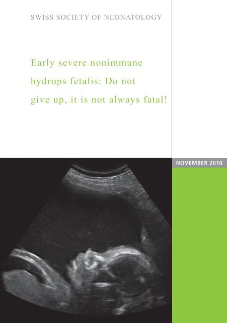
SWISS SOCIETY OF NEONATOLOGY<br />
<strong>Early</strong> <strong>severe</strong> <strong>nonimmune</strong><br />
<strong>hydrops</strong> <strong>fetalis</strong>: Do not<br />
give up, it is not always fatal!<br />
NOVEMBER 2010
Estermann K, Malzacher A, Drack G, Department <strong>of</strong><br />
Gynaecology and Obstetrics (DG), Department <strong>of</strong><br />
Neonatology (EK, MA), Kantonsspital St. Gallen,<br />
Switzerland<br />
© <strong>Swiss</strong> <strong>Society</strong> <strong>of</strong> Neonatology, Thomas M Berger, Webmaster<br />
2
3<br />
Hydrops <strong>fetalis</strong> (HF) is defined as the presence <strong>of</strong> ex-<br />
cessive fluid accumulation in at least two fetal body<br />
cavities (1). It occurs in 1/1‘500-4‘000 pregnancies (2,<br />
3). Two major groups can be differentiated: immune<br />
(IHF) and <strong>nonimmune</strong> <strong>hydrops</strong> <strong>fetalis</strong> (NIHF). IHF is almost<br />
always due to erythroblastosis caused by Rh alloimmunization.<br />
In the past, IHF was much more common<br />
than NIHF (incidence ratio <strong>of</strong> 9:1) (4). Nowadays,<br />
in the era <strong>of</strong> routine immunization <strong>of</strong> Rh negative mothers,<br />
about 80 percent <strong>of</strong> cases are NIHF (3).<br />
Although diagnosis and management has improved in<br />
recent years, NIHF is still associated with a high fetal<br />
mortality rate, which varies depending on the etiology<br />
between 50 to 90 percent (3, 6). To the best <strong>of</strong> our<br />
knowledge, thus far no case <strong>of</strong> spontaneous regression<br />
<strong>of</strong> idiopathic NIHF has been published. Therefore,<br />
we report on a patient with spontaneous regression<br />
<strong>of</strong> early <strong>severe</strong> and prolonged NIHF.<br />
At 19 2/7 weeks <strong>of</strong> gestation, a 34-year-old G3/P2 <strong>of</strong><br />
Tamil origin was referred to our perinatal center for<br />
further assessment <strong>of</strong> bilateral hydrothoraces and ascites.<br />
Her previous pregnancies had been uneventful. In<br />
the past she had suffered from grand mal epilepsy, but<br />
remained seizure free after discontinuing carbamazepine<br />
therapy three years ago. Prior to this pregnancy,<br />
she had been treated with infliximab, methotrexate<br />
and prednisolone for rheumatoid arthritis. Medica-<br />
INTRODUCTION<br />
CASE REPORT
tion was discontinued two months before conception<br />
without her rheumatologist‘s knowledge. Up to the<br />
20th week, her pregnancy had been complicated only<br />
by occasional dizziness which later turned out to be<br />
absence and later grand mal seizures. Carbamazepine<br />
was restarted at 36 weeks <strong>of</strong> gestation.<br />
Ultrasound results at 20 weeks <strong>of</strong> gestation confirmed ex-<br />
tensive fetal <strong>hydrops</strong> (Fig. 1) with normal heart function<br />
and brain anatomy. There were no skeletal abnormali-<br />
ties. Dimensions <strong>of</strong> the placenta and fetal organs were<br />
normal. Except for the massively enlarged abdominal<br />
circumference, biometrical findings were along the 10th<br />
percentile. There were no signs <strong>of</strong> fetal anemia with normal<br />
peak velocity in the middle cerebral artery.<br />
The mother‘s blood group was B Rh positive and the in-<br />
direct Coombs‘ test was negative. Serologies for toxo-<br />
plasmosis, parvovirus B19, herpes simplex virus 1 and<br />
2, cytomegalovirus, VZV, rubella, HIV-1/2, coxsackie<br />
B1-6, and treponema were negative. Amniocentesis<br />
was declined by the patient and her husband. Both<br />
parents were convinced that their child would recover.<br />
During the second trimester, fetal <strong>hydrops</strong> worsened.<br />
A head-lung-ratio <strong>of</strong> 0.27 suggested <strong>severe</strong> and most<br />
likely lethal lung hypoplasia. Therefore, it was decided<br />
that no caesarean section would be performed for<br />
fetal indications and spontaneous birth would not be<br />
monitored with fetal monitoring.<br />
4
5<br />
In the third trimester, signs <strong>of</strong> fetal <strong>hydrops</strong> improved.<br />
As a consequence, fetal lung maturation was induced<br />
at 28 weeks <strong>of</strong> gestation. Regular ultrasound exams<br />
documented further improvement <strong>of</strong> fetal <strong>hydrops</strong><br />
(Fig. 2) and, at 40 weeks, only a left-sided pleural<br />
effusion remained. Fetal growth continued along the<br />
10th percentile (Fig. 3, 4).<br />
Postterm amniotomy was performed at 41 4/7 weeks<br />
<strong>of</strong> gestation. A female neonate was born by spontaneous<br />
vaginal delivery. Apgar scores were 5, 7, 9 at<br />
1, 5, and 10 minutes, respectively. She received CPAP<br />
support for 10 minutes for mild respiratory distress.<br />
After bonding with the mother, the girl was transferred<br />
to our neonatal unit. The child had a birth weight<br />
<strong>of</strong> 2760 g (< P10) and a length <strong>of</strong> 47cm (< P10). The<br />
only remaining sign <strong>of</strong> the <strong>hydrops</strong> was loose skin over<br />
the abdomen with visible prominent intestinal loops<br />
(Fig. 5, 6). Echocardiography, cerebral and abdominal<br />
ultrasound examinations and chest X-ray were all<br />
normal. There was no evidence <strong>of</strong> pleural effusions.<br />
Albumin and total serum protein were within normal<br />
ranges. Her karyotype was normal. Histopathology <strong>of</strong><br />
the placenta was normal.<br />
After one week, mother and child were discharged<br />
home. At one year <strong>of</strong> age, the child was thriving and<br />
developing normally.
Fig. 1<br />
A<br />
Fetal ultrasound at 20 weeks <strong>of</strong> gestation: ascites (A)<br />
and bilateral hydrothoraces (B).<br />
6
7<br />
B
Fig. 2<br />
A<br />
A<br />
Fetal ultrasound at 33 weeks <strong>of</strong> gestation: improved<br />
<strong>hydrops</strong> with near complete resolution <strong>of</strong> ascites (A)<br />
and decreased pleural effusions (B).<br />
8
9<br />
B
Fig. 3<br />
450<br />
400<br />
350<br />
300<br />
250<br />
mm<br />
200<br />
150<br />
100<br />
50<br />
0<br />
head circumference abdominal circumference<br />
Biometric data: head circumference, abdominal<br />
circumference, femur length and estimated fetal<br />
weight.<br />
10<br />
450<br />
400<br />
350<br />
300<br />
250<br />
mm<br />
16 20 24 28 32 36 40<br />
200<br />
150<br />
100<br />
50<br />
0<br />
16 20 24 28 32 36 40<br />
weeks weeks<br />
90<br />
femur length estimated fetal weight<br />
6000<br />
80<br />
70<br />
5000<br />
60<br />
4000<br />
50<br />
mm<br />
40<br />
g 3000<br />
30<br />
2000<br />
20<br />
10<br />
1000<br />
0<br />
16 20 24 28 32 36 40<br />
0<br />
22 26 30 34 38 42<br />
weeks weeks
11<br />
aminotic fluid index V max syst MCY<br />
30.0<br />
120<br />
110<br />
25.0<br />
100<br />
90<br />
20.0<br />
80<br />
cm 15.0<br />
cm/s<br />
70<br />
60<br />
10.0<br />
50<br />
40<br />
5.0<br />
30<br />
20<br />
0.0<br />
10<br />
14 18 22 26 30 34 38 42<br />
15 19 23 27 31 35 39<br />
Biometric data: Amniotic fluid index and v max syst<br />
MCA.<br />
weeks weeks<br />
Fig. 4
Fig. 5<br />
First day <strong>of</strong> life: loose skin over abdomen with visible<br />
non-dilated loops <strong>of</strong> bowel.<br />
12
13<br />
First day <strong>of</strong> life: redundant loose skin over abdomen.<br />
Fig. 6
DISCUSSION NIHF has a variety <strong>of</strong> etiologies, consisting <strong>of</strong> fetal,<br />
placental, and maternal disorders (5). In 2008, Bellini<br />
and colleagues reviewed 6‘361 cases <strong>of</strong> NIHF published<br />
in the literature and suggested the following<br />
diagnostic categories: cardiovascular (21.7%), idiopathic<br />
(17.8%), chromosomal (13.4%), hematologic<br />
(10.4%), infections (6.7%), thoracic malformations<br />
(6.0%), lymphatic dysplasia (5.7%), TTTF-placental<br />
(5.6%), syndromic (4.4%), miscellaneous (3.7%),<br />
urinary tract malformations (2.3%), inborn errors <strong>of</strong><br />
metabolism (1.1%), extrathoracic tumors (0.7%), and<br />
gastrointestinal (0.5%) (2, 3).<br />
14<br />
The direct underlying mechanisms responsible for hy-<br />
drops are still unclear (7). Congestive heart failure, de-<br />
creased plasma osmotic pressure, increased capillary<br />
permeability, and obstructed lymphatic flow, all lead<br />
to abnormal water transport between the capillary and<br />
interstitial space (3). High right atrial pressures or volume<br />
overload with congestive heart failure might, for<br />
example, cause the edema. Hepatic venous congestion<br />
may lead to impaired hepatic function which in turn<br />
leads to hypoalbuminemia (8). Anemia causes <strong>hydrops</strong>,<br />
especially if it develops slowly (9). Inborn errors <strong>of</strong> metabolisms<br />
can lead to anemia or liver failure (10). Infectious<br />
agents associated with <strong>hydrops</strong> mainly affect<br />
bone marrow, myocardium, and vascular endothelium.<br />
The latter may lead to increased vascular permeability<br />
(11). Thoracic or diaphragmatic malformations can<br />
lead to intrathoracic masses, which compress the heart
15<br />
and limit its function (3). Twin-to-twin transfusion<br />
syndrome causes an imbalance in blood flow between<br />
donor and recipient (12). Finally, in maternal lupus<br />
erythematodes, antibodies crossing the placenta can<br />
lead to complete AV block (3).<br />
Our case illustrates that in medicine prognostication<br />
can be very difficult.
REFERENCES<br />
1. Forouzan I. Hydrops <strong>fetalis</strong>: recent advances. Obstet Gynecol<br />
Surv 1997;52:130-138<br />
2. Sohn C, Holzgreve W. Ultraschall in der Gynäkologie und<br />
Geburtshilfe. Thieme (1995), Stuttgart<br />
3. Bellini C, Hennekam RC, Fulcheri E, et al. Etiology <strong>of</strong><br />
<strong>nonimmune</strong> <strong>hydrops</strong> <strong>fetalis</strong>: a systemic review. Am J Med<br />
Genet A 2009;149:844-851<br />
4. Abrams ME, Meredith KS, Kinnard P, et al. Hydrops <strong>fetalis</strong>: A<br />
retrospective review <strong>of</strong> cases reported to a large national<br />
16<br />
database and identification <strong>of</strong> risk factors associated with death.<br />
Pediatrics 2007;120:84–89<br />
5. Wars<strong>of</strong> SL, Nicolaides KH, Rodeck C. Immune and non-immune<br />
<strong>hydrops</strong>. Clin Obst Gynecol 1986;29:533–536<br />
6. Schneider H, Husslein P, Schneider KTM. Die Geburtshilfe.<br />
Springer (2006), Heidelberg<br />
7. Bukowski R, Saade GR. Hydrops <strong>fetalis</strong>. Clin Perinatol<br />
2000;27:1007–1031<br />
8. Knilans TK. Cardiac abnormalities associated with <strong>hydrops</strong><br />
<strong>fetalis</strong>. Semin Perinatol 1995;19:483-492<br />
9. Arcasoy MO, Gallagher PG. Hematologic disorders and<br />
<strong>nonimmune</strong> <strong>hydrops</strong> <strong>fetalis</strong>. Semin Perinatol 1995;19:502–515<br />
10. Bellini C, Hennekam RC, Boccardo F, et al. Nonimmune<br />
idiopathic <strong>hydrops</strong> <strong>fetalis</strong> and congenital lymphatic dysplasia.<br />
Am J Med Genet 2006;Part A 140A:678–684<br />
11. Barron SD, Pass RF. Infectious causes <strong>of</strong> <strong>hydrops</strong> <strong>fetalis</strong>. Semin<br />
Perinatol 1995;19:493–501<br />
12. Mahieu-Caputo D, Salomon LJ, Le Bidois J, et al. Fetal<br />
hypertension: An insight into the pathogenesis <strong>of</strong> the twin-twin<br />
transfusion syndrome. Prenat Diagn 2003;23:640–645
SUPPORTED BY<br />
CONTACT<br />
<strong>Swiss</strong> <strong>Society</strong> <strong>of</strong> Neonatology<br />
www.neonet.ch<br />
webmaster@neonet.ch<br />
concept & design by mesch.ch