Neonatal ping pong fracture - Swiss Society of Neonatology
Neonatal ping pong fracture - Swiss Society of Neonatology
Neonatal ping pong fracture - Swiss Society of Neonatology
Create successful ePaper yourself
Turn your PDF publications into a flip-book with our unique Google optimized e-Paper software.
SWISS SOCIETY OF NEONATOLOGY<br />
<strong>Neonatal</strong> <strong>ping</strong> <strong>pong</strong> <strong>fracture</strong><br />
NOVEMBER 2006
Lunt CMB, Roth K, Eich G, Pasquier S, Zeilinger G,<br />
<strong>Neonatology</strong> (LCMB, PS, ZG), Pediatric Surgery (RK),<br />
Pediatric Radiology (EG), Children’s Hospital Aarau,<br />
Switzerland<br />
© <strong>Swiss</strong> <strong>Society</strong> <strong>of</strong> <strong>Neonatology</strong>, Thomas M Berger, Webmaster
A term baby-girl was delivered from a 6-year-old G /<br />
P1 by an elective Caesarian at 9 /7 weeks <strong>of</strong> gesta-<br />
tion after an uncomplicated pregnancy. She was in<br />
vertex presentation and the amniotic fluid was clear.<br />
Surgery was uneventful. There was no maternal hi-<br />
story <strong>of</strong> cephalopelvic disproportion and no history <strong>of</strong><br />
tauma or fall during antenatal period. The girl adap-<br />
ted well with an Apgar score <strong>of</strong> 9, 10, and 10 at 1, 5,<br />
and 10 minutes, respectively.<br />
The girl (birth weight <strong>of</strong> 1 0 g, normal length and<br />
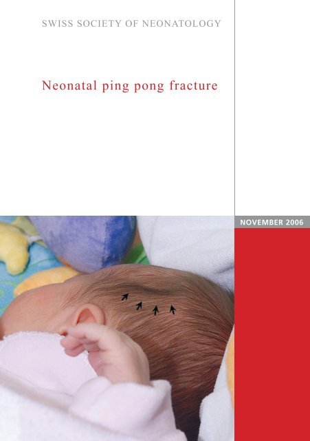
head circumference) had a depression on the left<br />
anterior temporo-parietal region measuring 4x4 cm<br />
without associated scalp swelling (Fig. 1). Her neuro-<br />
logical status was normal and there were no congeni-<br />
tal anomalies. The baby was not taking feeds adequa-<br />
tely and developed pallor and irregular breathing with<br />
an undulating oxygen saturation. She was then refer-<br />
red from the maternity hospital to our unit. Cerebral<br />
ultrasound was normal without signs <strong>of</strong> intracranial<br />
bleeding or contusions (Fig. , ). Skull x-ray showed<br />
a depression <strong>fracture</strong> the left temporo-parietal bone<br />
without <strong>fracture</strong> line (Fig. 4) which was confirmed by<br />
ultrasound (Fig. 5,6). A split-image procedure was re-<br />
fused by the parents. The breathing irregularity was<br />
interpreted as transient tachypnea <strong>of</strong> the newborn.<br />
Elevation <strong>of</strong> the depressed skull area through a bore-<br />
hole was suggested by the pediatric surgeon. The pa-<br />
rents, however, felt that such an operation wold be<br />
CASE REPORT
DISCUSSION<br />
too invasive and wanted to look for an alternative way<br />
to treat this greenstick <strong>fracture</strong>. The baby was dischar-<br />
ged after a three-day-hospitalization.<br />
Based on this case we would like to take the opportu-<br />
nity to discuss etiology, diagnostic evaluation, therapy<br />
and outcome <strong>of</strong> congenital depression <strong>fracture</strong>s <strong>of</strong> the<br />
skull.<br />
Traumatic injury to the skull and brain in utero is rare<br />
(1) and rarely needs intervention or causes persistent<br />
disability ( , ). Birth injury falls into categories: A)<br />
injuries produced by the normal force <strong>of</strong> labour and<br />
B) those produced by obstetric interventions. The inci-<br />
dence is higher in the instrumental deliveries and signi-<br />
ficantly more likely to be associated with intracranial<br />
lesions.<br />
The <strong>ping</strong> <strong>pong</strong> skull <strong>fracture</strong> is a green stick <strong>fracture</strong><br />
<strong>of</strong> the skull, it occurs when the skull bones are still<br />
s<strong>of</strong>t, thin and resilient. It presents as a depression de-<br />
formity <strong>of</strong> the skull – similar to a dent in a <strong>ping</strong> <strong>pong</strong><br />
ball – with no <strong>fracture</strong> line seen radiologically. It is<br />
thought that a <strong>ping</strong> <strong>pong</strong> <strong>fracture</strong> results from the<br />
pressure <strong>of</strong> the ischial tuberosity or sacral promontory<br />
symphisis pubis against the s<strong>of</strong>t skull during labor ( ).<br />
Associated intracranial injuries are rare. This condition<br />
can be diagnosed clinically. Plain x-ray may show the<br />
degree <strong>of</strong> deformation. An ultrasound <strong>of</strong> the skull is<br />
4
5<br />
Ping <strong>pong</strong> <strong>fracture</strong> in the left temporo-parietal<br />
region (arrow heads).<br />
Fig. 1
Fig. 2<br />
Cerebral ultrasound: coronal (2a) and paracoronal (2b)<br />
views with the latter demonstrating the <strong>ping</strong> <strong>pong</strong><br />
<strong>fracture</strong> (arrow).<br />
6
7<br />
Fig. 3
Fig. 4<br />
Skull X-ray.
9<br />
Cerebral ultrasound: Ping <strong>pong</strong> <strong>fracture</strong> in a<br />
transcranial scan (arrow).<br />
Fig. 5
Fig. 6<br />
Cerebral ultrasound: horizontal scan.<br />
10<br />
an easily available bedside tool for the diagnosis and<br />
management <strong>of</strong> intracerebral hematoma in neonates.<br />
Affected infants may be asymptomatic unless there is<br />
an associated intracranial injury. The management <strong>of</strong><br />
depressed skull <strong>fracture</strong>s neonates is discussed con-<br />
troversially. Small lesions are likely to resolve sponta-<br />
neously without any surgical intervention (4). Large<br />
lesions (> cm) with a mass effect leading to midline<br />
shift, require corrective surgery. In addition, it is advi-<br />
sable to elevate severe depressions to prevent cortical<br />
injury from sustained pressure. Such cases can be tre-<br />
ated easily by elevating the depressed bone fragment
11<br />
either by an obstetrical vacuum extractor (5, 6) or a<br />
minimally invasive borehole procedure adjacent to the<br />
depressed skull <strong>fracture</strong>.<br />
1. Eben Alexander, Jr., MD. Bourtland H. Davis. Intra-Uterine<br />
Fracture <strong>of</strong> Infant‘s Skull. J Neurosurgery 1969; 0:446-454<br />
. Dupuis O, Ruimark Silvera MS, Dupont C MW, Mottolese C,<br />
Kahn P, Dittmar A, Rudigoz RC. Comparison <strong>of</strong><br />
“instrument-associated” and “spontaneous” obstetric<br />
depressed skull <strong>fracture</strong>s in a cohort <strong>of</strong> 6 neonates. Am J<br />
Obstet Gynecol. 005;19 :165-170<br />
. Natelson SE, Sayers MP. The fate <strong>of</strong> children sustaining<br />
severe head trauma during birth. Pediatrics 197 ;51:169-174<br />
4. Ersahin Y, Mutluer S, Mirzai H, Palali I. Pediatric<br />
depressed skull <strong>fracture</strong>s:analysis <strong>of</strong> 5 0 cases. Childs Nerv<br />
Syst 1996;1 : - 1<br />
5. Pollak L, Raziel A, Ariely S, Schiffer J. Revival <strong>of</strong> non-surgical<br />
management <strong>of</strong> neonatal depressed skull <strong>fracture</strong>s. J Paediatr<br />
Child Health 1999; 5:96-97<br />
6. Tysvaer A, Nysted A. Depression <strong>fracture</strong>s <strong>of</strong> the skull in<br />
newborn infants. Treatment with vacuum extractor. Tidsskr<br />
Nor Laegeforen 1994;114: 15- 16<br />
REFERENCES
SUPPORTED BY<br />
CONTACT<br />
<strong>Swiss</strong> <strong>Society</strong> <strong>of</strong> <strong>Neonatology</strong><br />
www.neonet.ch<br />
webmaster@neonet.ch<br />
concept & design by mesch.ch


