

Physiological and molecular determinants of embryo implantation
Physiological and molecular determinants of embryo implantation
Physiological and molecular determinants of embryo implantation

Create successful ePaper yourself
Turn your PDF publications into a flip-book with our unique Google optimized e-Paper software.
Review<br />
<strong>Physiological</strong> <strong>and</strong> <strong>molecular</strong> <strong>determinants</strong> <strong>of</strong> <strong>embryo</strong><br />
<strong>implantation</strong><br />
Shuang Zhang a,b,1 , Haiyan Lin a,1 , Shuangbo Kong a,b,1 , Shumin Wang a , Hongmei Wang a ,<br />
Haibin Wang a,⇑ , D. R<strong>and</strong>all Armant c,d,⇑<br />
a<br />
State Key Laboratory <strong>of</strong> Reproductive Biology, Institute <strong>of</strong> Zoology, Chinese Academy <strong>of</strong> Sciences, Beijing 100101, PR China<br />
b<br />
Graduate School <strong>of</strong> the Chinese Academy <strong>of</strong> Sciences, Beijing 100039, PR China<br />
c<br />
Wayne State University School <strong>of</strong> Medicine, Detroit, MI 48201-1405, USA<br />
d<br />
Program in Reproductive <strong>and</strong> Adult Endocrinology, Eunice Kennedy Shriver National Institute for Child Health <strong>and</strong> Human Development, National Institutes<br />
<strong>of</strong> Health, Department <strong>of</strong> Health <strong>and</strong> Human Services, Bethesda, MD 20892, USA<br />
article info<br />
Article history:<br />
Available online xxxx<br />
Keywords:<br />
Blastocyst activation<br />
Uterine receptivity<br />
Blastocyst attachment<br />
Embryo <strong>implantation</strong><br />
Decidualization<br />
Contents<br />
abstract<br />
Embryo <strong>implantation</strong> involves the intimate interaction between an <strong>implantation</strong>-competent<br />
blastocyst <strong>and</strong> a receptive uterus, which occurs in a limited time period known as<br />
the window <strong>of</strong> <strong>implantation</strong>. Emerging evidence shows that defects originating during<br />
<strong>embryo</strong> <strong>implantation</strong> induce ripple effects with adverse consequences on later gestation<br />
events, highlighting the significance <strong>of</strong> this event for pregnancy success. Although a multitude<br />
<strong>of</strong> cellular events <strong>and</strong> <strong>molecular</strong> pathways involved in <strong>embryo</strong>–uterine crosstalk<br />
during <strong>implantation</strong> have been identified through gene expression studies <strong>and</strong> genetically<br />
engineered mouse models, a comprehensive underst<strong>and</strong>ing <strong>of</strong> the nature <strong>of</strong> <strong>embryo</strong><br />
<strong>implantation</strong> is still missing. This review focuses on recent progress with particular attention<br />
to physiological <strong>and</strong> <strong>molecular</strong> <strong>determinants</strong> <strong>of</strong> blastocyst activation, uterine receptivity,<br />
blastocyst attachment <strong>and</strong> uterine decidualization. A better underst<strong>and</strong>ing <strong>of</strong><br />
underlying mechanisms governing <strong>embryo</strong> <strong>implantation</strong> should generate new strategies<br />
to rectify <strong>implantation</strong> failure <strong>and</strong> improve pregnancy rates in women.<br />
Ó 2012 Elsevier Ltd. All rights reserved.<br />
1. Introduction . . . .......................................................................................... 00<br />
2. Maternal hormonal environment required for <strong>embryo</strong> <strong>implantation</strong> . . . . . . . . . . . . . . . ................................. 00<br />
3. Embryonic preparation for <strong>implantation</strong> necessitates ‘‘blastocyst activation’’ . . . . . . . . ................................. 00<br />
3.1. Estrogenic derivatives . . . ............................................................................. 00<br />
3.2. Cannabinoid signaling. . . ............................................................................. 00<br />
3.3. Wnt signaling . . . . . . . . . ............................................................................. 00<br />
3.4. Embryo-derived signals for <strong>implantation</strong> . . . ............................................................. 00<br />
4. Uterine receptivity: unique status <strong>of</strong> uterine differentiation conducive for <strong>embryo</strong> <strong>implantation</strong>. . . . . . . . ................. 00<br />
4.1. Steroid hormones . . . . . . ............................................................................. 00<br />
4.2. Cytokines . . . . . . . . . . . . . ............................................................................. 00<br />
⇑ Corresponding authors. Addresses: State Key Laboratory <strong>of</strong> Reproductive Biology, Institute <strong>of</strong> Zoology, Chinese Academy <strong>of</strong> Sciences, 1 Beichen West<br />
Road, Chaoyang District, Beijing 100101, PR China. Tel.: +86 10 64807868; fax: +86 10 64807099 (H. Wang), C.S. Mott Center for Human Growth <strong>and</strong><br />
Development, Wayne State University School <strong>of</strong> Medicine, 275 East Hancock Street, Detroit, MI 48201-1405, USA. Fax: +1 313 577 8554 (D.R. Armant).<br />
E-mail addresses: hbwang@ioz.ac.cn (H. Wang), D.Armant@wayne.edu (D.R. Armant).<br />
1 These authors contributed equally to this work.<br />
0098-2997/$ - see front matter Ó 2012 Elsevier Ltd. All rights reserved.<br />
http://dx.doi.org/10.1016/j.mam.2012.12.011<br />
Molecular Aspects <strong>of</strong> Medicine xxx (2013) xxx–xxx<br />
Contents lists available at SciVerse ScienceDirect<br />
Molecular Aspects <strong>of</strong> Medicine<br />
journal homepage: www.elsevier.com/locate/mam<br />
Please cite this article in press as: Zhang, S., et al. <strong>Physiological</strong> <strong>and</strong> <strong>molecular</strong> <strong>determinants</strong> <strong>of</strong> <strong>embryo</strong> <strong>implantation</strong>. Molecular Aspects <strong>of</strong><br />
Medicine (2013), http://dx.doi.org/10.1016/j.mam.2012.12.011
2 S. Zhang et al. / Molecular Aspects <strong>of</strong> Medicine xxx (2013) xxx–xxx<br />
4.3. Homeobox transcription factors . . ...................................................................... 00<br />
4.4. Developmental genes . . . . . . . . . . ...................................................................... 00<br />
5. Cell–cell interactions: the nature <strong>of</strong> <strong>embryo</strong> <strong>implantation</strong> . . . . . . . . . . . . . . .......................................... 00<br />
5.1. Uterine luminal closure for blastocyst apposition. . . . . . . ................................................... 00<br />
5.2. The trophectoderm–uterine epithelium interaction . . . . . ................................................... 00<br />
5.3. The epithelial–stromal interaction ...................................................................... 00<br />
5.3.1. Uterine epithelial responsiveness to estrogen signaling . . . . . . . . ..................................... 00<br />
5.3.2. Stromal responsiveness to progesterone signaling. . . . . . . . . . . . . ..................................... 00<br />
5.4. Uterine gl<strong>and</strong>s for <strong>embryo</strong> <strong>implantation</strong> . . . . . . . . . . . . . . ................................................... 00<br />
5.4.1. Uterine adenogenesis . . . . . . . . . . . . . . . . . ........................................................ 00<br />
5.4.2. Gl<strong>and</strong>ular–luminal epithelial interaction . ........................................................ 00<br />
5.4.3. Gl<strong>and</strong>ular–stromal interaction. . . . . . . . . . ........................................................ 00<br />
6. Molecular basis <strong>of</strong> decidualization . . . . . . . . . . . . . . ............................................................. 00<br />
6.1. Steroid hormones: central players in decidualization. . . . ................................................... 00<br />
6.2. Epithelial signals controlling stromal decidualization . . . ................................................... 00<br />
6.3. Cell-cycle regulators during decidualization . . . . . . . . . . . ................................................... 00<br />
6.4. Molecular <strong>and</strong> cellular aspects <strong>of</strong> immune tolerance during decidualization . . . . ................................ 00<br />
6.4.1. Genes regulating immune tolerance . . . . . ........................................................ 00<br />
6.4.2. Immune cells during decidualization . . . . ........................................................ 00<br />
7. Emerging concept: the quality <strong>of</strong> <strong>implantation</strong> determines the quality <strong>of</strong> ongoing pregnancy . .......................... 00<br />
8. Implications for human fertility . . . . . . . . . . . . . . . . ............................................................. 00<br />
9. Perspectives <strong>and</strong> closing remarks . . . . . . . . . . . . . . . ............................................................. 00<br />
Acknowledgements . . . . . . . ................................................................................ 00<br />
References . . . . . . . . . . . . . . ................................................................................ 00<br />
1. Introduction<br />
In mammals, a new life begins with the union <strong>of</strong> an egg with a sperm, a process known as fertilization (Wassarman,<br />
1999). Following fertilization, the zygote undergoes several rounds <strong>of</strong> divisions <strong>and</strong> morphogenesis to form the blastocyst,<br />
an <strong>embryo</strong>nic stage with two distinct cell lineages: the outer specialized trophectodermal epithelium <strong>and</strong> the inner cell mass<br />
(Cockburn <strong>and</strong> Rossant, 2010; Wang <strong>and</strong> Dey, 2006). The blastocyst participates in the first physical <strong>and</strong> physiological interaction<br />
with the maternal endometrium to initiate <strong>implantation</strong> (Red-Horse et al., 2004; Wang <strong>and</strong> Dey, 2006). A bidirectional<br />
crosstalk is essential for normal <strong>implantation</strong> thus the success <strong>of</strong> pregnancy, since perturbations will generate adverse outcomes<br />
for subsequent development, including decidualization <strong>and</strong> placentation, with potential loss <strong>of</strong> the pregnancy (Chen<br />
et al., 2011; Song et al., 2002; Wilcox et al., 1999; Ye et al., 2005).<br />
Early pregnancy loss, occurring during the peri<strong>implantation</strong> period before pregnancy is recognized clinically, is a relatively<br />
common phenomenon in humans (Cockburn <strong>and</strong> Rossant, 2010; Norwitz et al., 2001). For example, even in natural<br />
conception, the maximum chance <strong>of</strong> successful pregnancy occurring in a given menstrual cycle is limited to about 30% (Zinaman<br />
et al., 1996). Only 50–60% <strong>of</strong> all conceptions advance beyond 20 weeks <strong>of</strong> gestation (Norwitz et al., 2001). Among the<br />
pregnancies that are lost, <strong>implantation</strong> failure is the major cause, reaching approximate 75% (Wilcox et al., 1988). Furthermore,<br />
1 out <strong>of</strong> 7 couples worldwide are suffering from infertility (Forti <strong>and</strong> Krausz, 1998). Despite significant developments<br />
in in vitro fertilization <strong>and</strong> <strong>embryo</strong> transfer (IVF–ET) technology that have overcome many underlying causes <strong>of</strong> infertility,<br />
pregnancy success rates remain relatively low, mainly due to <strong>implantation</strong> failure (Miller et al., 2012; Norwitz et al., 2001;<br />
Wilcox et al., 1993). Therefore, it is imperative to address this global issue by investigating the mysteries <strong>of</strong> <strong>embryo</strong><br />
<strong>implantation</strong>.<br />
Successful <strong>implantation</strong> requires synchronization between the acquisition <strong>of</strong> <strong>implantation</strong> competency by the blastocyst<br />
<strong>and</strong> a receptive state in the uterine endometrium (Dey et al., 2004; Tranguch et al., 2005b; Wang <strong>and</strong> Dey, 2006). These two<br />
events are precisely regulated by maternal hormones, in particular, ovarian estrogen <strong>and</strong> progesterone (Conneely et al.,<br />
2002; Curtis Hewitt et al., 2002). Molecular <strong>and</strong> genetic evidence indicates that ovarian hormones together with locally produced<br />
signaling molecules, including cytokines, growth factors, homeobox transcription factors, lipid mediators <strong>and</strong> morphogen<br />
genes, function through autocrine, paracrine <strong>and</strong> juxtacrine interactions to specify the complex process <strong>of</strong><br />
<strong>implantation</strong> (Dey et al., 2004). However, the hierarchical l<strong>and</strong>scape <strong>of</strong> the <strong>molecular</strong> signaling pathways that govern <strong>embryo</strong>–uterine<br />
interactions during early pregnancy remains to be explored in depth.<br />
The crosstalk between the blastocyst <strong>and</strong> the uterus can only occur during a brief period, namely the ‘‘window <strong>of</strong> <strong>implantation</strong>’’<br />
(Ma et al., 2003; Paria et al., 1993; Rogers <strong>and</strong> Murphy, 1989; Yoshinaga, 1980). In response to the implanting <strong>embryo</strong>,<br />
the surrounding uterine stroma undergoes cellular transformation, a process known as decidualization, to<br />
accommodate <strong>embryo</strong>nic growth <strong>and</strong> invasion (Lim <strong>and</strong> Wang, 2010). Locally induced decidua provides a positive feedback<br />
to support <strong>embryo</strong> survival. It is also thought that the decidua functions as a barrier against maternal immunological responses<br />
to the semi-allogenic <strong>embryo</strong>. However, it remains largely unclear how the blastocyst escapes maternal immune<br />
surveillance at the time <strong>of</strong> <strong>implantation</strong>. With the emergence <strong>of</strong> advanced technologies, a global analysis <strong>of</strong> gene <strong>and</strong> protein<br />
Please cite this article in press as: Zhang, S., et al. <strong>Physiological</strong> <strong>and</strong> <strong>molecular</strong> <strong>determinants</strong> <strong>of</strong> <strong>embryo</strong> <strong>implantation</strong>. Molecular Aspects <strong>of</strong><br />
Medicine (2013), http://dx.doi.org/10.1016/j.mam.2012.12.011
S. Zhang et al. / Molecular Aspects <strong>of</strong> Medicine xxx (2013) xxx–xxx 3<br />
expression in the implanting <strong>embryo</strong> <strong>and</strong> uterus has been undertaken in several studies to unravel the <strong>molecular</strong> networks<br />
that control <strong>implantation</strong> in mice, as well as in humans (Hamatani et al., 2004b; Haouzi et al., 2011; Hu et al., 2008; Kao et al.,<br />
2002; Reese et al., 2001; Riesewijk et al., 2003; Yoon et al., 2004; Yoshioka et al., 2000). However, due to experimental difficulties<br />
<strong>and</strong> ethical restrictions, our underst<strong>and</strong>ing <strong>of</strong> human <strong>implantation</strong> still relies predominantly on animal models, particularly<br />
the mouse. Gene-knockout mouse models provide valuable information that has been used to construct a tentative<br />
<strong>molecular</strong> basis <strong>of</strong> <strong>implantation</strong>. Since <strong>embryo</strong> <strong>implantation</strong> is a dynamic developmental process that integrates many signaling<br />
molecules into a precisely orchestrated program, it is important to underst<strong>and</strong> the hierarchical l<strong>and</strong>scape <strong>of</strong> the pathways<br />
governing these processes to generate new strategies to correct <strong>implantation</strong> failure <strong>and</strong> improve pregnancy rates in<br />
women. This review will examine our underst<strong>and</strong>ing <strong>of</strong> signaling cascades that regulate <strong>embryo</strong> <strong>implantation</strong> <strong>and</strong> decidualization<br />
derived from gene expression studies <strong>and</strong> genetically engineered mouse models.<br />
2. Maternal hormonal environment required for <strong>embryo</strong> <strong>implantation</strong><br />
In the majority <strong>of</strong> eutherian mammals, <strong>implantation</strong> occurs in a fixed interval <strong>of</strong> time after ovulation when the corpus<br />
luteum is fully formed (Finn <strong>and</strong> Martin, 1974). In humans, this is during the luteal phase <strong>of</strong> the menstrual cycle, while<br />
in rodents, it is in the diestrous phase <strong>of</strong> the estrous cycle. It has been well established that estrogen <strong>and</strong> progesterone<br />
are principal hormones in this process. According to their dynamic fluctuating levels, the reproductive cycle is divided into<br />
three stages (Finn <strong>and</strong> Martin, 1974; Wang <strong>and</strong> Dey, 2006). The first stage is the proestrous or follicular phase in women<br />
during which estrogen levels are very high (Michael, 1976; Yoshinaga et al., 1969). The second stage is a period when the<br />
levels <strong>of</strong> both hormones are low immediately after ovulation. Finally, the luteal stage is when both progesterone <strong>and</strong> estrogen<br />
are secreted from the corpus luteum. Embryo <strong>implantation</strong> occurs at the luteal phase. For example, at this stage in mice,<br />
the level <strong>of</strong> progesterone is gradually increased, owing to an enhanced secretion from newly formed corpora luteum, accompanied<br />
by a pre<strong>implantation</strong> surge <strong>of</strong> estrogen on day 4 <strong>of</strong> pregnancy (day 1 = day <strong>of</strong> vaginal plug), while <strong>embryo</strong> <strong>implantation</strong><br />
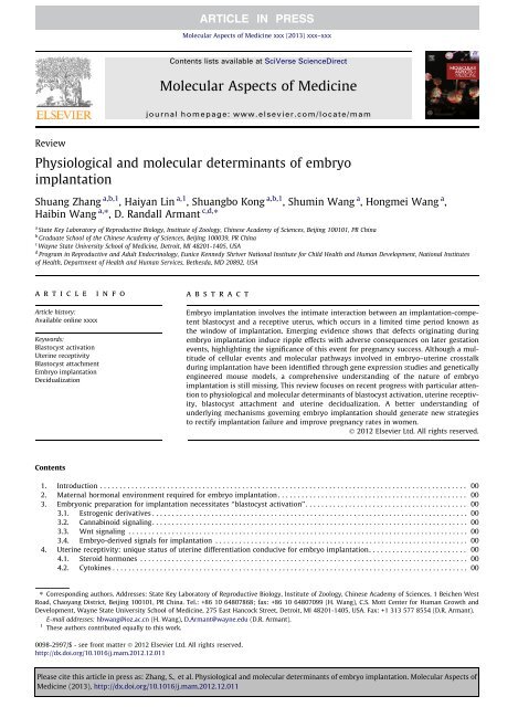
takes place at the midnight <strong>of</strong> day 4 (McCormack <strong>and</strong> Greenwald, 1974; Wang <strong>and</strong> Dey, 2006) (Fig. 1A). Based on<br />
Fig. 1. Hormonal control <strong>of</strong> <strong>embryo</strong> <strong>implantation</strong> in mice. (A) Steroid hormone patterns are illustrated during indicated days <strong>of</strong> the estrous cycle, uterine<br />
receptivity <strong>and</strong> early pregnancy. Estrogen secretion (red curve) is high at ovulation after the luteinizing hormone surge. Soon afterwards, progesterone (blue<br />
curve) increases beginning in the late afternoon <strong>of</strong> proestrus. If mating is successful, the newly formed corpora luteum, stimulated by mating behavior, will<br />
secrete progesterone from day 3 onward. On day 4, a small surge <strong>of</strong> estrogen cooperates with progesterone to induce uterine receptivity. Blastocyst<br />
<strong>implantation</strong> occurs at midnight <strong>of</strong> day 4. After <strong>implantation</strong>, progesterone is required for decidualization, placentation <strong>and</strong> completion <strong>of</strong> pregnancy. (B)<br />
Diagrams depicting cross-sections <strong>of</strong> the pre<strong>implantation</strong> uterus (day 1, day 4) <strong>and</strong> <strong>implantation</strong> sites (day 5, day 8). On day 1, the luminal epithelium <strong>of</strong> the<br />
non-receptive uterus is highly branched. On day 4, the uterus is receptive with the opposing luminal epithelium that closes around an implanting<br />
blastocyst. On day 5, the mural trophectoderm <strong>of</strong> the blastocyst attaches to the antimesometrial luminal epithelium. The stromal cells underlying the<br />
invading <strong>embryo</strong> then proliferate <strong>and</strong> differentiate to form an avascular primary decidual zone (PDZ) on the afternoon <strong>of</strong> day 5. Stroma cells next to the PDZ<br />
continue proliferation <strong>and</strong> differentiation to form a well-vascularized secondary decidual zone (SDZ) by day 8. AM, antimesometrial side; Bl, blastocyst; Em,<br />
<strong>embryo</strong>; E 2, estradiol-17b; GE, gl<strong>and</strong>ular epithelium; LE, luminal epithelium; M, mesometrial side; P 4, progesterone; S, stroma. (For interpretation <strong>of</strong> the<br />
references to colour in this figure legend, the reader is referred to the web version <strong>of</strong> this article.)<br />
Please cite this article in press as: Zhang, S., et al. <strong>Physiological</strong> <strong>and</strong> <strong>molecular</strong> <strong>determinants</strong> <strong>of</strong> <strong>embryo</strong> <strong>implantation</strong>. Molecular Aspects <strong>of</strong><br />
Medicine (2013), http://dx.doi.org/10.1016/j.mam.2012.12.011
4 S. Zhang et al. / Molecular Aspects <strong>of</strong> Medicine xxx (2013) xxx–xxx<br />
the pre<strong>implantation</strong> ovarian steroid pr<strong>of</strong>iles, priming with exogenous estrogen <strong>and</strong> progesterone can confer on the uterus <strong>of</strong><br />
ovariectomized mice a receptive state (Lim et al., 1997; Paria et al., 1999b). These hormones direct the preparation <strong>of</strong> the<br />
uterus for <strong>implantation</strong> in mice <strong>and</strong> rats (Harper <strong>and</strong> Walpole, 1967; Roblero et al., 1987; Vinijsanun <strong>and</strong> Martin, 1990).<br />
It is generally accepted that progesterone is required for <strong>implantation</strong> nearly in all the animals studied, while the role <strong>of</strong><br />
two estrogen surges at the proestrous <strong>and</strong> luteal phase prior to <strong>embryo</strong> <strong>implantation</strong> remains controversial (Dey et al.,<br />
2004; Finn <strong>and</strong> Martin, 1972; Tranguch et al., 2006; Wang <strong>and</strong> Dey, 2006).<br />
It was once thought that progesterone alone mediates <strong>implantation</strong>. However, the observation that suckling mice <strong>and</strong> rats<br />
with normal progesterone secretion have a facultative delayed <strong>implantation</strong> suggests that there may be another hormone<br />
involved in <strong>implantation</strong> (Malafaya et al., 2008; Mantalenakis <strong>and</strong> Ketchel, 1966; McLaren, 1968; Whitten, 1955; Yoshinaga<br />
<strong>and</strong> Adams, 1966). This suspicion is supported by the evidence that <strong>implantation</strong> can be induced in lactating rats by injection<br />
<strong>of</strong> a small dose <strong>of</strong> estrogen (Krehbiel, 1941). Later studies provide direct evidence for the role <strong>of</strong> luteal estrogen in normal<br />
<strong>implantation</strong> (Cochrane <strong>and</strong> Meyer, 1957; Whitten, 1958), showing that the timing <strong>of</strong> ovariectomy before or after luteal<br />
phase estrogen is critical for the induction <strong>of</strong> delayed <strong>implantation</strong>. For example, the blastocyst implants normally when<br />
ovariectomy is performed after pre<strong>implantation</strong> ovarian estrogen secretion, whereas if ovariectomy takes place before estrogen<br />
secretion, the <strong>embryo</strong> does not implant <strong>and</strong> the uterus enters into a condition <strong>of</strong> delayed <strong>implantation</strong>. With progesterone<br />
supplementation, the blastocyst remains quiescent, but it can be induced to implant by exogenous estrogen (Paria et al.,<br />
1993). These findings clearly indicate that pre<strong>implantation</strong> estrogen secretion is crucial for blastocyst <strong>implantation</strong> into a<br />
progesterone-primed receptive uterus. Since preovulatory estrogen is secreted in most species, it is thought that proestrous<br />
estrogen optimizes the subsequent response just before <strong>implantation</strong> (Barkley et al., 1979).<br />
Notably, the requirement for ovarian estrogen in <strong>implantation</strong> is species-specific. In species such as guinea pig, rhesus<br />
monkey, rabbit <strong>and</strong> golden hamster, progesterone alone is adequate for <strong>implantation</strong> (Harper et al., 1969; Heap <strong>and</strong><br />
Deanesly, 1967; Heap et al., 1981; Kwun <strong>and</strong> Emmens, 1974; Psychoyos, 1973, 1986). However, participation <strong>of</strong> estrogen<br />
in <strong>implantation</strong> <strong>of</strong> these species may not be completely excluded. A hypothesis has been proposed that blastocysts in these<br />
species could synthesize <strong>and</strong> secrete estrogen locally to initiate <strong>implantation</strong> (Dey et al., 2004; Wang <strong>and</strong> Dey, 2006). In<br />
agreement with this, aromatase, an enzyme for estrogen synthesis, is detected in the blastocyst <strong>of</strong> hamster <strong>and</strong> rabbit, while<br />
such an aromatase is absent in mice (Dickmann et al., 1975; Hoversl<strong>and</strong> et al., 1982a; Reese et al., 2008; Sengupta et al.,<br />
1983; Sholl et al., 1983). It remains unclear whether blastocyst–uterus attachment during <strong>implantation</strong> requires ovarian<br />
estrogen in humans or primates. There is evidence that mid-luteal estrogen is important for pregnancy establishment<br />
(Rao et al., 2007). However, contradictory evidence is also present (Ghosh et al., 1994; Smitz et al., 1993), showing that<br />
ovarian estrogen is not essential for <strong>implantation</strong> <strong>and</strong> pregnancy maintenance in IVF patients <strong>and</strong> non-human primates.<br />
It remains unclear whether the <strong>embryo</strong> or the endometrium could synthesize estrogen de novo that contributes to early<br />
pregnancy establishment in humans.<br />
In recent years, genetically engineered mouse models have provided valuable clues to underst<strong>and</strong> the roles <strong>of</strong> progesterone<br />
<strong>and</strong> estrogen during <strong>embryo</strong> <strong>implantation</strong>. The function <strong>of</strong> progesterone is mainly mediated by its receptor, PR (encoded<br />
by Pgr gene), which has two is<strong>of</strong>orms, PRA <strong>and</strong> PRB (Edwards, 2005). Both is<strong>of</strong>orms are expressed in the uterus (Mote et al.,<br />
2006). Female mice lacking both PRA <strong>and</strong> PRB are infertile with many defects in ovarian <strong>and</strong> uterine functions (Lydon et al.,<br />
1995), while these functions are normal in PRB deficient females (Mulac-Jericevic et al., 2000), indicating that essential progesterone-regulated<br />
functions in uteri are primarily mediated by PRA. Estrogen functions in the uterus primarily through<br />
nuclear estrogen receptors (Tan et al., 1999). ER also has two is<strong>of</strong>orms, known as ERa (encoded by Esr1 gene) <strong>and</strong> ERb (encoded<br />
by Esr2 gene) (Krege et al., 1998). Previous studies using knockout mice for ERs have demonstrated their differential<br />
functions in uterine biology (Hewitt et al., 2005; Lee et al., 2012a; Lubahn et al., 1993). ERa is the most important mediator <strong>of</strong><br />
estrogen signaling during early pregnancy since ERa knockout mice are unable to support <strong>implantation</strong> (Hewitt et al., 2005;<br />
Krege et al., 1998; Lee et al., 2012a; Lubahn et al., 1993). Although ERb knockout mice are fertile with normal <strong>implantation</strong>,<br />
evidence shows that it is also important in uterine biology (Krege et al., 1998; Lee et al., 2012a; Su et al., 2012; Wada-Hiraike<br />
et al., 2006). For example, ERb is expressed in the endometrial endothelium <strong>and</strong> may participate in <strong>implantation</strong> through<br />
regulating angiogenic <strong>and</strong> vasomotor changes (Huang et al., 2010; Su et al., 2012). In agreement with this, the expression<br />
level <strong>of</strong> ERb is significantly lower in women with infertility (Altmae et al., 2010). In addition, ERb is also believed to be a<br />
potent player in human labor onset due to its high expression in the myometrium <strong>and</strong> the cervix (Huang et al., 2010; Su<br />
et al., 2012).<br />
3. Embryonic preparation for <strong>implantation</strong> necessitates ‘‘blastocyst activation’’<br />
Acquisition <strong>of</strong> <strong>implantation</strong> competency by the blastocyst is a prerequisite for successful <strong>implantation</strong> (Paria et al., 1993).<br />
In mice, the blastocyst escapes from its zona pellucida <strong>and</strong> attaches to the uterine epithelium at day 4.5 <strong>of</strong> pregnancy (Das<br />
et al., 1994; McCormack <strong>and</strong> Greenwald, 1974; Wang <strong>and</strong> Dey, 2006). However, this sequence is interrupted in delayed<br />
<strong>implantation</strong>. Except for the facultative delay in lactating mice <strong>and</strong> rats (Enzmann et al., 1932; Hamlett, 1935; Kirkham,<br />
1918), <strong>implantation</strong> delay occurs naturally as an obligate delay in nearly 100 mammalian species, notably the mustelids<br />
<strong>and</strong> marsupials (Lopes et al., 2004; Mead, 1993; Renfree <strong>and</strong> Shaw, 2000; Thom et al., 2004). But in some species such as<br />
the hamster, guinea pig, rabbit <strong>and</strong> pig, delayed <strong>implantation</strong> does not occur (Dey et al., 2004). It is unclear whether this<br />
phenomenon exists in humans. Delayed <strong>implantation</strong> can be induced experimentally in mice <strong>and</strong> rats by ovariectomy before<br />
Please cite this article in press as: Zhang, S., et al. <strong>Physiological</strong> <strong>and</strong> <strong>molecular</strong> <strong>determinants</strong> <strong>of</strong> <strong>embryo</strong> <strong>implantation</strong>. Molecular Aspects <strong>of</strong><br />
Medicine (2013), http://dx.doi.org/10.1016/j.mam.2012.12.011
S. Zhang et al. / Molecular Aspects <strong>of</strong> Medicine xxx (2013) xxx–xxx 5<br />
the pre<strong>implantation</strong> ovarian estrogen surge <strong>and</strong> maintained by injection <strong>of</strong> progesterone (Huet <strong>and</strong> Dey, 1987; Yoshinaga<br />
<strong>and</strong> Adams, 1966), providing a powerful model for the study <strong>of</strong> <strong>implantation</strong>.<br />
During delayed <strong>implantation</strong>, the blastocyst is metabolically dormant <strong>and</strong> incompetent to initiate attachment in the<br />
uterus (Nieder <strong>and</strong> Weitlauf, 1985; Van Blerkom et al., 1978). Although <strong>embryo</strong>s develop into blastocysts <strong>and</strong> undergo zona<br />
dissolution, they show signs <strong>of</strong> being inactive with subnormal metabolic activity, reduced cell divisions <strong>and</strong> low DNA synthesis<br />
(Lopes et al., 2004). Ultrastructural observations <strong>of</strong> the trophoblast cells in dormant blastocysts reveal several morphological<br />
fine structure changes. For example, the ribosomes become monosomes, the endoplasmic reticulum is less pr<strong>of</strong>iled<br />
<strong>and</strong> the Golgi apparatus is not well developed (Renfree <strong>and</strong> Shaw, 2000; Wu <strong>and</strong> Meyer, 1974). The blastocyst can maintain<br />
this dormant state within the uterine cavity for days or even weeks (Lee et al., 2011a). But under appropriate conditions, the<br />
blastocyst is rapidly activated to resume its development with attachment <strong>and</strong> invasion. The active blastocyst also shows<br />
distinct morphological differences from the dormant blastocyst. For example, a more irregular surface with more microvilli<br />
is observed in activated trophoblast cells, with accumulating glycogen granules in the cytoplasm (Naeslund et al., 1980).<br />
Using the murine delayed <strong>implantation</strong> model <strong>and</strong> blastocyst transfer techniques, it has been demonstrated that dormant<br />
blastocysts fail to implant in the receptive uterus, highlighting the notion that the blastocyst’s state <strong>of</strong> activity determines<br />
the ‘‘window’’ <strong>of</strong> <strong>implantation</strong> in mice (Paria et al., 1993). However, it is still largely unknown how blastocysts undergo dormancy<br />
<strong>and</strong> survive for an extended period or how they become reactivated to acquire <strong>implantation</strong> competency.<br />
Since <strong>implantation</strong> is a two-way interaction, it has been speculated that there is a growth-impeding substance in the uterine<br />
secretions that target the <strong>embryo</strong>s (Surani, 1975). In support <strong>of</strong> this hypothesis, a blastocyst in delay that is subsequently<br />
activated reverts to its delayed state if replaced in the uterine cavity <strong>of</strong> a mouse in delayed <strong>implantation</strong> (Naeslund et al.,<br />
1980). Another explanation <strong>of</strong> <strong>embryo</strong> dormancy or diapause is that suboptimal conditions exist for blastocyst growth. Several<br />
in vitro experiments support this view. For instance, depletion <strong>of</strong> glucose <strong>and</strong>/or arginine <strong>and</strong> leucine from a conventional<br />
medium impedes blastocyst growth (Nieder <strong>and</strong> Weitlauf, 1985). Although these observations are valuable for the<br />
clues about blastocyst activation, the underlying <strong>molecular</strong> <strong>and</strong> cellular mechanisms are still unknown.<br />
The advent <strong>of</strong> cDNA microarray technology <strong>and</strong> genomic sequencing approaches has made global analysis <strong>of</strong> differential<br />
gene expression between dormant <strong>and</strong> active blastocysts possible (Hamatani et al., 2004b). Hamatani <strong>and</strong> coworkers find<br />
that 229 genes are differentially expressed between dormant <strong>and</strong> activated blastocysts, suggesting that the physiological<br />
states are <strong>molecular</strong>ly distinguishable. The major functional categories <strong>of</strong> altered genes include the cell cycle, cell signaling,<br />
<strong>and</strong> energy metabolic pathways. Current knowledge on mechanisms governing the process <strong>of</strong> blastocyst activation for<br />
<strong>implantation</strong> in mice is summarized in Fig. 2.<br />
It is not known whether delayed <strong>implantation</strong> exists in humans. However, insights into the <strong>molecular</strong> basis <strong>of</strong> blastocyst<br />
dormancy <strong>and</strong> activation in mice can provide valuable information for improving female fertility. For example, apart from<br />
providing a l<strong>and</strong>scape <strong>of</strong> specific genes <strong>and</strong> <strong>molecular</strong> pathways involved in blastocyst activation, the global gene study described<br />
above (Hamatani et al., 2004b) particularly highlights the importance <strong>of</strong> heparin-binding epidermal growth factor<br />
(EGF)-like growth factor (HB-EGF) signaling in <strong>embryo</strong>–uterine interactions required for <strong>implantation</strong>, which will be<br />
Fig. 2. Signals participating in blastocyst activation <strong>and</strong> uterine epithelial preparation for receptivity. Achievement <strong>of</strong> blastocyst <strong>implantation</strong> competency<br />
(blastocyst activation) involves steroid signaling, cannabinoid signaling <strong>and</strong> Wnt signaling pathways. Acquisition <strong>of</strong> uterine receptivity under the influence<br />
<strong>of</strong> ovarian progesterone <strong>and</strong> estrogen is associated with a flattening <strong>of</strong> the luminal epithelium <strong>and</strong> a loss <strong>of</strong> polarity at the site <strong>of</strong> <strong>embryo</strong> attachment.<br />
Several genes regulating uterine transformation are differentially regulated, as illustrated here <strong>and</strong> detailed in the text. CB1, brain-type cannabinoid<br />
receptor-1; ErbB1/4, epidermal growth factor receptor 1/4; ER, estrogen receptor; HB-EGF, heparin-binding EGF-like growth factor; ICM, inner cell mass;<br />
Klf5, kruppel-like factor 5; Msx1, muscle segment homeobox 1; 4-OH-E2, 4-hydroxyestradiol; PR, progesterone receptor; Tr, trophectoderm; Wnt5a,<br />
wingless-related MMTV integration site 5a.<br />
Please cite this article in press as: Zhang, S., et al. <strong>Physiological</strong> <strong>and</strong> <strong>molecular</strong> <strong>determinants</strong> <strong>of</strong> <strong>embryo</strong> <strong>implantation</strong>. Molecular Aspects <strong>of</strong><br />
Medicine (2013), http://dx.doi.org/10.1016/j.mam.2012.12.011
6 S. Zhang et al. / Molecular Aspects <strong>of</strong> Medicine xxx (2013) xxx–xxx<br />
discussed in details later. Furthermore, microarray analysis identifying multiple genes <strong>and</strong> signaling pathways involved in<br />
human pre<strong>implantation</strong> <strong>embryo</strong> development has been reported (Dobson et al., 2004; Hamatani et al., 2006). In IVF–ET programs,<br />
most pre<strong>implantation</strong> <strong>embryo</strong> arrest is due to dysregulation <strong>of</strong> individual genes or signaling pathways, since global<br />
analysis <strong>of</strong> the human pre<strong>implantation</strong> <strong>embryo</strong> transcriptome reveals that arrested <strong>embryo</strong>s nearly establish genome activation<br />
by day 3 following fertilization (Dobson et al., 2004). Thus, identification <strong>of</strong> dynamic gene expression networks would<br />
help to develop improved systems to assess <strong>embryo</strong> quality <strong>and</strong> enhance pregnancy success rates in IVF–ET programs.<br />
3.1. Estrogenic derivatives<br />
The dormant blastocysts in delayed <strong>implantation</strong> in mice will resume <strong>implantation</strong> upon an injection <strong>of</strong> estrogen, suggesting<br />
the importance <strong>of</strong> estrogen in mediating both the process <strong>of</strong> uterine receptivity <strong>and</strong> blastocyst activation (Paria<br />
et al., 1993). Since estrogen receptor is present in pre<strong>implantation</strong> mouse <strong>embryo</strong>s (Hou <strong>and</strong> Gorski, 1993; Hou et al.,<br />
1996), it was previously thought that estrogen could trigger blastocyst activation for <strong>implantation</strong>. However, a specific ER<br />
antagonist cannot inhibit the active status <strong>of</strong> the blastocyst, <strong>and</strong> estrogen fails to activate dormant blastocysts in culture,<br />
suggesting the participation <strong>of</strong> a pathway distinct from the classical nuclear ERs signaling (Paria et al., 1998). A further study<br />
shows that 4-hydroxyestradiol (4-OH-E2), a catechol metabolite <strong>of</strong> endogenous estrogen, is effective in initiating blastocyst<br />
activation through stimulating the synthesis <strong>of</strong> prostagl<strong>and</strong>in (PG) (Paria et al., 1998).<br />
A haloestrogen, 2-fluoroestrdiol-17b (2-FL-E2), is a potent estrogen but acts as an inhibitor <strong>of</strong> catecholestrogen synthesis.<br />
It has been previously shown to stimulate uterine growth <strong>and</strong> estrogen-targeted gene expression in mice (Dey et al., 1986;<br />
Mitchell et al., 1993; Paria et al., 1990, 1998). To distinguish the site-specific function <strong>of</strong> estrogen <strong>and</strong> catecholestrogen in<br />
uterine preparation versus blastocyst activation for <strong>implantation</strong>, experiments employing 2-FL-E2 have demonstrated that,<br />
unlike native estrogen, 2-FL-E2 fails to induce <strong>implantation</strong> in progesterone-primed delayed <strong>implantation</strong> mice, even at a<br />
relatively high dose (Paria et al., 1998). Moreover, dormant blastocysts fail to implant after their transfer into uteri <strong>of</strong> progesterone-treated<br />
delayed recipients receiving an injection <strong>of</strong> 2-FL-E2, whereas normal day 4 blastocysts show <strong>implantation</strong><br />
after transfer into recipient receiving the same treatment (Paria et al., 1998). These results indicate that 2-FL-E2 can induce<br />
the uterus into a receptive status, but fails to activate the dormant blastocyst mainly due to its inability to transform into<br />
catecholestrogen. In this respect, catecholestrogen alone can re-induce blastocyst activation <strong>and</strong> attachment reaction in delayed<br />
implanting mice (Hoversl<strong>and</strong> et al., 1982b; Kantor et al., 1985).<br />
Along with this finding, the receptive uterus is capable <strong>of</strong> transforming the native estrogen into catecholestrogen prior to<br />
<strong>embryo</strong> <strong>implantation</strong> (Paria et al., 1990, 1998). CYP1B1, an enzyme involved in NADPH-dependent 4-hydroxylation <strong>of</strong> estrogens,<br />
is expressed in uterine stroma cells on day 4 <strong>of</strong> pregnancy in mice (Paria et al., 1998; Reese et al., 2001). Observations <strong>of</strong><br />
the presence <strong>of</strong> CYP1B1 activity in the progesterone-treated uterus <strong>and</strong> Cyp1b1 transcripts in the receptive uterus strongly<br />
suggest that catecholestrogen locally produced in the uterus directs blastocyst activation for <strong>implantation</strong>. Collectively, ovarian<br />
estrogen interacting via the nuclear ERs is required for the preparation <strong>of</strong> the receptive uterus, whereas its catechol<br />
metabolite 4-hydroxyestradiol produced locally in the uterus activates the blastocyst for <strong>implantation</strong>. Future studies are<br />
warranted to further reveal the <strong>molecular</strong> mechanisms by which catecholestrogen exerts bioactivity <strong>and</strong> whether there is<br />
a specific receptor solely for catecholestrogen in the blastocyst <strong>and</strong> the uterus during <strong>implantation</strong>. It is also equally important<br />
to address whether <strong>and</strong> how ovarian steroid hormones <strong>and</strong> their metabolites interact with other local factors to initiate<br />
<strong>embryo</strong> <strong>implantation</strong> during early pregnancy.<br />
3.2. Cannabinoid signaling<br />
Psychoactive cannabinoids are major components in marijuana <strong>and</strong> exert most <strong>of</strong> their effects through activation <strong>of</strong> G<br />
protein-coupled cell surface receptors, cannabinoid receptor 1 (CB1) <strong>and</strong> cannabinoid receptor 2 (CB2), encoded in mice<br />
by the genes Cnr1 <strong>and</strong> Cnr2, respectively (Wang et al., 2006a). With the discovery <strong>of</strong> cannabinoid receptors, two endogenous<br />
cannabinoid lig<strong>and</strong>s, arachidonoylethanolamide (also known as an<strong>and</strong>amide) <strong>and</strong> 2-arachidonoylglycerol, have been identified<br />
that can be synthesized in the uterus during early pregnancy (De Petrocellis et al., 2004; Maccarrone <strong>and</strong> Finazzi-Agro,<br />
2002; Paria et al., 1996). The receptor-mediated activity <strong>of</strong> an<strong>and</strong>amide depends on its extracellular concentration, which<br />
largely depends on its intracellular degradation by the endocannabinoid-degrading enzyme, fatty acid amide hydrolase<br />
(FAAH) (Cravatt et al., 2001; Ueda et al., 2000; Wang et al., 2006c).<br />
Endocannabinoid signaling has recently been highlighted as an important lipid-mediated pathway that directs pre<strong>implantation</strong><br />
<strong>embryo</strong> development <strong>and</strong> the timely homing <strong>of</strong> <strong>embryo</strong>s into the uterus (Paria et al., 2001b; Wang <strong>and</strong> Dey,<br />
2005; Wang et al., 2004a, 2006b). Although both CB1 <strong>and</strong> CB2 are expressed in pre<strong>implantation</strong> <strong>embryo</strong>s (Paria et al.,<br />
1995), cellular <strong>and</strong> pharmological experiments have demonstrated that CB1 is the functional receptor subtype in the pre<strong>implantation</strong><br />
<strong>embryo</strong>s. For example, two-cell <strong>embryo</strong>s cultured in the presence <strong>of</strong> natural, synthetic or endocannabinoids fail to<br />
develop into the blastocyst stages, while a CB1-selective antagonist can reverse this inhibitory effect (Schmid et al., 1997;<br />
Wang et al., 1999). These findings indicate that the mouse <strong>embryo</strong> is a potential target for cannabinoid signaling. During<br />
the peri<strong>implantation</strong> period, the levels <strong>of</strong> uterine an<strong>and</strong>amide <strong>and</strong> blastocyst CB1 receptor are precisely regulated. Both<br />
the lig<strong>and</strong> <strong>and</strong> CB1 receptor are maintained at high levels in the non-receptive uterus <strong>and</strong> dormant blastocysts, whereas with<br />
the attainment <strong>of</strong> uterine receptivity <strong>and</strong> blastocyst activation, they are coordinately downregulated (Das et al., 1995; Guo<br />
et al., 2005; Schmid et al., 1997). This is further supported by the finding showing a higher level <strong>of</strong> an<strong>and</strong>amide in leukemia<br />
Please cite this article in press as: Zhang, S., et al. <strong>Physiological</strong> <strong>and</strong> <strong>molecular</strong> <strong>determinants</strong> <strong>of</strong> <strong>embryo</strong> <strong>implantation</strong>. Molecular Aspects <strong>of</strong><br />
Medicine (2013), http://dx.doi.org/10.1016/j.mam.2012.12.011
inhibitory factor deficient (Lif / ) mice with <strong>implantation</strong> failure than those in wild-type mice (Paria et al., 2001b). Similar<br />
fluctuations in an<strong>and</strong>amide levels during early pregnancy have been observed in women undergoing IVF–ET treatment,<br />
showing that a significantly lower level <strong>of</strong> an<strong>and</strong>amide during <strong>implantation</strong> is required for successful pregnancy (El-Talatini<br />
et al., 2009).<br />
Indeed, a very narrow range <strong>of</strong> an<strong>and</strong>amide regulates blastocyst function by differentially modulating mitogen-activated<br />
protein kinase (MAPK) signaling <strong>and</strong> Ca 2+ -channel activity via CB1 (Wang et al., 2003). For example, an<strong>and</strong>amide at a low<br />
concentration induces the activation <strong>of</strong> MAPK signaling <strong>and</strong> confers <strong>implantation</strong> competency on the blastocyst, whereas<br />
a higher concentration dampens this response by inhibiting Ca 2+ mobilization (Wang et al., 2003). Thus, it is conceivable that<br />
critical levels <strong>of</strong> endocannabinoids produced in the uterus synchronize blastocyst activation with uterine receptivity for<br />
<strong>implantation</strong>. Aberrant levels <strong>of</strong> uterine endocannabinoids or blastocyst CB1 receptors interfere with the normal <strong>embryo</strong>–<br />
uterine dialogue during <strong>implantation</strong>, resulting in early pregnancy loss. It is noteworthy that spontaneous pregnancy losses<br />
are associated with elevated an<strong>and</strong>amide levels in women (Habayeb et al., 2008; Maccarrone et al., 2002, 2000), reinforcing<br />
the concept that endocannabinoid signaling is an important determinant <strong>of</strong> <strong>embryo</strong>nic fate during <strong>implantation</strong>.<br />
3.3. Wnt signaling<br />
The highly conserved Wnt signaling pathway is a critical mediator <strong>of</strong> cell–cell interactions during <strong>embryo</strong>genesis (van<br />
Amerongen <strong>and</strong> Nusse, 2009). It is thus assumed to be involved in the process <strong>of</strong> <strong>implantation</strong>, where it is associated with<br />
intricate interplay between the blastocyst <strong>and</strong> the uterus. Wnt signaling can be divided into canonical <strong>and</strong> non-canonical<br />
pathways according to its distinct functions (van Amerongen <strong>and</strong> Nusse, 2009). With respect to the canonical Wnt pathway,<br />
the Wnt lig<strong>and</strong>s transduce their signals by combining with the frizzled (Fzd) receptor <strong>and</strong> low density lipoprotein receptorrelated<br />
protein (LRP) Lrp5/6 co-receptors complex (Logan <strong>and</strong> Nusse, 2004). This binding will liberate the cytoplasmic<br />
b-catenin, which is under continuous proteasome-mediated degradation before the pathway is activated (Metcalfe <strong>and</strong> Bienz,<br />
2011). Then cytoplasmic accumulated b-catenin will translocate into the nucleus <strong>and</strong> form a complex with the lymphoid<br />
enhancer-binding factor/T cell-specific transcription factor (LEF/TCF) to guide the transcription <strong>of</strong> target genes (Behrens<br />
et al., 1996). The non-canonical pathway is independent <strong>of</strong> b-catenin, involves only Fzd receptors, <strong>and</strong> the downstream effectors<br />
are diverse, including Ca 2+ /Planar Cell Polarity (PCP) pathway, Rho signaling, <strong>and</strong> Wnt-PKA signaling (Barrow, 2006;<br />
Semenov et al., 2007). The secreted frizzled related proteins (sFRPs), which have similar sequence signatures to Fzd receptors<br />
in the cysteine-rich lig<strong>and</strong>-binding domain <strong>and</strong> lack the transmembrane domain, will inhibit the bioactivities <strong>of</strong> Wnt proteins<br />
(Rattner et al., 1997; Suzuki et al., 2004). Alternatively, the co-receptor Lrp5/6 can also be regulated by the Dickkopf<br />
proteins (Dkks) to downregulate cell-surface LRP receptors by interacting with Kremen (Bafico et al., 2001; Glinka et al.,<br />
1998; Mao <strong>and</strong> Niehrs, 2003; Mao et al., 2002, 2001; Semenov et al., 2001). The complexity <strong>and</strong> redundancy <strong>of</strong> the Wnt family<br />
<strong>of</strong> proteins, receptors, extracellular antagonists <strong>and</strong> intracellular signaling components suggest that the nature <strong>of</strong> the inter-<br />
<strong>and</strong> intra-cellular Wnt machinery determines the orchestration <strong>of</strong> this signaling pathway during development.<br />
Previous studies have revealed specific expression patterns <strong>of</strong> Wnt pathway members in early <strong>embryo</strong>s <strong>and</strong> uteri during<br />
the peri<strong>implantation</strong> period in mice (Hamatani et al., 2004a; Kemp et al., 2005; Mohamed et al., 2004; Mohamed et al., 2005;<br />
Paria et al., 2001a; Wang et al., 2004c; Zeng et al., 2004), <strong>and</strong> the dispensable role <strong>of</strong> canonical Wnt signaling in blastocyst<br />
formation is well studied <strong>and</strong> reviewed previously (Chen et al., 2009; Sonderegger et al., 2010). Notably, during <strong>implantation</strong>,<br />
the canonical Wnt signaling is required for blastocyst competency. Female mice with oocyte specific deletion <strong>of</strong> b-catenin<br />
produce fewer numbers <strong>of</strong> pups compared with the wild-type, as <strong>embryo</strong>s are lost during the blastocyst stage, implying a<br />
role for b-catenin during the peri<strong>implantation</strong> stage (De Vries et al., 2004). Interfering with the Wnt pathway with the sFRP<br />
molecule also disturbs the <strong>implantation</strong> process (Mohamed et al., 2005). Using the strategy <strong>of</strong> adenoviral mediated Dkk1, our<br />
previous study has clearly demonstrated that silencing canonical Wnt/b-catenin signaling does not adversely affect the uterine<br />
preparation for receptivity, but remarkably blocks blastocyst competency for <strong>implantation</strong> in mice (Xie et al., 2008). The<br />
significance <strong>of</strong> this pathway in blastocysts is suggested by the delayed <strong>implantation</strong> model, showing that the activity <strong>of</strong> nuclear<br />
b-catenin signaling is different between the dormant <strong>and</strong> reactivated blastocysts (Xie et al., 2008). Moreover, Wnt3a is<br />
able to induce intracellular accumulation <strong>and</strong> nuclear translocation <strong>of</strong> b-catenin in trophectoderm cells <strong>and</strong> concurrently induces<br />
the expression <strong>of</strong> peroxisome proliferator-activated receptor (PPAR) d, a nuclear receptor for prostacyclin (Xie et al.,<br />
2008). Through pharmological gain <strong>of</strong> function experiments we further revealed that canonical Wnt signaling synergizes<br />
with PG signaling to confer blastocyst competency to <strong>implantation</strong> (Xie et al., 2008). This is consistent with the early finding<br />
that expression <strong>of</strong> cyclooxygenase 2 (COX-2), a rate-limiting enzyme <strong>of</strong> PG biosynthesis, is substantially upregulated in activated<br />
blastocysts after treatment with catecholestrogen (Paria et al., 1998). Collectively, these findings constitute direct evidence<br />
that Wnt signaling is at least one pathway determining blastocyst competency for <strong>implantation</strong>.<br />
3.4. Embryo-derived signals for <strong>implantation</strong><br />
S. Zhang et al. / Molecular Aspects <strong>of</strong> Medicine xxx (2013) xxx–xxx 7<br />
Although it is clear that a maternal–<strong>embryo</strong>nic dialogue is essential for the initiation <strong>of</strong> <strong>embryo</strong> <strong>implantation</strong> <strong>and</strong> that the<br />
state <strong>of</strong> the blastocyst determines the <strong>implantation</strong> window (Paria et al., 1993), there is a long-st<strong>and</strong>ing quest for specific<br />
<strong>embryo</strong>-derived signaling molecules that can functionally influence the uterus, as well as other reproductive organs (Lim<br />
<strong>and</strong> Dey, 2009). Global gene analysis <strong>of</strong> the dormant versus active blastocysts demonstrates that HB-EGF encoded by Hbegf<br />
gene is significantly up-regulated during blastocyst activation (Hamatani et al., 2004b). Prior studies demonstrate that Hbegf<br />
Please cite this article in press as: Zhang, S., et al. <strong>Physiological</strong> <strong>and</strong> <strong>molecular</strong> <strong>determinants</strong> <strong>of</strong> <strong>embryo</strong> <strong>implantation</strong>. Molecular Aspects <strong>of</strong><br />
Medicine (2013), http://dx.doi.org/10.1016/j.mam.2012.12.011
8 S. Zhang et al. / Molecular Aspects <strong>of</strong> Medicine xxx (2013) xxx–xxx<br />
is expressed in the luminal epithelium at the site <strong>of</strong> blastocyst apposition approximate 6 h prior to the blastocyst attachment<br />
reaction in mice (Das et al., 1994; Paria et al., 1999a). Moreover, increasing expression <strong>of</strong> its receptors (ErbB1 <strong>and</strong> ErbB4) <strong>and</strong><br />
lig<strong>and</strong> binding activity has been observed in the blastocysts that are competent for <strong>implantation</strong> (Das et al., 1997; Paria et al.,<br />
1993; Raab et al., 1996). Most interestingly, blastocyst-size Affi-gel beads presoaked with HB-EGF protein that are transferred<br />
intraluminally into a pseudopregnant uterus can induce its own gene expression in uterine cells surrounding the<br />
beads <strong>and</strong> increase vascular permeability, similar to the physiological changes induced by normal blastocysts (Hamatani<br />
et al., 2004b; Paria et al., 2001a). Signaling by HB-EGF back to the <strong>embryo</strong>, in turn, activates the program <strong>of</strong> trophoblast differentiation<br />
required for adhesive functions during subsequent attachment <strong>and</strong> invasion (Wang et al., 2000). These observations<br />
suggest that HB-EGF conducts an auto-induction loop between the implanting blastocyst <strong>and</strong> the uterus via paracrine<br />
<strong>and</strong> juxtacrine signaling. Maternal deficiency <strong>of</strong> HB-EGF targeted to the uterus defers the window <strong>of</strong> <strong>implantation</strong> <strong>and</strong> compromises<br />
pregnancy outcome, while amphiregulin, another heparin-binding member <strong>of</strong> the EGF family, partially compensates<br />
for the loss <strong>of</strong> HB-EGF during <strong>implantation</strong> (Xie et al., 2007). In human, HB-EGF is highly expressed in the receptive<br />
endometrium (Birdsall et al., 1996; Leach et al., 1999; Stavreus-Evers et al., 2002; Yoo et al., 1997) <strong>and</strong> its receptor ErbB4<br />
is localized on the surface <strong>of</strong> the trophectoderm in peri<strong>implantation</strong> blastocysts (Chobotova et al., 2002b), indicating that<br />
HB-EGF-ErbB4 signaling also mediates the trophectoderm–uterine epithelium interaction during <strong>implantation</strong> in humans.<br />
HB-EGF continues to regulate trophoblast development <strong>and</strong> survival during placentation in the first trimester <strong>of</strong> human gestation<br />
(Jessmon et al., 2009).<br />
As described above, aberrant endocannabinoid signaling is detrimental for pre<strong>implantation</strong> development <strong>and</strong> blastocyst<br />
<strong>implantation</strong> (Paria et al., 2001b; Wang et al., 1999). As an endogenous cannabinoid lig<strong>and</strong>, an<strong>and</strong>amide is spatiotemporally<br />
regulated in the uterus during early pregnancy, i.e., its expression levels are lower in the receptive uterus than for non-receptive<br />
uterus <strong>and</strong> at the <strong>implantation</strong> site than for inter<strong>implantation</strong> site (Paria et al., 2001b; Wang et al., 1999). Regarding the<br />
machinery controlling appropriate levels <strong>of</strong> an<strong>and</strong>amide in the implanting uterus, it is worth noting that the blastocyst can<br />
rapidly release a non-identified, cell permeable lipid that is able to activate uterine FAAH, protecting against the detrimental<br />
effects <strong>of</strong> excessive uterine endocannabinoids (Maccarrone et al., 2004).<br />
Another intriguing example <strong>of</strong> <strong>embryo</strong>nic regulation <strong>of</strong> uterine function during <strong>implantation</strong> is revealed by asynchronous<br />
<strong>embryo</strong> transfer studies. When pre<strong>implantation</strong> <strong>embryo</strong>s at different developmental stage are transferred into the oviducts<br />
<strong>of</strong> the same recipient, <strong>embryo</strong>s <strong>of</strong> advanced stage implant in advance <strong>of</strong> younger <strong>embryo</strong>s prior to the anticipated time when<br />
<strong>implantation</strong> normally occurs (Ueda et al., 2003). This finding indicates that the presence <strong>of</strong> <strong>embryo</strong>s in the reproductive<br />
tract can prime the endometrium through unknown <strong>embryo</strong>nic factors, leading to an expansion <strong>of</strong> the <strong>implantation</strong> window.<br />
A previous study employing non-human primates reports that super-imposition <strong>of</strong> endometrial receptivity on <strong>embryo</strong>nic<br />
stimuli causes remarkable changes in endometrial expression <strong>of</strong> pro-inflammatory cytokines <strong>and</strong> immunosuppressive factors,<br />
which regulate endometrial transformation conducive for <strong>embryo</strong> survival, growth <strong>and</strong> development (Nimbkar-Joshi<br />
et al., 2009). A similar phenomenon has been observed in Lif null pregnant mice, in which <strong>implantation</strong> fails to occur, but<br />
the uterine luminal epithelium exhibits COX-2 expression at the apposition site <strong>of</strong> blastocysts (Fouladi-Nashta et al.,<br />
2005; Song et al., 2000), reinforcing the notion that <strong>embryo</strong>-derived signaling molecules are able to regulate uterine preparation<br />
for <strong>implantation</strong>. It is a challenge to unravel the nature <strong>of</strong> <strong>embryo</strong>nic signals influencing uterine functions. One <strong>of</strong> the<br />
limiting factors is the availability <strong>of</strong> adequate amount <strong>of</strong> tissues for analysis. With the advent <strong>of</strong> microscale proteomics <strong>and</strong><br />
genomics, it becomes possible to identify <strong>embryo</strong>nic signals during <strong>implantation</strong> (Aghajanova et al., 2012; Dominguez et al.,<br />
2010; Katz-Jaffe et al., 2006; Tang et al., 2009). Identification <strong>of</strong> putative secreted <strong>embryo</strong>nic signaling molecules could be<br />
validated by employing the uterine blastocyst-size bead transfer approach, as previously described (Hamatani et al.,<br />
2004b; Paria et al., 2001a).<br />
After the attachment, the <strong>embryo</strong> directly contacts with the endometrium <strong>and</strong> the uterine stroma cells initiate the decidualization<br />
that embed <strong>and</strong> protect the <strong>embryo</strong> in rodents. Decidualization can also occur in response to artificial stimulus by<br />
oil injection or endometrium scratch in the receptive uterus to form deciduoma, which exclude the absolute requirement <strong>of</strong><br />
the conceptus (Loeb, 1906, 1908). However, some studies uncover the morphological <strong>and</strong> temporal difference between the<br />
deciduoma <strong>and</strong> the decidua, which hint the involvement <strong>of</strong> <strong>embryo</strong>-derived signals (Deanesly, 1971; Lundkvist <strong>and</strong> Nilsson,<br />
1982; Welsh <strong>and</strong> Enders, 1985). The disappearance <strong>of</strong> primary decidual zone which forms a barrier surrounding the <strong>embryo</strong><br />
<strong>and</strong> reduced immune cell population in deciduoma further emphasize the importance <strong>of</strong> <strong>embryo</strong>–uterus dialogue (Herington<br />
<strong>and</strong> Bany, 2007b; Wang et al., 2004d). Using microarray approaches, the differentially expressed genes responsive to the<br />
living <strong>embryo</strong> versus artificial stimuli further prove the paracrine signals from the <strong>embryo</strong> regulating the decidua development<br />
(Bany <strong>and</strong> Cross, 2006; Herington <strong>and</strong> Bany, 2007a,b, 2009; Herington et al., 2009; Kashiwagi et al., 2007; McConaha<br />
et al., 2011). Collectively, these findings highlight the necessity <strong>of</strong> <strong>embryo</strong>-derived signals for normal <strong>implantation</strong> <strong>and</strong><br />
decidualization.<br />
4. Uterine receptivity: unique status <strong>of</strong> uterine differentiation conducive for <strong>embryo</strong> <strong>implantation</strong><br />
In placental mammals, the uterus is receptive to blastocyst <strong>implantation</strong> during a spatiotemporally restricted ‘‘window’’<br />
when the uterine environment is favorable for blastocyst <strong>implantation</strong> (Yoshinaga, 1988). In mice, this period is limited to<br />
day 4 <strong>of</strong> pregnancy (Wang <strong>and</strong> Dey, 2006). The uterus cannot initiate <strong>implantation</strong> before this period (days 1–3). Immediately<br />
after the receptive state, the uterus spontaneously enters a refractory phase (day 5 onward) where the uterine<br />
Please cite this article in press as: Zhang, S., et al. <strong>Physiological</strong> <strong>and</strong> <strong>molecular</strong> <strong>determinants</strong> <strong>of</strong> <strong>embryo</strong> <strong>implantation</strong>. Molecular Aspects <strong>of</strong><br />
Medicine (2013), http://dx.doi.org/10.1016/j.mam.2012.12.011
environment is hostile for blastocyst survival (Carson et al., 2000; Yoshinaga, 1988). Similarly, in humans, the receptivity<br />
period occurs between days 20 <strong>and</strong> 24 <strong>of</strong> a regular menstrual cycle (days 6 to 10 after ovulation LH surge) (Bergh <strong>and</strong> Navot,<br />
1992; Lessey, 2011; Noyes et al., 1975; Psychoyos, 1973; Rashid et al., 2011). The key to uterine receptivity is the dynamic<br />
<strong>and</strong> precisely controlled <strong>molecular</strong> <strong>and</strong> cellular events that drive blastocyst growth, attachment <strong>and</strong> the subsequent events<br />
<strong>of</strong> <strong>implantation</strong>. This dynamic process involves a variety <strong>of</strong> genes that include cytokines, homeobox transcription factors, <strong>and</strong><br />
developmental genes working together with ovarian steroids to specify uterine receptivity.<br />
4.1. Steroid hormones<br />
Ovarian steroids progesterone <strong>and</strong> estrogen are principal hormones that direct uterine receptivity. Synchronized production<br />
<strong>of</strong> progesterone <strong>and</strong> estrogen mediates structural <strong>and</strong> functional changes in the uterus that enable the blastocyst to attach<br />
<strong>and</strong> initiate the process <strong>of</strong> <strong>implantation</strong>. On day 1 <strong>of</strong> pregnancy in mice, under the influence <strong>of</strong> preovulatory ovarian<br />
estrogen, the uterine epithelial cells undergo extensive proliferation that to some extent continues through day 2. Rising<br />
progesterone levels secreted from the newly formed corpora luteum initiate stromal cell proliferation from day 3 onward<br />
(Huet-Hudson et al., 1989; Huet et al., 1989). On the morning <strong>of</strong> day 4, when the uterus enters the pre-receptive stage,<br />
the production <strong>of</strong> a small amount <strong>of</strong> estrogen is crucial for the uterus to attain receptivity (Tranguch et al., 2005b). At that<br />
time, the uterine epithelial cells gradually lose their polarity, <strong>and</strong> meanwhile, the plasma membranes <strong>of</strong> the epithelial cells<br />
become smooth <strong>and</strong> flattened at the site <strong>of</strong> blastocyst apposition (Martin et al., 1970).<br />
To some extent, the window <strong>of</strong> uterine receptivity is flexible <strong>and</strong> can be modified under different hormonal environments.<br />
In mice, the blastocyst can initiate <strong>implantation</strong> outside <strong>of</strong> the normal ‘‘window’’ <strong>of</strong> uterine receptivity (Song et al., 2007). For<br />
example, blastocysts can initiate the attachment reaction in the non-receptive uterus when transferred on day 5 <strong>of</strong> pseudopregnancy,<br />
but <strong>implantation</strong> will not occur when normal blastocysts are transferred into the day 6 pseudopregnant uterus<br />
(Song et al., 2007). Exogenous progesterone supplementation can prolong the <strong>implantation</strong> window to day 6 with sustained<br />
LIF expression (Song et al., 2007). However, this deferred <strong>embryo</strong> <strong>implantation</strong> leads to <strong>embryo</strong>nic demise before birth in<br />
mice, <strong>and</strong> is <strong>of</strong>ten associated with higher risk <strong>of</strong> early pregnancy losses in humans (Wilcox et al., 1999).<br />
Estrogen is a critical determinant specifying the duration <strong>of</strong> the window <strong>of</strong> uterine receptivity for <strong>implantation</strong>. Using different<br />
doses <strong>of</strong> estrogens in the delayed <strong>implantation</strong> model, estrogen extends the window <strong>of</strong> uterine receptivity at a low<br />
threshold, whereas physiologically higher levels rapidly close the <strong>implantation</strong> window, transforming the uterus into a<br />
refractory state (Ma et al., 2003). Although whether blastocyst attachment reaction requires ovarian estrogen or not is still<br />
ambiguous in humans, exposure to high levels <strong>of</strong> estrogen, which could be resulting from ovarian stimulation with clomiphene<br />
citrate in IVF, leads to <strong>implantation</strong> failure <strong>and</strong> <strong>embryo</strong> resorption (Ertzeid <strong>and</strong> Storeng, 2001; Shapiro et al., 2011).<br />
This reduced <strong>implantation</strong> rate in IVF cycles could be due to asynchrony between the endometrium <strong>and</strong> the blastocyst when<br />
exposed to high levels <strong>of</strong> estrogen (Devroey et al., 2004). In fact, high expression level <strong>of</strong> endometrial aromatase is associated<br />
with poor IVF outcome (Brosens et al., 2004). Furthermore, there is evidence that decreasing estradiol levels during the pre<strong>implantation</strong><br />
period by using a follicle-stimulating hormone step-down regimen or aromatase inhibitor can improve uterine<br />
receptivity <strong>and</strong> pregnancy rates in IVF patients (de Ziegler et al., 2004; Mitwally et al., 2005; Simon et al., 1998). Therefore,<br />
the remarkable sensitivity <strong>of</strong> the uterus to estrogen is one <strong>of</strong> the important <strong>determinants</strong> for successful pregnancy establishment<br />
in IVF–ET programs.<br />
4.2. Cytokines<br />
S. Zhang et al. / Molecular Aspects <strong>of</strong> Medicine xxx (2013) xxx–xxx 9<br />
It is generally accepted that a class <strong>of</strong> cytokines, IL-6 family, is <strong>of</strong> great importance during <strong>embryo</strong>nic <strong>implantation</strong> (Dimitriadis<br />
et al., 2005). IL-6 family involves several cytokines such as LIF, IL-6, IL-11, <strong>and</strong> neurothrophic factor that all share<br />
intracellular signaling through gp130 <strong>and</strong> mediate the activation <strong>of</strong> STAT3 (Yun et al., 2012). Of the cytokines that have been<br />
studied, LIF is most pertinent to <strong>implantation</strong> (Kimber, 2005; Stewart et al., 1992; White et al., 2007). The LIF receptor shares<br />
gp130 as a common signal-transduction partner with other cytokine receptors. Expression <strong>of</strong> LIF is biphasic on day 4, initially<br />
in the uterine gl<strong>and</strong>s <strong>and</strong> later in the stromal cells surrounding the blastocyst during the attachment reaction (Ni et al., 2002;<br />
Song <strong>and</strong> Lim, 2006; Song et al., 2000). This expression pattern indicates that LIF has dual roles, first in uterine preparation<br />
<strong>and</strong> later in the attachment reaction (Song et al., 2000; Stewart et al., 1992). Lif-deficient female mice exhibit <strong>implantation</strong><br />
failure <strong>and</strong> supplementation with LIF rescues this defect (Chen et al., 2000; Stewart et al., 1992). Moreover, pharmologically<br />
blocking LIF action in the uterus significantly reduces the phosphorylation <strong>of</strong> the downstream signaling molecule, signal<br />
transducer <strong>and</strong> activator <strong>of</strong> transcription (STAT) 3, in the uterus, resulting in <strong>implantation</strong> failure (Menkhorst et al., 2011;<br />
Mohamet et al., 2009; White et al., 2007). The importance <strong>of</strong> LIF signaling in <strong>implantation</strong> is further evidenced by the observations<br />
that inactivation <strong>of</strong> gp130 <strong>and</strong> STAT3 also causes <strong>implantation</strong> failure (Catalano et al., 2005; Cheng et al., 2001;<br />
Daikoku et al., 2011; Ernst et al., 2001). In humans, LIF is expressed at high levels in the gl<strong>and</strong>ular epithelium <strong>of</strong> the secretory<br />
endometrium (Rashid et al., 2011), with a significant increase in the luminal <strong>and</strong> gl<strong>and</strong>ular epithelium at mid-secretory<br />
phase (Leach et al., 2012). It is reported that an optimal level <strong>of</strong> LIF is required for blastocyst <strong>implantation</strong> in women<br />
(Menkhorst et al., 2011; Terakawa et al., 2011). Moreover, clinical evidence shows that LIF deficiency is associated with<br />
unexplained recurrent abortion <strong>and</strong> infertility in women (Dey et al., 2004; Ernst et al., 2001; Hambartsoumian, 1998). Overall,<br />
significant gains have been made in our underst<strong>and</strong>ing <strong>of</strong> the function <strong>of</strong> LIF, a critical determinant <strong>of</strong> <strong>embryo</strong><br />
<strong>implantation</strong>.<br />
Please cite this article in press as: Zhang, S., et al. <strong>Physiological</strong> <strong>and</strong> <strong>molecular</strong> <strong>determinants</strong> <strong>of</strong> <strong>embryo</strong> <strong>implantation</strong>. Molecular Aspects <strong>of</strong><br />
Medicine (2013), http://dx.doi.org/10.1016/j.mam.2012.12.011
10 S. Zhang et al. / Molecular Aspects <strong>of</strong> Medicine xxx (2013) xxx–xxx<br />
Besides LIF, IL-6 is another important cytokine for successful pregnancy. In mice, it is secreted by the epithelial <strong>and</strong> stromal<br />
cell in uterus <strong>and</strong> regulated by the ovarian steroid hormones (Prins et al., 2012; Robertson et al., 1992; Sanford et al.,<br />
1992). Mice lacking IL-6 exhibit an impaired <strong>implantation</strong> <strong>and</strong> a deferred labor onset comprising term pregnancy outcome<br />
(Dimitriadis et al., 2005; Robertson et al., 2000b, 2010). In humans, IL-6 is mainly produced by endometrial epithelium <strong>and</strong><br />
stromal cells in a cyclical manner, <strong>and</strong> the levels <strong>of</strong> IL-6 are relatively low during the proliferative phase <strong>and</strong> rise steadily<br />
during the secretory phase (Champion et al., 2012; Jasper et al., 2007), suggesting its important role during <strong>implantation</strong>.<br />
Furthermore, low IL-6 production in the endometrium is highly associated with recurrent miscarriages, whereas elevated<br />
levels <strong>of</strong> IL-6 are also reported in women with recurrent abortions, preeclampsia <strong>and</strong> preterm delivery (Dimitriadis et al.,<br />
2005; Prins et al., 2012).<br />
4.3. Homeobox transcription factors<br />
Homeobox genes are evolutionarily conserved transcriptional regulators that control <strong>embryo</strong>nic morphogenesis <strong>and</strong> differentiation<br />
(Krumlauf, 1994). In mice, homeobox A (Hoxa) genes, Hoxa10 <strong>and</strong> Hoxa11, are expressed in uterine stromal cells<br />
during receptivity <strong>and</strong> upregulated upon decidualization in a steroid hormone-responsive manner (Benson et al., 1996; Gendron<br />
et al., 1997; Hsieh-Li et al., 1995). This specific expression pattern indicates that these two transcription factors may<br />
have dual roles, first in uterine receptivity <strong>and</strong> then in decidualization. Both Hoxa10 <strong>and</strong> Hoxa11 mutant mice are infertile<br />
due to <strong>implantation</strong> defects (Bagot et al., 2001; Gendron et al., 1997; Lim et al., 1999b). Hoxa11 / uteri have a more severe<br />
phenotype than Hoxa10 / mice, <strong>and</strong> the absence <strong>of</strong> Lif expression in Hoxa11 / uteri reinforces its role as a crucial participant<br />
in uterine receptivity <strong>and</strong> later events <strong>of</strong> <strong>implantation</strong> (Gendron et al., 1997). Regarding the pathophysiological significance<br />
<strong>of</strong> Hox genes in human <strong>implantation</strong>, expression <strong>of</strong> HOXA10 <strong>and</strong> HOXA11 in the endometrium increases significantly<br />
in the midluteal phase when the uterus is receptive for <strong>embryo</strong> attachment (Gui et al., 1999; Taylor et al., 1998, 1999b), <strong>and</strong><br />
is significantly lower in infertile women (Eun Kwon <strong>and</strong> Taylor, 2004; Fischer et al., 2011; Matsuzaki et al., 2009; Taylor et al.,<br />
1999a).<br />
Non-classical Hox genes may also be important in <strong>implantation</strong>. Ablation <strong>of</strong> H6 homeobox 3 (Hmx3) in mice leads to<br />
<strong>implantation</strong> failure (Wang et al., 1998). Msx1, another homeobox gene, is transiently expressed in the mouse luminal epithelium<br />
<strong>and</strong> gl<strong>and</strong>ular epithelium on the morning <strong>of</strong> day 4 <strong>of</strong> pregnancy (Daikoku et al., 2004; Pavlova et al., 1994), but its<br />
expression is dramatically downregulated to undetectable levels upon the termination <strong>of</strong> uterine receptivity (Daikoku<br />
et al., 2004; Pavlova et al., 1994). However, in Lif / mice, Msx1 is consistently expressed in the uterine epithelium even<br />
on day 6 <strong>of</strong> pregnancy, suggesting that LIF signaling is essential for the down-regulation <strong>of</strong> Msx1 (Daikoku et al., 2004).<br />
This is confirmed by the observation <strong>of</strong> sustained Msx1 expression in uteri with conditional depletion <strong>of</strong> gp130 (Daikoku<br />
et al., 2011). Conditional deletion <strong>of</strong> uterine Msx1 impairs <strong>implantation</strong>. Histological analysis <strong>of</strong> Msx1 /<br />
<strong>implantation</strong> sites<br />
reveals that the luminal epithelium lacks well-defined crypts for blastocyst homing <strong>and</strong> attachment (Daikoku et al., 2011).<br />
Moreover, a double knockout <strong>of</strong> uterine Msx1 <strong>and</strong> Msx2 results in complete <strong>implantation</strong> failure with altered luminal<br />
epithelial cell polarity <strong>and</strong> impaired stromal–epithelial communication (Daikoku et al., 2011; Nallasamy et al., 2012),<br />
pointing toward a compensatory role <strong>of</strong> Msx2 in the establishment <strong>of</strong> uterine receptivity in the absence <strong>of</strong> Msx1. Nonetheless,<br />
these results suggest that Msx1/2 genes are critical for conferring uterine epithelial integrity <strong>and</strong> uterine receptivity in<br />
mice.<br />
4.4. Developmental genes<br />
The dialogue between the blastocyst <strong>and</strong> uterus shares features with reciprocal epithelial–mesenchymal interactions that<br />
occur during <strong>embryo</strong>genesis, <strong>and</strong> involves evolutionarily conserved signaling pathways mediated by Indian hedgehog (IHH)<br />
proteins, bone morphogenetic proteins (BMPs), Wnts, <strong>and</strong> their cognate receptors (Paria et al., 2001a).<br />
IHH is a member <strong>of</strong> the hedgehog family which fine-tune cell proliferation, differentiation <strong>and</strong> cell–cell communication in<br />
many processes such as organogenesis, stem cell maintenance <strong>and</strong> oncogenesis (Ingham <strong>and</strong> McMahon, 2001; Ryan <strong>and</strong><br />
Chiang, 2012). The progesterone-dependent expression <strong>of</strong> Ihh is restricted to the epithelium, whereas the effectors <strong>of</strong><br />
IHH, patched-1 (ptch1) <strong>and</strong> transcription factors GLI-Krüppel family member (Gli) 1–3 are coordinately expressed in the<br />
underlying stroma (Matsumoto et al., 2002; Takamoto et al., 2002). Conditional deletion <strong>of</strong> Ihh in the uterus results in<br />
<strong>implantation</strong> failure due to a lack <strong>of</strong> progesterone-facilitated stromal proliferation, angiogenesis <strong>and</strong> decidualization, phenotypically<br />
mimicking that <strong>of</strong> PR knockout mice (Franco et al., 2010a; Lee et al., 2006). These findings indicate that IHH is an<br />
essential mediator for PR action in the uterus, participating in the communication between the uterine epithelium <strong>and</strong> the<br />
stroma required for <strong>embryo</strong> <strong>implantation</strong>. Microarray analysis demonstrates that IHH mRNA is synchronously regulated<br />
with PR during the human menstrual cycle (Talbi et al., 2006). Indeed, the temporal elevation <strong>of</strong> endometrial IHH <strong>and</strong><br />
GLI1 during the secretory phase, <strong>and</strong> their modulation by CDB-2914, a selective PR modulator (Wei et al., 2010), suggest that<br />
progesterone regulated hedgehog signaling is a key regulator <strong>of</strong> human endometrial differentiation <strong>and</strong> <strong>implantation</strong>. Moreover,<br />
endometrial IHH expression is reduced in women with endometriosis, indicative <strong>of</strong> progesterone resistance (Smith<br />
et al., 2011).<br />
BMPs are the largest family <strong>of</strong> morphogens belonging to the transforming growth factor-b (TGF-b) superfamily <strong>of</strong> growth<br />
modulators (Zamani <strong>and</strong> Brown, 2011), <strong>and</strong> transcripts corresponding to several BMP family members are expressed in<br />
mouse uteri (Paria et al., 2001a; Ying <strong>and</strong> Zhao, 2000). Among all the BMPs expressed in the uterus, only BMP2 is induced<br />
Please cite this article in press as: Zhang, S., et al. <strong>Physiological</strong> <strong>and</strong> <strong>molecular</strong> <strong>determinants</strong> <strong>of</strong> <strong>embryo</strong> <strong>implantation</strong>. Molecular Aspects <strong>of</strong><br />
Medicine (2013), http://dx.doi.org/10.1016/j.mam.2012.12.011
in response to progesterone, with intense expression in the stromal cells surrounding the implanted <strong>embryo</strong> (Paria et al.,<br />
2001a). In vitro studies have demonstrated that addition <strong>of</strong> recombinant BMP2 to undifferentiated stromal cells markedly<br />
enhances the decidualization by stimulating Smad signaling pathway, whereas silencing the expression <strong>of</strong> BMP2 in these<br />
cells efficiently blocks the decidualization in both mice <strong>and</strong> humans (Lee et al., 2007; Li et al., 2007). Moreover, the biological<br />
activity <strong>of</strong> BMP2 is mediated by proprotein convertase 6 (PC6) during decidualization; therefore, addition <strong>of</strong> recombinant<br />
active BMP2 partially rescues the decidualization arrest caused by PC6 inhibition (Heng et al., 2010). This is consistent with<br />
the previous findings that PC6 is highly expressed <strong>and</strong> regulated during <strong>implantation</strong> <strong>and</strong> decidualization, <strong>and</strong> that deletion<br />
or knockdown <strong>of</strong> PC6 inhibits decidualization, causing <strong>implantation</strong> failure <strong>and</strong> female infertility (Nie et al., 2005; Okada<br />
et al., 2005; Tang et al., 2005).<br />
Interestingly, uterine expression <strong>of</strong> the PR-interacting protein, Krüppel-like factor (KLF) 9, is significantly enhanced upon<br />
conditional ablation <strong>of</strong> Bmp2 (Lee et al., 2007; Li et al., 2007). Previous studies have demonstrated that KLF9 is highly expressed<br />
in the predecidual stroma <strong>and</strong> becomes undetectable in the decidua, while its deficiency induces subfertility in mice<br />
<strong>and</strong> reduces uterine progesterone sensitivity (Simmen et al., 2004; Velarde et al., 2005). Increasing evidence points toward<br />
the existence <strong>of</strong> a negative feedback loop between KLF9 <strong>and</strong> BMP2 to maintain PR function in stromal cells for successful<br />
<strong>implantation</strong>, where deregulation <strong>of</strong> this interaction can compromise the attainment <strong>of</strong> uterine receptivity <strong>and</strong> cause infertility<br />
(Pabona et al., 2010).<br />
Gene expression pr<strong>of</strong>iling experiments have identified Wnt4 as a downstream target <strong>of</strong> BMP2-induced decidualization (Li<br />
et al., 2007). Wnt4 is expressed primarily in the luminal epithelium during the pre<strong>implantation</strong> period <strong>and</strong> then re-localizes<br />
to the stromal cells surrounding the implanting <strong>embryo</strong> <strong>and</strong> exp<strong>and</strong>s its expression to the decidua (Daikoku et al., 2004;<br />
Hayashi et al., 2009). Conditional deletion <strong>of</strong> Wnt4 in the uterus renders female mice subfertile due to defective <strong>embryo</strong><br />
<strong>implantation</strong> <strong>and</strong> subsequent decidualization (Franco et al., 2011a). Molecular analysis demonstrates that Bmp2 along with<br />
other decidual marker genes such as follistatin are downregulated in Wnt4-null uteri (Franco et al., 2011a). These data indicate<br />
a complex correlation between Wnt4 <strong>and</strong> BMP2 during decidualization.<br />
In addition to Wnt4, many other components <strong>of</strong> Wnt signaling pathway are spatiotemporally regulated in the peri<strong>implantation</strong><br />
uterus <strong>and</strong> are believed to be crucial to <strong>implantation</strong> (Chen et al., 2009; Hayashi et al., 2009). For example,<br />
Wnt5a is expressed in the uterine subepithelial stroma albeit at lower levels in the epithelium (Hayashi et al., 2009;<br />
Hou et al., 2004; Mericskay et al., 2004). However, its expression is upregulated in both the epithelium <strong>and</strong> stroma in<br />
Msx1 /<br />
<strong>and</strong> Msx1/Msx2 / uteri (Daikoku et al., 2011; Nallasamy et al., 2012). In addition, application <strong>of</strong> Wnt5a<br />
in vitro compromises blastocyst invasion <strong>and</strong> trophoblast outgrowth when co-cultured with uterine epithelial cells<br />
(Daikoku et al., 2011). These findings suggest that Wnt5a may affect epithelial function <strong>and</strong>, thus, direct blastocyst trophectoderm<br />
differentiation. A recent study show that Wnt7b is strongly upregulated in uterine epithelia after peri<strong>implantation</strong><br />
estrogen secretion <strong>and</strong> that its expression can be induce by injection <strong>of</strong> estrogen in ovariectomized mice (Hayashi<br />
et al., 2009). Therefore, it is likely that Wnt7b is a direct mediator <strong>of</strong> uterine function in response to ovarian estrogen.<br />
However, a definitive role <strong>of</strong> Wnt7b in uterine receptivity warrants further investigation through the use <strong>of</strong> a conditional<br />
deletion mouse model, since Wnt7b null mice die during midgestation due to a failure <strong>of</strong> chorioallantoic fusion (Parr et al.,<br />
2001). Wnt7a is also expressed in the luminal epithelium with maximal expression on day 5 <strong>and</strong> declines thereafter at<br />
both <strong>implantation</strong> <strong>and</strong> inter-<strong>implantation</strong> sites (Hayashi et al., 2009). In contrast to Wnt7b, uterine expression <strong>of</strong> Wnt7a<br />
is not under the control <strong>of</strong> ovarian estrogen. Adult Wnt7a-deficient mice are viable, but infertile, owing to a lack <strong>of</strong> endometrial<br />
gl<strong>and</strong>s, suggesting that Wnt7a is crucial for normal formation <strong>and</strong> maintenance <strong>of</strong> uterine cellular architecture<br />
(Carta <strong>and</strong> Sassoon, 2004; Miller <strong>and</strong> Sassoon, 1998; Parr <strong>and</strong> McMahon, 1998). Sfrp4, a Wnt inhibitory protein, is expressed<br />
in the uterine stroma during the receptive phase (Daikoku et al., 2004). A marked downregulation <strong>of</strong> sfrp4 in<br />
Lif / uteri indicates that sFRP4 could participate in regulating uterine preparation for <strong>implantation</strong> (Daikoku et al.,<br />
2004). This is further supported by the observation that sFPR4 expression remains low in the endometrium <strong>of</strong> women<br />
with recurrent <strong>implantation</strong> failure (Revel et al., 2011). Unlike sFRP4, expression <strong>of</strong> sFRP2 is downregulated at the <strong>implantation</strong><br />
site, <strong>and</strong> intraluminal infusion <strong>of</strong> sFRP2 protein disrupts normal <strong>implantation</strong> (Mohamed et al., 2005). This differential<br />
expression <strong>of</strong> Wnts <strong>and</strong> their inhibitory molecules underscores that a precisely regulated Wnt system is crucial to<br />
uterine preparation for <strong>implantation</strong>.<br />
5. Cell–cell interactions: the nature <strong>of</strong> <strong>embryo</strong> <strong>implantation</strong><br />
S. Zhang et al. / Molecular Aspects <strong>of</strong> Medicine xxx (2013) xxx–xxx 11<br />
Embryo <strong>implantation</strong> is a dynamic developmental event that involves a series <strong>of</strong> physical <strong>and</strong> physiological interactions<br />
among the blastocyst trophectoderm <strong>and</strong> various endometrial cell-types, including the luminal <strong>and</strong> gl<strong>and</strong>ular epithelial <strong>and</strong><br />
stromal cells (Aplin <strong>and</strong> Kimber, 2004; Carson, 2008; Enders, 2000; Lim <strong>and</strong> Dey, 2009). Implantation proceeds through three<br />
stages: apposition, adhesion/attachment, <strong>and</strong> penetration (Carson et al., 2000; Schlafke <strong>and</strong> Enders, 1975). During the apposition<br />
stage, the trophectoderm comes into close proximity with the luminal epithelium. With uterine lumen closure, firm<br />
attachment between the trophectoderm <strong>and</strong> luminal epithelium is initiated. In mice, the attachment reaction occurs on day<br />
4 <strong>of</strong> pregnancy at midnight, coinciding with a localized increase <strong>of</strong> endometrial vascular permeability. Intravenous injection<br />
<strong>of</strong> macro<strong>molecular</strong> blue dye will clearly mark the sites <strong>of</strong> blastocyst <strong>implantation</strong> along the uterine horn due to this vascular<br />
permeability (Lundkvist, 1978a,b, 1979; Lundkvist <strong>and</strong> Ljungkvist, 1977). After the attachment reaction, the implanting <strong>embryo</strong><br />
initiates penetration through the luminal epithelium into the stromal bed (Carson et al., 2000; Schlafke <strong>and</strong> Enders,<br />
Please cite this article in press as: Zhang, S., et al. <strong>Physiological</strong> <strong>and</strong> <strong>molecular</strong> <strong>determinants</strong> <strong>of</strong> <strong>embryo</strong> <strong>implantation</strong>. Molecular Aspects <strong>of</strong><br />
Medicine (2013), http://dx.doi.org/10.1016/j.mam.2012.12.011
12 S. Zhang et al. / Molecular Aspects <strong>of</strong> Medicine xxx (2013) xxx–xxx<br />
Fig. 3. Signaling networks that regulate <strong>embryo</strong> <strong>implantation</strong>. Embryo <strong>implantation</strong> is a dynamic developmental event which involves physical <strong>and</strong><br />
physiological interactions among the blastocyst trophectoderm <strong>and</strong> the uterine luminal <strong>and</strong> gl<strong>and</strong>ular epithelial cells, as well as participation by stromal<br />
cells. In this cartoon, critical signals that regulate these interactions are portrayed, as described in the text. AA, arachidonic acid; cPLA2a, cytosolic<br />
phospholipase A 2a; COUP-TFII, chicken ovalbumin upstream promoter transcription factor-2; COX2, cyclooxygenase-2; E2, 17b -estradiol; ER, estrogen<br />
receptor; H<strong>and</strong>2, Heart- <strong>and</strong> neural crest derivatives-expressed protein 2; Hoxa10/11, homeobox A10/11; ICM, inner cell mass; LIF, leukemia inhibitory<br />
factor; LIFR, LIF receptor; LPA3, lysophosphatidic acid receptor 3; PG, prostagl<strong>and</strong>in; PPARd; peroxisome proliferator-activating receptor d; PR, progesterone<br />
receptor; STAT3, signal transducers <strong>and</strong> activators <strong>of</strong> Transcription 3; Tr, trophectoderm.<br />
1975; Welsh <strong>and</strong> Enders, 1987). Therefore, <strong>implantation</strong> involves multiple cell–cell interactions between the blastocyst <strong>and</strong><br />
the uterus under the primary influence <strong>of</strong> ovarian steroids (Fig. 3).<br />
5.1. Uterine luminal closure for blastocyst apposition<br />
During the pre<strong>implantation</strong> period, uterine luminal fluid provides a medium for transporting pre<strong>implantation</strong> <strong>embryo</strong>s<br />
into the uterine horn, <strong>and</strong> absorption <strong>of</strong> uterine luminal fluid on day 4 <strong>of</strong> pregnancy facilitates the process <strong>of</strong> luminal closure<br />
<strong>and</strong> blastocyst apposition within the uterine cavity, establishing intimate contact <strong>of</strong> the blastocyst <strong>and</strong> the uterine epithelium<br />
(Fig. 1B). In mice <strong>and</strong> rats, ovarian estrogen stimulates fluid secretion, while progesterone induces fluid absorption prior<br />
to attachment reaction. Interaction between the cystic fibrosis transmembrane conductance regulator (CFTR), a cAMP-activated<br />
Cl channel, <strong>and</strong> the epithelial Na + channel (ENaC) has been proposed as the major mechanism regulating uterine fluid<br />
secretion <strong>and</strong> reabsorption (Salleh et al., 2005). Previous studies demonstrate that ENaC is primarily localized to the apical<br />
membrane <strong>of</strong> both luminal <strong>and</strong> gl<strong>and</strong>ular epithelia, while CFTR is predominantly expressed in the stromal cells (Chan et al.,<br />
2002; Ruan et al., 2012; Yang et al., 2004). Estrogen induces CFTR expression, but represses ENaC, resulting in fluid accumulation<br />
in the uterine lumen, whereas progesterone acts oppositely in regulating these two genes for fluid reabsorption in the<br />
uterus (Nobuzane et al., 2008; Zheng et al., 2004). Abnormal uterine fluid accumulation <strong>and</strong> <strong>implantation</strong> failure have also<br />
been observed when CFTR expression is aberrantly upregulated by inflammation in mice (He et al., 2010).<br />
The invading <strong>embryo</strong>s can release trypsin, a serine protease known to activate ENaC (Kleyman et al., 2009; Vallet et al.,<br />
1997). A most recent study demonstrates that the activation <strong>of</strong> ENaC in the mouse uterus is critical for <strong>implantation</strong> due to<br />
its requirement for PG production <strong>and</strong> release (Ruan et al., 2012). A related study is conducted on serum <strong>and</strong> glucocorticoid<br />
inducible kinase-1 (SGK1), a key regulator <strong>of</strong> sodium transport in mammalian epithelia (Fejes-Toth et al., 2008). SGK1 functions<br />
through directly activating ENaC <strong>and</strong> enhancing ENaC expression by inhibiting the ubiquitin ligase, neural precursor<br />
cell expressed developmentally down-regulated protein (NEDD) 4–2 (Lang et al., 2006). In mice, Sgk1 mRNA levels transiently<br />
decline in the luminal epithelium during the window <strong>of</strong> endometrial receptivity (Fisher <strong>and</strong> Giudice, 2011; Salker<br />
et al., 2011). Intraluminal delivery <strong>of</strong> an overexpressing Sgk1 vector abolishes normal <strong>implantation</strong>, with markedly upregulated<br />
expression <strong>of</strong> ENaC a subunit (Salker et al., 2011). In this respect, SGK1 functions as an important enzyme to modulate<br />
the appropriate expression <strong>and</strong> activity <strong>of</strong> ENaC, which is conducive to uterine fluid absorption prior to <strong>implantation</strong>. Overall,<br />
these findings add a new line <strong>of</strong> evidence showing that a compensatory expression pr<strong>of</strong>ile <strong>and</strong> balanced activity <strong>of</strong> uterine<br />
Please cite this article in press as: Zhang, S., et al. <strong>Physiological</strong> <strong>and</strong> <strong>molecular</strong> <strong>determinants</strong> <strong>of</strong> <strong>embryo</strong> <strong>implantation</strong>. Molecular Aspects <strong>of</strong><br />
Medicine (2013), http://dx.doi.org/10.1016/j.mam.2012.12.011
CFTR <strong>and</strong> ENaC provide a <strong>molecular</strong> mechanism by which maximal fluid absorption <strong>and</strong> uterine luminal closure can be<br />
achieved to facilitate blastocyst apposition.<br />
5.2. The trophectoderm–uterine epithelium interaction<br />
S. Zhang et al. / Molecular Aspects <strong>of</strong> Medicine xxx (2013) xxx–xxx 13<br />
It is thought that adhesion molecules play key roles in blastocyst apposition <strong>and</strong> attachment during <strong>implantation</strong>. Previous<br />
studies have demonstrated a group <strong>of</strong> adhesion molecules, such as integrins, the trophinin–bystin–tastin complex, selectins<br />
<strong>and</strong> cadherins, are involved in these processes (Armant, 2011; Kimber <strong>and</strong> Spanswick, 2000; Singh <strong>and</strong> Aplin, 2009).<br />
The integrins are versatile <strong>and</strong> have the ability to bind arginine–glycine–aspartic acid (RGD) sequences in extracellular<br />
lig<strong>and</strong>s including fibronectin, osteopontin, vitronectin <strong>and</strong> others (Giancotti <strong>and</strong> Ruoslahti, 1999). An attachment mechanism<br />
involving the integrins might require a bifunctional bridging lig<strong>and</strong> to span between the receptors on the <strong>embryo</strong>nic<br />
<strong>and</strong> uterine cell surfaces (Singh <strong>and</strong> Aplin, 2009). Many integrins have been proposed to have important roles during<br />
<strong>implantation</strong>, including avb3, a9b1, avb1, a1b1, a3b1, a6b1, avb5, <strong>and</strong> avb6 (Aplin, 1997). Among these integrins, the integrin<br />
b3 subunit is most broadly studied in both humans <strong>and</strong> mice. In the human endometrium, the integrin b3 subunit is<br />
highly expressed in the luminal <strong>and</strong> gl<strong>and</strong>ular epithelium during the mid-secretory phase, suggesting its potential function<br />
in endometrial receptivity (Lessey et al., 1995; Liu et al., 2012). Indeed, it has been demonstrated that the aberrant expression<br />
<strong>of</strong> integrin avb3 is associated with infertility <strong>and</strong> recurrent pregnancy loss (Lessey et al., 1995; Liu et al., 2012). In<br />
mice, avb3 is expressed in the uterine luminal epithelium <strong>and</strong> the blastocyst during <strong>implantation</strong>, <strong>and</strong> injection <strong>of</strong> RGD<br />
peptides or neutralizing antibody against the av or b3 subunit into the mouse uterine cavity reduces the rate <strong>of</strong><br />
<strong>implantation</strong> compared to injection <strong>of</strong> BSA or non-RGD peptides, suggesting its participation in mediating the trophectoderm–luminal<br />
epithelium interaction (Aplin et al., 1996; Illera et al., 2000; Sutherl<strong>and</strong> et al., 1993). There is evidence that<br />
integrin signaling modulates trophoblast adhesion to extracellular matrices via activation <strong>of</strong> phospholipase C-c to initiate<br />
phosphoinositide signaling <strong>and</strong> intracellular calcium mobilization during blastocyst <strong>implantation</strong> (Wang et al., 2002,<br />
2007b). ‘‘Outside-in’’ integrin signaling could select appropriate ‘‘inside-out’’ integrin mobilization depending on the extracellular<br />
matrix composition (Armant, 2005). Recent evidence shows that integrin b3 expression in the trophectoderm can<br />
be repressed by microRNA lethal-7a (let-7a) during blastocyst dormancy with reversal during reactivation for <strong>implantation</strong><br />
(Liu et al., 2012).<br />
Trophinin <strong>and</strong> tastin form a cell adhesion complex through an intermediary protein bystin that directly binds trophinin<br />
<strong>and</strong> tastin (Suzuki et al., 1998). Previous studies have demonstrated that the trophinin–bystin–tastin complex mediates a<br />
unique homophilic adhesion mechanism between trophoblasts <strong>and</strong> endometrial epithelial cells at their respective apical cell<br />
membranes (Fukuda et al., 1995; Suzuki et al., 1998). Trophinin, an intrinsic membrane protein, is expressed in trophectoderm<br />
cells <strong>of</strong> <strong>embryo</strong>s <strong>and</strong> in uterine epithelial cells in mice during the peri<strong>implantation</strong> period (Fukuda et al., 1995; Suzuki<br />
et al., 2000). In humans, trophinin expression is restricted to the apical plasma membranes <strong>of</strong> the endometrial epithelium at<br />
the early secretory phase (Sugihara et al., 2007). In a non-human primate model, trophinin is strongly expressed in the<br />
trophectoderm <strong>of</strong> the rhesus macaque blastocyst (Fukuda et al., 1995). Blastocyst-secreted human chorionic gonadotropin<br />
(hCG) induces endometrial expression <strong>of</strong> trophinin, indicative <strong>of</strong> the spatiotemporal mechanisms that govern <strong>embryo</strong>–uterine<br />
crosstalk for their synchronous development during <strong>implantation</strong> (Nakayama et al., 2003; Sugihara et al., 2008). Bystin is<br />
expressed in the luminal <strong>and</strong> gl<strong>and</strong>ular epithelium in the mouse uterus during the peri<strong>implantation</strong> period <strong>and</strong> is detected in<br />
hatching blastocysts (Fukuda et al., 2008; Fukuda <strong>and</strong> Nozawa, 1999). However, its expression apparently disappears from<br />
the blastocyst during <strong>implantation</strong>, suggesting that its absence is permissive for blastocyst activation (Aoki et al., 2006). Indeed,<br />
dissociation <strong>of</strong> trophinin from bystin by the trophinin-binding peptide, GWRQ, which mimics trophinin homophilic<br />
binding, permits activation <strong>of</strong> ErbB4 to induce trophectoderm activation in human <strong>embryo</strong> <strong>implantation</strong> (Sugihara et al.,<br />
2007). At the same time, homophilic ligation <strong>of</strong> trophinin induces tyrosine phosphorylation <strong>of</strong> PKC-d <strong>and</strong> its nuclear translocation<br />
in uterine epithelial cells, leading to their apoptosis that is conducive for trophoblast invasion (Armant, 2011;<br />
Tamura et al., 2011). These findings collectively suggest that trophinin, through its dynamic ligation <strong>and</strong> dissociation from<br />
interacting proteins bystin <strong>and</strong> tastin, potentially mediates the attachment <strong>of</strong> the blastocyst to uterine epithelial cells at the<br />
time <strong>of</strong> <strong>implantation</strong> <strong>and</strong> initiates key physiological changes in both maternal <strong>and</strong> <strong>embryo</strong>nic cells.<br />
L-selectin, a carbohydrate-binding protein, has been proposed as part <strong>of</strong> a system that mediates the initial adhesion <strong>of</strong><br />
human blastocysts to the uterine epithelium (Genbacev et al., 2003; Wang et al., 2008). L-selectin was previously thought<br />
to be expressed only in hematopoietic cells due to its essential role in adhesion between lymphocytes <strong>and</strong> high endothelial<br />
venules (Berg et al., 1993). Intriguingly, human trophoblasts also express functional L-selectin, while its lig<strong>and</strong> oligosaccharides<br />
are detected mainly in pinopodes, the apical cellular protrusions <strong>of</strong> the endometrial epithelium where blastocyst adhesion<br />
initiates (Genbacev et al., 2003; Nejatbakhsh et al., 2012). These findings indicate a potential interaction between Lselectin<br />
in human blastocysts <strong>and</strong> oligosaccharide lig<strong>and</strong>s on the endometrial epithelium as an initial step in human <strong>implantation</strong>.<br />
Blocking L-selectin with specific antibodies leads to impaired adhesion <strong>of</strong> trophoblasts to the endometrial epithelium<br />
(Genbacev et al., 2003). Moreover, endometrial expression <strong>of</strong> L-selectin lig<strong>and</strong>s is significantly different between fertile <strong>and</strong><br />
infertile women in natural cycles (Foulk et al., 2007; Margarit et al., 2009), <strong>and</strong> impaired expression <strong>of</strong> L-selectin lig<strong>and</strong>s reduces<br />
the chance <strong>of</strong> successful <strong>implantation</strong> (Genbacev et al., 2003; Shamonki et al., 2006). High expression <strong>of</strong> L-selectin lig<strong>and</strong>s<br />
in the secretory endometrium is likely associated with robust endometrial receptivity for <strong>embryo</strong> <strong>implantation</strong> in<br />
humans (Shamonki et al., 2006; Wang et al., 2008). However, gene knockout mice deficient in fucosyltransferases<br />
responsible for the synthesis <strong>of</strong> fucosylated oligosaccharides lig<strong>and</strong> <strong>of</strong> L-selectin are fertile, suggesting that fucosylated<br />
Please cite this article in press as: Zhang, S., et al. <strong>Physiological</strong> <strong>and</strong> <strong>molecular</strong> <strong>determinants</strong> <strong>of</strong> <strong>embryo</strong> <strong>implantation</strong>. Molecular Aspects <strong>of</strong><br />
Medicine (2013), http://dx.doi.org/10.1016/j.mam.2012.12.011
14 S. Zhang et al. / Molecular Aspects <strong>of</strong> Medicine xxx (2013) xxx–xxx<br />
carbohydrates, including L-selectin, are dispensable for <strong>implantation</strong> in mice, although they are expressed in the uterine epithelial<br />
cells (Domino et al., 2001).<br />
E-cadherin, a Ca 2+ -dependent transmembrane adhesion molecule, mediates intercellular adhesion <strong>and</strong> dynamic changes<br />
to the cytoskeleton through its interaction with cytoplasmic catenins (Goodrich <strong>and</strong> Strutt, 2011). E-cadherin is a critical factor<br />
for blastocyst formation, since <strong>embryo</strong>s lacking the E-cadherin gene fail to establish adhesion junctions in the trophectoderm<br />
<strong>and</strong> die in the peri<strong>implantation</strong> period (De Vries et al., 2004; Larue et al., 1994). On the maternal side, E-cadherin is<br />
highly expressed in the luminal epithelium prior to <strong>implantation</strong>, but is transiently downregulated before blastocyst invasion<br />
into the stroma, suggesting that remodeling the adhesion junctions between epithelial cells is a critical event during<br />
<strong>embryo</strong> <strong>implantation</strong> (Paria et al., 1999c; Thie et al., 1996, 1995, 1998). There is evidence that loosening <strong>of</strong> cell–cell junctions<br />
in the mouse uterine epithelium through downregulation <strong>of</strong> E-cadherin is a prerequisite for blastocyst attachment (Li et al.,<br />
2002; Thie et al., 1996). Indeed, E-cadherin is persistently expressed in the luminal epithelium <strong>of</strong> uterine-specific Msx1/Msx2<br />
ablated mice which show <strong>implantation</strong> failure (Daikoku et al., 2011; Nallasamy et al., 2012). Similarly, a recent observation<br />
shows that the expression <strong>of</strong> E-cadherin is remarkably downregulated in endometrial epithelial cells <strong>of</strong> the mid-secretory<br />
endometrium in women with endometriosis (Matsuzaki et al., 2010). Uterine-specific deletion <strong>of</strong> E-cadherin results in female<br />
infertility due to defective <strong>implantation</strong> <strong>and</strong> decidualization. The deficient mice lose adhesion junctions <strong>and</strong> tight junctions<br />
in the uterine epithelium, creating a disorganized cellular structure that is incapable <strong>of</strong> supporting <strong>embryo</strong> attachment<br />
<strong>and</strong> invasion (Reardon et al., 2012). Collectively, these findings indicate that E-cadherin plays critical roles during <strong>embryo</strong>nic<br />
<strong>and</strong> uterine preparation for <strong>implantation</strong>.<br />
5.3. The epithelial–stromal interaction<br />
Uterine tissue consists <strong>of</strong> three major layers: an outer muscle layer, the inner luminal epithelium <strong>and</strong> the stromal bed in<br />
between (Wang <strong>and</strong> Dey, 2006). Synchronization <strong>of</strong> estrogen <strong>and</strong> progesterone directs the uterus into the receptive state,<br />
accompanied by morphological <strong>and</strong> functional changes in the epithelium <strong>and</strong> the stroma. Increasing attention has been paid<br />
to underst<strong>and</strong> how these two hormones execute their differential effects on the two major endometrial cell types, <strong>and</strong> the<br />
<strong>molecular</strong> basis <strong>of</strong> stromal–epithelial interactions essential for uterine receptivity (Cooke et al., 1986; Cunha et al., 2004;<br />
Kurita et al., 2000b). The synergistic <strong>and</strong> antagonistic interactions <strong>of</strong> ovarian progesterone <strong>and</strong> estrogen on uterine cell proliferation<br />
<strong>and</strong> differentiation are portrayed in Fig. 4.<br />
5.3.1. Uterine epithelial responsiveness to estrogen signaling<br />
ER is expressed in both epithelial <strong>and</strong> stromal cells <strong>of</strong> adult uteri, <strong>and</strong> it was initially assumed that estrogen exerts its<br />
function directly through ER in the corresponding compartments (Cooke et al., 1998). The crucial finding that estrogen stimulates<br />
the proliferation <strong>of</strong> neonatal mouse uterine epithelium, which does not express ER, indicates that estrogen might<br />
stimulate uterine epithelial mitogenesis indirectly (Cooke et al., 1998). Employing stroma–epithelium separation/recombination<br />
systems (Cunha, 2008) using uteri from adult ERa-deficient mice <strong>and</strong> neonatal ER intact wild-type mice, a previous<br />
study finds that estrogen-induced epithelial proliferation is a paracrine event mediated by stromal ER, not epithelial ER<br />
Fig. 4. Synergistic <strong>and</strong> antagonistic interactions <strong>of</strong> ovarian progesterone <strong>and</strong> estrogen on uterine cell proliferation <strong>and</strong> differentiation. Estrogen-stimulated<br />
uterine epithelium proliferation requires the presence <strong>of</strong> functional ER in the stroma through a paracrine/autocrine manner. Moreover, estrogen-induced<br />
differentiation <strong>of</strong> the uterine epithelium requires ER in both the epithelium <strong>and</strong> the stroma. Progesterone acts through stromal <strong>and</strong> epithelial PRs to inhibit<br />
the proliferative response <strong>of</strong> epithelium to estrogen, while inducing the proliferation <strong>of</strong> the underlying stroma. E 2,17b-estradiol; ERa, nuclear estrogen<br />
receptor-a; LE, luminal epithelium; P 4, progesterone; PF, paracrine factor; PR, progesterone receptor; S, stroma.<br />
Please cite this article in press as: Zhang, S., et al. <strong>Physiological</strong> <strong>and</strong> <strong>molecular</strong> <strong>determinants</strong> <strong>of</strong> <strong>embryo</strong> <strong>implantation</strong>. Molecular Aspects <strong>of</strong><br />
Medicine (2013), http://dx.doi.org/10.1016/j.mam.2012.12.011
S. Zhang et al. / Molecular Aspects <strong>of</strong> Medicine xxx (2013) xxx–xxx 15<br />
(Cooke et al., 1997). The tissue-specific knockout technique provides an excellent approach to systematically study the uterine<br />
responsiveness to estrogen. Selective deletion <strong>of</strong> ERa in the uterine epithelium (UtEpiaERKO) using Wnt7a-Cre <strong>and</strong> ERloxp<br />
mouse models demonstrates that stromal ERa is responsible for estrogen-induced epithelial proliferation (Winuthayanon<br />
et al., 2010). A most recent study shows that uterine selective deletion <strong>of</strong> nuclear receptor coactivator-6 (Ncoa6) induces<br />
an impaired <strong>implantation</strong> with persistent epithelial proliferation resulting from increased ERa accumulation in<br />
stromal cells (Kawagoe et al., 2012). However, an immediately arising question is how does estrogen act through stromal<br />
ERa induction <strong>of</strong> epithelial proliferation.<br />
Paracrine actions <strong>of</strong> polypeptide growth factors, such as insulin-like growth factor (IGF) 1, EGF or TGFa, are believed to be<br />
integral components <strong>of</strong> uterine responses to estrogens. IGF1, a key growth factor induced <strong>and</strong> activated by estrogen in the<br />
uterine stroma, is necessary for estrogen-induced uterine epithelial DNA synthesis through IGF1 receptor signaling in the<br />
luminal epithelium (Chen et al., 2005; Kapur et al., 1992; Kurita et al., 2005; Zhu <strong>and</strong> Pollard, 2007). A previous study has<br />
shown that estrogen-stimulated proliferation <strong>of</strong> uterine epithelial cells is compromised in Igf1 knockout mice, suggesting<br />
the role <strong>of</strong> IGF1 in mediating estrogen action in the endometrium (Adesanya et al., 1999; Sato et al., 2002). These studies<br />
collectively support a paracrine mechanism <strong>of</strong> estrogen-mediated epithelial proliferation that solely requires functional<br />
ERa in the underlying stroma. After estrogen treatment, PR is dramatically downregulated in the epithelium <strong>and</strong> increased<br />
in the stroma in wild-type <strong>and</strong> UtEpiaERKO mice, whereas the ER antagonist ICI 182,780 (ICI) inhibits this effect in both<br />
genotypes (Kurita et al., 2001; Winuthayanon et al., 2010), suggesting that stromal ERa is also required for estrogen-induced<br />
down-regulation <strong>of</strong> uterine epithelial PR. These findings indicate that estrogen acts on stromal ERa to stimulate the proliferation<br />
<strong>of</strong> uterine epithelium through production <strong>of</strong> paracrine factors, such as IGF1.<br />
Although uterine epithelial ERa is dispensable for estrogen-induced epithelial proliferation, it is essential for complete<br />
biological <strong>and</strong> biochemical responses, since selective deletion <strong>of</strong> uterine epithelial ERa results in compromised uterine<br />
weight increase in response to estrogen <strong>and</strong> epithelial apoptosis after initial proliferation (Winuthayanon et al., 2010). Differentiation<br />
<strong>of</strong> the uterine epithelium, as indicated by secretory products, such as lact<strong>of</strong>errin (LF) <strong>and</strong> Mucin-1, requires functional<br />
ERa in both the stroma <strong>and</strong> the epithelium, <strong>and</strong> may be a direct effect via ERa plus a paracrine/autocrine effect via<br />
synthesis <strong>of</strong> secreted factors (Buchanan et al., 1999; Kurita et al., 2000a,b). These observations suggest that differentiation<br />
<strong>of</strong> the uterine epithelium requires functional ER in both the epithelium <strong>and</strong> the stroma.<br />
5.3.2. Stromal responsiveness to progesterone signaling<br />
PR-null uteri exhibit a phenotype similar to ovariectomized mice exposed to prolonged estrogen treatment, which is ascribed<br />
to an essential modulatory action <strong>of</strong> PR in the uterus (Lydon et al., 1995). Recombination experiments using uterine<br />
tissues from PR-null <strong>and</strong> wild-type mice demonstrate that stromal PR is required to attenuate estrogen’s proliferative effect<br />
on the endometrial epithelium (Kurita et al., 1998). In recent years, numerous genes have been identified that mediate progesterone<br />
activity via PR. Immunophilin FK506 binding protein-4 (FKbp52), a co-chaperone required for appropriate uterine<br />
PR function (Daikoku et al., 2005), has overlapping expression with PR in uterine stroma (Tranguch et al., 2005a). Fkbp52 /<br />
mice exhibit <strong>implantation</strong> failure with an exaggerated estrogenic influence in the epithelium (Tranguch et al., 2005a; Yang<br />
et al., 2006). At the histological <strong>and</strong> cellular level, Fkbp52 / uteri display aberrant epithelial proliferation <strong>and</strong> lower stromal<br />
proliferation on day 4 <strong>of</strong> pregnancy, pointing towards a phenomenon <strong>of</strong> progesterone resistance (Tranguch et al., 2005a;<br />
Yang et al., 2006). Moreover, ER activity is mostly unaffected <strong>and</strong> the <strong>implantation</strong> defect can be rescued by treatment with<br />
high dose <strong>of</strong> progesterone in Fkbp52 null females (Tranguch et al., 2007).<br />
Chicken ovalbumin upstream promoter transcription factor II (Coup-TFII, also known as NR2F2), a member <strong>of</strong> the nuclear<br />
receptor superfamily, is highly expressed in the uterine stroma (Takamoto et al., 2005), <strong>and</strong> its expression is controlled by<br />
progesterone–IHH–PTCH1 signaling from the epithelium to the stroma (Franco et al., 2011b; Kurihara et al., 2007). In addition,<br />
uterine conditional knockout <strong>of</strong> the Coup-TFII gene results in <strong>implantation</strong> <strong>and</strong> decidualization failure <strong>and</strong> enhanced<br />
epithelial estrogen receptor activity (Kurihara et al., 2007; Lee et al., 2010; Simon et al., 2009). These findings suggest that<br />
stromal Coup-TFII is an essential PR mediator during <strong>implantation</strong> <strong>and</strong> decidualization.<br />
The basic helix-loop-helix transcription factor, heart <strong>and</strong> neural crest derivatives expressed transcript 2 (H<strong>and</strong>2), is disclosed<br />
by microarray gene pr<strong>of</strong>iling analysis <strong>of</strong> progesterone-responsive transcription in mouse uteri (Bagchi et al., 2005; Li<br />
et al., 2011). Progesterone induces the expression <strong>of</strong> H<strong>and</strong>2 in the uterine stroma (Li et al., 2011). Selective ablation <strong>of</strong> H<strong>and</strong>2<br />
in uterine cells induces sustained fibroblast growth factor (FGF) expression <strong>and</strong> its activities in stimulating pre<strong>implantation</strong><br />
estrogen-induced epithelial proliferation, leading to <strong>implantation</strong> failure in mice (Li et al., 2011). This finding indicates that<br />
H<strong>and</strong>2 is a critical regulator <strong>of</strong> uterine stromal–epithelial communication initiated by steroid signaling that fosters uterine<br />
receptivity for <strong>implantation</strong>.<br />
Despite the well-established concept that stromal PR mediates progesterone activity to counter the proliferative response<br />
<strong>of</strong> the epithelium to estrogen, specific roles <strong>of</strong> epithelial PR in uterine biology have been largely ignored. A most recent study<br />
using Wnt7a-Cre/PR loxp mouse models for uterine epithelial PR ablation demonstrates that epithelial PR is essential for uterine<br />
epithelial–stromal crosstalk via directly regulating the transcription <strong>of</strong> progesterone target genes in the epithelium, such<br />
as Ihh (Franco et al., 2011b). Furthermore, loss <strong>of</strong> epithelial PR results in a complete pregnancy failure due to impaired decidualization<br />
<strong>and</strong> uncontrolled estrogen-induced epithelial cell proliferation (Franco et al., 2011b), highlighting that the epithelium<br />
is essential for normal stromal–decidual transformation. This finding clearly demonstrates that epithelial PR is an<br />
essential regulator <strong>of</strong> the stromal–epithelial interaction. In conclusion, progesterone acts through both epithelial <strong>and</strong> stromal<br />
PRs to antagonize the proliferative response <strong>of</strong> the epithelium to estrogen, while it induces the stromal proliferation.<br />
Please cite this article in press as: Zhang, S., et al. <strong>Physiological</strong> <strong>and</strong> <strong>molecular</strong> <strong>determinants</strong> <strong>of</strong> <strong>embryo</strong> <strong>implantation</strong>. Molecular Aspects <strong>of</strong><br />
Medicine (2013), http://dx.doi.org/10.1016/j.mam.2012.12.011
16 S. Zhang et al. / Molecular Aspects <strong>of</strong> Medicine xxx (2013) xxx–xxx<br />
5.4. Uterine gl<strong>and</strong>s for <strong>embryo</strong> <strong>implantation</strong><br />
The initial development <strong>of</strong> the female reproductive tract occurs during <strong>embryo</strong>genesis soon after gastrulation (Kobayashi<br />
<strong>and</strong> Behringer, 2003; Orvis <strong>and</strong> Behringer, 2007; Spencer et al., 2012). In laboratory rodents, domestic animals <strong>and</strong> humans,<br />
the uterus is not fully developed or differentiated at birth; thus, postnatal uterine morphogenesis is a critical event for normal<br />
uterine functions in adult life (Gray et al., 2001a). Uterine gl<strong>and</strong>s are thought to be the principal source <strong>of</strong> uterine secretions<br />
that are required for blastocyst survival <strong>and</strong> <strong>implantation</strong>, as well as the establishment <strong>and</strong> maintenance <strong>of</strong> pregnancy<br />
(Bazer, 1975). Using the uterine gl<strong>and</strong> knockout ewe model, it has been demonstrated that uterine gl<strong>and</strong> <strong>and</strong> its secretion<br />
during early pregnancy are essential for conceptus survival (Gray et al., 2001a,b,c). Molecular <strong>and</strong> genetic studies have demonstrated<br />
that growth factors, cytokine factors <strong>and</strong> potential histotrophic factors produced by uterine gl<strong>and</strong>s are able to regulate<br />
the endometrial function.<br />
5.4.1. Uterine adenogenesis<br />
Coordinated development <strong>of</strong> the endometrial gl<strong>and</strong>s from the luminal epithelium, known as uterine adenogenesis, is a<br />
predominant event during postnatal uterine morphogenesis. The process <strong>of</strong> adenogenesis involves three typical steps, budding,<br />
penetration <strong>and</strong> branching. The gl<strong>and</strong>ular epithelium derived from the luminal epithelium differentiates <strong>and</strong> buds, followed<br />
by the penetration <strong>of</strong> the gl<strong>and</strong>ular tubes into the uterine stroma, <strong>and</strong> then the gl<strong>and</strong>ular elements keep branching<br />
<strong>and</strong> coiling to exp<strong>and</strong> throughout the endometrial stroma (Bartol et al., 1999, 1993). In mice, epithelial invaginations or<br />
gl<strong>and</strong> buds appear at postnatal day (PND) 5, nascent endometrial gl<strong>and</strong>s are present at PND 7, <strong>and</strong> complex gl<strong>and</strong>s extending<br />
from the luminal epithelium into the endometrial stroma are established by PND 12 (Branham et al., 1985; Brody <strong>and</strong> Cunha,<br />
1989). In contrast, endometrial gl<strong>and</strong> development in humans begins during fetal development within the womb, continues<br />
from birth to age 6, <strong>and</strong> is fully developed by puberty (K<strong>of</strong>f, 1933).<br />
The process <strong>of</strong> uterine adenogenesis involves intrinsic epithelial–stromal interactions <strong>and</strong> is precisely orchestrated by<br />
multifactorial gene networks (Baker et al., 1996; Miller <strong>and</strong> Sassoon, 1998; Reardon et al., 2012). Exposure <strong>of</strong> mice to native<br />
estrogen, estrogen analogs or progesterone during critical developmental periods inhibits endometrial adenogenesis by disrupting<br />
the normal expression <strong>of</strong> morphoregulatory genes (Cooke et al., 2012; Filant et al., 2012; Hayashi et al., 2011; Miller<br />
et al., 1998; Stewart et al., 2011; Talbi et al., 2006). Ablation <strong>of</strong> these key genes in mice reduces or even eliminates uterine<br />
gl<strong>and</strong>s (Table 1). Interestingly, abnormalities <strong>of</strong> uterine gl<strong>and</strong>s are <strong>of</strong>ten accompanied by defects in the uterine stroma <strong>and</strong><br />
luminal epithelium, leading to <strong>implantation</strong> failure. For example, uterine conditional depletion <strong>of</strong> Dicer, an RNase III endonuclease<br />
that is essential for the biogenesis <strong>of</strong> microRNAs <strong>and</strong> small interfering RNAs, leads to female sterility <strong>and</strong> underdeveloped<br />
small uteri (Hawkins et al., 2012). The most prominent features <strong>of</strong> Dicer-deficient uteri are a complete lack <strong>of</strong> uterine<br />
gl<strong>and</strong>s <strong>and</strong> enhanced stromal apoptosis with failure to proliferate in response to progesterone (Hawkins et al., 2012). Females<br />
with conditional knockout <strong>of</strong> prohibitin 2 (also known as repressor <strong>of</strong> estrogen receptor activity, Rea) in the uterus<br />
show <strong>implantation</strong> <strong>and</strong> decidualization failure due to altered endometrial gl<strong>and</strong> morphogenesis (Park et al., 2012b). In addition,<br />
Wnt5a plays a crucial role in the generation <strong>of</strong> uterine gl<strong>and</strong>s <strong>and</strong> is required for cellular <strong>and</strong> <strong>molecular</strong> responses to<br />
exogenous estrogen (Mericskay et al., 2004). During postnatal uterine development, Wnt5a participates in a regulatory loop<br />
with other uterine patterning genes, including Wnt7a, Hoxa10 <strong>and</strong> Hoxa11 (Mericskay et al., 2004). Mice with uterine-specific<br />
ablation <strong>of</strong> Wnt4, Wnt7a or E-cadherin show defective <strong>implantation</strong> associated with a reduction or absence <strong>of</strong> uterine<br />
gl<strong>and</strong>s (Dunlap et al., 2011; Franco et al., 2011a; Hayashi et al., 2011).<br />
5.4.2. Gl<strong>and</strong>ular–luminal epithelial interaction<br />
Of the products secreted by uterine gl<strong>and</strong>s, LIF is one <strong>of</strong> the most important molecules. It has been well documented that<br />
membrane-associated LIFR <strong>and</strong> gp130 are required for LIF signaling (Layton et al., 1994; Narazaki et al., 1994). In mice, the<br />
Table 1<br />
Genes required for uterine adenogenesis.<br />
Gene Gene encoded Knockout phenotype in females Ref.<br />
Ctnnb1 b-Catenin Few uterine gl<strong>and</strong>s Jeong et al. (2009)<br />
Cdh1 E-cadherin A disorganized cellular structure <strong>of</strong> the epithelium <strong>and</strong><br />
ablation <strong>of</strong> endometrial gl<strong>and</strong>s<br />
Reardon et al. (2012)<br />
Dicer Dicer No gl<strong>and</strong>s Hawkins et al. (2012)<br />
Foxa2 Forkhead box A2 Severe reduction in the number <strong>of</strong> endometrial gl<strong>and</strong>s Jeong et al. (2010)<br />
Hoxa11 Homeobox A11 (TF) Decreased number <strong>of</strong> endometrial gl<strong>and</strong>s <strong>and</strong> abnormal<br />
gl<strong>and</strong>ular differentiation<br />
Gendron et al. (1997)<br />
Igf1 Insulin-like growth factor 1 (IGF1) Reduction in abundance <strong>and</strong> complexity <strong>of</strong> the secretory<br />
gl<strong>and</strong>ular elements<br />
Baker et al. (1996)<br />
REA Repressor <strong>of</strong> estrogen receptor activity Severely compromised gl<strong>and</strong> morphogenesis Park et al. (2012b)<br />
Wnt4 Wingless-related MMTV integration site 4 Decreased number <strong>of</strong> uterine gl<strong>and</strong>s Franco et al. (2011a)<br />
Wnt5a Wingless-related MMTV integration site 5a Uterine gl<strong>and</strong> formation failure due to lack <strong>of</strong> stromal<br />
signals<br />
Mericskay et al. (2004)<br />
Wnt7a Wingless-related MMTV integration site 7a No uterine gl<strong>and</strong>s due to lack <strong>of</strong> epithelial signals Dunlap et al. (2011),<br />
Miller <strong>and</strong> Sassoon (1998)<br />
Please cite this article in press as: Zhang, S., et al. <strong>Physiological</strong> <strong>and</strong> <strong>molecular</strong> <strong>determinants</strong> <strong>of</strong> <strong>embryo</strong> <strong>implantation</strong>. Molecular Aspects <strong>of</strong><br />
Medicine (2013), http://dx.doi.org/10.1016/j.mam.2012.12.011
expression <strong>of</strong> LIFR is restricted to the luminal epithelium during the pre<strong>implantation</strong> period <strong>and</strong> remains highly intense on<br />
day 5 when <strong>implantation</strong> occurs (Cheng et al., 2001; Ni et al., 2002; Song <strong>and</strong> Lim, 2006). In contrast, gp130 is expressed in<br />
the gl<strong>and</strong> <strong>and</strong> the stroma on day 4, however, its expression is induced in the luminal epithelium, similar to LIFR, on day 5<br />
(Bhatt et al., 1991; Song <strong>and</strong> Lim, 2006). This overlapping expression pattern <strong>of</strong> LIFR <strong>and</strong> gp130 in the luminal epithelium<br />
at the attachment site provides robust evidence for the paracrine nature <strong>of</strong> gl<strong>and</strong>ular LIF acting on uterine epithelial cells<br />
(Cheng et al., 2001; Stewart et al., 1992). Failure <strong>of</strong> gp130 activation in the luminal epithelium in Lif / mice reinforces<br />
the notion that LIF-driven gl<strong>and</strong>ular–epithelial interaction is essential for normal <strong>implantation</strong>. Moreover, gl<strong>and</strong>ular LIF is<br />
essential for the downregulation <strong>of</strong> Msx1 expression in the uterine luminal epithelium, since the expression <strong>of</strong> Msx1 persists<br />
in the epithelium in Lif null mice during the peri<strong>implantation</strong> period (Daikoku et al., 2004). Reciprocally, the expression <strong>of</strong> Lif<br />
in the gl<strong>and</strong>ular epithelium is also downregulated with loss <strong>of</strong> uterine Msx1 gene (Daikoku et al., 2011), suggesting that there<br />
is a regulatory loop between Lif <strong>and</strong> Msx1 genes. However, <strong>implantation</strong> failure resulting from Msx1 deletion cannot be rescued<br />
by administration <strong>of</strong> LIF, suggesting that this mutual regulation is not mediated by the same mechanism (Daikoku et al.,<br />
2011). The remarkable positive–negative feedback loop between Msx1 <strong>and</strong> LIF reflects, to some extent, the complexity <strong>of</strong><br />
gl<strong>and</strong>ular–epithelial interactions during <strong>implantation</strong>.<br />
5.4.3. Gl<strong>and</strong>ular–stromal interaction<br />
Since uterine gl<strong>and</strong>s are embedded within the stromal bed, it is conceivable that gl<strong>and</strong>ular-derived signaling can influence<br />
stromal function in a paracrine manner, <strong>and</strong> vice versa. Forkhead box A2 (Foxa2) is expressed specifically in the uterine<br />
gl<strong>and</strong>ular epithelium (Besnard et al., 2004). Uterine-specific ablation <strong>of</strong> this gene in mice results in defective uterine gl<strong>and</strong><br />
formation <strong>and</strong> dysfunctional uterine stroma unable to support <strong>embryo</strong> invasion <strong>and</strong> decidualization (Bazer, 2010; Jeong<br />
et al., 2010). Notably, LIF expression is significantly decreased in Foxa2 deficient uteri, whereas administration <strong>of</strong> recombinant<br />
LIF can partially restore decidualization in null females (Bazer, 2010; Jeong et al., 2010), suggesting that Foxa2 may be<br />
required for normal gl<strong>and</strong>ular LIF expression. Mice systemically lacking Wnt7a have a stratified luminal epithelium surrounded<br />
by a shallow stroma layer devoid <strong>of</strong> gl<strong>and</strong>s, in contrast to the simple columnar structure in the wild-type (Miller<br />
<strong>and</strong> Sassoon, 1998). The conditional uterine ablation <strong>of</strong> Wnt7a after birth disrupts endometrial gl<strong>and</strong> development, resulting<br />
in a failure <strong>of</strong> blastocyst <strong>implantation</strong> (Dunlap et al., 2011). Both Foxa2 <strong>and</strong> LIF are severely reduced in the Wnt7a / uterus<br />
during the peri<strong>implantation</strong> period (Dunlap et al., 2011). These findings collectively highlight the necessity <strong>of</strong> endometrial<br />
gl<strong>and</strong>-derived signals for normal stromal function during <strong>implantation</strong>.<br />
The tumor suppressor p53 is crucial for <strong>embryo</strong>nic <strong>implantation</strong> <strong>and</strong> decidual development (Hirota et al., 2010; Hu et al.,<br />
2007). Loss <strong>of</strong> p53 decreases uterine LIF levels, whereas supplementation <strong>of</strong> exogenous LIF restores normal <strong>implantation</strong> in<br />
p53 /<br />
females (Hu et al., 2007). Experiments using human p53 knock-in mice carrying a single nucleotide polymorphism<br />
(SNP) at codon 72 (arginine/proline) reveal that mice with the arginine allele exhibit higher uterine LIF levels during <strong>implantation</strong><br />
than those with the proline allele (Feng et al., 2011), indicating that p53 can regulate the transcription <strong>of</strong> Lif. However,<br />
it is worth noting that p53 is primarily expressed in uterine stromal cells during the peri<strong>implantation</strong> period (Yue et al.,<br />
2005), whereas Lif is initially expressed in the uterine gl<strong>and</strong>ular epithelium (Bhatt et al., 1991; Song et al., 2000; Stewart<br />
et al., 1992). These studies hint that signals from the stroma can also influence the physiological function <strong>of</strong> uterine gl<strong>and</strong>s<br />
during <strong>implantation</strong>.<br />
6. Molecular basis <strong>of</strong> decidualization<br />
In response to <strong>implantation</strong>, stromal cells surrounding the implanting <strong>embryo</strong> undergo extensive proliferation <strong>and</strong> subsequent<br />
differentiation into Polyploid decidual cells in mice (Lim <strong>and</strong> Wang, 2010; Ramathal et al., 2010). The differentiating<br />
stromal cells initially form an avascular primary decidual zone (PDZ) encasing the fetus in the afternoon <strong>of</strong> day 5 (Paria et al.,<br />
1999c; Wang et al., 2004d). Stromal cells next to the PDZ continue to proliferate <strong>and</strong> then differentiate to from a wellvascularized<br />
secondary decidual zone (SDZ) (Fig. 1B). It is believed that a fully developed decidua is important for the<br />
provision <strong>of</strong> nutrition to the developing <strong>embryo</strong> <strong>and</strong> to provide a barrier that prevents uncontrolled trophoblast invasion<br />
<strong>and</strong> protects the <strong>embryo</strong> from the maternal rejection prior to the formation <strong>of</strong> functional placenta (Peng et al., 2008; Rogers<br />
et al., 1983; Wang et al., 2004d; Welsh <strong>and</strong> Enders, 1987).<br />
6.1. Steroid hormones: central players in decidualization<br />
S. Zhang et al. / Molecular Aspects <strong>of</strong> Medicine xxx (2013) xxx–xxx 17<br />
Ovarian progesterone signaling through PRA is essential for post<strong>implantation</strong> uterine development <strong>and</strong> decidualization<br />
(Conneely et al., 2002). However, the potential role <strong>of</strong> PRB during decidualization cannot be ruled out since mice lacking both<br />
PRB <strong>and</strong> p21, a cyclin dependent kinase (CDK) inhibitor, are infertile with decidualization defects (Das, 2009). In addition to<br />
FKBP52, transcriptional coregulators are also crucial in mediating PR activity, such as the steroid receptor coactivator (SRC)-<br />
1, SRC-2 <strong>and</strong> SRC-3 (McKenna <strong>and</strong> O’Malley, 2002; Spencer et al., 1997). These c<strong>of</strong>actors have certain overlapping <strong>and</strong> distinct<br />
physiological functions in the female reproductive system. For example, mice with a null mutation <strong>of</strong> either SRC-1 or<br />
SRC-2 show progesterone resistance <strong>and</strong> compromised decidualization, whereas mice lacking both SRC-1 <strong>and</strong> SRC-2 are<br />
infertile with a complete blockage <strong>of</strong> decidualization (Jeong et al., 2007; Mukherjee et al., 2006a,b; Xu <strong>and</strong> Li, 2003; Xu<br />
et al., 1998). In contrast, SRC-3 knockout mice display delayed puberty, but have normal decidualization (Xu et al.,<br />
Please cite this article in press as: Zhang, S., et al. <strong>Physiological</strong> <strong>and</strong> <strong>molecular</strong> <strong>determinants</strong> <strong>of</strong> <strong>embryo</strong> <strong>implantation</strong>. Molecular Aspects <strong>of</strong><br />
Medicine (2013), http://dx.doi.org/10.1016/j.mam.2012.12.011
18 S. Zhang et al. / Molecular Aspects <strong>of</strong> Medicine xxx (2013) xxx–xxx<br />
2000b). Interestingly, SRC-2 also acts as a coactivator <strong>of</strong> PPARd, a nuclear hormone receptor activated during stromal differentiation<br />
by COX-2-derived prostacyclin during <strong>implantation</strong> <strong>and</strong> decidualization (Lim et al., 1999a, 2004).<br />
Since progesterone alone is adequate to maintain the decidual response in experimentally induced decidualization after<br />
ovariectomy (Paria et al., 1999b), it was thought that estrogen is dispensable in this process. Conversely, decidualization is<br />
compromised by administration <strong>of</strong> ICI after <strong>embryo</strong> attachment, suggesting a contribution <strong>of</strong> an intrauterine source <strong>of</strong> estrogen<br />
in the decidua (Curtis et al., 1999). Moreover, later studies show that blocking P450 aromatase, a key decidual enzyme<br />
that converts <strong>and</strong>rogen to estrogen (Simpson et al., 1994), inhibits the process <strong>of</strong> decidualization, suggesting that de novo<br />
synthesized estrogen in the uterus is essential for normal decidualization (Das et al., 2009). As a downstream target <strong>of</strong> estrogen,<br />
Fos-related antigen 1 (FRA-1), a member <strong>of</strong> the Fos family <strong>of</strong> transcription factors, has recently been shown to be a critical<br />
estrogen mediator during decidualization (Das et al., 2012). FRA-1 is expressed in the differentiating uterine stroma<br />
surrounding the implanted <strong>embryo</strong> <strong>and</strong> this expression is significantly repressed in response to letrozole. Loss <strong>of</strong> FRA-1<br />
inhibits stromal cell differentiation <strong>and</strong> migration (Das et al., 2012).<br />
In humans, decidualization is initiated independently <strong>of</strong> the implanting <strong>embryo</strong> in the late secretory phase <strong>of</strong> the menstrual<br />
cycle, <strong>and</strong> continues after successful <strong>implantation</strong> to regulate trophoblast invasion <strong>and</strong> placentation (Brosens <strong>and</strong><br />
Gellersen, 2006; Brosens et al., 2002). Therefore, the key difference is that it is an autonomous maternally driven process<br />
as part <strong>of</strong> the menstrual cycle (Cha et al., 2012; Dey et al., 2004). However, a full decidual transformation with extensive<br />
stromal cell proliferation <strong>and</strong> differentiation will only occur upon blastocyst embedding into the endometrial bed in humans.<br />
PR has also been proven to be critical during human decidualization, <strong>and</strong> some transcription factors, such as CCAAT/enhancer<br />
binding protein b (C/EBPb) <strong>and</strong> the forkhead transcription factor forkhead box O1A (FOXO1), engage in transcriptional crosstalk<br />
with PR to coordinate stromal differentiation in humans <strong>and</strong> rodents (Brosens <strong>and</strong> Gellersen, 2006; Brosens et al., 1999;<br />
Christian et al., 2002a; Christian et al., 2002b; Schneider-Merck et al., 2006). Moreover, the cAMP-PKA pathway is another<br />
determinant for human decidualization (Tanaka et al., 1993; Telgmann et al., 1997).<br />
6.2. Epithelial signals controlling stromal decidualization<br />
By employing rodent models with experimentally ablated uterine epithelium, evidence has been produced that the uterine<br />
luminal epithelium transmits signals required for the induction <strong>of</strong> decidualization (Lejeune et al., 1981). After blastocyst<br />
attachment, the luminal epithelial cells surrounding the invading blastocyst undergo apoptosis (Parr et al., 1987; Schlafke<br />
et al., 1985; Welsh <strong>and</strong> Enders, 1991) to facilitate <strong>embryo</strong> invasion <strong>and</strong> access to maternal blood supply (Pampfer <strong>and</strong><br />
Donnay, 1999; Parr et al., 1987). The apoptotic degeneration <strong>of</strong> the uterine epithelium involves <strong>embryo</strong>-derived signaling<br />
molecules, such as transforming growth factor b (Kamijo et al., 1998; Mahesh et al., 1996) <strong>and</strong> trophinin (Tamura et al.,<br />
2011), <strong>and</strong> is mediated by caspase 3 (Joswig et al., 2003; Zhang <strong>and</strong> Paria, 2006). Recent studies have demonstrated that<br />
KLF5, a zinc finger-containing transcription factor is primarily expressed in the luminal epithelium throughout the peri<strong>implantation</strong><br />
stage (Ema et al., 2008; Sun et al., 2012). Conditional ablation <strong>of</strong> Klf5 leads to <strong>implantation</strong> failure, suggesting that<br />
KLF5 contributes to the establishment <strong>of</strong> uterine receptivity. In addition, Klf5-null mice show decidualization defects, primarily<br />
due to a failure <strong>of</strong> the trophoblast cells to penetrate the luminal epithelium (Sun et al., 2012). Conversely, premature<br />
death <strong>of</strong> endometrial epithelium from aberrantly upregulated expression <strong>of</strong> CFTR in response to high levels <strong>of</strong> estrogen contributes<br />
to impaired <strong>embryo</strong> <strong>implantation</strong> (Yang et al., 2011). These findings highlight the necessity <strong>of</strong> appropriately differentiated<br />
luminal epithelium for inducing the decidual response.<br />
With the advancement <strong>of</strong> uterine specific gene depletion models, a number <strong>of</strong> key regulators have been identified that are<br />
involved in epithelial–stromal interactions that initiate decidualization (Lim <strong>and</strong> Wang, 2010; Ramathal et al., 2010; Wetendorf<br />
<strong>and</strong> DeMayo, 2012; Yang et al., 2011)(Fig. 5). Ihh is expressed in the uterine epithelium under the control <strong>of</strong> progesterone<br />
(Lee et al., 2006; Matsumoto et al., 2002). In addition to its essential role in uterine receptivity, Ihh-null mice also exhibit<br />
decidualization failure in response to an artificial stimulus (Lee et al., 2006). Since both the IHH receptor Ptch1 <strong>and</strong> the downstream<br />
effectors, Gli1–3 <strong>and</strong> COUP-TFII, are expressed in the uterine stroma, it is presumed that IHH signals from the luminal<br />
epithelium to the stroma through Ptch1 <strong>and</strong> activates downstream transcription factors. Indeed, ablation <strong>of</strong> COUP-TFII also<br />
results in severe decidual defects, suggesting that this cascade is required for decidualization (Kurihara et al., 2007; Lee<br />
et al., 2006). Interestingly, the decidual defects in these mice can be partially rescued by intraluminal supplementation <strong>of</strong> recombinant<br />
BMP2, suggesting that BMP2 also participates in IHH signaling <strong>and</strong> operates as a downstream effector <strong>of</strong> COUP-TFII<br />
in decidualization (Kurihara et al., 2007). Collectively, these studies indicate that the progesterone-induced IHH/Ptch1/COUP-<br />
TFII/BMP2 pathway mediates the epithelial–stromal crosstalk that is critical for <strong>implantation</strong> <strong>and</strong> decidualization.<br />
6.3. Cell-cycle regulators during decidualization<br />
Polyploidization is a hallmark <strong>of</strong> decidualization that utilizes a specialized cell cycle progression (Das, 2009; Dey et al.,<br />
2004). It is speculated that polyploidy limits the life span <strong>of</strong> decidual cells, but ensures that these cells meet the dem<strong>and</strong><br />
for post<strong>implantation</strong> <strong>embryo</strong>nic growth (Ansell et al., 1974; Davoli <strong>and</strong> de Lange, 2011; Hemberger, 2008; Sachs <strong>and</strong> Shelesnyak,<br />
1955). Cyclin D3 is a key cell cycle regulator in stromal cell proliferation, differentiation <strong>and</strong> polyploidization<br />
(Das, 2009; Das et al., 1999). Its deficiency in mice leads to defective decidualization (Das, 2009; Das et al., 1999). Moreover,<br />
previous study has identified several other regulators crucial for decidualization executing their function through cyclin<br />
D3. For instance, female deficiency <strong>of</strong> Hoxa10, which is highly expressed in developing decidua, exhibits impaired<br />
Please cite this article in press as: Zhang, S., et al. <strong>Physiological</strong> <strong>and</strong> <strong>molecular</strong> <strong>determinants</strong> <strong>of</strong> <strong>embryo</strong> <strong>implantation</strong>. Molecular Aspects <strong>of</strong><br />
Medicine (2013), http://dx.doi.org/10.1016/j.mam.2012.12.011
S. Zhang et al. / Molecular Aspects <strong>of</strong> Medicine xxx (2013) xxx–xxx 19<br />
Fig. 5. Factors governing decidualization <strong>and</strong> immune tolerance after <strong>embryo</strong> invasion. Decidualization involves coordination <strong>of</strong> several processes,<br />
including luminal epithelium apoptosis at the site <strong>of</strong> <strong>embryo</strong> invasion, epithelial–stromal dialogue initiating decidualization <strong>and</strong> decidual cell<br />
polyploidization. Many molecules, such as cytokine receptors, morphogens, hormones <strong>and</strong> transcription factors, regulate this process. The primary role <strong>of</strong><br />
decidua is to protect the genetically ‘foreign’ semi-allogeneic <strong>embryo</strong> from attack by the maternal immune system. Several immune-related molecules <strong>and</strong><br />
cells that contribute to decidual development <strong>and</strong> immune tolerance are depicted in this cartoon <strong>and</strong> detailed in the text. BMP2, bone morphogenetic<br />
protein 2; BTEB1, basic transcription element-binding protein1; COUP-TFII, chicken ovalbumin upstream promoter transcription factor-2; DC: dendritic<br />
cell; DEDD, death effector domain-containing protein; ER, estrogen receptor; HB-EGF, heparin-binding EGF-like growth factor; Hoxa10, homeobox A10;<br />
Gal1, galectin-1; Hurp, hepatoma upregulated protein; IDO, indoleamine 2,3-dioxygenase 1; IFNc, interferon c; Ihh, Indian hedgehog; IL11Ra, interleukin-<br />
11 receptor alpha; Klf5, Kruppel-like factor 5; uNK, uterine nature killer cell; PR, progesterone receptor; pRB, retinoblastoma protein; p21, cyclin-dependent<br />
kinase inhibitor 1A; Ptch, patched homolog 1; TDO, tryptophan 2,3-dioxygenase; Wnt4, wingless-related MMTV integration site 4.<br />
decidualization with aberrant regulation <strong>of</strong> cyclin D3 <strong>and</strong> loss <strong>of</strong> region-specific expression <strong>of</strong> CDK4 <strong>and</strong> CDK6 in the decidua<br />
bed (Rahman et al., 2006). Female deficiency <strong>of</strong> basic transcription element-binding protein 1 (BTEB1), a potential upstream<br />
regulator <strong>of</strong> Hoxa10, also exhibits decidualization defects with dysregulation <strong>of</strong> both Hoxa10 <strong>and</strong> cyclin D3 expression in the<br />
uterus (Pabona et al., 2012; Simmen et al., 2004). Interleukine-11 receptor a (IL-11Ra) is the a-chain <strong>of</strong> the receptor for IL-11.<br />
A null mutant <strong>of</strong> this gene in mice results in decidual degeneration with derailed endoreplication due to a reduced cyclin D3<br />
expression (Bilinski et al., 1998; Li et al., 2008; Menkhorst et al., 2009; Robb et al., 1998). In addition, p21 <strong>and</strong> survivin are<br />
downstream targets <strong>of</strong> IL-11-stimulated decidualization (Li et al., 2008). Observation <strong>of</strong> decidual defects in mice lacking both<br />
p21 <strong>and</strong> PRB indicates a critical role <strong>of</strong> p21 in decidualization (Li et al., 2008). A recent study has demonstrated that the death<br />
effector domain-containing protein (DEDD), which can stabilize cyclin D3, is essential for normal uterine decidualization. Its<br />
deficiency leads to impaired decidual development accompanied by attenuated polyploidy (Arai et al., 2007; Mori et al.,<br />
2011). HB-EGF also facilitates stromal cell polyploidy by upregulating cyclin D3 expression (Das, 2009; Tan et al., 2004).<br />
All these findings point towards the central role <strong>of</strong> cyclin D3 during decidual cells proliferation <strong>and</strong> polyploidization.<br />
In addition, many cell-cycle associated genes, such as hepatoma upregulated protein (Hurp) <strong>and</strong> C/EBPb, have also been<br />
proved to be essential for decidual development (Tsou et al., 2003). Hurp is highly expressed in G2/M phase <strong>of</strong> the cell cycle<br />
<strong>and</strong> participates in regulating spindle formation <strong>and</strong> chromosome congression during mitosis (Bassal et al., 2001). Female<br />
mice lacking Hurp are completely infertile due to a failed decidual reaction caused by a defect in stromal proliferation that<br />
leads to <strong>implantation</strong> failure (Tsai et al., 2008). C/EBPb is rapidly induced in the stromal cells surrounding the implanted blastocyst<br />
<strong>and</strong> the expression is highly increased during decidualization. Ablation <strong>of</strong> C/EBPb gene in female mice results in infertility<br />
with a complete lack <strong>of</strong> decidual formation (Bagchi et al., 2006; Ramathal et al., 2011; Wang et al., 2010a). Further<br />
studies show that C/EBPb functions during decidualization through modulating the expression <strong>of</strong> multiple key cell cycle regulatory<br />
factors that control G2 to M transition <strong>of</strong> the proliferating stromal cells (Bagchi et al., 2006; Ramathal et al., 2011;<br />
Wang et al., 2010a) (Fig. 5).<br />
6.4. Molecular <strong>and</strong> cellular aspects <strong>of</strong> immune tolerance during decidualization<br />
Decidua is a privileged site for coordinating the process <strong>of</strong> immune tolerance to protect the genetically ‘foreign’ semiallogeneic<br />
<strong>embryo</strong> from attack by the maternal immune system. Indeed, the avascular PDZ, composed <strong>of</strong> several layers <strong>of</strong><br />
Please cite this article in press as: Zhang, S., et al. <strong>Physiological</strong> <strong>and</strong> <strong>molecular</strong> <strong>determinants</strong> <strong>of</strong> <strong>embryo</strong> <strong>implantation</strong>. Molecular Aspects <strong>of</strong><br />
Medicine (2013), http://dx.doi.org/10.1016/j.mam.2012.12.011
20 S. Zhang et al. / Molecular Aspects <strong>of</strong> Medicine xxx (2013) xxx–xxx<br />
semi-epithelialized stromal cells, acts as a physical barrier to restrict the access <strong>of</strong> immunologically competent maternal<br />
cells <strong>and</strong>/or their secretions (Tung et al., 1986; Wang et al., 2004d). A large number <strong>of</strong> molecules <strong>and</strong> immune cell types<br />
participate in immune surveillance <strong>and</strong> tolerance during pregnancy (Dosiou <strong>and</strong> Giudice, 2005; M<strong>of</strong>fett <strong>and</strong> Loke, 2006;<br />
Taglauer et al., 2010; Trowsdale <strong>and</strong> Betz, 2006; Yoshinaga, 2012) (Fig. 5).<br />
6.4.1. Genes regulating immune tolerance<br />
The maternal circulating tryptophan concentration falls with the progression <strong>of</strong> pregnancy <strong>and</strong> the establishment <strong>of</strong><br />
maternal tolerance, <strong>and</strong> this tryptophan catabolism can suppress the T cell activity <strong>and</strong> defend the conceptus against maternal<br />
rejection (Fuchs et al., 1996; Kamimura et al., 1991; Munn et al., 1996, 1998). The first clear example <strong>of</strong> allospecific abortion<br />
during early pregnancy comes from the study on the effect <strong>of</strong> 1-methyl-tryptophan, a pharmacologic agent that inhibits<br />
the activity <strong>of</strong> indoleamine 2,3-dioxygenase (IDO) enzyme, which catabolizes tryptophan, on murine pregnancy (Cady <strong>and</strong><br />
Sono, 1991; Munn et al., 1998). A systemic administration <strong>of</strong> 1-methyl-tryptophan that causes continuously high tryptophan<br />
concentration induces fetal deterioration by <strong>embryo</strong>nic day 9.5 in female mice mated with allogeneic males but not with<br />
syngeneic males (Munn et al., 1998). IDO is expressed in dendritic cells, macrophages <strong>and</strong> trophoblasts in mice <strong>and</strong> humans<br />
(Munn et al., 1998; Saito et al., 2007), <strong>and</strong> its activity is also detectable in the decidua (Jeddi-Tehrani et al., 2009; Kudo et al.,<br />
2004). A high tryptophan-degrading activity is present in the early conceptus on days 7 <strong>and</strong> 8 <strong>of</strong> pregnancy in mice, whereas<br />
IDO is not expressed during this early gestational stage, but appeared 2–3 days later concomitant with the formation <strong>of</strong> the<br />
placenta (Suzuki et al., 2001). Tryptophan 2,3-dioxygenase (TDO), another key catabolizing enzyme <strong>of</strong> tryptophan, contributes<br />
to this early tryptophan-degrading activity (Suzuki et al., 2001). Expression <strong>of</strong> TDO is induced in the decidualizing stroma<br />
surrounding the implanted <strong>embryo</strong>. With the progression <strong>of</strong> pregnancy, its expression is further upregulated <strong>and</strong> shifts to<br />
the mesometrial decidua, a site where future placenta will form (Suzuki et al., 2001; Tatsumi et al., 2000).These findings<br />
demonstrate that TDO <strong>and</strong> IDO present in the endometrium <strong>and</strong> trophoblasts concomitant with decidualization is necessary<br />
to prevent immunological rejection <strong>of</strong> fetal allografts during pregnancy recognition <strong>and</strong> maintenance. Moreover, 1-methyltryptophan-induced<br />
abortion occurs when the T <strong>and</strong> B cell-deficient (Rag-I / ) mother carries CD8 + T cells specific for a fetal<br />
major histocompatibility complex (MHC) antigen inherited from the father, whereas does not occur when the mother carries<br />
syngeneic fetuses, suggesting that this fetal allograft rejection triggered by 1-methyl-tryptophan is T cell-dependent <strong>and</strong><br />
antibody-independent activation <strong>of</strong> complement deposition (Mellor et al., 2001). Interestingly, IDO-deficient females produce<br />
litters <strong>of</strong> normal sizes at normal rates, <strong>and</strong> resistance to 1-methyl-tryptophan injection (Baban et al., 2004), implying<br />
that some other molecules, such as uterine TDO, elicit the compensatory function to protect the allogeneic fetuses. Alternatively,<br />
this phenomenon can be argued that abortion induced by IDO-catabolized metabolic products from 1-methyl-tryptophan<br />
is allospecific <strong>and</strong> T cell-dependent.<br />
Other molecules associated with immune tolerance have been found in succession, such as programmed death lig<strong>and</strong> 1<br />
(PDL1). PDL1 is an inhibitory T cell costimulatory molecule, which has been proved to be pivotal in regulating immune responses<br />
in many systems (Khoury <strong>and</strong> Sayegh, 2004; Rothstein <strong>and</strong> Sayegh, 2003). Interestingly, its expression in the uterus<br />
is restricted to the decidua basalis (Guleria et al., 2005). This specific expression pattern suggests a role <strong>of</strong> PDL1 in regulating<br />
the maternal alloimmune response. Blockage <strong>of</strong> PDL1 signaling with specific monoclonal antibody leads to a significant increase<br />
in the rejection rate <strong>of</strong> allogeneic concepti in B cell-deficient but not in Rag-I / mothers, <strong>and</strong> therefore this fetal rejection<br />
was T cell-but not B cell-dependent (Guleria et al., 2005; Guleria <strong>and</strong> Sayegh, 2007). Furthermore, ablation <strong>of</strong> PDL1 in<br />
female mice results in severe reduction in allogeneic fetal survival rates (Guleria et al., 2005; Guleria <strong>and</strong> Sayegh, 2007).<br />
These observations together indicate that the PDL1 is crucial in fetomaternal immune tolerance.<br />
Galectins (Gal) are a family <strong>of</strong> b-galactoside-binding lectins which execute their immune functions through orchestrating<br />
the proliferation <strong>and</strong> survival <strong>of</strong> effector T cells, <strong>and</strong> among them, Gal-1 can directly induce the death <strong>of</strong> T cells, which is<br />
favorable to fetal survival in the maternal uterus (Blaser et al., 1998; Chung et al., 2000; Rabinovich et al., 2007; Stillman<br />
et al., 2006). In mouse uteri, the expression <strong>of</strong> Gal-1 is regulated by ovarian steroids <strong>and</strong> increased in the decidua during early<br />
pregnancy (Hirota et al., 2012). Gal-1-deficient mice show higher rates <strong>of</strong> fetal loss just when the female mice are mated with<br />
the allogeneic males (Blois et al., 2007). A functional crosstalk between the immune <strong>and</strong> endocrine systems exists in the regulation<br />
<strong>of</strong> fetomaternal tolerance (Arck et al., 2007). For example, Gal-1 treatment prevents the stress-induced decrease <strong>of</strong><br />
circulating progesterone (Blois et al., 2007). Reciprocally, treatment with progesterone increases Gal-1 expression in the<br />
myometrium <strong>and</strong> decidua <strong>of</strong> stress-challenged pregnancies (Blois et al., 2007). Previous studies have shown that exogenous<br />
progesterone can rescue <strong>implantation</strong> failure in Fkbp52 / mice, but the <strong>embryo</strong> resorption rate remains high at midgestation<br />
(Tranguch et al., 2005a, 2007; Yang et al., 2006). This is consistent with recent findings that Fkbp52 deficiency results in<br />
significantly down-regulated Gal-1 expression, whereas concomitant supplementation <strong>of</strong> recombinant Gal-1 can suppress<br />
the high incidence <strong>of</strong> fetal resorptions in Fkbp52 /<br />
females (Hirota et al., 2012). These results provide robust evidence<br />
for the crosstalk between progesterone–FKBP52-PR signaling <strong>and</strong> Gal-1 in immune surveillance during midgestation<br />
pregnancy.<br />
Complement activation is a component <strong>of</strong> innate T-cell driven immunity <strong>and</strong> its accurate regulation is necessary for protecting<br />
tissues from inflammation (Mellor et al., 2001; Molina, 2005). Complement component receptor 1-like protein (Crry),<br />
a murine negative regulator <strong>of</strong> complement activation, is highly expressed in the trophoblast cells as early as <strong>embryo</strong>nic day<br />
7.5, as well as in the decidual tissues during early pregnancy (Hagmann, 2000; Xu et al., 2000a). Systemic deletion <strong>of</strong> Crry<br />
leads to <strong>embryo</strong>nic lethality due to abnormal complement deposition in the placenta (Mellor et al., 2001; Molina, 2005;<br />
Xu et al., 2000a). However, when this mutation is backcrossed into complement component 3 (C3)-deficient background,<br />
Please cite this article in press as: Zhang, S., et al. <strong>Physiological</strong> <strong>and</strong> <strong>molecular</strong> <strong>determinants</strong> <strong>of</strong> <strong>embryo</strong> <strong>implantation</strong>. Molecular Aspects <strong>of</strong><br />
Medicine (2013), http://dx.doi.org/10.1016/j.mam.2012.12.011
S. Zhang et al. / Molecular Aspects <strong>of</strong> Medicine xxx (2013) xxx–xxx 21<br />
the knockout fetus can survive until adulthood, indicating that the Crry / <strong>embryo</strong>nic lethality is resulted from the activation<br />
<strong>of</strong> complement system (Xu et al., 2000a). It is further proved that the Crry / <strong>embryo</strong>nic lethality is mediated exclusively by<br />
the activation <strong>of</strong> maternal C3, independent <strong>of</strong> another component C5 (Mao et al., 2003). Moreover, C3 activation has been<br />
identified as the main cause <strong>of</strong> pathogenesis <strong>of</strong> antiphospholipid syndrome with recurrent fetal loss in humans (Holers<br />
et al., 2002).<br />
6.4.2. Immune cells during decidualization<br />
Stromal differentiation <strong>and</strong> angiogenesis during decidualization are crucial for pregnancy establishment <strong>and</strong> maintenance.<br />
Decidual leukocytes are thought to play a role not only in creating maternal immune tolerance, but also in decidual<br />
development <strong>and</strong> remodeling during early pregnancy. Leukocytes comprise a large part <strong>of</strong> cell population in decidual bed<br />
during early pregnancy both in humans <strong>and</strong> mice (Croy et al., 2012; Lash et al., 2010a). Among these leukocytes, the major<br />
player involved in maternal immune tolerance is the uterine natural killer (uNK) cells, which become activated <strong>and</strong> dramatically<br />
increased during decidualization, accounting for about 65–70% <strong>of</strong> the decidual immune cells (Croy et al., 2002; Dosiou<br />
<strong>and</strong> Giudice, 2005; Lash et al., 2010b). Unlike T <strong>and</strong> B lymphocytes, uNK cells are devoid <strong>of</strong> somatically rearranged antigensensing<br />
receptors (Natarajan et al., 2002), but express CD56 brightly <strong>and</strong> lack CD16 <strong>and</strong> L-selectin, which distinguish them<br />
from peripheral NK cells (Searle et al., 1999). uNK cells proliferate rapidly between days 5–7 <strong>of</strong> pregnancy in mice within the<br />
uterus, where they execute the differentiation program under the control <strong>of</strong> local microenvironment (Chantakru et al., 2002;<br />
Croy et al., 2006). Although murine trophoblast cells express target molecules recognized by the uNK cells (Chatterjee-Hasrouni<br />
et al., 1984), they remain unharmed in the presence <strong>of</strong> the decidual uNK cells, which is also blocked by prostagl<strong>and</strong>in<br />
secreted from the decidual cells (Parhar et al., 1989). The physiological role <strong>of</strong> uNK cells is to provide growth support for<br />
stromal cells to differentiate into decidual cells by modifying spiral arteries (Charalambous et al., 2012; Greenwood et al.,<br />
2000; Hanna et al., 2006; Herington <strong>and</strong> Bany, 2007b), which are critical for sufficient fetus nutrition supply (Adamson<br />
et al., 2002). Ablation <strong>of</strong> uNK cells in mice <strong>of</strong>ten induces severe abnormalities in the uterus, such as absence <strong>of</strong> metrial gl<strong>and</strong>,<br />
hypocellularity <strong>and</strong> edema <strong>of</strong> decidual bed <strong>and</strong> blockade <strong>of</strong> normal modification <strong>of</strong> spiral arteries (Ashkar et al., 2000;<br />
Guimond et al., 1998). This defect is uNK-specific as bone marrow transplants from severe combined immunodeficient<br />
(SCID) mice, which are T <strong>and</strong> B cell-deficient, can rescue this phenotype (Guimond et al., 1998). In humans, extravillous cytotrophoblast<br />
cells express a unique combination <strong>of</strong> MHC class I molecules, namely human leukocyte antigen (HLA)-C, -E <strong>and</strong><br />
-G, <strong>and</strong> among these only HLA-C is polymorphic (M<strong>of</strong>fett-King, 2002). Another crucial phenotype <strong>of</strong> uNK cells is that they<br />
express highly diverse killer immunoglobulin-like receptors (KIRs) that recognize polymorphic HLA-C. Different KIR <strong>and</strong><br />
HLA-C haplotype combinations can limit or foster uNK activation <strong>and</strong> uterine vasculature remodeling, <strong>and</strong> notably, are associated<br />
with the etiology <strong>of</strong> pre-eclampsia (Hiby et al., 2010; Redman <strong>and</strong> Sargent, 2005). In humans, the differentiating stromal<br />
cells produce some chemokines such as macrophage-derived chemokine, monocyte chemotactic protein-3, fractalkine<br />
<strong>and</strong> macrophage inhibitory protein 1b, which can be potential chemoattractants for NK cells to infiltrate into decidual tissue<br />
(Jones et al., 2004; Polentarutti et al., 1997; Robertson et al., 2000a).<br />
Antigen-presenting cells (APCs) are the second abundant leukocyte population in the decidua, displaying foreign antigen<br />
complexes with MHC on their surfaces, <strong>and</strong> these complexes bind to T-cell receptors (TCRs) to activate T cells (Cella et al.,<br />
1997). Among APCs, CD11c hi dendritic cells (DCs) expressing both MHC class I <strong>and</strong> II molecules are the most potent inducers<br />
<strong>of</strong> immune responses. DCs have been confirmed to exist in the non-pregnant <strong>and</strong> pregnanant endometrium in both humans<br />
<strong>and</strong> rodents (Blois et al., 2004; Gardner <strong>and</strong> M<strong>of</strong>fett, 2003; Zarnani et al., 2007). In mice, DCs are located subadjacent to the<br />
luminal epithelium at early pregnancy, <strong>and</strong> r<strong>and</strong>omly distributed in the stroma <strong>and</strong> around the epithelium afterwards<br />
(Zarnani et al., 2007). Inducible ablation <strong>of</strong> DCs can be achieved by the administration <strong>of</strong> diphtheria toxin (DT) in transgenic<br />
mice carrying diphtheria toxin receptor (DTR) under the control <strong>of</strong> CD11c promoter, <strong>and</strong> therefore the CD11c-expressing DC<br />
cells are susceptible to acute ablation (Jung et al., 2002; Saito et al., 2001). By using this approach, several groups demonstrate<br />
that depletion <strong>of</strong> DCs impairs the proliferation <strong>and</strong> differentiation <strong>of</strong> endometrial stromal cells during decidulization<br />
(Krey et al., 2008; Plaks et al., 2008; Pollard, 2008). Moreover, abalation <strong>of</strong> DCs also affects the accumulation <strong>of</strong> mature uNK<br />
cells (Krey et al., 2008). An impaired trafficking <strong>of</strong> CD11c + CD11b + DC cells to the pregnant uterus are accompanied with<br />
aberrant uNK differentiation in lower numbers <strong>and</strong> with smaller cell size (Behrends et al., 2008; Karsten et al., 2009), suggesting<br />
that DC-derived signals may directly mediate uNK homing <strong>and</strong> maturation in the pregnant endometrium, as reviewed<br />
(Blois et al., 2011). A milestone for underst<strong>and</strong>ing the fetomaternal immune tolerance is a unique Act-mOVA<br />
transgenic mouse model, which express the transmembrane form <strong>of</strong> ovalbumin (OVA) under the control <strong>of</strong> the ubiquitous<br />
b-actin promoter. Unexpectly, the OVA transgene is expressed in the labyrinthine trophoblasts <strong>and</strong> particularly by invasive<br />
trophoblasts at the fetomaternal interface (Ehst et al., 2003; Erlebacher et al., 2007). As a result, the non-transgenic females<br />
bearing the transgenic conceptus will encounter the antigen OVA, which serves as a surrogate fetal or placental alloantigen<br />
<strong>and</strong> allows for visualizing antigen prsentation at mouse fetomaternal interface (Erlebacher et al., 2007). Employing this model,<br />
Erlebacher et al. demonstrate that T cells have a specific <strong>and</strong> robust proliferative responses in female mice bearing OVAexpressing<br />
conceptus (Erlebacher et al., 2007). However, the OVA antigen presentation is mediated exclusively by APCs <strong>of</strong><br />
maternal rather than fetal origin, <strong>and</strong> this is important because allorecognition relying on maternal APCs is minor <strong>and</strong> avoids<br />
a major threat to fetal survival by T cells recognizing fetal MHC complexes (Erlebacher et al., 2007; Moldenhauer et al., 2009).<br />
Moreover, CD8 + cytoxic T cells that indirectly recognize fetal antigen can undergo clonal deletion <strong>and</strong> cannot prime for their<br />
effector function (Erlebacher et al., 2007). More strikingly, the decidual DCs become trapped <strong>and</strong> immobile within the pregnant<br />
uterus, <strong>and</strong> as a result, these resident DCs cannot reach the lymphatic vessels, which solely exist in the myometrium<br />
Please cite this article in press as: Zhang, S., et al. <strong>Physiological</strong> <strong>and</strong> <strong>molecular</strong> <strong>determinants</strong> <strong>of</strong> <strong>embryo</strong> <strong>implantation</strong>. Molecular Aspects <strong>of</strong><br />
Medicine (2013), http://dx.doi.org/10.1016/j.mam.2012.12.011
22 S. Zhang et al. / Molecular Aspects <strong>of</strong> Medicine xxx (2013) xxx–xxx<br />
(Chakraborty <strong>and</strong> Pulendran, 2009; Collins et al., 2009). Therefore, maternal T cells appear to be ignorant <strong>of</strong> fetal antigens<br />
<strong>and</strong> immnue surveilance at the fetomaternal interface, as comprehensively reviewed by Erlebacher recently (Erlebacher,<br />
2010).<br />
Regulatory T cells (Treg cells), also known as suppressor T cells, are a population <strong>of</strong> CD4 + CD25 + T cells, which contribute<br />
to maintaining in vivo tolerance by suppressing immune response in an antigen-nonspecific manner (Sakaguchi et al., 1995;<br />
Shevach, 2011). Such immune activities <strong>of</strong> Treg cells suggest their involvement in impeding the maternal allogeneic response<br />
against the fetal antigen. Recent studies suggest that circulating <strong>and</strong> uterine CD4 + CD25 + Treg cells increase during pregnancy<br />
in both humans <strong>and</strong> mice (Aluvihare et al., 2004; Heikkinen et al., 2004; Robertson et al., 2009; Sasaki et al., 2004;<br />
Zenclussen, 2005; Zhu et al., 2005). Therefore, it is not surprising that absence <strong>of</strong> Treg cells will lead to pregnancy loss<br />
due to uncontrolled immune rejection <strong>of</strong> the fetus. For example, in Treg-deficient BALB/c nude (nu/nu) females, transferring<br />
Treg-depleted polyclonal T cells leads to fetal resorption in allogeneic pregnancy (Aluvihare et al., 2004). Consistently, Treg<br />
depletion with anti-CD25 monoclonal antibodies during <strong>implantation</strong> phase or early pregnancy also results in <strong>implantation</strong><br />
failure or rapid <strong>embryo</strong> resorption in allogeneic pregnancy (Darrasse-Jeze et al., 2006; Shima et al., 2010), whereas Treg cells<br />
are dispensible for the maintenance <strong>of</strong> the late stage <strong>of</strong> maternal tolerance to the allogeneic fetus (Shima et al., 2010). Notably,<br />
seminal fluid at mating also contributes to maternal immune tolerance to paternal alloantigens via mediating the expansion<br />
<strong>of</strong> Treg cell pool in the uterine draining lymph nodes at the onset <strong>of</strong> pregnancy (Robertson et al., 2009). In addition to<br />
CD4 <strong>and</strong> CD25, endogenous Treg cells also specifically express the forkhead box P3 (Foxp3), the key control gene in their<br />
development (Fontenot et al., 2003; Hori et al., 2003), <strong>and</strong> Foxp3 dysfunction causes autoimmune disease in humans <strong>and</strong><br />
mice (Sakaguchi, 2005). Ablation <strong>of</strong> Foxp3-expressing Treg cells with diphtheria toxin also fractures maternal tolerance to<br />
fetal antigen <strong>and</strong> triggers fetal resorption with robust expansion <strong>of</strong> fetal antigen-specific CD4 + <strong>and</strong> CD8 + T cells (Rowe<br />
et al., 2011). In humans, a decrease <strong>of</strong> Treg cell population <strong>and</strong> their expression <strong>of</strong> transcript factor Foxp3 has been reported<br />
in paitents with miscarriage or unexplained infertility (Jasper et al., 2006; Sasaki et al., 2004; Yang et al., 2008). Remarkably,<br />
conserved non-coding DNA sequence 1 (CNS1) present only in placental mammals, has recently been characterized as a<br />
Foxp3 enhancer (Zheng et al., 2010), <strong>and</strong> CNS1-deficient females exhibit increased fetal resorption in allogeneic pregnancy,<br />
resulting from failed accumulation <strong>of</strong> paternal alloantigen-specific Treg cells in the decidua (Samstein et al., 2012). During<br />
the pregnancy, sustained expansion <strong>of</strong> Treg cells contributes to the maternal immune tolerance, thus it seems that the ability<br />
<strong>of</strong> host defense against infection will be reduced (Rowe et al., 2011, 2012a). A recent study highlights the specificity <strong>of</strong> Treg<br />
cells during pregnancy, which uncovers the myth to dissociate their beneficial <strong>and</strong> detrimental impacts. Treg cells have a<br />
memory after the first pregnancy <strong>and</strong> these fetal-specific Treg cells can maintain tolerance to pre-existing fetal antigen more<br />
rapidly <strong>and</strong> effectly, which could be resistant to partial maternal Foxp3 cell ablation (Rowe et al., 2012b).<br />
The T helper type 1 (Th1)-Th2 theory proposed in 1986 (Mosmann et al., 1986) defines that Th1 cells-derived cytokines<br />
such as IFN-c predominantly promote cell-mediated immunity, whereas cytokines produced by Th2 cells such as IL-4 induce<br />
humoral immunity <strong>and</strong> the production <strong>of</strong> immunoglobulin antibodies. It has been once considered that successful pregnancy<br />
is dependent upon the Th1/Th2 balance, in that Th2 cytokines favor the fetal growth whereas Th1 cytokines hamper it<br />
(Piccinni <strong>and</strong> Romagnani, 1996; Saito, 2000; Zenclussen, 2005; Zenclussen et al., 2005). T cells from the decidua <strong>of</strong> women<br />
with miscarriage can produce predominant Th1-type cytokines with decreased Th2-type cytokines (Piccinni et al., 1998;<br />
Piccinni <strong>and</strong> Romagnani, 1996; Raghupathy et al., 1999), <strong>and</strong> this is also the case in mice with impaired gestation (Zenclussen,<br />
2005). Moreover, previous studies show that Th1/Th2 cytokines can regulate uterine expression <strong>of</strong> FAAH <strong>and</strong> LIF <strong>and</strong><br />
trophoblast release <strong>of</strong> hCG (Karasu et al., 2011; Saito, 2000), all <strong>of</strong> which are known to play roles during <strong>implantation</strong>. However,<br />
the Th1–Th2 paradigm is questioned since IFN-c that is detrimental to human pregnancy facilitates uterine vascular<br />
modification, decidualization <strong>and</strong> uNK cells differentiation in mice (Ashkar et al., 2000). Mice lacking Th2-type cytokines<br />
IL-10 <strong>and</strong> IL-4 show apparently normal pregnancy (Svensson et al., 2001), further shaking the Th1–Th2 theory.<br />
In fact, this traditional Th1–Th2 paradigm has evolved into a more complicated theory. Recent evidence shows that naive<br />
CD4 + T helper cells differentiate into four effector subsets, Th1, Th2, Th17 <strong>and</strong> Treg cells, which secret distinct combinations<br />
<strong>of</strong> cytokines (Tato <strong>and</strong> O’Shea, 2006). It is becoming extensively recognized that cytokines are <strong>of</strong> multiple cellular originations,<br />
rather than solely secreted from one subset <strong>of</strong> leukocyte (C<strong>of</strong>fman, 2006). This view helps to resolve the longst<strong>and</strong>ing<br />
paradox <strong>of</strong> Th1–Th2 theory. Adaptive immune defense tailored to different pathogens is orchestrated by Th1, Th2 <strong>and</strong> Th17,<br />
whereas Treg cells perform an immunosuppression role, as discussed above. The development <strong>of</strong> these subsets is under the<br />
control <strong>of</strong> distinct lineage specifying genes. For example, STATs, T-box transcription factor (T-bet) <strong>and</strong> GATA binding protein<br />
3 (GATA-3) are crucial for the differentiation along the Th1 <strong>and</strong> Th2 pathways, while retinoic acid receptor (RAR)-related<br />
orphan nuclear receptor RORc t is the master regulator <strong>of</strong> Th17 cells, as reviewed excellently (Lehar <strong>and</strong> Bevan, 2004; Reiner,<br />
2007; Tato <strong>and</strong> O’Shea, 2006). Notably, a very recent study shows the differentiation <strong>of</strong> Th2 lineage is controlled by an epigenetic<br />
transcriptional silencing pathway (Allan et al., 2012). The Th1/Th2/Th17 <strong>and</strong> Treg paradigm in <strong>implantation</strong> has also<br />
drawn extensive attention <strong>of</strong> reproductive immunologists (Lee et al., 2012b; Saito et al., 2010). Increasing evidence shows an<br />
elevated prevalence <strong>of</strong> IL-17-producing cells <strong>and</strong>/or Th17 immune response in patients with pregnancy complications, miscarriage<br />
(Nakashima et al., 2012; Wang et al., 2010b) <strong>and</strong> preeclampsia (Darmochwal-Kolarz et al., 2012, 2002; Santner-Nanan<br />
et al., 2009; Toldi et al., 2011). Similar as Th1 cells, Th17 cells tend to reject the conceptus alloantigens <strong>and</strong> are thus<br />
harmful to pregnancy maintenance in humans (Liu et al., 2011). Interestingly, progesterone suppresses the development<br />
<strong>of</strong> Th17 cells via inhibiting STAT3 activation in response to IL-6 (Lee et al., 2011b). However, a recent study fails to observe<br />
a predominant Th1 <strong>and</strong> Th17 response in women with repeated IVF failure (Persson et al., 2012), suggesting a diversified<br />
Please cite this article in press as: Zhang, S., et al. <strong>Physiological</strong> <strong>and</strong> <strong>molecular</strong> <strong>determinants</strong> <strong>of</strong> <strong>embryo</strong> <strong>implantation</strong>. Molecular Aspects <strong>of</strong><br />
Medicine (2013), http://dx.doi.org/10.1016/j.mam.2012.12.011
S. Zhang et al. / Molecular Aspects <strong>of</strong> Medicine xxx (2013) xxx–xxx 23<br />
etiology <strong>of</strong> gestation diseases. Overall, the hitherto useful Th1/Th2/Th17 <strong>and</strong> Treg paradigm can be exp<strong>and</strong>ed into a wellorchestrated<br />
cytokine network conducive for maternal immune tolerance during pregnancy recognition <strong>and</strong> adaption.<br />
Effector T cells are the main cell type causing immune rejection owing to their cytolytic <strong>and</strong> cytokine-producing effector<br />
function. At the fetomaternal interface, as mentioned, decidua-entraped DC cells contribute to the prevention <strong>of</strong> fetal rejection<br />
during early pregnancy via minimizing the activation <strong>of</strong> naive T cells (Collins et al., 2009; Erlebacher et al., 2007). Moreover,<br />
the fetus can aviod the T cell attack even when antigen-specific cytotoxic T lymphocyte (CTL) activity is experimentally<br />
induced in late gestation (Collins et al., 2009), pointing towards the existence <strong>of</strong> a fail-safe mechanism protecting the conceptus<br />
from activated CTLs. In support <strong>of</strong> this, a recent study demonstrates that effector T cells fail to infiltrate into the decidua<br />
in response to fetal challenge, <strong>and</strong> instead are trapped within the myometeial layer <strong>of</strong> the pregnant uteri (Nancy et al.,<br />
2012; Reinhardt et al., 2003). This impaired accumulation <strong>of</strong> cytolytic T cells in the decidua is in part attributable to the epigenetic<br />
silencing <strong>of</strong> key T cell-attracting chemokine genes, <strong>and</strong> as a result the process <strong>of</strong> decidualization reduces the capacity<br />
<strong>of</strong> the decidual cells to produce T cell chemoattractants (Nancy et al., 2012; Reinhardt et al., 2003). For example, the expression<br />
<strong>of</strong> chemoattractants for T cell infiltration, such as CCL5 <strong>and</strong> CXCL9, are synergistically induced in the myometrium,<br />
whereas their expression remains at low levels in the decidua, via a repressive promoter regulatory status presented by<br />
the histone H3 trimethyl lysine 27 (H3K27me3) mark (Nancy et al., 2012; Reinhardt et al., 2003). In humans, T cells have<br />
also been reported to be relatively rare in the decidua, <strong>and</strong> chemoattractant CXCL10 is expressed only locally in decidual tissues,<br />
in association with perigl<strong>and</strong>ular leukocyte aggregations (Bulmer et al., 2010; Marlin et al., 2011; Red-Horse et al.,<br />
2001; Volchek et al., 2010).<br />
7. Emerging concept: the quality <strong>of</strong> <strong>implantation</strong> determines the quality <strong>of</strong> ongoing pregnancy<br />
Although our underst<strong>and</strong>ing <strong>of</strong> reciprocal interactions between the blastocyst <strong>and</strong> the uterus during <strong>implantation</strong> has advanced<br />
during past decades through the identification <strong>of</strong> numerous molecules involved in the process (Dey et al., 2004; Lim<br />
<strong>and</strong> Wang, 2010; Paria et al., 2002; Wang <strong>and</strong> Dey, 2006), these discoveries may be just the tip <strong>of</strong> the iceberg. Recent progress<br />
highlights an emerging concept that the quality <strong>of</strong> <strong>implantation</strong> determines the quality <strong>of</strong> ongoing pregnancy. Embryo<br />
<strong>implantation</strong> initiates the first physical interaction between the blastocyst <strong>and</strong> the uterus, thus, programming all subsequent<br />
development <strong>of</strong> both the <strong>embryo</strong> <strong>and</strong> uterus. Any errors at this early stage will pr<strong>of</strong>oundly contribute to early pregnancy loss<br />
or various pregnancy complications (Dey, 2005).<br />
Previous studies using <strong>embryo</strong> transfer technique demonstrate that normal mouse blastocysts transferred after the window<br />
<strong>of</strong> <strong>implantation</strong> exhibit a dramatic increase <strong>of</strong> <strong>implantation</strong> failure <strong>and</strong> fetal absorption (Song et al., 2002), establishing<br />
the concept that a short delay in the <strong>embryo</strong> attachment to the uterus creates adverse effects that are compounded in later<br />
development. This view is further supported by findings from genetically engineered mouse models.<br />
For example, a dysfunctional cytoplasmic phospholipase A 2a (cPLA2a)-COX-2-PG signaling axis defers on-time <strong>implantation</strong>,<br />
coupled with impaired decidualization <strong>and</strong> placentation, that eventually compromises term pregnancy in mice (Lim<br />
et al., 1999a, 1997; Song et al., 2002; Wang et al., 2004b, 2007a). Female mice lacking lysophosphatidic acid receptor 3<br />
(LPA3) also exhibit a deferral <strong>of</strong> <strong>embryo</strong> <strong>implantation</strong> with a crowded <strong>embryo</strong> distribution <strong>and</strong> retarded feto-placental development<br />
(Ye et al., 2005), phenotypically mimicking cPLA2a deficient mice (Song et al., 2002). The spatiotemporal expression<br />
<strong>of</strong> LPA3 receptor partially overlaps with that <strong>of</strong> cPLA2a <strong>and</strong> COX-2 at the site <strong>of</strong> <strong>implantation</strong> (Chakraborty et al., 1996; Lim<br />
et al., 1997; Song et al., 2002; Wang et al., 2004b; Ye et al., 2005). More strikingly, Lpa3 /<br />
females show an aberrant expression<br />
<strong>of</strong> COX-2 <strong>and</strong> treatment with PGs can restore the on-time <strong>implantation</strong> in mutant females (Chen et al., 2011; Daikoku<br />
et al., 2011; Hama et al., 2007; Liu <strong>and</strong> Armant, 2004; Nallasamy et al., 2012; Sun et al., 2012; Xie et al., 2007; Ye et al., 2011),<br />
highlighting the necessity <strong>of</strong> this lipid signaling for on-time <strong>implantation</strong>. This new concept has high clinical relevance since<br />
the risk <strong>of</strong> early pregnancy loss increases with later <strong>implantation</strong> in humans (Liu <strong>and</strong> Rosenwaks, 1991; Wilcox et al., 1999).<br />
In women during normal menstrual cycle or undergoing IVF treatment, if <strong>implantation</strong> occurs beyond the normal window,<br />
pregnancy loss is frequently observed, indicating a high association <strong>of</strong> occult pregnancy failure with deferral <strong>of</strong> <strong>implantation</strong><br />
(Macklon et al., 2002). However, these data also demonstrate a considerable range in the time, up to several days, e.g. days<br />
6–8 after ovulation, when human <strong>implantation</strong> can take place without negative consequences, emphasizing the latitude that<br />
must be programmed into mammalian development to account for the uncertainties <strong>of</strong> when interactions between the<br />
maternal <strong>and</strong> <strong>embryo</strong>nic components will occur. The necessity to synchronize the independently developing <strong>embryo</strong> <strong>and</strong><br />
female reproductive tract is, therefore, paramount <strong>and</strong> facilitated through rapid post-transcriptional regulation <strong>of</strong> physiological<br />
responses to crucial elements in the local microenvironment (Armant, 2005).<br />
Interestingly, a recent study further demonstrates that aberrant <strong>implantation</strong> caused by transient adrenoceptor activation<br />
retains normal on-time <strong>implantation</strong> but disrupts <strong>embryo</strong> spacing, with comprised <strong>embryo</strong> development <strong>and</strong> pregnancy loss<br />
occurred at midgestation (Chen et al., 2011), reinforcing the concept that <strong>embryo</strong> <strong>implantation</strong> at the right time <strong>and</strong> the right<br />
place are the prerequisites for successful pregnancy. Moreover, IVF pregnancies are <strong>of</strong>ten complicated by a high risk <strong>of</strong> the<br />
vanishing twin syndrome (Pinborg et al., 2006), one cause <strong>of</strong> which might be <strong>embryo</strong> crowding at <strong>implantation</strong> <strong>and</strong> subsequent<br />
resorption <strong>of</strong> one fetus by the twin (Chen et al., 2011; Ye et al., 2011; Song et al., 2002). Placenta previa is a human<br />
pregnancy complication in which the placenta is situated close to or completely covering the cervix (Allahdin et al., 2011;<br />
Bulletti <strong>and</strong> de Ziegler, 2006; Rao et al., 2012), affecting approximate 0.5% <strong>of</strong> human pregnancies. One risk factor for this<br />
complication could be aberrant <strong>embryo</strong> spacing (Wang <strong>and</strong> Dey, 2006). Therefore, answers to the question <strong>of</strong> how <strong>embryo</strong><br />
Please cite this article in press as: Zhang, S., et al. <strong>Physiological</strong> <strong>and</strong> <strong>molecular</strong> <strong>determinants</strong> <strong>of</strong> <strong>embryo</strong> <strong>implantation</strong>. Molecular Aspects <strong>of</strong><br />
Medicine (2013), http://dx.doi.org/10.1016/j.mam.2012.12.011
24 S. Zhang et al. / Molecular Aspects <strong>of</strong> Medicine xxx (2013) xxx–xxx<br />
spaces in mouse models will provide clues into the etiology <strong>of</strong> pregnancy disease in humans, such as vanishing twin syndrome<br />
<strong>and</strong> placenta previa.<br />
While extensive efforts have been devoted in exploring the <strong>molecular</strong> basis <strong>of</strong> endometrial receptivity, limited attention<br />
was paid to unravel the <strong>embryo</strong>nic contribution for on-time <strong>implantation</strong>. With the technological advance, many strategies,<br />
including non-invasive <strong>and</strong> invasive evaluation <strong>of</strong> human blastocysts for transfer, have been developed <strong>and</strong> applied for the<br />
evaluation human blastocyst viability. Non-invasive assessment <strong>of</strong> blastocyst quality involving morphologically grading<br />
(Gardner et al., 2000), <strong>embryo</strong> metabolomics assessment (Conaghan et al., 1993; Gardner et al., 2001; Leese et al., 2008;<br />
Turner et al., 1994; Urbanski et al., 2008), <strong>and</strong> <strong>embryo</strong> secretome evaluation (Dominguez et al., 2008) is currently widely<br />
used. Invasive assessment <strong>of</strong> <strong>embryo</strong>s involves investigation <strong>of</strong> chromosomal status, the nuclear status, the mitochondrial<br />
status, the cytoskeleton, <strong>and</strong> the cell organelles (Van Soom et al., 2001). In humans, the frequency <strong>of</strong> chromosomal abnormalities<br />
increases with the advancing <strong>of</strong> female age (Fishel et al., 2010; Fragouli et al., 2011, 2006). The mitochondria <strong>and</strong><br />
cytoskeleton can also be assessed by fluorescent staining (Acton et al., 2004; Wakefield et al., 2011; Warner et al., 2004).<br />
Epigenetic DNA methylation analysis reveals that intracytoplasmic sperm injection (ICSI) does not increase incidence <strong>of</strong> epigenetic<br />
errors in human <strong>embryo</strong>s (Santos et al., 2010). Although <strong>implantation</strong> rates have been improved by these non-invasive<br />
<strong>and</strong> invasive assessments, the mechanisms determining <strong>embryo</strong> quality need to be understood in depth to develop<br />
reliable indicators for human blastocyst quality. Recently, it has been reported that the decidua can sensor the quality <strong>of</strong><br />
<strong>embryo</strong> as a natural selection mechanism (Salker et al., 2010; Teklenburg et al., 2010; Weimar et al., 2012). Therefore,<br />
<strong>implantation</strong> defaults originating from either the <strong>embryo</strong> or the endometrium will lead to later pregnancy failure (Koot<br />
et al., 2012).<br />
8. Implications for human fertility<br />
Most <strong>of</strong> what is understood about the <strong>molecular</strong> signaling pathways that govern <strong>embryo</strong>–uterine interactions has been<br />
obtained using animal models because human experimentation is limited by ethical considerations <strong>and</strong> restrictions on research<br />
with human <strong>embryo</strong>s (Berlanga et al., 2011; Lim <strong>and</strong> Wang, 2010). Although humans <strong>and</strong> rodents are evolutionarily<br />
different, they share many physiological similarities with respect to <strong>embryo</strong> <strong>implantation</strong>. For example, blastocyst hatching<br />
or zona pellucida dissolution occurs in both species to ensure proper contact <strong>and</strong> orientation <strong>of</strong> the blastocyst within the<br />
uterine lumen (Chen et al., 2012). After blastocyst apposition <strong>and</strong> spacing, blastocyst attachment initiates a precisely regulated<br />
<strong>embryo</strong>–uterine dialogue that occurs within a defined window <strong>of</strong> <strong>implantation</strong> in both species (Bazer et al., 2009; Singh<br />
<strong>and</strong> Aplin, 2009; Wang <strong>and</strong> Dey, 2006). Stroma cells underlying the luminal epithelium undergo extensive decidualization<br />
upon trophoblast penetration to protect <strong>and</strong> support the development <strong>of</strong> <strong>embryo</strong>s in both humans <strong>and</strong> mice, although stromal–decidual<br />
transformation begins independently <strong>of</strong> an <strong>embryo</strong> in women during the mid-secretory phase <strong>of</strong> the menstrual<br />
cycle (Bazer et al., 2009). These similarities strengthen the value <strong>of</strong> the tremendous advances in <strong>implantation</strong><br />
research achieved using mouse models.<br />
Many signaling molecules found to be crucial for <strong>embryo</strong> <strong>implantation</strong> in mice are <strong>of</strong> high clinical significance for human<br />
<strong>implantation</strong> <strong>and</strong> pregnancy outcome (Table 2). For example, LIF, a crucial cytokine in mouse <strong>implantation</strong>, appears essential<br />
in humans, since LIF gene mutations are <strong>of</strong>ten associated with unexplained infertility <strong>and</strong> repeated <strong>implantation</strong> failures<br />
(Aghajanova, 2004; Aghajanova et al., 2009; Steck et al., 2004). Moreover, women with high endometrial LIF immunoreactivity<br />
during the window <strong>of</strong> <strong>implantation</strong> are more likely to become pregnant than those with weaker expression in IVF–ET<br />
program (Serafini et al., 2009). IL-11 can be produced by uterine stromal <strong>and</strong> epithelial cells, <strong>and</strong> its production reaches the<br />
highest level during decidualization (Cork et al., 2002; Dimitriadis et al., 2006; Robb et al., 2002). In vitro experiments<br />
demonstrate that IL-11 is involved in human endometrial stromal differentiation (Bao et al., 2006; Cork et al., 2002). The<br />
Table 2<br />
Molecules implicated in human infertility: clues from mice.<br />
Factors Putative function in human fertility Ref.<br />
An<strong>and</strong>amide High levels <strong>of</strong> an<strong>and</strong>amide correlate with recurrent pregnancy losses Bari et al. (2011), Maccarrone (2009)<br />
Cox Expressed in the endometrium during the <strong>implantation</strong> period, participate in PG Achache et al. (2010), Marions <strong>and</strong> Danielsson<br />
synthesis in the endometrium, associated with endometrial receptivity<br />
(1999)<br />
Hbegf Highly expressed in the receptive endometrium, suggesting a role in<br />
<strong>implantation</strong>; Preeclampsia<br />
Leach et al. (2002)<br />
Hoxa10/ Upregulated during the secretory phase, suggesting a potential role in uterine Taylor (2000), Taylor et al. (1998, 1997)<br />
Hoxa11 receptivity<br />
IL-11Ra Dysregulated expression in the endometrium is related with infertility Dimitriadis et al. (2006, 2005)<br />
Integrin a vb 3 Aberrant expression correlated with infertility <strong>and</strong> recurrent pregnancy loss Illera et al. (2000)<br />
Lif Lif mutation associated with unexplained infertility <strong>and</strong> repeated <strong>implantation</strong><br />
failure occurred<br />
Serafini et al. (2009)<br />
Msx1 Dynamically expressed in the human endometrium around the time <strong>of</strong><br />
<strong>implantation</strong>, suggesting a potential role in determining uterine receptivity<br />
Kao et al. (2002), Mirkin et al. (2005)<br />
p53 Reduced expression level in preterm labor Balasch <strong>and</strong> Gratacos (2012), Roberts et al. (1994)<br />
Sgk1 Markedly low expression in recurrent pregnancy loss endometrium Salker et al. (2011)<br />
Please cite this article in press as: Zhang, S., et al. <strong>Physiological</strong> <strong>and</strong> <strong>molecular</strong> <strong>determinants</strong> <strong>of</strong> <strong>embryo</strong> <strong>implantation</strong>. Molecular Aspects <strong>of</strong><br />
Medicine (2013), http://dx.doi.org/10.1016/j.mam.2012.12.011
production <strong>of</strong> both IL-11 <strong>and</strong> IL-11Ra is dysregulated in women with infertility, suggesting the important role <strong>of</strong> IL-11 during<br />
human <strong>implantation</strong> (Dimitriadis et al., 2006; Dimitriadis et al., 2005). An<strong>and</strong>amide at high levels is detrimental to blastocyst<br />
differentiation <strong>and</strong> uterine receptivity in mice (Saito, 2000; Sun <strong>and</strong> Dey, 2009; Wang et al., 2006a). Clinical evidence suggests<br />
that high peripheral levels <strong>of</strong> an<strong>and</strong>amide are correlated with recurrent pregnancy losses in women (Bari et al., 2011;<br />
Habayeb et al., 2008; Maccarrone, 2009; Maccarrone et al., 2002, 2000). While the lipid signaling cascade <strong>of</strong> LPA3-cPLA2a-<br />
COX-2-PG is essential for normal <strong>implantation</strong> <strong>and</strong> decidualization in mice (Lim et al., 1999a, 1997; Song et al., 2002; Wang<br />
et al., 2004b, 2007a; Ye et al., 2005), the physiological significance <strong>of</strong> PG <strong>and</strong> LPA signaling in human <strong>embryo</strong> <strong>implantation</strong> is<br />
supported by observations that women with repeated <strong>implantation</strong> failure have reduced levels <strong>of</strong> PG <strong>and</strong> LPA (Achache et al.,<br />
2010; Marions <strong>and</strong> Danielsson, 1999). HB-EGF activation <strong>of</strong> ErbB1/4 receptors involves bidirectional interactions between<br />
the blastocyst <strong>and</strong> the uterus prior to <strong>implantation</strong> in mice (Das et al., 1994; Hamatani et al., 2004b; Paria et al., 1999a,<br />
2001a; Raab et al., 1996; Wang et al., 2000; Xie et al., 2007), <strong>and</strong> a similar machinery functions during human <strong>implantation</strong><br />
<strong>and</strong> placentation (Birdsall et al., 1996; Chobotova et al., 2005, 2002a,b; Gonzalez et al., 2011; Jessmon et al., 2009; Leach<br />
et al., 1999; Lessey et al., 2002; Yoo et al., 1997). Moreover, aberrant expression <strong>of</strong> HB-EGF in human trophoblasts is associated<br />
with preeclampsia (Aghajanova et al., 2008a; Leach et al., 2002). In addition, homeobox transcription factors <strong>and</strong><br />
developmental morphogenesis genes essential for uterine function <strong>and</strong> <strong>implantation</strong> in mice exhibit dynamic expression<br />
patterns in human endometrium <strong>and</strong> potential association with certain pregnancy complications (Bombail et al., 2010;<br />
Cheng et al., 2008; Kodama et al., 2010; Koler et al., 2009; Li et al., 2007; Mirkin et al., 2005; Taylor, 2000; Taylor et al.,<br />
1998, 1997; Tulac et al., 2006).<br />
9. Perspectives <strong>and</strong> closing remarks<br />
S. Zhang et al. / Molecular Aspects <strong>of</strong> Medicine xxx (2013) xxx–xxx 25<br />
Human population growth is a global issue that has become increasingly important since the last half <strong>of</strong> the twentieth<br />
century. By 2020, there will be approximate 1.2 billion people (15% <strong>of</strong> the world’s population) entering their child-bearing<br />
years (Rao, 2008). Meanwhile, about 15% <strong>of</strong> couples at reproductive age face the risk <strong>of</strong> infertility. Moreover, human reproduction<br />
is an inefficient process with only about 30% success rate in each normal menstrual cycle, while <strong>embryo</strong> <strong>implantation</strong><br />
is a major limiting event. Therefore, unraveling the complexities <strong>of</strong> <strong>implantation</strong> will help to address these two<br />
contrasting global issues (Wang <strong>and</strong> Dey, 2006).<br />
Mapping the <strong>molecular</strong> l<strong>and</strong>scape <strong>of</strong> <strong>implantation</strong> is necessary for fully underst<strong>and</strong>ing this process. Although studies in<br />
the past few years have established the role <strong>of</strong> various signaling pathways, whether these pathways function independently<br />
or converge into larger networks needs to be further explored. In recent years, the identification <strong>of</strong> specific genes that regulate<br />
<strong>implantation</strong> has been largely advanced by the PR-Cre mouse model (Soyal et al., 2005), where c<strong>and</strong>idate genes can be<br />
ablated in PR-expressing tissues, including the uterus. Employing this mouse model, a number <strong>of</strong> genes with <strong>embryo</strong>nic<br />
lethal phenotypes have been recognized to be essential for adult uterine biology <strong>and</strong> <strong>implantation</strong> (Table 3) (Afshar et al.,<br />
2012; Daikoku et al., 2008; Das, 2012; Hawkins et al., 2012; Kim et al., 2010a,b; Laws et al., 2008; Park et al., 2012a,b). In<br />
addition, transgenic or knock-in lines with Cre recombinase driven by the promoter <strong>of</strong> Wnt7a, small proline-rich protein<br />
2F (Sprr2f) or anti-Müllerian hormone receptor type 2 (AMHR2) known respectively as Wnt7a-Cre, Sprr2f-Cre <strong>and</strong><br />
AMHR2-Cre mice, have been used for conditional depletion <strong>of</strong> genes in the uterine epithelium (Contreras et al., 2010; Franco<br />
et al., 2011b; Winuthayanon et al., 2010) or in mesenchyme-derived stroma (Deutscher <strong>and</strong> Hung-Chang Yao, 2007; Jamin<br />
et al., 2002; Orvis et al., 2008). However, currently available Cre mouse lines are not ideal for investigating the potential roles<br />
<strong>of</strong> genes involved in the development <strong>of</strong> the female reproductive tract during <strong>embryo</strong> <strong>implantation</strong> in adulthood, since it will<br />
be difficult to distinguish the outcome <strong>of</strong> any early pregnancy defects from an impaired postnatal uterine development<br />
(Dunlap et al., 2011; Jeong et al., 2009). Therefore, development <strong>of</strong> new Cre mouse lines is warranted for conditional gene<br />
deletion in epithelial <strong>and</strong> stromal cells, respectively.<br />
To improve the diagnosis <strong>and</strong> treatment <strong>of</strong> infertility is one <strong>of</strong> the ultimate goals <strong>of</strong> underst<strong>and</strong>ing the <strong>molecular</strong> basis <strong>of</strong><br />
<strong>implantation</strong>. Embryo quality <strong>and</strong> endometrial receptivity are two <strong>determinants</strong> for pregnancy success in patients undergoing<br />
IVF–ET program. With respect to <strong>embryo</strong>s, clinical data show that 70% <strong>of</strong> <strong>embryo</strong>s selected for transfer fail to implant. To<br />
increase pregnancy rates, multiple <strong>embryo</strong>s are transferred, which is responsible for complications due to multiple gestation<br />
pregnancy (Hern<strong>and</strong>ez, 2001; Tiitinen, 2012). A pre-selection <strong>of</strong> high quality <strong>embryo</strong>s by monitoring <strong>embryo</strong>nic metabolic<br />
activity <strong>and</strong> morphology can significantly reduce IVF complications, while maintaining acceptable pregnancy rates (Gardner<br />
<strong>and</strong> Sakkas, 2003; Giorgetti et al., 1995; Rijnders <strong>and</strong> Jansen, 1998). Newly-developed time-lapse imaging <strong>and</strong> <strong>embryo</strong> secretomics<br />
technologies can be useful in assessing <strong>embryo</strong> quality. In addition, improvement <strong>of</strong> culture medium <strong>and</strong> <strong>embryo</strong><br />
freezing techniques are <strong>of</strong> equal importance (Ciray et al., 2012; Pool et al., 2012; Rato et al., 2012), particularly since the risk<br />
<strong>of</strong> low birth weight in IVF–ET conceived newborns is greatly reduced in frozen <strong>embryo</strong> cycles (Kalra et al., 2011).<br />
Transferring the <strong>embryo</strong> into the non-receptive endometrium largely accounts for low pregnancy rates in IVF–ET programs<br />
(Miller et al., 2012). Despite numerous advances in our underst<strong>and</strong>ing on the underlying <strong>molecular</strong> <strong>and</strong> cellular mechanisms<br />
that are essential for endometrial receptivity, it has not yet been possible for clinicians to accurately predict when<br />
receptivity is attained in each individual. Thus, it is essential to identify reliable biomarkers to define the status <strong>of</strong> endometrial<br />
receptivity. Large-scale technologies, such as proteomics <strong>and</strong> secretomics, can also be used to screen novel biomarkers<br />
for dating endometrium (Aghajanova et al., 2008b; Borthwick et al., 2003; Carson et al., 2002; Diaz-Gimeno et al., 2011;<br />
Hannan et al., 2010; Haouzi et al., 2009; Horcajadas et al., 2007, 2004; Mirkin et al., 2005; Riesewijk et al., 2003).<br />
Please cite this article in press as: Zhang, S., et al. <strong>Physiological</strong> <strong>and</strong> <strong>molecular</strong> <strong>determinants</strong> <strong>of</strong> <strong>embryo</strong> <strong>implantation</strong>. Molecular Aspects <strong>of</strong><br />
Medicine (2013), http://dx.doi.org/10.1016/j.mam.2012.12.011
26 S. Zhang et al. / Molecular Aspects <strong>of</strong> Medicine xxx (2013) xxx–xxx<br />
Table 3<br />
Genes critical to <strong>implantation</strong> <strong>and</strong> decidualization: results from conditional knockout models.<br />
Gene Gene product Knockout phenotype in females Refs<br />
Bmp2 Bone morphogenetic protein 2 Incapable <strong>of</strong> undergoing the decidual reaction Lee et al. (2007)<br />
Cdh1 E-cadherin Implantation failure; failed to response to artificially induced<br />
decidualization<br />
Reardon et al. (2012)<br />
Ctnnb1 b-catenin Implantation failure Jeong et al. (2009)<br />
Dicer Dicer Enhanced stromal apoptosis; impaired uterine stromal cell<br />
proliferation in response to progesterone<br />
Hawkins et al. (2012)<br />
Errfi1 ERBB receptor feedback inhibitor 1 Implantation failure due to enhanced ER activity in epithelium Kim et al. (2010a)<br />
Foxa2 Forkhead box A2 Implantation failure, severe impairment to respond to the<br />
artificially induced decidualization<br />
Jeong et al. (2010)<br />
Gja1 Connexin 43 Comprised decidualization; neovascularization defects Laws et al. (2008)<br />
Hbegf Heparin-binding EGF-like growth factor Sub-fertile with deferred <strong>implantation</strong> Xie et al. (2007)<br />
H<strong>and</strong>2 Heart <strong>and</strong> neural crest derivatives<br />
expressed transcript 2<br />
Impaired PR function; uterus receptivity defects Li et al. (2011)<br />
Ihh Indian hedgehog Implantation failure Lee et al. (2006)<br />
Src2 Steroid receptor coactivator 2 Infertile due to impaired PR function mediated by SRC2 Mukherjee et al. (2006a,b)<br />
Klf5 Kruppel-like factor 5 Defective <strong>implantation</strong>; comprised decidualization Sun et al. (2012)<br />
K-ras v-Ki-ras2 Kirsten rat sarcoma viral<br />
oncogene homolog<br />
Endometrial cancer Kim et al. (2010b)<br />
Pten Phosphatase <strong>and</strong> tensin homolog Endometrial cancer Daikoku et al. (2008)<br />
Msx1/ Muscle segment homeobox gene (Msh) Implantation failure as altered uterine luminal epithelial cell Daikoku et al. (2011),<br />
2 family members 1/2;<br />
polarity<br />
Nallasamy et al. (2012)<br />
Nodal NODAL Abnormal decidua basalis at midgestation <strong>and</strong> aberrant placental<br />
development<br />
Park et al. (2012a)<br />
Notch1 Notch1 Comprised decidualization Afshar et al. (2012)<br />
Nr2f2 Chicken ovalbumin upstream promoter<br />
transcription factor II<br />
Implantation failure Kurihara et al. (2007)<br />
p53 Transformation related protein 53 Uterine decidual senescence; preterm birth Hirota et al. (2010)<br />
Rea Repressor <strong>of</strong> estrogen receptor activity Implantation <strong>and</strong> decidualization failure due to uterine<br />
development defects<br />
Park et al. (2012b)<br />
Smo Smoothened Uterine hypertrophy; luminal epithelial stratify; impaired<br />
decidualization<br />
Franco et al. (2010b)<br />
Wnt4 Wingless-related MMTV integration Implantation defect; failed to undergo the artificially induced Franco et al. (2011a)<br />
site 4<br />
decidual response<br />
Wnt7a Wingless-related MMTV integration<br />
site 7a<br />
Implantation failure Dunlap et al. (2011)<br />
Characterization <strong>of</strong> biomarkers <strong>of</strong> endometrial receptivity will help to formulate new strategies <strong>of</strong> non-hormonal contraception.<br />
Moreover, a better underst<strong>and</strong>ing <strong>of</strong> the window <strong>of</strong> <strong>implantation</strong> in humans will suggest new interventions to correct<br />
receptivity defects <strong>and</strong> extend the receptivity window in IVF–ET programs.<br />
There is an urgent need to verify in humans the role <strong>of</strong> molecules known to be essential for <strong>implantation</strong> in mice through<br />
expression <strong>and</strong> functional analysis under various pathophysiological conditions. These efforts will be advanced through<br />
large-scale approaches, using mass spectrometry or genome-wide screening to discover essential regulators in humans.<br />
Mouse models will continue to be important as functional tools to demonstrate the involvement <strong>of</strong> <strong>embryo</strong>–uterine crosstalk<br />
during early pregnancy. A better underst<strong>and</strong>ing <strong>of</strong> the physiological <strong>and</strong> <strong>molecular</strong> mechanisms responsible for <strong>implantation</strong><br />
will, thus, help improve modern reproductive therapies <strong>and</strong> develop novel contraceptives.<br />
Acknowledgements<br />
Work incorporated in this article was partially supported by the National Basic Research Program <strong>of</strong> China<br />
(2011CB944400), the National Natural Science Foundation (30825015, 81130009), Beijing Natural Science Foundation<br />
(5091002), the National Institutes <strong>of</strong> Health (HD071408) <strong>and</strong> the Intramural Research Program <strong>of</strong> the National Institutes<br />
<strong>of</strong> Health, Eunice Kennedy Shriver National Institute <strong>of</strong> Child Health <strong>and</strong> Human Development. We apologize for unintended<br />
omission <strong>of</strong> any relevant references.<br />
References<br />
Achache, H., Tsafrir, A., Prus, D., Reich, R., Revel, A., 2010. Defective endometrial prostagl<strong>and</strong>in synthesis identified in patients with repeated <strong>implantation</strong><br />
failure undergoing in vitro fertilization. Fertil. Steril. 94 (4), 1271–1278.<br />
Acton, B.M., Jurisicova, A., Jurisica, I., Casper, R.F., 2004. Alterations in mitochondrial membrane potential during pre<strong>implantation</strong> stages <strong>of</strong> mouse <strong>and</strong><br />
human <strong>embryo</strong> development. Mol. Hum. Reprod. 10 (1), 23–32.<br />
Adamson, S.L., Lu, Y., Whiteley, K.J., Holmyard, D., Hemberger, M., Pfarrer, C., Cross, J.C., 2002. Interactions between trophoblast cells <strong>and</strong> the maternal <strong>and</strong><br />
fetal circulation in the mouse placenta. Dev. Biol. 250 (2), 358–373.<br />
Adesanya, O.O., Zhou, J., Samathanam, C., Powell-Braxton, L., Bondy, C.A., 1999. Insulin-like growth factor 1 is required for G2 progression in the estradiolinduced<br />
mitotic cycle. Proc. Natl. Acad. Sci. USA 96 (6), 3287–3291.<br />
Please cite this article in press as: Zhang, S., et al. <strong>Physiological</strong> <strong>and</strong> <strong>molecular</strong> <strong>determinants</strong> <strong>of</strong> <strong>embryo</strong> <strong>implantation</strong>. Molecular Aspects <strong>of</strong><br />
Medicine (2013), http://dx.doi.org/10.1016/j.mam.2012.12.011
S. Zhang et al. / Molecular Aspects <strong>of</strong> Medicine xxx (2013) xxx–xxx 27<br />
Afshar, Y., Jeong, J.W., Roqueiro, D., DeMayo, F., Lydon, J., Radtke, F., Radnor, R., Miele, L., Fazleabas, A., 2012. Notch1 mediates uterine stromal differentiation<br />
<strong>and</strong> is critical for complete decidualization in the mouse. FASEB J. 26 (1), 282–294.<br />
Aghajanova, L., 2004. Leukemia inhibitory factor <strong>and</strong> human <strong>embryo</strong> <strong>implantation</strong>. Ann. NY Acad. Sci. 1034, 176–183.<br />
Aghajanova, L., Altmae, S., Bjuresten, K., Hovatta, O., L<strong>and</strong>gren, B.M., Stavreus-Evers, A., 2009. Disturbances in the LIF pathway in the endometrium among<br />
women with unexplained infertility. Fertil. Steril. 91 (6), 2602–2610.<br />
Aghajanova, L., Bjuresten, K., Altmae, S., L<strong>and</strong>gren, B.M., Stavreus-Evers, A., 2008a. HB-EGF but not amphiregulin or their receptors HER1 <strong>and</strong> HER4 is altered<br />
in endometrium <strong>of</strong> women with unexplained infertility. Reprod. Sci. 15 (5), 484–492.<br />
Aghajanova, L., Hamilton, A.E., Giudice, L.C., 2008b. Uterine receptivity to human <strong>embryo</strong>nic <strong>implantation</strong>: histology, biomarkers, <strong>and</strong> transcriptomics.<br />
Semin. Cell Dev. Biol. 19 (2), 204–211.<br />
Aghajanova, L., Shen, S., Rojas, A.M., Fisher, S.J., Irwin, J.C., Giudice, L.C., 2012. Comparative transcriptome analysis <strong>of</strong> human trophectoderm <strong>and</strong> <strong>embryo</strong>nic<br />
stem cell-derived trophoblasts reveal key participants in early <strong>implantation</strong>. Biol. Reprod. 86 (1), 1–21.<br />
Allahdin, S., Voigt, S., Htwe, T.T., 2011. Management <strong>of</strong> placenta praevia <strong>and</strong> accreta. J. Obstet. Gynaecol. 31 (1), 1–6.<br />
Allan, R.S., Zueva, E., Cammas, F., Schreiber, H.A., Masson, V., Belz, G.T., Roche, D., Maison, C., Quivy, J.P., Almouzni, G., Amigorena, S., 2012. An epigenetic<br />
silencing pathway controlling T helper 2 cell lineage commitment. Nature 487 (7406), 249–253.<br />
Altmae, S., Martinez-Conejero, J.A., Salumets, A., Simon, C., Horcajadas, J.A., Stavreus-Evers, A., 2010. Endometrial gene expression analysis at the time <strong>of</strong><br />
<strong>embryo</strong> <strong>implantation</strong> in women with unexplained infertility. Mol. Hum. Reprod. 16 (3), 178–187.<br />
Aluvihare, V.R., Kallikourdis, M., Betz, A.G., 2004. Regulatory T cells mediate maternal tolerance to the fetus. Nat. Immunol. 5 (3), 266–271.<br />
Ansell, J.D., Barlow, P.W., McLaren, A., 1974. Binucleate <strong>and</strong> polyploid cells in the decidua <strong>of</strong> the mouse. J. Embryol. Exp. Morphol. 31 (1), 223–227.<br />
Aoki, R., Suzuki, N., Paria, B.C., Sugihara, K., Akama, T.O., Raab, G., Miyoshi, M., Nadano, D., Fukuda, M.N., 2006. The Bysl gene product, bystin, is essential for<br />
survival <strong>of</strong> mouse <strong>embryo</strong>s. FEBS Lett. 580 (26), 6062–6068.<br />
Aplin, J.D., 1997. Adhesion molecules in <strong>implantation</strong>. Rev. Reprod. 2 (2), 84–93.<br />
Aplin, J.D., Kimber, S.J., 2004. Trophoblast–uterine interactions at <strong>implantation</strong>. Reprod. Biol. Endocrinol. 2, 48.<br />
Aplin, J.D., Spanswick, C., Behzad, F., Kimber, S.J., Vicovac, L., 1996. Integrins beta 5, beta 3 <strong>and</strong> alpha v are apically distributed in endometrial epithelium.<br />
Mol. Hum. Reprod. 2 (7), 527–534.<br />
Arai, S., Miyake, K., Voit, R., Nemoto, S., Wakel<strong>and</strong>, E.K., Grummt, I., Miyazaki, T., 2007. Death-effector domain-containing protein DEDD is an inhibitor <strong>of</strong><br />
mitotic Cdk1/cyclin B1. Proc. Natl. Acad. Sci. USA 104 (7), 2289–2294.<br />
Arck, P., Hansen, P.J., Mulac Jericevic, B., Piccinni, M.P., Szekeres-Bartho, J., 2007. Progesterone during pregnancy: endocrine-immune cross talk in<br />
mammalian species <strong>and</strong> the role <strong>of</strong> stress. Am. J. Reprod. Immunol. 58 (3), 268–279.<br />
Armant, D.R., 2005. Blastocysts don’t go it alone. Extrinsic signals fine-tune the intrinsic developmental program <strong>of</strong> trophoblast cells. Dev. Biol. 280 (2), 260–<br />
280.<br />
Armant, D.R., 2011. Life <strong>and</strong> death responses to trophinin-mediated adhesion during blastocyst <strong>implantation</strong>. Cell Cycle 10 (4), 574–575.<br />
Ashkar, A.A., Di Santo, J.P., Croy, B.A., 2000. Interferon gamma contributes to initiation <strong>of</strong> uterine vascular modification, decidual integrity, <strong>and</strong> uterine<br />
natural killer cell maturation during normal murine pregnancy. J. Exp. Med. 192 (2), 259–270.<br />
Baban, B., Ch<strong>and</strong>ler, P., McCool, D., Marshall, B., Munn, D.H., Mellor, A.L., 2004. Indoleamine 2,3-dioxygenase expression is restricted to fetal trophoblast<br />
giant cells during murine gestation <strong>and</strong> is maternal genome specific. J. Reprod. Immunol. 61 (2), 67–77.<br />
Bafico, A., Liu, G., Yaniv, A., Gazit, A., Aaronson, S.A., 2001. Novel mechanism <strong>of</strong> Wnt signalling inhibition mediated by Dickkopf-1 interaction with LRP6/<br />
Arrow. Nat. Cell Biol. 3 (7), 683–686.<br />
Bagchi, I.C., Li, Q., Cheon, Y.P., Mantena, S.R., Kannan, A., Bagchi, M.K., 2005. Use <strong>of</strong> the progesterone receptor antagonist RU 486 to identify novel<br />
progesterone receptor-regulated pathways in <strong>implantation</strong>. Semin. Reprod. Med. 23 (1), 38–45.<br />
Bagchi, M.K., Mantena, S.R., Kannan, A., Bagchi, I.C., 2006. Control <strong>of</strong> uterine cell proliferation <strong>and</strong> differentiation by C/EBPb: functional implications for<br />
establishment <strong>of</strong> early pregnancy. Cell Cycle 5 (9), 922–925.<br />
Bagot, C.N., Kliman, H.J., Taylor, H.S., 2001. Maternal Hoxa10 is required for pinopod formation in the development <strong>of</strong> mouse uterine receptivity to <strong>embryo</strong><br />
<strong>implantation</strong>. Dev. Dyn. 222 (3), 538–544.<br />
Baker, J., Hardy, M.P., Zhou, J., Bondy, C., Lupu, F., Bellve, A.R., Efstratiadis, A., 1996. Effects <strong>of</strong> an Igf1 gene null mutation on mouse reproduction. Mol.<br />
Endocrinol. 10 (7), 903–918.<br />
Balasch, J., Gratacos, E., 2012. Delayed childbearing: effects on fertility <strong>and</strong> the outcome <strong>of</strong> pregnancy. Curr. Opin. Obstet. Gynecol. 24 (3), 187–193.<br />
Bany, B.M., Cross, J.C., 2006. Post-<strong>implantation</strong> mouse conceptuses produce paracrine signals that regulate the uterine endometrium undergoing<br />
decidualization. Dev. Biol. 294 (2), 445–456.<br />
Bao, L., Devi, S., Bowen-Shauver, J., Ferguson-Gottschall, S., Robb, L., Gibori, G., 2006. The role <strong>of</strong> interleukin-11 in pregnancy involves up-regulation <strong>of</strong><br />
alpha2-macroglobulin gene through janus kinase 2-signal transducer <strong>and</strong> activator <strong>of</strong> transcription 3 pathway in the decidua. Mol. Endocrinol. 20 (12),<br />
3240–3250.<br />
Bari, M., Battista, N., Pirazzi, V., Maccarrone, M., 2011. The manifold actions <strong>of</strong> endocannabinoids on female <strong>and</strong> male reproductive events. Front. Biosci. 16,<br />
498–516.<br />
Barkley, M.S., Geschwind, I.I., Bradford, G.E., 1979. The gestational pattern <strong>of</strong> estradiol, testosterone <strong>and</strong> progesterone secretion in selected strains <strong>of</strong> mice.<br />
Biol. Reprod. 20 (4), 733–738.<br />
Barrow, J.R., 2006. Wnt/PCP signaling: a veritable polar star in establishing patterns <strong>of</strong> polarity in <strong>embryo</strong>nic tissues. Semin. Cell Dev. Biol. 17 (2),<br />
185–193.<br />
Bartol, F.F., Wiley, A.A., Floyd, J.G., Ott, T.L., Bazer, F.W., Gray, C.A., Spencer, T.E., 1999. Uterine differentiation as a foundation for subsequent fertility. J.<br />
Reprod. Fertil. Suppl. 54, 287–302.<br />
Bartol, F.F., Wiley, A.A., Spencer, T.E., Vallet, J.L., Christenson, R.K., 1993. Early uterine development in pigs. J. Reprod. Fertil. Suppl. 48, 99–116.<br />
Bassal, S., Nomura, N., Venter, D., Br<strong>and</strong>, K., McKay, M.J., van der Spek, P.J., 2001. Characterization <strong>of</strong> a novel human cell-cycle-regulated homologue <strong>of</strong><br />
Drosophila dlg1. Genomics 77 (1–2), 5–7.<br />
Bazer, F.W., 1975. Uterine protein secretions: relationship to development <strong>of</strong> the conceptus. J. Anim. Sci. 41 (5), 1376–1382.<br />
Bazer, F.W., 2010. Uterine adenogenesis <strong>and</strong> pregnancy: multiple roles for Foxa2 in mice. Biol. Reprod. 83 (3), 319–321.<br />
Bazer, F.W., Spencer, T.E., Johnson, G.A., Burghardt, R.C., Wu, G., 2009. Comparative aspects <strong>of</strong> <strong>implantation</strong>. Reproduction 138 (2), 195–209.<br />
Behrends, J., Karsten, C.M., Wilke, S., Robke, A., Kruse, A., 2008. Identification <strong>of</strong> ITGA4/ITGB7 <strong>and</strong> ITGAE/ITGB7 expressing subsets <strong>of</strong> decidual dendritic-like<br />
cells within distinct microdomains <strong>of</strong> the pregnant mouse uterus. Biol. Reprod. 79 (4), 624–632.<br />
Behrens, J., von Kries, J.P., Kühl, M., Bruhn, L., Wedlich, D., Grosschedl, R., Birchmeier, W., 1996. Functional interaction <strong>of</strong> b-catenin with the transcription<br />
factor LEF-1. Nature 382 (6592), 638–642.<br />
Benson, G.V., Lim, H., Paria, B.C., Satokata, I., Dey, S.K., Maas, R.L., 1996. Mechanisms <strong>of</strong> reduced fertility in Hoxa-10 mutant mice: uterine homeosis <strong>and</strong> loss<br />
<strong>of</strong> maternal Hoxa-10 expression. Development 122 (9), 2687–2696.<br />
Berg, E., McEvoy, L., Berlin, C., Bargatze, R., Butcher, E., 1993. L-selectin-mediated lymphocyte rolling on MAdCAM-1. Nature 366 (6456), 695–698.<br />
Bergh, P., Navot, D., 1992. The impact <strong>of</strong> <strong>embryo</strong>nic development <strong>and</strong> endometrial maturity on the timing <strong>of</strong> <strong>implantation</strong>. Fertil. Steril. 58 (3), 537.<br />
Berlanga, O., Bradshaw, H.B., Vilella-Mitjana, F., Garrido-Gomez, T., Simon, C., 2011. How endometrial secretomics can help in predicting <strong>implantation</strong>.<br />
Placenta 32 (Suppl. 3), S271–S275.<br />
Besnard, V., Wert, S.E., Hull, W.M., Whitsett, J.A., 2004. Immunohistochemical localization <strong>of</strong> Foxa1 <strong>and</strong> Foxa2 in mouse <strong>embryo</strong>s <strong>and</strong> adult tissues. Gene<br />
Expr. Patterns 5 (2), 193–208.<br />
Bhatt, H., Brunet, L.J., Stewart, C.L., 1991. Uterine expression <strong>of</strong> leukemia inhibitory factor coincides with the onset <strong>of</strong> blastocyst <strong>implantation</strong>. Proc. Natl.<br />
Acad. Sci. USA 88 (24), 11408–11412.<br />
Please cite this article in press as: Zhang, S., et al. <strong>Physiological</strong> <strong>and</strong> <strong>molecular</strong> <strong>determinants</strong> <strong>of</strong> <strong>embryo</strong> <strong>implantation</strong>. Molecular Aspects <strong>of</strong><br />
Medicine (2013), http://dx.doi.org/10.1016/j.mam.2012.12.011
28 S. Zhang et al. / Molecular Aspects <strong>of</strong> Medicine xxx (2013) xxx–xxx<br />
Bilinski, P., Roopenian, D., Gossler, A., 1998. Maternal IL-11Ralpha function is required for normal decidua <strong>and</strong> fetoplacental development in mice. Genes<br />
Dev. 12 (14), 2234–2243.<br />
Birdsall, M.A., Hopkisson, J.F., Grant, K.E., Barlow, D.H., Mardon, H.J., 1996. Expression <strong>of</strong> heparin-binding epidermal growth factor messenger RNA in the<br />
human endometrium. Mol. Hum. Reprod. 2 (1), 31–34.<br />
Blaser, C., Kaufmann, M., Muller, C., Zimmermann, C., Wells, V., Mallucci, L., Pircher, H., 1998. Beta-galactoside-binding protein secreted by activated T cells<br />
inhibits antigen-induced proliferation <strong>of</strong> T cells. Eur. J. Immunol. 28 (8), 2311–2319.<br />
Blois, S.M., Alba Soto, C.D., Tometten, M., Klapp, B.F., Margni, R.A., Arck, P.C., 2004. Lineage, maturity, <strong>and</strong> phenotype <strong>of</strong> uterine murine dendritic cells<br />
throughout gestation indicate a protective role in maintaining pregnancy. Biol. Reprod. 70 (4), 1018–1023.<br />
Blois, S.M., Ilarregui, J.M., Tometten, M., Garcia, M., Orsal, A.S., Cordo-Russo, R., Toscano, M.A., Bianco, G.A., Kobelt, P., H<strong>and</strong>jiski, B., Tirado, I., Markert, U.R.,<br />
Klapp, B.F., Poirier, F., Szekeres-Bartho, J., Rabinovich, G.A., Arck, P.C., 2007. A pivotal role for galectin-1 in fetomaternal tolerance. Nat. Med. 13 (12),<br />
1450–1457.<br />
Blois, S.M., Klapp, B.F., Barrientos, G., 2011. Decidualization <strong>and</strong> angiogenesis in early pregnancy: unravelling the functions <strong>of</strong> DC <strong>and</strong> NK cells. J. Reprod.<br />
Immunol. 88 (2), 86–92.<br />
Bombail, V., Gibson, D.A., Collins, F., MacPherson, S., Critchley, H.O., Saunders, P.T., 2010. A role for the orphan nuclear receptor estrogen-related receptor<br />
alpha in endometrial stromal cell decidualization <strong>and</strong> expression <strong>of</strong> genes implicated in energy metabolism. J. Clin. Endocrinol. Metab. 95 (10), E224–<br />
228.<br />
Borthwick, J.M., Charnock-Jones, D.S., Tom, B.D., Hull, M.L., Teirney, R., Phillips, S.C., Smith, S.K., 2003. Determination <strong>of</strong> the transcript pr<strong>of</strong>ile <strong>of</strong> human<br />
endometrium. Mol. Hum. Reprod. 9 (1), 19–33.<br />
Branham, W.S., Sheehan, D.M., Zehr, D.R., Ridlon, E., Nelson, C.J., 1985. The postnatal ontogeny <strong>of</strong> rat uterine gl<strong>and</strong>s <strong>and</strong> age-related effects <strong>of</strong> 17 betaestradiol.<br />
Endocrinology 117 (5), 2229–2237.<br />
Brody, J.R., Cunha, G.R., 1989. Histologic, morphometric, <strong>and</strong> immunocytochemical analysis <strong>of</strong> myometrial development in rats <strong>and</strong> mice: I. Normal<br />
development. Am. J. Anat. 186 (1), 1–20.<br />
Brosens, J., Verhoeven, H., Campo, R., Gianaroli, L., Gordts, S., Hazekamp, J., Hagglund, L., Mardesic, T., Varila, E., Zech, J., Brosens, I., 2004. High endometrial<br />
aromatase P450 mRNA expression is associated with poor IVF outcome. Hum. Reprod. 19 (2), 352–356.<br />
Brosens, J.J., Gellersen, B., 2006. Death or survival–progesterone-dependent cell fate decisions in the human endometrial stroma. J. Mol. Endocrinol. 36 (3),<br />
389–398.<br />
Brosens, J.J., Hayashi, N., White, J.O., 1999. Progesterone receptor regulates decidual prolactin expression in differentiating human endometrial stromal cells.<br />
Endocrinology 140 (10), 4809–4820.<br />
Brosens, J.J., Pijnenborg, R., Brosens, I.A., 2002. The myometrial junctional zone spiral arteries in normal <strong>and</strong> abnormal pregnancies: a review <strong>of</strong> the<br />
literature. Am. J. Obstet. Gynecol. 187 (5), 1416–1423.<br />
Buchanan, D.L., Setiawan, T., Lubahn, D.B., Taylor, J.A., Kurita, T., Cunha, G.R., Cooke, P.S., 1999. Tissue compartment-specific estrogen receptor-alpha<br />
participation in the mouse uterine epithelial secretory response. Endocrinology 140 (1), 484–491.<br />
Bulletti, C., de Ziegler, D., 2006. Uterine contractility <strong>and</strong> <strong>embryo</strong> <strong>implantation</strong>. Curr. Opin. Obstet. Gynecol. 18 (4), 473–484.<br />
Bulmer, J.N., Williams, P.J., Lash, G.E., 2010. Immune cells in the placental bed. Int. J. Dev. Biol. 54 (2–3), 281–294.<br />
Cady, S.G., Sono, M., 1991. 1-Methyl-DL-tryptophan, beta-(3-benz<strong>of</strong>uranyl)-DL-alanine (the oxygen analog <strong>of</strong> tryptophan), <strong>and</strong> beta-[3-benzo(b)thienyl]-DLalanine<br />
(the sulfur analog <strong>of</strong> tryptophan) are competitive inhibitors for indoleamine 2,3-dioxygenase. Arch. Biochem. Biophys. 291 (2), 326–333.<br />
Carson, D.D., 2008. Molecular <strong>and</strong> cell biology <strong>of</strong> <strong>embryo</strong>–uterine interactions: mammalian <strong>embryo</strong> <strong>implantation</strong>. Semin. Cell Dev. Biol. 19 (2), 160.<br />
Carson, D.D., Bagchi, I., Dey, S.K., Enders, A.C., Fazleabas, A.T., Lessey, B.A., Yoshinaga, K., 2000. Embryo <strong>implantation</strong>. Dev. Biol. 223 (2), 217–237.<br />
Carson, D.D., Lagow, E., Thathiah, A., Al-Shami, R., Farach-Carson, M.C., Vernon, M., Yuan, L., Fritz, M.A., Lessey, B., 2002. Changes in gene expression during<br />
the early to mid-luteal (receptive phase) transition in human endometrium detected by high-density microarray screening. Mol. Hum. Reprod. 8 (9),<br />
871–879.<br />
Carta, L., Sassoon, D., 2004. Wnt7a is a suppressor <strong>of</strong> cell death in the female reproductive tract <strong>and</strong> is required for postnatal <strong>and</strong> estrogen-mediated growth.<br />
Biol. Reprod. 71 (2), 444–454.<br />
Catalano, R.D., Johnson, M.H., Campbell, E.A., Charnock-Jones, D.S., Smith, S.K., Sharkey, A.M., 2005. Inhibition <strong>of</strong> Stat3 activation in the endometrium<br />
prevents <strong>implantation</strong>: a nonsteroidal approach to contraception. Proc. Natl. Acad. Sci. USA 102 (24), 8585–8590.<br />
Cella, M., Sallusto, F., Lanzavecchia, A., 1997. Origin, maturation <strong>and</strong> antigen presenting function <strong>of</strong> dendritic cells. Curr. Opin. Immunol. 9 (1), 10–16.<br />
Cha, J., Sun, X., Dey, S.K., 2012. Mechanisms <strong>of</strong> <strong>implantation</strong>: strategies for successful pregnancy. Nat. Med. 18 (12), 1754–1767.<br />
Chakraborty, I., Das, S.K., Wang, J., Dey, S.K., 1996. Developmental expression <strong>of</strong> the cyclo-oxygenase-1 <strong>and</strong> cyclo-oxygenase-2 genes in the peri<strong>implantation</strong><br />
mouse uterus <strong>and</strong> their differential regulation by the blastocyst <strong>and</strong> ovarian steroids. J. Mol. Endocrinol. 16 (2), 107–122.<br />
Chakraborty, R., Pulendran, B., 2009. Restraining order for dendritic cells: all quiet on the fetal front. J. Clin. Invest. 119 (7), 1854–1857.<br />
Champion, H., Innes, B.A., Robson, S.C., Lash, G.E., Bulmer, J.N., 2012. Effects <strong>of</strong> interleukin-6 on extravillous trophoblast invasion in early human pregnancy.<br />
Mol. Hum. Reprod. 18 (8), 391–400.<br />
Chan, L.N., Tsang, L.L., Rowl<strong>and</strong>s, D.K., Rochelle, L.G., Boucher, R.C., Liu, C.Q., Chan, H.C., 2002. Distribution <strong>and</strong> regulation <strong>of</strong> ENaC subunit <strong>and</strong> CFTR mRNA<br />
expression in murine female reproductive tract. J. Membr. Biol. 185 (2), 165–176.<br />
Chantakru, S., Miller, C., Roach, L.E., Kuziel, W.A., Maeda, N., Wang, W.C., Evans, S.S., Croy, B.A., 2002. Contributions from self-renewal <strong>and</strong> trafficking to the<br />
uterine NK cell population <strong>of</strong> early pregnancy. J. Immunol. 168 (1), 22–28.<br />
Charalambous, F., Elia, A., Georgiades, P., 2012. Decidual spiral artery remodeling during early post-<strong>implantation</strong> period in mice: investigation <strong>of</strong><br />
associations with decidual uNK cells <strong>and</strong> invasive trophoblast. Biochem. Biophys. Res. Commun. 417 (2), 847–852.<br />
Chatterjee-Hasrouni, S., Parhar, R., Lala, P.K., 1984. An evaluation <strong>of</strong> the maternal natural killer cell population during the course <strong>of</strong> murine pregnancy. Cell.<br />
Immunol. 84 (2), 264–275.<br />
Chen, B., Pan, H., Zhu, L., Deng, Y., Pollard, J.W., 2005. Progesterone inhibits the estrogen-induced phosphoinositide 3-kinase–>AKT–>GSK-3beta–>cyclin D1–<br />
>pRB pathway to block uterine epithelial cell proliferation. Mol. Endocrinol. 19 (8), 1978–1990.<br />
Chen, J.R., Cheng, J.G., Shatzer, T., Sewell, L., Hern<strong>and</strong>ez, L., Stewart, C.L., 2000. Leukemia inhibitory factor can substitute for nidatory estrogen <strong>and</strong> is essential<br />
to inducing a receptive uterus for <strong>implantation</strong> but is not essential for subsequent <strong>embryo</strong>genesis. Endocrinology 141 (12), 4365–4372.<br />
Chen, Q., Zhang, Y., Elad, D., Jaffa, A.J., Cao, Y., Ye, X., Duan, E., 2012. Navigating the site for <strong>embryo</strong> <strong>implantation</strong>: biomechanical <strong>and</strong> <strong>molecular</strong> regulation <strong>of</strong><br />
intrauterine <strong>embryo</strong> distribution. Mol. Aspects Med. http://dx.doi.org/10.1016/j.mam.2012.07.017.<br />
Chen, Q., Zhang, Y., Lu, J., Wang, Q., Wang, S., Cao, Y., Wang, H., Duan, E., 2009. Embryo–uterine cross-talk during <strong>implantation</strong>: the role <strong>of</strong> Wnt signaling.<br />
Mol. Hum. Reprod. 15 (4), 215–221.<br />
Chen, Q., Zhang, Y., Peng, H., Lei, L., Kuang, H., Zhang, L., Ning, L., Cao, Y., Duan, E., 2011. Transient {beta}2-adrenoceptor activation confers pregnancy loss by<br />
disrupting <strong>embryo</strong> spacing at <strong>implantation</strong>. J. Biol. Chem. 286 (6), 4349–4356.<br />
Cheng, C.W., Smith, S.K., Charnock-Jones, D.S., 2008. Transcript pr<strong>of</strong>ile <strong>and</strong> localization <strong>of</strong> Wnt signaling-related molecules in human endometrium. Fertil.<br />
Steril. 90 (1), 201–204.<br />
Cheng, J.G., Chen, J.R., Hern<strong>and</strong>ez, L., Alvord, W.G., Stewart, C.L., 2001. Dual control <strong>of</strong> LIF expression <strong>and</strong> LIF receptor function regulate Stat3 activation at the<br />
onset <strong>of</strong> uterine receptivity <strong>and</strong> <strong>embryo</strong> <strong>implantation</strong>. Proc. Natl. Acad. Sci. USA 98 (15), 8680–8685.<br />
Chobotova, K., Karpovich, N., Carver, J., Manek, S., Gullick, W.J., Barlow, D.H., Mardon, H.J., 2005. Heparin-binding epidermal growth factor <strong>and</strong> its receptors<br />
mediate decidualization <strong>and</strong> potentiate survival <strong>of</strong> human endometrial stromal cells. J. Clin. Endocrinol. Metab. 90 (2), 913–919.<br />
Chobotova, K., Muchmore, M.E., Carver, J., Yoo, H.J., Manek, S., Gullick, W.J., Barlow, D.H., Mardon, H.J., 2002a. The mitogenic potential <strong>of</strong> heparin-binding<br />
epidermal growth factor in the human endometrium is mediated by the epidermal growth factor receptor <strong>and</strong> is modulated by tumor necrosis factoralpha.<br />
J. Clin. Endocrinol. Metab. 87 (12), 5769–5777.<br />
Please cite this article in press as: Zhang, S., et al. <strong>Physiological</strong> <strong>and</strong> <strong>molecular</strong> <strong>determinants</strong> <strong>of</strong> <strong>embryo</strong> <strong>implantation</strong>. Molecular Aspects <strong>of</strong><br />
Medicine (2013), http://dx.doi.org/10.1016/j.mam.2012.12.011
S. Zhang et al. / Molecular Aspects <strong>of</strong> Medicine xxx (2013) xxx–xxx 29<br />
Chobotova, K., Spyropoulou, I., Carver, J., Manek, S., Heath, J.K., Gullick, W.J., Barlow, D.H., Sargent, I.L., Mardon, H.J., 2002b. Heparin-binding epidermal<br />
growth factor <strong>and</strong> its receptor ErbB4 mediate <strong>implantation</strong> <strong>of</strong> the human blastocyst. Mech. Dev. 119 (2), 137–144.<br />
Christian, M., Pohnke, Y., Kempf, R., Gellersen, B., Brosens, J.J., 2002a. Functional association <strong>of</strong> PR <strong>and</strong> CCAAT/enhancer-binding protein beta is<strong>of</strong>orms:<br />
promoter-dependent cooperation between PR-B <strong>and</strong> liver-enriched inhibitory protein, or liver-enriched activatory protein <strong>and</strong> PR-A in human<br />
endometrial stromal cells. Mol. Endocrinol. 16 (1), 141–154.<br />
Christian, M., Zhang, X., Schneider-Merck, T., Unterman, T.G., Gellersen, B., White, J.O., Brosens, J.J., 2002b. Cyclic AMP-induced forkhead transcription factor,<br />
FKHR, cooperates with CCAAT/enhancer-binding protein beta in differentiating human endometrial stromal cells. J. Biol. Chem. 277 (23), 20825–20832.<br />
Chung, C.D., Patel, V.P., Moran, M., Lewis, L.A., Miceli, M.C., 2000. Galectin-1 induces partial TCR zeta-chain phosphorylation <strong>and</strong> antagonizes processive TCR<br />
signal transduction. J. Immunol. 165 (7), 3722–3729.<br />
Ciray, H.N., Aksoy, T., Goktas, C., Ozturk, B., Bahceci, M., 2012. Time-lapse evaluation <strong>of</strong> human <strong>embryo</strong> development in single versus sequential culture<br />
media-a sibling oocyte study. J. Assist. Reprod. Genet.<br />
Cochrane, R.L., Meyer, R.K., 1957. Delayed nidation in the rat induced by progesterone. Proc. Soc. Exp. Biol. Med. 96 (1), 155–159.<br />
Cockburn, K., Rossant, J., 2010. Making the blastocyst: lessons from the mouse. J. Clin. Invest. 120 (4), 995–1003.<br />
C<strong>of</strong>fman, R.L., 2006. Origins <strong>of</strong> the T(H)1–T(H)2 model: a personal perspective. Nat. Immunol. 7 (6), 539–541.<br />
Collins, M.K., Tay, C.S., Erlebacher, A., 2009. Dendritic cell entrapment within the pregnant uterus inhibits immune surveillance <strong>of</strong> the maternal/fetal<br />
interface in mice. J. Clin. Invest. 119 (7), 2062–2073.<br />
Conaghan, J., Hardy, K., H<strong>and</strong>yside, A.H., Winston, R.M., Leese, H.J., 1993. Selection criteria for human <strong>embryo</strong> transfer: a comparison <strong>of</strong> pyruvate uptake <strong>and</strong><br />
morphology. J. Assist. Reprod. Genet. 10 (1), 21–30.<br />
Conneely, O.M., Mulac-Jericevic, B., DeMayo, F., Lydon, J.P., O’Malley, B.W., 2002. Reproductive functions <strong>of</strong> progesterone receptors. Recent. Prog. Horm. Res.<br />
57, 339–355.<br />
Contreras, C.M., Akbay, E.A., Gallardo, T.D., Haynie, J.M., Sharma, S., Tagao, O., Bardeesy, N., Takahashi, M., Settleman, J., Wong, K.K., Castrillon, D.H., 2010.<br />
Lkb1 inactivation is sufficient to drive endometrial cancers that are aggressive yet highly responsive to mTOR inhibitor monotherapy. Dis. Models Mech.<br />
3 (3–4), 181–193.<br />
Cooke, P.S., Buchanan, D.L., Lubahn, D.B., Cunha, G.R., 1998. Mechanism <strong>of</strong> estrogen action: lessons from the estrogen receptor-alpha knockout mouse. Biol.<br />
Reprod. 59 (3), 470–475.<br />
Cooke, P.S., Buchanan, D.L., Young, P., Setiawan, T., Brody, J., Korach, K.S., Taylor, J., Lubahn, D.B., Cunha, G.R., 1997. Stromal estrogen receptors mediate<br />
mitogenic effects <strong>of</strong> estradiol on uterine epithelium. Proc. Natl. Acad. Sci. USA 94 (12), 6535–6540.<br />
Cooke, P.S., Ekman, G.C., Kaur, J., Davila, J., Bagchi, I.C., Clark, S.G., Dziuk, P.J., Hayashi, K., Bartol, F.F., 2012. Brief exposure to progesterone during a critical<br />
neonatal window prevents uterine gl<strong>and</strong> formation in mice. Biol. Reprod. 86 (3), 63.<br />
Cooke, P.S., Uchima, F.D., Fujii, D.K., Bern, H.A., Cunha, G.R., 1986. Restoration <strong>of</strong> normal morphology <strong>and</strong> estrogen responsiveness in cultured vaginal <strong>and</strong><br />
uterine epithelia transplanted with stroma. Proc. Natl. Acad. Sci. USA 83 (7), 2109–2113.<br />
Cork, B.A., Tuckerman, E.M., Li, T.C., Laird, S.M., 2002. Expression <strong>of</strong> interleukin (IL)-11 receptor by the human endometrium in vivo <strong>and</strong> effects <strong>of</strong> IL-11, IL-6<br />
<strong>and</strong> LIF on the production <strong>of</strong> MMP <strong>and</strong> cytokines by human endometrial cells in vitro. Mol. Hum. Reprod. 8 (9), 841–848.<br />
Cravatt, B.F., Demarest, K., Patricelli, M.P., Bracey, M.H., Giang, D.K., Martin, B.R., Lichtman, A.H., 2001. Supersensitivity to an<strong>and</strong>amide <strong>and</strong> enhanced<br />
endogenous cannabinoid signaling in mice lacking fatty acid amide hydrolase. Proc. Natl. Acad. Sci. USA 98 (16), 9371–9376.<br />
Croy, B.A., Chantakru, S., Esadeg, S., Ashkar, A.A., Wei, Q., 2002. Decidual natural killer cells: key regulators <strong>of</strong> placental development (a review). J. Reprod.<br />
Immunol. 57 (1-2), 151–168.<br />
Croy, B.A., Chen, Z., H<strong>of</strong>mann, A.P., Lord, E.M., Sedlacek, A.L., Gerber, S.A., 2012. Imaging <strong>of</strong> vascular development in early mouse decidua <strong>and</strong> its association<br />
with leukocytes <strong>and</strong> trophoblasts. Biol. Reprod.<br />
Croy, B.A., van den Heuvel, M.J., Borzychowski, A.M., Tayade, C., 2006. Uterine natural killer cells: a specialized differentiation regulated by ovarian<br />
hormones. Immunol. Rev. 214, 161–185.<br />
Cunha, G.R., 2008. Mesenchymal–epithelial interactions: past, present, <strong>and</strong> future. Differentiation 76 (6), 578–586.<br />
Cunha, G.R., Cooke, P.S., Kurita, T., 2004. Role <strong>of</strong> stromal–epithelial interactions in hormonal responses. Arch. Histol. Cytol. 67 (5), 417–434.<br />
Curtis Hewitt, S., Goulding, E.H., Eddy, E.M., Korach, K.S., 2002. Studies using the estrogen receptor alpha knockout uterus demonstrate that <strong>implantation</strong><br />
but not decidualization-associated signaling is estrogen dependent. Biol. Reprod. 67 (4), 1268–1277.<br />
Curtis, S.W., Clark, J., Myers, P., Korach, K.S., 1999. Disruption <strong>of</strong> estrogen signaling does not prevent progesterone action in the estrogen receptor alpha<br />
knockout mouse uterus. Proc. Natl. Acad. Sci. USA 96 (7), 3646–3651.<br />
Daikoku, T., Cha, J., Sun, X., Tranguch, S., Xie, H., Fujita, T., Hirota, Y., Lydon, J., DeMayo, F., Maxson, R., Dey, S.K., 2011. Conditional deletion <strong>of</strong> Msx homeobox<br />
genes in the uterus inhibits blastocyst <strong>implantation</strong> by altering uterine receptivity. Dev. Cell 21 (6), 1014–1025.<br />
Daikoku, T., Hirota, Y., Tranguch, S., Joshi, A.R., DeMayo, F.J., Lydon, J.P., Ellenson, L.H., Dey, S.K., 2008. Conditional loss <strong>of</strong> uterine Pten unfailingly <strong>and</strong> rapidly<br />
induces endometrial cancer in mice. Cancer Res. 68 (14), 5619–5627.<br />
Daikoku, T., Song, H., Guo, Y., Riesewijk, A., Mosselman, S., Das, S.K., Dey, S.K., 2004. Uterine Msx-1 <strong>and</strong> Wnt4 signaling becomes aberrant in mice with the<br />
loss <strong>of</strong> leukemia inhibitory factor or Hoxa-10: evidence for a novel cytokine-homeobox-Wnt signaling in <strong>implantation</strong>. Mol. Endocrinol. 18 (5), 1238–<br />
1250.<br />
Daikoku, T., Tranguch, S., Friedman, D.B., Das, S.K., Smith, D.F., Dey, S.K., 2005. Proteomic analysis identifies immunophilin FK506 binding protein 4 (FKBP52)<br />
as a downstream target <strong>of</strong> Hoxa10 in the peri<strong>implantation</strong> mouse uterus. Mol. Endocrinol. 19 (3), 683–697.<br />
Darmochwal-Kolarz, D., Kludka-Sternik, M., Tabarkiewicz, J., Kolarz, B., Rolinski, J., Leszczynska-Gorzelak, B., Oleszczuk, J., 2012. The predominance <strong>of</strong> Th17<br />
lymphocytes <strong>and</strong> decreased number <strong>and</strong> function <strong>of</strong> Treg cells in preeclampsia. J. Reprod. Immunol. 93 (2), 75–81.<br />
Darmochwal-Kolarz, D., Rolinski, J., Leszczynska-Gorzelak, B., Oleszczuk, J., 2002. The expressions <strong>of</strong> intracellular cytokines in the lymphocytes <strong>of</strong><br />
preeclamptic patients. Am. J. Reprod. Immunol. 48 (6), 381–386.<br />
Darrasse-Jeze, G., Klatzmann, D., Charlotte, F., Salomon, B.L., Cohen, J.L., 2006. CD4+CD25+ regulatory/suppressor T cells prevent allogeneic fetus rejection in<br />
mice. Immunol. Lett. 102 (1), 106–109.<br />
Das, A., Li, Q., Laws, M.J., Kaya, H., Bagchi, M.K., Bagchi, I.C., 2012. Estrogen-induced expression <strong>of</strong> Fos-related antigen 1 (FRA-1) regulates uterine stromal<br />
differentiation <strong>and</strong> remodeling. J. Biol. Chem. 287 (23), 19622–19630.<br />
Das, A., Mantena, S.R., Kannan, A., Evans, D.B., Bagchi, M.K., Bagchi, I.C., 2009. De novo synthesis <strong>of</strong> estrogen in pregnant uterus is critical for stromal<br />
decidualization <strong>and</strong> angiogenesis. Proc. Natl. Acad. Sci. USA 106 (30), 12542–12547.<br />
Das, S.K., 2009. Cell cycle regulatory control for uterine stromal cell decidualization in <strong>implantation</strong>. Reproduction 137 (6), 889–899.<br />
Das, S.K., 2012. Contribution <strong>of</strong> maternal NODAL to term pregnancy. Biol. Reprod. 86 (6), 193.<br />
Das, S.K., Das, N., Wang, J., Lim, H., Schryver, B., Plowman, G.D., Dey, S.K., 1997. Expression <strong>of</strong> betacellulin <strong>and</strong> epiregulin genes in the mouse uterus<br />
temporally by the blastocyst solely at the site <strong>of</strong> its apposition is coincident with the ‘‘window’’ <strong>of</strong> <strong>implantation</strong>. Dev. Biol. 190 (2), 178–190.<br />
Das, S.K., Lim, H., Paria, B.C., Dey, S.K., 1999. Cyclin D3 in the mouse uterus is associated with the decidualization process during early pregnancy. J. Mol.<br />
Endocrinol. 22 (1), 91–101.<br />
Das, S.K., Paria, B.C., Chakraborty, I., Dey, S.K., 1995. Cannabinoid lig<strong>and</strong>–receptor signaling in the mouse uterus. Proc. Natl. Acad. Sci. USA 92 (10), 4332–<br />
4336.<br />
Das, S.K., Wang, X.N., Paria, B.C., Damm, D., Abraham, J.A., Klagsbrun, M., Andrews, G.K., Dey, S.K., 1994. Heparin-binding EGF-like growth factor gene is<br />
induced in the mouse uterus temporally by the blastocyst solely at the site <strong>of</strong> its apposition: a possible lig<strong>and</strong> for interaction with blastocyst EGFreceptor<br />
in <strong>implantation</strong>. Development 120 (5), 1071–1083.<br />
Davoli, T., de Lange, T., 2011. The causes <strong>and</strong> consequences <strong>of</strong> polyploidy in normal development <strong>and</strong> cancer. Annu. Rev. Cell Dev. Biol. 27, 585–610.<br />
De Petrocellis, L., Cascio, M.G., Di Marzo, V., 2004. The endocannabinoid system: a general view <strong>and</strong> latest additions. Br. J. Pharmacol. 141 (5), 765–774.<br />
Please cite this article in press as: Zhang, S., et al. <strong>Physiological</strong> <strong>and</strong> <strong>molecular</strong> <strong>determinants</strong> <strong>of</strong> <strong>embryo</strong> <strong>implantation</strong>. Molecular Aspects <strong>of</strong><br />
Medicine (2013), http://dx.doi.org/10.1016/j.mam.2012.12.011
30 S. Zhang et al. / Molecular Aspects <strong>of</strong> Medicine xxx (2013) xxx–xxx<br />
De Vries, W.N., Evsikov, A.V., Haac, B.E., Fancher, K.S., Holbrook, A.E., Kemler, R., Solter, D., Knowles, B.B., 2004. Maternal beta-catenin <strong>and</strong> E-cadherin in<br />
mouse development. Development 131 (18), 4435–4445.<br />
de Ziegler, D., Mattenberger, C., Schwarz, C., Ibecheole, V., Fournet, N., Bianchi-Demicheli, F., 2004. New tools for optimizing endometrial receptivity in<br />
controlled ovarian hyperstimulation: aromatase inhibitors <strong>and</strong> LH/(mini)hCG. Ann. NY Acad. Sci. 1034, 262–277.<br />
Deanesly, R., 1971. The differentiation <strong>of</strong> the decidua at ovo-<strong>implantation</strong> in the guinea-pig contrasted with that <strong>of</strong> the traumatic deciduoma. J. Reprod.<br />
Fertil. 26 (1), 91–97.<br />
Deutscher, E., Hung-Chang Yao, H., 2007. Essential roles <strong>of</strong> mesenchyme-derived beta-catenin in mouse Mullerian duct morphogenesis. Dev. Biol. 307 (2),<br />
227–236.<br />
Devroey, P., Bourgain, C., Macklon, N.S., Fauser, B.C., 2004. Reproductive biology <strong>and</strong> IVF: ovarian stimulation <strong>and</strong> endometrial receptivity. Trends<br />
Endocrinol. Metab. 15 (2), 84–90.<br />
Dey, S., Johnson, D., Pakrasi, P., Liehr, J., 1986. Estrogens with reduced catechol-forming capacity fail to induce <strong>implantation</strong> in the rat. R. Soc. Med. 1, 215–<br />
218.<br />
Dey, S.K., 2005. Reproductive biology: fatty link to fertility. Nature 435 (7038), 34–35.<br />
Dey, S.K., Lim, H., Das, S.K., Reese, J., Paria, B.C., Daikoku, T., Wang, H., 2004. Molecular cues to <strong>implantation</strong>. Endocr. Rev. 25 (3), 341–373.<br />
Diaz-Gimeno, P., Horcajadas, J.A., Martinez-Conejero, J.A., Esteban, F.J., Alama, P., Pellicer, A., Simon, C., 2011. A genomic diagnostic tool for human<br />
endometrial receptivity based on the transcriptomic signature. Fertil. Steril. 95 (1), 50–60, 60 e51–e15.<br />
Dickmann, Z., Dey, S.K., Gupta, J.S., 1975. Steroidogenesis in rabbit pre<strong>implantation</strong> <strong>embryo</strong>s. Proc. Natl. Acad. Sci. USA 72 (1), 298–300.<br />
Dimitriadis, E., Stoikos, C., Stafford-Bell, M., Clark, I., Paiva, P., Kovacs, G., Salamonsen, L.A., 2006. Interleukin-11, IL-11 receptoralpha <strong>and</strong> leukemia inhibitory<br />
factor are dysregulated in endometrium <strong>of</strong> infertile women with endometriosis during the <strong>implantation</strong> window. J. Reprod. Immunol. 69 (1), 53–64.<br />
Dimitriadis, E., White, C.A., Jones, R.L., Salamonsen, L.A., 2005. Cytokines, chemokines <strong>and</strong> growth factors in endometrium related to <strong>implantation</strong>. Hum.<br />
Reprod. Update 11 (6), 613–630.<br />
Dobson, A.T., Raja, R., Abeyta, M.J., Taylor, T., Shen, S., Haqq, C., Pera, R.A., 2004. The unique transcriptome through day 3 <strong>of</strong> human pre<strong>implantation</strong><br />
development. Hum. Mol. Genet. 13 (14), 1461–1470.<br />
Dominguez, F., Gadea, B., Esteban, F.J., Horcajadas, J.A., Pellicer, A., Simon, C., 2008. Comparative protein-pr<strong>of</strong>ile analysis <strong>of</strong> implanted versus non-implanted<br />
human blastocysts. Hum. Reprod. 23 (9), 1993–2000.<br />
Dominguez, F., Gadea, B., Mercader, A., Esteban, F.J., Pellicer, A., Simon, C., 2010. Embryologic outcome <strong>and</strong> secretome pr<strong>of</strong>ile <strong>of</strong> implanted blastocysts<br />
obtained after coculture in human endometrial epithelial cells versus the sequential system. Fertil. Steril. 93 (3), 774–782, e771.<br />
Domino, S.E., Zhang, L., Gillespie, P.J., Saunders, T.L., Lowe, J.B., 2001. Deficiency <strong>of</strong> reproductive tract alpha(1,2)fucosylated glycans <strong>and</strong> normal fertility in<br />
mice with targeted deletions <strong>of</strong> the FUT1 or FUT2 alpha(1,2)fucosyltransferase locus. Mol. Cell. Biol. 21 (24), 8336–8345.<br />
Dosiou, C., Giudice, L.C., 2005. Natural killer cells in pregnancy <strong>and</strong> recurrent pregnancy loss: endocrine <strong>and</strong> immunologic perspectives. Endocr. Rev. 26 (1),<br />
44–62.<br />
Dunlap, K.A., Filant, J., Hayashi, K., Rucker 3rd, E.B., Song, G., Deng, J.M., Behringer, R.R., DeMayo, F.J., Lydon, J., Jeong, J.W., Spencer, T.E., 2011. Postnatal<br />
deletion <strong>of</strong> Wnt7a inhibits uterine gl<strong>and</strong> morphogenesis <strong>and</strong> compromises adult fertility in mice. Biol. Reprod. 85 (2), 386–396.<br />
Edwards, D.P., 2005. Regulation <strong>of</strong> signal transduction pathways by estrogen <strong>and</strong> progesterone. Annu. Rev. Physiol. 67, 335–376.<br />
Ehst, B.D., Ingulli, E., Jenkins, M.K., 2003. Development <strong>of</strong> a novel transgenic mouse for the study <strong>of</strong> interactions between CD4 <strong>and</strong> CD8 T cells during graft<br />
rejection. Am. J. Transplant. 3 (11), 1355–1362.<br />
El-Talatini, M.R., Taylor, A.H., Konje, J.C., 2009. Fluctuation in an<strong>and</strong>amide levels from ovulation to early pregnancy in in-vitro fertilization-<strong>embryo</strong> transfer<br />
women, <strong>and</strong> its hormonal regulation. Hum. Reprod. 24 (8), 1989–1998.<br />
Ema, M., Mori, D., Niwa, H., Hasegawa, Y., Yamanaka, Y., Hitoshi, S., Mimura, J., Kawabe, Y., Hosoya, T., Morita, M., Shimosato, D., Uchida, K., Suzuki, N.,<br />
Yanagisawa, J., Sogawa, K., Rossant, J., Yamamoto, M., Takahashi, S., Fujii-Kuriyama, Y., 2008. Kruppel-like factor 5 is essential for blastocyst<br />
development <strong>and</strong> the normal self-renewal <strong>of</strong> mouse ESCs. Cell Stem Cell 3 (5), 555–567.<br />
Enders, A.C., 2000. Trophoblast–uterine interactions in the first days <strong>of</strong> <strong>implantation</strong>: models for the study <strong>of</strong> <strong>implantation</strong> events in the human. Semin.<br />
Reprod. Med. 18 (3), 255–263.<br />
Enzmann, E., Saphir, N., Pincus, G., 1932. Delayed pregnancy in mice. Anat. Rec. 54 (3), 325–341.<br />
Erlebacher, A., 2010. Immune surveillance <strong>of</strong> the maternal/fetal interface: controversies <strong>and</strong> implications. Trends Endocrinol. Metab. 21 (7), 428–434.<br />
Erlebacher, A., Vencato, D., Price, K.A., Zhang, D., Glimcher, L.H., 2007. Constraints in antigen presentation severely restrict T cell recognition <strong>of</strong> the allogeneic<br />
fetus. J. Clin. Invest. 117 (5), 1399–1411.<br />
Ernst, M., Inglese, M., Waring, P., Campbell, I.K., Bao, S., Clay, F.J., Alex<strong>and</strong>er, W.S., Wicks, I.P., Tarlinton, D.M., Novak, U., Heath, J.K., Dunn, A.R., 2001.<br />
Defective gp130-mediated signal transducer <strong>and</strong> activator <strong>of</strong> transcription (STAT) signaling results in degenerative joint disease, gastrointestinal<br />
ulceration, <strong>and</strong> failure <strong>of</strong> uterine <strong>implantation</strong>. J. Exp. Med. 194 (2), 189–203.<br />
Ertzeid, G., Storeng, R., 2001. The impact <strong>of</strong> ovarian stimulation on <strong>implantation</strong> <strong>and</strong> fetal development in mice. Hum. Reprod. 16 (2), 221–225.<br />
Eun Kwon, H., Taylor, H.S., 2004. The role <strong>of</strong> HOX genes in human <strong>implantation</strong>. Ann. NY Acad. Sci. 1034, 1–18.<br />
Fejes-Toth, G., Frindt, G., Naray-Fejes-Toth, A., Palmer, L.G., 2008. Epithelial Na+ channel activation <strong>and</strong> processing in mice lacking SGK1. Am. J. Physiol.<br />
Renal Physiol. 294 (6), F1298–F1305.<br />
Feng, Z., Zhang, C., Kang, H.J., Sun, Y., Wang, H., Naqvi, A., Frank, A.K., Rosenwaks, Z., Murphy, M.E., Levine, A.J., Hu, W., 2011. Regulation <strong>of</strong> female<br />
reproduction by p53 <strong>and</strong> its family members. FASEB J. 25 (7), 2245–2255.<br />
Filant, J., Zhou, H., Spencer, T.E., 2012. Progesterone inhibits uterine gl<strong>and</strong> development in the neonatal mouse uterus. Biol. Reprod. 86 (5), 146, 141–149.<br />
Finn, C., Martin, L., 1972. Endocrine control <strong>of</strong> the timing <strong>of</strong> endometrial sensitivity to a decidual stimulus. Biol. Reprod. 7 (1), 82–86.<br />
Finn, C.A., Martin, L., 1974. The control <strong>of</strong> <strong>implantation</strong>. J. Reprod. Fertil. 39 (1), 195–206.<br />
Fischer, C.P., Kayisili, U., Taylor, H.S., 2011. HOXA10 expression is decreased in endometrium <strong>of</strong> women with adenomyosis. Fertil. Steril. 95 (3), 1133–1136.<br />
Fishel, S., Gordon, A., Lynch, C., Dowell, K., Ndukwe, G., Kelada, E., Thornton, S., Jenner, L., Cater, E., Brown, A., Garcia-Bernardo, J., 2010. Live birth after polar<br />
body array comparative genomic hybridization prediction <strong>of</strong> <strong>embryo</strong> ploidy – the future <strong>of</strong> IVF? Fertil. Steril. 93 (3), 1006 e1007–1006 e1010.<br />
Fisher, S.J., Giudice, L.C., 2011. SGK1: a fine balancing act for human pregnancy. Nat. Med. 17 (11), 1348–1349.<br />
Fontenot, J.D., Gavin, M.A., Rudensky, A.Y., 2003. Foxp3 programs the development <strong>and</strong> function <strong>of</strong> CD4+ CD25+ regulatory T cells. Nat. Immunol. 4 (4), 330–<br />
336.<br />
Forti, G., Krausz, C., 1998. Clinical review 100: evaluation <strong>and</strong> treatment <strong>of</strong> the infertile couple. J. Clin. Endocrinol. Metab. 83 (12), 4177–4188.<br />
Fouladi-Nashta, A.A., Jones, C.J., Nijjar, N., Mohamet, L., Smith, A., Chambers, I., Kimber, S.J., 2005. Characterization <strong>of</strong> the uterine phenotype during the peri<strong>implantation</strong><br />
period for LIF-null, MF1 strain mice. Dev. Biol. 281 (1), 1–21.<br />
Foulk, R.A., Zdravkovic, T., Genbacev, O., Prakobphol, A., 2007. Expression <strong>of</strong> L-selectin lig<strong>and</strong> MECA-79 as a predictive marker <strong>of</strong> human uterine receptivity.<br />
J. Assist. Reprod. Genet. 24 (7), 316–321.<br />
Fragouli, E., Alfarawati, S., Daphnis, D.D., Goodall, N.N., Mania, A., Griffiths, T., Gordon, A., Wells, D., 2011. Cytogenetic analysis <strong>of</strong> human blastocysts with the<br />
use <strong>of</strong> FISH, CGH <strong>and</strong> aCGH: scientific data <strong>and</strong> technical evaluation. Hum. Reprod. 26 (2), 480–490.<br />
Fragouli, E., Wells, D., Thornhill, A., Serhal, P., Faed, M.J., Harper, J.C., Delhanty, J.D., 2006. Comparative genomic hybridization analysis <strong>of</strong> human oocytes <strong>and</strong><br />
polar bodies. Hum. Reprod. 21 (9), 2319–2328.<br />
Franco, H.L., Dai, D., Lee, K.Y., Rubel, C.A., Roop, D., Boerboom, D., Jeong, J.W., Lydon, J.P., Bagchi, I.C., Bagchi, M.K., DeMayo, F.J., 2011a. WNT4 is a key<br />
regulator <strong>of</strong> normal postnatal uterine development <strong>and</strong> progesterone signaling during <strong>embryo</strong> <strong>implantation</strong> <strong>and</strong> decidualization in the mouse. FASEB J.<br />
25 (4), 1176–1187.<br />
Franco, H.L., Lee, K.Y., Broaddus, R.R., White, L.D., Lanske, B., Lydon, J.P., Jeong, J.W., DeMayo, F.J., 2010a. Ablation <strong>of</strong> Indian hedgehog in the murine uterus<br />
results in decreased cell cycle progression, aberrant epidermal growth factor signaling, <strong>and</strong> increased estrogen signaling. Biol. Reprod. 82 (4), 783–790.<br />
Please cite this article in press as: Zhang, S., et al. <strong>Physiological</strong> <strong>and</strong> <strong>molecular</strong> <strong>determinants</strong> <strong>of</strong> <strong>embryo</strong> <strong>implantation</strong>. Molecular Aspects <strong>of</strong><br />
Medicine (2013), http://dx.doi.org/10.1016/j.mam.2012.12.011
S. Zhang et al. / Molecular Aspects <strong>of</strong> Medicine xxx (2013) xxx–xxx 31<br />
Franco, H.L., Lee, K.Y., Rubel, C.A., Creighton, C.J., White, L.D., Broaddus, R.R., Lewis, M.T., Lydon, J.P., Jeong, J.W., DeMayo, F.J., 2010b. Constitutive activation<br />
<strong>of</strong> smoothened leads to female infertility <strong>and</strong> altered uterine differentiation in the mouse. Biol. Reprod. 82 (5), 991–999.<br />
Franco, H.L., Rubel, C.A., Large, M.J., Wetendorf, M., Fern<strong>and</strong>ez-Valdivia, R., Jeong, J.W., Spencer, T.E., Behringer, R.R., Lydon, J.P., Demayo, F.J., 2011b.<br />
Epithelial progesterone receptor exhibits pleiotropic roles in uterine development <strong>and</strong> function. FASEB J. 26 (3), 1218–1227.<br />
Fuchs, D., Schrocksnadel, H., Baier-Bitterlich, G., Dapunt, O., Wachter, H., 1996. Activated cellular immunity <strong>and</strong> decreased serum tryptophan in healthy<br />
pregnancy. Adv. Exp. Med. Biol. 398, 149–153.<br />
Fukuda, M., Miyoshi, M., Nadano, D., 2008. The role <strong>of</strong> bystin in <strong>embryo</strong> <strong>implantation</strong> <strong>and</strong> in ribosomal biogenesis. Cell. Mol. Life Sci. 65 (1), 92–99.<br />
Fukuda, M.N., Nozawa, S., 1999. Trophinin, tastin, <strong>and</strong> bystin: a complex mediating unique attachment between trophoblastic <strong>and</strong> endometrial epithelial<br />
cells at their respective apical cell membranes. p. 229.<br />
Fukuda, M.N., Sato, T., Nakayama, J., Klier, G., Mikami, M., Aoki, D., Nozawa, S., 1995. Trophinin <strong>and</strong> tastin, a novel cell adhesion molecule complex with<br />
potential involvement in <strong>embryo</strong> <strong>implantation</strong>. Genes Dev. 9 (10), 1199–1210.<br />
Gardner, D., Sakkas, D., 2003. Assessment <strong>of</strong> <strong>embryo</strong> viability: the ability to select a single <strong>embryo</strong> for transfer – a review. Placenta 24, S5–S12.<br />
Gardner, D.K., Lane, M., Stevens, J., Schlenker, T., Schoolcraft, W.B., 2000. Blastocyst score affects <strong>implantation</strong> <strong>and</strong> pregnancy outcome: towards a single<br />
blastocyst transfer. Fertil. Steril. 73 (6), 1155–1158.<br />
Gardner, D.K., Lane, M., Stevens, J., Schoolcraft, W.B., 2001. Noninvasive assessment <strong>of</strong> human <strong>embryo</strong> nutrient consumption as a measure <strong>of</strong> developmental<br />
potential. Fertil. Steril. 76 (6), 1175–1180.<br />
Gardner, L., M<strong>of</strong>fett, A., 2003. Dendritic cells in the human decidua. Biol. Reprod. 69 (4), 1438–1446.<br />
Genbacev, O.D., Prakobphol, A., Foulk, R.A., Krtolica, A.R., Ilic, D., Singer, M.S., Yang, Z.Q., Kiessling, L.L., Rosen, S.D., Fisher, S.J., 2003. Trophoblast L-selectinmediated<br />
adhesion at the maternal–fetal interface. Science 299 (5605), 405–408.<br />
Gendron, R.L., Paradis, H., Hsieh-Li, H.M., Lee, D.W., Potter, S.S., Mark<strong>of</strong>f, E., 1997. Abnormal uterine stromal <strong>and</strong> gl<strong>and</strong>ular function associated with maternal<br />
reproductive defects in Hoxa-11 null mice. Biol. Reprod. 56 (5), 1097–1105.<br />
Ghosh, D., De, P., Sengupta, J., 1994. Luteal phase ovarian oestrogen is not essential for <strong>implantation</strong> <strong>and</strong> maintenance <strong>of</strong> pregnancy from surrogate <strong>embryo</strong><br />
transfer in the rhesus monkey. Hum. Reprod. 9 (4), 629–637.<br />
Giancotti, F.G., Ruoslahti, E., 1999. Integrin signaling. Science 285 (5430), 1028–1032.<br />
Giorgetti, C., Terriou, P., Auquier, P., Hans, E., Spach, J.L., Salzmann, J., Roulier, R., 1995. Embryo score to predict <strong>implantation</strong> after in-vitro fertilization: based<br />
on 957 single <strong>embryo</strong> transfers. Hum. Reprod. 10 (9), 2427–2431.<br />
Glinka, A., Wu, W., Delius, H., Monaghan, A.P., Blumenstock, C., Niehrs, C., 1998. Dickkopf-1 is a member <strong>of</strong> a new family <strong>of</strong> secreted proteins <strong>and</strong> functions<br />
in head induction. Nature 391 (6665), 357–362.<br />
Gonzalez, M., Neufeld, J., Reimann, K., Wittmann, S., Samalecos, A., Wolf, A., Bamberger, A.M., Gellersen, B., 2011. Expansion <strong>of</strong> human trophoblastic<br />
spheroids is promoted by decidualized endometrial stromal cells <strong>and</strong> enhanced by heparin-binding epidermal growth factor-like growth factor <strong>and</strong><br />
interleukin-1 beta. Mol. Hum. Reprod. 17 (7), 421–433.<br />
Goodrich, L.V., Strutt, D., 2011. Principles <strong>of</strong> planar polarity in animal development. Development 138 (10), 1877–1892.<br />
Gray, C.A., Bartol, F.F., Tarleton, B.J., Wiley, A.A., Johnson, G.A., Bazer, F.W., Spencer, T.E., 2001a. Developmental biology <strong>of</strong> uterine gl<strong>and</strong>s. Biol. Reprod. 65 (5),<br />
1311–1323.<br />
Gray, C.A., Bazer, F.W., Spencer, T.E., 2001b. Effects <strong>of</strong> neonatal progestin exposure on female reproductive tract structure <strong>and</strong> function in the adult ewe. Biol.<br />
Reprod. 64 (3), 797–804.<br />
Gray, C.A., Taylor, K.M., Ramsey, W.S., Hill, J.R., Bazer, F.W., Bartol, F.F., Spencer, T.E., 2001c. Endometrial gl<strong>and</strong>s are required for pre<strong>implantation</strong> conceptus<br />
elongation <strong>and</strong> survival. Biol. Reprod. 64 (6), 1608–1613.<br />
Greenwood, J.D., Minhas, K., di Santo, J.P., Makita, M., Kiso, Y., Croy, B.A., 2000. Ultrastructural studies <strong>of</strong> <strong>implantation</strong> sites from mice deficient in uterine<br />
natural killer cells. Placenta 21 (7), 693–702.<br />
Gui, Y., Zhang, J., Yuan, L., Lessey, B.A., 1999. Regulation <strong>of</strong> HOXA-10 <strong>and</strong> its expression in normal <strong>and</strong> abnormal endometrium. Mol. Hum. Reprod. 5 (9),<br />
866–873.<br />
Guimond, M.J., Wang, B., Croy, B.A., 1998. Engraftment <strong>of</strong> bone marrow from severe combined immunodeficient (SCID) mice reverses the reproductive<br />
deficits in natural killer cell-deficient tg epsilon 26 mice. J. Exp. Med. 187 (2), 217–223.<br />
Guleria, I., Khosroshahi, A., Ansari, M.J., Habicht, A., Azuma, M., Yagita, H., Noelle, R.J., Coyle, A., Mellor, A.L., Khoury, S.J., Sayegh, M.H., 2005. A critical role for<br />
the programmed death lig<strong>and</strong> 1 in fetomaternal tolerance. J. Exp. Med. 202 (2), 231–237.<br />
Guleria, I., Sayegh, M.H., 2007. Maternal acceptance <strong>of</strong> the fetus: true human tolerance. J. Immunol. 178 (6), 3345–3351.<br />
Guo, Y., Wang, H., Okamoto, Y., Ueda, N., Kingsley, P.J., Marnett, L.J., Schmid, H.H., Das, S.K., Dey, S.K., 2005. N-Acylphosphatidylethanolamine-hydrolyzing<br />
phospholipase D is an important determinant <strong>of</strong> uterine an<strong>and</strong>amide levels during <strong>implantation</strong>. J. Biol. Chem. 280 (25), 23429–23432.<br />
Habayeb, O.M., Taylor, A.H., Finney, M., Evans, M.D., Konje, J.C., 2008. Plasma an<strong>and</strong>amide concentration <strong>and</strong> pregnancy outcome in women with threatened<br />
miscarriage. JAMA 299 (10), 1135–1136.<br />
Hagmann, M., 2000. Embryos attacked by mom’s natural defenses. Science 287 (5452), 408.<br />
Hama, K., Aoki, J., Inoue, A., Endo, T., Amano, T., Motoki, R., Kanai, M., Ye, X., Chun, J., Matsuki, N., Suzuki, H., Shibasaki, M., Arai, H., 2007. Embryo spacing <strong>and</strong><br />
<strong>implantation</strong> timing are differentially regulated by LPA3-mediated lysophosphatidic acid signaling in mice. Biol. Reprod. 77 (6), 954–959.<br />
Hamatani, T., Carter, M.G., Sharov, A.A., Ko, M.S., 2004a. Dynamics <strong>of</strong> global gene expression changes during mouse pre<strong>implantation</strong> development. Dev. Cell<br />
6 (1), 117–131.<br />
Hamatani, T., Daikoku, T., Wang, H., Matsumoto, H., Carter, M.G., Ko, M.S., Dey, S.K., 2004b. Global gene expression analysis identifies <strong>molecular</strong> pathways<br />
distinguishing blastocyst dormancy <strong>and</strong> activation. Proc. Natl. Acad. Sci. USA 101 (28), 10326–10331.<br />
Hamatani, T., Ko, M., Yamada, M., Kuji, N., Mizusawa, Y., Shoji, M., Hada, T., Asada, H., Maruyama, T., Yoshimura, Y., 2006. Global gene expression pr<strong>of</strong>iling <strong>of</strong><br />
pre<strong>implantation</strong> <strong>embryo</strong>s. Hum. Cell 19 (3), 98–117.<br />
Hambartsoumian, E., 1998. Endometrial leukemia inhibitory factor (LIF) as a possible cause <strong>of</strong> unexplained infertility <strong>and</strong> multiple failures <strong>of</strong> <strong>implantation</strong>.<br />
Am. J. Reprod. Immunol. 39 (2), 137–143.<br />
Hamlett, G., 1935. Delayed <strong>implantation</strong> <strong>and</strong> discontinuous development in the mammals. Q. Rev. Biol. 10 (4), 432–447.<br />
Hanna, J., Goldman-Wohl, D., Hamani, Y., Avraham, I., Greenfield, C., Natanson-Yaron, S., Prus, D., Cohen-Daniel, L., Arnon, T.I., Manaster, I., Gazit, R., Yutkin,<br />
V., Benharroch, D., Porgador, A., Keshet, E., Yagel, S., M<strong>and</strong>elboim, O., 2006. Decidual NK cells regulate key developmental processes at the human fetal–<br />
maternal interface. Nat. Med. 12 (9), 1065–1074.<br />
Hannan, N.J., Stephens, A.N., Rainczuk, A., Hincks, C., Rombauts, L.J., Salamonsen, L.A., 2010. 2D-DiGE analysis <strong>of</strong> the human endometrial secretome reveals<br />
differences between receptive <strong>and</strong> nonreceptive states in fertile <strong>and</strong> infertile women. J. Proteome Res. 9 (12), 6256–6264.<br />
Haouzi, D., Assou, S., Mahmoud, K., Tondeur, S., Reme, T., Hedon, B., De Vos, J., Hamamah, S., 2009. Gene expression pr<strong>of</strong>ile <strong>of</strong> human endometrial<br />
receptivity: comparison between natural <strong>and</strong> stimulated cycles for the same patients. Hum. Reprod. 24 (6), 1436–1445.<br />
Haouzi, D., Dechaud, H., Assou, S., Monzo, C., de Vos, J., Hamamah, S., 2011. Transcriptome analysis reveals dialogues between human trophectoderm <strong>and</strong><br />
endometrial cells during the <strong>implantation</strong> period. Hum. Reprod. 26 (6), 1440–1449.<br />
Harper, M.J., Dowd, D., Elliott, A.S., 1969. Implantation <strong>and</strong> <strong>embryo</strong>nic development in the ovariectomized-adrenalectomized hamster. Biol. Reprod. 1 (3),<br />
253–257.<br />
Harper, M.J., Walpole, A.L., 1967. Mode <strong>of</strong> action <strong>of</strong> I.C.I. 46,474 in preventing <strong>implantation</strong> in rats. J. Endocrinol. 37 (1), 83–92.<br />
Hawkins, S.M., Andreu-Vieyra, C.V., Kim, T.H., Jeong, J.W., Hodgson, M.C., Chen, R., Creighton, C.J., Lydon, J.P., Gunaratne, P.H., Demayo, F.J., Matzuk, M.M.,<br />
2012. Dysregulation <strong>of</strong> uterine signaling pathways in progesterone receptor-Cre knockout <strong>of</strong> dicer. Mol. Endocrinol.<br />
Hayashi, K., Erikson, D.W., Tilford, S.A., Bany, B.M., Maclean 2nd, J.A., Rucker 3rd, E.B., Johnson, G.A., Spencer, T.E., 2009. Wnt genes in the mouse uterus:<br />
potential regulation <strong>of</strong> <strong>implantation</strong>. Biol. Reprod. 80 (5), 989–1000.<br />
Please cite this article in press as: Zhang, S., et al. <strong>Physiological</strong> <strong>and</strong> <strong>molecular</strong> <strong>determinants</strong> <strong>of</strong> <strong>embryo</strong> <strong>implantation</strong>. Molecular Aspects <strong>of</strong><br />
Medicine (2013), http://dx.doi.org/10.1016/j.mam.2012.12.011
32 S. Zhang et al. / Molecular Aspects <strong>of</strong> Medicine xxx (2013) xxx–xxx<br />
Hayashi, K., Yoshioka, S., Reardon, S.N., Rucker 3rd, E.B., Spencer, T.E., DeMayo, F.J., Lydon, J.P., MacLean 2nd, J.A., 2011. WNTs in the neonatal mouse uterus:<br />
potential regulation <strong>of</strong> endometrial gl<strong>and</strong> development. Biol. Reprod. 84 (2), 308–319.<br />
He, Q., Tsang, L.L., Ajonuma, L.C., Chan, H.C., 2010. Abnormally up-regulated cystic fibrosis transmembrane conductance regulator expression <strong>and</strong> uterine<br />
fluid accumulation contribute to Chlamydia trachomatis-induced female infertility. Fertil. Steril. 93 (8), 2608–2614.<br />
Heap, R.B., Deanesly, R., 1967. The increase in plasma progesterone levels in the pregnant guinea-pig <strong>and</strong> its possible significance. J. Reprod. Fertil. 14 (2),<br />
339–341.<br />
Heap, R.B., Flint, A.P., Hartmann, P.E., Gadsby, J.E., Staples, L.D., Ackl<strong>and</strong>, N., Hamon, M., 1981. Oestrogen production in early pregnancy. J. Endocrinol. 89<br />
(Suppl.), 77P–94P.<br />
Heikkinen, J., Mottonen, M., Alanen, A., Lassila, O., 2004. Phenotypic characterization <strong>of</strong> regulatory T cells in the human decidua. Clin. Exp. Immunol. 136 (2),<br />
373–378.<br />
Hemberger, M., 2008. IFPA award in placentology lecture – characteristics <strong>and</strong> significance <strong>of</strong> trophoblast giant cells. Placenta 29 (Suppl. A), S4–S9.<br />
Heng, S., Paule, S., Hardman, B., Li, Y., Singh, H., Rainczuk, A., Stephens, A.N., Nie, G., 2010. Posttranslational activation <strong>of</strong> bone morphogenetic protein 2 is<br />
mediated by proprotein convertase 6 during decidualization for pregnancy establishment. Endocrinology 151 (8), 3909–3917.<br />
Herington, J.L., Bany, B.M., 2007a. The conceptus increases secreted phosphoprotein 1 gene expression in the mouse uterus during the progression <strong>of</strong><br />
decidualization mainly due to its effects on uterine natural killer cells. Reproduction 133 (6), 1213–1221.<br />
Herington, J.L., Bany, B.M., 2007b. Effect <strong>of</strong> the conceptus on uterine natural killer cell numbers <strong>and</strong> function in the mouse uterus during decidualization.<br />
Biol. Reprod. 76 (4), 579–588.<br />
Herington, J.L., Bany, B.M., 2009. Do <strong>molecular</strong> signals from the conceptus influence endometrium decidualization in rodents? J. Exp. Zool. B. Mol. Dev. Evol.<br />
312 (8), 797–816.<br />
Herington, J.L., Underwood, T., McConaha, M., Bany, B.M., 2009. Paracrine signals from the mouse conceptus are not required for the normal progression <strong>of</strong><br />
decidualization. Endocrinology 150 (9), 4404–4413.<br />
Hern<strong>and</strong>ez, E.R., 2001. Avoiding multiple pregnancies: sailing uncharted seas. Hum. Reprod. 16 (4), 615–616.<br />
Hewitt, S.C., Harrell, J.C., Korach, K.S., 2005. Lessons in estrogen biology from knockout <strong>and</strong> transgenic animals. Annu. Rev. Physiol. 67, 285–308.<br />
Hiby, S.E., Apps, R., Sharkey, A.M., Farrell, L.E., Gardner, L., Mulder, A., Claas, F.H., Walker, J.J., Redman, C.W., Morgan, L., Tower, C., Regan, L., Moore, G.E.,<br />
Carrington, M., M<strong>of</strong>fett, A., 2010. Maternal activating KIRs protect against human reproductive failure mediated by fetal HLA-C2. J. Clin. Invest. 120 (11),<br />
4102–4110.<br />
Hirota, Y., Burnum, K.E., Acar, N., Rabinovich, G.A., Daikoku, T., Dey, S.K., 2012. Galectin-1 markedly reduces the incidence <strong>of</strong> resorptions in mice missing<br />
immunophilin FKBP52. Endocrinology 153 (5), 2486–2493.<br />
Hirota, Y., Daikoku, T., Tranguch, S., Xie, H., Bradshaw, H.B., Dey, S.K., 2010. Uterine-specific p53 deficiency confers premature uterine senescence <strong>and</strong><br />
promotes preterm birth in mice. J. Clin. Invest. 120 (3), 803–815.<br />
Holers, V.M., Girardi, G., Mo, L., Guthridge, J.M., Molina, H., Pierangeli, S.S., Espinola, R., Xiaowei, L.E., Mao, D., Vialp<strong>and</strong>o, C.G., Salmon, J.E., 2002. Complement<br />
C3 activation is required for antiphospholipid antibody-induced fetal loss. J. Exp. Med. 195 (2), 211–220.<br />
Horcajadas, J.A., Pellicer, A., Simon, C., 2007. Wide genomic analysis <strong>of</strong> human endometrial receptivity: new times, new opportunities. Hum. Reprod. Update<br />
13 (1), 77–86.<br />
Horcajadas, J.A., Riesewijk, A., Dominguez, F., Cervero, A., Pellicer, A., Simon, C., 2004. Determinants <strong>of</strong> endometrial receptivity. Ann. NY Acad. Sci. 1034, 166–<br />
175.<br />
Hori, S., Nomura, T., Sakaguchi, S., 2003. Control <strong>of</strong> regulatory T cell development by the transcription factor Foxp3. Science 299 (5609), 1057.<br />
Hou, Q., Gorski, J., 1993. Estrogen receptor <strong>and</strong> progesterone receptor genes are expressed differentially in mouse <strong>embryo</strong>s during pre<strong>implantation</strong><br />
development. Proc. Natl. Acad. Sci. USA 90 (20), 9460–9464.<br />
Hou, Q., Paria, B.C., Mui, C., Dey, S.K., Gorski, J., 1996. Immunolocalization <strong>of</strong> estrogen receptor protein in the mouse blastocyst during normal <strong>and</strong> delayed<br />
<strong>implantation</strong>. Proc. Natl. Acad. Sci. USA 93 (6), 2376–2381.<br />
Hou, X., Tan, Y., Li, M., Dey, S.K., Das, S.K., 2004. Canonical Wnt signaling is critical to estrogen-mediated uterine growth. Mol. Endocrinol. 18 (12), 3035–<br />
3049.<br />
Hoversl<strong>and</strong>, R.C., Dey, S.K., Johnson, D.C., 1982a. Aromatase activity in the rabbit blastocyst. J. Reprod. Fertil. 66 (1), 259–263.<br />
Hoversl<strong>and</strong>, R.C., Dey, S.K., Johnson, D.C., 1982b. Catechol estradiol induced <strong>implantation</strong> in the mouse. Life Sci. 30 (21), 1801–1804.<br />
Hsieh-Li, H.M., Witte, D.P., Weinstein, M., Branford, W., Li, H., Small, K., Potter, S.S., 1995. Hoxa 11 structure, extensive antisense transcription, <strong>and</strong> function<br />
in male <strong>and</strong> female fertility. Development 121 (5), 1373–1385.<br />
Hu, S.J., Ren, G., Liu, J.L., Zhao, Z.A., Yu, Y.S., Su, R.W., Ma, X.H., Ni, H., Lei, W., Yang, Z.M., 2008. MicroRNA expression <strong>and</strong> regulation in mouse uterus during<br />
<strong>embryo</strong> <strong>implantation</strong>. J. Biol. Chem. 283 (34), 23473–23484.<br />
Hu, W., Feng, Z., Teresky, A.K., Levine, A.J., 2007. p53 Regulates maternal reproduction through LIF. Nature 450 (7170), 721–724.<br />
Huang, G.S., Gunter, M.J., Arend, R.C., Li, M., Arias-Pulido, H., Prossnitz, E.R., Goldberg, G.L., Smith, H.O., 2010. Co-expression <strong>of</strong> GPR30 <strong>and</strong> ERbeta <strong>and</strong> their<br />
association with disease progression in uterine carcinosarcoma. Am. J. Obstet. Gynecol. 203 (3), 242, e241–245.<br />
Huet-Hudson, Y.M., Andrews, G.K., Dey, S.K., 1989. Cell type-specific localization <strong>of</strong> c-myc protein in the mouse uterus: modulation by steroid hormones <strong>and</strong><br />
analysis <strong>of</strong> the peri<strong>implantation</strong> period. Endocrinology 125 (3), 1683–1690.<br />
Huet, Y.M., Andrews, G.K., Dey, S.K., 1989. Modulation <strong>of</strong> c-myc protein in the mouse uterus during pregnancy <strong>and</strong> by steroid hormones. Prog. Clin. Biol. Res.<br />
294, 401–412.<br />
Huet, Y.M., Dey, S.K., 1987. Role <strong>of</strong> early <strong>and</strong> late oestrogenic effects on <strong>implantation</strong> in the mouse. J. Reprod. Fertil. 81 (2), 453–458.<br />
Illera, M.J., Cullinan, E., Gui, Y., Yuan, L., Beyler, S.A., Lessey, B.A., 2000. Blockade <strong>of</strong> the alpha(v)beta(3) integrin adversely affects <strong>implantation</strong> in the mouse.<br />
Biol. Reprod. 62 (5), 1285–1290.<br />
Ingham, P.W., McMahon, A.P., 2001. Hedgehog signaling in animal development: paradigms <strong>and</strong> principles. Genes Dev. 15 (23), 3059–3087.<br />
Jamin, S.P., Arango, N.A., Mishina, Y., Hanks, M.C., Behringer, R.R., 2002. Requirement <strong>of</strong> Bmpr1a for Mullerian duct regression during male sexual<br />
development. Nat. Genet. 32 (3), 408–410.<br />
Jasper, M.J., Tremellen, K.P., Robertson, S.A., 2006. Primary unexplained infertility is associated with reduced expression <strong>of</strong> the T-regulatory cell<br />
transcription factor Foxp3 in endometrial tissue. Mol. Hum. Reprod. 12 (5), 301–308.<br />
Jasper, M.J., Tremellen, K.P., Robertson, S.A., 2007. Reduced expression <strong>of</strong> IL-6 <strong>and</strong> IL-1alpha mRNAs in secretory phase endometrium <strong>of</strong> women with<br />
recurrent miscarriage. J. Reprod. Immunol. 73 (1), 74–84.<br />
Jeddi-Tehrani, M., Abbasi, N., Dokouhaki, P., Ghasemi, J., Rezania, S., Ostadkarampour, M., Rabbani, H., Akhondi, M., Tahmasebi Fard, Z., Zarnani, A., 2009.<br />
Indoleamine 2,3-dioxygenase is expressed in the endometrium <strong>of</strong> cycling mice throughout the oestrous cycle. J. Reprod. Immunol. 80 (1), 41–48.<br />
Jeong, J.W., Kwak, I., Lee, K.Y., Kim, T.H., Large, M.J., Stewart, C.L., Kaestner, K.H., Lydon, J.P., DeMayo, F.J., 2010. Foxa2 is essential for mouse endometrial<br />
gl<strong>and</strong> development <strong>and</strong> fertility. Biol. Reprod. 83 (3), 396–403.<br />
Jeong, J.W., Lee, H.S., Franco, H.L., Broaddus, R.R., Taketo, M.M., Tsai, S.Y., Lydon, J.P., DeMayo, F.J., 2009. beta-catenin mediates gl<strong>and</strong>ular formation <strong>and</strong><br />
dysregulation <strong>of</strong> beta-catenin induces hyperplasia formation in the murine uterus. Oncogene 28 (1), 31–40.<br />
Jeong, J.W., Lee, K.Y., Han, S.J., Aronow, B.J., Lydon, J.P., O’Malley, B.W., DeMayo, F.J., 2007. The p160 steroid receptor coactivator 2, SRC-2, regulates murine<br />
endometrial function <strong>and</strong> regulates progesterone-independent <strong>and</strong> -dependent gene expression. Endocrinology 148 (9), 4238–4250.<br />
Jessmon, P., Leach, R.E., Armant, D.R., 2009. Diverse functions <strong>of</strong> HBEGF during pregnancy. Mol. Reprod. Dev. 76 (12), 1116–1127.<br />
Jones, R.L., Hannan, N.J., Kaitu’u, T.J., Zhang, J., Salamonsen, L.A., 2004. Identification <strong>of</strong> chemokines important for leukocyte recruitment to the human<br />
endometrium at the times <strong>of</strong> <strong>embryo</strong> <strong>implantation</strong> <strong>and</strong> menstruation. J. Clin. Endocrinol. Metab. 89 (12), 6155–6167.<br />
Joswig, A., Gabriel, H.D., Kibschull, M., Winterhager, E., 2003. Apoptosis in uterine epithelium <strong>and</strong> decidua in response to <strong>implantation</strong>: evidence for two<br />
different pathways. Reprod. Biol. Endocrinol. 1, 44.<br />
Please cite this article in press as: Zhang, S., et al. <strong>Physiological</strong> <strong>and</strong> <strong>molecular</strong> <strong>determinants</strong> <strong>of</strong> <strong>embryo</strong> <strong>implantation</strong>. Molecular Aspects <strong>of</strong><br />
Medicine (2013), http://dx.doi.org/10.1016/j.mam.2012.12.011
S. Zhang et al. / Molecular Aspects <strong>of</strong> Medicine xxx (2013) xxx–xxx 33<br />
Jung, S., Unutmaz, D., Wong, P., Sano, G., De los Santos, K., Sparwasser, T., Wu, S., Vuthoori, S., Ko, K., Zavala, F., Pamer, E.G., Littman, D.R., Lang, R.A., 2002. In<br />
vivo depletion <strong>of</strong> CD11c+ dendritic cells abrogates priming <strong>of</strong> CD8+ T cells by exogenous cell-associated antigens. Immunity 17 (2), 211–220.<br />
Kalra, S.K., Ratcliffe, S.J., Coutifaris, C., Molinaro, T., Barnhart, K.T., 2011. Ovarian stimulation <strong>and</strong> low birth weight in newborns conceived through in vitro<br />
fertilization. Obstet. Gynecol. 118 (4), 863–871.<br />
Kamijo, T., Rajabi, M.R., Mizunuma, H., Ibuki, Y., 1998. Biochemical evidence for autocrine/paracrine regulation <strong>of</strong> apoptosis in cultured uterine epithelial<br />
cells during mouse <strong>embryo</strong> <strong>implantation</strong> in vitro. Mol. Hum. Reprod. 4 (10), 990–998.<br />
Kamimura, S., Eguchi, K., Yonezawa, M., Sekiba, K., 1991. Localization <strong>and</strong> developmental change <strong>of</strong> indoleamine 2,3-dioxygenase activity in the human<br />
placenta. Acta Med. Okayama 45 (3), 135–139.<br />
Kantor, B.S., Dey, S.K., Johnson, D.C., 1985. Catechol oestrogen induced initiation <strong>of</strong> <strong>implantation</strong> in the delayed implanting rat. Acta Endocrinol.<br />
(Copenhagen) 109 (3), 418–422.<br />
Kao, L.C., Tulac, S., Lobo, S., Imani, B., Yang, J.P., Germeyer, A., Osteen, K., Taylor, R.N., Lessey, B.A., Giudice, L.C., 2002. Global gene pr<strong>of</strong>iling in human<br />
endometrium during the window <strong>of</strong> <strong>implantation</strong>. Endocrinology 143 (6), 2119–2138.<br />
Kapur, S., Tamada, H., Dey, S.K., Andrews, G.K., 1992. Expression <strong>of</strong> insulin-like growth factor-I (IGF-I) <strong>and</strong> its receptor in the peri-<strong>implantation</strong> mouse<br />
uterus, <strong>and</strong> cell-specific regulation <strong>of</strong> IGF-I gene expression by estradiol <strong>and</strong> progesterone. Biol. Reprod. 46 (2), 208–219.<br />
Karasu, T., Marczylo, T., Maccarrone, M., Konje, J., 2011. The role <strong>of</strong> sex steroid hormones, cytokines <strong>and</strong> the endocannabinoid system in female fertility.<br />
Hum. Reprod. Update 17 (3), 347–361.<br />
Karsten, C.M., Behrends, J., Wagner, A.K., Fuchs, F., Figge, J., Schmudde, I., Hellberg, L., Kruse, A., 2009. DC within the pregnant mouse uterus influence growth<br />
<strong>and</strong> functional properties <strong>of</strong> uterine NK cells. Eur. J. Immunol. 39 (8), 2203–2214.<br />
Kashiwagi, A., DiGirolamo, C.M., K<strong>and</strong>a, Y., Niikura, Y., Esmon, C.T., Hansen, T.R., Shioda, T., Pru, J.K., 2007. The post<strong>implantation</strong> <strong>embryo</strong> differentially<br />
regulates endometrial gene expression <strong>and</strong> decidualization. Endocrinology 148 (9), 4173–4184.<br />
Katz-Jaffe, M.G., Schoolcraft, W.B., Gardner, D.K., 2006. Analysis <strong>of</strong> protein expression (secretome) by human <strong>and</strong> mouse pre<strong>implantation</strong> <strong>embryo</strong>s. Fertil.<br />
Steril. 86 (3), 678–685.<br />
Kawagoe, J., Li, Q., Mussi, P., Liao, L., Lydon, J.P., DeMayo, F.J., Xu, J., 2012. Nuclear receptor coactivator-6 attenuates uterine estrogen sensitivity to permit<br />
<strong>embryo</strong> <strong>implantation</strong>. Dev. Cell 23 (4), 858–865.<br />
Kemp, C., Willems, E., Abdo, S., Lambiv, L., Leyns, L., 2005. Expression <strong>of</strong> all Wnt genes <strong>and</strong> their secreted antagonists during mouse blastocyst <strong>and</strong><br />
post<strong>implantation</strong> development. Dev. Dyn. 233 (3), 1064–1075.<br />
Khoury, S.J., Sayegh, M.H., 2004. The roles <strong>of</strong> the new negative T cell costimulatory pathways in regulating autoimmunity. Immunity 20 (5), 529–538.<br />
Kim, T.H., Lee, D.K., Franco, H.L., Lydon, J.P., Jeong, J.W., 2010a. ERBB receptor feedback inhibitor 1 regulation <strong>of</strong> estrogen receptor activity is critical for<br />
uterine <strong>implantation</strong> in mice. Biol. Reprod. 82 (4), 706–713.<br />
Kim, T.H., Wang, J., Lee, K.Y., Franco, H.L., Broaddus, R.R., Lydon, J.P., Jeong, J.W., Demayo, F.J., 2010b. The synergistic effect <strong>of</strong> conditional Pten loss <strong>and</strong><br />
oncogenic K-ras mutation on endometrial cancer development occurs via decreased progesterone receptor action. J. Oncol. 2010, 139087.<br />
Kimber, S.J., 2005. Leukaemia inhibitory factor in <strong>implantation</strong> <strong>and</strong> uterine biology. Reproduction 130 (2), 131–145.<br />
Kimber, S.J., Spanswick, C., 2000. Blastocyst <strong>implantation</strong>: the adhesion cascade. Semin. Cell Dev. Biol. 11 (2), 77–92.<br />
Kirkham, W.B., 1918. Observations on the relation between suckling <strong>and</strong> the rate <strong>of</strong> <strong>embryo</strong>nic development in mice. J. Exp. Zool. 27 (1), 49–55.<br />
Kleyman, T.R., Carattino, M.D., Hughey, R.P., 2009. ENaC at the cutting edge: regulation <strong>of</strong> epithelial sodium channels by proteases. J. Biol. Chem. 284 (31),<br />
20447–20451.<br />
Kobayashi, A., Behringer, R.R., 2003. Developmental genetics <strong>of</strong> the female reproductive tract in mammals. Nat. Rev. Genet. 4 (12), 969–980.<br />
Kodama, A., Yoshino, O., Osuga, Y., Harada, M., Hasegawa, A., Hamasaki, K., Takamura, M., Koga, K., Hirota, Y., Hirata, T., Takemura, Y., Yano, T., Taketani, Y.,<br />
2010. Progesterone decreases bone morphogenetic protein (BMP) 7 expression <strong>and</strong> BMP7 inhibits decidualization <strong>and</strong> proliferation in endometrial<br />
stromal cells. Hum. Reprod. 25 (3), 751–756.<br />
K<strong>of</strong>f, A.K., 1933. Development <strong>of</strong> the vagina in the human fetus. Contrib. Embryol. 24 (140), 59–91.<br />
Koler, M., Achache, H., Tsafrir, A., Smith, Y., Revel, A., Reich, R., 2009. Disrupted gene pattern in patients with repeated in vitro fertilization (IVF) failure. Hum.<br />
Reprod. 24 (10), 2541–2548.<br />
Koot, Y.E., Teklenburg, G., Salker, M.S., Brosens, J.J., Macklon, N.S., 2012. Molecular aspects <strong>of</strong> <strong>implantation</strong> failure. Biochim. Biophys. Acta 1822 (12), 1943–<br />
1950.<br />
Krege, J.H., Hodgin, J.B., Couse, J.F., Enmark, E., Warner, M., Mahler, J.F., Sar, M., Korach, K.S., Gustafsson, J.A., Smithies, O., 1998. Generation <strong>and</strong> reproductive<br />
phenotypes <strong>of</strong> mice lacking estrogen receptor beta. Proc. Natl. Acad. Sci. USA 95 (26), 15677–15682.<br />
Krehbiel, R.H., 1941. The effects <strong>of</strong> theelin on delayed <strong>implantation</strong> in the pregnant lactating rat. Anat. Rec. 81 (3), 381–392.<br />
Krey, G., Frank, P., Shaikly, V., Barrientos, G., Cordo-Russo, R., Ringel, F., Moschansky, P., Chernukhin, I.V., Metodiev, M., Fern<strong>and</strong>ez, N., Klapp, B.F., Arck, P.C.,<br />
Blois, S.M., 2008. In vivo dendritic cell depletion reduces breeding efficiency, affecting <strong>implantation</strong> <strong>and</strong> early placental development in mice. J. Mol.<br />
Med. (Berlin) 86 (9), 999–1011.<br />
Krumlauf, R., 1994. Hox genes in vertebrate development. Cell 78 (2), 191–201.<br />
Kudo, Y., Boyd, C.A., Spyropoulou, I., Redman, C.W., Takikawa, O., Katsuki, T., Hara, T., Ohama, K., Sargent, I.L., 2004. Indoleamine 2,3-dioxygenase:<br />
distribution <strong>and</strong> function in the developing human placenta. J. Reprod. Immunol. 61 (2), 87–98.<br />
Kurihara, I., Lee, D.K., Petit, F.G., Jeong, J., Lee, K., Lydon, J.P., DeMayo, F.J., Tsai, M.J., Tsai, S.Y., 2007. COUP-TFII mediates progesterone regulation <strong>of</strong> uterine<br />
<strong>implantation</strong> by controlling ER activity. PLoS Genet. 3 (6), e102.<br />
Kurita, T., Lee, K., Saunders, P.T., Cooke, P.S., Taylor, J.A., Lubahn, D.B., Zhao, C., Makela, S., Gustafsson, J.A., Dahiya, R., Cunha, G.R., 2001. Regulation <strong>of</strong><br />
progesterone receptors <strong>and</strong> decidualization in uterine stroma <strong>of</strong> the estrogen receptor-alpha knockout mouse. Biol. Reprod. 64 (1), 272–283.<br />
Kurita, T., Lee, K.J., Cooke, P.S., Lydon, J.P., Cunha, G.R., 2000a. Paracrine regulation <strong>of</strong> epithelial progesterone receptor <strong>and</strong> lact<strong>of</strong>errin by progesterone in the<br />
mouse uterus. Biol. Reprod. 62 (4), 831–838.<br />
Kurita, T., Lee, K.J., Cooke, P.S., Taylor, J.A., Lubahn, D.B., Cunha, G.R., 2000b. Paracrine regulation <strong>of</strong> epithelial progesterone receptor by estradiol in the<br />
mouse female reproductive tract. Biol. Reprod. 62 (4), 821–830.<br />
Kurita, T., Medina, R., Schabel, A.B., Young, P., Gama, P., Parekh, T.V., Brody, J., Cunha, G.R., Osteen, K.G., Bruner-Tran, K.L., Gold, L.I., 2005. The activation<br />
function-1 domain <strong>of</strong> estrogen receptor alpha in uterine stromal cells is required for mouse but not human uterine epithelial response to estrogen.<br />
Differentiation 73 (6), 313–322.<br />
Kurita, T., Young, P., Brody, J.R., Lydon, J.P., O’Malley, B.W., Cunha, G.R., 1998. Stromal progesterone receptors mediate the inhibitory effects <strong>of</strong> progesterone<br />
on estrogen-induced uterine epithelial cell deoxyribonucleic acid synthesis. Endocrinology 139 (11), 4708–4713.<br />
Kwun, J.K., Emmens, C.W., 1974. Hormonal requirements for <strong>implantation</strong> <strong>and</strong> pregnancy in the ovariectomized rabbit. Aust. J. Biol. Sci. 27 (3), 275–283.<br />
Lang, F., Bohmer, C., Palmada, M., Seebohm, G., Strutz-Seebohm, N., Vallon, V., 2006. (Patho)physiological significance <strong>of</strong> the serum- <strong>and</strong> glucocorticoidinducible<br />
kinase is<strong>of</strong>orms. Physiol. Rev. 86 (4), 1151–1178.<br />
Larue, L., Ohsugi, M., Hirchenhain, J., Kemler, R., 1994. E-cadherin null mutant <strong>embryo</strong>s fail to form a trophectoderm epithelium. Proc. Natl. Acad. Sci. USA 91<br />
(17), 8263.<br />
Lash, G., Robson, S., Bulmer, J., 2010a. Review: functional role <strong>of</strong> uterine natural killer (uNK) cells in human early pregnancy decidua. Placenta 31, S87–S92.<br />
Lash, G.E., Robson, S.C., Bulmer, J.N., 2010b. Review: functional role <strong>of</strong> uterine natural killer (uNK) cells in human early pregnancy decidua. Placenta 31<br />
(Suppl.), S87–S92.<br />
Laws, M.J., Taylor, R.N., Sidell, N., DeMayo, F.J., Lydon, J.P., Gutstein, D.E., Bagchi, M.K., Bagchi, I.C., 2008. Gap junction communication between uterine<br />
stromal cells plays a critical role in pregnancy-associated neovascularization <strong>and</strong> <strong>embryo</strong> survival. Development 135 (15), 2659–2668.<br />
Layton, M.J., Lock, P., Metcalf, D., Nicola, N.A., 1994. Cross-species receptor binding characteristics <strong>of</strong> human <strong>and</strong> mouse leukemia inhibitory factor suggest a<br />
complex binding interaction. J. Biol. Chem. 269 (25), 17048–17055.<br />
Please cite this article in press as: Zhang, S., et al. <strong>Physiological</strong> <strong>and</strong> <strong>molecular</strong> <strong>determinants</strong> <strong>of</strong> <strong>embryo</strong> <strong>implantation</strong>. Molecular Aspects <strong>of</strong><br />
Medicine (2013), http://dx.doi.org/10.1016/j.mam.2012.12.011
34 S. Zhang et al. / Molecular Aspects <strong>of</strong> Medicine xxx (2013) xxx–xxx<br />
Leach, R.E., Jessmon, P., Coutifaris, C., Kruger, M., Myers, E.R., Ali-Fehmi, R., Carson, S.A., Legro, R.S., Schlaff, W.D., Carr, B.R., Steinkampf, M.P., Silva, S., Leppert,<br />
P.C., Giudice, L., Diamond, M.P., Armant, D.R., 2012. High throughput, cell type-specific analysis <strong>of</strong> key proteins in human endometrial biopsies <strong>of</strong><br />
women from fertile <strong>and</strong> infertile couples. Hum. Reprod. 27 (3), 814–828.<br />
Leach, R.E., Khalifa, R., Ramirez, N.D., Das, S.K., Wang, J., Dey, S.K., Romero, R., Armant, D.R., 1999. Multiple roles for heparin-binding epidermal growth<br />
factor-like growth factor are suggested by its cell-specific expression during the human endometrial cycle <strong>and</strong> early placentation. J. Clin. Endocrinol.<br />
Metab. 84 (9), 3355–3363.<br />
Leach, R.E., Romero, R., Kim, Y.M., Chaiworapongsa, T., Kilburn, B., Das, S.K., Dey, S.K., Johnson, A., Qureshi, F., Jacques, S., Armant, D.R., 2002. Pre-eclampsia<br />
<strong>and</strong> expression <strong>of</strong> heparin-binding EGF-like growth factor. Lancet 360 (9341), 1215–1219.<br />
Lee, D.K., Kurihara, I., Jeong, J.W., Lydon, J.P., DeMayo, F.J., Tsai, M.J., Tsai, S.Y., 2010. Suppression <strong>of</strong> ERalpha activity by COUP-TFII is essential for successful<br />
<strong>implantation</strong> <strong>and</strong> decidualization. Mol. Endocrinol. 24 (5), 930–940.<br />
Lee, H.R., Kim, T.H., Choi, K.C., 2012a. Functions <strong>and</strong> physiological roles <strong>of</strong> two types <strong>of</strong> estrogen receptors, ERalpha <strong>and</strong> ERbeta, identified by estrogen<br />
receptor knockout mouse. Lab. Anim. Res. 28 (2), 71–76.<br />
Lee, J.E., Oh, H.A., Song, H., Jun, J.H., Roh, C.R., Xie, H., Dey, S.K., Lim, H.J., 2011a. Autophagy regulates <strong>embryo</strong>nic survival during delayed <strong>implantation</strong>.<br />
Endocrinology 152 (5), 2067–2075.<br />
Lee, J.H., Ulrich, B., Cho, J., Park, J., Kim, C.H., 2011b. Progesterone promotes differentiation <strong>of</strong> human cord blood fetal T cells into T regulatory cells but<br />
suppresses their differentiation into Th17 cells. J. Immunol. 187 (4), 1778–1787.<br />
Lee, K., Jeong, J., Kwak, I., Yu, C.T., Lanske, B., Soegiarto, D.W., T<strong>of</strong>tgard, R., Tsai, M.J., Tsai, S., Lydon, J.P., DeMayo, F.J., 2006. Indian hedgehog is a major<br />
mediator <strong>of</strong> progesterone signaling in the mouse uterus. Nat. Genet. 38 (10), 1204–1209.<br />
Lee, K.Y., Jeong, J.W., Wang, J., Ma, L., Martin, J.F., Tsai, S.Y., Lydon, J.P., DeMayo, F.J., 2007. Bmp2 is critical for the murine uterine decidual response. Mol. Cell.<br />
Biol. 27 (15), 5468–5478.<br />
Lee, S.K., Kim, J.Y., Lee, M., Gilman-Sachs, A., Kwak-Kim, J., 2012b. Th17 <strong>and</strong> regulatory T cells in women with recurrent pregnancy loss. Am. J. Reprod.<br />
Immunol. 67 (4), 311–318.<br />
Leese, H.J., Baumann, C.G., Brison, D.R., McEvoy, T.G., Sturmey, R.G., 2008. Metabolism <strong>of</strong> the viable mammalian <strong>embryo</strong>: quietness revisited. Mol. Hum.<br />
Reprod. 14 (12), 667–672.<br />
Lehar, S.M., Bevan, M.J., 2004. Polarizing a T cell response. Nature 430 (6996), 150–151.<br />
Lejeune, B., Van Hoeck, J., Leroy, F., 1981. Transmitter role <strong>of</strong> the luminal uterine epithelium in the induction <strong>of</strong> decidualization in rats. J. Reprod. Fertil. 61<br />
(1), 235–240.<br />
Lessey, B.A., 2011. Assessment <strong>of</strong> endometrial receptivity. Fertil. Steril. 96 (3), 522–529.<br />
Lessey, B.A., Castelbaum, A.J., Sawin, S.W., Sun, J., 1995. Integrins as markers <strong>of</strong> uterine receptivity in women with primary unexplained infertility. Fertil.<br />
Steril. 63 (3), 535–542.<br />
Lessey, B.A., Gui, Y., Apparao, K.B., Young, S.L., Mulholl<strong>and</strong>, J., 2002. Regulated expression <strong>of</strong> heparin-binding EGF-like growth factor (HB-EGF) in the human<br />
endometrium: a potential paracrine role during <strong>implantation</strong>. Mol. Reprod. Dev. 62 (4), 446–455.<br />
Li, F., Devi, Y.S., Bao, L., Mao, J., Gibori, G., 2008. Involvement <strong>of</strong> cyclin D3, CDKN1A (p21), <strong>and</strong> BIRC5 (Survivin) in interleukin 11 stimulation <strong>of</strong><br />
decidualization in mice. Biol. Reprod. 78 (1), 127–133.<br />
Li, Q., Kannan, A., DeMayo, F.J., Lydon, J.P., Cooke, P.S., Yamagishi, H., Srivastava, D., Bagchi, M.K., Bagchi, I.C., 2011. The antiproliferative action <strong>of</strong><br />
progesterone in uterine epithelium is mediated by H<strong>and</strong>2. Science 331 (6019), 912–916.<br />
Li, Q., Kannan, A., Wang, W., Demayo, F.J., Taylor, R.N., Bagchi, M.K., Bagchi, I.C., 2007. Bone morphogenetic protein 2 functions via a conserved signaling<br />
pathway involving Wnt4 to regulate uterine decidualization in the mouse <strong>and</strong> the human. J. Biol. Chem. 282 (43), 31725–31732.<br />
Li, Q., Wang, J., Armant, D.R., Bagchi, M.K., Bagchi, I.C., 2002. Calcitonin down-regulates E-cadherin expression in rodent uterine epithelium during<br />
<strong>implantation</strong>. J. Biol. Chem. 277 (48), 46447–46455.<br />
Lim, H., Gupta, R.A., Ma, W.G., Paria, B.C., Moller, D.E., Morrow, J.D., DuBois, R.N., Trzaskos, J.M., Dey, S.K., 1999a. Cyclo-oxygenase-2-derived prostacyclin<br />
mediates <strong>embryo</strong> <strong>implantation</strong> in the mouse via PPARdelta. Genes Dev. 13 (12), 1561–1574.<br />
Lim, H., Ma, L., Ma, W.G., Maas, R.L., Dey, S.K., 1999b. Hoxa-10 regulates uterine stromal cell responsiveness to progesterone during <strong>implantation</strong> <strong>and</strong><br />
decidualization in the mouse. Mol. Endocrinol. 13 (6), 1005–1017.<br />
Lim, H., Paria, B.C., Das, S.K., Dinchuk, J.E., Langenbach, R., Trzaskos, J.M., Dey, S.K., 1997. Multiple female reproductive failures in cyclooxygenase 2-deficient<br />
mice. Cell 91 (2), 197–208.<br />
Lim, H.J., Dey, S.K., 2009. HB-EGF: a unique mediator <strong>of</strong> <strong>embryo</strong>–uterine interactions during <strong>implantation</strong>. Exp. Cell Res. 315 (4), 619–626.<br />
Lim, H.J., Moon, I., Han, K., 2004. Transcriptional c<strong>of</strong>actors exhibit differential preference toward peroxisome proliferator-activated receptors alpha <strong>and</strong> delta<br />
in uterine cells. Endocrinology 145 (6), 2886–2895.<br />
Lim, H.J., Wang, H., 2010. Uterine disorders <strong>and</strong> pregnancy complications: insights from mouse models. J. Clin. Invest. 120 (4), 1004–1015.<br />
Liu, H.C., Rosenwaks, Z., 1991. Early pregnancy wastage in IVF (in vitro fertilization) patients. J. Assisted Reprod. Genet. 8 (2), 65–72.<br />
Liu, W.M., Pang, R.T., Cheong, A.W., Ng, E.H., Lao, K., Lee, K.F., Yeung, W.S., 2012. Involvement <strong>of</strong> microRNA lethal-7a in the regulation <strong>of</strong> <strong>embryo</strong><br />
<strong>implantation</strong> in mice. PLoS One 7 (5), e37039.<br />
Liu, Y.S., Wu, L., Tong, X.H., Wu, L.M., He, G.P., Zhou, G.X., Luo, L.H., Luan, H.B., 2011. Study on the relationship between Th17 cells <strong>and</strong> unexplained recurrent<br />
spontaneous abortion. Am. J. Reprod. Immunol. 65 (5), 503–511.<br />
Liu, Z., Armant, D.R., 2004. Lysophosphatidic acid regulates murine blastocyst development by transactivation <strong>of</strong> receptors for heparin-binding EGF-like<br />
growth factor. Exp. Cell Res. 296 (2), 317–326.<br />
Loeb, L., 1906. Wounds <strong>of</strong> the pregnant uterus. R. Soc. Med., 93–94.<br />
Loeb, L., 1908. The production <strong>of</strong> Deciduomata. J. Am. Med. Assoc. 50 (23), 1897.<br />
Logan, C.Y., Nusse, R., 2004. The Wnt signaling pathway in development <strong>and</strong> disease. Annu. Rev. Cell Dev. Biol. 20, 781–810.<br />
Lopes, F.L., Desmarais, J.A., Murphy, B.D., 2004. Embryonic diapause <strong>and</strong> its regulation. Reproduction 128 (6), 669–678.<br />
Lubahn, D.B., Moyer, J.S., Golding, T.S., Couse, J.F., Korach, K.S., Smithies, O., 1993. Alteration <strong>of</strong> reproductive function but not prenatal sexual development<br />
after insertional disruption <strong>of</strong> the mouse estrogen receptor gene. Proc. Natl. Acad. Sci. USA 90 (23), 11162–11166.<br />
Lundkvist, O., 1978a. Morphological studies <strong>of</strong> the stromal changes after artificial induction <strong>of</strong> the decidual reaction in rats. Cell Tissue Res. 194 (2), 287–<br />
296.<br />
Lundkvist, O., 1978b. Ultrastructural studies <strong>of</strong> the endometrial stromal cells in rats during estradiol-induced <strong>implantation</strong> after an experimental delay. Biol.<br />
Reprod. 18 (2), 306–316.<br />
Lundkvist, O., 1979. Morphometric estimation <strong>of</strong> stromal edema during delayed <strong>implantation</strong> in the rat. Cell Tissue Res. 199 (2), 339–348.<br />
Lundkvist, O., Ljungkvist, I., 1977. Morphology <strong>of</strong> the rat endometrial stroma at the appearance <strong>of</strong> the pontamine blue reaction during <strong>implantation</strong> after an<br />
experimental delay. Cell Tissue Res. 184 (4), 453–466.<br />
Lundkvist, Ö., Nilsson, B.O., 1982. Endometrial ultrastructure in the early uterine response to blastocysts <strong>and</strong> artificial deciduogenic stimuli in rats. Cell<br />
Tissue Res. 225 (2), 355–364.<br />
Lydon, J.P., DeMayo, F.J., Funk, C.R., Mani, S.K., Hughes, A.R., Montgomery Jr., C.A., Shyamala, G., Conneely, O.M., O’Malley, B.W., 1995. Mice lacking<br />
progesterone receptor exhibit pleiotropic reproductive abnormalities. Genes Dev. 9 (18), 2266–2278.<br />
Ma, W.G., Song, H., Das, S.K., Paria, B.C., Dey, S.K., 2003. Estrogen is a critical determinant that specifies the duration <strong>of</strong> the window <strong>of</strong> uterine receptivity for<br />
<strong>implantation</strong>. Proc. Natl. Acad. Sci. USA 100 (5), 2963–2968.<br />
Maccarrone, M., 2009. Endocannabinoids: friends <strong>and</strong> foes <strong>of</strong> reproduction. Prog. Lipid Res. 48 (6), 344–354.<br />
Maccarrone, M., Bisogno, T., Valensise, H., Lazzarin, N., Fezza, F., Manna, C., Di Marzo, V., Finazzi-Agro, A., 2002. Low fatty acid amide hydrolase <strong>and</strong> high<br />
an<strong>and</strong>amide levels are associated with failure to achieve an ongoing pregnancy after IVF <strong>and</strong> <strong>embryo</strong> transfer. Mol. Hum. Reprod. 8 (2), 188–195.<br />
Please cite this article in press as: Zhang, S., et al. <strong>Physiological</strong> <strong>and</strong> <strong>molecular</strong> <strong>determinants</strong> <strong>of</strong> <strong>embryo</strong> <strong>implantation</strong>. Molecular Aspects <strong>of</strong><br />
Medicine (2013), http://dx.doi.org/10.1016/j.mam.2012.12.011
S. Zhang et al. / Molecular Aspects <strong>of</strong> Medicine xxx (2013) xxx–xxx 35<br />
Maccarrone, M., DeFelici, M., Klinger, F.G., Battista, N., Fezza, F., Dainese, E., Siracusa, G., Finazzi-Agro, A., 2004. Mouse blastocysts release a lipid which<br />
activates an<strong>and</strong>amide hydrolase in intact uterus. Mol. Hum. Reprod. 10 (4), 215–221.<br />
Maccarrone, M., Finazzi-Agro, A., 2002. Endocannabinoids <strong>and</strong> their actions. Vitam. Horm. 65, 225–255.<br />
Maccarrone, M., Valensise, H., Bari, M., Lazzarin, N., Romanini, C., Finazzi-Agro, A., 2000. Relation between decreased an<strong>and</strong>amide hydrolase concentrations<br />
in human lymphocytes <strong>and</strong> miscarriage. Lancet 355 (9212), 1326–1329.<br />
Macklon, N.S., Geraedts, J.P., Fauser, B.C., 2002. Conception to ongoing pregnancy: the ‘black box’ <strong>of</strong> early pregnancy loss. Hum. Reprod. Update 8 (4), 333–<br />
343.<br />
Mahesh, V.B., Brann, D.W., Hendry, L.B., 1996. Diverse modes <strong>of</strong> action <strong>of</strong> progesterone <strong>and</strong> its metabolites. J. Steroid. Biochem. Mol. Biol. 56 (1–6), 209–219.<br />
Malafaya, P.B., Santos, T.C., van Griensven, M., Reis, R.L., 2008. Morphology, mechanical characterization <strong>and</strong> in vivo neo-vascularization <strong>of</strong> chitosan particle<br />
aggregated scaffolds architectures. Biomaterials 29 (29), 3914–3926.<br />
Mantalenakis, S.J., Ketchel, M.M., 1966. Frequency <strong>and</strong> extent <strong>of</strong> delayed <strong>implantation</strong> in lactating rats <strong>and</strong> mice. J. Reprod. Fertil. 12 (2), 391–394.<br />
Mao, B., Niehrs, C., 2003. Kremen2 modulates Dickkopf2 activity during Wnt/LRP6 signaling. Gene 302 (1–2), 179–183.<br />
Mao, B., Wu, W., Davidson, G., Marhold, J., Li, M., Mechler, B.M., Delius, H., Hoppe, D., Stannek, P., Walter, C., Glinka, A., Niehrs, C., 2002. Kremen proteins are<br />
Dickkopf receptors that regulate Wnt/beta-catenin signalling. Nature 417 (6889), 664–667.<br />
Mao, B., Wu, W., Li, Y., Hoppe, D., Stannek, P., Glinka, A., Niehrs, C., 2001. LDL-receptor-related protein 6 is a receptor for Dickkopf proteins. Nature 411<br />
(6835), 321–325.<br />
Mao, D., Wu, X., Deppong, C., Friend, L.D., Dolecki, G., Nelson, D.M., Molina, H., 2003. Negligible role <strong>of</strong> antibodies <strong>and</strong> C5 in pregnancy loss associated<br />
exclusively with C3-dependent mechanisms through complement alternative pathway. Immunity 19 (6), 813–822.<br />
Margarit, L., Gonzalez, D., Lewis, P.D., Hopkins, L., Davies, C., Conlan, R.S., Joels, L., White, J.O., 2009. L-selectin lig<strong>and</strong>s in human endometrium: comparison <strong>of</strong><br />
fertile <strong>and</strong> infertile subjects. Hum. Reprod. 24 (11), 2767–2777.<br />
Marions, L., Danielsson, K.G., 1999. Expression <strong>of</strong> cyclo-oxygenase in human endometrium during the <strong>implantation</strong> period. Mol. Hum. Reprod. 5 (10), 961–<br />
965.<br />
Marlin, R., Nugeyre, M.T., Duriez, M., Cannou, C., Le Breton, A., Berkane, N., Barre-Sinoussi, F., Menu, E., 2011. Decidual soluble factors participate in the<br />
control <strong>of</strong> HIV-1 infection at the matern<strong>of</strong>etal interface. Retrovirology 8, 58.<br />
Martin, L., Finn, C.A., Carter, J., 1970. Effects <strong>of</strong> progesterone <strong>and</strong> oestradiol-17 beta on the luminal epithelium <strong>of</strong> the mouse uterus. J. Reprod. Fertil. 21 (3),<br />
461–469.<br />
Matsumoto, H., Zhao, X., Das, S.K., Hogan, B.L., Dey, S.K., 2002. Indian hedgehog as a progesterone-responsive factor mediating epithelial–mesenchymal<br />
interactions in the mouse uterus. Dev. Biol. 245 (2), 280–290.<br />
Matsuzaki, S., Canis, M., Darcha, C., Pouly, J.L., Mage, G., 2009. HOXA-10 expression in the mid-secretory endometrium <strong>of</strong> infertile patients with either<br />
endometriosis, uterine fibromas or unexplained infertility. Hum. Reprod. 24 (12), 3180–3187.<br />
Matsuzaki, S., Darcha, C., Maleysson, E., Canis, M., Mage, G., 2010. Impaired down-regulation <strong>of</strong> E-cadherin <strong>and</strong> beta-catenin protein expression in<br />
endometrial epithelial cells in the mid-secretory endometrium <strong>of</strong> infertile patients with endometriosis. J. Clin. Endocrinol. Metab. 95 (7), 3437–3445.<br />
McConaha, M.E., Eckstrum, K., An, J., Steinle, J.J., Bany, B.M., 2011. Microarray assessment <strong>of</strong> the influence <strong>of</strong> the conceptus on gene expression in the mouse<br />
uterus during decidualization. Reproduction 141 (4), 511–527.<br />
McCormack, J.T., Greenwald, G.S., 1974. Evidence for a pre<strong>implantation</strong> rise in oestradiol-17beta levels on day 4 <strong>of</strong> pregnancy in the mouse. J. Reprod. Fertil.<br />
41 (2), 297–301.<br />
McKenna, N.J., O’Malley, B.W., 2002. Combinatorial control <strong>of</strong> gene expression by nuclear receptors <strong>and</strong> coregulators. Cell 108 (4), 465–474.<br />
McLaren, A., 1968. A study <strong>of</strong> balstocysts during delay <strong>and</strong> subsequent <strong>implantation</strong> in lactating mice. J. Endocrinol. 42 (3), 453–463.<br />
Mead, R.A., 1993. Embryonic diapause in vertebrates. J. Exp. Zool. 266 (6), 629–641.<br />
Mellor, A.L., Sivakumar, J., Ch<strong>and</strong>ler, P., Smith, K., Molina, H., Mao, D., Munn, D.H., 2001. Prevention <strong>of</strong> T cell-driven complement activation <strong>and</strong> inflammation<br />
by tryptophan catabolism during pregnancy. Nat. Immunol. 2 (1), 64–68.<br />
Menkhorst, E., Salamonsen, L., Robb, L., Dimitriadis, E., 2009. IL11 antagonist inhibits uterine stromal differentiation, causing pregnancy failure in mice. Biol.<br />
Reprod. 80 (5), 920–927.<br />
Menkhorst, E., Zhang, J.G., Sims, N.A., Morgan, P.O., Soo, P., Poulton, I.J., Metcalf, D., Alex<strong>and</strong>rou, E., Gresle, M., Salamonsen, L.A., Butzkueven, H., Nicola, N.A.,<br />
Dimitriadis, E., 2011. Vaginally administered PEGylated LIF antagonist blocked <strong>embryo</strong> <strong>implantation</strong> <strong>and</strong> eliminated non-target effects on bone in mice.<br />
PLoS One 6 (5), e19665.<br />
Mericskay, M., Kitajewski, J., Sassoon, D., 2004. Wnt5a is required for proper epithelial–mesenchymal interactions in the uterus. Development 131 (9),<br />
2061–2072.<br />
Metcalfe, C., Bienz, M., 2011. Inhibition <strong>of</strong> GSK3 by Wnt signalling – two contrasting models. J. Cell Sci. 124 (Pt. 21), 3537–3544.<br />
Michael, S.D., 1976. Plasma prolactin <strong>and</strong> progesterone during the estrous cycle in the mouse. R. Soc. Med., 254–257.<br />
Miller, C., Degenhardt, K., Sassoon, D.A., 1998. Fetal exposure to DES results in de-regulation <strong>of</strong> Wnt7a during uterine morphogenesis. Nat. Genet. 20 (3),<br />
228.<br />
Miller, C., Sassoon, D.A., 1998. Wnt-7a maintains appropriate uterine patterning during the development <strong>of</strong> the mouse female reproductive tract.<br />
Development 125 (16), 3201–3211.<br />
Miller, P.B., Parnell, B.A., Bushnell, G., Tallman, N., Forstein, D.A., Higdon 3rd, H.L., Kitawaki, J., Lessey, B.A., 2012. Endometrial receptivity defects during IVF<br />
cycles with <strong>and</strong> without letrozole. Hum. Reprod. 27 (3), 881–888.<br />
Mirkin, S., Arslan, M., Churikov, D., Corica, A., Diaz, J.I., Williams, S., Bocca, S., Oehninger, S., 2005. In search <strong>of</strong> c<strong>and</strong>idate genes critically expressed in the<br />
human endometrium during the window <strong>of</strong> <strong>implantation</strong>. Hum. Reprod. 20 (8), 2104–2117.<br />
Mitchell, J.A., Akarasereenont, P., Thiemermann, C., Flower, R.J., Vane, J.R., 1993. Selectivity <strong>of</strong> nonsteroidal antiinflammatory drugs as inhibitors <strong>of</strong><br />
constitutive <strong>and</strong> inducible cyclooxygenase. Proc. Natl. Acad. Sci. USA 90 (24), 11693–11697.<br />
Mitwally, M.F., Casper, R.F., Diamond, M.P., 2005. The role <strong>of</strong> aromatase inhibitors in ameliorating deleterious effects <strong>of</strong> ovarian stimulation on outcome <strong>of</strong><br />
infertility treatment. Reprod. Biol. Endocrinol. 3, 54.<br />
M<strong>of</strong>fett-King, A., 2002. Natural killer cells <strong>and</strong> pregnancy. Nat. Rev. Immunol. 2 (9), 656–663.<br />
M<strong>of</strong>fett, A., Loke, C., 2006. Immunology <strong>of</strong> placentation in eutherian mammals. Nat. Rev. Immunol. 6 (8), 584–594.<br />
Mohamed, O.A., Dufort, D., Clarke, H.J., 2004. Expression <strong>and</strong> estradiol regulation <strong>of</strong> Wnt genes in the mouse blastocyst identify a c<strong>and</strong>idate pathway for<br />
<strong>embryo</strong>–maternal signaling at <strong>implantation</strong>. Biol. Reprod. 71 (2), 417–424.<br />
Mohamed, O.A., Jonnaert, M., Labelle-Dumais, C., Kuroda, K., Clarke, H.J., Dufort, D., 2005. Uterine Wnt/beta-catenin signaling is required for <strong>implantation</strong>.<br />
Proc. Natl. Acad. Sci. USA 102 (24), 8579–8584.<br />
Mohamet, L., Heath, J.K., Kimber, S.J., 2009. Determining the LIF-sensitive period for <strong>implantation</strong> using a LIF-receptor antagonist. Reproduction 138 (5),<br />
827–836.<br />
Moldenhauer, L.M., Diener, K.R., Thring, D.M., Brown, M.P., Hayball, J.D., Robertson, S.A., 2009. Cross-presentation <strong>of</strong> male seminal fluid antigens elicits T cell<br />
activation to initiate the female immune response to pregnancy. J. Immunol. 182 (12), 8080–8093.<br />
Molina, H., 2005. Complement regulation during pregnancy. Immunol. Res. 32 (1-3), 187–192.<br />
Mori, M., Kitazume, M., Ose, R., Kurokawa, J., Koga, K., Osuga, Y., Arai, S., Miyazaki, T., 2011. Death effector domain-containing protein (DEDD) is required for<br />
uterine decidualization during early pregnancy in mice. J. Clin. Invest. 121 (1), 318–327.<br />
Mosmann, T.R., Cherwinski, H., Bond, M., Giedlin, M., C<strong>of</strong>fman, R., 1986. Two types <strong>of</strong> murine helper T cell clone. I. Definition according to pr<strong>of</strong>iles <strong>of</strong><br />
lymphokine activities <strong>and</strong> secreted proteins. J. Immunol. 136 (7), 2348–2357.<br />
Mote, P.A., Arnett-Mansfield, R.L., Gava, N., deFazio, A., Mulac-Jericevic, B., Conneely, O.M., Clarke, C.L., 2006. Overlapping <strong>and</strong> distinct expression <strong>of</strong><br />
progesterone receptors A <strong>and</strong> B in mouse uterus <strong>and</strong> mammary gl<strong>and</strong> during the estrous cycle. Endocrinology 147 (12), 5503–5512.<br />
Please cite this article in press as: Zhang, S., et al. <strong>Physiological</strong> <strong>and</strong> <strong>molecular</strong> <strong>determinants</strong> <strong>of</strong> <strong>embryo</strong> <strong>implantation</strong>. Molecular Aspects <strong>of</strong><br />
Medicine (2013), http://dx.doi.org/10.1016/j.mam.2012.12.011
36 S. Zhang et al. / Molecular Aspects <strong>of</strong> Medicine xxx (2013) xxx–xxx<br />
Mukherjee, A., Amato, P., Allred, D.C., Fern<strong>and</strong>ez-Valdivia, R., Nguyen, J., O’Malley, B.W., DeMayo, F.J., Lydon, J.P., 2006a. Steroid receptor coactivator 2 is<br />
essential for progesterone-dependent uterine function <strong>and</strong> mammary morphogenesis: insights from the mouse – implications for the human. J. Steroid.<br />
Biochem. Mol. Biol. 102 (1-5), 22–31.<br />
Mukherjee, A., Soyal, S.M., Fern<strong>and</strong>ez-Valdivia, R., Gehin, M., Chambon, P., Demayo, F.J., Lydon, J.P., O’Malley, B.W., 2006b. Steroid receptor coactivator 2 is<br />
critical for progesterone-dependent uterine function <strong>and</strong> mammary morphogenesis in the mouse. Mol. Cell. Biol. 26 (17), 6571–6583.<br />
Mulac-Jericevic, B., Mullinax, R.A., DeMayo, F.J., Lydon, J.P., Conneely, O.M., 2000. Subgroup <strong>of</strong> reproductive functions <strong>of</strong> progesterone mediated by<br />
progesterone receptor-B is<strong>of</strong>orm. Science 289 (5485), 1751–1754.<br />
Munn, D.H., Pressey, J., Beall, A.C., Hudes, R., Alderson, M.R., 1996. Selective activation-induced apoptosis <strong>of</strong> peripheral T cells imposed by macrophages. A<br />
potential mechanism <strong>of</strong> antigen-specific peripheral lymphocyte deletion. J. Immunol. 156 (2), 523–532.<br />
Munn, D.H., Zhou, M., Attwood, J.T., Bondarev, I., Conway, S.J., Marshall, B., Brown, C., Mellor, A.L., 1998. Prevention <strong>of</strong> allogeneic fetal rejection by<br />
tryptophan catabolism. Science 281 (5380), 1191–1193.<br />
Naeslund, G., Lundkvist, O., Nilsson, B.O., 1980. Transmission electron microscopy <strong>of</strong> mouse blastocysts activated <strong>and</strong> growth-arrested in vivo <strong>and</strong> in vitro.<br />
Anat. Embryol. (Berlin) 159 (1), 33–48.<br />
Nakashima, A., Shima, T., Inada, K., Ito, M., Saito, S., 2012. The balance <strong>of</strong> the immune system between T cells <strong>and</strong> NK cells in miscarriage. Am. J. Reprod.<br />
Immunol. 67 (4), 304–310.<br />
Nakayama, J., Aoki, D., Suga, T., Akama, T.O., Ishizone, S., Yamaguchi, H., Imakawa, K., Nadano, D., Fazleabas, A.T., Katsuyama, T., Nozawa, S., Fukuda, M.N.,<br />
2003. Implantation-dependent expression <strong>of</strong> trophinin by maternal fallopian tube epithelia during tubal pregnancies: possible role <strong>of</strong> human chorionic<br />
gonadotrophin on ectopic pregnancy. Am. J. Pathol. 163 (6), 2211–2219.<br />
Nallasamy, S., Li, Q., Bagchi, M.K., Bagchi, I.C., 2012. Msx homeobox genes critically regulate <strong>embryo</strong> <strong>implantation</strong> by controlling paracrine signaling<br />
between uterine stroma <strong>and</strong> epithelium. PLoS Genet. 8 (2), e1002500.<br />
Nancy, P., Tagliani, E., Tay, C.S., Asp, P., Levy, D.E., Erlebacher, A., 2012. Chemokine gene silencing in decidual stromal cells limits T cell access to the<br />
maternal–fetal interface. Science 336 (6086), 1317–1321.<br />
Narazaki, M., Witthuhn, B.A., Yoshida, K., Silvennoinen, O., Yasukawa, K., Ihle, J.N., Kishimoto, T., Taga, T., 1994. Activation <strong>of</strong> JAK2 kinase mediated by the<br />
interleukin 6 signal transducer gp130. Proc. Natl. Acad. Sci. USA 91 (6), 2285–2289.<br />
Natarajan, K., Dimasi, N., Wang, J., Mariuzza, R.A., Margulies, D.H., 2002. Structure <strong>and</strong> function <strong>of</strong> natural killer cell receptors: multiple <strong>molecular</strong> solutions<br />
to self, nonself discrimination. Annu. Rev. Immunol. 20, 853–885.<br />
Nejatbakhsh, R., Kabir-Salmani, M., Dimitriadis, E., Hosseini, A., Taheripanah, R., Sadeghi, Y., Akimoto, Y., Iwashita, M., 2012. Subcellular localization <strong>of</strong> Lselectin<br />
lig<strong>and</strong> in the endometrium implies a novel function for pinopodes in endometrial receptivity. Reprod. Biol. Endocrinol. 10 (1), 46.<br />
Ni, H., Ding, N.Z., Harper, M.J., Yang, Z.M., 2002. Expression <strong>of</strong> leukemia inhibitory factor receptor <strong>and</strong> gp130 in mouse uterus during early pregnancy. Mol.<br />
Reprod. Dev. 63 (2), 143–150.<br />
Nie, G., Li, Y., Wang, M., Liu, Y.X., Findlay, J.K., Salamonsen, L.A., 2005. Inhibiting uterine PC6 blocks <strong>embryo</strong> <strong>implantation</strong>: an obligatory role for a proprotein<br />
convertase in fertility. Biol. Reprod. 72 (4), 1029–1036.<br />
Nieder, G.L., Weitlauf, H.M., 1985. Effects <strong>of</strong> metabolic substrates <strong>and</strong> ionic environment on in-vitro activation <strong>of</strong> delayed implanting mouse blastocysts. J.<br />
Reprod. Fertil. 73 (1), 151–157.<br />
Nimbkar-Joshi, S., Rosario, G., Katkam, R.R., Manjramkar, D.D., Metkari, S.M., Puri, C.P., Sachdeva, G., 2009. Embryo-induced alterations in the <strong>molecular</strong><br />
phenotype <strong>of</strong> primate endometrium. J. Reprod. Immunol. 83 (1-2), 65–71.<br />
Nobuzane, T., Tashiro, S., Kudo, Y., 2008. Morphologic effects <strong>of</strong> epithelial ion channels on the mouse uterus: differences between raloxifene analog<br />
(LY117018) <strong>and</strong> estradiol treatments. Am. J. Obstet. Gynecol. 199 (4), 363, e361–366.<br />
Norwitz, E.R., Schust, D.J., Fisher, S.J., 2001. Implantation <strong>and</strong> the survival <strong>of</strong> early pregnancy. N. Engl. J. Med. 345 (19), 1400–1408.<br />
Noyes, R.W., Hertig, A.T., Rock, J., 1975. Dating the endometrial biopsy. Am. J. Obstet. Gynecol. 122 (2), 262–263.<br />
Okada, H., Nie, G., Salamonsen, L.A., 2005. Requirement for proprotein convertase 5/6 during decidualization <strong>of</strong> human endometrial stromal cells in vitro. J.<br />
Clin. Endocrinol. Metab. 90 (2), 1028–1034.<br />
Orvis, G.D., Behringer, R.R., 2007. Cellular mechanisms <strong>of</strong> Mullerian duct formation in the mouse. Dev. Biol. 306 (2), 493–504.<br />
Orvis, G.D., Jamin, S.P., Kwan, K.M., Mishina, Y., Kaartinen, V.M., Huang, S., Roberts, A.B., Umans, L., Huylebroeck, D., Zwijsen, A., Wang, D., Martin, J.F.,<br />
Behringer, R.R., 2008. Functional redundancy <strong>of</strong> TGF-beta family type I receptors <strong>and</strong> receptor-Smads in mediating anti-Mullerian hormone-induced<br />
Mullerian duct regression in the mouse. Biol. Reprod. 78 (6), 994–1001.<br />
Pabona, J.M., Simmen, F.A., Nikiforov, M.A., Zhuang, D., Shankar, K., Velarde, M.C., Zelenko, Z., Giudice, L.C., Simmen, R.C., 2012. Kruppel-like factor 9 <strong>and</strong><br />
progesterone receptor coregulation <strong>of</strong> decidualizing endometrial stromal cells: implications for the pathogenesis <strong>of</strong> endometriosis. J. Clin. Endocrinol.<br />
Metab. 97 (3), E376–E392.<br />
Pabona, J.M., Zeng, Z., Simmen, F.A., Simmen, R.C., 2010. Functional differentiation <strong>of</strong> uterine stromal cells involves cross-regulation between bone<br />
morphogenetic protein 2 <strong>and</strong> Kruppel-like factor (KLF) family members KLF9 <strong>and</strong> KLF13. Endocrinology 151 (7), 3396–3406.<br />
Pampfer, S., Donnay, I., 1999. Apoptosis at the time <strong>of</strong> <strong>embryo</strong> <strong>implantation</strong> in mouse <strong>and</strong> rat. Cell Death Differ. 6 (6), 533–545.<br />
Parhar, R.S., Yagel, S., Lala, P.K., 1989. PGE2-mediated immunosuppression by first trimester human decidual cells blocks activation <strong>of</strong> maternal leukocytes<br />
in the decidua with potential anti-trophoblast activity. Cell. Immunol. 120 (1), 61–74.<br />
Paria, B.C., Chakraborty, C., Dey, S.K., 1990. Catechol estrogen formation in the mouse uterus <strong>and</strong> its role in <strong>implantation</strong>. Mol. Cell. Endocrinol. 69 (1), 25–32.<br />
Paria, B.C., Das, S.K., Dey, S.K., 1995. The pre<strong>implantation</strong> mouse <strong>embryo</strong> is a target for cannabinoid lig<strong>and</strong>–receptor signaling. Proc. Natl. Acad. Sci. USA 92<br />
(21), 9460–9464.<br />
Paria, B.C., Deutsch, D.D., Dey, S.K., 1996. The uterus is a potential site for an<strong>and</strong>amide synthesis <strong>and</strong> hydrolysis: differential pr<strong>of</strong>iles <strong>of</strong> an<strong>and</strong>amide synthase<br />
<strong>and</strong> hydrolase activities in the mouse uterus during the peri<strong>implantation</strong> period. Mol. Reprod. Dev. 45 (2), 183–192.<br />
Paria, B.C., Elenius, K., Klagsbrun, M., Dey, S.K., 1999a. Heparin-binding EGF-like growth factor interacts with mouse blastocysts independently <strong>of</strong> ErbB1: a<br />
possible role for heparan sulfate proteoglycans <strong>and</strong> ErbB4 in blastocyst <strong>implantation</strong>. Development 126 (9), 1997–2005.<br />
Paria, B.C., Huet-Hudson, Y.M., Dey, S.K., 1993. Blastocyst’s state <strong>of</strong> activity determines the ‘‘window’’ <strong>of</strong> <strong>implantation</strong> in the receptive mouse uterus. Proc.<br />
Natl. Acad. Sci. USA 90 (21), 10159–10162.<br />
Paria, B.C., Lim, H., Wang, X.N., Liehr, J., Das, S.K., Dey, S.K., 1998. Coordination <strong>of</strong> differential effects <strong>of</strong> primary estrogen <strong>and</strong> catecholestrogen on two<br />
distinct targets mediates <strong>embryo</strong> <strong>implantation</strong> in the mouse. Endocrinology 139 (12), 5235–5246.<br />
Paria, B.C., Ma, W., Tan, J., Raja, S., Das, S.K., Dey, S.K., Hogan, B.L., 2001a. Cellular <strong>and</strong> <strong>molecular</strong> responses <strong>of</strong> the uterus to <strong>embryo</strong> <strong>implantation</strong> can be<br />
elicited by locally applied growth factors. Proc. Natl. Acad. Sci. USA 98 (3), 1047–1052.<br />
Paria, B.C., Reese, J., Das, S.K., Dey, S.K., 2002. Deciphering the cross-talk <strong>of</strong> <strong>implantation</strong>: advances <strong>and</strong> challenges. Science 296 (5576), 2185–2188.<br />
Paria, B.C., Song, H., Wang, X., Schmid, P.C., Krebsbach, R.J., Schmid, H.H., Bonner, T.I., Zimmer, A., Dey, S.K., 2001b. Dysregulated cannabinoid signaling<br />
disrupts uterine receptivity for <strong>embryo</strong> <strong>implantation</strong>. J. Biol. Chem. 276 (23), 20523–20528.<br />
Paria, B.C., Tan, J., Lubahn, D.B., Dey, S.K., Das, S.K., 1999b. Uterine decidual response occurs in estrogen receptor-alpha-deficient mice. Endocrinology 140<br />
(6), 2704–2710.<br />
Paria, B.C., Zhao, X., Das, S.K., Dey, S.K., Yoshinaga, K., 1999c. Zonula occludens-1 <strong>and</strong> E-cadherin are coordinately expressed in the mouse uterus with the<br />
initiation <strong>of</strong> <strong>implantation</strong> <strong>and</strong> decidualization. Dev. Biol. 208 (2), 488–501.<br />
Park, C.B., DeMayo, F.J., Lydon, J.P., Dufort, D., 2012a. NODAL in the uterus is necessary for proper placental development <strong>and</strong> maintenance <strong>of</strong> pregnancy.<br />
Biol. Reprod. 86 (6), 194.<br />
Park, S., Yoon, S., Zhao, Y., Park, S.E., Liao, L., Xu, J., Lydon, J.P., Demayo, F.J., O’Malley, B.W., Bagchi, M.K., Katzenellenbogen, B.S., 2012b. Uterine development<br />
<strong>and</strong> fertility are dependent on gene dosage <strong>of</strong> the nuclear receptor coregulator REA. Endocrinology 153 (8), 3982–3994.<br />
Parr, B.A., Cornish, V.A., Cybulsky, M.I., McMahon, A.P., 2001. Wnt7b regulates placental development in mice. Dev. Biol. 237 (2), 324–332.<br />
Please cite this article in press as: Zhang, S., et al. <strong>Physiological</strong> <strong>and</strong> <strong>molecular</strong> <strong>determinants</strong> <strong>of</strong> <strong>embryo</strong> <strong>implantation</strong>. Molecular Aspects <strong>of</strong><br />
Medicine (2013), http://dx.doi.org/10.1016/j.mam.2012.12.011
S. Zhang et al. / Molecular Aspects <strong>of</strong> Medicine xxx (2013) xxx–xxx 37<br />
Parr, B.A., McMahon, A.P., 1998. Sexually dimorphic development <strong>of</strong> the mammalian reproductive tract requires Wnt-7a. Nature 395 (6703), 707–710.<br />
Parr, E.L., Tung, H.N., Parr, M.B., 1987. Apoptosis as the mode <strong>of</strong> uterine epithelial cell death during <strong>embryo</strong> <strong>implantation</strong> in mice <strong>and</strong> rats. Biol. Reprod. 36<br />
(1), 211–225.<br />
Pavlova, A., Boutin, E., Cunha, G., Sassoon, D., 1994. Msx1 (Hox-7.1) in the adult mouse uterus: cellular interactions underlying regulation <strong>of</strong> expression.<br />
Development 120 (2), 335–345.<br />
Peng, S., Li, J., Miao, C., Jia, L., Hu, Z., Zhao, P., Zhang, Y., Chen, Q., Duan, E., 2008. Dickkopf-1 secreted by decidual cells promotes trophoblast cell invasion<br />
during murine placentation. Reproduction 135 (3), 367–375.<br />
Persson, M., Ekerfelt, C., Jablonowska, B., Jonsson, Y., Ernerudh, J., Jenmalm, M.C., Berg, G., 2012. Immunological status in patients undergoing in vitro<br />
fertilisation: responses to hormone treatment <strong>and</strong> relation SHIP to outcome. J. Reprod. Immunol.<br />
Piccinni, M.P., Beloni, L., Livi, C., Maggi, E., Scarselli, G., Romagnani, S., 1998. Defective production <strong>of</strong> both leukemia inhibitory factor <strong>and</strong> type 2 T-helper<br />
cytokines by decidual T cells in unexplained recurrent abortions. Nat. Med. 4 (9), 1020–1024.<br />
Piccinni, M.P., Romagnani, S., 1996. Regulation <strong>of</strong> fetal allograft survival by hormone-controlled Th1-<strong>and</strong> Th2-type cytokines. Immunol. Res. 15 (2), 141–<br />
150.<br />
Pinborg, A., Lidegaard, Ø., Andersen, A.N., 2006. The vanishing twin: a major determinant <strong>of</strong> infant outcome in IVF singleton births. Br. J. Hosp. Med. 67 (8),<br />
417–420.<br />
Plaks, V., Birnberg, T., Berkutzki, T., Sela, S., BenYashar, A., Kalchenko, V., Mor, G., Keshet, E., Dekel, N., Neeman, M., Jung, S., 2008. Uterine DCs are crucial for<br />
decidua formation during <strong>embryo</strong> <strong>implantation</strong> in mice. J. Clin. Invest. 118 (12), 3954–3965.<br />
Polentarutti, N., Allavena, P., Bianchi, G., Giardina, G., Basile, A., Sozzani, S., Mantovani, A., Introna, M., 1997. IL-2-regulated expression <strong>of</strong> the monocyte<br />
chemotactic protein-1 receptor (CCR2) in human NK cells: characterization <strong>of</strong> a predominant 3.4-kilobase transcript containing CCR2B <strong>and</strong> CCR2A<br />
sequences. J. Immunol. 158 (6), 2689–2694.<br />
Pollard, J.W., 2008. Uterine DCs are essential for pregnancy. J. Clin. Invest. 118 (12), 3832–3835.<br />
Pool, T.B., Schoolfield, J., Han, D., 2012. Human <strong>embryo</strong> culture media comparisons. Methods Mol. Biol. 912, 367–386.<br />
Prins, J.R., Gomez-Lopez, N., Robertson, S.A., 2012. Interleukin-6 in pregnancy <strong>and</strong> gestational disorders. J. Reprod. Immunol. 95 (1-2), 1–14.<br />
Psychoyos, A., 1973. Hormonal control <strong>of</strong> ovo<strong>implantation</strong>. Vitam. Horm. 31, 201–256.<br />
Psychoyos, A., 1986. Uterine Receptivity for Nidationa. Ann. NY Acad. Sci. 476 (1), 36–42.<br />
Raab, G., Kover, K., Paria, B.C., Dey, S.K., Ezzell, R.M., Klagsbrun, M., 1996. Mouse pre<strong>implantation</strong> blastocysts adhere to cells expressing the transmembrane<br />
form <strong>of</strong> heparin-binding EGF-like growth factor. Development 122 (2), 637–645.<br />
Rabinovich, G.A., Liu, F.T., Hirashima, M., Anderson, A., 2007. An emerging role for galectins in tuning the immune response: lessons from experimental<br />
models <strong>of</strong> inflammatory disease, autoimmunity <strong>and</strong> cancer. Sc<strong>and</strong>. J. Immunol. 66 (2-3), 143–158.<br />
Raghupathy, R., Makhseed, M., Azizieh, F., Hassan, N., Al-Azemi, M., Al-Shamali, E., 1999. Maternal Th1- <strong>and</strong> Th2-type reactivity to placental antigens in<br />
normal human pregnancy <strong>and</strong> unexplained recurrent spontaneous abortions. Cell. Immunol. 196 (2), 122.<br />
Rahman, M.A., Li, M., Li, P., Wang, H., Dey, S.K., Das, S.K., 2006. Hoxa-10 deficiency alters region-specific gene expression <strong>and</strong> perturbs differentiation <strong>of</strong><br />
natural killer cells during decidualization. Dev. Biol. 290 (1), 105–117.<br />
Ramathal, C., Wang, W., Hunt, E., Bagchi, I.C., Bagchi, M.K., 2011. Transcription factor CCAAT enhancer-binding protein beta (C/EBPbeta) regulates the<br />
formation <strong>of</strong> a unique extracellular matrix that controls uterine stromal differentiation <strong>and</strong> <strong>embryo</strong> <strong>implantation</strong>. J. Biol. Chem. 286 (22), 19860–19871.<br />
Ramathal, C.Y., Bagchi, I.C., Taylor, R.N., Bagchi, M.K., 2010. Endometrial decidualization: <strong>of</strong> mice <strong>and</strong> men. Semin. Reprod. Med. 28 (1), 17–26.<br />
Rao, A.J., Ramach<strong>and</strong>ra, S.G., Ramesh, V., Krishnamurthy, H.N., Ravindranath, N., Moudgal, N.R., 2007. Establishment <strong>of</strong> the need for oestrogen during<br />
<strong>implantation</strong> in non-human primates. Reprod. Biomed. Online 14 (5), 563–571.<br />
Rao, K.A., 2008. Putting sexual <strong>and</strong> reproductive health on the agenda. Int. J. Gynaecol. Obstet. 102 (3), 221–222.<br />
Rao, K.P., Belogolovkin, V., Yankowitz, J., Spinnato 2nd, J.A., 2012. Abnormal placentation: evidence-based diagnosis <strong>and</strong> management <strong>of</strong> placenta previa,<br />
placenta accreta, <strong>and</strong> vasa previa. Obstet. Gynecol. Surv. 67 (8), 503–519.<br />
Rashid, N.A., Lalitkumar, S., Lalitkumar, P.G., Gemzell-Danielsson, K., 2011. Endometrial receptivity <strong>and</strong> human <strong>embryo</strong> <strong>implantation</strong>. Am. J. Reprod.<br />
Immunol. 66 (Suppl. 1), 23–30.<br />
Rato, M.L., Gouveia-Oliveira, A., Plancha, C.E., 2012. Influence <strong>of</strong> post-thaw culture on the developmental potential <strong>of</strong> human frozen <strong>embryo</strong>s. J. Assist.<br />
Reprod. Genet. 29 (8), 789–795.<br />
Rattner, A., Hsieh, J.C., Smallwood, P.M., Gilbert, D.J., Copel<strong>and</strong>, N.G., Jenkins, N.A., Nathans, J., 1997. A family <strong>of</strong> secreted proteins contains homology to the<br />
cysteine-rich lig<strong>and</strong>-binding domain <strong>of</strong> frizzled receptors. Proc. Natl. Acad. Sci. USA 94 (7), 2859–2863.<br />
Reardon, S.N., King, M.L., MacLean 2nd, J.A., Mann, J.L., DeMayo, F.J., Lydon, J.P., Hayashi, K., 2012. CDH1 is essential for endometrial differentiation, gl<strong>and</strong><br />
development, <strong>and</strong> adult function in the mouse uterus. Biol. Reprod. 86 (5), 141, 141–110.<br />
Red-Horse, K., Drake, P.M., Gunn, M.D., Fisher, S.J., 2001. Chemokine lig<strong>and</strong> <strong>and</strong> receptor expression in the pregnant uterus: reciprocal patterns in<br />
complementary cell subsets suggest functional roles. Am. J. Pathol. 159 (6), 2199–2213.<br />
Red-Horse, K., Zhou, Y., Genbacev, O., Prakobphol, A., Foulk, R., McMaster, M., Fisher, S.J., 2004. Trophoblast differentiation during <strong>embryo</strong> <strong>implantation</strong> <strong>and</strong><br />
formation <strong>of</strong> the maternal–fetal interface. J. Clin. Invest. 114 (6), 744–754.<br />
Redman, C.W., Sargent, I.L., 2005. Latest advances in underst<strong>and</strong>ing preeclampsia. Science 308 (5728), 1592–1594.<br />
Reese, J., Das, S.K., Paria, B.C., Lim, H., Song, H., Matsumoto, H., Knudtson, K.L., DuBois, R.N., Dey, S.K., 2001. Global gene expression analysis to identify<br />
<strong>molecular</strong> markers <strong>of</strong> uterine receptivity <strong>and</strong> <strong>embryo</strong> <strong>implantation</strong>. J. Biol. Chem. 276 (47), 44137–44145.<br />
Reese, J., Wang, H., Ding, T., Paria, B.C., 2008. The hamster as a model for <strong>embryo</strong> <strong>implantation</strong>: insights into a multifaceted process. Semin. Cell Dev. Biol. 19<br />
(2), 194–203.<br />
Reiner, S.L., 2007. Development in motion: helper T cells at work. Cell 129 (1), 33–36.<br />
Reinhardt, R.L., Bullard, D.C., Weaver, C.T., Jenkins, M.K., 2003. Preferential accumulation <strong>of</strong> antigen-specific effector CD4 T cells at an antigen injection site<br />
involves CD62E-dependent migration but not local proliferation. J. Exp. Med. 197 (6), 751–762.<br />
Renfree, M.B., Shaw, G., 2000. Diapause. Annu. Rev. Physiol. 62, 353–375.<br />
Revel, A., Achache, H., Stevens, J., Smith, Y., Reich, R., 2011. MicroRNAs are associated with human <strong>embryo</strong> <strong>implantation</strong> defects. Hum. Reprod. 26 (10),<br />
2830–2840.<br />
Riesewijk, A., Martin, J., van Os, R., Horcajadas, J.A., Polman, J., Pellicer, A., Mosselman, S., Simon, C., 2003. Gene expression pr<strong>of</strong>iling <strong>of</strong> human endometrial<br />
receptivity on days LH+2 versus LH+7 by microarray technology. Mol. Hum. Reprod. 9 (5), 253–264.<br />
Rijnders, P.M., Jansen, C.A., 1998. The predictive value <strong>of</strong> day 3 <strong>embryo</strong> morphology regarding blastocyst formation, pregnancy <strong>and</strong> <strong>implantation</strong> rate after<br />
day 5 transfer following in-vitro fertilization or intracytoplasmic sperm injection. Hum. Reprod. 13 (10), 2869–2873.<br />
Robb, L., Dimitriadis, E., Li, R., Salamonsen, L.A., 2002. Leukemia inhibitory factor <strong>and</strong> interleukin-11: cytokines with key roles in <strong>implantation</strong>. J. Reprod.<br />
Immunol. 57 (1–2), 129–141.<br />
Robb, L., Li, R., Hartley, L., N<strong>and</strong>urkar, H.H., Koentgen, F., Begley, C.G., 1998. Infertility in female mice lacking the receptor for interleukin 11 is due to a<br />
defective uterine response to <strong>implantation</strong>. Nat. Med. 4 (3), 303–308.<br />
Roberts, C.L., Algert, C.S., March, L.M., 1994. Delayed childbearing – are there any risks? Med. J. Aust. 160 (9), 539.<br />
Robertson, M.J., Williams, B.T., Christopherson 2nd, K., Brahmi, Z., Hromas, R., 2000a. Regulation <strong>of</strong> human natural killer cell migration <strong>and</strong> proliferation by<br />
the exodus subfamily <strong>of</strong> CC chemokines. Cell. Immunol. 199 (1), 8–14.<br />
Robertson, S., O’Connell, A., Ramsey, A., 2000b. The effect <strong>of</strong> interleukin-6 deficiency on <strong>implantation</strong>, fetal development <strong>and</strong> parturition in mice. Proc. Aust.<br />
Soc. Reprod. Biol., 97.<br />
Robertson, S.A., Christiaens, I., Dorian, C.L., Zaragoza, D.B., Care, A.S., Banks, A.M., Olson, D.M., 2010. Interleukin-6 is an essential determinant <strong>of</strong> on-time<br />
parturition in the mouse. Endocrinology 151 (8), 3996–4006.<br />
Please cite this article in press as: Zhang, S., et al. <strong>Physiological</strong> <strong>and</strong> <strong>molecular</strong> <strong>determinants</strong> <strong>of</strong> <strong>embryo</strong> <strong>implantation</strong>. Molecular Aspects <strong>of</strong><br />
Medicine (2013), http://dx.doi.org/10.1016/j.mam.2012.12.011
38 S. Zhang et al. / Molecular Aspects <strong>of</strong> Medicine xxx (2013) xxx–xxx<br />
Robertson, S.A., Guerin, L.R., Bromfield, J.J., Branson, K.M., Ahlstrom, A.C., Care, A.S., 2009. Seminal fluid drives expansion <strong>of</strong> the CD4+CD25+ T regulatory cell<br />
pool <strong>and</strong> induces tolerance to paternal alloantigens in mice. Biol. Reprod. 80 (5), 1036–1045.<br />
Robertson, S.A., Mayrh<strong>of</strong>er, G., Seamark, R.F., 1992. Uterine epithelial cells synthesize granulocyte-macrophage colony-stimulating factor <strong>and</strong> interleukin-6<br />
in pregnant <strong>and</strong> nonpregnant mice. Biol. Reprod. 46 (6), 1069–1079.<br />
Roblero, L.S., Fern<strong>and</strong>ez, O., Croxatto, H.B., 1987. The effect <strong>of</strong> RU486 on transport, development <strong>and</strong> <strong>implantation</strong> <strong>of</strong> mouse <strong>embryo</strong>s. Contraception 36 (5),<br />
549–555.<br />
Rogers, P., Murphy, C., 1989. Uterine receptivity for <strong>implantation</strong>: human studies, pp. 231–238.<br />
Rogers, P.A., Murphy, C.R., Rogers, A.W., Gannon, B.J., 1983. Capillary patency <strong>and</strong> permeability in the endometrium surrounding the implanting rat<br />
blastocyst. Int. J. Microcirc. Clin. Exp. 2 (3), 241–249.<br />
Rothstein, D.M., Sayegh, M.H., 2003. T-cell costimulatory pathways in allograft rejection <strong>and</strong> tolerance. Immunol. Rev. 196, 85–108.<br />
Rowe, J.H., Ertelt, J.M., Aguilera, M.N., Farrar, M.A., Way, S.S., 2011. Foxp3(+) regulatory T cell expansion required for sustaining pregnancy compromises host<br />
defense against prenatal bacterial pathogens. Cell. Host Microbe 10 (1), 54–64.<br />
Rowe, J.H., Ertelt, J.M., Xin, L., Way, S.S., 2012a. Listeria monocytogenes cytoplasmic entry induces fetal wastage by disrupting maternal Foxp3(+) regulatory<br />
T cell-sustained fetal tolerance. PLoS Pathog. 8 (8), e1002873.<br />
Rowe, J.H., Ertelt, J.M., Xin, L., Way, S.S., 2012b. Pregnancy imprints regulatory memory that sustains anergy to fetal antigen. Nature. Advance online<br />
publication.<br />
Ruan, Y.C., Guo, J.H., Liu, X., Zhang, R., Tsang, L.L., Dong, J.D., Chen, H., Yu, M.K., Jiang, X., Zhang, X.H., Fok, K.L., Chung, Y.W., Huang, H., Zhou, W.L., Chan, H.C.,<br />
2012. Activation <strong>of</strong> the epithelial Na(+) channel triggers prostagl<strong>and</strong>in E(2) release <strong>and</strong> production required for <strong>embryo</strong> <strong>implantation</strong>. Nat. Med. 18 (7),<br />
1112–1117.<br />
Ryan, K.E., Chiang, C., 2012. Hedgehog secretion <strong>and</strong> signal transduction in vertebrates. J. Biol. Chem. 287 (22), 17905–17913.<br />
Sachs, L., Shelesnyak, M.C., 1955. The development <strong>and</strong> suppression <strong>of</strong> polyploidy in the developing <strong>and</strong> suppressed deciduoma in the rat. J. Endocrinol. 12<br />
(2), 146–151.<br />
Saito, M., Iwawaki, T., Taya, C., Yonekawa, H., Noda, M., Inui, Y., Mekada, E., Kimata, Y., Tsuru, A., Kohno, K., 2001. Diphtheria toxin receptor-mediated<br />
conditional <strong>and</strong> targeted cell ablation in transgenic mice. Nat. Biotechnol. 19 (8), 746–750.<br />
Saito, S., 2000. Cytokine network at the feto-maternal interface. J. Reprod. Immunol. 47 (2), 87–103.<br />
Saito, S., Nakashima, A., Shima, T., Ito, M., 2010. Review article: Th1/Th2/Th17 <strong>and</strong> regulatory T-cell paradigm in pregnancy. Am. J. Reprod. Immunol. 63 (6),<br />
601–610.<br />
Saito, S., Shima, T., Nakashima, A., Shiozaki, A., Ito, M., Sasaki, Y., 2007. What is the role <strong>of</strong> regulatory T cells in the success <strong>of</strong> <strong>implantation</strong> <strong>and</strong> early<br />
pregnancy? J. Assist. Reprod. Genet. 24 (9), 379–386.<br />
Sakaguchi, S., 2005. Naturally arising Foxp3-expressing CD25+ CD4+ regulatory T cells in immunological tolerance to self <strong>and</strong> non-self. Nat. Immunol. 6 (4),<br />
345–352.<br />
Sakaguchi, S., Sakaguchi, N., Asano, M., Itoh, M., Toda, M., 1995. Immunologic self-tolerance maintained by activated T cells expressing IL-2 receptor alphachains<br />
(CD25). Breakdown <strong>of</strong> a single mechanism <strong>of</strong> self-tolerance causes various autoimmune diseases. J. Immunol. 155 (3), 1151–1164.<br />
Salker, M., Teklenburg, G., Molokhia, M., Lavery, S., Trew, G., Aojanepong, T., Mardon, H.J., Lokugamage, A.U., Rai, R., L<strong>and</strong>les, C., Roelen, B.A., Quenby, S.,<br />
Kuijk, E.W., Kavelaars, A., Heijnen, C.J., Regan, L., Macklon, N.S., Brosens, J.J., 2010. Natural selection <strong>of</strong> human <strong>embryo</strong>s: impaired decidualization <strong>of</strong><br />
endometrium disables <strong>embryo</strong>–maternal interactions <strong>and</strong> causes recurrent pregnancy loss. PLoS One 5 (4), e10287.<br />
Salker, M.S., Christian, M., Steel, J.H., Nautiyal, J., Lavery, S., Trew, G., Webster, Z., Al-Sabbagh, M., Puchchakayala, G., Foller, M., L<strong>and</strong>les, C., Sharkey, A.M.,<br />
Quenby, S., Aplin, J.D., Regan, L., Lang, F., Brosens, J.J., 2011. Deregulation <strong>of</strong> the serum- <strong>and</strong> glucocorticoid-inducible kinase SGK1 in the endometrium<br />
causes reproductive failure. Nat. Med. 17 (11), 1509–1513.<br />
Salleh, N., Baines, D.L., Naftalin, R.J., Milligan, S.R., 2005. The hormonal control <strong>of</strong> uterine luminal fluid secretion <strong>and</strong> absorption. J. Membr. Biol. 206 (1), 17–<br />
28.<br />
Samstein, R.M., Josefowicz, S.Z., Arvey, A., Treuting, P.M., Rudensky, A.Y., 2012. Extrathymic generation <strong>of</strong> regulatory T cells in placental mammals mitigates<br />
maternal–fetal conflict. Cell 150 (1), 29–38.<br />
Sanford, T.R., De, M., Wood, G.W., 1992. Expression <strong>of</strong> colony-stimulating factors <strong>and</strong> inflammatory cytokines in the uterus <strong>of</strong> CD1 mice during days 1 to 3<strong>of</strong><br />
pregnancy. J. Reprod. Fertil. 94 (1), 213–220.<br />
Santner-Nanan, B., Peek, M.J., Khanam, R., Richarts, L., Zhu, E., de St Groth, B.F., Nanan, R., 2009. Systemic increase in the ratio between Foxp3+ <strong>and</strong> IL-17producing<br />
CD4+ T cells in healthy pregnancy but not in preeclampsia. J. Immunol. 183 (11), 7023–7030.<br />
Santos, F., Hyslop, L., Stojkovic, P., Leary, C., Murdoch, A., Reik, W., Stojkovic, M., Herbert, M., Dean, W., 2010. Evaluation <strong>of</strong> epigenetic marks in human<br />
<strong>embryo</strong>s derived from IVF <strong>and</strong> ICSI. Hum. Reprod. 25 (9), 2387–2395.<br />
Sasaki, Y., Sakai, M., Miyazaki, S., Higuma, S., Shiozaki, A., Saito, S., 2004. Decidual <strong>and</strong> peripheral blood CD4+CD25+ regulatory T cells in early pregnancy<br />
subjects <strong>and</strong> spontaneous abortion cases. Mol. Hum. Reprod. 10 (5), 347–353.<br />
Sato, T., Wang, G., Hardy, M.P., Kurita, T., Cunha, G.R., Cooke, P.S., 2002. Role <strong>of</strong> systemic <strong>and</strong> local IGF-I in the effects <strong>of</strong> estrogen on growth <strong>and</strong> epithelial<br />
proliferation <strong>of</strong> mouse uterus. Endocrinology 143 (7), 2673–2679.<br />
Schlafke, S., Enders, A.C., 1975. Cellular basis <strong>of</strong> interaction between trophoblast <strong>and</strong> uterus at <strong>implantation</strong>. Biol. Reprod. 12 (1), 41–65.<br />
Schlafke, S., Welsh, A.O., Enders, A.C., 1985. Penetration <strong>of</strong> the basal lamina <strong>of</strong> the uterine luminal epithelium during <strong>implantation</strong> in the rat. Anat. Rec. 212<br />
(1), 47–56.<br />
Schmid, P.C., Paria, B.C., Krebsbach, R.J., Schmid, H.H., Dey, S.K., 1997. Changes in an<strong>and</strong>amide levels in mouse uterus are associated with uterine receptivity<br />
for <strong>embryo</strong> <strong>implantation</strong>. Proc. Natl. Acad. Sci. USA 94 (8), 4188–4192.<br />
Schneider-Merck, T., Pohnke, Y., Kempf, R., Christian, M., Brosens, J.J., Gellersen, B., 2006. Physical interaction <strong>and</strong> mutual transrepression between CCAAT/<br />
enhancer-binding protein beta <strong>and</strong> the p53 tumor suppressor. J. Biol. Chem. 281 (1), 269–278.<br />
Searle, R.F., Jones, R.K., Bulmer, J.N., 1999. Phenotypic analysis <strong>and</strong> proliferative responses <strong>of</strong> human endometrial granulated lymphocytes during the<br />
menstrual cycle. Biol. Reprod. 60 (4), 871–878.<br />
Semenov, M.V., Habas, R., Macdonald, B.T., He, X., 2007. SnapShot: noncanonical Wnt signaling pathways. Cell 131 (7), 1378.<br />
Semenov, M.V., Tamai, K., Brott, B.K., Kuhl, M., Sokol, S., He, X., 2001. Head inducer Dickkopf-1 is a lig<strong>and</strong> for Wnt coreceptor LRP6. Curr. Biol. 11 (12), 951–<br />
961.<br />
Sengupta, J., Paria, B.C., Manch<strong>and</strong>a, S.K., 1983. Effect <strong>of</strong> an estrogen antagonist on development <strong>of</strong> blastocysts <strong>and</strong> <strong>implantation</strong> in the hamster. J. Exp. Zool.<br />
225 (1), 119–122.<br />
Serafini, P.C., Silva, I.D., Smith, G.D., Motta, E.L., Rocha, A.M., Baracat, E.C., 2009. Endometrial claudin-4 <strong>and</strong> leukemia inhibitory factor are associated with<br />
assisted reproduction outcome. Reprod. Biol. Endocrinol. 7, 30.<br />
Shamonki, M.I., Kligman, I., Shamonki, J.M., Schattman, G.L., Hyjek, E., Sp<strong>and</strong>orfer, S.D., Zaninovic, N., Rosenwaks, Z., 2006. Immunohistochemical expression<br />
<strong>of</strong> endometrial L-selectin lig<strong>and</strong> is higher in donor egg recipients with <strong>embryo</strong>nic <strong>implantation</strong>. Fertil. Steril. 86 (5), 1365–1375.<br />
Shapiro, B.S., Daneshm<strong>and</strong>, S.T., Garner, F.C., Aguirre, M., Hudson, C., Thomas, S., 2011. Evidence <strong>of</strong> impaired endometrial receptivity after ovarian<br />
stimulation for in vitro fertilization: a prospective r<strong>and</strong>omized trial comparing fresh <strong>and</strong> frozen-thawed <strong>embryo</strong> transfer in normal responders. Fertil.<br />
Steril. 96 (2), 344–348.<br />
Shevach, E.M., 2011. The resurrection <strong>of</strong> T cell-mediated suppression. J. Immunol. 186 (7), 3805–3807.<br />
Shima, T., Sasaki, Y., Itoh, M., Nakashima, A., Ishii, N., Sugamura, K., Saito, S., 2010. Regulatory T cells are necessary for <strong>implantation</strong> <strong>and</strong> maintenance <strong>of</strong> early<br />
pregnancy but not late pregnancy in allogeneic mice. J. Reprod. Immunol. 85 (2), 121–129.<br />
Sholl, S.A., Orsini, M.W., Hitchins, D.J., 1983. Estrogen synthesis <strong>and</strong> metabolism in the hamster blastocyst, uterus <strong>and</strong> liver near the time <strong>of</strong> <strong>implantation</strong>. J.<br />
Steroid. Biochem. 19 (2), 1153–1161.<br />
Please cite this article in press as: Zhang, S., et al. <strong>Physiological</strong> <strong>and</strong> <strong>molecular</strong> <strong>determinants</strong> <strong>of</strong> <strong>embryo</strong> <strong>implantation</strong>. Molecular Aspects <strong>of</strong><br />
Medicine (2013), http://dx.doi.org/10.1016/j.mam.2012.12.011
S. Zhang et al. / Molecular Aspects <strong>of</strong> Medicine xxx (2013) xxx–xxx 39<br />
Simmen, R.C., Eason, R.R., McQuown, J.R., Linz, A.L., Kang, T.J., Chatman Jr., L., Till, S.R., Fujii-Kuriyama, Y., Simmen, F.A., Oh, S.P., 2004. Subfertility, uterine<br />
hypoplasia, <strong>and</strong> partial progesterone resistance in mice lacking the Kruppel-like factor 9/basic transcription element-binding protein-1 (Bteb1) gene. J.<br />
Biol. Chem. 279 (28), 29286–29294.<br />
Simon, C., Garcia Velasco, J.J., Valbuena, D., Peinado, J.A., Moreno, C., Remohi, J., Pellicer, A., 1998. Increasing uterine receptivity by decreasing estradiol levels<br />
during the pre<strong>implantation</strong> period in high responders with the use <strong>of</strong> a follicle-stimulating hormone step-down regimen. Fertil. Steril. 70 (2), 234–239.<br />
Simon, L., Spiewak, K.A., Ekman, G.C., Kim, J., Lydon, J.P., Bagchi, M.K., Bagchi, I.C., DeMayo, F.J., Cooke, P.S., 2009. Stromal progesterone receptors mediate<br />
induction <strong>of</strong> Indian Hedgehog (IHH) in uterine epithelium <strong>and</strong> its downstream targets in uterine stroma. Endocrinology 150 (8), 3871–3876.<br />
Simpson, E.R., Mahendroo, M.S., Means, G.D., Kilgore, M.W., Hinshelwood, M.M., Graham-Lorence, S., Amarneh, B., Ito, Y., Fisher, C.R., Michael, M.D., et al,<br />
1994. Aromatase cytochrome P450, the enzyme responsible for estrogen biosynthesis. Endocr. Rev. 15 (3), 342–355.<br />
Singh, H., Aplin, J.D., 2009. Adhesion molecules in endometrial epithelium: tissue integrity <strong>and</strong> <strong>embryo</strong> <strong>implantation</strong>. J. Anat. 215 (1), 3–13.<br />
Smith, K., Alnifaidy, R., Wei, Q., Nieman, L.K., 2011. Endometrial Indian hedgehog expression is decreased in women with endometriosis. Fertil. Steril. 95 (8),<br />
2738–2741, e2731–2733.<br />
Smitz, J., Bourgain, C., Van Waesberghe, L., Camus, M., Devroey, P., Van Steirteghem, A.C., 1993. A prospective r<strong>and</strong>omized study on oestradiol valerate<br />
supplementation in addition to intravaginal micronized progesterone in buserelin <strong>and</strong> HMG induced superovulation. Hum. Reprod. 8 (1), 40–45.<br />
Sonderegger, S., Pollheimer, J., Kn<strong>of</strong>ler, M., 2010. Wnt signalling in <strong>implantation</strong>, decidualisation <strong>and</strong> placental differentiation – review. Placenta 31 (10),<br />
839–847.<br />
Song, H., Han, K., Lim, H., 2007. Progesterone supplementation extends uterine receptivity for blastocyst <strong>implantation</strong> in mice. Reproduction 133 (2), 487–<br />
493.<br />
Song, H., Lim, H., 2006. Evidence for heterodimeric association <strong>of</strong> leukemia inhibitory factor (LIF) receptor <strong>and</strong> gp130 in the mouse uterus for LIF signaling<br />
during blastocyst <strong>implantation</strong>. Reproduction 131 (2), 341–349.<br />
Song, H., Lim, H., Das, S.K., Paria, B.C., Dey, S.K., 2000. Dysregulation <strong>of</strong> EGF family <strong>of</strong> growth factors <strong>and</strong> COX-2 in the uterus during the preattachment <strong>and</strong><br />
attachment reactions <strong>of</strong> the blastocyst with the luminal epithelium correlates with <strong>implantation</strong> failure in LIF-deficient mice. Mol. Endocrinol. 14 (8),<br />
1147–1161.<br />
Song, H., Lim, H., Paria, B.C., Matsumoto, H., Swift, L.L., Morrow, J., Bonventre, J.V., Dey, S.K., 2002. Cytosolic phospholipase A2alpha is crucial [correction <strong>of</strong><br />
A2alpha deficiency is crucial] for ‘on-time’ <strong>embryo</strong> <strong>implantation</strong> that directs subsequent development. Development 129 (12), 2879–2889.<br />
Soyal, S.M., Mukherjee, A., Lee, K.Y., Li, J., Li, H., DeMayo, F.J., Lydon, J.P., 2005. Cre-mediated recombination in cell lineages that express the progesterone<br />
receptor. Genesis 41 (2), 58–66.<br />
Spencer, T.E., Dunlap, K.A., Filant, J., 2012. Comparative developmental biology <strong>of</strong> the uterus: insights into mechanisms <strong>and</strong> developmental disruption. Mol.<br />
Cell. Endocrinol. 354 (1-2), 34–53.<br />
Spencer, T.E., Jenster, G., Burcin, M.M., Allis, C.D., Zhou, J., Mizzen, C.A., McKenna, N.J., Onate, S.A., Tsai, S.Y., Tsai, M.J., O’Malley, B.W., 1997. Steroid receptor<br />
coactivator-1 is a histone acetyltransferase. Nature 389 (6647), 194–198.<br />
Stavreus-Evers, A., Aghajanova, L., Brismar, H., Eriksson, H., L<strong>and</strong>gren, B.M., Hovatta, O., 2002. Co-existence <strong>of</strong> heparin-binding epidermal growth factor-like<br />
growth factor <strong>and</strong> pinopodes in human endometrium at the time <strong>of</strong> <strong>implantation</strong>. Mol. Hum. Reprod. 8 (8), 765–769.<br />
Steck, T., Giess, R., Suetterlin, M.W., Boll<strong>and</strong>, M., Wiest, S., Poehls, U.G., Dietl, J., 2004. Leukaemia inhibitory factor (LIF) gene mutations in women with<br />
unexplained infertility <strong>and</strong> recurrent failure <strong>of</strong> <strong>implantation</strong> after IVF <strong>and</strong> <strong>embryo</strong> transfer. Eur. J. Obstet. Gynecol. Reprod. Biol. 112 (1), 69–73.<br />
Stewart, C.A., Fisher, S.J., Wang, Y., Stewart, M.D., Hewitt, S.C., Rodriguez, K.F., Korach, K.S., Behringer, R.R., 2011. Uterine gl<strong>and</strong> formation in mice is a<br />
continuous process, requiring the ovary after puberty, but not after parturition. Biol. Reprod. 85 (5), 954–964.<br />
Stewart, C.L., Kaspar, P., Brunet, L.J., Bhatt, H., Gadi, I., Kontgen, F., Abbondanzo, S.J., 1992. Blastocyst <strong>implantation</strong> depends on maternal expression <strong>of</strong><br />
leukaemia inhibitory factor. Nature 359 (6390), 76–79.<br />
Stillman, B.N., Hsu, D.K., Pang, M., Brewer, C.F., Johnson, P., Liu, F.T., Baum, L.G., 2006. Galectin-3 <strong>and</strong> galectin-1 bind distinct cell surface glycoprotein<br />
receptors to induce T cell death. J. Immunol. 176 (2), 778–789.<br />
Su, E.J., Xin, H., Monsivais, D., 2012. The emerging role <strong>of</strong> estrogen receptor-beta in human reproduction. Semin. Reprod. Med. 30 (1), 62–70.<br />
Sugihara, K., Kabir-Salmani, M., Byrne, J., Wolf, D.P., Lessey, B., Iwashita, M., Aoki, D., Nakayama, J., Fukuda, M.N., 2008. Induction <strong>of</strong> trophinin in human<br />
endometrial surface epithelia by CGbeta <strong>and</strong> IL-1beta. FEBS Lett. 582 (2), 197–202.<br />
Sugihara, K., Sugiyama, D., Byrne, J., Wolf, D.P., Lowitz, K.P., Kobayashi, Y., Kabir-Salmani, M., Nadano, D., Aoki, D., Nozawa, S., Nakayama, J., Mustelin, T.,<br />
Ruoslahti, E., Yamaguchi, N., Fukuda, M.N., 2007. Trophoblast cell activation by trophinin ligation is implicated in human <strong>embryo</strong> <strong>implantation</strong>. Proc.<br />
Natl. Acad. Sci. USA 104 (10), 3799–3804.<br />
Sun, X., Dey, S.K., 2009. Cannabinoid/endocannabinoid signaling impact on early pregnancy events. Curr. Top. Behav. Neurosci. 1, 255–273.<br />
Sun, X., Zhang, L., Xie, H., Wan, H., Magella, B., Whitsett, J.A., Dey, S.K., 2012. Kruppel-like factor 5 (KLF5) is critical for conferring uterine receptivity to<br />
<strong>implantation</strong>. Proc. Natl. Acad. Sci. USA 109 (4), 1145–1150.<br />
Surani, M.A., 1975. Hormonal regulation <strong>of</strong> proteins in the uterine secretion <strong>of</strong> ovariectomized rats <strong>and</strong> the implications for <strong>implantation</strong> <strong>and</strong> <strong>embryo</strong>nic<br />
diapause. J. Reprod. Fertil. 43 (3), 411–417.<br />
Sutherl<strong>and</strong>, A.E., Calarco, P.G., Damsky, C.H., 1993. Developmental regulation <strong>of</strong> integrin expression at the time <strong>of</strong> <strong>implantation</strong> in the mouse <strong>embryo</strong>.<br />
Development 119 (4), 1175–1186.<br />
Suzuki, H., Watkins, D.N., Jair, K.W., Schuebel, K.E., Markowitz, S.D., Chen, W.D., Pretlow, T.P., Yang, B., Akiyama, Y., Van Engel<strong>and</strong>, M., 2004. Epigenetic<br />
inactivation <strong>of</strong> SFRP genes allows constitutive WNT signaling in colorectal cancer. Nat. Genet. 36 (4), 417–422.<br />
Suzuki, N., Nadano, D., Paria, B.C., Kupriyanov, S., Sugihara, K., Fukuda, M.N., 2000. Trophinin expression in the mouse uterus coincides with <strong>implantation</strong><br />
<strong>and</strong> is hormonally regulated but not induced by implanting blastocysts. Endocrinology 141 (11), 4247–4254.<br />
Suzuki, N., Zara, J., Sato, T., Ong, E., Bakhiet, N., Oshima, R.G., Watson, K.L., Fukuda, M.N., 1998. A cytoplasmic protein, bystin, interacts with trophinin, tastin,<br />
<strong>and</strong> cytokeratin <strong>and</strong> may be involved in trophinin-mediated cell adhesion between trophoblast <strong>and</strong> endometrial epithelial cells. Proc. Natl. Acad. Sci.<br />
USA 95 (9), 5027–5032.<br />
Suzuki, S., Tone, S., Takikawa, O., Kubo, T., Kohno, I., Minatogawa, Y., 2001. Expression <strong>of</strong> indoleamine 2,3-dioxygenase <strong>and</strong> tryptophan 2,3-dioxygenase in<br />
early concepti. Biochem. J. 355 (Pt 2), 425–429.<br />
Svensson, L., Arvola, M., Sällström, M.A., Holmdahl, R., Mattsson, R., 2001. The Th2 cytokines IL-4 <strong>and</strong> IL-10 are not crucial for the completion <strong>of</strong> allogeneic<br />
pregnancy in mice. J. Reprod. Immunol. 51 (1), 3–7.<br />
Taglauer, E.S., Adams Waldorf, K.M., Petr<strong>of</strong>f, M.G., 2010. The hidden maternal–fetal interface: events involving the lymphoid organs in maternal–fetal<br />
tolerance. Int. J. Dev. Biol. 54 (2–3), 421–430.<br />
Takamoto, N., Kurihara, I., Lee, K., Demayo, F.J., Tsai, M.J., Tsai, S.Y., 2005. Haploinsufficiency <strong>of</strong> chicken ovalbumin upstream promoter transcription factor II<br />
in female reproduction. Mol. Endocrinol. 19 (9), 2299–2308.<br />
Takamoto, N., Zhao, B., Tsai, S.Y., DeMayo, F.J., 2002. Identification <strong>of</strong> Indian hedgehog as a progesterone-responsive gene in the murine uterus. Mol.<br />
Endocrinol. 16 (10), 2338–2348.<br />
Talbi, S., Hamilton, A.E., Vo, K.C., Tulac, S., Overgaard, M.T., Dosiou, C., Le Shay, N., Nezhat, C.N., Kempson, R., Lessey, B.A., Nayak, N.R., Giudice, L.C., 2006.<br />
Molecular phenotyping <strong>of</strong> human endometrium distinguishes menstrual cycle phases <strong>and</strong> underlying biological processes in normo-ovulatory women.<br />
Endocrinology 147 (3), 1097–1121.<br />
Tamura, N., Sugihara, K., Akama, T.O., Fukuda, M.N., 2011. Trophinin-mediated cell adhesion induces apoptosis <strong>of</strong> human endometrial epithelial cells<br />
through PKC-delta. Cell Cycle 10 (1), 135–143.<br />
Tan, J., Paria, B.C., Dey, S.K., Das, S.K., 1999. Differential uterine expression <strong>of</strong> estrogen <strong>and</strong> progesterone receptors correlates with uterine preparation for<br />
<strong>implantation</strong> <strong>and</strong> decidualization in the mouse. Endocrinology 140 (11), 5310–5321.<br />
Please cite this article in press as: Zhang, S., et al. <strong>Physiological</strong> <strong>and</strong> <strong>molecular</strong> <strong>determinants</strong> <strong>of</strong> <strong>embryo</strong> <strong>implantation</strong>. Molecular Aspects <strong>of</strong><br />
Medicine (2013), http://dx.doi.org/10.1016/j.mam.2012.12.011
40 S. Zhang et al. / Molecular Aspects <strong>of</strong> Medicine xxx (2013) xxx–xxx<br />
Tan, Y., Li, M., Cox, S., Davis, M.K., Tawfik, O., Paria, B.C., Das, S.K., 2004. HB-EGF directs stromal cell polyploidy <strong>and</strong> decidualization via cyclin D3 during<br />
<strong>implantation</strong>. Dev. Biol. 265 (1), 181–195.<br />
Tanaka, N., Miyazaki, K., Tashiro, H., Mizutani, H., Okamura, H., 1993. Changes in adenylyl cyclase activity in human endometrium during the menstrual<br />
cycle <strong>and</strong> in human decidua during pregnancy. J. Reprod. Fertil. 98 (1), 33–39.<br />
Tang, F., Barbacioru, C., Wang, Y., Nordman, E., Lee, C., Xu, N., Wang, X., Bodeau, J., Tuch, B.B., Siddiqui, A., Lao, K., Surani, M.A., 2009. mRNA-Seq wholetranscriptome<br />
analysis <strong>of</strong> a single cell. Nat. Methods 6 (5), 377–382.<br />
Tang, M., Mikhailik, A., Pauli, I., Giudice, L.C., Fazelabas, A.T., Tulac, S., Carson, D.D., Kaufman, D.G., Barbier, C., Creemers, J.W., Tabibzadeh, S., 2005. Decidual<br />
differentiation <strong>of</strong> stromal cells promotes Proprotein Convertase 5/6 expression <strong>and</strong> lefty processing. Endocrinology 146 (12), 5313–5320.<br />
Tato, C.M., O’Shea, J.J., 2006. Immunology: what does it mean to be just 17? Nature 441 (7090), 166–168.<br />
Tatsumi, K., Higuchi, T., Fujiwara, H., Nakayama, T., Egawa, H., Itoh, K., Fujii, S., Fujita, J., 2000. Induction <strong>of</strong> tryptophan 2,3-dioxygenase in the mouse<br />
endometrium during <strong>implantation</strong>. Biochem. Biophys. Res. Commun. 274 (1), 166–170.<br />
Taylor, H.S., 2000. The role <strong>of</strong> HOX genes in human <strong>implantation</strong>. Hum. Reprod. Update 6 (1), 75–79.<br />
Taylor, H.S., Arici, A., Olive, D., Igarashi, P., 1998. HOXA10 is expressed in response to sex steroids at the time <strong>of</strong> <strong>implantation</strong> in the human endometrium. J.<br />
Clin. Invest. 101 (7), 1379–1384.<br />
Taylor, H.S., Bagot, C., Kardana, A., Olive, D., Arici, A., 1999a. HOX gene expression is altered in the endometrium <strong>of</strong> women with endometriosis. Hum.<br />
Reprod. 14 (5), 1328–1331.<br />
Taylor, H.S., Igarashi, P., Olive, D.L., Arici, A., 1999b. Sex steroids mediate HOXA11 expression in the human peri-<strong>implantation</strong> endometrium. J. Clin.<br />
Endocrinol. Metab. 84 (3), 1129–1135.<br />
Taylor, H.S., V<strong>and</strong>en Heuvel, G.B., Igarashi, P., 1997. A conserved Hox axis in the mouse <strong>and</strong> human female reproductive system: late establishment <strong>and</strong><br />
persistent adult expression <strong>of</strong> the Hoxa cluster genes. Biol. Reprod. 57 (6), 1338–1345.<br />
Teklenburg, G., Salker, M., Molokhia, M., Lavery, S., Trew, G., Aojanepong, T., Mardon, H.J., Lokugamage, A.U., Rai, R., L<strong>and</strong>les, C., Roelen, B.A., Quenby, S.,<br />
Kuijk, E.W., Kavelaars, A., Heijnen, C.J., Regan, L., Brosens, J.J., Macklon, N.S., 2010. Natural selection <strong>of</strong> human <strong>embryo</strong>s: decidualizing endometrial<br />
stromal cells serve as sensors <strong>of</strong> <strong>embryo</strong> quality upon <strong>implantation</strong>. PLoS One 5 (4), e10258.<br />
Telgmann, R., Maronde, E., Tasken, K., Gellersen, B., 1997. Activated protein kinase A is required for differentiation-dependent transcription <strong>of</strong> the decidual<br />
prolactin gene in human endometrial stromal cells. Endocrinology 138 (3), 929–937.<br />
Terakawa, J., Wakitani, S., Sugiyama, M., Inoue, N., Ohmori, Y., Kiso, Y., Hosaka, Y.Z., Hondo, E., 2011. Embryo <strong>implantation</strong> is blocked by intraperitoneal<br />
injection with anti-LIF antibody in mice. J. Reprod. Dev. 57 (6), 700–707.<br />
Thie, M., Fuchs, P., Denker, H.W., 1996. Epithelial cell polarity <strong>and</strong> <strong>embryo</strong> <strong>implantation</strong> in mammals. Int. J. Dev. Biol. 40 (1), 389–393.<br />
Thie, M., Harrach-Ruprecht, B., Sauer, H., Fuchs, P., Albers, A., Denker, H.W., 1995. Cell adhesion to the apical pole <strong>of</strong> epithelium: a function <strong>of</strong> cell polarity.<br />
Eur. J. Cell. Biol. 66 (2), 180–191.<br />
Thie, M., Rospel, R., Dettmann, W., Benoit, M., Ludwig, M., Gaub, H.E., Denker, H.W., 1998. Interactions between trophoblast <strong>and</strong> uterine epithelium:<br />
monitoring <strong>of</strong> adhesive forces. Hum. Reprod. 13 (11), 3211–3219.<br />
Thom, M.D., Johnson, D.D.P., Macdonald, D.W., 2004. The evolution <strong>and</strong> maintenance <strong>of</strong> delayed <strong>implantation</strong> in the Mustelidae (Mammalia: Carnivora).<br />
Evolution 58 (1), 175–183.<br />
Tiitinen, A., 2012. Prevention <strong>of</strong> multiple pregnancies in infertility treatment. Best Pract. Res. Clin. Obstet. Gynaecol.<br />
Toldi, G., Rigó Jr., J., Stenczer, B., Vásárhelyi, B., Molvarec, A., 2011. Increased prevalence <strong>of</strong> IL-17-producing peripheral blood lymphocytes in pre-eclampsia.<br />
Am. J. Reprod. Immunol. 66 (3), 223–229.<br />
Tranguch, S., Cheung-Flynn, J., Daikoku, T., Prapapanich, V., Cox, M.B., Xie, H., Wang, H., Das, S.K., Smith, D.F., Dey, S.K., 2005a. Cochaperone immunophilin<br />
FKBP52 is critical to uterine receptivity for <strong>embryo</strong> <strong>implantation</strong>. Proc. Natl. Acad. Sci. USA 102 (40), 14326–14331.<br />
Tranguch, S., Daikoku, T., Guo, Y., Wang, H., Dey, S.K., 2005b. Molecular complexity in establishing uterine receptivity <strong>and</strong> <strong>implantation</strong>. Cell Mol. Life Sci. 62<br />
(17), 1964–1973.<br />
Tranguch, S., Smith, D.F., Dey, S.K., 2006. Progesterone receptor requires a co-chaperone for signalling in uterine biology <strong>and</strong> <strong>implantation</strong>. Reprod. Biomed.<br />
Online 13 (5), 651–660.<br />
Tranguch, S., Wang, H., Daikoku, T., Xie, H., Smith, D.F., Dey, S.K., 2007. FKBP52 deficiency-conferred uterine progesterone resistance is genetic background<br />
<strong>and</strong> pregnancy stage specific. J. Clin. Invest. 117 (7), 1824–1834.<br />
Trowsdale, J., Betz, A.G., 2006. Mother’s little helpers: mechanisms <strong>of</strong> maternal–fetal tolerance. Nat. Immunol. 7 (3), 241–246.<br />
Tsai, C.Y., Chou, C.K., Yang, C.W., Lai, Y.C., Liang, C.C., Chen, C.M., Tsai, T.F., 2008. Hurp deficiency in mice leads to female infertility caused by an <strong>implantation</strong><br />
defect. J. Biol. Chem. 283 (39), 26302–26306.<br />
Tsou, A.P., Yang, C.W., Huang, C.Y., Yu, R.C., Lee, Y.C., Chang, C.W., Chen, B.R., Chung, Y.F., Fann, M.J., Chi, C.W., Chiu, J.H., Chou, C.K., 2003. Identification <strong>of</strong> a<br />
novel cell cycle regulated gene, HURP, overexpressed in human hepatocellular carcinoma. Oncogene 22 (2), 298–307.<br />
Tulac, S., Overgaard, M.T., Hamilton, A.E., Jumbe, N.L., Suchanek, E., Giudice, L.C., 2006. Dickkopf-1, an inhibitor <strong>of</strong> Wnt signaling, is regulated by<br />
progesterone in human endometrial stromal cells. J. Clin. Endocrinol. Metab. 91 (4), 1453–1461.<br />
Tung, H.N., Parr, M.B., Parr, E.L., 1986. The permeability <strong>of</strong> the primary decidual zone in the rat uterus: an ultrastructural tracer <strong>and</strong> freeze-fracture study.<br />
Biol. Reprod. 35 (4), 1045–1058.<br />
Turner, K., Martin, K.L., Woodward, B.J., Lenton, E.A., Leese, H.J., 1994. Comparison <strong>of</strong> pyruvate uptake by <strong>embryo</strong>s derived from conception <strong>and</strong> nonconception<br />
natural cycles. Hum. Reprod. 9 (12), 2362–2366.<br />
Ueda, N., Puffenbarger, R.A., Yamamoto, S., Deutsch, D.G., 2000. The fatty acid amide hydrolase (FAAH). Chem. Phys. Lipids 108 (1-2), 107–121.<br />
Ueda, O., Yorozu, K., Kamada, N., Jishage, K., Kawase, Y., Toyoda, Y., Suzuki, H., 2003. Possible expansion <strong>of</strong> ‘‘Window <strong>of</strong> Implantation’’ in pseudopregnant<br />
mice: time <strong>of</strong> <strong>implantation</strong> <strong>of</strong> <strong>embryo</strong>s at different stages <strong>of</strong> development transferred into the same recipient. Biol. Reprod. 69 (3), 1085–1090.<br />
Urbanski, J.P., Johnson, M.T., Craig, D.D., Potter, D.L., Gardner, D.K., Thorsen, T., 2008. Noninvasive metabolic pr<strong>of</strong>iling using micr<strong>of</strong>luidics for analysis <strong>of</strong><br />
single pre<strong>implantation</strong> <strong>embryo</strong>s. Anal. Chem. 80 (17), 6500–6507.<br />
Vallet, V., Chraibi, A., Gaeggeler, H.P., Horisberger, J.D., Rossier, B.C., 1997. An epithelial serine protease activates the amiloride-sensitive sodium channel.<br />
Nature 389 (6651), 607–610.<br />
van Amerongen, R., Nusse, R., 2009. Towards an integrated view <strong>of</strong> Wnt signaling in development. Development 136 (19), 3205–3214.<br />
Van Blerkom, J., Chavez, D.J., Bell, H., 1978. Molecular <strong>and</strong> cellular aspects <strong>of</strong> facultative delayed <strong>implantation</strong> in the mouse. Ciba Found Symp. (64), 141–<br />
172.<br />
Van Soom, A., Vanroose, G., de Kruif, A., 2001. Blastocyst evaluation by means <strong>of</strong> differential staining: a practical approach. Reprod. Domest. Anim. 36 (1),<br />
29–35.<br />
Velarde, M.C., Geng, Y., Eason, R.R., Simmen, F.A., Simmen, R.C., 2005. Null mutation <strong>of</strong> Kruppel-like factor9/basic transcription element binding protein-1<br />
alters peri-<strong>implantation</strong> uterine development in mice. Biol. Reprod. 73 (3), 472–481.<br />
Vinijsanun, A., Martin, L., 1990. Effects <strong>of</strong> progesterone antagonists RU486 <strong>and</strong> ZK98734 on <strong>embryo</strong> transport, development <strong>and</strong> <strong>implantation</strong> in laboratory<br />
mice. Reprod. Fertil. Dev. 2 (6), 713–727.<br />
Volchek, M., Girling, J.E., Lash, G.E., Cann, L., Kumar, B., Robson, S.C., Bulmer, J.N., Rogers, P.A., 2010. Lymphatics in the human endometrium disappear during<br />
decidualization. Hum. Reprod. 25 (10), 2455–2464.<br />
Wada-Hiraike, O., Hiraike, H., Okinaga, H., Imamov, O., Barros, R.P., Morani, A., Omoto, Y., Warner, M., Gustafsson, J.A., 2006. Role <strong>of</strong> estrogen receptor beta in<br />
uterine stroma <strong>and</strong> epithelium: insights from estrogen receptor beta / mice. Proc. Natl. Acad. Sci. USA 103 (48), 18350–18355.<br />
Wakefield, S.L., Lane, M., Mitchell, M., 2011. Impaired mitochondrial function in the pre<strong>implantation</strong> <strong>embryo</strong> perturbs fetal <strong>and</strong> placental development in<br />
the mouse. Biol. Reprod. 84 (3), 572–580.<br />
Please cite this article in press as: Zhang, S., et al. <strong>Physiological</strong> <strong>and</strong> <strong>molecular</strong> <strong>determinants</strong> <strong>of</strong> <strong>embryo</strong> <strong>implantation</strong>. Molecular Aspects <strong>of</strong><br />
Medicine (2013), http://dx.doi.org/10.1016/j.mam.2012.12.011
S. Zhang et al. / Molecular Aspects <strong>of</strong> Medicine xxx (2013) xxx–xxx 41<br />
Wang, B., Sheng, J.Z., He, R.H., Qian, Y.L., Jin, F., Huang, H.F., 2008. High expression <strong>of</strong> L-selectin lig<strong>and</strong> in secretory endometrium is associated with better<br />
endometrial receptivity <strong>and</strong> facilitates <strong>embryo</strong> <strong>implantation</strong> in human being. Am. J. Reprod. Immunol. 60 (2), 127–134.<br />
Wang, H., Dey, S.K., 2005. Lipid signaling in <strong>embryo</strong> <strong>implantation</strong>. Prostagl<strong>and</strong>ins Other Lipid Mediat. 77 (1–4), 84–102.<br />
Wang, H., Dey, S.K., 2006. Roadmap to <strong>embryo</strong> <strong>implantation</strong>: clues from mouse models. Nat. Rev. Genet. 7 (3), 185–199.<br />
Wang, H., Dey, S.K., Maccarrone, M., 2006a. Jekyll <strong>and</strong> hyde: two faces <strong>of</strong> cannabinoid signaling in male <strong>and</strong> female fertility. Endocr. Rev. 27 (5),<br />
427–448.<br />
Wang, H., Guo, Y., Wang, D., Kingsley, P.J., Marnett, L.J., Das, S.K., DuBois, R.N., Dey, S.K., 2004a. Aberrant cannabinoid signaling impairs oviductal transport <strong>of</strong><br />
<strong>embryo</strong>s. Nat. Med. 10 (10), 1074–1080.<br />
Wang, H., Ma, W.G., Tejada, L., Zhang, H., Morrow, J.D., Das, S.K., Dey, S.K., 2004b. Rescue <strong>of</strong> female infertility from the loss <strong>of</strong> cyclooxygenase-2 by<br />
compensatory up-regulation <strong>of</strong> cyclooxygenase-1 is a function <strong>of</strong> genetic makeup. J. Biol. Chem. 279 (11), 10649–10658.<br />
Wang, H., Matsumoto, H., Guo, Y., Paria, B.C., Roberts, R.L., Dey, S.K., 2003. Differential G protein-coupled cannabinoid receptor signaling by an<strong>and</strong>amide<br />
directs blastocyst activation for <strong>implantation</strong>. Proc. Natl. Acad. Sci. USA 100 (25), 14914–14919.<br />
Wang, H., Xie, H., Dey, S.K., 2006b. Endocannabinoid signaling directs peri<strong>implantation</strong> events. AAPS J. 8 (2), E425–E432.<br />
Wang, H., Xie, H., Guo, Y., Zhang, H., Takahashi, T., Kingsley, P.J., Marnett, L.J., Das, S.K., Cravatt, B.F., Dey, S.K., 2006c. Fatty acid amide hydrolase deficiency<br />
limits early pregnancy events. J. Clin. Invest. 116 (8), 2122–2131.<br />
Wang, H., Xie, H., Sun, X., Tranguch, S., Zhang, H., Jia, X., Wang, D., Das, S.K., Desvergne, B., Wahli, W., DuBois, R.N., Dey, S.K., 2007a. Stage-specific integration<br />
<strong>of</strong> maternal <strong>and</strong> <strong>embryo</strong>nic peroxisome proliferator-activated receptor delta signaling is critical to pregnancy success. J. Biol. Chem. 282 (52), 37770–<br />
37782.<br />
Wang, J., Mayernik, L., Armant, D.R., 2002. Integrin signaling regulates blastocyst adhesion to fibronectin at <strong>implantation</strong>: intracellular calcium transients<br />
<strong>and</strong> vesicle trafficking in primary trophoblast cells. Dev. Biol. 245 (2), 270–279.<br />
Wang, J., Mayernik, L., Armant, D.R., 2007b. Trophoblast adhesion <strong>of</strong> the peri-<strong>implantation</strong> mouse blastocyst is regulated by integrin signaling that targets<br />
phospholipase C. Dev. Biol. 302 (1), 143–153.<br />
Wang, J., Mayernik, L., Schultz, J.F., Armant, D.R., 2000. Acceleration <strong>of</strong> trophoblast differentiation by heparin-binding EGF-like growth factor is dependent<br />
on the stage-specific activation <strong>of</strong> calcium influx by ErbB receptors in developing mouse blastocysts. Development 127 (1), 33–44.<br />
Wang, J., Paria, B.C., Dey, S.K., Armant, D.R., 1999. Stage-specific excitation <strong>of</strong> cannabinoid receptor exhibits differential effects on mouse <strong>embryo</strong>nic<br />
development. Biol. Reprod. 60 (4), 839–844.<br />
Wang, Q.T., Piotrowska, K., Ciemerych, M.A., Milenkovic, L., Scott, M.P., Davis, R.W., Zernicka-Goetz, M., 2004c. A genome-wide study <strong>of</strong> gene activity reveals<br />
developmental signaling pathways in the pre<strong>implantation</strong> mouse <strong>embryo</strong>. Dev. Cell 6 (1), 133–144.<br />
Wang, W., Li, Q., Bagchi, I.C., Bagchi, M.K., 2010a. The CCAAT/enhancer binding protein beta is a critical regulator <strong>of</strong> steroid-induced mitotic expansion <strong>of</strong><br />
uterine stromal cells during decidualization. Endocrinology 151 (8), 3929–3940.<br />
Wang, W., Van De Water, T., Lufkin, T., 1998. Inner ear <strong>and</strong> maternal reproductive defects in mice lacking the Hmx3 homeobox gene. Development 125 (4),<br />
621–634.<br />
Wang, W.J., Hao, C.F., Yin, G.J., Bao, S.H., Qiu, L.H., Lin, Q.D., 2010b. Increased prevalence <strong>of</strong> T helper 17 (Th17) cells in peripheral blood <strong>and</strong> decidua in<br />
unexplained recurrent spontaneous abortion patients. J. Reprod. Immunol. 84 (2), 164–170.<br />
Wang, X., Matsumoto, H., Zhao, X., Das, S.K., Paria, B.C., 2004d. Embryonic signals direct the formation <strong>of</strong> tight junctional permeability barrier in the<br />
decidualizing stroma during <strong>embryo</strong> <strong>implantation</strong>. J. Cell Sci. 117 (Pt. 1), 53–62.<br />
Warner, C.M., Newmark, J.A., Comiskey, M., De Fazio, S.R., O’Malley, D.M., Rajadhyaksha, M., Townsend, D.J., McKnight, S., Roysam, B., Dwyer, P.J., DiMarzio,<br />
C.A., 2004. Genetics <strong>and</strong> imaging to assess oocyte <strong>and</strong> pre<strong>implantation</strong> <strong>embryo</strong> health. Reprod. Fertil. Dev. 16 (7), 729–741.<br />
Wassarman, P.M., 1999. Mammalian fertilization: <strong>molecular</strong> aspects <strong>of</strong> gamete adhesion, exocytosis, <strong>and</strong> fusion. Cell 96 (2), 175–183.<br />
Wei, Q., Levens, E.D., Stefansson, L., Nieman, L.K., 2010. Indian Hedgehog <strong>and</strong> its targets in human endometrium: menstrual cycle expression <strong>and</strong> response<br />
to CDB-2914. J. Clin. Endocrinol. Metab. 95 (12), 5330–5337.<br />
Weimar, C.H., Kavelaars, A., Brosens, J.J., Gellersen, B., de Vreeden-Elbertse, J.M., Heijnen, C.J., Macklon, N.S., 2012. Endometrial stromal cells <strong>of</strong> women with<br />
recurrent miscarriage fail to discriminate between high- <strong>and</strong> low-quality human <strong>embryo</strong>s. PLoS One 7 (7), e41424.<br />
Welsh, A.O., Enders, A.C., 1985. Light <strong>and</strong> electron microscopic examination <strong>of</strong> the mature decidual cells <strong>of</strong> the rat with emphasis on the antimesometrial<br />
decidua <strong>and</strong> its degeneration. Am. J. Anat. 172 (1), 1–29.<br />
Welsh, A.O., Enders, A.C., 1987. Trophoblast–decidual cell interactions <strong>and</strong> establishment <strong>of</strong> maternal blood circulation in the parietal yolk sac placenta <strong>of</strong><br />
the rat. Anat. Rec. 217 (2), 203–219.<br />
Welsh, A.O., Enders, A.C., 1991. Chorioallantoic placenta formation in the rat: I. Luminal epithelial cell death <strong>and</strong> extracellular matrix modifications in the<br />
mesometrial region <strong>of</strong> <strong>implantation</strong> chambers. Am. J. Anat. 192 (3), 215–231.<br />
Wetendorf, M., DeMayo, F.J., 2012. The progesterone receptor regulates <strong>implantation</strong>, decidualization, <strong>and</strong> gl<strong>and</strong>ular development via a complex paracrine<br />
signaling network. Mol. Cell. Endocrinol. 357 (1-2), 108–118.<br />
White, C.A., Zhang, J.G., Salamonsen, L.A., Baca, M., Fairlie, W.D., Metcalf, D., Nicola, N.A., Robb, L., Dimitriadis, E., 2007. Blocking LIF action in the uterus by<br />
using a PEGylated antagonist prevents <strong>implantation</strong>: a nonhormonal contraceptive strategy. Proc. Natl. Acad. Sci. USA 104 (49), 19357–19362.<br />
Whitten, W.K., 1955. Endocrine studies on delayed <strong>implantation</strong> in lactating mice. J. Endocrinol. 13 (1), 1–6.<br />
Whitten, W.K., 1958. Endocrine studies on delayed <strong>implantation</strong> in lactating mice; role <strong>of</strong> the pituitary in <strong>implantation</strong>. J. Endocrinol. 16 (4), 435–440.<br />
Wilcox, A.J., Baird, D.D., Weinberg, C.R., 1999. Time <strong>of</strong> <strong>implantation</strong> <strong>of</strong> the conceptus <strong>and</strong> loss <strong>of</strong> pregnancy. N. Engl. J. Med. 340 (23), 1796–1799.<br />
Wilcox, A.J., Weinberg, C.R., O’Connor, J.F., Baird, D.D., Schlatterer, J.P., Canfield, R.E., Armstrong, E.G., Nisula, B.C., 1988. Incidence <strong>of</strong> early loss <strong>of</strong> pregnancy.<br />
N. Engl. J. Med. 319 (4), 189–194.<br />
Wilcox, L.S., Peterson, H.B., Haseltine, F.P., Martin, M.C., 1993. Defining <strong>and</strong> interpreting pregnancy success rates for in vitro fertilization. Fertil. Steril. 60 (1),<br />
18–25.<br />
Winuthayanon, W., Hewitt, S.C., Orvis, G.D., Behringer, R.R., Korach, K.S., 2010. Uterine epithelial estrogen receptor alpha is dispensable for proliferation but<br />
essential for complete biological <strong>and</strong> biochemical responses. Proc. Natl. Acad. Sci. USA 107 (45), 19272–19277.<br />
Wu, J.T., Meyer, R.K., 1974. Ultrastructural changes <strong>of</strong> rat blastocysts induced by estrogen during delayed <strong>implantation</strong>. Anat. Rec. 179 (2), 253–271.<br />
Xie, H., Tranguch, S., Jia, X., Zhang, H., Das, S.K., Dey, S.K., Kuo, C.J., Wang, H., 2008. Inactivation <strong>of</strong> nuclear Wnt-beta-catenin signaling limits blastocyst<br />
competency for <strong>implantation</strong>. Development 135 (4), 717–727.<br />
Xie, H., Wang, H., Tranguch, S., Iwamoto, R., Mekada, E., Demayo, F.J., Lydon, J.P., Das, S.K., Dey, S.K., 2007. Maternal heparin-binding-EGF deficiency limits<br />
pregnancy success in mice. Proc. Natl. Acad. Sci. USA 104 (46), 18315–18320.<br />
Xu, C., Mao, D., Holers, V.M., Palanca, B., Cheng, A.M., Molina, H., 2000a. A critical role for murine complement regulator crry in fetomaternal tolerance.<br />
Science 287 (5452), 498–501.<br />
Xu, J., Li, Q., 2003. Review <strong>of</strong> the in vivo functions <strong>of</strong> the p160 steroid receptor coactivator family. Mol. Endocrinol. 17 (9), 1681–1692.<br />
Xu, J., Liao, L., Ning, G., Yoshida-Komiya, H., Deng, C., O’Malley, B.W., 2000b. The steroid receptor coactivator SRC-3 (p/CIP/RAC3/AIB1/ACTR/TRAM-1) is<br />
required for normal growth, puberty, female reproductive function, <strong>and</strong> mammary gl<strong>and</strong> development. Proc. Natl. Acad. Sci. USA 97 (12), 6379.<br />
Xu, J., Qiu, Y., DeMayo, F.J., Tsai, S.Y., Tsai, M.J., O’Malley, B.W., 1998. Partial hormone resistance in mice with disruption <strong>of</strong> the steroid receptor coactivator-1<br />
(SRC-1) gene. Science 279 (5358), 1922–1925.<br />
Yang, H., Qiu, L., Chen, G., Ye, Z., Lu, C., Lin, Q., 2008. Proportional change <strong>of</strong> CD4+CD25+ regulatory T cells in decidua <strong>and</strong> peripheral blood in unexplained<br />
recurrent spontaneous abortion patients. Fertil. Steril. 89 (3), 656–661.<br />
Yang, J.Z., Ajonuma, L.C., Tsang, L.L., Lam, S.Y., Rowl<strong>and</strong>s, D.K., Ho, L.S., Zhou, C.X., Chung, Y.W., Chan, H.C., 2004. Differential expression <strong>and</strong> localization <strong>of</strong><br />
CFTR <strong>and</strong> ENaC in mouse endometrium during pre-<strong>implantation</strong>. Cell Biol. Int. 28 (6), 433–439.<br />
Please cite this article in press as: Zhang, S., et al. <strong>Physiological</strong> <strong>and</strong> <strong>molecular</strong> <strong>determinants</strong> <strong>of</strong> <strong>embryo</strong> <strong>implantation</strong>. Molecular Aspects <strong>of</strong><br />
Medicine (2013), http://dx.doi.org/10.1016/j.mam.2012.12.011
42 S. Zhang et al. / Molecular Aspects <strong>of</strong> Medicine xxx (2013) xxx–xxx<br />
Yang, J.Z., Jiang, X., Dong, J., Guo, J., Chen, H., Tsang, L.L., Chung, Y.W., Zhang, X., Chan, H.C., 2011. Abnormally enhanced cystic fibrosis transmembrane<br />
conductance regulator-mediated apoptosis in endometrial cells contributes to impaired <strong>embryo</strong> <strong>implantation</strong> in controlled ovarian hyperstimulation.<br />
Fertil. Steril. 95 (6), 2100–2106, 2106 e2101–2102.<br />
Yang, Z., Wolf, I.M., Chen, H., Periyasamy, S., Chen, Z., Yong, W., Shi, S., Zhao, W., Xu, J., Srivastava, A., Sanchez, E.R., Shou, W., 2006. FK506-binding protein 52<br />
is essential to uterine reproductive physiology controlled by the progesterone receptor A is<strong>of</strong>orm. Mol. Endocrinol. 20 (11), 2682–2694.<br />
Ye, X., Hama, K., Contos, J.J., Anliker, B., Inoue, A., Skinner, M.K., Suzuki, H., Amano, T., Kennedy, G., Arai, H., Aoki, J., Chun, J., 2005. LPA3-mediated<br />
lysophosphatidic acid signalling in <strong>embryo</strong> <strong>implantation</strong> <strong>and</strong> spacing. Nature 435 (7038), 104–108.<br />
Ye, X., Herr, D.R., Diao, H., Rivera, R., Chun, J., 2011. Unique uterine localization <strong>and</strong> regulation may differentiate LPA3 from other lysophospholipid receptors<br />
for its role in <strong>embryo</strong> <strong>implantation</strong>. Fertil. Steril. 95 (6), 2107–2113, 2113 e2101–2104.<br />
Ying, Y., Zhao, G.Q., 2000. Detection <strong>of</strong> multiple bone morphogenetic protein messenger ribonucleic acids <strong>and</strong> their signal transducer, Smad1, during mouse<br />
decidualization. Biol. Reprod. 63 (6), 1781–1786.<br />
Yoo, H.J., Barlow, D.H., Mardon, H.J., 1997. Temporal <strong>and</strong> spatial regulation <strong>of</strong> expression <strong>of</strong> heparin-binding epidermal growth factor-like growth factor in<br />
the human endometrium: a possible role in blastocyst <strong>implantation</strong>. Dev. Genet. 21 (1), 102–108.<br />
Yoon, S.J., Choi, D.H., Lee, W.S., Cha, K.Y., Kim, S.N., Lee, K.A., 2004. A <strong>molecular</strong> basis for <strong>embryo</strong> apposition at the luminal epithelium. Mol. Cell. Endocrinol.<br />
219 (1–2), 95–104.<br />
Yoshinaga, K., 1980. Inhibition <strong>of</strong> <strong>implantation</strong> by advancement <strong>of</strong> uterine sensitivity <strong>and</strong> refractoriness. In: Leroy, F., Finn, C.A., Psychoyos, A., Hubinot, P.O.<br />
(Eds.), Blastocysts-Endometrium Relationships: Progress in Reproductive Biology. Karger, Basel, Switzerl<strong>and</strong>, pp. 189–199.<br />
Yoshinaga, K., 1988. Uterine receptivity for blastocyst <strong>implantation</strong>. Ann. NY Acad. Sci. 541, 424–431.<br />
Yoshinaga, K., 2012. Two concepts on the immunological aspect <strong>of</strong> blastocyst <strong>implantation</strong>. J. Reprod. Dev. 58 (2), 196–203.<br />
Yoshinaga, K., Adams, C.E., 1966. Delayed <strong>implantation</strong> in the spayed, progesterone treated adult mouse. J. Reprod. Fertil. 12 (3), 593–595.<br />
Yoshinaga, K., Hawkins, R., Stocker, J., 1969. Estrogen secretion by the rat ovary in vivo during the estrous cycle <strong>and</strong> pregnancy. Endocrinology 85 (1), 103–<br />
112.<br />
Yoshioka, K., Matsuda, F., Takakura, K., Noda, Y., Imakawa, K., Sakai, S., 2000. Determination <strong>of</strong> genes involved in the process <strong>of</strong> <strong>implantation</strong>: application <strong>of</strong><br />
GeneChip to scan 6500 genes. Biochem. Biophys. Res. Commun. 272 (2), 531–538.<br />
Yue, L., Daikoku, T., Hou, X., Li, M., Wang, H., Nojima, H., Dey, S.K., Das, S.K., 2005. Cyclin G1 <strong>and</strong> cyclin G2 are expressed in the peri<strong>implantation</strong> mouse<br />
uterus in a cell-specific <strong>and</strong> progesterone-dependent manner: evidence for aberrant regulation with Hoxa-10 deficiency. Endocrinology 146 (5), 2424–<br />
2433.<br />
Yun, U.J., Park, S.E., Jo, Y.S., Kim, J., Shin, D.Y., 2012. DNA damage induces the IL-6/STAT3 signaling pathway, which has anti-senescence <strong>and</strong> growthpromoting<br />
functions in human tumors. Cancer Lett. 323 (2), 155–160.<br />
Zamani, N., Brown, C.W., 2011. Emerging roles for the transforming growth factor-{beta} superfamily in regulating adiposity <strong>and</strong> energy expenditure.<br />
Endocr. Rev. 32 (3), 387–403.<br />
Zarnani, A.H., Moazzeni, S.M., Shokri, F., Salehnia, M., Jeddi-Tehrani, M., 2007. Kinetics <strong>of</strong> murine decidual dendritic cells. Reproduction 133 (1), 275–283.<br />
Zenclussen, A.C., 2005. CD4(+)CD25+ T regulatory cells in murine pregnancy. J. Reprod. Immunol. 65 (2), 101–110.<br />
Zenclussen, A.C., Gerl<strong>of</strong>, K., Zenclussen, M.L., Ritschel, S., Zambon Bertoja, A., Fest, S., Hontsu, S., Ueha, S., Matsushima, K., Leber, J., 2005. Regulatory T cells<br />
induce a privileged tolerant microenvironment at the fetal–maternal interface. Eur. J. Immunol. 36 (1), 82–94.<br />
Zeng, F., Baldwin, D.A., Schultz, R.M., 2004. Transcript pr<strong>of</strong>iling during pre<strong>implantation</strong> mouse development. Dev. Biol. 272 (2), 483.<br />
Zhang, Q., Paria, B.C., 2006. Importance <strong>of</strong> uterine cell death, renewal, <strong>and</strong> their hormonal regulation in hamsters that show progesterone-dependent<br />
<strong>implantation</strong>. Endocrinology 147 (5), 2215–2227.<br />
Zheng, X.Y., Chen, G.A., Wang, H.Y., 2004. Expression <strong>of</strong> cystic fibrosis transmembrane conductance regulator in human endometrium. Hum. Reprod. 19 (12),<br />
2933–2941.<br />
Zheng, Y., Josefowicz, S., Chaudhry, A., Peng, X.P., Forbush, K., Rudensky, A.Y., 2010. Role <strong>of</strong> conserved non-coding DNA elements in the Foxp3 gene in<br />
regulatory T-cell fate. Nature 463 (7282), 808–812.<br />
Zhu, L., Pollard, J.W., 2007. Estradiol-17beta regulates mouse uterine epithelial cell proliferation through insulin-like growth factor 1 signaling. Proc. Natl.<br />
Acad. Sci. USA 104 (40), 15847–15851.<br />
Zhu, X.Y., Zhou, Y.H., Wang, M.Y., Jin, L.P., Yuan, M.M., Li, D.J., 2005. Blockade <strong>of</strong> CD86 signaling facilitates a Th2 bias at the maternal–fetal interface <strong>and</strong><br />
exp<strong>and</strong>s peripheral CD4+CD25+ regulatory T cells to rescue abortion-prone fetuses. Biol. Reprod. 72 (2), 338–345.<br />
Zinaman, M.J., Clegg, E., Brown, C.C., O’connor, J., Selevan, S., 1996. Estimates <strong>of</strong> human fertility <strong>and</strong> pregnancy loss. Fertil. Steril. 65 (3), 503.<br />
Please cite this article in press as: Zhang, S., et al. <strong>Physiological</strong> <strong>and</strong> <strong>molecular</strong> <strong>determinants</strong> <strong>of</strong> <strong>embryo</strong> <strong>implantation</strong>. Molecular Aspects <strong>of</strong><br />
Medicine (2013), http://dx.doi.org/10.1016/j.mam.2012.12.011