Mitochondrial Fission, Fusion, and Stress
Mitochondrial Fission, Fusion, and Stress
Mitochondrial Fission, Fusion, and Stress

You also want an ePaper? Increase the reach of your titles
YUMPU automatically turns print PDFs into web optimized ePapers that Google loves.
<strong>Mitochondrial</strong> <strong>Fission</strong>, <strong>Fusion</strong>, <strong>and</strong> <strong>Stress</strong><br />
Richard J. Youle <strong>and</strong> Alex<strong>and</strong>er M. van der Bliek<br />
Science 337,<br />
1062 (2012);<br />
DOI: 10.1126/science.1219855<br />
This copy is for your personal, non-commercial use only.<br />
If you wish to distribute this article to others,<br />
you can order high-quality copies for your<br />
colleagues, clients, or customers by clicking here.<br />
Permission to republish or repurpose articles or portions of articles can be obtained by<br />
following the guidelines here.<br />
The following resources related to this article are available online at<br />
www.sciencemag.org (this information is current as of September 1, 2012 ):<br />
Updated information <strong>and</strong> services, including high-resolution figures, can be found in the online<br />
version of this article at:<br />
http://www.sciencemag.org/content/337/6098/1062.full.html<br />
A list of selected additional articles on the Science Web sites related to this article can be<br />
found at:<br />
http://www.sciencemag.org/content/337/6098/1062.full.html#related<br />
This article cites 45 articles,<br />
22 of which can be accessed free:<br />
http://www.sciencemag.org/content/337/6098/1062.full.html#ref-list-1<br />
This article has been cited by 1 articles hosted by HighWire Press; see:<br />
http://www.sciencemag.org/content/337/6098/1062.full.html#related-urls<br />
This article appears in the following subject collections:<br />
Cell Biology<br />
http://www.sciencemag.org/cgi/collection/cell_biol<br />
Science (print ISSN 0036-8075; online ISSN 1095-9203) is published weekly, except the last week in December, by the<br />
American Association for the Advancement of Science, 1200 New York Avenue NW, Washington, DC 20005. Copyright<br />
2012 by the American Association for the Advancement of Science; all rights reserved. The title Science is a<br />
registered trademark of AAAS.<br />
on September 2, 2012<br />
www.sciencemag.org<br />
Downloaded from
1062<br />
REVIEW<br />
<strong>Mitochondrial</strong> <strong>Fission</strong>, <strong>Fusion</strong>,<br />
<strong>and</strong> <strong>Stress</strong><br />
Richard J. Youle 1 * <strong>and</strong> Alex<strong>and</strong>er M. van der Bliek 2 *<br />
<strong>Mitochondrial</strong> fission <strong>and</strong> fusion play critical roles in maintaining functional mitochondria when cells<br />
experience metabolic or environmental stresses. <strong>Fusion</strong> helps mitigate stress by mixing the contents<br />
of partially damaged mitochondria as a form of complementation. <strong>Fission</strong> is needed to create new<br />
mitochondria, but it also contributes to quality control by enabling the removal of damaged mitochondria<br />
<strong>and</strong> can facilitate apoptosis during high levels of cellular stress. Disruptions in these processes affect<br />
normal development, <strong>and</strong> they have been implicated in neurodegenerative diseases, such as Parkinson’s.<br />
Mitochondria are double-membrane–<br />
bound subcellular organelles that provide<br />
a host of metabolic functions,<br />
including energy production through oxidative<br />
phosphorylation. <strong>Mitochondrial</strong> morphologies<br />
vary widely among different cell types. Fibroblast<br />
mitochondria, for example, are usually long<br />
filaments (1 to 10 mm inlengthwithafairly<br />
constant diameter of ~700 nm), whereas hepatocyte<br />
mitochondria are more uniformly spheres or<br />
ovoids. When mitochondria are viewed in live<br />
cells, it becomes immediately apparent that their<br />
morphologies are far from static. Their shapes<br />
change continually through the combined actions<br />
of fission, fusion, <strong>and</strong> motility. Rapid fission <strong>and</strong><br />
fusion of mitochondria in cultured fibroblasts<br />
allows for the complete redistribution of mitochondrial<br />
green fluorescent protein (GFP) from<br />
one mitochondrion to all the other mitochondria<br />
of a cell within an hour. The wide range of<br />
mitochondrial lengths observed in different cell<br />
types <strong>and</strong> under different conditions results from<br />
changes in the balance between the rates of mitochondrial<br />
fission <strong>and</strong> fusion. Here, we discuss<br />
how fission <strong>and</strong> fusion contribute to mitochondrial<br />
quality control <strong>and</strong> the responses of mammalian<br />
cells to stress.<br />
<strong>Mitochondrial</strong> <strong>Fusion</strong> <strong>and</strong> <strong>Fission</strong> Proteins<br />
<strong>Mitochondrial</strong> fission <strong>and</strong> fusion processes are<br />
both mediated by large guanosine triphosphatases<br />
(GTPases) in the dynamin family that are well<br />
conserved between yeast, flies, <strong>and</strong> mammals<br />
(1). Their combined actions divide <strong>and</strong> fuse the<br />
two lipid bilayers that surround mitochondria.<br />
The mitochondrial inner membrane, which encloses<br />
the matrix, is folded into cristae that contain<br />
membrane-bound oxidative phosphorylation<br />
enzyme complexes <strong>and</strong> the bulk of the soluble<br />
electron transport proteins such as cytochrome c,<br />
whereas the smooth mitochondrial outer mem-<br />
1 Biochemistry Section, Surgical Neurology Branch, National Institute<br />
of Neurological Disorders <strong>and</strong> Stroke, National Institutes<br />
of Health, Bethesda, MD 20892, USA. 2 Department of Biological<br />
Chemistry, David Geffen School of Medicine at University of<br />
California–Los Angeles, Los Angeles, CA 90095, USA.<br />
*To whom correspondence should be addressed. E-mail: youler@<br />
ninds.nih.gov (R.J.Y.); avan@mednet.ucla.edu (A.M.v.d.B.)<br />
brane encapsulates the inner membrane <strong>and</strong> an<br />
intermembrane space.<br />
<strong>Fission</strong> is mediated by a cytosolic dynamin<br />
family member (Drp1 in worms, flies, <strong>and</strong> mammals<br />
<strong>and</strong> Dnm1 in yeast). Drp1 is recruited from<br />
the cytosol to form spirals around mitochondria<br />
that constrict to sever both inner <strong>and</strong> outer membranes.<br />
Yeast share with mammals this core function<br />
of Drp1 but have distinct accessory proteins.<br />
Mdv1 recruits Dnm1 to mitochondrial fission<br />
sites in yeast, whereas Mid49, Mid51, <strong>and</strong> Mff<br />
recruit Drp1 to mitochondria in mammals (2),<br />
often at sites where mitochondria make contact<br />
with the endoplasmic reticulum (3). <strong>Fusion</strong> between<br />
mitochondrial outer membranes is mediated<br />
by membrane-anchored dynamin family members<br />
named Mfn1 <strong>and</strong> Mfn2 in mammals, whereas<br />
fusion between mitochondrial inner membranes<br />
is mediated by a single dynamin family member<br />
called Opa1 in mammals. <strong>Mitochondrial</strong> fission<br />
<strong>and</strong> fusion machineries are regulated by proteolysis<br />
<strong>and</strong> posttranslational modifications (1).<br />
<strong>Mitochondrial</strong> fission is essential for growing<br />
<strong>and</strong> dividing cells to populate them with adequate<br />
numbers of mitochondria. It has been less clear<br />
why mitochondrial fission <strong>and</strong> fusion are also<br />
needed for nonproliferating cells, but the importance<br />
of these processes is evident from nonproliferating<br />
neurons, which cannot survive<br />
Damaged<br />
without mitochondrial fission, <strong>and</strong> from two human<br />
diseases, dominant optic atrophy <strong>and</strong> Charcot<br />
Marie Tooth disease type 2A, which are caused<br />
by fusion defects. The importance of mitochondrial<br />
fusion for embryogenesis was shown with<br />
Mfn1 <strong>and</strong> Mfn2 knock-out mice, which die in<br />
utero at midgestation because of a placental deficiency,<br />
whereas the Mfn1 Mfn2 double knockout<br />
mice die even earlier in development (4).<br />
Mouse embryo fibroblasts (MEFs) derived from<br />
the double knock-out mice do survive in culture,<br />
despite a complete absence of fusion, but some of<br />
their mitochondria display a reduced mitochondrial<br />
DNA (mtDNA) copy number <strong>and</strong> lose membrane<br />
potential, causing problems with adenosine<br />
triphosphate (ATP) synthesis (5). <strong>Mitochondrial</strong><br />
fusion is therefore not absolutely essential for cell<br />
survival in vitro, but it is required for embryonic<br />
development <strong>and</strong> for cell survival at later stages<br />
in development (4). These differential requirements<br />
for fusion may stem from higher dem<strong>and</strong>s<br />
on oxidative metabolism in different cell types or<br />
on other functions that are indirectly affected by<br />
fusion, such as mitochondrial motility in neurons.<br />
<strong>Fusion</strong> Promotes Complementation<br />
Between Damaged Mitochondria<br />
Mitochondria have their own small circular genomes,<br />
encoding select subunits of ATP synthesis<br />
<strong>and</strong> electron transport proteins that form oxidative<br />
phosphorylation complexes with other subunits<br />
encoded by the nuclear genome, as well as<br />
transfer <strong>and</strong> ribosomal RNAs (tRNAs <strong>and</strong> rRNAs)<br />
needed for their translation. A single somatic cell<br />
can have thous<strong>and</strong>s of copies of these genomes,<br />
whicharegroupedinprotein-richcomplexescalled<br />
nucleoids, with between one <strong>and</strong> eight genome<br />
copies per nucleoid (6). Mutations <strong>and</strong> deletions<br />
that occasionally arise in mitochondrial DNA<br />
yield a heteroplasmic mixture of wild-type <strong>and</strong><br />
mutant mitochondrial genomes within one cell.<br />
Maternal inheritance of these mutations can cause<br />
mitochondrial diseases, such as mitochondrial<br />
encephalomyopathy with lactic acidosis <strong>and</strong><br />
strokelike episodes (MELAS) <strong>and</strong> myoclonus<br />
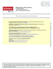
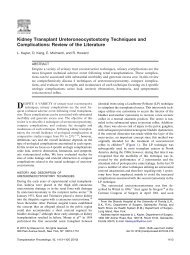
Complementation of mitochondrial function by fusion<br />
Healthy<br />
<strong>Fusion</strong> is stimulated<br />
by energy dem<strong>and</strong><br />
<strong>and</strong> stress<br />
<strong>Fission</strong> generates<br />
new organelles<br />
<strong>and</strong> facilitates<br />
quality control<br />
Fig. 1. <strong>Fusion</strong> rescues stress by allowing functional mitochondria (green) to complement dysfunctional<br />
mitochondria (yellow) by diffusion <strong>and</strong> sharing of components between organelles. <strong>Stress</strong>-induced hyperfusion<br />
yields maximal potential (light green), whereasunderrelaxedconditionscellsareabletosegregate<br />
the damaged (yellow) ones.<br />
31 AUGUST 2012 VOL 337 SCIENCE www.sciencemag.org<br />
on September 2, 2012<br />
www.sciencemag.org<br />
Downloaded from
epilepsy with ragged-red fibers (MERRF). Fortunately,<br />
mitochondria with mutant DNA can still<br />
fuse with other mitochondria in the same cell, allowing<br />
mitochondria with wild-type DNA to compensate<br />
for defects in mitochondria with mutant<br />
DNA by sharing components as long as the mutationloadremainsbelow80to90%percell(7,<br />
8).<br />
Because nucleoids do not appear to exchange<br />
DNA (6), mitochondria in heteroplasmic cells complement<br />
one another by sharing RNA or protein<br />
components. <strong>Fusion</strong> between mitochondria can<br />
also rescue two mitochondria with mutations in<br />
different genes by cross-complementation to one<br />
another, <strong>and</strong> it can mitigate the effects of environmental<br />
damage through the exchange of proteins<br />
<strong>and</strong> lipids with other mitochondria. <strong>Mitochondrial</strong><br />
fusion can therefore maximize oxidative capacity<br />
in response to toxic stress, as long as the stress is<br />
below a critical threshold (Fig. 1).<br />
<strong>Mitochondrial</strong> Morphology Is Controlled<br />
by Metabolism<br />
Rates of mitochondrial fission<br />
<strong>and</strong> fusion respond to changes<br />
in metabolism. Mitochondria<br />
become more fused when they<br />
are forced to rely on oxidative<br />
phosphorylation by withdrawing<br />
glucose as a carbon source<br />
(9). Increased fusion may be necessary<br />
to maximize the fidelity<br />
for oxidative phosphorylation<br />
by stimulating complementation<br />
among mitochondria (Fig.<br />
1). <strong>Fusion</strong> is also enhanced by<br />
treatments that directly or indirectly<br />
inhibit protein synthesis<br />
<strong>and</strong> by starvation <strong>and</strong> mTOR<br />
(mammalian target of rapamycin)<br />
inhibition–induced autophagy<br />
(10–12). Starvation-induced autophagy<br />
may enhance fusion by<br />
increasing the reliance on oxidative<br />
phosphorylation through the<br />
metabolism of lipids <strong>and</strong> proteins<br />
(9). Alternatively, starvation<br />
may evoke a specific stress response called stressinduced<br />
mitochondrial hyperfusion (10), or it may<br />
inhibit fission to protect mitochondria from autophagic<br />
catabolism when they are most needed<br />
(11, 12). Each of these effects is consistent with a<br />
model in which mitochondrial dynamics help<br />
maximize the capacity for oxidative phosphorylation<br />
under stressful conditions (Fig. 1).<br />
Repairing Small Amounts of<br />
<strong>Mitochondrial</strong> Damage<br />
Mitochondria continually produce highly reactive<br />
superoxide anions as a byproduct of electron transport<br />
during oxidative phosphorylation. These reactive<br />
oxygen species (ROS) damage proteins, lipids, <strong>and</strong><br />
DNA (Box 1). Damage to proteins in the electron<br />
transport chain may worsen the situation by producing<br />
even more ROS (13). Mitochondria use qualitycontrol<br />
proteases to eliminate damaged proteins (14)<br />
<strong>and</strong> respond to unfolded protein stress in the matrix<br />
through transcriptional induction of chaperone expression<br />
(15). Damaged mitochondrial outer membrane<br />
proteins also may be removed by the ubiquitin<br />
proteasome quality-control pathway (16). Mitochondria<br />
respond to genotoxic damage by some,<br />
but not all, of the DNA repair pathways found in the<br />
nucleus. These proteotoxic <strong>and</strong> genotoxic damageresponse<br />
pathways target individual molecules for<br />
quality control, thereby rescuing mitochondria with<br />
minor damage without the need for altered fission<br />
or fusion rates (14). Another level of quality control<br />
entails the wholesale elimination of mitochondria<br />
by autophagy, a process that is linked to mitochondrial<br />
fission <strong>and</strong> fusion.<br />
Scrapping Mitochondria That<br />
Are Beyond Repair<br />
Autophagy is a well-established mechanism to<br />
compensate for nutrient depletion by degrading<br />
cellular components <strong>and</strong> to protect cells from del-<br />
Box 1. <strong>Mitochondrial</strong> <strong>Stress</strong><br />
Various insults can cause damage:<br />
Environmental (radiation, toxic chemicals)<br />
Genetic (mutations in genes for metabolic processes or repair pathways)<br />
Spontaneous (ROS generated as byproduct of electron transport)<br />
Types of damage:<br />
DNA<br />
Proteins<br />
Lipids<br />
Problems caused by damage:<br />
Loss of metabolic functions (ATP synthesis, etc.)<br />
More ROS made by defective mitochondria<br />
F 1F 0-ATPase may, instead of making ATP, consume ATP to generate<br />
membrane potential<br />
Cellular responses to damage:<br />
DNA repair<br />
Proteases<br />
Lipases<br />
<strong>Mitochondrial</strong> unfolded protein response<br />
Mitophagy<br />
Apoptosis<br />
eterious protein aggregates by encapsulating <strong>and</strong><br />
degrading them. Autophagy is also required for<br />
maintaining a healthy mitochondrial network, presumably<br />
by eliminating old <strong>and</strong> damaged mitochondria<br />
(17, 18). The importance of this process<br />
is shown by the accumulation of swollen <strong>and</strong> defective<br />
mitochondria in hepatocytes <strong>and</strong> MEFs<br />
from mice lacking the key autophagy gene Ulk1<br />
(17) <strong>and</strong> the appearance of deformed mitochondria<br />
in hepatic cells in Atg7-deficient mice (18).<br />
The autophagic elimination of mitochondria,<br />
mitophagy, appears to be intimately linked to mitochondrial<br />
fission <strong>and</strong> fusion processes. A study of<br />
fibroblast mitochondrial dynamics showed that one<br />
in five daughter mitochondria is depolarized <strong>and</strong><br />
eliminated by mitophagy (19). In most fission events,<br />
one daughter mitochondrion is transiently hyperpolarized<br />
while the sister mitochondrion is hypopolarized,<br />
suggesting that fission embodies a “stress<br />
REVIEW<br />
test” that could push a daughter mitochondrion to<br />
completely depolarize if it functions suboptimally.<br />
Mitophagy could be prevented with a dominantnegative<br />
mutant of Drp1, suggesting that fission<br />
is required for mitophagy (19). Photodamaged<br />
mitochondria undergo selective mitophagy (20),<br />
which is also consistent with the model that fission<br />
provides a form of quality control by segregating<br />
damaged parts of mitochondria <strong>and</strong> targeting<br />
them for elimination by autophagy (Fig. 2).<br />
Recent work on two gene products mutated in<br />
familial Parkinson’s disease, PINK1 <strong>and</strong> Parkin,<br />
yields insight into a molecular mechanism of quality<br />
control via the elimination of damaged mitochondria<br />
(Fig. 3). The abundance of the kinase<br />
PINK1 is constitutively repressed in healthy<br />
mitochondria by import into the inner mitochondrial<br />
membrane <strong>and</strong> degradation by the rhomboid<br />
protease PARL. When a mitochondrion becomes<br />
uncoupled, protein import to the inner mitochondrial<br />
membrane is prevented so PINK1 is diverted<br />
from PARL <strong>and</strong> accumulates on<br />
the outer mitochondrial membrane.<br />
This yields a sensor of mitochondrial<br />
damage that can flag an individual<br />
impaired mitochondrion<br />
in a milieu of healthy ones. PINK1<br />
on a damaged mitochondrion,<br />
through its kinase activity, recruits<br />
the E3 ligase Parkin from the cytosol<br />
specifically to that impaired<br />
mitochondrion (Fig. 3). Once there,<br />
Parkin ubiquitinates outer mitochondrial<br />
membrane proteins <strong>and</strong><br />
induces autophagic elimination of<br />
the flagged mitochondrion (21).<br />
This molecular pathway fits<br />
nicely with the fission model (19)<br />
(Fig. 2) to yield the mitochondrial<br />
quality-control model (Fig. 3).<br />
However, mitochondria have to<br />
be severely depolarized to accumulate<br />
PINK1, <strong>and</strong> the degree to<br />
which this happens physiologically<br />
is not clear. At least in cultured tumor<br />
cells that can maintain robust<br />
ATP levels by glycolysis, mitochondrial F 1F 0 ATPase<br />
can cleave ATP derived from glycolysis <strong>and</strong> reconstitute<br />
membrane potential despite the complete<br />
loss of membrane potential maintenance through<br />
respiration (22). Furthermore, mitochondrial fusion<br />
as discussed previously can lead to compensation<br />
for missing components, thereby rescuing impaired<br />
organelles. These forces would be expected<br />
to counteract damage-induced depolarization of<br />
mitochondria <strong>and</strong> mitigate PINK1-mediated mitophagy.<br />
The stress test on membrane potential during<br />
fission (Fig. 2), however, might overcome those<br />
forces to trigger complete depolarization.<br />
Mutations in PINK1 (23)<strong>and</strong>Parkin(24) lead<br />
to early-onset autosomal recessive Parkinson’s<br />
disease, suggesting that defects in mitochondrial<br />
quality control could cause certain forms of parkinsonism<br />
<strong>and</strong> supporting more general models<br />
that mitochondrial dysfunction is an etiology of<br />
www.sciencemag.org SCIENCE VOL 337 31 AUGUST 2012 1063<br />
Downloaded from<br />
www.sciencemag.org on September 2, 2012
REVIEW<br />
1064<br />
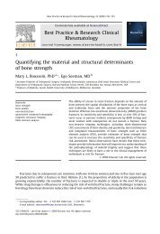
Debris<br />
segregation<br />
Damage<br />
accumulation<br />
<strong>Mitochondrial</strong><br />
maintenance<br />
<strong>Fission</strong><br />
substantia nigral neuron degeneration. PINK1<strong>and</strong><br />
Parkin-deficient Drosophila display muscle<br />
<strong>and</strong> neuron degeneration that is associated with<br />
swollen <strong>and</strong> defective mitochondria (25–27).<br />
Consistent with the model that mitochondrial<br />
fission <strong>and</strong> fusion promotes mitochondrial quality<br />
control, inhibition of mitochondrial fusion or<br />
promotion of mitochondrial fission compensates<br />
for deficiencies of PINK1 <strong>and</strong> Parkin in flies.<br />
Furthermore, Parkin overexpression in flies rescues<br />
unfolded protein stress of mitochondria<br />
through autophagy (28), <strong>and</strong> stimulation of<br />
autophagy rescues depolarized mitochondria<br />
accumulation in dopaminergic neurons from<br />
Parkin-deficient Drosophila (29).<br />
Banish Mitochondria That Truly<br />
Are Uncoupled<br />
Defective mitochondria can be toxic by generating<br />
excessive amounts of ROS, by consuming ATP<br />
through reversal of ATP synthase, <strong>and</strong> by interfering<br />
with a host of other metabolic processes (Box 1).<br />
Low levels of damage might be corrected by complementation<br />
through mitochondrial fusion, but<br />
badly damaged mitochondria will contaminate other<br />
mitochondria if they are allowed to rejoin the<br />
mitochondrial network before their elimination<br />
by autophagy. Several mechanisms are at work to<br />
stop this from happening. A first line of defense<br />
is provided by a built-in requirement of the mitochondrial<br />
inner membrane fusion machinery for<br />
membrane potential (30). Vertebrates have elaborated<br />
on this mechanism by providing a second line<br />
of defense through proteolytic inactivation of the<br />
inner membrane fusion dynamin OPA1. Proteolysis<br />
is mediated by the mitochondrial inner membrane<br />
protease OMA1, which is rapidly activated<br />
by low membrane potential <strong>and</strong> low levels of ATP<br />
(31, 32). The outer membranes of these mitochondria<br />
can still fuse, even without functional OPA1<br />
or membrane potential, but the inner membrane–<br />
bound matrix compartments do not fuse, resulting<br />
Biogenesis<br />
in several matrix compartments surrounded by a<br />
common outer membrane, like peas in a pod.<br />
The last line of defense is provided by the Pink1<br />
<strong>and</strong> Parkin pathway through the ubiquitination of<br />
the mitochondrial outer membrane fusion proteins<br />
Mfn1 <strong>and</strong> Mfn2. Ubiquitination of these proteins<br />
leads to their extraction from the membrane by p97<br />
<strong>and</strong>theirdegradationbyproteasomes(16). In addition,<br />
Pink1 <strong>and</strong> Parkin disrupt mitochondrial motility<br />
by degrading the small GTPase Miro, which<br />
serves as an adaptor for kinesin-dependent transport<br />
Autophagosome<br />
UPS<br />
p97<br />
Mitophagy<br />
Ub<br />
Ub<br />
Mfn-Ub<br />
Autophagosome<br />
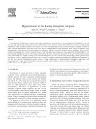
Fig. 2. Autophagy could purify the cellular pool of mitochondria if debris is aggregated <strong>and</strong> segregated<br />
byfissioninasubsetofmitochondria.Ifdeleterious components (black fibers) are asymmetrically<br />
distributed or aggregated, fission could lead to cleansing of daughter mitochondrion (green) by preventing<br />
fusion <strong>and</strong> inducing mitophagy of the impaired ones (yellow).<br />
<strong>and</strong> is also needed for mitochondrial fusion (33).<br />
Ultimately, uncoupled mitochondria lose both their<br />
inner <strong>and</strong> outer membrane fusion machineries, thereby<br />
preventing them from fusing with <strong>and</strong> poisoning<br />
the healthy mitochondrial network. Purposeful<br />
segregation <strong>and</strong> disposal of damaged mitochondria<br />
through changes in fission <strong>and</strong> fusion pathways<br />
are therefore integral parts of mitochondrial qualitycontrol<br />
mechanisms.<br />
Is Debris Also Sorted Inside Mitochondria?<br />
The gradual accumulation of damaged components<br />
poses a problem for the mitophagic disposal<br />
process. If damaged components were evenly<br />
distributed, then the simple act of fission through<br />
Drp1 would not generate the asymmetry needed<br />
for inducing mitophagy by selective loss of membrane<br />
potential. It seems that asymmetric sorting<br />
of debris would be needed to generate the differences<br />
in membrane potential between daughter<br />
mitochondria that have been observed immediately<br />
after fission (19). Accumulation of damaged<br />
components in a subset of daughter mitochondria<br />
would enable their selective disposal, thus helping<br />
to rejuvenate the remaining population of<br />
mitochondria (Fig. 2).<br />
How might mitochondria achieve this type of<br />
asymmetric fission? The mechanism is not yet<br />
known, but it seems likely that damaged proteins<br />
form aggregates within the mitochondrial matrix.<br />
Perhaps there is a way to stow these aggregates at<br />
the tips of mitochondria, thus providing a starting<br />
point for polarized fission. A precedent for this<br />
was set by bacteria, which remove aggregates by<br />
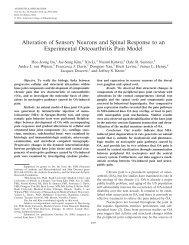
Model of<br />
Parkin-induced<br />
mitophagy<br />
mtDNA mutation<br />
ROS<br />
∆ψ<br />
Pink 1<br />
accumulation<br />
Parkin<br />
recruitment<br />
Lysosomal<br />
hydrolases<br />
31 AUGUST 2012 VOL 337 SCIENCE www.sciencemag.org<br />
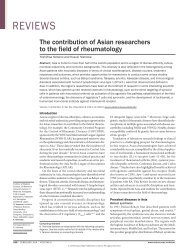
Fig. 3. PINK1 is constitutively degraded<br />
by the inner mitochondrial<br />
membrane protease PARL <strong>and</strong><br />
maintained at low levels on healthy<br />
mitochondria. When a mitochondrion<br />
becomes damaged to the<br />
point of depolarizing the membrane<br />
potential across the inner<br />
membrane, PINK1 import to the<br />
inner membrane is prevented,<br />
thereby sequestering it on the<br />
outer mitochondrial membrane<br />
<strong>and</strong> away from PARL. PINK1 accumulates<br />
there <strong>and</strong> recruits the E3<br />
ligase Parkin from the cytosol via<br />
PINK1 kinase activity. Parkin conjugates<br />
ubiquitin (Ub) to a variety<br />
of proteins on the outer mitochondrial<br />
membrane <strong>and</strong> mediates the<br />
proteosomal elimination of mitofusins<br />
1 <strong>and</strong> 2. Lastly, Parkin induces<br />
autophagic elimination of<br />
the dysfunctional mitochondria. This<br />
pathway may constitute a qualitycontrol<br />
mechanism to eliminate damaged<br />
mitochondria. UPS, ubiquitin<br />
proteasome system.<br />
Downloaded from<br />
www.sciencemag.org on September 2, 2012
asymmetric fission, thus enhancing the growth<br />
rates of those daughter cells that do not receive<br />
aggresomes (34). A similar asymmetry was observed<br />
during mammalian cell division, where<br />
aggregates accumulate at the centrosome <strong>and</strong> are<br />
selectively inherited by one of the two daughter<br />
cells (35). If mitochondria also have such a<br />
deliberate mechanism, then they might have a<br />
mechanism for inducing fission when too many<br />
aggregates are formed inside mitochondria.<br />
Such an inducing mechanism is suggested by<br />
genetic studies showing that Pink1 <strong>and</strong> Parkin act<br />
upstream of the fission machinery in Drosophila.<br />
However, studies with mammalian cells have<br />
only shown effects of Pink1 <strong>and</strong> Parkin after<br />
fission is completed. Mammalian cells may have<br />
developed an additional, as yet undiscovered,<br />
mechanism to induce fission when mitochondrial<br />
aggregates accumulate, analogous to the rapid<br />
proteolytic inactivation of the fusion machinery<br />
through Oma1-mediated proteolysis when mitochondria<br />
lose membrane potential or ATP.<br />
Aggregation of misfolded proteins in the cytosol<br />
is facilitated by p62 <strong>and</strong> NBR1, which can lead<br />
to their disposal by autophagy (36). Interestingly,<br />
p62 also accumulates on mitochondria after Pink1<br />
<strong>and</strong> Parkin activation. Once there, p62 triggers<br />
mitochondrial aggregation through its oligomerizationdomain(36).<br />
<strong>Mitochondrial</strong> aggregation<br />
may be an indirect result of aggregating ubiquitinated<br />
proteins on the mitochondrial outer membrane<br />
to segregate debris before fission. When<br />
protein damage accumulates, small vesicles bud<br />
from the outer mitochondrial surface. The trafficking<br />
of these vesicles to lysosomes suggests another<br />
<strong>and</strong> surprisingly direct pathway of mitochondrial<br />
debris removal that is independent of Drp1,<br />
therefore independent of classic mitochondrial<br />
fission, <strong>and</strong> also independent of autophagy (37).<br />
Selective Removal of Mutant mtDNA<br />
Can mitophagy cleanse genotoxic stress in addition<br />
to proteotoxic stress? Mutations in mtDNA accumulate<br />
as mammals age <strong>and</strong> could accumulate<br />
generation after generation were it not for germline<br />
purification of mtDNA. Although the mechanisms<br />
are not yet known, mitochondrial genomes with<br />
strong deleterious mutations can be removed during<br />
oogenesis (38, 39). Models for this cleansing<br />
mechanism include selective expansion of less impaired<br />
mitochondria to populate oocytes, apoptosis<br />
of oocytes with excessive mutant mtDNAs,<br />
<strong>and</strong> removal of poorly functioning mitochondria<br />
by mitophagy. Whether mutated mtDNA is selectively<br />
removed from somatic cells is not known.<br />
A requisite for elimination of deleterious mitochondrial<br />
DNA mutations by mitophagy, be it in<br />
the germ line or soma, is physical linkage between<br />
the mutated mtDNA <strong>and</strong> the mutated gene product<br />
(40). Might there be a mechanism to identify malfunctioning<br />
nucleoids through their defective protein<br />
products, for example, through physical association<br />
with protein aggregates? Integral inner membrane<br />
proteins diffuse much more slowly than soluble<br />
matrix or intermembrane space proteins <strong>and</strong> there-<br />
forearemorelikelytoberetainedwiththeirparental<br />
nucleoid than soluble tRNAs after mitochondrial<br />
fusion <strong>and</strong> fission events. This physical proximity<br />
might link nucleoids with mutant gene products<br />
that affect protein coding sequences <strong>and</strong> facilitate<br />
their autophagic purification. Such differential diffusibility<br />
between integral membrane proteins <strong>and</strong><br />
tRNAs might explain why mutations in tRNAs are<br />
much more common in human diseases than mutations<br />
in the integral protein components of the<br />
oxidative phosphorylation machinery. Also, protein<br />
aggregates may start to form immediately during<br />
protein synthesis, which is physically linked with<br />
nucleoids (41). A mechanism for purifying mtDNA,<br />
by retaining mutant proteins with their genome, may<br />
prove to be an unexpected bonus of coupled transcription<br />
<strong>and</strong> translation to mitochondrial nucleoids.<br />
<strong>Mitochondrial</strong> <strong>Fission</strong> <strong>and</strong> Apoptosis<br />
When all else fails, stressed cells undergo apoptosis.<br />
In the past decade, many connections have been<br />
discovered between apoptosis <strong>and</strong> mitochondrial<br />
dynamics, as discussed more fully in this issue by<br />
Hoppins <strong>and</strong> Nunnari. High levels of cell stress<br />
that lead to apoptosis also lead to excessive fission<br />
of mitochondria. This occurs almost simultaneously<br />
with two steps of apoptosis that involve<br />
mitochondria: translocation from the cytosol to<br />
mitochondria of the pro-apoptotic Bcl-2 family<br />
member Bax <strong>and</strong> cytochrome c release. When Bax<br />
translocates to mitochondria, it accumulates in<br />
concentrated foci that colocalize with Drp1 <strong>and</strong><br />
mitofusins. Inhibition of mitochondrial fission by<br />
Drp1 knock-down delays cytochrome c release,<br />
indicating that mitochondrial fission participates in<br />
Bax-mediated permeabilization of the outer mitochondrial<br />
membrane (42). The link may be that<br />
Bax is activated to oligomerize <strong>and</strong> release cytochrome<br />
c by membrane hemifusion intermediates<br />
that are formed during mitochondrial fission (43).<br />
Intriguingly, Bcl-2 family members also participate<br />
in mitochondrial fission <strong>and</strong> fusion in nonapoptotic<br />
cells (44). Thus, mitochondrial dynamics are involved<br />
not only in regulating individual mitochondrial<br />
fidelity within cells but also at the whole-cell<br />
level by participating in apoptotic cell death.<br />
Outlook<br />
<strong>Fusion</strong> allows mitochondria to compensate for one<br />
another’s defects by sharing components <strong>and</strong> thereby<br />
helps maintain energy output in the face of<br />
stress. However, when a certain threshold of damage<br />
is reached, mitochondria are eliminated wholesale<br />
by autophagy. <strong>Fission</strong> segregates the most<br />
seriously damaged mitochondria to preserve the<br />
health of the mitochondrial network in addition to<br />
regulating morphology <strong>and</strong> facilitating mitochondrial<br />
trafficking. The highly dynamic mitochondrial<br />
fusion <strong>and</strong> fission cycle is proposed to balance<br />
two competing processes: compensation of damage<br />
by fusion <strong>and</strong> elimination of damage by fission.<br />
Failure of these stress responses may lead to<br />
neuron death <strong>and</strong> neurodegenerative disorders. Indepth<br />
underst<strong>and</strong>ing of mitophagic processes could<br />
aid the development of new treatments for mito-<br />
chondrial <strong>and</strong> neurodegenerative diseases: It was<br />
recently shown that reactivation of autophagy can<br />
mitigate certain other diseases, such as muscular<br />
dystrophies associated with mitophagy (45).<br />
References <strong>and</strong> Notes<br />
1. S. Hoppins, L. Lackner, J. Nunnari, Annu. Rev. Biochem.<br />
76, 751 (2007).<br />
2. K. Elgass, J. Pakay, M. T. Ryan, C. S. Palmer, Biochim.<br />
Biophys. Acta, published online 10 May 2012.<br />
3. J. R. Friedman et al., Science 334, 358 (2011); 10.1126/<br />
science.1207385.<br />
4. H. Chen, D. C. Chan, Ann. N. Y. Acad. Sci. 1201, 21 (2010).<br />
5. H. Chen, A. Chomyn, D. C. Chan, J. Biol. Chem. 280,<br />
26185 (2005).<br />
6. E. A. Schon, R. W. Gilkerson, Biochim. Biophys. Acta<br />
1800, 245 (2010).<br />
7. M. Yoneda, T. Miyatake, G. Attardi, Mol. Cell. Biol. 14,<br />
2699 (1994).<br />
8. K. Nakada et al., Nat. Med. 7, 934 (2001).<br />
9. R. Rossignol et al., Cancer Res. 64, 985 (2004).<br />
10. D. Tondera et al., EMBO J. 28, 1589 (2009).<br />
11. A. S. Rambold, B. Kostelecky, N. Elia, J. Lippincott-Schwartz,<br />
Proc. Natl. Acad. Sci. U.S.A. 108, 10190 (2011).<br />
12. L. C. Gomes, G. Di Benedetto, L. Scorrano, Nat. Cell Biol.<br />
13, 589 (2011).<br />
13. R. S. Balaban, S. Nemoto, T. Finkel, Cell 120, 483 (2005).<br />
14. M. J. Baker, T. Tatsuta, T. Langer, Cold Spring Harbor<br />
Perspect. Biol. 3, a007559 (2011).<br />
15. A. M. Nargund, M. W. Pellegrino, C. J. Fiorese,<br />
B. M. Baker, C. M. Haynes, Science 337, 587 (2012);<br />
10.1126/science.1223560.<br />
16. A. Tanaka et al., J. Cell Biol. 191, 1367 (2010).<br />
17. D. F. Egan et al., Science 331, 456 (2011); 10.1126/<br />
science.1196371.<br />
18. M. Komatsu et al., J. Cell Biol. 169, 425 (2005).<br />
19. G. Twig et al., EMBO J. 27, 433 (2008).<br />
20. I. Kim, J. J. Lemasters, Antioxid. Redox Signal. 14, 1919<br />
(2011).<br />
21. D. Narendra, A. Tanaka, D. F. Suen, R. J. Youle,<br />
J. Cell Biol. 183, 795 (2008).<br />
22. K. Buchet, C. Godinot, J. Biol. Chem. 273, 22983 (1998).<br />
23. E. M. Valente et al., Science 304, 1158 (2004); 10.1126/<br />
science.1096284.<br />
24. T. Kitada et al., Nature 392, 605 (1998).<br />
25. J. C. Greene et al., Proc. Natl. Acad. Sci. U.S.A. 100,<br />
4078 (2003).<br />
26. J. Park et al., Nature 441, 1157 (2006).<br />
27. I. E. Clark et al., Nature 441, 1162 (2006).<br />
28. I. Pimenta de Castro et al., Cell Death Differ. 19, 1308 (2012).<br />
29. J. L. Burman, S. Yu, A. C. Poole, R. B. Decal, L. Pallanck,<br />
Proc. Natl. Acad. Sci. U.S.A. 109, 10438 (2012).<br />
30. S. Meeusen et al., Cell 127, 383 (2006).<br />
31. S. Ehses et al., J. Cell Biol. 187, 1023 (2009).<br />
32. B. Head, L. Griparic, M. Amiri, S. G<strong>and</strong>re-Babbe,<br />
A. M. van der Bliek, J. Cell Biol. 187, 959 (2009).<br />
33. X. Wang et al., Cell 147, 893 (2011).<br />
34. A. B. Lindner, R. Madden, A. Demarez, E. J. Stewart,<br />
F. Taddei, Proc. Natl. Acad. Sci. U.S.A. 105, 3076 (2008).<br />
35. L. C. Fuentealba, E. Eivers, D. Geissert, V. Taelman,<br />
E. M. De Robertis, Proc. Natl. Acad. Sci. U.S.A. 105, 7732<br />
(2008).<br />
36. T. Johansen, T. Lamark, Autophagy 7, 279 (2011).<br />
37. V. Soubannier et al., Curr. Biol. 22, 135 (2012).<br />
38. J. B. Stewart et al., PLoS Biol. 6, e10 (2008).<br />
39. W. Fan et al., Science 319, 958 (2008).<br />
40. A. Kowald, T. B. Kirkwood, Commun. Integr. Biol. 4, 627<br />
(2011).<br />
41. J. He et al., Nucleic Acids Res. 40, 6109 (2012).<br />
42. D. F. Suen, K. L. Norris, R. J. Youle, Genes Dev. 22,<br />
1577 (2008).<br />
43. S. Montessuit et al., Cell 142, 889 (2010).<br />
44. A. Autret, S. J. Martin, Mol. Cell 36, 355 (2009).<br />
45. P. Grumati et al., Nat. Med. 16, 1313 (2010).<br />
Acknowledgments: We thank members of the Youle lab for<br />
thoughtful comments. This work was supported by Intramural<br />
Program of the National Institute of Neurological Disorders<br />
<strong>and</strong> Stroke <strong>and</strong> grants from the NIH (GM051866) <strong>and</strong> the<br />
NSF (0552271) to A.M.v.d.B.<br />
10.1126/science.1219855<br />
REVIEW<br />
www.sciencemag.org SCIENCE VOL 337 31 AUGUST 2012 1065<br />
Downloaded from<br />
www.sciencemag.org on September 2, 2012