Endoscopic Photocoagulation - Meridian
Endoscopic Photocoagulation - Meridian
Endoscopic Photocoagulation - Meridian

Create successful ePaper yourself
Turn your PDF publications into a flip-book with our unique Google optimized e-Paper software.
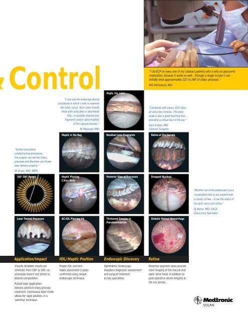
Control<br />
“Unlike transscleral<br />
cylodestructive procedures,<br />
the surgeon can see the ciliary<br />
processes and therefore can titrate<br />
laser delivery properly.”<br />
M Uram, MD, MPH<br />
Visually titratable results are<br />
achieved, from 180 o to 360 o , as<br />
processes blanch and shrink to<br />
desired composition.<br />
Pulsed laser application<br />
delivers selective ciliary process<br />
treatment. Continuous laser mode<br />
allows for rapid ablation, in a<br />
'painting' technique.<br />
“I also use the endoscope during<br />
procedures in which I wish to examine<br />
the ciliary sulcus. Such cases include<br />
those with dislocated or decentered<br />
IOLs, or possible retained lens<br />
fragments and/or abnormalities<br />
of the capsule/zonules.”<br />
RJ Mackool, MD<br />
Haptic in the Bag<br />
180 o -360 o Range Haptic Piercing<br />
Ciliary Body<br />
Laser Treated Processes AC-IOL Piercing Iris<br />
Proper IOL and lens<br />
haptic placement is easily<br />
confirmed using simple<br />
endoscopic technique.<br />
Angle, Iris, Lens<br />
Residual Lens Fragments<br />
Posterior View of Processes<br />
Thickened Zonules in<br />
Pseudoexfoliation<br />
Application/Impact IOL/Haptic Position <strong>Endoscopic</strong> Discovery Retina<br />
Ophthalmic Endoscopy<br />
broadens diagnostic assessment<br />
and surgical treatment<br />
across specialties.<br />
“I do ECP on every one of my cataract patients who is also on glaucoma<br />
medication, because it works so well... through a single incision I can<br />
initially treat approximately 210 o to 240 o of ciliary processes.”<br />
MS McFarland, MD<br />
“Combined with phaco, ECP takes<br />
an extra two minutes. The endoscope<br />
is also a great teaching tool...<br />
providing a virtual tour of the eye.”<br />
Alan B Aker, MD<br />
Cataract Surgeon<br />
Retina at Ora Serrata<br />
Dropped Nucleus<br />
Diabetic Retinal Hemorrhage<br />
Posterior segment views provide<br />
clear imaging of the macula and<br />
optic nerve head, in addition to<br />
post-operative retinal integrity at<br />
the ora serrata.<br />
“Another use of the endoscope is as a<br />
visualization tool in any patient with<br />
a cloudy cornea... to see the status of<br />
the optic nerve and retina.”<br />
SJ Berke, MD, FACS<br />
Glaucoma Specialist