this download - University of Utah
this download - University of Utah
this download - University of Utah

Create successful ePaper yourself
Turn your PDF publications into a flip-book with our unique Google optimized e-Paper software.
The<br />
John A. Moran<br />
Eye Center<br />
The John A. Moran<br />
Eye Center is<br />
committed to the<br />
goal that no person<br />
with a blinding<br />
condition, eye<br />
disease or visual<br />
impairment should<br />
be without hope,<br />
understanding and<br />
treatment.<br />
Research and<br />
Clinical Abstracts<br />
2009 - 2010
2<br />
17<br />
18<br />
32<br />
33<br />
35<br />
36<br />
37<br />
39<br />
41<br />
42<br />
45<br />
48<br />
51<br />
56<br />
58<br />
Contents<br />
ARVO 2010 Abstracts<br />
Research at the<br />
Moran Eye Center<br />
Retinal Research<br />
Angiogenesis Research<br />
Highlight:<br />
The Connectome<br />
Visual Cortex Research<br />
Implants Research<br />
Developmental Research<br />
Neuro-Ophthalmology<br />
Research<br />
Highlight:<br />
Intermountain Ocular<br />
Research Laboratory<br />
Intermountain Ocular<br />
Research Abstracts<br />
Translational Research<br />
Clinical Research<br />
Abstracts:<br />
Anterior Segment<br />
Posterior Segment<br />
Donor Report<br />
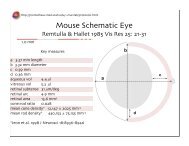
Photos Courtesy <strong>of</strong> Bryan William Jones and Ann Torrence.<br />
Collaborators: Brenda Stringham, Greg Jones, Bryan<br />
William Jones, and Nathan Galli.
Randall J. Olson, MD<br />
Chair, Presidential Pr<strong>of</strong>essor<br />
The John Moran Eye Center was born as the new home for the Department <strong>of</strong> Ophthalmology<br />
and Visual Sciences at the <strong>University</strong> <strong>of</strong> <strong>Utah</strong> School <strong>of</strong> Medicine in July <strong>of</strong> 1993. The one person<br />
Division <strong>of</strong> Ophthalmology in the Department <strong>of</strong> Surgery in 1979 had grown to need <strong>this</strong><br />
new 82,000 square feet facility. Sadly, we had dedicated only 20,000 square feet to research and<br />
the space was obviously inadequate after living in our new building only a few short years. After<br />
years <strong>of</strong> planning and fund raising we were able to move into our present building in August<br />
<strong>of</strong> 2006. This time we dedicated over half <strong>of</strong> a 210,000 square feet building to research, which<br />
space includes over 30,000 square feet roughed in and ready for program expansion. With the<br />
new building, the research program has expanded nicely, and we are very proud <strong>of</strong> our research<br />
team. This publication is our opportunity to showcase some <strong>of</strong> the work and effort ongoing here.<br />
While the work represented spans the field from very clinical to very basic, major emphasis has<br />
been placed on retinal research. As part <strong>of</strong> <strong>this</strong> we are pleased to announce our new Center for<br />
Translational Medicine under our John A. Moran Presidential Endowed Pr<strong>of</strong>essor, Greg Hageman.<br />
We will be announcing soon other members that will be joining his team.<br />
As Chair <strong>of</strong> the Department and CEO <strong>of</strong> the John A. Moran Eye Center, I am most pleased with<br />
the research effort and the proposed expansion as we look to the future. The size <strong>of</strong> the research<br />
mission now represents close to half our budget and our total NIH funding for the next academic<br />
year will exceed $10 million a year. Our real hope is that our institution takes the translational<br />
mission to heart and that our research effort here will lead to new treatments for our patients as<br />
we look to the future.<br />
Sincerely,<br />
Randall J Olson, MD<br />
Robert Marc, PhD<br />
Director <strong>of</strong> Research<br />
The Moran Eye Center houses researchers whose studies span the development <strong>of</strong> the eye to<br />
the organization <strong>of</strong> cortex; the basis <strong>of</strong> phototransduction to the genetics <strong>of</strong> retinal diseases; the<br />
synaptology <strong>of</strong> the retina to neuroprosthetics. These efforts are enhanced by strong graduate<br />
programs and a tradition <strong>of</strong> interdepartmental collaboration. Our principles include maintaining<br />
basic science expertise, fostering research alliances, and accelerating the evolution <strong>of</strong> translational<br />
science programs.<br />
Paul Bernstein, MD, PhD<br />
Director <strong>of</strong> Clinical Research<br />
The Clinical Research Group at the Moran Eye Center focuses on research that directly impacts<br />
the patient. Whether <strong>this</strong> means working with industry sponsors to run a clinical trial investigating<br />
a new drug or a clinician initiated study to systematically compare standard <strong>of</strong> care<br />
treatments, the Clinical Research Group is focused on finding the optimal in patient care. In <strong>this</strong><br />
year alone, we have 45 clinical trials and research studies underway involving more than 550<br />
patients. This work is shared with the global ophthalmic community in over 27 publications just<br />
<strong>this</strong> year.<br />
Gregory Hageman, PhD<br />
Director <strong>of</strong> Translational Research<br />
The Moran Center for Translational Medicine (CTM) was recently created to expedite the pace<br />
at which basic scientific discoveries are translated into clinically effective diagnostics and therapies<br />
for the treatment <strong>of</strong> devastating eye disorders such as age-related macular degeneration and<br />
glaucoma, as well as other diseases. The conceptual framework for the CTM derived from a<br />
growing realization that seemingly disparate diseases likely share common etiologies and thus,<br />
common therapeutic targets. The CTM will draw upon the collective strengths and expertise <strong>of</strong><br />
a collaborative team <strong>of</strong> cell biologists, molecular microbiologists, pathologists and clinicians to<br />
expedite its translationalmission. The unique resources, clinical acumen and scientific expertise<br />
<strong>of</strong> the CTM will complement the core competecies <strong>of</strong> collaborating corporate and academic<br />
partners to insure its success.
The Association for<br />
Research in Vision and<br />
Ophthalmology Honors<br />
the ARVO Fellows<br />
Inaugural Class <strong>of</strong> 2009<br />
2009 Silver Fellows Wolfgang Baehr, PhD<br />
Robert E. Marc, PhD<br />
Class <strong>of</strong> 2010<br />
2010 ARVO Abstracts<br />
Antisense Morpholine Oligonucleotide<br />
Against FLT Splice Junction Inhibits<br />
Murine Corneal Hemangiogenesis<br />
(HA) and Lymphangiogenesis (LA)<br />
Y. Qazi 1,2 , H. Uehara 1 , B.K. Ambati 1 .<br />
1 Ophthalmology, John Moran Eye Center/<br />
<strong>University</strong> <strong>of</strong> <strong>Utah</strong>, Salt Lake City, UT;<br />
2 Ophthalmology, Emory Eye Center/Emory<br />
<strong>University</strong>, Atlanta, GA<br />
Purpose. To design and test a molecular<br />
tool to convert the pro-angiogenic microenvironment<br />
<strong>of</strong> vascularised corneas by<br />
manipulating post-transcriptional processing<br />
<strong>of</strong> the FLT gene towards its anti-angiogenic<br />
is<strong>of</strong>orm, sFLT-1.<br />
Methods. Antisense morpholino oligonucleotide<br />
(morpholino, MO) was designed<br />
complementary to a sequence within the<br />
FLT splice junction <strong>of</strong> exon13-intron13 at<br />
the 5’ splice site. Gene Tools LLC. prepared<br />
the morpholino. Fluorescein-tagged<br />
standard morpholino (STD MO) was transfected<br />
(electroporation) into HUVECs to<br />
test and confirm delivery <strong>of</strong> morpholinos<br />
2<br />
2010 Gold Fellow Gregory S. Hageman, PhD<br />
2010 Silver Fellow Paul S. Bernstein, MD, PhD<br />
into the nucleus using fluorescence microscopy.<br />
A suture-induced model <strong>of</strong> corneal neovascularization<br />
(KNV) was used. BALB/<br />
CJ mice had 3 sutures placed intrastromally.1<br />
week after suture placement, the<br />
mice were randomly divided into 3 groups,<br />
their corneas photographed and each <strong>of</strong> the<br />
groups received one <strong>of</strong> the following interventions<br />
intracorneally: 200ng sFlt-MO;<br />
200ng STD MO; 5ul phosphate-buffered<br />
saline (PBS).1week post-injection, the corneas<br />
were photographed and the mice sacrificed.<br />
The harvested corneas were were<br />
used for flatmounts, ELISA and RT-PCR.<br />
Immunostaining was performed for vascular<br />
endothelial cells (CD31) and lymphatic<br />
endothelial cells (LYVE1). Scion Image<br />
was used to quantitate HA and LA from<br />
flatmounts imaged by fluorescence microscopy.<br />
KNV was induced in mice as above and 1<br />
week after suture placement, the mice were<br />
randomly divided into 2 groups, each <strong>of</strong><br />
which received an intracorneal injection<br />
<strong>of</strong> a combination <strong>of</strong> either 2ug siRNA.sFlt<br />
+ 200ng sFlt-MO, or, 2ug siRNA negative<br />
control + 200ng sFlt-MO. 1 week postinjection,<br />
the corneas were harvested and<br />
processed as mentioned above.<br />
Results. Morpholinos designed to increase<br />
sFlt-1 (sFlt-MO) significantly inhibited<br />
the progression <strong>of</strong> both blood and lymph<br />
vessels.Treated corneas had 6.9% HA (p=<br />
0.017, n=8) and 4.6% LA (p
Designing and Manufacturing a<br />
Refillable Multi-Drug Capsule Ring<br />
Platform<br />
C.J. Bishop 1A , H.J. Sant 1B , S.A. Molokhia<br />
2 , R.M. Burr 1A , B.K. Gale 1B , B.K.<br />
Ambati 2 .<br />
A Biomedical Engineering, B Mechanical<br />
Engineering, 1 <strong>University</strong> <strong>of</strong> <strong>Utah</strong>, Salt<br />
Lake City, UT; 2 Ophthalmology, Moran<br />
Eye Center, Salt Lake City, UT.<br />
Purpose. AMD treatment requires<br />
monthly intravitreal injections which are<br />
uncomfortable, risky, and costly. Because<br />
glaucoma is treated using topical polypharmaceutic<br />
regimens, patient compliance<br />
and drug-target interactions are low.<br />
A controlled delivery device would benefit<br />
both ocular diseases and transcend<br />
the patient interface. Our Capsule Drug<br />
Rings (CDR) are implanted in the capsule<br />
bag periphery during IOL placement. The<br />
CDRs are to be refilled every 6 months to<br />
1 year in situ.<br />
Methods. CDRs are made <strong>of</strong> well-established<br />
biocompatible materials. The<br />
inflatable shell <strong>of</strong> the CDR is a flexible<br />
polycarbonate-based polyether urethane<br />
which resists hydrolytic degradation improving<br />
longevity in vivo. A CO2 laser<br />
was used to laminate the shell edge <strong>of</strong> the<br />
CDR. Refilling Ports were made <strong>of</strong> polyimide<br />
tubing and resealing polydimethylsiloxane<br />
plugs acting as a one-way valve.<br />
One side <strong>of</strong> the shell consisted <strong>of</strong> a 1 mm2<br />
oval window, covered by a 30 nm diameter<br />
pore-size polyethersulfone filter. Islets<br />
were placed at the ends <strong>of</strong> the device for<br />
surgical manipulation. CDRs were implanted<br />
in rabbits during intraocular lens<br />
implantation.<br />
Results. Each port re-seals and holds at<br />
least 40 mm Hg up to 30 insertions. The<br />
carbothane shell’s inner and outer diameters<br />
are 11.0 and 13.0 mm, respectively.<br />
The thickness <strong>of</strong> the shell pre and postinflation<br />
is 200 µm and 750 µm, respectively.<br />
The drug reservoir holds 80 µL.<br />
Significantly, various sections <strong>of</strong> the CDR<br />
shell were laminated using different laser<br />
parameters. The edges with greater curvature<br />
required greater speed or less power.<br />
Radial lamination (vs horizontal or vertical)<br />
due to the symmetry and curvature <strong>of</strong><br />
the ring device was far superior for proper<br />
sealing.<br />
Conclusions. The CDR multi-drug platform<br />
is a promising device for treating multiple<br />
diseases simultaneously (AMD, Glaucoma,<br />
etc.). The CDR filter placement, thickness,<br />
pore size/density, window size, and drug<br />
concentration can be altered to optimize<br />
near zero order pharmacokinetics for a given<br />
drug molecule.<br />
Program#/Poster#: 5331/A259<br />
Higher Levels <strong>of</strong> Soluble NRP-1 May<br />
be Associated With Lesser Corneal<br />
Neovascularization in MRL6/MPJ Mice<br />
Compared to Their Background Strain<br />
C57BL/6J Mice<br />
N. Singh, L. Luo, J.M. Simonis, B.K.<br />
Ambati<br />
Ophthalmology-<strong>University</strong> <strong>of</strong> <strong>Utah</strong>, John<br />
Moran Eye Center, Salt Lake City, UT.<br />
Purpose. To determine whether soluble<br />
neuropilin-1 is elevated in MRL6/MpJ<br />
mice compared to their background strain<br />
C57BL/6J mice.<br />
Methods. C57BL/6J and MRL6/MpJ mice<br />
corneas were injected with plasmid expressing<br />
SiRNA against soluble NRP. The corneas<br />
were imaged after every 3 days and<br />
harvested 10 days after injections. C57BL6<br />
mice developed significant higher level<br />
<strong>of</strong> blood vessels compared to MRL/MpJ<br />
mice. Corneas were immunostained with<br />
the CD31 antibodies and confocal images<br />
were obtained using an Olympus confocal<br />
microscope. RNA was isolated from the<br />
MRL6/MpJ and C57BL/6 mice and Real<br />
time PCR was performed using primers designed<br />
against N- terminal region present in<br />
soluble NRP-1 and C-terminal end present<br />
only in membrane NRP-1.<br />
Results. Normal MRL6/MpJ mice had significant<br />
higher levels <strong>of</strong> soluble NRP 1 compared<br />
to background strain C57BL/6J mice<br />
(p=0.00018). Vascularization <strong>of</strong> cornea<br />
leads to significant decrease in soluble NRP-<br />
1 (p=0.0153) with a concurrent increase in<br />
membrane NRP-1 (p=0.0374) in C57BL/6J<br />
mice, while there was no significant change<br />
in levels <strong>of</strong> soluble NRP-1 is MRL6/MpJ<br />
mice.<br />
Conclusions. Higher level <strong>of</strong> soluble NRP1<br />
may be associated with a resistance to injury-induced<br />
corneal neovascularization in<br />
MRL6/MpJ mice.<br />
Program#/Poster#: 5701/D865<br />
In vitro Diffusion and Permeability<br />
<strong>of</strong> a Novel Intraocular Drug Delivery<br />
Implant<br />
R.M. Burr 1A , S.A. Molokhia 1B , C.J.<br />
Bishop 1A , H.J. Sant 1C , J.M. Simonis 1B ,<br />
B.K. Gale 1C , B.K. Ambati 2 .<br />
A Bioengineering, B Ophthalmology, C Mechanical<br />
Engineering, 1 <strong>University</strong> <strong>of</strong> <strong>Utah</strong>,<br />
Salt Lake City, UT; 2 Ophthalmology, John<br />
Moran Eye Center, Salt Lake City, UT.<br />
Methods. To control the release <strong>of</strong> Avastin®<br />
we have investigated 3 commercially<br />
available hydrophilic and biocompatible<br />
membranes: Polyethersulfone (PES), Polycarbonate<br />
(PC), and Polyvinylidene Fluoride.<br />
The membrane is attached to a 1 - 4<br />
mm2 hole that has been previously cut in<br />
a controlled drug delivery ring (CDR). An<br />
rhVEGF ELISA with known concentrations<br />
for calibration was used to assay samples<br />
collected. Side by side diffusion chambers<br />
<strong>of</strong> 2 ml each (donor and receiver) were<br />
used to identify the permeability coefficients<br />
<strong>of</strong> our Avastin® formulation for each<br />
membrane. 1 ml samples were taken from<br />
the receiver and replaced with an equal volume<br />
<strong>of</strong> fresh PVA every 2 to 6 hours for a<br />
maximum <strong>of</strong> 24 hours. We also conducted<br />
drug release studies using the PES and PC<br />
membranes to determine diffusion coefficients<br />
for later translation into a suitable in<br />
vivo Avastin® formulation. Samples were<br />
taken and replaced with an equal volume <strong>of</strong><br />
fresh BSS daily for the first week and 1 - 2<br />
times per week thereafter until no Avastin®<br />
was detected.<br />
Results. The CDR will provide long-term<br />
sustained release <strong>of</strong> drug from a 100 µl reservoir.<br />
One candidate drug that we are investigating<br />
is Avastin® which is currently<br />
administered via intravitreal (IV) injections<br />
at 1.25 mg/month. We have formulated<br />
Avastin® with a polyvinyl alcohol (PVA)<br />
polymer to increase Avastin® stability and<br />
slow the release rate from the CDR. CDRs<br />
were filled with 50 - 100 µl <strong>of</strong> 12.5 and 25<br />
mg/ml Avastin® solutions and placed in 4<br />
ml <strong>of</strong> balanced salt solution. We found continuous<br />
elution for >2 months and therefore<br />
the CDR is a plausible alternative to<br />
monthly IV injections.<br />
Conclusions. In vitro diffusion studies<br />
confirm long-term release <strong>of</strong> Avastin®<br />
from the CDR.<br />
Program#/Poster#: 5299/A227<br />
3<br />
2010 ARVO
2010 ARVO<br />
Nanoparticles Delivering Anti-VEGF-<br />
A Plasmid Regress Murine Corneal<br />
Neovascularization<br />
B.C. Stagg 1 , Y. Qazi 2 , S. Singh 3 , N. Singh 2 ,<br />
E. Pearson 1 , U. Kompella 3 , B.K. Ambati 2 .<br />
1 <strong>University</strong> <strong>of</strong> <strong>Utah</strong> School <strong>of</strong> Medicine,<br />
Salt Lake City, UT; 2 Moran Eye Center,<br />
Salt Lake City, UT; 3 Department <strong>of</strong><br />
Pharmaceutical Sciences, <strong>University</strong> <strong>of</strong><br />
Colorado - Denver, Denver, CO.<br />
Purpose. To determine the efficacy <strong>of</strong><br />
pSEC.siRNA.VEGFA loaded Poly Lactic<br />
Co-Glycolic Acid (PLGA) nanoparticles<br />
(NPs) in the regression <strong>of</strong> murine corneal<br />
neovascularization (KNV).<br />
Methods. Plasmid-loaded nile red PLGA<br />
nanoparticles were prepared using the double<br />
emulsion solvent evaporation method.<br />
KNV was induced in BALB/C mice by mechanical-alkali<br />
injury using 2 ul <strong>of</strong> 0.15M<br />
NaOH for 10 seconds followed by scraping<br />
<strong>of</strong> the corneal epithelium with a Tooke<br />
corneal knife. Vessels were allowed to<br />
mature over 4 weeks after which the mice<br />
were randomly divided into 4 groups, each<br />
<strong>of</strong> which received one <strong>of</strong> the following<br />
interventions: pSEC.siRNA.VEGFA NR<br />
PLGA NPs (2ug plasmid), naked pSEC.<br />
siRNA.VEGFA plasmid (2ug plasmid),<br />
blank NR PLGA NPs, and DMSO. The<br />
plasmid-loaded NPs were prepared in sterile<br />
DMSO to a concentration <strong>of</strong> 1 ug/ul and<br />
2 ul were injected intracorneally using a 33<br />
gauge needle. 4 weeks after intervention,<br />
the mice were sacrificed and the corneas<br />
were harvested for flatmounts, RT-PCR,<br />
and VEGF-A ELISA. Flatmounted corneas<br />
were immunostained for CD31 (endothelial<br />
cell marker) and the neovascular area<br />
was quantitated using Scion Image. VEGF-<br />
A gene expression was evaluated using RT-<br />
PCR. Protein levels were determined using<br />
VEGF-A ELISA.<br />
Results. siRNA.VEGFA loaded PLGA<br />
NPs showed significant regression <strong>of</strong> KNV<br />
compared to naked plasmid and controls.<br />
siRNA.VEGFA loaded PLGA NPs regressed<br />
KNV to 12.5%. Naked plasmid<br />
treatment resulted in a KNV area <strong>of</strong> 28%.<br />
The two control groups had highly vascular<br />
corneas with 53% KNV for DMSO and<br />
55% KNV for blank NPs. VEGF-A protein<br />
and RNA expression were reduced significantly<br />
in siRNA.VEGFA loaded PLGA<br />
NP-treated corneas.<br />
4<br />
Conclusion. pSEC.siRNA.VEGFA -loaded<br />
PLGA NPs are an effective, non-viral,<br />
non-toxic and sustainable form <strong>of</strong> gene<br />
therapy for the regression <strong>of</strong> murine KNV.<br />
Program#/Poster#: 440/D1144<br />
Noncnzo10/ltj Mouse, a Model <strong>of</strong> Type<br />
2 Diabetes, May Not Be Suitable for<br />
Diabetic Retinopathy<br />
H. Uehara, J. Cahoon, S. Oblad, J. Simonis,<br />
L. Luo, B.K. Ambati.<br />
Ophthalmology, <strong>University</strong> <strong>of</strong> <strong>Utah</strong>, Salt<br />
Lake City, UT.<br />
Purpose. Diabetic retinopathy is one complication<br />
<strong>of</strong> diabetes mellitus. Many rodent<br />
models <strong>of</strong> diabetes have been used to understand<br />
the mechanism <strong>of</strong>, and improve,<br />
diabetic retinopathy. In <strong>this</strong> study, we<br />
characterized retina <strong>of</strong> NONcNZO10/Ltj<br />
males, which was recently generated as a<br />
mouse model <strong>of</strong> Type 2 diabetes mellitus.<br />
Methods. NONcNZO10/Ltj males were<br />
obtained from Jackson laboratory and fed<br />
with a high fat diet. To observe retina vascularity<br />
in vivo, fluorescein angiography<br />
was used. Retina flatmounts were stained<br />
with isolectin GS-IB4 for vessel endothelial<br />
cells and α-SMA for pericytes. They<br />
were then observed with confocal microscopy.<br />
To determine retina thickness and<br />
examine retina layer composition, we used<br />
optical coherence tomography (OCT) and<br />
cryosection. In addition, we examined expression<br />
<strong>of</strong> PDE6α, PDE6β, and PDE6γ in<br />
the retina by RT-PCR and checked rd1 mutation<br />
by genotyping.<br />
Results. Fluorescein angiography and retina<br />
flat mount analysis showed that NONcNZO10/Ltj<br />
retina has less vasculature<br />
compared with control mouse, but pericytes<br />
still exist with retina blood vessels. In<br />
addition, high backgrounds were observed<br />
in NONcNZO10/Ltj mouse by fluorescein<br />
angiography. Retina thickness <strong>of</strong> NONcNZO10/Ltj<br />
was dramatically thinner than<br />
control mouse. OCT showed the thickness<br />
<strong>of</strong> NONcNZO10/Ltj and control retina as<br />
137±13μm and 281±25 μm respectively.<br />
Cryosection <strong>of</strong> NONcNZO10/Ltj eye also<br />
showed thin retina (NONcNZO10/Ltj retina:<br />
93±12μm; Control retina: 203±18μm).<br />
Nuclear observation indicated that the<br />
outer nuclear layer <strong>of</strong> NONcNZO10/Ltj<br />
was atrophic. From RT-PCR, PDE6α and<br />
PDE6β did not express in NONcNZO10/<br />
Ltj retina, but PDE6γ was still expressed.<br />
Genotyping for rd1 indicated NONcN-<br />
ZO10/Ltj is a rd1/rd1 mouse.<br />
Conclusions. From these results, we confirmed<br />
NONcNZO10/Ltj mouse retina<br />
degeneration comes from rd1 mutation.<br />
Although fluorescein angiography indicated<br />
NONcNZO10/Ltj retina has leakage,<br />
which is a manifestation <strong>of</strong> diabetic<br />
retinopathy, NONcNZO10/Ltj mouse<br />
may not be suitable for diabetic retinopathy<br />
studies because the outer nuclear layer<br />
progressively atrophies.<br />
Program#/Poster#: 118/A246<br />
Ocular Bioimaging <strong>of</strong> a Murine Model<br />
<strong>of</strong> Macular Degeneration<br />
C.A. Mamalis 1 , L. Luo 2 , S.A. Molokhia 1 ,<br />
K. Jackman 1 , M. Romanowski 3 , B.K.<br />
Ambati 4 . 1 Ophthalmology, Moran Eye<br />
Center, Salt Lake City, UT; 2 Ophthalmology,<br />
<strong>University</strong> <strong>of</strong> <strong>Utah</strong>, Salt Lake City,<br />
UT; 3 Biomedical Engineering, <strong>University</strong><br />
<strong>of</strong> Arizona, Tucson, AZ; 4 Ophthalmology,<br />
John Moran Eye Center, Salt Lake<br />
City, UT.<br />
Purpose. To determine if anti-Vascular<br />
Endothelial Growth Factor antibody<br />
fragments (fabs) conjugated with Indocyanine<br />
Green (ICG) and gold nanorods<br />
reveal the presence <strong>of</strong> subretinal injection<br />
-induced choroidal neovascularization<br />
(CNV) in a murine model.<br />
Methods. Using the Thermo Scientific<br />
Fab Preparation kit (#44885), anti- mouse<br />
VEGF IgG underwent papain digestion<br />
into Fab fragments which were isolated<br />
from the remaining IgG and Fc particles.<br />
Purified Fab fragments were lyophilized<br />
and conjugated to ICG and aminated gold<br />
nanorods in separate aliquots. The prepared<br />
bioconjugates were combined and<br />
injected systemically into the tail vein <strong>of</strong><br />
Balb/C mice which had previously been<br />
injected with AAV.siRNA.sFlt to induce<br />
CNV. The posterior segment was evaluated<br />
using Ocular Coherence Tomography<br />
(OCT) and ICG imaging capabilities<br />
<strong>of</strong> the Heidelberg Spectralis. Images were<br />
obtained immediately, 2 hours, 4 hours,<br />
24 hours, and 2 weeks post injection, for<br />
evidence <strong>of</strong> contrast indicating focally elevated<br />
concentrations <strong>of</strong> VEGF.<br />
Results. The bioconjugation <strong>of</strong> ICG and
gold nanorods as contrast agents to anti-<br />
VEGF Fab fragments successfully identified<br />
regions <strong>of</strong> CNV (a model <strong>of</strong> exudative<br />
macular degeneration) induced via<br />
subretinal injection in Balb/C mice. The<br />
ICG images revealed a localized signal 2<br />
and 4 hours post injection highlighting areas<br />
<strong>of</strong> suspected CNV. OCT images were<br />
then obtained immediately in the same location<br />
in order to confirm that areas highlighted<br />
by the ICG bioconjugate were in<br />
fact areas <strong>of</strong> CNV. The OCT images confirmed<br />
that the anti-VEGF-Fab-ICG bioconjugate<br />
was in areas <strong>of</strong> induced CNV.<br />
ICG became visible in the vasculature 2<br />
hours post injection and remained until 4<br />
hours post-injection. The localized ICG<br />
signal faded within 24 hours after injection.<br />
Conclusions. ICG-anti-VEGF-fab and<br />
anti-VEGF-fab-nanorod bioconjugates<br />
highlighted regions <strong>of</strong> CNV in a murine<br />
model. Using novel bio-conjugated markers<br />
with SLO technology could provide<br />
convenient detection <strong>of</strong> VEGF elevation,<br />
a key pathogenic event in age-related<br />
macular degeneration, perhaps before<br />
neovascularization has caused clinical<br />
damage.<br />
Program#/Poster#: 6145/D657<br />
Knock-down <strong>of</strong> GCAP1 by RNA<br />
interference delays photoreceptor<br />
degeneration in GCAP-Y99C transgenic<br />
mice<br />
Li Jiang, Sanford L. Boye, Alexander<br />
Dizhoor, William Hauswirth, Wolfgang<br />
Baehr<br />
Department <strong>of</strong> Ophthalmology, <strong>University</strong><br />
<strong>of</strong> <strong>Utah</strong> Health Science Center, Salt<br />
Lake City, UT; Pennsylvania College<br />
<strong>of</strong> Optometry, Salus <strong>University</strong>, Elkins<br />
Park, PA; Department <strong>of</strong> Ophthalmology,<br />
<strong>University</strong> <strong>of</strong> Florida College <strong>of</strong> Medicine,<br />
Gainesville, FL<br />
Purpose. Transgenic mice expressing<br />
GCAP1 carrying mutations in high affinity<br />
Ca2+ binding sites EF3 and EF4 exhibit<br />
dominant rod and cone dystrophies.<br />
Our goal was to develop a virus-mediated<br />
RNA interference that would efficiently<br />
knock down both mutant and WT Guca1a<br />
mRNA in mutant mice carrying<br />
the Y99C mutation (L53 line). The virus<br />
is predicted to delay or prevent onset <strong>of</strong><br />
photoreceptor degeneration since GCAP<br />
knockouts have no retinal degeneration phenotype.<br />
Results. Four GCAP1 specific shRNAs<br />
were cloned into an shRNA expression vector.<br />
These consisted <strong>of</strong> 21 bp sense and 21 bp<br />
antis ense strands connected by a 9 bp loop<br />
under the control <strong>of</strong> the human H1(RNA<br />
polymerase III) promoter. The constructs<br />
also expressed mCherry as a reporter driven<br />
by the CMV promoter. The most efficient<br />
GCAP1-shRNA (bG1hp4) knocked down<br />
GCAP1 with >80% efficiency as judged by<br />
western blotting and qRT/PCR. This shR-<br />
NA expression cassette was cloned into a<br />
scAAV8 shuttle vector (scAAV8_bG1hp4),<br />
packaged, and subretinally injected into WT<br />
and Y99C-GCAP1 transgenic mice. The virus<br />
showed robust expression <strong>of</strong> the reporter<br />
mCherry as early as 5 days after injection.<br />
When injected into the subretinal space <strong>of</strong><br />
GCAP1-Y99C transgenics at P12, the ONL<br />
thickness was roughly 8 nuclei 45 days post<br />
treatment while untreated mutant retinas had<br />
completely degenerated. Currently control<br />
shRNA viruses carrying mutant sense and<br />
antisense sequences are being generated and<br />
tested.<br />
Conclusion. The results show that a rapidly<br />
progressing dominant retinal dystrophy in<br />
GCAP1-Y99C transgenic mice can be delayed<br />
by a recombinant scAAV2/8 expressing<br />
an efficient GCAP1 shRNA. Future<br />
experiments will test the virus on animal<br />
models with a slowly progressing dominant<br />
cone/rod dystrophy (GCAP1-y99c L52 line,<br />
GCAP1-L151F transgenics).<br />
Program#/Poster#: 4488/A460<br />
AAV-Mediated Gene Replacement<br />
Therapy in a Mouse Model <strong>of</strong> Usher<br />
Syndrome Type II Lacking Whirlin<br />
J. H. Zou 1 , L. Luo 1 , B.K. Ambati 1 , V.A.<br />
Chiodo 2 , W. W. Hauswirth 2 , J. Yang 1<br />
1 Department <strong>of</strong> Ophthalmology and Visual<br />
Sciences, Moran Eye Center, <strong>University</strong><br />
<strong>of</strong> <strong>Utah</strong>, Salt Lake City, UT; 2 Department<br />
<strong>of</strong> Ophthalmology, <strong>University</strong> <strong>of</strong> Florida,<br />
Gainesville, FL<br />
Purpose. Whirlin, USH2A and VLGR1 are<br />
the three causative genes <strong>of</strong> Usher syndrome<br />
type II, a condition with both retinitis<br />
pigmentosa and congenital deafness.<br />
It has been demonstrated that the proteins<br />
encoded by these three genes form a multiprotein<br />
complex at the periciliary membrane<br />
complex (PMC) in photoreceptors.<br />
Loss <strong>of</strong> any one <strong>of</strong> these three proteins<br />
causes disruption <strong>of</strong> <strong>this</strong> protein complex<br />
and retinal degeneration in mice. In <strong>this</strong><br />
study, we evaluated the therapeutic effect<br />
<strong>of</strong> whirlin replacement using recombinant<br />
adeno-associated virus (AAV) in whirlin<br />
knockout mice.<br />
Methods. Murine whirlin cDNA driven<br />
by a human rhodopsin kinase (hRK) promoter<br />
was packaged into an AAV5 vector<br />
(AAV5-hRK-whirlin) and delivered into<br />
whirlin knockout mice by subretinal injection<br />
at postnatal day 18. At 8 weeks postinjection,<br />
the localization and expression<br />
level <strong>of</strong> whirlin, USH2A and VLGR1 in the<br />
retina were examined by immunostaining<br />
and western blotting analyses. The DNA<br />
plasmid without whirlin cDNA packaged<br />
into the same AAV vector was used as a<br />
negative control.<br />
5<br />
2010 ARVO
2010 ARVO<br />
Results. In whirlin knockout mice injected<br />
with AAV5-hRK-whirlin, the transduced<br />
whirlin was expressed throughout the entire<br />
retina at its normal cellular location, the<br />
PMC, in photoreceptors. It had a molecular<br />
size and an expression level close to those<br />
in wildtype retinas. Importantly, while<br />
USH2A and VLGR1 were mislocalized<br />
and their expression decreased in whirlin<br />
knockout retinas, expression <strong>of</strong> whirlin delivered<br />
by the AAV5 vector was found to<br />
restore both the localization and expression<br />
levels <strong>of</strong> USH2A and VLGR1. No difference<br />
in the expression <strong>of</strong> whirlin, USH2A<br />
and VLGR1 was detected in whirlin knockout<br />
mice injected with the control AAV5<br />
vector and in uninjected whirlin knockout<br />
mice.<br />
Conclusion. This study further confirmed<br />
that whirlin, USH2A and VLGR1 form<br />
a multi-protein complex at the PMC in<br />
photoreceptors. The successful delivery<br />
<strong>of</strong> whirlin into photoreceptors by an AAV<br />
vector and the resulting expression <strong>of</strong><br />
whirlin driven by the hRK promoter close<br />
to the endogenous level suggest that <strong>this</strong><br />
AAV vector gene delivery system can be<br />
an effective gene therapy approach to treat<br />
retinal degeneration in patients with Usher<br />
syndrome type II caused by whirlin mutations<br />
and, possibly, other forms <strong>of</strong> retinal<br />
degeneration as well.<br />
Program#/Poster#: 3102/A326<br />
Targeting <strong>of</strong> mouse guanylate cyclase<br />
1 (Gucy2e) to Xenopus laevis rod<br />
outer segments<br />
S. Karan 1 , B. M. Tam 2 , O. L. Moritz 2 , W.<br />
Baehr 1 ,<br />
1 Department <strong>of</strong> Ophthalmology, John A.<br />
Moran Eye Center, <strong>University</strong> <strong>of</strong> <strong>Utah</strong><br />
Health Science Center, Salt Lake City,<br />
UT, USA; 2 Department <strong>of</strong> Ophthalmology,<br />
<strong>University</strong> <strong>of</strong> British Columbia, Vancouver,<br />
BC, Canada<br />
Purpose. It is unknown how guanylate cyclase<br />
1 (GC1) targets to the outer segments<br />
where it resides. To identify a putative GC1<br />
targeting signal, we generated a series <strong>of</strong><br />
peripheral membrane (PM) and transmembrane<br />
(TM) constructs encoding extracellular<br />
and intracellular mouse GC1 fragments<br />
fused to eGFP for expression in X. laevis<br />
rod photoreceptors.<br />
6<br />
Results. GC1 consists <strong>of</strong> an extracellular<br />
domain (ECD), a transmembrane domain<br />
(TM), a kinase-like homology domain<br />
(KHD), a dimerization domain (DD), and a<br />
catalytic domain (CAT). Eight PM fusion<br />
proteins (GCct1-GCct8) contained combinations<br />
<strong>of</strong> the immediate C-terminus, CAT,<br />
DD, or KHD. Additionally, four TM constructs<br />
(GCtm9-12) consisted <strong>of</strong> GC1 fused<br />
to eGFP; in GCtm9 the ECD was replaced<br />
by eGFP; in GCtm11 the cytoplasmic domain<br />
was replaced by eGFP, and in GCtm12<br />
the rhodopsin targeting signal TETSQVAPA<br />
UNC119 regulates G protein trafficking in sensory neurons<br />
was added to the C-terminus <strong>of</strong> GCtm9. Of<br />
the eight PM fusions, none targeted perfectly<br />
to the outer segments (OS), seven<br />
showed significant mislocalization to the<br />
inner segment (IS) and synapse, only one<br />
containing the entire cytoplasmic domain<br />
targeted primarily to OS. Three fusion proteins<br />
containing either C-term+CAT, KHD<br />
or KHD+DD, were excluded from the OS.<br />
Of the TM constructs, GCtm10 and GCtm9<br />
showed near perfect targeting to the OS<br />
while GCtm11 mistargeted to the OS, IS<br />
and synapse. Addition <strong>of</strong> TETSQVSPA in<br />
GCtm12 did not prevent mistargeting in<br />
some rods, presumably because <strong>of</strong> overexpression/misfolding<br />
<strong>of</strong> the fusion protein.<br />
As a group, fusion proteins containing the<br />
entire cytoplasmic domain <strong>of</strong> GC1 (GCct4,<br />
GCtm9, GCtm10 and GCtm12) targeted to<br />
the OS nearly correctly.<br />
Conclusions. GC1 likely has no single linear<br />
peptide-based OS targeting signal in<br />
the cytoplasmic or extracellular domains.<br />
Our results suggest targeting is due to either<br />
multiple weak signals in the cytoplasmic<br />
domain <strong>of</strong> GC1, or co-transport to the<br />
OS with another protein.<br />
Program#/Poster#: 1103/D769<br />
H. Zhang, R. Constantine, S. Vorobiev, Y. Chen, J. Seetharaman, Y. J. Huang, R. Xiao,<br />
G. T. Montelione, M. W. Davis, G. Inana, E. M. Jorgensen, L. Tong, W. Baehr<br />
SUMMARY. UNC119, a protein <strong>of</strong> unknown function, is expressed widely among vertebrates<br />
and invertebrates. Here we report that UNC119 recognizes the acylated N-terminus<br />
<strong>of</strong> the rod photoreceptor transducin α-subunit (Tα) as well as other Gα subunits, and that<br />
<strong>this</strong> interaction is crucial for the transport <strong>of</strong> G proteins to the sensory cilia <strong>of</strong> the neurons<br />
in which they reside. Pulldowns and isothermal titration calorimetry reveal a tight interaction<br />
between UNC119 and Gα peptides in the presence <strong>of</strong> N-terminal acylation. The<br />
crystal structure <strong>of</strong> human UNC119 at 1.95 Å resolution reveals an immunoglobulin-like<br />
β-sandwich fold, with a prominent hydrophobic cavity capable <strong>of</strong> accommodating lipids.<br />
UNC119 deletion in both mouse and<br />
C. elegans leads to G protein trafficking<br />
defects, ultimately resulting<br />
in photoreceptor degeneration and<br />
deficiencies in chemosensation, respectively.<br />
These results establish<br />
UNC119 as a novel c<strong>of</strong>actor/chaperone<br />
<strong>of</strong> G protein α-subunits, which<br />
is essential for their transport to sensory<br />
cilia.<br />
Program#/Poster#: 1083/D749
Carotenoid Bioavailability in C57BL/6<br />
Mouse Eyes After High-Dose Supplementation<br />
A. Liu, P.P. Vachali, Z. Shen, P.S. Bernstein.<br />
Ophthal & Visual Sci, <strong>University</strong> <strong>of</strong><br />
<strong>Utah</strong>/Moran Eye Center, Salt Lake City,<br />
UT.<br />
Purpose. Lutein and zeaxanthin, the<br />
principal carotenoids in human eyes, are<br />
thought to play a protective role against<br />
age-related macular degeneration and<br />
cataract. A number <strong>of</strong> recent studies have<br />
examined carotenoid effects in various<br />
mouse models <strong>of</strong> eye disease, but no study<br />
has convincingly demonstrated that carotenoids<br />
actually accumulate in the mouse<br />
eye. After studying a variety <strong>of</strong> delivery<br />
forms and dosages, we have developed a<br />
method for reliable uptake <strong>of</strong> carotenoids<br />
into mouse eyes.<br />
Methods. A 20% emulsion <strong>of</strong> purified<br />
marigold carotenoids (92% 3R,3’R,6’Rlutein<br />
and 8% 3R,3’R-zeaxanthin) dissolved<br />
in safflower oil was obtained from<br />
Kemin Health (Des Moines, Iowa). 2g/<br />
kg <strong>of</strong> carotenoid was orally administrated<br />
daily to 2 month old C57BL/6 mice, while<br />
control mice were fed the same volume <strong>of</strong><br />
safflower oil. Eyes, liver, and serum were<br />
harvested after 4 weeks <strong>of</strong> supplementation.<br />
All samples were extracted with tetrahydr<strong>of</strong>uran<br />
containing 0.1% BHT. Carotenoids<br />
were then analyzed by HPLC<br />
on cyano and chiral columns with diodearray<br />
and mass spectral detection.<br />
Results. Control mice had barely detectable<br />
lutein and zeaxanthin in serum and<br />
liver, and none was detectable in the<br />
eye. With supplementation, we could<br />
achieve serum levels ~1/5 <strong>of</strong> normal human<br />
serum, and liver levels increased<br />
substantially. Both serum and liver had<br />
lutein:zeaxanthin ratios similar to the administered<br />
material. The lutein:zeaxanthin<br />
ratio was dramatically different in the<br />
supplemented mice with 3.50 ± 1.90 ng<br />
lutein and 6.17 ± 1.67 ng zeaxanthin detected<br />
per pair <strong>of</strong> eyes. Chiral HPLC confirmed<br />
that no 3R,3’S-meso-zeaxanthin<br />
was produced.<br />
Conclusions. These results indicate that<br />
with high-dose supplementation, carotenoids<br />
can be delivered to the mouse<br />
eye. The altered lutein:zeaxanthin ratio is<br />
consistent with the essential role <strong>of</strong> specific<br />
transport and binding proteins in mediating<br />
carotenoid uptake into the mammalian<br />
eye. With further optimization <strong>of</strong> carotenoid<br />
delivery and uptake, the mouse could serve<br />
as a useful model for lutein and zeaxanthin<br />
function in the retina.<br />
Program#/Poster#: 1290/A102<br />
Differential Genetic Susceptibility to<br />
Geographic Atrophy and Choroidal<br />
Neovascularization in Age-Related<br />
Macular Degeneration<br />
L. Sobrin 1 , R.C. Reynolds 2 , J.A. Fagerness 3 ,<br />
B.M. Neale 3 , N. Leveziel 4 , P.S. Bernstein 5 ,<br />
E.H. Souied 4 , M.J. Daly 3 , J.M. Seddon 6 .<br />
1 Retina/Uveitis, Mass Eye & Ear Inf., Boston,<br />
MA; 2 Ophthal. Epidem. and Genetics,<br />
Tufts Med. Ctr., Boston, MA; 3 Ctr. Human<br />
Gen. Res., Mass. Gen. Hosp., Boston, MA;<br />
4 Creteil Univ. Hosp., Creteil, France; 5 Moran<br />
Eye Ctr., Univ. <strong>Utah</strong> Sch. Med., Salt<br />
Lake City, UT; 6 Ophthalmic Epidemiology<br />
and Genetics, Tufts <strong>University</strong> School <strong>of</strong><br />
Medicine, Boston, MA.<br />
Purpose. To determine if genetic variants<br />
that have been reliably associated with advanced<br />
age-related macular degeneration<br />
(AMD) have a differential effect on the risk<br />
<strong>of</strong> geographic atrophy (GA) and choroidal<br />
neovascularization (NV) in a large sample<br />
size <strong>of</strong> both phenotypes.<br />
Methods. Participants were derived from<br />
ongoing AMD study protocols with similar<br />
procedures including the Progression <strong>of</strong><br />
AMD Study, AMD Registry Study, Family<br />
Study <strong>of</strong> AMD, the US Twin Study <strong>of</strong><br />
AMD, the Age-Related Eye Disease Study,<br />
<strong>University</strong> <strong>of</strong> <strong>Utah</strong>, and Hopital Intercommunal<br />
de Creteil. AMD grade was assigned<br />
based on fundus photography and ocular<br />
examination using the clinical age-related<br />
maculopathy grading system (CARMS) in<br />
which grade 4 is GA anywhere within the<br />
macula (central or non-central) and grade<br />
5 is NV. Participants were assigned a grade<br />
based on the highest grade in either eye. All<br />
samples were genotyped on the Sequenom<br />
Iplex platform for previously associated<br />
single nucleotide polymorphism (SNPs), including<br />
CFH rs1061170, CFH rs1410996,<br />
CFI rs10033900, CFB/C2 rs641153, and<br />
C3 rs2230199 in the complement pathway;<br />
ARMS2/HTRA1 rs10490924; and new loci<br />
TIMP3 rs9621532; and LIPC rs493258.<br />
We performed association testing compar-<br />
ing allele frequencies between participants<br />
with GA and participants with NV using<br />
PLINK. We performed stratified analyses<br />
by recruitment site.<br />
Results. 748 participants with GA and<br />
2775 participants with NV were included<br />
in the analysis. The frequency <strong>of</strong> the T allele<br />
<strong>of</strong> ARMS2/HTRA1 rs10490924 was<br />
significantly more common in participants<br />
with NV than those with GA (odds ratio,<br />
1.41; 95% confidence interval, 1.24 -1.60;<br />
p value = 2.5 X 10⁻⁷). This result remained<br />
statistically significant when the association<br />
testing was performed excluding individuals<br />
who had GA in one eye and NV<br />
in the contralateral eye. None <strong>of</strong> the other<br />
SNPs examined showed a differential effect<br />
for NV vs. GA.<br />
Conclusions. Genetic variation at the<br />
ARMS2/HTRA1 locus confers a differential<br />
risk for NV vs. GA in a well-powered<br />
sample. Future identification <strong>of</strong> other loci<br />
with similar differential effects could lead<br />
to biological insights into the mechanisms<br />
associated with development <strong>of</strong> NV vs. GA<br />
in patients with AMD.<br />
Program#/Poster#: 1624<br />
Genome-Wide Association Study <strong>of</strong><br />
Advanced Age-Related Macular<br />
Degeneration Identifies a New Susceptibility<br />
Locus in the Lipid Metabolism<br />
Pathway, Hepatic Lipase (LIPC)<br />
J.M. Seddon 1 , R. Reynolds 2 , J. Fagerness 3 ,<br />
L. Sobrin 4 , E.H. Souied 5 , P. Bernstein 6 , M.<br />
Brantley, Jr. 7 , N. Katsanis 8 , R. Allikmets 9 ,<br />
M. Daly 3 .<br />
1 Ophthal.Epidem. and Genetics, Tufts<br />
Univ. Sch. Med., Boston, MA; 2 Tufts<br />
Med. Ctr., Boston, MA; 3 Mass. Gen.Hosp.<br />
and Broad Inst., Boston, MA; 4 Mass. Eye<br />
& Ear Inf., Boston, MA; 5 Creteil Univ<br />
Hospital, Creteil, France; 6 Moran Eye Ctr.,<br />
Univ. <strong>Utah</strong> Sch. Med., Salt Lake City, UT;<br />
7 Wash. Univ Sch. Med., St Louis, MO;<br />
8 Wilmer Eye Inst. Johns Hopkins, Baltimore,<br />
MD; 9 Columbia Univ., New York,<br />
NY.<br />
Purpose. We conducted a large genomewide<br />
association study (GWAS) <strong>of</strong> advanced<br />
age-related macular degeneration<br />
(AMD) to identify new genetic pathways<br />
contributing to the development <strong>of</strong> <strong>this</strong><br />
complex disease.<br />
7<br />
2010 ARVO
2010 ARVO<br />
Methods. The GWAS included 979 cases <strong>of</strong><br />
geographic atrophy and neovascular AMD<br />
and 1709 controls all <strong>of</strong> whom were Caucasian<br />
and unrelated, and used the Affymetrix<br />
6.0 platform (906,000 single nucleotide<br />
polymorphisms (SNPs) and 946,000 genotyped<br />
copy number variations. Replication<br />
genotyping was performed using 4337 advanced<br />
cases and 2077 controls from six<br />
independent samples with similar phenotypes<br />
and ethnicity.<br />
Results. Our discovery scan implicated a<br />
strong association between AMD and the<br />
hepatic lipase gene LIPC with P=4.5 x 10<br />
-5. We conducted several stages <strong>of</strong> replication<br />
<strong>of</strong> <strong>this</strong> and other significant findings<br />
(P < 10-4). The association with LIPC remained<br />
our most significant finding and<br />
was genome-wide significant with replication<br />
P= 3.3 x 10⁻⁷ for the discovery SNP<br />
and P= 1.2 x 10⁻⁸ for the functional variant<br />
in <strong>this</strong> gene. The odds ratio for the minor T<br />
allele (which raises HDL) was 0.76 (95%<br />
confidence interval 0.69-0.87) suggesting<br />
a decreased risk <strong>of</strong> AMD related to each<br />
copy <strong>of</strong> <strong>this</strong> allele, with consistent effects<br />
for geographic atrophy and neovascular<br />
disease. We also found strong associations<br />
between advanced AMD and other SNPs<br />
in the same lipid pathway including CETP<br />
and ABCA1, but these were not genomewide<br />
significant and the direction <strong>of</strong> effect<br />
was not consistent among the lipid SNPs.<br />
We confirmed reported loci including two<br />
CFH loci, ARMS2/HTRA1, CFB/C2, CFI,<br />
and C3. No new copy number variations<br />
were shown to have genome-wide significant<br />
associations with AMD.<br />
Conclusion. LIPC, encoding hepatic lipase,<br />
a critical enzyme in HDL metabolism,<br />
is a new gene associated with AMD.<br />
Several related mechanisms are plausible.<br />
This locus provides a new pathway for consideration<br />
in the pathogenesis <strong>of</strong> AMD, and<br />
may lead to new avenues for prevention<br />
and treatment.<br />
Program#/Poster#: 2475<br />
Retinal Pathology Linked to<br />
Nephropathy in Alport Syndrome<br />
P.S. Bernstein 1 , F. Ahmed 1 , M. Gregory 2 .<br />
1 Ophthal and Visual Sciences, Univ <strong>of</strong><br />
<strong>Utah</strong>/Moran Eye Center, Salt Lake City,<br />
UT; 2 Nephrology, Univ <strong>of</strong> <strong>Utah</strong>, Salt Lake<br />
City, UT.<br />
8<br />
Purpose. Many inherited systemic diseases<br />
are associated with varying degrees <strong>of</strong> corneal,<br />
lens, and retinal degeneration. Alport<br />
syndrome is characterized by a juvenile onset<br />
<strong>of</strong> hematuria followed by changes in glomerular<br />
basement membranes (GBM). The<br />
hallmark <strong>of</strong> glomerular changes in Alport<br />
syndrome is irregular thickening, thinning,<br />
and splitting <strong>of</strong> the GBM. X-linked Alport<br />
syndrome is caused by mutations in the CO-<br />
L4A5 gene. The rarer autosomal recessive<br />
form is due to mutations in other basement<br />
membrane collagen genes: COL4A3 or CO-<br />
L4A4. Alport syndrome’s best known ocular<br />
manifestation is anterior lenticonus, but<br />
retinal pathology is increasingly recognized<br />
as well, especially since retinal pigment epithelial<br />
basement membrane and GBM are<br />
structurally similar.<br />
Methods. Subjects had complete eye examinations<br />
and retinal imaging using aut<strong>of</strong>luorescence,<br />
fundus photography, and optical<br />
coherence tomography (OCT). Blood<br />
samples were collected for genotyping <strong>of</strong><br />
COL4A5 mutations.<br />
Results. 21 subjects (11 female and 10 male)<br />
patients have currently enrolled in <strong>this</strong> ongoing<br />
study. Genetic analyses showed 96%<br />
<strong>of</strong> study patients have a mutation associated<br />
with the COL4A5 gene. Of these patients,<br />
the 10 expressing L1649R and C1654S mutations<br />
had no retinal pathology with the exception<br />
for one patient with vitreomacular<br />
traction syndrome. Patients with other CO-<br />
L4A5 mutations: G576S, G96A, G1060X,<br />
Lys1097ter, 3528+T, del ex2 had significant<br />
retinal pathology. Retinal pathology included<br />
peri-macular flecks, pigmentary changes,<br />
and reduced temporal OCT thickness.<br />
Conclusions. The L1649R and C1564S mutations<br />
in the COL4A5 gene, which cause a<br />
relatively mild form <strong>of</strong> Alport syndrome<br />
characterized by late onset renal failure,<br />
were not associated with substantial retinal<br />
pathology. Other COL4A5 mutations associated<br />
with more significant renal pathology<br />
exhibited numerous retinal abnormalities<br />
including reduced temporal retina thickness<br />
and pigmentary changes. There was strong<br />
concordance <strong>of</strong> retinal phenotypes between<br />
Alport siblings. The pathological basis for<br />
Alport syndrome’s retinal abnormalities remains<br />
to be explored.<br />
Program#/Poster#: 1395/A408<br />
Surface Plasmon Resonance (SPR)<br />
Studies <strong>of</strong> the Interactions <strong>of</strong> Carotenoids<br />
and Their Binding<br />
P.P. Vachali1, B. Li 1 , P.S. Bernstein 2 .<br />
1 Moran Eye Center, <strong>University</strong> <strong>of</strong> <strong>Utah</strong>,<br />
Salt Lake City, UT; 2 Ophthal and Visual<br />
Sciences, Univ <strong>of</strong> <strong>Utah</strong>/Moran Eye Center,<br />
Salt Lake City, UT.<br />
Purpose. SPR-based biosensors have<br />
drawn attention in recent years because<br />
<strong>of</strong> their ability to analyze protein-ligand<br />
interactions rapidly and sensitively. The<br />
main advantages <strong>of</strong> <strong>this</strong> technology are<br />
that assays can be performed directly and<br />
that the kinetics <strong>of</strong> analyte-target interaction<br />
can be easily determined. In <strong>this</strong><br />
study, we explored the binding interactions<br />
<strong>of</strong> a recombinant CBP, a new member<br />
<strong>of</strong> the steroidogenic acute regulatory<br />
(StAR) protein family from silk worm<br />
(Bombyx mori) gland with significant homology<br />
to many human StAR proteins,<br />
and GSTP1 a xanthophyll-binding protein<br />
in human macula with different carotenoid<br />
ligands.<br />
Methods. Purified rCBP and GSTP1<br />
were immobilized on sensor chip (a planar<br />
mixture <strong>of</strong> hydroxyls and carboxyls<br />
in a 4:1 ratio <strong>of</strong> hydroxyl to carboxyl)<br />
using standard amine-coupling protocols<br />
to obtain a surface density <strong>of</strong> 1000-1200<br />
RU. The carotenoids were tested in 2-fold<br />
serial dilutions. The running buffer contained<br />
50 mM Tris pH 8 and 5% DMSO.<br />
All surface plasmon resonance measurements<br />
were recorded on a SensiQ SPR<br />
instrument (Icx Nomadics) at a controlled<br />
temperature <strong>of</strong> 25ºC.<br />
Results.The rCBP binding responses<br />
were analyzed using Qdat S<strong>of</strong>tware (Icx<br />
Nomadics). Lutein showed a high affinity<br />
towards rCBP with a KD <strong>of</strong> 130 nM. The<br />
rCBP-lutein binding result is in excellent<br />
agreement with the earlier reports using<br />
Protein A -sepharose-rCBP pull down assay.<br />
The GSTP1 binding responses were<br />
analyzed with a heterogenous binding<br />
model using GraphPad Prism s<strong>of</strong>tware.<br />
Out <strong>of</strong> the five carotenoids tested with<br />
GSTP1, (3R, 3’R)-zeaxanthin showed<br />
the highest affinity toward GSTP1 with a<br />
KD <strong>of</strong> 52.9 nM, followed by (3R, 3’S)meso-zeaxanthin<br />
and astaxanthin with<br />
KD <strong>of</strong> 55.2nM and146 nM respectively.<br />
Zeaxanthin, meso-zeaxanthin and astaxanthin<br />
showed a second low affinity site
with a KD <strong>of</strong> 5.29 µM, 5.17 µM and 4.13<br />
µM respectively. Beta-carotene and lutein<br />
did not show a significant affinity towards<br />
GSTP1.<br />
Conclusions. The results demonstrate<br />
that biosensor technology can be employed<br />
to study carotenoid affinities with<br />
target proteins reliably and reproducibly.<br />
The GSTP1 results confirm our published<br />
findings that GSTP1 is the physiologically<br />
relevant binding protein for zeaxanthin<br />
in the human macula. We recently<br />
reported a Human Retinal Lutein binding<br />
protein (HR-LBP), which share many features<br />
similar to CBP. Biosensor-based assays<br />
should facilitate further study <strong>of</strong> the<br />
functional roles <strong>of</strong> xanthophyll-binding<br />
proteins in the human retina.<br />
Program#/Poster#: 1291/A103<br />
Determination and Assessment <strong>of</strong><br />
Extended Haplotypes Spanning the<br />
Chromosome 1q32 CFH-To-CFHR5<br />
Locus<br />
E.N. Brown 1 , L.S. Hancox 1 , N.J. Miller<br />
2A , J.L. Hageman 2A , C.M. Pappas 2B,2A ,<br />
D.A. Hutcheson 2A , M.A. Morrison 3 ,<br />
M.F. Leppert 2B , M.M. DeAngelis 3 , G.S.<br />
Hageman 2A .<br />
1 College <strong>of</strong> Medicine, <strong>University</strong> <strong>of</strong><br />
Iowa, Iowa City, IA; A John A. Moran Eye<br />
Center, B Human Genetics, 2 <strong>University</strong> <strong>of</strong><br />
<strong>Utah</strong>, Salt Lake City, UT; 3 Dept <strong>of</strong> Ophthalmology,<br />
Harvard Medical School,<br />
Boston, MA.<br />
Purpose. Variations and haplotypes <strong>of</strong><br />
the complement factor H (CFH) and the<br />
five CFH-related genes (CFHR-1 through<br />
CFHR-5) are significantly associated<br />
with the risk <strong>of</strong> developing complementmediated<br />
diseases, including age-related<br />
macular degeneration (AMD). Due to the<br />
strong homology between these six genes,<br />
and substantial copy number variation<br />
within the 1q32 region, it has been difficult<br />
to determine the causal association <strong>of</strong><br />
individual variants, genes, and haplotypes<br />
to disease. To address <strong>this</strong> deficiency,<br />
haplotypes encompassing CFH and the<br />
five CFHR genes were determined, and<br />
their association with AMD and other diseases<br />
assessed.<br />
Methods. Sixty-three SNPs in 1,073 white<br />
individuals were genotyped. The deletion<br />
status <strong>of</strong> the CFHR-3/CFHR-1 genes was<br />
assessed using SSCP and a novel qPCR<br />
assay developed to detect the CFHR-3/<br />
CFHR-1 deletion on single chromosomes.<br />
Genotypes were imputed and phased and<br />
haplotypes were subsequently constructed.<br />
Haplotype validity was confirmed in multiple<br />
three-generation families, including<br />
<strong>Utah</strong> CEPH families. Disease association<br />
was assessed by constructing haplotypes<br />
in a backwards step-wise selection method<br />
with individual SNPs, in 657 white siblings<br />
(average age = 83 yrs), who had normal<br />
maculas and the diagnosis <strong>of</strong> AREDS categories<br />
2, 3, and neovascular AMD.<br />
Results. Nine major haplotypes (>1% frequency)<br />
within the 260kb region were identified;<br />
they are tagged by eight SNPs. The deletion<br />
status <strong>of</strong> the CFHR-3/CFHR-1 genes<br />
could be predicted with 97% specificity and<br />
96% sensitivity based on the genotype <strong>of</strong><br />
a single SNP. Fourteen percent <strong>of</strong> chromosomes<br />
in the discovery cohort contained <strong>this</strong><br />
deletion. Haplotype fidelity was confirmed<br />
in large multi-generation families. Unique<br />
associations <strong>of</strong> six <strong>of</strong> the nine major haplotypes<br />
with different AMD risk phenotypes<br />
(both as a binary and quantitative trait) were<br />
found (p < 10-7), after permutation testing<br />
correction and controlling for age, sex and<br />
smoking status. In addition, haplotype associations<br />
with other complement-mediated<br />
diseases were found.<br />
Conclusions. Haplotypes spanning the<br />
extended CFH-to-CFHR-5 locus and including<br />
CFHR-3/CFHR-1 deletion were<br />
determined. These 1q32 haplotypes will be<br />
valuable in refining our understanding <strong>of</strong> the<br />
causal association <strong>of</strong> individual gene variations<br />
and haplotypes in disease risk and protection.<br />
Program#/Poster#: 1262/A44<br />
Further Identification and Characterization<br />
<strong>of</strong> the Lutein-Binding Protein in<br />
Human and Monkey Retina<br />
B. Li, I.L. Pop, J.M. Frederick, P.S. Bernstein.<br />
<strong>University</strong> <strong>of</strong> <strong>Utah</strong>, Moran Eye Ctr, Salt<br />
Lake City, UT.<br />
Purpose. We have previously reported that<br />
the lutein-binding protein in human retina is<br />
a member <strong>of</strong> the steroidogenic acute regulatory<br />
(StAR) protein family with significant<br />
cross reactivity with CBP, the StAR protein<br />
responsible for lutein uptake in the silkworm<br />
gut and silk gland (Biochemistry, 2009, 48<br />
(22), pp 4798-4807). The human genome<br />
contains fifteen genes encoding StAR domain<br />
(StARD) proteins. Here we identify<br />
which StARD protein is responsible for lutein<br />
binding in the human retina.<br />
Methods. Western blots were performed<br />
using fifteen StAR protein antibodies<br />
against total protein extracts from human<br />
and mouse retina, RPE and liver. Corresponding<br />
mRNA expressions <strong>of</strong> lutein<br />
binding protein candidates were tested by<br />
RT-PCR using human retina total RNA.<br />
Tissue distributions <strong>of</strong> lutein binding protein<br />
candidates and CBP were determined<br />
by immunohistochemistry <strong>of</strong> monkey retina<br />
sections.<br />
Results. Immunoblots revealed that<br />
StARD3 and StARD8 are the two best<br />
lutein-binding protein candidates <strong>of</strong> the<br />
fifteen human StARD proteins. StARD3<br />
labels human macula with a band ~48kD,<br />
while retina and RPE labeling is weak.<br />
StARD8 labels human macula, human peripheral<br />
retinal and human RPE with a band<br />
~118kD. mRNA expressions <strong>of</strong> StARD3<br />
and StARD8 were demonstrated by RT-<br />
PCR with bands <strong>of</strong> 728 bp and 654 bp, respectively.<br />
RT-PCR product identities were<br />
confirmed by DNA sequencing. Although<br />
broadly distributed in neurons, StARD3<br />
and CBP colocalize prominently in photoreceptors<br />
<strong>of</strong> monkey retina. By contrast,<br />
StARD8 distribution was strong in selected<br />
cell bodies <strong>of</strong> the inner nuclear layer (INL)<br />
with axons extending into the inner plexiform<br />
layer ( IPL). StARD8 did not colocalize<br />
with the Müller cell marker, glutamine<br />
synthetase.<br />
Conclusions. StARD3 and StARD8 are<br />
both found in human retina. Based on the<br />
immunolocalization and immunoblot experiments,<br />
StARD3 is the best candidate<br />
for the human retinal lutein-binding protein,<br />
especially since it has significant sequence<br />
homology with silkworm CBP.<br />
Protein expression and quantitative binding<br />
studies are required to confirm its physiological<br />
function.<br />
Program#/Poster#: 4814<br />
9<br />
2010 ARVO
2010 ARVO<br />
Soluble Vascular Endothelial Growth<br />
Factor Receptor 1 (sflt-1) is Essential<br />
for Subretinal Vascular Zoning<br />
L. Luo 1 , H. Uehara 1 , N. Singh 1 , V.<br />
Chiodo 2 , W. Hauswirth 2 , N. Ferrara 3 , J.<br />
Ambati 4 , Y. Fu 1 , W. Baehr 1 , B.K. Ambati 1 .<br />
1 Moran Eye Center, <strong>University</strong> <strong>of</strong> <strong>Utah</strong>,<br />
Salt Lake City, UT; 2 <strong>University</strong> <strong>of</strong> Florida-<br />
Gainesville, Gainesville, FL; 3 Genentech,<br />
South San Francisco, CA; 4 <strong>University</strong> <strong>of</strong><br />
Kentucky, Lexington, KY.<br />
Purpose. The retinal pigment epithelium<br />
(RPE)-Bruch’s membrane (BrM)choroicapillaris(CC)<br />
complex is a crucial<br />
anatomic structure for subretinal vascular<br />
zoning that presents highly vascularized,<br />
highly permeable fenestrated CC on its outer<br />
basal aspect, whereas the photoreceptor<br />
layer is completely avascular. The pathological<br />
conditions <strong>of</strong> RPE- BrM-CC arouse<br />
choroidal neovascularization(CNV) which<br />
breach the subretinal vascular barrier. The<br />
molecular underpinnings <strong>of</strong> maintaining<br />
the normal subretinal vascular zoning have<br />
remained obscure. VEGF- A is a potent<br />
stimulator <strong>of</strong> angiogenesis. sFlt-1 specifically<br />
binds VEGF and inhibits its activity.<br />
We previously showed corneal avascularity<br />
is due to sFlt-1. Here we sought to determine<br />
whether sFlt-1 in subretinal space is<br />
vital for subretinal vascular zoning.<br />
Methods. Three independent strategies<br />
were used to test if CNV can be induced<br />
by suppressing sflt-1. First, a neutralizing<br />
antibody against sflt-1 was injected into<br />
subretinal space, isotype IgG performed as<br />
control. The second strategy was knockdown<br />
<strong>of</strong> sflt-1 using adeno-associated viral<br />
deliveryviral delivery <strong>of</strong> small RNA interference<br />
targeted to soluble VEGF receptor<br />
1(AAV.siRNA.sFLT-1) by subretinal injection.<br />
PBS, aav.GFP and aav.nonspecific<br />
siRNA served as controls. The third strategy<br />
was genomic deletion: in FltloxP/loxP<br />
mice, subretinal NLS-Cre would reduces<br />
sFlt expression and then CNV would be<br />
expected. CNV was observed and evaluated<br />
by FA and ICG angiography and OCT<br />
using the Heidelberg Spectralis. ERG and<br />
histology were also used to evaluate functional<br />
and anatomic status after sacrifice.<br />
RT-PCR , Western blot, IHC and ELISA<br />
were performed to confirm sflt knocked<br />
down or cre expression and VEGF-A level.<br />
Results. RPE expresses sflt-1 and suppression<br />
<strong>of</strong> <strong>this</strong> endogenous VEGF-A trap by<br />
10<br />
neutralizing antibodies, RNA interference<br />
or Cre-lox-mediated gene ablation in Fltloxp<br />
mice induced CNV(P < 0.05). RPE<br />
secretes VEGF toward its basal side where<br />
its receptor sflt is located on the apical RPE.<br />
Free VEGF-A was elevated in these models.<br />
Subretinal pCre injection in VEGF-loxp<br />
animals prevented CNV. AAV.siRNA.sFltinduced<br />
CNV more closely resembles human<br />
CNV than laser-induced CNV.<br />
Conclusions. sflt-1 is essential for subretinal<br />
vascular zoning. Aav.sirna.sflt induced<br />
CNV has significant correlates to human<br />
CNV that the laser-induced CNV model<br />
does not.<br />
Program#/Poster#: 6385<br />
A New Era For Age-related Macular<br />
Degeneration<br />
G.S. Hageman.<br />
John Moran Eye Center, Ophthalmology &<br />
Visual Sciences, <strong>University</strong> <strong>of</strong> <strong>Utah</strong>, Salt<br />
Lake City, UT.<br />
Abstract. Great strides have been made<br />
during the past ten years in the identification<br />
<strong>of</strong> genetic and environmental factors that<br />
give rise to age-related macular degeneration<br />
(AMD), as well as the ensuing cellular<br />
events that characterize the disease process.<br />
A definitive body <strong>of</strong> evidence has emerged<br />
that implicates a role for immune-mediated<br />
processes, specifically the complement system<br />
in disease pathogenesis and progression.<br />
Still unresolved are the specific roles<br />
that complement, its effector pathways and<br />
other inflammation-related pathways, including<br />
the adaptive immune system, play<br />
in the development <strong>of</strong> early AMD and its<br />
progression to late stage disease, including<br />
choroidal neovascularization and geographic<br />
atrophy. Other unresolved issues relate<br />
to the primary tissue target <strong>of</strong> AMD associated<br />
immune-mediated pathways, how to<br />
explain the fact that AMD is age-related in<br />
the context <strong>of</strong> established immune-mediated<br />
pathways, why the macula is uniquely susceptibility<br />
to degeneration and many others.<br />
These issues will be addressed in the presentation<br />
as an integrated model <strong>of</strong> AMD<br />
pathobiology.<br />
Program#/Poster#: 1602<br />
Müller Cells in Macular Pathology<br />
M.B. Powner 1 , M.C. Gillies 2 , M. Tretiach<br />
2 , A. Scott 1 , R.H. Guymer 3 , G.S.<br />
Hageman 4 , M. Fruttiger 1 .<br />
1 Cell Biology, UCL Institute <strong>of</strong> Ophthalmology,<br />
London, United Kingdom;<br />
2 Save Sight Institute, <strong>University</strong> <strong>of</strong><br />
Sydney, Sydney, Australia; 3 Centre for<br />
Eye Research Australia, <strong>University</strong> <strong>of</strong><br />
Melbourne, Royal Victorian Eye and Ear<br />
Hospital, Melbourne, Australia; 4 John<br />
A. Moran Eye Center, Department <strong>of</strong><br />
Ophthalmology and Visual Sciences,<br />
<strong>University</strong> <strong>of</strong> <strong>Utah</strong>, <strong>Utah</strong>, UT<br />
Purpose. To assess the histopathological<br />
changes in a postmortem sample derived<br />
from an eye donor with Macular Telangiectasia<br />
Type 2 (MacTel type 2) to gain<br />
further insight into the cause <strong>of</strong> the disease.<br />
MacTel type 2 affects such a specific<br />
region around the fovea that is consistent<br />
in terms <strong>of</strong> clinical observations between<br />
patients, the specificity <strong>of</strong> <strong>this</strong> region<br />
might be due to anatomical/biochemical<br />
differences in the macula compared to the<br />
rest <strong>of</strong> the retina.<br />
Methods. Macular pigment distribution<br />
<strong>of</strong> freshly dissected eyes was photographed.<br />
Sections <strong>of</strong> the retina-RPEchoroid<br />
complex from both the macular<br />
and peripheral regions were assessed using<br />
antigen retrieval and immunohistochemistry<br />
to study the distribution <strong>of</strong><br />
cell-specific markers for blood vessels,<br />
glial cells, microglia and photoreceptors.<br />
Using anatomical landmarks the sections<br />
were matched with the macular pigment<br />
distribution and a fluorescein angiogram<br />
that was taken before the donors’ death.<br />
Results. Macular pigment was absent in<br />
the macula. Abnormally dilated capillaries<br />
were indentified in a macula that correlated<br />
spatially with regions <strong>of</strong> fluorescein<br />
leakage in an angiogram that was taken<br />
12 years prior to death. These telangiectatic<br />
vessels displayed a marked reduction<br />
<strong>of</strong> the basement membrane component<br />
collagen IV, indicating vascular pathology.<br />
GFAP was limited to retinal astrocytes<br />
and no reactive Müller cells were identified.<br />
Importantly, reduced immunoreactivity<br />
with Müller cell markers (vimentin,<br />
glutamine synthetase and retinaldehyde<br />
binding protein) in the macula was observed,<br />
which correlated to the region <strong>of</strong><br />
depleted macular pigment.
Conclusions. These findings suggest that<br />
macular Müller cell loss or dysfunction<br />
is a critical component <strong>of</strong> MacTel type 2,<br />
which may have implications for future<br />
treatment strategies. Due to the spatially<br />
restricted pathology in MacTel type 2, we<br />
also conclude that the specificity <strong>of</strong> the<br />
disease area implies that there is a fundamental<br />
biological or biochemical difference<br />
between the retina in the macula and<br />
the periphery.<br />
Program#/Poster#: 5761/D983<br />
Retinal Plasticity and Restoration <strong>of</strong><br />
Function After Photocoagulation<br />
A. Sher 1 , L.-S. Leung 2 , T. Leng 2 , H. Nomoto<br />
2 , Y. Paulus 2 , R. Gariano 2 ,<br />
B.W. Jones 3 , A.M. Litke 1 , D.V. Palanker 2 .<br />
1 Santa Cruz Institute for Particle Physic,<br />
<strong>University</strong> <strong>of</strong> California, Santa Cruz,<br />
Santa Cruz, CA; 2 Ophthalmology, Stanford<br />
<strong>University</strong>, Stanford, CA; 3 Ophthalmology,<br />
Moran Eye Center, Salt Lake<br />
City, UT.<br />
Purpose. While retinal photocoagulation<br />
is an effective treatment for a variety<br />
<strong>of</strong> retinopathies, its side effects include<br />
secondary loss <strong>of</strong> visual field and sensitivity,<br />
and retinal scarring. Histological<br />
studies show that in laser lesions milder<br />
than those in current clinical use photoreceptors<br />
from surrounding retina fill-in<br />
the damage zone without scarring. In order<br />
to evaluate the functional significance<br />
<strong>of</strong> <strong>this</strong> phenomenon, we characterized<br />
changes in retinal visual response during<br />
the healing <strong>of</strong> photocoagulation lesions.<br />
Methods. Retinal photocoagulation lesions<br />
<strong>of</strong> Moderate and Barely Visible<br />
clinical grades were produced in rabbits<br />
with a 532-nm Nd:YAG laser, using beam<br />
diameter <strong>of</strong> 250 and 400 μm. Retinal<br />
functional properties were characterized<br />
by in-vitro multielectrode array (512 electrodes,<br />
1.7 mm2 area) recording <strong>of</strong> retinal<br />
ganglion cells’ (RGC) responses to spatiotemporal<br />
white noise stimulus.<br />
Results. One day after photocoagulation,<br />
the receptive fields <strong>of</strong> the RGCs had nonsensitive<br />
regions corresponding to the<br />
lesions’ size and shape, confirming the<br />
photoreceptor layer destruction. At one<br />
week the non-sensitive areas significantly<br />
decreased. Finally, after 2 months sensitivity<br />
was restored for the lesions with<br />
a smaller initial size while non-responsive<br />
areas over larger lesions were retained. The<br />
results were similar for the on-set and the<br />
<strong>of</strong>f-set <strong>of</strong> light sensitive RGCs.<br />
Conclusions. Filling <strong>of</strong> the damage zone by<br />
photoreceptors in smaller lesions results in<br />
the restoration <strong>of</strong> visual sensitivity for ON<br />
and OFF retinal pathways. This may allow<br />
for significant improvements in the treatment<br />
<strong>of</strong> retinopathy by photocoagulation.<br />
The phenomenon also establishes a model<br />
for study <strong>of</strong> the dynamic changes in retinal<br />
circuitry associated with loss and restoration<br />
<strong>of</strong> the photoreceptors, thus providing<br />
a valuable insight into processes occurring<br />
in degenerated and remodeling adult retinas,<br />
important for studies <strong>of</strong> retinal rescue<br />
through surgical or genetic means. The full<br />
extent and functional consequences <strong>of</strong> the<br />
observed retinal plasticity remain to be further<br />
explored.<br />
Program#/Poster#: 2482<br />
Computational Molecular Phenotyping<br />
and Excitation Mapping in a Human<br />
Patient With Retinitis Pigmentosa<br />
B.W. Jones 1 , R.E. Marc 2 .<br />
1 Ophthalmology, Moran Eye Center, Salt<br />
Lake City, UT; 2 Ophthalmology-Sch <strong>of</strong><br />
Med, Univ <strong>of</strong> <strong>Utah</strong>/Moran Eye Center, Salt<br />
Lake City, UT.<br />
Purpose. Evaluation <strong>of</strong> animals models<br />
<strong>of</strong> human retinitis pigmentosa have been<br />
extensively documented. However, substantive<br />
documentation <strong>of</strong> human samples<br />
<strong>of</strong> retinitis pigmentosa have not been introduced<br />
into the literature. Our goal was to assess<br />
the state and condition <strong>of</strong> the late stage<br />
human retina with retinitis pigmentosa and<br />
evaluate the dependance <strong>of</strong> retinal remodeling<br />
on cone survival.<br />
Methods. Samples from human subjects<br />
with retinitis pigmentosa were collected<br />
post-mortem through the <strong>Utah</strong> Lions Eye<br />
Bank and incubated with 1-amino-4- guanidobutane<br />
(AGB). Retinal fragments were<br />
incubated 10 minutes at 35 degrees C in<br />
oxygenated Ames-Hepes medium with<br />
5mM AGB with and without iGluR agonists<br />
(KA 50uM, NMDA 1mM), followed<br />
by conven-tional fixation in buffered aldehydes<br />
and embedding in epoxy resins. Tissues<br />
were sectioned at 200nm followed by<br />
classification with computational molecular<br />
phenotyping (CMP) using an array <strong>of</strong> small<br />
and macromolecular signatures (aspartate,<br />
glutamate, glycine, glutamine, glutathione,<br />
GABA, taurine, CRALBP, GS, rhodopsin,<br />
LWS1 cone opsin, tyrosine hydroxylase).<br />
Results. As in animal models <strong>of</strong> retinitis<br />
pigmentosa and most notably the P347L<br />
transgenic rabbit, progressive rodspecific<br />
degeneration leads to complete elimination<br />
<strong>of</strong> rods and rod signaling. There is extensive<br />
survival <strong>of</strong> substantially altered cones.<br />
However, the presence <strong>of</strong><br />
cones prevents the onset <strong>of</strong> radical remodeling<br />
in the retina, also preserving iGluR<br />
mediated signaling to surviving horizontal<br />
and bipolar cells. Those surviving bipolar<br />
and horizontal cells demonstrate neurite<br />
sprouting and potential bipolar cell and retinal<br />
reprogramming through upregulation<br />
<strong>of</strong> iGluR expression.<br />
Conclusions. Disease progression observed<br />
in the human condition shows that<br />
animal models <strong>of</strong> retinal degeneration recapitulate<br />
the human condition. Cone mediated<br />
preservation <strong>of</strong> bipolar cell signaling,<br />
retinal reprogramming and retinal remodeling<br />
seen in the human retinitis pigmentosa<br />
retina are all duplicated in animal models.<br />
Program#/Poster#: 4045/A438<br />
Metabolic Biomarkers <strong>of</strong> Photoreceptor<br />
Degeneration<br />
W.D. Ferrell, F.R. Vazquez-Chona, B.W.<br />
Jones, R.E. Marc.<br />
Department <strong>of</strong> Ophthalmology <strong>University</strong><br />
<strong>of</strong> <strong>Utah</strong>, John A. Moran Eye Center, Salt<br />
Lake City, UT.<br />
Purpose. The goals <strong>of</strong> <strong>this</strong> study are to relate<br />
the classic sequelae <strong>of</strong> photoreceptor<br />
degeneration to small molecular metabolic<br />
signals and define metabolic states that define<br />
survival or apoptotic pathways in photoreceptors.<br />
Methods. The light-induced retinal damage<br />
(LIRD) model in albino mice has been<br />
used for decades <strong>of</strong> biochemical, genetic,<br />
molecular and pharmacological investigations<br />
and ensures an adult onset, coherent<br />
timing <strong>of</strong> photoreceptor stress, built<br />
in controls in dorsal retina and extreme<br />
retinal remodeling similar to remodeling<br />
induced by age-related macular degeneration.<br />
Comparing metabolic states with specific<br />
cellular transformations is possible<br />
through the integration <strong>of</strong> high resolution,<br />
11<br />
2010 ARVO
2010 ARVO<br />
N-dimensional metabolic pr<strong>of</strong>iling (Computational<br />
Molecular Phenotyping, CMP)<br />
with electron microscopy, classic photoreceptor<br />
proteome pr<strong>of</strong>iling, as well as assays<br />
for mitochondrial function, proliferation,<br />
and cell death.<br />
Results. We found classic features <strong>of</strong> photoreceptor<br />
degeneration including rhodopsin<br />
mislocalization, membrane blebbing,<br />
and nuclear fragmentation correlated with<br />
specific metabolic pr<strong>of</strong>iles. An early metabolic<br />
anomaly was the redistribution <strong>of</strong> mitochondrial<br />
metabolites to the cytoplasm:<br />
mainly glutamate and aspartate. Late stage<br />
apoptotic events <strong>of</strong> nuclear fragmentation<br />
correlate with depletion <strong>of</strong> glutamate and<br />
aspartate in photoreceptors.<br />
Conclusions. Current research aims to<br />
discover how changes in glutamate and aspartate<br />
distribution contribute to mitochondrial<br />
dysfunction and energy depravation.<br />
CMP is an ideal platform to integrate various<br />
levels <strong>of</strong> cell regulation (metabolism,<br />
energetics and proteomics) with high spatial<br />
resolution and quantitative analyses.<br />
Thus, CMP is paving the way toward a<br />
unified explanation <strong>of</strong> how oxidative stress<br />
imposes degeneration and ultimately cell<br />
death in photoreceptors.<br />
Program#/Poster#: 5202<br />
Roles <strong>of</strong> Retinoic Acid Signaling During<br />
Neuritogenesis in Light-Induced<br />
Retinal Degeneration<br />
Y. Lin, B.W. Jones, K. Rapp, M.V. Shaw,<br />
J. Yang, C.B. Watt, R.E. Marc.<br />
Department <strong>of</strong> Ophthalmology, <strong>University</strong><br />
<strong>of</strong> <strong>Utah</strong>, John A Moran Eye Center, Salt<br />
Lake City, UT.<br />
Purpose. Work in our laboratory and others<br />
has revealed striking neuritogenesis in<br />
the neural retina subsequent to photoreceptor<br />
stress and degeneration. While the initiators<br />
<strong>of</strong> <strong>this</strong> process remain unknown, our<br />
data indicate that major changes in retinoid<br />
processing occur prior to neuritogenesis.<br />
We hypothesize that alterations in retinoic<br />
acid (RA) signaling may influence the evolution<br />
<strong>of</strong> neuritogenesis and subsequent<br />
retinal remodeling.<br />
Methods. Adult balb/c albino mice were<br />
exposed to constant intense light (24 h) by<br />
excluding one normal night cycle (12 h) to<br />
establish the light-induced retinal degen-<br />
12<br />
eration (LIRD) animal model. Retinas were<br />
harvested at post-light exposure day (pLX)<br />
0, 1, 7, 30, 60, 90 and 120 for RA signaling<br />
analysis with morphological, metabolic pr<strong>of</strong>iling<br />
and biochemical parameters.<br />
Results. Cellular retinoic acid-binding<br />
proteins II (CRABP II), RA receptors α,<br />
β (RARα, β), and retinoid receptors β, γ<br />
(RXRβ, γ) were expressed in inner nuclear<br />
layer (INL) and ganglion cell layer (GCL).<br />
RARγ and RXRα were not detected in control<br />
retina, but RARγ expression was immediately<br />
initiated by light stress and peaked<br />
at pLX7. The levels <strong>of</strong> CRABP II, RARβ,<br />
RXRβ, RXRγ, and müller cells marker cellular<br />
retinaldehyde-binding protein (CRAL-<br />
BP) were up-regulated then fluctuated in the<br />
neural retina after light stress, while RARα<br />
level had no alteration. These changes were<br />
followed by bipolar cell neuritogenesis revealed<br />
by PKCα staining in the survivor<br />
zone where the gross histology <strong>of</strong> the neural<br />
retina seemed normal. However, as expected,<br />
signals <strong>of</strong> the examined RA signaling<br />
components were reduced in INL <strong>of</strong> the<br />
LIRD zone, where photoreceptor loss and<br />
early dendritic remodeling occurred.<br />
Conclusions. Even though the gross histology<br />
<strong>of</strong> the neural retina in the survivor zone<br />
seems normal early in LIRD, RA signaling<br />
displays large alterations, suggesting potential<br />
RA signaling pathways responsible for<br />
neuritogenesis and reactive neuronal plasticity.<br />
Deficiency <strong>of</strong> RA signaling may contribute<br />
to neurite degeneration.<br />
Program#/Poster#: 5592/D641<br />
The Metabolic Response <strong>of</strong> Müller Glial<br />
Cells to Photoreceptor Degeneration<br />
F.R. Vazquez-Chona 1 , W.D. Ferrell 2 , B.W.<br />
Jones 2 , R.E. Marc 3 .<br />
1 Ophthalmology, Univ <strong>of</strong> <strong>Utah</strong>, Salt Lake<br />
City, UT; 2 Ophthalmology, Moran Eye<br />
Center, Salt Lake City, UT; 3 Ophthalmology-Sch<br />
<strong>of</strong> Med, Univ <strong>of</strong> <strong>Utah</strong>/Moran Eye<br />
Center, Salt Lake City, UT.<br />
Purpose. To test the hypothesis that glial<br />
cells provide neuroprotection, we are visualizing<br />
the metabolic response <strong>of</strong> Müller<br />
glial cells to photoreceptor degeneration.<br />
Methods. Visualizing and quantifying the<br />
metabolic states <strong>of</strong> activated glia is possible<br />
through the integration <strong>of</strong> high resolution,<br />
N-dimensional metabolic pr<strong>of</strong>iling (Compu-<br />
tational Molecular Phenotyping, CMP),<br />
electron microscopy, classic glial proteome<br />
pr<strong>of</strong>iling, and proliferation assays.<br />
We are using the classic light-induced<br />
retinal damage model in albino mice to<br />
characterize the glial response to photoreceptor<br />
degeneration.<br />
Results. CMP is capable <strong>of</strong> visualizing<br />
thousands <strong>of</strong> Müller cells and photoreceptors<br />
covering large stretches <strong>of</strong> retina<br />
while resolving the metabolic response <strong>of</strong><br />
individual Müller cells and photoreceptors,<br />
preserving all histological context.<br />
We found that glial processes surrounding<br />
stressed photoreceptors exhibit changes<br />
in metabolic envelopes, displaying altered<br />
metabolic signals for glutamate metabolism,<br />
osmoregulation, anti-oxidation<br />
and retinoid metabolism. These metabolic<br />
pr<strong>of</strong>iles may reveal altered metabolic stabilities,<br />
altered metabolic programming<br />
or biochemical response pr<strong>of</strong>iles indicative<br />
<strong>of</strong> cell stress.<br />
Conclusions. This work is aimed at investigating<br />
the metabolic relationships<br />
between Müller cells and photoreceptors<br />
in retinal stress situations. Metabolic networks<br />
are likely complex and we propose<br />
that the metabolic response <strong>of</strong> activated<br />
Müller cells assists metabolically stressed<br />
or challenged photoreceptors. The power<br />
<strong>of</strong> CMP to integrate various levels <strong>of</strong> cell<br />
regulation (metabolism, energetics and<br />
proteomics) with high spatial resolution<br />
is paving the way to discover the molecular<br />
glial transformations that confer neuroprotection.<br />
Program#/Poster#: 4058/A451<br />
The Viking Viewer for Connectomics:<br />
Scalable Multiuser Annotation and<br />
Summarization <strong>of</strong> Large Volume<br />
Datasets<br />
J.R. Anderson 1 , B. Grimm 2 , S. Mohammed<br />
1 , B.W. Jones 1 , P. Koshevoy 2,3 ,<br />
T. Tasdizen 2,4 , R. Whitaker 2 , R.E. Marc 1<br />
1 Department <strong>of</strong> Ophthalmology, Moran<br />
Eye Center, <strong>University</strong> <strong>of</strong> <strong>Utah</strong>, Salt<br />
Lake City, UT, 2 Scientific Computing<br />
and Imaging Institute, <strong>University</strong> <strong>of</strong><br />
<strong>Utah</strong>, Salt Lake City, UT, 3 Sorenson<br />
Media, Salt Lake City, UT, 4 Department<br />
Electrical and Computer Engineering,<br />
<strong>University</strong> <strong>of</strong> <strong>Utah</strong>, Salt Lake City, UT.<br />
Abstract. Modern microscope automa-
tion permits the collection <strong>of</strong> vast amounts<br />
<strong>of</strong> continuous anatomical imagery in both<br />
two and three dimensions. These large<br />
datasets present significant challenges<br />
for data storage, access, viewing, annotation,<br />
and analysis. The cost and overhead<br />
<strong>of</strong> collecting and storing the data can be<br />
extremely high. Large datasets quickly<br />
exceed an individual’s capability for<br />
timely analysis and present challenges in<br />
efficiently applying transforms, if needed.<br />
Finally annotated anatomical datasets can<br />
represent a significant investment <strong>of</strong> resources<br />
and should be easily accessible<br />
to the scientific community. The Viking<br />
application was our solution created to<br />
view and annotate a 16.5 TB ultrastructural<br />
retinal connectome volume and we<br />
demonstrate its utility in reconstructing<br />
neural networks for a distinctive <strong>of</strong> retinal<br />
amacrine cell class. Viking has several<br />
key features. (1) It works over the internet<br />
using HTTP and supports many concurrent<br />
users limited only by hardware. (2) It<br />
supports a multi-user, collaborative annotation<br />
strategy. (3) It cleanly demarcates<br />
viewing and analysis from data collection<br />
and hosting. (4) It is capable <strong>of</strong> applying<br />
transformations in real-time. (5) It has an<br />
easily extensible user interface, allowing<br />
addition <strong>of</strong> specialized modules without<br />
rewriting the viewer.<br />
Calcium Stores and Glaucoma<br />
W. Huang 1 , C. Punzo 2 , W. Xing 1 , P. Barabas<br />
1 , G.R. Howell 3 , S.W.M. John 3 , D.<br />
Krizaj 1 . 1 Ophthalmology, <strong>University</strong> <strong>of</strong><br />
<strong>Utah</strong>, Salt Lake City, UT; 2 Genetics, Harvard<br />
<strong>University</strong>, Boston, .MA; 3 Jackson<br />
Laboratory & HHMI, Boston, MA<br />
Purpose. The rearrangement <strong>of</strong> cytoskeletal<br />
elements in any cellular remodeling<br />
is calcium-dependent. Ca release from<br />
internal stores, mediated by ryanodine receptors<br />
(RyRs) and inositol triphosphate<br />
receptors (IP3Rs) represents the hub for<br />
intracellular Ca homeostasis in both neurons<br />
and glia. Our aim was to characterize<br />
the relationship between Ca stores and the<br />
intense reorganization <strong>of</strong> neuronal-glial<br />
interfaces in glaucoma.<br />
Methods. Expression <strong>of</strong> calcium signaling<br />
proteins together with RGC and glial<br />
markers were examined in age-matched<br />
retinas from wild type C57BL/6, DBA/2J<br />
and DBA/2J-Gpnmb + mice using quanti-<br />
tative RT-PCR, in situ hybridization, immunohistochemistry<br />
(IHC) and calcium imaging<br />
methods. The severity <strong>of</strong> glaucoma was<br />
assessed in PPD-labeled nerves using an<br />
established grading scheme. Eyes were classified<br />
with no or early (less than 10% axons<br />
damage), moderate (10-50% axons damage)<br />
or severe (more than 50% axons damage)<br />
glaucoma.<br />
Results. As glaucoma severity increased,<br />
there was an upregulation <strong>of</strong> astrocyte<br />
(Gfap; 5.2 ± 1.2 -fold in moderate; 10.6 ±<br />
2.5 -fold in severe glaucoma) and microglial<br />
(Iba1 and Cd11) markers and downregulation<br />
<strong>of</strong> RGC markers (Brn3a and Vglut2;<br />
Vglut2 changed to 0.93 ± 0.19 in moderate;<br />
0.13 ± 0.04 in severe glaucoma). There was<br />
a marked upregulation <strong>of</strong> RyR1 receptors in<br />
DBA/2J retinas (7.95 ± 2.56 -fold for moderate<br />
glaucoma; 6.94 ± 1.07 -fold for severe<br />
respectively). In situ hybridization and IHC<br />
revealed RyR1 expression in all retinal layers,<br />
with prominent signals in the ONL, sublamina<br />
b <strong>of</strong> the IPL and the GCL. Endfeet<br />
<strong>of</strong> Müller cells, putative astrocyte processes<br />
at the ILM and at the optic nerve head were<br />
strongly labeled by RyR1 antibodies. RyR2<br />
was localized to GCL and proximal INL;<br />
no significant differences between controls<br />
and DBA/2J retinas were observed with respect<br />
to RyR2 is<strong>of</strong>orm expression/localization<br />
(1.0 ± 0.2 in moderate; 1.5 ± 0.4 -fold<br />
in severe glaucoma). IP3R1 was localized<br />
to RGC layer and the proximal INL and<br />
was significantly more upregulated in severe<br />
(1.5 ± 0.2 -fold) and moderate (2.1 ±<br />
0.2 -fold) glaucoma. IP3R2 was localized<br />
to RGCs and in the inner retina. Expression<br />
<strong>of</strong> IP3R2 was decreased 0.38 ± 0.02 -fold<br />
in moderate and 0.44 ± 0.08 -fold in severe<br />
glaucoma.<br />
Conclusions. Severe glaucoma is associated<br />
with upregulation <strong>of</strong> RyR1 and IP3R1<br />
genes in glial cells and with downregulation<br />
<strong>of</strong> IP3R2 in RGCs. This data suggests<br />
that cytoskeletal rearrangements in<br />
DBA/2J glaucomatous remodeling may be<br />
driven in part by increased Ca release from<br />
intracellular store compartments.<br />
Program#/Poster#: 2127/A226<br />
13<br />
2010 ARVO
2010 ARVO<br />
Expression and Function <strong>of</strong> TRPV4<br />
Channels in the Vertebrate Retina<br />
D. Krizaj 1 , P. Witkovsky 2 , P. Barabas 1 , C.<br />
Punzo 3 , R.C. Renteria 4 , W. Liedtke 5 , W.H.<br />
Huang 1 . 1 Ophthalmology and Visual Science,<br />
<strong>University</strong> <strong>of</strong> <strong>Utah</strong> School <strong>of</strong> Medicine,<br />
Salt Lake City, UT; 2 Ophthalmology,<br />
New York <strong>University</strong>, New York, NY;<br />
3 Genetics, Harvard <strong>University</strong>, Cambridge,<br />
MA; 4 Physiology, Univ Texas Health Sci<br />
Ctr San Antonio, San Antonio, TX; 5 Duke<br />
<strong>University</strong>, Durham, NC.<br />
Purpose. TRPV4 ion channels integrate<br />
extracellular signals (osmotic pressure,<br />
stretch, temperature), plasma membrane<br />
calcium fluxes with the activity <strong>of</strong> intracellular<br />
signaling pathways. TRPV4 channels<br />
are ubiquitous across the CNS where they<br />
function as osmotic and mechanosensors.<br />
We examined expression and localization<br />
<strong>of</strong> TRPV4 mRNA and protein in the mouse<br />
retina and investigated their contribution to<br />
calcium signaling and spontaneous activity<br />
in retinal ganglion neurons.<br />
Methods. Retinas from wild type mice,<br />
DBA/2J and transgenic mice expressing<br />
GFP under the TRPV4 promoter were investigated<br />
with in situ hybridization, RT-<br />
PCR, immunohistochemistry, calcium imaging<br />
and multielectrode arrays (MEAs).<br />
Imaging was performed in isolated cells<br />
and slices from tiger salamander and<br />
mouse retinas. MEAs were utilized in the<br />
retinal wholemount preparation.<br />
Results. TRPV4 mRNA was localized to<br />
the outer and inner nuclear layers and to<br />
prominently labeled RGCs. The signal in<br />
transgenic mice expressing GFP driven<br />
by the TRPV4 promotor was confined to<br />
RGC perikaryal cytoplasm. Exposure to<br />
the TRPV4 agonist 4-alpha PDD (10 uM)<br />
transiently elevated intracellular calcium<br />
concentration in subsets <strong>of</strong> putative salamander<br />
and mouse RGCs. 4 -alpha PDDevoked<br />
increases were antagonized by 20<br />
uM Ruthenium Red. The agonist also induced<br />
a transient increase in firing in a subset<br />
<strong>of</strong> RGCs recorded with MEAs. TRPV4<br />
mRNA content was significantly increased<br />
in the DBA/2J mouse glaucoma model relative<br />
to age-matched control retinas.<br />
Conclusions. These results suggest that<br />
TRPV4 is localized to retinal ganglion<br />
neurons. The upregulation <strong>of</strong> TRPV4 gene<br />
expression in DBA/2J retinas potentially<br />
14<br />
implicates <strong>this</strong> calcium-permeable osmosensitive<br />
channel in cellular remodeling observed<br />
in glaucoma.<br />
Program#/Poster#: 1860/D741<br />
Store-Operated Calcium Entry Sustains<br />
Increased Calcium Level in<br />
Photoreceptor Cells and Affects Vision<br />
in the Mouse<br />
P. Barabas 1 , W. Huang 1 , W. Xing 1 , A. Rao 2 ,<br />
Y.-Z. Le 3 , C.-K.J. Chen 4 , D. Krizaj 1 .<br />
1 Department <strong>of</strong> Ophthalmology, <strong>University</strong><br />
<strong>of</strong> <strong>Utah</strong>, Salt Lake City, UT; 2 Department<br />
<strong>of</strong> Pathology, Harvard Medical School and<br />
Immune Disease Institute, Boston, MA;<br />
3 Medicine, Univ <strong>of</strong> Oklahoma Hlth Sci<br />
Ctr, Oklahoma City, OK; 4 Biochemistry<br />
and Molecular Biology, Virginia Commonwealth<br />
<strong>University</strong>, Richmond, VA.<br />
Purpose. Photoreceptor cells contain a<br />
store-operated system, which consists <strong>of</strong><br />
Ca-permeable channels in the plasma membrane<br />
and STIM1 (stromal interaction molecule<br />
1), a store-depletion EF sensor within<br />
the endoplasmic reticulum. Depletion <strong>of</strong><br />
calcium stores activates store-operated calcium<br />
entry (SOCE) across plasma membrane<br />
SOC channels through STIM1 activation.<br />
We characterized the functional role <strong>of</strong><br />
SOCE in mammalian vision by selectively<br />
eliminating STIM1 from rod and cone photoreceptors.<br />
Methods. We developed mouse lines in<br />
which STIM1 was conditionally knocked<br />
out (cKO) in either rods or cones. STIM1<br />
and Cre recombinase expression was determined<br />
with RT-PCR, Western blot and<br />
immun<strong>of</strong>luorescence. Fura-2 calcium imaging<br />
was performed in dissociated cells from<br />
control and cKO mouse retinas. The visual<br />
function was tested using flash ERG and the<br />
optomotor tracking response.<br />
Results. IHC revealed photoreceptor-specific<br />
expression <strong>of</strong> Cre and deletion <strong>of</strong> the<br />
STIM1 signals from targeted photoreceptor<br />
populations. Significant changes in visual<br />
acuity were observed for both rod and<br />
cone STIM1 cKO mouse lines. Rod cKOs<br />
exhibited lower scotopic acuity (0.239 ±<br />
0.013 c/deg) compared to wild type (0.256<br />
± 0.004 c/deg) and Cre+fl-/fl- control animals<br />
(0.254 ± 0.007 c/deg) with no difference<br />
in photopic acuity. Cone cKOs were<br />
characterized by a decreased photopic acuity<br />
(0.358 ± 0.007 c/deg vs 0.384 ± 0.007 c/<br />
deg in controls) and no change in scotopic<br />
acuity. Photopic vision deficits developed<br />
over 6-15 weeks after birth. Baseline<br />
[Ca2+]i in light-adapted rods was substantially<br />
decreased in cKO rods from 97<br />
± 6 nM in control wild type to 64 ± 11<br />
nM in rod cKO animals whereas no such<br />
difference was observed in Müller cells.<br />
SOCE responses in isolated cKO photoreceptors<br />
were significantly decreased with<br />
respect to controls in both response amplitude<br />
and ratio <strong>of</strong> cells expressing <strong>this</strong><br />
phenomenon.<br />
Conclusions. Loss <strong>of</strong> STIM1 from rods<br />
selectively compromises rod vision<br />
whereas selective elimination <strong>of</strong> STIM1<br />
from cones causes photopic vision deficits.<br />
The observed visual phenotypes<br />
suggest that STIM1-stimulated storeoperated<br />
calcium entry in photoreceptors<br />
significantly contributes to the regulation<br />
<strong>of</strong> steady-state [Ca2+]i and the flow <strong>of</strong> visual<br />
information from photoreceptors to<br />
higher order visual centers.<br />
Program#/Poster#: 4133/D629<br />
Generation and Analysis <strong>of</strong> Two<br />
Mutant Alleles <strong>of</strong> Vsx2 That Cause<br />
Microphthalmia in Humans and Mice<br />
C. Zou, Sr., E. Levine.<br />
Dept. <strong>of</strong> Ophthalmology, <strong>University</strong> <strong>of</strong><br />
<strong>Utah</strong>, Salt Lake City, UT.<br />
Purpose. Mutations in the Vsx2 homeobox<br />
gene cause non-syndromic congenital<br />
microphthalmia in humans and mice. Because<br />
the mouse mutation, a spontaneous<br />
null mutation named ocular retardation<br />
J (orJ), is genetically distinct from those<br />
found in humans, the phenotypic pr<strong>of</strong>iles<br />
caused by each <strong>of</strong> the mutations might<br />
vary. To address <strong>this</strong> and to gain further<br />
insight into the etiology <strong>of</strong> human micropthalmia,<br />
we generated knock-in mice<br />
with the missense mutations Arg200Glu<br />
(R200Q) and Arg227Trp (R227W), both<br />
<strong>of</strong> which are identical to those found in<br />
humans. R200Q is located in the homeodomain<br />
and disrupts DNA binding.<br />
R227W is located in the CVC domain,<br />
which is <strong>of</strong> unknown function.<br />
Methods. Each mouse line was generated<br />
by gene targeting. The phenotypes<br />
<strong>of</strong> these mutants and the Vsx2 orJ/orJ mutant<br />
were examined by immunohistochemistry<br />
and quantitative RT-PCR.
Results. The extent <strong>of</strong> microphthalmia<br />
and spectrum <strong>of</strong> ocular phenotypes<br />
are similar between the orJ and R200Q<br />
mutant mice, indicating that the homeodomain<br />
is essential for Vsx2 function.<br />
Surprisingly, the phenotypes caused by<br />
the R227W mutation are more severe as<br />
indicated by smaller eyes and highly penetrant<br />
transformation <strong>of</strong> neural retina into<br />
pigmented tissue. The more severe phenotypes<br />
are attributed to novel changes<br />
in the expression levels <strong>of</strong> the transcription<br />
factor Mitf and the cyclin dependent<br />
kinase inhibitor p27Kip1, two genes<br />
previously identified as causal factors in<br />
the ocular phenotypes caused by the orJ<br />
mutation. Genetic interaction studies indicate<br />
a pathway by which p27Kip1 mediates<br />
the pigment-transforming effect <strong>of</strong><br />
Mitf on the neural retina.<br />
Conclusion. Our results indicate that the<br />
expression levels <strong>of</strong> Mitf or P27 in Vsx2<br />
mutant retinal progenitor cells determine<br />
the severity <strong>of</strong> microphthalmia. Our results<br />
also reveal that the homeo- and CVC<br />
domains are essential for Vsx2 function.<br />
Program#/Poster#: 2938<br />
The Lim-Homeodomain Gene Lhx2<br />
Is Required for the Self-Renewal <strong>of</strong><br />
Retinal Progenitor Cells (RPCs) During<br />
Development<br />
E.M. Levine, S. Yun.<br />
Ophthalmology & Visual Science, Moran<br />
Eye Center at <strong>University</strong> <strong>of</strong> <strong>Utah</strong>, Salt<br />
Lake City, UT.<br />
Purpose. Lhx2 is a key regulator <strong>of</strong><br />
early eye development. Lhx2 knockout<br />
mice have anophthalmia and we recently<br />
showed that Lhx2 acts as a molecular node<br />
linking eye field specification with lens<br />
formation and the patterning <strong>of</strong> the optic<br />
neuroepithelium. Lhx2 is also expressed<br />
in the retina during the subsequent stages<br />
<strong>of</strong> eye development, but the anophthalmic<br />
phenotype precludes studying its later requirements.<br />
We therefore generated mice<br />
carrying an Lhx2 conditional allele (Lhx-<br />
2 flox ) with the Pax6 alpha enhancer cre<br />
driver (alpha-cre) and a tamoxifen (TM)<br />
regulated cre driver active in RPCs (Hes1<br />
creERT2 ) to determine the requirements <strong>of</strong><br />
Lhx2 during retinal histogenesis.<br />
Methods. Lhx2 flox ; alpha-cre and Lhx-<br />
2 flox ; Hes1 creERT2 embryos (TM exposure<br />
starting at E10.5) were harvested at several<br />
developmental timepoints. Phenotypes were<br />
examined by immunohistochemistry and insitu<br />
hybridization.<br />
Results. Lhx2 is expressed in the majority<br />
<strong>of</strong> RPCs during retinal histogenesis. Loss <strong>of</strong><br />
Lhx2 resulted in an extensive depletion <strong>of</strong><br />
RPCs due to a failure to remain in the cell<br />
cycle regardless <strong>of</strong> the cre driver utilized or<br />
the time <strong>of</strong> Lhx2 inactivation. Interestingly,<br />
the exited RPCs adopted fates that were<br />
characteristic <strong>of</strong> the stage when Lhx2 was<br />
eliminated and <strong>this</strong> occurred at the expense<br />
<strong>of</strong> later generated cell types.<br />
Conclusions. Our results suggest that Lhx2<br />
regulates histogenesis by preventing RPCs<br />
from exiting the cell cycle and from acquiring<br />
a state that predisposes or biases them<br />
toward the cell fates being generated at that<br />
time. By acting in <strong>this</strong> manner, Lhx2 maintains<br />
the RPC pool by actively promoting<br />
the self-renewal <strong>of</strong> an otherwise uncommitted<br />
state.<br />
Program#/Poster#: 1231<br />
An Anterior Chamber Toxicity Study<br />
Evaluating Besivance, Azasite and<br />
Cipr<strong>of</strong>loxacin<br />
P.J. Ness 1 , N. Mamalis 1 , L. Werner 1 , S.<br />
Maddula 1 , D.K. Davis 1 , E. Donnenfeld 2 ,<br />
R.J. Olson 1 .<br />
1 Ophthalmology, Univ <strong>of</strong> <strong>Utah</strong>/Moran Eye<br />
Center, Salt Lake City, UT; 2 Ophthalmic<br />
Consultants <strong>of</strong> Long Island, Rockville<br />
Center, NY.<br />
Purpose. We determine whether<br />
Besivance® and AzaSite® (both<br />
with DuraSite® bioadhesive), and cipr<strong>of</strong>loxacin<br />
are toxic inside the anterior chamber.<br />
Methods. 20 New Zealand white rabbits (40<br />
eyes) were randomized to one <strong>of</strong> four study<br />
groups: Besivance, AzaSite, cipr<strong>of</strong>loxacin<br />
and balanced salt solution (BSS). Each eye<br />
was injected with 0.1 mL <strong>of</strong> the study medication.<br />
Clinical slit lamp examinations were<br />
conducted at 24 and 48 hours post-injection.<br />
All rabbits were then sacrificed and all eyes<br />
enucleated. We randomized eyes to either<br />
corneal vital staining or histopathologic<br />
examination. The main outcome measures<br />
were clinical and pathologic signs <strong>of</strong> toxicity.<br />
Results. The two DuraSite-based study<br />
groups (Besivance and AzaSite) showed<br />
both clinically and pathologically significant<br />
differences when compared to cipr<strong>of</strong>loxacin<br />
and BSS groups. Besivance and<br />
AzaSite eyes exhibited significantly similar<br />
and severe clinical damage, including<br />
severe corneal edema. Cipr<strong>of</strong>loxacin and<br />
BSS eyes appeared very similar and had<br />
only mild conjunctival injection and limbal<br />
vascularity. Vital staining and histopathologic<br />
evaluation revealed glaucomatous<br />
and toxic damage in eyes given DuraSitebased<br />
medications, while non-DuraSite<br />
groups showed minimal changes.<br />
Conclusions. DuraSite blocks the trabecular<br />
meshwork and may be additionally<br />
toxic. Until the safety <strong>of</strong> these medications<br />
is established, we recommend they not be<br />
used after a clear corneal incision unless a<br />
suture is placed.<br />
Program#/Poster#: 6420/D1038<br />
Histopathologic Evaluation <strong>of</strong> LASIK<br />
Donor Corneal Caps in Descemet’s<br />
Stripping Automated Endothelial Keratoplasty<br />
Using a Microkeratome<br />
Y.M. Khalifa, D. Davis, M.D. Mifflin, M.<br />
Moshirfar, N. Mamalis.<br />
Ophthalmology, <strong>University</strong> <strong>of</strong> <strong>Utah</strong>, Salt<br />
Lake City, UT.<br />
Purpose. To compare Descemet’s stripping<br />
automated endothelial keratoplasty<br />
(DSAEK) donor corneal caps from LASIK<br />
and non-LASIK donors and assess cut<br />
smoothness and LASIK flap stability.<br />
Methods. Review <strong>of</strong> donor caps submitted<br />
for pathologic evaluation from 2005<br />
through 2009 showed three LASIK donors<br />
and four non-LASIK donors. Tissue<br />
was prepared using an anterior chamber<br />
maintainer and Moria microkeratome.<br />
Specimens had been previously prepared<br />
with hematoxylin/eosin staining <strong>of</strong> slides<br />
following sectioning and processing for<br />
histopathologic evaluation. Examination<br />
<strong>of</strong> the LASIK flap interface and posterior<br />
cut smoothness was performed with light<br />
microscopy at 100X magnification in each<br />
specimen. Posterior cut smoothness was<br />
assessed centrally and peripherally by<br />
evaluating the number <strong>of</strong> frayed collagen<br />
fibers emanating from the posterior stromal<br />
surface. This was thought to be proportional<br />
to tissue planes invaded by the<br />
15<br />
2010 ARVO
2010 ARVO<br />
microkeratome. Corneal cap curvature and<br />
stromal thickness were likely altered by<br />
tissue processing, and therefore, were not<br />
used as comparison criteria. Comparison<br />
<strong>of</strong> smoothness was statistically analyzed<br />
between LASIK and non-LASIK donor<br />
groups with non-parametric assays.<br />
Results. Light microscopy revealed two<br />
<strong>of</strong> the three LASIK donor caps with large<br />
dehiscence <strong>of</strong> the flap, and the third with<br />
micro-dehiscence. Although the LASIK<br />
donor caps showed flap dehiscence anteriorly,<br />
the number <strong>of</strong> frayed posterior<br />
collagen fibers in LASIK vs. non-LASIK<br />
donor caps was not statistically significant<br />
centrally nor peripherally. Microkeratome<br />
cut smoothness appears similar between<br />
LASIK and non-LASIK donor caps.<br />
Conclusions. LASIK donor tissue for<br />
DSAEK appears not to hinder microkeratome<br />
cut smoothness. The shearing force<br />
<strong>of</strong> the microkeratome pass does appear to<br />
create flap dehiscence.<br />
Program#/Poster#: 780/D932<br />
Cataract Practice Patterns and Correlation<br />
With Wound Burns<br />
R.J. Olson.<br />
Ophthalmology and Visual Sciences, Univ<br />
<strong>of</strong> <strong>Utah</strong> School <strong>of</strong> Medicine<br />
Purpose. To determine the percentage <strong>of</strong><br />
wound burns associated with various phaco<br />
machines and incision sizes in the U.S. and<br />
Canada.<br />
Methods. Web-based survey <strong>of</strong> practices<br />
across the U.S. and Canada. Data analyzed<br />
included the number <strong>of</strong> incision wound<br />
burns, presence <strong>of</strong> ophthalmic viscosurgical<br />
devices (OVDs), phaco system employed,<br />
cataract removal technique, phaco system<br />
settings, and tip occlusion.<br />
Results. There were a total <strong>of</strong> 957,323<br />
phaco procedures and 415 wound burns reported<br />
(0.04%). 157 (38%) burns were associated<br />
with continuous, 140 (34%) with<br />
OZiL, 51 (12%) with pulse, 44 (11%) with<br />
hyperpulse, 4 (1%) with burst and 0 with<br />
Ellips ultrasound. For each system, the inci-<br />
Enhanced Data Visualization In Anatomy<br />
dence was 0% for Ellips for about 12116<br />
surgeries vs OZil incidence <strong>of</strong> 0.030%<br />
(172,723 surgeries) and 0.072% for OZil<br />
+ longitudinal (173,460 surgeries). Of the<br />
wound burns, 211 occurred with incisions<br />
between 2.41-2.79mm, 132 occurred with<br />
incisions ≥ 2.8 mm. 244 (59%) <strong>of</strong> burns<br />
occurred with OVD present in the anterior<br />
chamber.<br />
Conclusions. Although wound burn is<br />
uncommon in modern cataract surgery,<br />
the type <strong>of</strong> machine used, power modality,<br />
incision size and presence <strong>of</strong> OVD are<br />
related to <strong>this</strong> problem.<br />
Program#/Poster#: 5452/A468<br />
R.E. Marc.<br />
Ophthalmology-Sch <strong>of</strong> Med, Univ <strong>of</strong> <strong>Utah</strong>/Moran Eye Center, Salt Lake City, UT.<br />
Abstract. A range <strong>of</strong> data-linked tools have been developed in computer science to augment<br />
analysis, navigate data spaces, allow structured queries, facilitate annotation, and<br />
enhance didactic display. Though anatomy has made modest use <strong>of</strong> these visualizations,<br />
new web-based journal formats, “open data” models, and improved imaging standards now<br />
permit interactive, multi-dimensional and dynamic “pretty” graph displays. These provide<br />
both compact data summaries and access to real data. I will address several approaches<br />
using examples from the emerging field <strong>of</strong> retinal connectomics: (1) 3D rendering for data<br />
display, discovery and validation; (2) movies as surrogates for data mining; (3) visualization<br />
and open-data; (4) annotation as a resource to drive display and as a tool for presentation;<br />
(5) network tools from graph theory; (6) and how life in N-space has evolved towards<br />
use <strong>of</strong> projective geometries and parallel coordinate systems.
Research at the Moran Eye Center<br />
The John A. Moran Eye Center<br />
hosts a collection <strong>of</strong> collegial and imaginative<br />
scientists whose work ranges from<br />
the development <strong>of</strong> the eye and retinal<br />
pigmented epithelium to the organization<br />
<strong>of</strong> visual cortex; from basic mechanisms<br />
<strong>of</strong> retinal phototransduction and the visual<br />
cycle, to the genetics and plasticity<br />
<strong>of</strong> retinal diseases; from synaptic interactions<br />
to immune modulation. Based<br />
in the L.S. and Aline Skaggs Research<br />
Pavilion, and joined by multiple walkways<br />
to the John A. Moran Clinical Pavilion,<br />
the Moran Eye Center research<br />
effort spans seven floors <strong>of</strong> laboratories,<br />
support resources and expansion space.<br />
Community engagement in our research<br />
programs is reflected in the establishment<br />
<strong>of</strong> the William and Pat Child and<br />
Ida Smith Vision Research Floors, and<br />
the Thomas Dee II & Family Center for<br />
Macular Degeneration Research.<br />
The Moran Eye Center currently houses<br />
over 38,000 sq. ft. <strong>of</strong> dedicated, secure<br />
research space with primary state-<strong>of</strong>the-art<br />
research space dedicated to individual<br />
research scientists. Each floor also<br />
houses over <strong>of</strong> core facilities including<br />
cold rooms, dark rooms, tissue culture<br />
facilities, ultracold freezer storage, <strong>of</strong>fice<br />
space students and fellows, conference<br />
and administrative space. Support<br />
include a dishwashing and in-house pr<strong>of</strong>essional<br />
building management.<br />
The Moran Eye Center houses a secure, accredited<br />
state-<strong>of</strong>-the-art vivarium managed<br />
and staffed by the <strong>University</strong>. With capacity<br />
<strong>of</strong> over 3000 mouse cages in a sealed system,<br />
the facility has procedure rooms, surgery,<br />
cage washing, and staff <strong>of</strong>fices.<br />
Major Moran Eye Center Core equipment<br />
includes two electron microscopes including<br />
an automated JEOL JEM 1400 with capacity<br />
for 5000 images/day; confocal systems;<br />
research cryostats, spectrophotometers, gel<br />
imagers, sequencing, mass spectrometry,<br />
terabyte scale data storage, optical scanning<br />
light microscopy, imaging workstations.<br />
Computational resources include a high<br />
density <strong>of</strong> gigabit ports (over 50/floor), secure<br />
wireless at all locations, and encrypted<br />
communication options.<br />
Our basic and translational science programs<br />
include:<br />
• The development and phenotypic maturation<br />
<strong>of</strong> the retinal pigmented epithelium<br />
• Developmental mechanisms <strong>of</strong> cell proliferation<br />
and differentiation in the neural<br />
retina<br />
• The genetics and biology <strong>of</strong> developmental<br />
eye defects<br />
• The genetics, molecular and cell biology<br />
<strong>of</strong> macular degenerations<br />
• Macular degeneration therapeutics<br />
• Animal models <strong>of</strong> macular degeneration<br />
• The molecular biology <strong>of</strong> phototransduction<br />
• The molecular biology and biochemistry<br />
<strong>of</strong> retinoid metabolism<br />
• The molecular biology and neurobiology<br />
<strong>of</strong> inherited retinal degenerations<br />
• Gene therapy for Usher syndrome and<br />
other forms <strong>of</strong> retinitis pigmentosa<br />
• The molecular and cellular basis <strong>of</strong><br />
glaucoma<br />
• Calcium signaling and regulation in the<br />
neural retina<br />
• Regulation <strong>of</strong> vessel growth in retina<br />
and cornea<br />
• Nanoparticle drug delivery mechanisms<br />
• Synaptic plasticity in the developing<br />
and mature retina<br />
• Retinal connectomics<br />
• Retinal metabolomics<br />
• The basis <strong>of</strong> excitatory signaling in the<br />
neural retina<br />
• Parallel processing in visual cortex<br />
• Cortical neuroprosthetics<br />
• Tissue - intraocular lens implant interactions<br />
These programs benefit from a strong<br />
collaborative tradition <strong>of</strong> the <strong>University</strong><br />
<strong>of</strong> <strong>Utah</strong> that includes the Departments <strong>of</strong><br />
Biology, Biochemistry, Biomedical Engineering,<br />
Electrical and Computer Engineering,<br />
Human Genetics, Internal Medicine,<br />
Mathematics, Medicinal Chemistry,<br />
Neurobiology and Anatomy, Neurology,<br />
Pediatrics, Physics, and Physiology; the<br />
School <strong>of</strong> Computing; and the Scientific<br />
Computing and Imaging Institute. In addition,<br />
strong graduate programs in Neuroscience,<br />
Biomedical Engineering and<br />
Molecular Biology, as well as MD/PhD<br />
programs through the <strong>University</strong> <strong>of</strong> <strong>Utah</strong><br />
School <strong>of</strong> Medicine provide a vibrant collection<br />
<strong>of</strong> students for the John A. Moran<br />
Eye Center.<br />
Our objectives in building a research team include maintaining diversity in basic science<br />
expertise, encouraging long-term research alliances among faculty, and providing an<br />
environment that supports the evolution <strong>of</strong> basic science to translational programs.<br />
17
Retinal Research at Moran<br />
Bala Ambati M.D., Ph.D, has<br />
joined the Moran faculty as a researcher<br />
and physician. We are honored to have<br />
Dr. Ambati, who has the distinction <strong>of</strong><br />
being the world’s youngest person to<br />
graduate from medical school at 17. He<br />
received his ophthalmology training at<br />
Harvard and Duke Universities. Dr. Ambati<br />
is experienced in cornea transplants,<br />
cataract extraction, keratoprosthesis<br />
(artificial cornea), LASIK, and other<br />
complex procedures <strong>of</strong> the cornea and<br />
anterior segment <strong>of</strong> the eye. He plans on<br />
building a practice welcoming patients in<br />
these areas as well as general ophthalmic<br />
issues. He has been an invited speaker<br />
at the World Ophthalmology Congress,<br />
American Society <strong>of</strong> Cataract & Refractive<br />
Surgery, International Congress <strong>of</strong><br />
Eye Research, and other national and<br />
international conferences. He donates<br />
his time overseas on missions with OR-<br />
BIS, a nonpr<strong>of</strong>it organization with a Flying<br />
Eye Hospital, on which Dr. Ambati<br />
has operated and trained local surgeons<br />
in Ghana and Malaysia. With respect<br />
to clinical research, Dr. Ambati is committed<br />
to constant analysis <strong>of</strong> results <strong>of</strong><br />
cornea transplants, LASIK, cataract extraction,<br />
and other anterior segment procedures<br />
with a view towards optimization<br />
<strong>of</strong> patient outcomes.<br />
18<br />
CCR3 is a target for age-related<br />
macular degeneration diagnosis and<br />
therapy.<br />
Takeda A, Baffi JZ, Kleinman ME, Cho WG,<br />
Nozaki M,Yamada K, Kaneko H, Albuquerque<br />
RJ, Dridi S, Saito K, Raisler BJ, Budd SJ,<br />
Geisen P, Munitz A, Ambati BK, Green MG,<br />
Ishibashi T, Wright JD, Humbles AA, Gerard<br />
CJ, Ogura Y, Pan Y, Smith JR, Grisanti S,<br />
Hartnett ME, Rothenberg ME, Ambati J.<br />
Journal: Nature. 2009 Jul 9; 460(7252): 225-<br />
30. Epub 2009 Jun 14.<br />
Comment in: Nature. 2009 Jul<br />
9;460(7252):182-3.<br />
ABSTRACT: Age-related macular degeneration<br />
(AMD), a leading cause <strong>of</strong> blindness<br />
worldwide, is as prevalent as cancer<br />
in industrialized nations. Most blindness in<br />
AMD results from invasion <strong>of</strong> the retina<br />
by choroidal neovascularisation (CNV).<br />
Here we show that the eosinophil/mast cell<br />
chemokine receptor CCR3 is specifically<br />
expressed in choroidal neovascular endothelial<br />
cells in humans with AMD, and<br />
that despite the expression <strong>of</strong> its ligands<br />
eotaxin-1, -2 and -3, neither eosinophils<br />
nor mast cells are present in human CNV.<br />
Genetic or pharmacological targeting <strong>of</strong><br />
CCR3 or eotaxins inhibited injury-induced<br />
CNV in mice. CNV suppression by CCR3<br />
blockade was due to direct inhibition <strong>of</strong><br />
endothelial cell proliferation, and was un-<br />
coupled from inflammation because it<br />
occurred in mice lacking eosinophils or<br />
mast cells, and was independent <strong>of</strong> macrophage<br />
and neutrophil recruitment.<br />
Functional kinase homology domain<br />
is essential for the activity <strong>of</strong> photoreceptor<br />
guanylate cyclase 1<br />
Author: Bereta G, Wang B, Kiser PD, Baehr<br />
W, Jang GF, Palczewski K.<br />
Journal: J Biol Chem. 2009 Nov 9.<br />
ABSTRACT: Phototransduction is carried<br />
out by a signaling pathway that links<br />
photoactivation <strong>of</strong> visual pigments in<br />
retinal photoreceptor cells to a change in<br />
their membrane potential. Upon photoactivation,<br />
the second messenger <strong>of</strong> phototransduction,<br />
cyclinc GMP, is rapidly<br />
degraded and mut be replenished during<br />
the recovery phase <strong>of</strong> phototransduction<br />
by photoreceptor guanylate cyclases<br />
(GCs), GC1 (or GC-E) and GC2 (or GC-<br />
F), to maintain vision. Here, we present<br />
data that address the role <strong>of</strong> the GC kinase<br />
homology (KH) domain in cyclic GMP<br />
production by GC1, the major cyclase in<br />
photoreceptors. First, experiments were<br />
done to test which GC1 residues undergo<br />
phosphorylation and whether such phosphorylation<br />
affects cyclase activity. Using<br />
mass spectrometry, we show that GC1<br />
residues S530, S532, S533, and S538,<br />
located within the KH domain, undego<br />
light- and signal transduction- independent<br />
phosphorylation in vivo. Mutations<br />
in the putative Mg2+ binding site <strong>of</strong> the<br />
KH domain abolished phosphorylation,<br />
indicating that GC1 undergoes autophosphorylation.<br />
The dramatically reduced<br />
GC activity <strong>of</strong> these mutants suggests that<br />
a functional KH domai is essential for cyclic<br />
GMP production. However, evidence<br />
is presented that autophospghorylation<br />
does not regulate GC1 activity, in contrast<br />
to phsphorylation <strong>of</strong> other members<br />
<strong>of</strong> <strong>this</strong> cyclase family.
Purification and partial characterization<br />
<strong>of</strong> a lutein-binding protein from<br />
human retina.<br />
Author: Bhosale P, Li B, Sharifzadeh M,<br />
Gellermann W, Frederick JM, Tsuchida K,<br />
Bernstein PS.<br />
Journal: Biochemistry. 2009 Jun<br />
9;48(22):4798-807.<br />
ABSTRACT: Dietary intake <strong>of</strong> lutein and<br />
zeaxanthin appears to be advantageous for<br />
protecting human retinal and macular tissues<br />
from degenerative disorders such as<br />
age-related macular degeneration. Selective<br />
concentration <strong>of</strong> just two <strong>of</strong> the many<br />
dietary carotenoids suggests that uptake<br />
and transport <strong>of</strong> the xanthophylls carotenoids<br />
into the human foveal region are<br />
mediated by specific xanthophylls-binding<br />
proteins such as GSTP1 which has previously<br />
been identified as the zeaxanthinbinding<br />
protein <strong>of</strong> the primate macula.<br />
Here, a membrane-associated human retinal<br />
lutein-binding protein (HR-LBP) was<br />
purified from human peripheral retina<br />
using ion-exchange chromatography followed<br />
by size-exclusion chromatography.<br />
After attaining 83-fold enrichment <strong>of</strong> HR-<br />
LBP, <strong>this</strong> protein exhibited a significant<br />
bathochromic shift <strong>of</strong> approximately 90<br />
nm in association with lutein, and equilibrium<br />
binding studies demonstrated saturable,<br />
specific binding toward lutein with<br />
a K8(D) <strong>of</strong> 0.45 muM. Examination for<br />
cross-reactivity with antibodies raised<br />
Wolfgang Baehr, Ph.D, studied organic<br />
chemistry at the <strong>University</strong> <strong>of</strong> Heidelberg.<br />
His career in retinal research was launched<br />
in the Department <strong>of</strong> Biochemistry, Princeton<br />
<strong>University</strong>, in 1976. He was recruited<br />
from the Cullen Eye Institute at Baylor College<br />
<strong>of</strong> Medicine where he was a Jules and<br />
Doris Stein Research to Prevent Blindness<br />
Pr<strong>of</strong>essor from 1987—1994, and joined<br />
the Moran Eye Center as Pr<strong>of</strong>essor <strong>of</strong> Ophthalmology<br />
and Director <strong>of</strong> our Foundation<br />
Fighting Blindness Center.<br />
Dr. Baehr’s career work addresses the biochemistry<br />
and molecular biology <strong>of</strong> the capture<br />
<strong>of</strong> light by photoreceptors in the eye,<br />
and the biochemistry <strong>of</strong> the key elements<br />
in that process with a focus on gene defects<br />
causative for human retinal disease.<br />
against known lutein-binding proteins from<br />
other organisms revealed consistent labeling<br />
<strong>of</strong> a major protein band <strong>of</strong> purified HR_LBP<br />
at approximately 29 KDa with an antibody<br />
raised against silkworm (Bombyx mori) carotenoid-binding<br />
protein (CBP), a member<br />
<strong>of</strong> steroidogenic acute regulatory (StAR)<br />
protein family with significant homology to<br />
many human StAR proteins.<br />
Immunolocalization with antibodies directed<br />
against either CBP or GSTPf1 showed<br />
specific labeling <strong>of</strong> rod and cone inner segments,,<br />
especially in the mitochondria-rich<br />
ellipsoid region. There was also strong labeling<br />
<strong>of</strong> the outer plexiform (Henle fiber)<br />
layer with anti-GSTP1. Such localizations<br />
compare favorably with the distribution <strong>of</strong><br />
macular carotenoids as revealed by resonance<br />
Raman microscopy. Our results suggest<br />
that HR-LBP may facilitate lutein’s localization<br />
to a region <strong>of</strong> the cell subject to<br />
considerable oxidative stress<br />
Paul S. Bernstein, M.D., Ph.D,<br />
joined the faculty <strong>of</strong> the Moran Eye Center<br />
in 1995, where he currently divides his<br />
time equally between basic science retina<br />
research and a clinical practice devoted to<br />
medical and surgical treatment <strong>of</strong> disease<br />
<strong>of</strong> the retina and vitreous, with special<br />
emphasis on macular and retinal degenerations.<br />
His academic training at Harvard<br />
<strong>University</strong> included a summa cum laude<br />
undergraduate degree in chemistry, a Ph.D.<br />
with Robert Rando, and an M.D. from the<br />
Division <strong>of</strong> Health Sciences and Technology,<br />
and a joint program between Harvard<br />
Medical School and MIT. He did a postdoctoral<br />
fellowship with Dr. Dean Bok in<br />
retinal cell biology and a residency in ophthalmology<br />
at the UCLA Jules Stein Eye<br />
Institute.<br />
Retinal Research
Jeanne M. Frederick, Ph.D.<br />
Identification <strong>of</strong> genes involved in inherited<br />
human retinal disorders is the goal <strong>of</strong><br />
research conducted by Jeanne Frederick,<br />
Ph.D. and Wolfgang Baehr, Ph.D. One <strong>of</strong><br />
the best known heritable retinal diseases<br />
is retinitis pigmentosa (RP). Multiple genetic<br />
causes, each identified at the molecular<br />
level, are responsible for RP. The<br />
general approach to <strong>this</strong> research is tw<strong>of</strong>old:<br />
to understand the molecular mechanism<br />
responsible for degeneration, and<br />
to design rational strategies to intervene<br />
and, eventually, retard or reverse disease<br />
onset. One method involves development<br />
<strong>of</strong> an animal strain that mimics a human<br />
disease. For example, knowing a specific<br />
DNA alteration in human visual pigment<br />
that is associated with RP, the same alteration<br />
can produce a comparable retinal<br />
degeneration in mice which can then be<br />
used to study the physiology, biochemistry<br />
and morphology <strong>of</strong> the retina.<br />
Naturally occurring animal models with outer retina phenotypes.<br />
Baehr W, Frederick JM.<br />
Journal: Vision Res. 2009 Nov;49(22):2636-52. Epub 2009 Apr 16.<br />
ABSTRACT: Naturally occurring and laboratory generated animal models serve as powerful<br />
tools with which to investigate the etiology <strong>of</strong> human retinal degeenerations, especially<br />
retinitis pigmentosa (RP), Leber congenital amaurosis (LCA), cone dystrophies (CD) and<br />
macular degeneration (MD). Much progress has been made in elucidating gene defects<br />
underlying disease, in understanding mechanisms leading to disease, and in designing molecules<br />
for translational research and gene-based therapy to interfere with the progression <strong>of</strong><br />
disease. Key to <strong>this</strong> progress has been study <strong>of</strong> naturally occurring murine and canine retinal<br />
degeneration mutants. This article will review the history, phenoteypes and gene defects<br />
<strong>of</strong> select animal models with outer retina (photoreceptor and retinal pigment epithelium)<br />
degeneration phenotypes.<br />
Trafficking <strong>of</strong> Membrane Proteins to Cone but not Rod Outer Segments is Dependent<br />
on Heterotrimeric Kinesin-II<br />
Avasthi P, Watt CB, Williams DS, Le YZ, Li S, Chen CK, Marc RE, Frederick JM, Baehr W.<br />
Journal: J Neurosci. 2009 No 11;29(45):14287-98.<br />
SUMMARY: Heterotrimeric Kinesin-II is a molecular motor localized to the inner segment,<br />
connecting cilium and axoneme <strong>of</strong> mammalian photoreceptors. Our purpose was to identify<br />
the role <strong>of</strong> kinesin-II in anterograde intraflagellar transport (IFT) by photoreceptor-specific<br />
deletions <strong>of</strong> KIF3A, its obligatory motor subunit. In cones lacking KIF3A, membrane proteins<br />
involved in phototransduction did not traffic to the outer segments resulting in complete<br />
absence <strong>of</strong> a photopic electroretinogram and progressive cone degeneration. Rod photoreceptors<br />
lacking KIF3A degenerated rapidly between two and four weeks postnatal, but<br />
the phototransduction components including rhodopsin trafficked to the outer segments during<br />
the course <strong>of</strong> degeneration. Further, KIF3A deletion did not affect synaptic anterograde<br />
trafficking. The results indicate that trafficking <strong>of</strong> membrane proteins to the outer segment<br />
is dependent on kinesin-II in cone, but not rod photoreceptors, even though rods and cones<br />
share similar structures, and closely related photo transduction polypeptides.<br />
Jun Yang, Ph.D.<br />
Dr Yang’s laboratory is focused on the disease mechanisms and therapeutic treatments for<br />
retinal degenerative diseases using mouse models. Her research group investigates the biological<br />
functions <strong>of</strong> genes whose mutations are known to cause human retinal disease. Using<br />
mouse models for these diseases, the group also studies treatment <strong>of</strong> these diseases by means<br />
<strong>of</strong> gene therapy. Dr.Yang’s team is also interested in the cell biology <strong>of</strong> photoreceptors, especially<br />
the cellular processes <strong>of</strong> intracellular trafficking, structural maintenance, and calcium<br />
regulation.
Yingbin Fu, Ph.D, received his B.S<br />
in Biochemistry at Peking <strong>University</strong>,<br />
Beijing, China. He received his Ph.D. in<br />
Biochemistry at Michigan State <strong>University</strong>,<br />
East Lansing, Michigan, where he<br />
was a member <strong>of</strong> the Honor Society for<br />
International Scholars. Prior to coming<br />
to the Moran Eye Center, Dr. Fu worked<br />
as a postdoc fellow at Dr. King-Wai<br />
Yau’s lab at The Johns Hopkins <strong>University</strong><br />
School <strong>of</strong> Medicine in Baltimore,<br />
Maryland.<br />
Genetic and functional dissection <strong>of</strong><br />
HTRA1 and LOC387715 in age-related<br />
macular degeneration.<br />
Authors: Yang Z, Tong Z, Chen Y, Zeng J,<br />
Sun X, Zhao C, Davey L, Wang K, Chen<br />
H, London N, Muramatsu D, Salasar F,<br />
Kasuga D, Wang X, Dixie M, Zhao P, Yang<br />
R, Gibbs D, Lu F, Liu X, Li Y, Li B, Li C,<br />
Li Y, Campochiaro B, Constantine R, Zack<br />
D, Campochiaro P, Fu Y, Li D, Katsanis N,<br />
and Zhang K.<br />
Journal: PLoS Genet. 2010 Feb 5;6(2):<br />
: e1000836. doi:10.1371/journal.<br />
pgen.1000836<br />
ABSTRACT: A common haplotype on<br />
10q26 influences the risk <strong>of</strong> age-related<br />
macular degeneration (AMD) and encompasses<br />
two genes, LOC387715 and HTRA1.<br />
Recent data have suggested that loss <strong>of</strong><br />
LOC387715, mediated by an insertion/deletion<br />
(in/del) that destabilizes its message,<br />
is causally related with the disorder. Here<br />
we show that loss <strong>of</strong> LOC387715 is insufficient<br />
to explain AMD susceptibility, since<br />
a nonsense mutation (R38X) in <strong>this</strong> gene<br />
that leads to loss <strong>of</strong> its message resides in<br />
a protective haplotype. At the same time,<br />
the common disease haplotype tagged by<br />
the in/del and rs11200638 has an effect on<br />
the transcriptional upregulation <strong>of</strong> the adjacent<br />
gene, HTRA1. These data implicate increased<br />
HTRA1 expression in the pathogenesis<br />
<strong>of</strong> AMD and highlight the importance <strong>of</strong><br />
exploring multiple functional consequences<br />
<strong>of</strong> alleles in haplotypes that confer susceptibility<br />
to complex traits.<br />
Phototransduction: Phototransduction<br />
in Rods<br />
Author: Fu, Y.<br />
Encyclopedia <strong>of</strong> the Eye. Molecular<br />
Mechanisms <strong>of</strong> Visual Transduction,<br />
Chapter 206. 2010. Academic Press.<br />
ISBN-13: 978-0-12-374198-1<br />
ABSTRACT: Rods are extremely sensitive<br />
to light to the extent that they can<br />
detect single photons. The biochemical<br />
cascade responsible for rod phototransduction<br />
is established. The exquisite<br />
sensitivity <strong>of</strong> the rod system is achieved<br />
through high quantum efficiency <strong>of</strong><br />
photoactivation, multiple amplification<br />
steps in combination with a very low<br />
noise design. This chapter will describe<br />
the current understanding on each <strong>of</strong> these<br />
features.<br />
21<br />
Retinal Research
The Pivotal Role <strong>of</strong> the Complement System in Aging and<br />
Age-related Macular Degeneration: Hypothesis Re-visited.<br />
Author: Anderson DH, Radeke MJ, Gallo NB, Chapin EA,<br />
Johnson PT, Curletti CR, Hancox LS, Hu J, Ebright JN, Malek<br />
G, Hauser MA, Rickman CB, Bok D, Hageman GS, Johnson<br />
LV.<br />
Journal: Prog Retin Eye Res. 2010 Mar;29(2):95-112. Epub<br />
2009 Dec 2.<br />
ABSTRACT: During the past ten years, dramatic advances<br />
have been made in unraveling the biological bases <strong>of</strong> age-related<br />
macular degeneration (AMD), the most common cause <strong>of</strong> irreversible<br />
blindness in western populations. In that timeframe, two<br />
distinct lines <strong>of</strong> evidence emerged which implicated chonic local<br />
inflammation and activation <strong>of</strong> the complement cascade in AMD<br />
pathogenesis. First, a number <strong>of</strong> complement system proteins,<br />
complement activators, and complement regulatory proteins<br />
were identified as molecular constituents <strong>of</strong> drusen, the hallmark<br />
extracellular deposits associated with early AMD. Subsequently,<br />
genetic studies revealed highly significant statistical associations<br />
between AMD and variants <strong>of</strong> several complement pathway-associated<br />
genes including: Complement factor H (CFH), complement<br />
factor H-related 1 and 3 (CFHR1 and CFHR3), complement<br />
factor B (CFB), complement component 2 (C2), and complement<br />
component 3 (C3).<br />
In <strong>this</strong> article, we revisit our original hypothesis that chronic local<br />
inflammatory and immune-mediated events at the level <strong>of</strong><br />
Bruch’s membrane play critical roles in drusen biogenesis and,<br />
by extension, in the pathobiology <strong>of</strong> AMD. Secondly, we report<br />
the results <strong>of</strong> a new screening for additional AMD-associated<br />
polymorphisms in a batter <strong>of</strong> 63 complement-related genes.<br />
Third, we identify and characterize the local complement system<br />
in the RPE-choroid complex – thus adding a new dimension <strong>of</strong><br />
biological complexity to the role <strong>of</strong> the complement system in<br />
ocular aging and AMD. Finally, we evaluate the most salient,<br />
recent evidence that bears directly on the role <strong>of</strong> complement<br />
in AMD pathogenesis and progression. Collectively, these re-<br />
22<br />
Gregory Hageman, Ph.D, is the new John A. Moran Presidential<br />
Pr<strong>of</strong>essor <strong>of</strong> Ophthalmology and Visual Sciences, and Director<br />
<strong>of</strong> the Center for Translational Medicine. Prior to his move to the<br />
Moran Eye Center, Dr. Hageman served as the Iowa Entrepreneurial<br />
Endowed Pr<strong>of</strong>essor and Pr<strong>of</strong>essor <strong>of</strong> Ophthalmology & Visual<br />
Sciences at the <strong>University</strong> <strong>of</strong> Iowa, Carver College <strong>of</strong> Medicine. At<br />
Iowa he directed the Cell Biology and Functional Genomics Laboratory.<br />
He held additional appointments as a Senior Member <strong>of</strong> the<br />
<strong>University</strong> <strong>of</strong> Iowa Center for Macular Degeneration, an Associate<br />
Faculty Member in the Center for the Study <strong>of</strong> Macular Degeneration,<br />
<strong>University</strong> <strong>of</strong> California, Santa Barbara, and Honorary Pr<strong>of</strong>essorships<br />
at Queens <strong>University</strong>, Belfast, UK and Shandong Eye Institute,<br />
Qingdao, China.<br />
cent findings strongly re-affirm the importance <strong>of</strong> the complement<br />
system in AMD. They lay the groundwork for further studies that<br />
may lead to the identification <strong>of</strong> a transcriptional disease signature<br />
<strong>of</strong> AMD, and hasten the development <strong>of</strong> new therapeutic approaches<br />
that will restore the complement-modulating activity that appears to<br />
be compromised in genetically susceptible individuals.
Convergence <strong>of</strong> linkage, gene<br />
expression and association data<br />
demonstrates the influence <strong>of</strong> the<br />
RAR-related orphan receptor alpha<br />
(RORA) gene on neovascular AMD: A<br />
systems biology based approach.<br />
Silveira AC, Morrison MA, Ji F, Xu H,<br />
Reinecke JB, Adams SM, Arneberg TM,<br />
Janssian M, Lee JE, Yuan Y, Schaumberg<br />
DA, Kotoula MG, Tsironi EE, Tsiloulis<br />
AN, Chatzoulis DZ, Miller JW, Kim IK,<br />
Hageman GS, Farrer LA, Haider NB,<br />
Deangelis MM.<br />
Journal: Vision Res. 2010 Mar<br />
31;50(7):698-715. Epub 2009 Sep 26.<br />
ABSTRACT: To identify novel genes<br />
and pathways associated with AMD, we<br />
performed microarray gene expression<br />
and linkage analysis which implicated the<br />
candidate gene, retinoic acid receptor-related<br />
orphan receptor alpha (RORA, 15q).<br />
Subsequent genotyping <strong>of</strong> 159 RORA single<br />
nucleotide plymorphisms (SNPs) in a<br />
family-based cohort, followed by replication<br />
in an unrelated case-control cohort,<br />
demonstrated that SNPs and haplotypes<br />
located in intron 1 were significantly associated<br />
with neovascular AMD risk in both<br />
cohorts. This is the first report demonstrating<br />
a possible role for RORA, a receptor<br />
for cholesterol, in the pathophysiology<br />
<strong>of</strong> AMD. Moreover, we found a significant<br />
interaction between RORA and the<br />
ARMS2/HTRA1 locus suggesting a novel<br />
pathway underlying AMD pathophysiology.<br />
Complement factor H gene polymorphisms<br />
and Chlamydia pneumoniae<br />
infection in age-related macular<br />
degeneration.<br />
Authors: Haas P, Steindl K, Schmid-<br />
Kubista KE, Aggermann T, Krugluger W,<br />
Hageman GS, Binder S.<br />
Journal: Eye (Lond). 2009<br />
Dec;23(12):2228-32.<br />
PURPOSE: To investigate the association<br />
<strong>of</strong> the complement factor H gene<br />
(CFH)Y402H polymorphism and age-related<br />
macular degeneration (AMD) in the<br />
Austrian population (Caucasoid descent),<br />
and to determine whether there is an association<br />
between exposure to Chlamydia<br />
pneumoniae-responsible or up to 20% <strong>of</strong><br />
community-acquired pneumoniae-and<br />
the AMD-associated CFH risk polymorphism.<br />
METHODS: Genotypes were determined<br />
by polymerase chain reaction-restriction<br />
fragment length polymorphism analysis in<br />
75 unrelated AMD patients and compared<br />
with 75 healthy, age-matched control subjects.<br />
C. pneumoniae serum IgG was tested<br />
by ELISA (R&D) in both groups. The<br />
association between the CFHY402H genetic<br />
polymorphism and the disease was examined<br />
by chi (2)ptest and logistic regression.<br />
RESULTS: CFH Y402H genotype frequencies<br />
differed significantly between<br />
AMD patients and healthy controls (1277<br />
TT, 22.7%; 1277 TC, 53.3%; and 1277 CC,<br />
22.7% in the AMD group; 1277 TT, 48.0%;<br />
1277 TC, 38.7%; and 1277 CC, 13.3% in the<br />
control group) showing a P-value
Mary Elizabeth Hartnett, M.D.<br />
Our lab is using molecular techniques to<br />
study growth factor mechanisms involved<br />
in cell-cell interactions and models <strong>of</strong> retinal<br />
diseases associated with abnormal or<br />
unwanted angiogenesis.<br />
We are investigating causes <strong>of</strong> retinal avascularity,<br />
or lack <strong>of</strong> blood vessel support in<br />
areas <strong>of</strong> the inner retina, which leads to<br />
retinal hypoxia. Retinal avascularity is a<br />
common finding prior to the formation <strong>of</strong><br />
damaging, abnormal blood vessel growth<br />
(abnormal angiogenesis) into the vitreous<br />
gel with later blinding consequences <strong>of</strong> vitreous<br />
hemorrhage and retinal detachment.<br />
Understanding why blood vessels do not<br />
grow into the avascular and hypoxic retinal<br />
areas may allow us to find methods to<br />
promote helpful blood vessel support <strong>of</strong> the<br />
inner retina and reduce abnormal damaging<br />
blood vessel growth into the vitreous. Ultimately,<br />
<strong>this</strong> could be helpful in diseases associated<br />
with abnormal angiogenesis such<br />
as in diseases such as retinopathy <strong>of</strong> prematurity<br />
and diabetic retinopathy.<br />
24<br />
Plasma complement components and<br />
activation fragments: associations with<br />
age-related macular degeneration<br />
genotypes and phenotypes.<br />
Authors: Reynolds R, Hartnett ME, Atkinson<br />
JP, Giclas PC, Rosner B, Seddon JM.<br />
Journal: Invest Ophthalmol Vis Sci. 2009<br />
Dec;50(12):5818-27. Epub 2009 Aug 6.<br />
PURPOSE: Several genes encoding complement<br />
system components and fragments<br />
are associated with age-related macular<br />
degeneration (AMD). This study was conducted<br />
to determine whether alterations in<br />
circulating levels <strong>of</strong> these markers <strong>of</strong> complement<br />
activation and regulation are also<br />
independently associated with advanced<br />
AMD and whether they are related to AMD<br />
genotypes.<br />
METHODS: Plasma and DNA samples<br />
were selected from individuals in our AMD<br />
registry who had progressed to or developed<br />
the advanced stages <strong>of</strong> AMD, including 58<br />
with geographic atrophy and 62 with neovascular<br />
disease. Subjects <strong>of</strong> similar age<br />
and sex, but without AMD, and who did not<br />
progress were included as controls (n = 60).<br />
Plasma complement components (C3, CFB,<br />
CFI, CFH, and factor D) and activation fragments<br />
(Bb, C3a, C5a, iC3b, and SC5b-9)<br />
were analyzed. DNA samples were genotyped<br />
for seven single-nucleotide polymorphisms<br />
in six genes previously shown to be<br />
associated with AMD: CFB, CFH, C2, C3,<br />
and CFI and the LOC387715/ARMS2 gene<br />
region. The association between AMD and<br />
each complement biomarker was assessed<br />
by using logistic regression, controlling for<br />
age, sex, and proinflammatory risk factors:<br />
smoking and body mass index (BMI). Functional<br />
genomic analyses were performed to<br />
assess the relationship between the complement<br />
markers and genotypes. Concordance,<br />
or C, statistics were calculated to assess the<br />
effect <strong>of</strong> complement components and activation<br />
fragments in an AMD gene-environment<br />
prediction model.<br />
RESULTS: The highest quartiles <strong>of</strong> Bb and<br />
C5a were significantly associated with advanced<br />
AMD, when compared with the lowest<br />
quartiles. In multivariate models without<br />
genetic variants, the odds ratio (OR) for Bb<br />
was 3.3 (95% confidence interval [CI] = 1.3-<br />
8.6), and the OR for C5a was 3.6 (95% CI =<br />
1.2-10.3). With adjustment for genetic variants,<br />
these ORs were substantially higher.<br />
The alternative pathway regulator CFH<br />
was inversely associated with AMD in<br />
the model without genotypes (OR = 0.3;<br />
P = 0.01). Positive associations were<br />
found between BMI and plasma C3,<br />
CFB, CFH, iC3b, and C3a. There were<br />
also significant associations between C5a<br />
fragment and LOC387715/ARMS2 and<br />
C3 genotypes (P for trend = 0.02, 0.04),<br />
respectively. C statistics for models with<br />
behavioral and genetic factors increased<br />
to 0.94 +/- 0.20 with the addition <strong>of</strong> C3a,<br />
Bb, and C5a.<br />
CONCLUSIONS: Increased levels <strong>of</strong><br />
activation fragments Bb and C5a are<br />
independently associated with AMD.<br />
Higher BMI is related to increased levels<br />
<strong>of</strong> complement components. C5a is<br />
associated with AMD genotypes. C statistics<br />
are stronger with the addition <strong>of</strong><br />
C3a, Bb, and C5a in predictive models.<br />
Results implicate ongoing activation <strong>of</strong><br />
the alternative complement pathway in<br />
AMD pathogenesis.<br />
The effects <strong>of</strong> oxygen stresses<br />
on the development <strong>of</strong> features <strong>of</strong><br />
severe retinopathy <strong>of</strong> prematurity:<br />
knowledge from the 50/10 OIR<br />
model.<br />
Author: Hartnett ME.<br />
Journal: Doc Ophthalmol. 2010<br />
Feb;120(1):25-39. Epub 2009 Jul 29.<br />
ABSTRACT: The objective <strong>of</strong> <strong>this</strong> study<br />
is to determine growth factor expression<br />
and activation <strong>of</strong> signaling pathways associated<br />
with intravitreous neovascularization<br />
and peripheral avascular retina<br />
using a model <strong>of</strong> retinopathy <strong>of</strong> prematurity<br />
(ROP) relevant to today with oxygen<br />
monitoring in neonatal units. Studies<br />
using 50/10 oxygen-induced retinopathy<br />
(OIR) and 50/10 OIR+SO models were<br />
reviewed. Repeated fluctuations in oxygen<br />
increased retinal vascular endothelial<br />
growth factor (VEGF) even while<br />
peripheral avascular retina persisted and<br />
prior to the development <strong>of</strong> intravitreous<br />
neovascularization. Repeated fluctuations<br />
in oxygen increased VEGF(164)<br />
expression but not VEGF(120). Neutralizing<br />
VEGF bioactivity significantly reduced<br />
intravitreous neovascularization<br />
and arteriolar tortuosity without interfering<br />
with ongoing retinal vasculariza-
tion. Repeated oxygen fluctuations led<br />
to retinal hypoxia and increased reactive<br />
oxygen species (ROS). Inhibiting ROS<br />
with NADPH oxidase inhibitor, apocynin,<br />
reduced avascular retina by interfering<br />
with apoptosis. Supplemental oxygen<br />
reduced retinal VEGF concentration and<br />
exacerbated NADPH oxidase activation<br />
to contribute to intravitreous neovascularization<br />
through activation <strong>of</strong> the JAK/<br />
STAT pathway. Oxygen stresses relevant<br />
to those experienced by preterm infants<br />
today trigger signaling <strong>of</strong> different pathways<br />
to cause avascular retina and intravitreous<br />
neovascularization. Increased<br />
signaling <strong>of</strong> VEGF appears important to<br />
the development <strong>of</strong> both avascular retina<br />
and intravitreous neovascularization.<br />
Reduction in endothelial tip cell<br />
filopodia corresponds to reduced<br />
intravitreous but not intraretinal vascularization<br />
in a model <strong>of</strong> ROP.<br />
Authors: Budd S, Byfield G, Martiniuk<br />
D, Geisen P, Hartnett ME.<br />
Journal: Exp Eye Res. 2009<br />
Nov;89(5):718-27. Epub 2009 Jul 1.<br />
ABSTRACT: To determine the effect<br />
<strong>of</strong> a vascular endothelial growth factor<br />
receptor 2 tyrosine kinase (VEGFR2)<br />
inhibitor on intravitreous neovascularization<br />
(IVNV), endothelial tip cell filopodia,<br />
and intraretinal vascularization<br />
in a rat model <strong>of</strong> retinopathy <strong>of</strong> prematurity<br />
(ROP). Within 4h <strong>of</strong> birth, newborn<br />
Sprague-Dawley rat pups and their<br />
mothers were cycled between 50% and<br />
10% oxygen daily until postnatal day<br />
(p)12. Pups were given intravitreous injections<br />
<strong>of</strong> VEGFR2 inhibitor, SU5416,<br />
or control (dimethyl sulfoxide, DMSO)<br />
and returned to oxygen cycling until p14,<br />
then placed into room air. Intravitreous<br />
neovascularization (IVNV), avascular/<br />
total retinal areas, and endothelial tip<br />
cell filopodial number and length were<br />
determined in lectin-labeled neurosensory<br />
retinal flat mounts. Cryosections or<br />
fresh tissue were analyzed for phospho-<br />
VEGFR1, phospho-VEGFR2, activated<br />
caspase-3, or phospho-beta3 integrin.<br />
Human umbilical venous (HUVECs) and<br />
human choroidal endothelial cells (ECs)<br />
were treated with VEGFR2 inhibitor to<br />
determine effect on VEGFR2 phosphorylation<br />
and on directed EC migration toward<br />
a VEGF gradient. Filopodial length<br />
and number <strong>of</strong> migrated ECs were also measured.<br />
Compared to control, the VEGFR2<br />
inhibitor reduced VEGFR2 phosphorylation<br />
in HUVECs in vitro and clock hours and areas<br />
<strong>of</strong> IVNV but not percent avascular retina<br />
in vivo. Filopodial length and number <strong>of</strong> filopodia/EC<br />
tip cell were reduced in retinal<br />
flat mounts at doses that inhibited IVNV,<br />
whereas at lower doses, only a reduction<br />
in filopodial length/EC tip cell was found.<br />
There was no difference in phosphorylated<br />
beta3 integrin and cleaved caspase-3 labeling<br />
in VEGFR2 inhibitor-treated compared<br />
to control in vivo. Doses <strong>of</strong> the VEGFR2<br />
inhibitor that reduced filopodial length and<br />
number <strong>of</strong> filopodia/migrating EC corresponded<br />
to reduced EC migration in in<br />
vitro models. VEGFR2 inhibitor reduced<br />
IVNV and filopodial number and length/EC<br />
tip cell without interfering with intraretinal<br />
vascularization. Reducing the number and<br />
length <strong>of</strong> filopodia/endothelial tip cell may<br />
reduce guidance cues for endothelial cells<br />
to migrate into the vitreous without interfering<br />
with migration into the retina toward a<br />
VEGF gradient.<br />
The role <strong>of</strong> supplemental oxygen and<br />
JAK/STAT signaling in intravitreous<br />
neovascularization in a ROP rat model.<br />
Authors: Byfield G, Budd S, Hartnett ME.<br />
Journal: Invest Ophthalmol Vis Sci. 2009<br />
Jul;50(7):3360-5. Epub 2009 Mar 5.<br />
PURPOSE: To investigate whether oxygen<br />
stresses experienced in retinopathy <strong>of</strong> prematurity<br />
(ROP) trigger signaling through re-<br />
active oxygen species (ROS) and whether<br />
the Janus kinase-signal transducer and activator<br />
<strong>of</strong> transcription (JAK/STAT) pathway<br />
lead to intravitreous neovascularization<br />
(IVNV) in an oxygen-induced retinopathy<br />
(OIR) rat model.<br />
METHODS: Newborn rat pups exposed<br />
to repeated fluctuations in oxygen and rescued<br />
in supplemental oxygen (28% O(2),<br />
50/10 OIR+SO) were treated with apocynin,<br />
an NADPH oxidase and ROS inhibitor<br />
(10 mg/kg/d), AG490, a JAK2 inhibitor<br />
(5 mg/kg/d), or phosphate-buffered saline.<br />
Intraperitoneal injections were given from<br />
postnatal day (P)12 to P17 (apocynin), or<br />
from P3 to P17 (AG490). Outcomes were<br />
intravitreous neovascularization and avascular/total<br />
retinal areas, vascular endothelial<br />
growth factor, phosphorylated JAK2,<br />
and phosphorylated STAT3.<br />
RESULTS: Apocynin significantly reduced<br />
phosphorylated STAT3 in 50/10<br />
OIR+SO (P = 0.04), in association with<br />
previously reported inhibition <strong>of</strong> the IVNV<br />
area. Inhibition <strong>of</strong> JAK with AG490 significantly<br />
reduced phosphorylated JAK2 (P <<br />
0.001), phosphorylated STAT3 (P = 0.002),<br />
and IVNV area (P = 0.033) in the 50/10<br />
OIR+SO model compared with control.<br />
CONCLUSIONS: Activation <strong>of</strong> NADPH<br />
oxidase from supplemental oxygen works<br />
through activated STAT3 to lead to IVNV.<br />
In addition, inhibition <strong>of</strong> the JAK/STAT<br />
pathway reduces IVNV. Further studies are<br />
needed to determine the effects and relationships<br />
<strong>of</strong> oxygen stresses on JAK/STAT<br />
and NAPDH oxidase signaling.<br />
Retinal Research
Bryan William Jones, Ph.D, joined the Research Faculty <strong>of</strong> the Moran Eye Center<br />
in 2006. Originally coming to science through the study <strong>of</strong> epilepsy and sleep medicine, he<br />
became fascinated by the beauty <strong>of</strong> the retina and the complexity <strong>of</strong> blinding diseases. His<br />
work in the laboratory <strong>of</strong> Dr. Robert E. Marc revealed the nature and extent <strong>of</strong> pathology<br />
seen in retinal degenerative diseases such as retinitis pigmentosa and macular degeneration,<br />
now known as retinal remodeling. This work helped to refine approaches to vision rescue<br />
through both bionic and biological approaches. Continued work will further define the time<br />
lines <strong>of</strong> retinal remodeling in an effort to determine windows <strong>of</strong> opportunity for intervention<br />
to limit, prevent or exploit retinal remodeling. Other work in the Marc Laboratory focuses<br />
on applying novel molecular and computational approaches to resolving the identities<br />
and connectivities <strong>of</strong> neurons in the retina to discover how the normal retina is wired, how<br />
that circuitry is altered in degenerative diseases, and how to potentially engineer artificial<br />
biological retinas.<br />
The Viking Viewer for Connectomics:<br />
Scalable Multiuser Annotation and<br />
Summarization <strong>of</strong> Large Volume Datasets<br />
Authors: Anderson JR, Grimm B, Mohammed<br />
S, Jones BW, Koshevoy P, Tasdizen<br />
T, Whitaker R, Marc RE.<br />
Journal: Journal <strong>of</strong> Microscopy 2010<br />
ABSTRACT: Modern microscope automation<br />
permits the collection <strong>of</strong> vast<br />
amounts <strong>of</strong> continuous anatomical imagery<br />
in both two and three dimensions. These<br />
large datasets present significant challenges<br />
for data storage, access, viewing, annotation,<br />
and analysis. The cost and overhead<br />
<strong>of</strong> collecting and storing the data can be<br />
extremely high. Large datasets quickly<br />
exceed an individual’s capability for timely<br />
analysis and present challenges in efficiently<br />
applying transforms, if needed.<br />
Finally annotated anatomical datasets can<br />
represent a significant investment <strong>of</strong> resources<br />
and should be easily accessible to<br />
26<br />
the scientific community. The Viking application<br />
was our solution created to view and<br />
annotate a 16.5 TB ultrastructural retinal<br />
connectome volume and we demonstrate its<br />
utility in reconstructing neural networks for<br />
a distinctive <strong>of</strong> retinal amacrine cell class.<br />
Viking has several key features. (1) It works<br />
over the internet using HTTP and supports<br />
many concurrent users limited only by<br />
hardware. (2) It supports a multi-user, collaborative<br />
annotation strategy. (3) It cleanly<br />
demarcates viewing and analysis from data<br />
collection and hosting. (4) It is capable <strong>of</strong><br />
applying transformations in real-time. (5)<br />
It has an easily extensible user interface,<br />
allowing addition <strong>of</strong> specialized modules<br />
without rewriting the viewer.<br />
A computational framework for ultrastructural<br />
mapping <strong>of</strong> neural circuitry.<br />
Authors: Anderson JR, Jones BW, Yang JH,<br />
Shaw MV, Watt CB, Koshevoy P, Spaltenstein<br />
J, Jurrus E, U V K, Whitaker RT,<br />
Mastronarde D, Tasdizen T, Marc RE.<br />
Journal: PLoS Biol. 2009<br />
Mar;7(3):e1000074.<br />
ABSTRACT: Circuitry mapping <strong>of</strong> metazoan<br />
neural systems is difficult because<br />
canonical neural regions (regions containing<br />
one or more copies <strong>of</strong> all components)<br />
are large, regional borders are uncertain,<br />
neuronal diversity is high, and potential<br />
network topologies so numerous that only<br />
anatomical ground truth can resolve them.<br />
Complete mapping <strong>of</strong> a specific network<br />
requires synaptic resolution, canonical region<br />
coverage, and robust neuronal classification.<br />
Though transmission electron<br />
microscopy (TEM) remains the optimal<br />
tool for network mapping, the process <strong>of</strong><br />
building large serial section TEM (ssTEM)<br />
image volumes is rendered difficult by the<br />
need to precisely mosaic distorted image<br />
tiles and register distorted mosaics.<br />
Moreover, most molecular neuronal class<br />
markers are poorly compatible with optimal<br />
TEM imaging. Our objective was<br />
to build a complete framework for ultrastructural<br />
circuitry mapping. This framework<br />
combines strong TEM-compliant<br />
small molecule pr<strong>of</strong>iling with automated<br />
image tile mosaicking, automated sliceto-slice<br />
image registration, and gigabytescale<br />
image browsing for volume annotation.<br />
Specifically we show how ultrathin<br />
molecular pr<strong>of</strong>iling datasets and their resultant<br />
classification maps can be embedded<br />
into ssTEM datasets and how scripted<br />
acquisition tools (SerialEM), mosaicking<br />
and registration (ir-tools), and large slice<br />
viewers (MosaicBuilder, Viking) can be<br />
used to manage terabyte-scale volumes.<br />
These methods enable large-scale connectivity<br />
analyses <strong>of</strong> new and legacy data. In<br />
well-posed tasks (e.g., complete network<br />
mapping in retina), terabyte-scale image<br />
volumes that previously would require decades<br />
<strong>of</strong> assembly can now be completed<br />
in months. Perhaps more importantly, the<br />
fusion <strong>of</strong> molecular pr<strong>of</strong>iling, image acquisition<br />
by SerialEM, ir-tools volume assembly,<br />
and data viewers/annotators also<br />
allow ssTEM to be used as a prospective<br />
tool for discovery in nonneural systems<br />
and a practical screening methodology<br />
for neurogenetics. Finally, <strong>this</strong> framework<br />
provides a mechanism for parallelization<br />
<strong>of</strong> ssTEM imaging, volume assembly, and<br />
data analysis across an international user<br />
base, enhancing the productivity <strong>of</strong> a large<br />
cohort <strong>of</strong> electron microscopists.
Calcium homeostasis and cone signaling<br />
are regulated by interactions<br />
between calcium stores and plasma<br />
membrane ion channels.<br />
Author: Szikra T, Barabas P, Bartoletti<br />
TM, Huang W, Akopian A, Thoreson<br />
WB, Krizaj D.<br />
Journal: PLoS One. 2009 Aug<br />
21;4(8):e6723.<br />
ABSTRACT: Calcium is a messenger<br />
ion that controls all aspects <strong>of</strong> cone photoreceptor<br />
function, including synaptic<br />
release. The dynamic range <strong>of</strong> the cone<br />
output extends beyond the activation<br />
threshold for voltage-operated calcium<br />
entry, suggesting another calcium influx<br />
mechanism operates in cones hyperpolarized<br />
by light.<br />
We have used optical imaging and wholecell<br />
voltage clamp to measure the contribution<br />
<strong>of</strong> store-operated Ca(2+) entry<br />
(SOCE) to Ca(2+) homeostasis and its<br />
role in regulation <strong>of</strong> neurotransmission<br />
at cone synapses. Mn(2+) quenching <strong>of</strong><br />
Fura-2 revealed sustained divalent cation<br />
entry in hyperpolarized cones. Ca(2+)<br />
stores unaffected by pharmacological manipulation<br />
<strong>of</strong> voltage-operated or cyclic<br />
nucleotide-gated Ca(2+) channels and suppressed<br />
by lanthanides, 2-APB, MRS 1845<br />
and SKF 96365.<br />
However, cation influx through store-operated<br />
channels crossed the threshold for activation<br />
<strong>of</strong> voltage-operated Ca(+) entry in<br />
a subset <strong>of</strong> cones, indicating that the operating<br />
range <strong>of</strong> inner segment signals is set by<br />
interactions between store- and voltage-operated<br />
Ca(2+) channels. Exposure to MRS<br />
1845 resulted in approximately 40% reduction<br />
<strong>of</strong> light-evoked postsynaptic currents<br />
in photopic horizontal cells without affecting<br />
the light responses or voltage-operated<br />
Ca(2+) currents in simultaneously recorded<br />
cones.<br />
The spatial pattern <strong>of</strong> store-operated calcium<br />
entry in cones matched immunolocalization<br />
<strong>of</strong> the store-operated sensor STIM1. These<br />
findings show that store-operated channels<br />
regulate spatial and temporal properties <strong>of</strong><br />
Ca(2+) homeostasis in vertebrate cones and<br />
demonstrate their role in generation <strong>of</strong> sustained<br />
excitatory signals across the first retinal<br />
synapse.<br />
Glutamate-induced internalization<br />
<strong>of</strong> Ca(v)1.3 L-type Ca(2+) channels<br />
protects retinal neurons against excitotoxicity.<br />
Authors: Mizuno F, Barabas P, Krizaj D,<br />
Akopian A.<br />
Journal: J Physiol. 2010 Mar 15;588(Pt<br />
6):953-66. Epub 2010 Feb 1.<br />
David Krizaj, Ph.D, did graduate<br />
training at New York <strong>University</strong> with Paul<br />
Witkovsky focusing on synaptic signaling<br />
between retinal cells and postdoctoral work<br />
with David Copenhagen at <strong>University</strong> <strong>of</strong><br />
California San Francisco School <strong>of</strong> Medicine,<br />
working on intracellular signaling in<br />
photoreceptors. He spent six years as faculty<br />
at the at UCSF Dept. <strong>of</strong> Ophthalmology<br />
before joining the Moran Eye Center <strong>of</strong> the<br />
<strong>University</strong> <strong>of</strong> <strong>Utah</strong> in 2007.<br />
ABSTRACT: Glutamate-induced rise in<br />
the intracellular Ca(2+) level is thought to<br />
be a major cause <strong>of</strong> excitotoxic cell death,<br />
but the mechanisms that control the Ca(2+)<br />
overload are poorly understood. Using immunocytochemistry,<br />
electrophysiology and<br />
Ca(2+) imaging, we show that activation<br />
<strong>of</strong> ionotropic glutamate receptors induces<br />
a selective internalization <strong>of</strong> Ca(v)1.3 Ltype<br />
Ca(2+) channels in salamander retinal<br />
neurons. The effect <strong>of</strong> glutamate on<br />
Ca(v)1.3 internalization was blocked in<br />
Ca(2+)-free external solution, or by strong<br />
buffering <strong>of</strong> internal Ca(2+) with BAPTA.<br />
Downregulation <strong>of</strong> L-type Ca(2+) channel<br />
activity in retinal ganglion cells by glutamate<br />
was suppressed by inhibitors <strong>of</strong> dynamin-dependent<br />
endocytosis. Stabilization<br />
<strong>of</strong> F-actin by jasplakinolide significantly<br />
reduced the ability <strong>of</strong> glutamate to induce<br />
internalization suggesting it is mediated by<br />
Ca(2+)-dependent reorganization <strong>of</strong> actin<br />
cytoskeleton. We showed that the Ca(v)1.3<br />
is the primary L-type Ca(2+) channel<br />
contributing to kainate-induced excitotoxic<br />
death <strong>of</strong> amacrine and ganglion cells.<br />
Block <strong>of</strong> Ca(v)1.3 internalization by either<br />
dynamin inhibition or F-actin stabilization<br />
increased vulnerability <strong>of</strong> retinal amacrine<br />
and ganglion cells to kainate-induced excitotoxicity.<br />
Our data show for the first time<br />
that Ca(v)1.3 L-type Ca(2+) channels are<br />
subject to rapid glutamate-induced internalization,<br />
which may serve as a negative<br />
feedback mechanism protecting retinal<br />
neurons against glutamate-induced excitotoxicity.<br />
27<br />
Retinal Research
Rlbp1 promoter drives robust Müller<br />
glial GFP expression in transgenic<br />
mice.<br />
Author: Vázquez-Chona FR, Clark AM,<br />
Levine EM.<br />
Journal: Invest Ophthalmoml Vis Sci.<br />
2009 Aug;50(8):3996-4003. Epub 2009<br />
Mar 25.<br />
PURPOSE: Müller glia are essential for<br />
maintaining retinal homeostasis and exhibit<br />
neuroprotective and deleterious responses<br />
during retinal degeneration. Having the<br />
abililty to visualize and genetically manipulate<br />
Müller glia in vivo will facilitate<br />
a better understanding <strong>of</strong> how these cells<br />
contribute to these processes. The goal <strong>of</strong><br />
<strong>this</strong> study was to determine whether regulatory<br />
elements <strong>of</strong> the retinaldehyde bindg<br />
protein 1 (Rlbp1; formerly Cralbp) gene<br />
can drive robust Müller glial gene expression<br />
in vivo.<br />
METHODS: Transgenic mice were generated<br />
by pronuclear injection <strong>of</strong> a construct<br />
carrying a 3-kilobase (kb) region <strong>of</strong><br />
the Rlbp1 gene and 5’-flanking sequences<br />
linked to the enhanced green fluorescent<br />
protein (GFP) cDNA. GFP expression was<br />
analyzed by immunohistology in regions <strong>of</strong><br />
the central nervous system in which RLBP1<br />
protein is expressed, in retinas from wildtype<br />
and retinal degeneration 1 (rd1) mice,<br />
and during retinal development.<br />
RESULTS: Three transgenic lines were<br />
generated, and the one with the strongest<br />
28<br />
and most consistent GFP expression was<br />
characterized further. Müller glia displayed<br />
robust GFP expression at all postnatal developmental<br />
stages and in the rd1 retina.<br />
Onset <strong>of</strong> expression occurred by birth in<br />
retinal progenitor cells.<br />
CONCLUSIONS: Regulatory elements<br />
in a restricted region <strong>of</strong> the Rlbp1 gene are<br />
sufficient to drive GFP expression in vivo.<br />
Edward Levine, Ph.D, joined the<br />
Moran Eye Center in 2000. His laboratory<br />
is focused on understanding the molecular<br />
and cellular mechanisms <strong>of</strong> retinal development,<br />
as well as determining the contributions<br />
<strong>of</strong> developmental mechanisms<br />
to the progression and treatment <strong>of</strong> retinal<br />
degenerative diseases. His research uses<br />
the mouse retina because its developmental<br />
progression is well understood and several<br />
genetic models <strong>of</strong> retinal degeneration are<br />
available, thus facilitating the identification<br />
and characterization <strong>of</strong> important regulatory<br />
molecules. These studies enable direct<br />
tests <strong>of</strong> the roles <strong>of</strong> these molecules in retinal<br />
degenerations.<br />
This transgenic line provides robust GFP<br />
expression that can be used to visualize<br />
retinal progenitor cells during postnatal<br />
development and Müller glia during their<br />
differentiation and in the healthy or degenerating<br />
adult retina.<br />
Below: Expression <strong>of</strong> Green Fluorescent<br />
Protein in Muller Glia
Robert Marc, Ph.D, joined the Research Faculty <strong>of</strong> the Moran Eye Center in 1993<br />
after 15 years at the <strong>University</strong> <strong>of</strong> Texas at Houston, where he was the Robert Greer Pr<strong>of</strong>essor<br />
<strong>of</strong> Biomedical Science. Dr. Marc’s early research provided the first maps <strong>of</strong> the different<br />
color varieties <strong>of</strong> photoreceptors in the retina. It is now clear that it is <strong>this</strong> unique pattern<br />
<strong>of</strong> color sensitive cones which dictates many features <strong>of</strong> our perception <strong>of</strong> color and form.<br />
And as retinal disease inexorably disassembles the retina, these exquisite sensors are <strong>of</strong>ten<br />
among the first to fail.<br />
After more than 30 years <strong>of</strong> continuous NIH funding, Dr. Marc’s laboratory now exploits<br />
advanced molecular detection, imaging, and computational technologies to produce new,<br />
richer visualizations <strong>of</strong> neurons and how they are connected. These new approaches allow<br />
the Marc Laboratory to track disruptions in these connections triggered by retinal diseases<br />
such as retinitis pigmentosa and macular degeneration. The ultimate goal <strong>of</strong> <strong>this</strong> research<br />
is to learn enough about the assembly, function and disassembly <strong>of</strong> these networks to guide<br />
the development <strong>of</strong> strategies to repair defects triggered by retinal diseases.<br />
A computational framework for<br />
ultrastructural mapping <strong>of</strong> neural<br />
circuitry.<br />
Author: Anderson JR, Jones BW, Yang<br />
JH, Shaw MV, Matt CB, Koshevoy P,<br />
Spaltenstein J, Jurrus E, U V K, Whitaker<br />
RT, Mastronarde D, Tasdizen T, Marc RE<br />
Journal: PLoS Biol. 2009<br />
Mar;7(3):e1000074. Epub 2009 Mar 31.<br />
ABSTRACT: Circuitry mapping <strong>of</strong> metazoan<br />
neural systems is difficult because canonical<br />
neural regions (regions containing<br />
one or more copies <strong>of</strong> all components) are<br />
large, regional borders are uncertain, neuronal<br />
diversity is high, and potential network<br />
topologies so numerous that only anatomical<br />
ground truth can resolve them. Complete<br />
mapping <strong>of</strong> a specific network requires<br />
synaptic resolution, canonical region coverage,<br />
and robust neuronal classification.<br />
Though transmission electron microscopy<br />
(TEM) remains the optimal tool for network<br />
mapping, the process <strong>of</strong> building<br />
large serial section TEM (ssTEM) image<br />
volumes is rendered difficult by the need to<br />
precisely mosaic distorted image tiles and<br />
register distorted mosaics Moreover, most<br />
molecular neuronal class markers are poorly<br />
compatible with optimal TEM imagine.<br />
Our objective was to build a complete<br />
framework for ultrastructural circuitry<br />
mapping. This framework combines strong<br />
TEM-compliant small molecule pr<strong>of</strong>iling<br />
with automated image tile mosaicking, automated<br />
slice-to-slice image registration,<br />
and gigabyte-scale image browsing for<br />
volume annotation. Specifically we show<br />
how ultrathin molecular pr<strong>of</strong>iling datasets<br />
and their resultant classification maps can<br />
be embedded into ssTEM datasets and<br />
how scripted acquisition tools (SerialEM),<br />
mosaicking and registration (ir-tools), and<br />
large slice viewers (MosaicBuilder, Viking)<br />
can be used to manage terabyte-scale<br />
volumes.<br />
These methods enable large-scale connectivity<br />
analyses <strong>of</strong> new and legacy data. In<br />
well-posed tasks (e.g., complete network<br />
mapping in retina), terabyte-scale image<br />
volumes that previously would require decades<br />
<strong>of</strong> assembly can now be completed<br />
in months. Perhaps more importantly, the<br />
fusion <strong>of</strong> molecular pr<strong>of</strong>iling, image acquisition<br />
by SerialEMM, ir-tools volume assembly,<br />
and data viewers/annotators also<br />
allow ssTEM to be used as a prospective<br />
tool for discovery in nonneural systems<br />
and a practical screening methodology for<br />
neurogenetics. Finally, <strong>this</strong> framework<br />
provides a mechanism for parallelization<br />
<strong>of</strong> ssTEM imaging, volume assembly, and<br />
data analysis across an international user<br />
base, enhancing the productivity <strong>of</strong> a large<br />
cohort <strong>of</strong> electron microscopists.<br />
Retinal Research
Retinal Remodeling.<br />
Author: Marc RE.<br />
The 114th Annual Meeting <strong>of</strong> the Japanese<br />
Ophthalmological Society<br />
Motivation: We seek to understand the<br />
nature and scope <strong>of</strong> cellular remodeling in<br />
retinal degenerations such as RP, neovascular<br />
disease and AMD. The failure <strong>of</strong> neural<br />
integration is likely an early cause <strong>of</strong> vision<br />
loss and, ultimately, anomalous rewiring<br />
corrupts the surviving retina.<br />
Methods: We combined advanced animal<br />
models (e.g. the transgenic P347L rabbit<br />
developed by Pr<strong>of</strong>. Mineo Kondo, Nagoya<br />
<strong>University</strong>), new global molecular tagging<br />
methods, high-throughput imaging, functional<br />
imaging, computational molecular<br />
phenotyping and connectomics analysis.<br />
Results: We have been screening response<br />
pathways triggered by photoreceptor stress<br />
and death in rodent, rabbit and human retinal<br />
degenerations and have discovered five<br />
major kinds <strong>of</strong> remodeling events. 1. Photoreceptor<br />
stress and death trigger repro-<br />
30<br />
gramming <strong>of</strong> retinal neurons. Retinal bipolar<br />
cells driven by dying rods switch their<br />
alliegences to cones but also switch their<br />
polarities from ON to OFF. 2. In regions<br />
<strong>of</strong> stressed retina that survive photoreceptor<br />
challenges, neurons show regulation<br />
<strong>of</strong> function. Upregulations in protective<br />
GluR2 receptor subunits and supression <strong>of</strong><br />
non-protective GluR5 subunits, imply that<br />
the neural retina is highly plastic even in<br />
early degeneration. 3. After both rods and<br />
cones are lost, bipolar cells deconstruct<br />
their dendrites. 4. Many neurons generate<br />
new axons throughout photoreceptor<br />
degeneration and rewire into corruptive<br />
networks. The expression <strong>of</strong> retinoic acid<br />
signaling pathways may drive new <strong>this</strong><br />
neurite growth. 5. Breakdown <strong>of</strong> the RPEchoriocapillaris<br />
interface not only permits<br />
vascular invasion, it allows Müller cells,<br />
surviving cone photoreceptors and neurons<br />
to emigrate into the choroid.<br />
Conclusions: These tranformations show<br />
that human retinal degenerations are more<br />
than simple photoreceptor or RPE-vascular<br />
defects. The entire retina responds, similar<br />
to the brain’s response to neurodegenerative<br />
challenges. It is clear that connectivity<br />
in the mature retina is as dynamically<br />
regulated as in adult hippocampus or cortex.<br />
This capacity to rewire challenges the<br />
abilities <strong>of</strong> cellular, bionic, molecular and<br />
genetic interventions to restore visual capacity.<br />
Remodeling is also an opportunity,<br />
as molecular techniques to guide and inform<br />
neurons may permit the generation <strong>of</strong><br />
interfaces between the remnant retina and<br />
bionic devices.<br />
Support: NIH, NSF, Research to Prevent<br />
Blindess.<br />
Disclosure: REM is a princiapl <strong>of</strong> Signature<br />
Immunologics.<br />
Scalable Multiuser Annotation and<br />
Summarization <strong>of</strong> Large Terabyte<br />
Scale Volumes<br />
Authors: Anderson J, Mohammed S,<br />
Marc, RE.<br />
Janelia Farm Conference 2010. Turning<br />
Images to Knowledge: Large-Scale 3D<br />
Image Annotation, Management and Visualization<br />
conference.<br />
ABSTRACT: Our group has assembled a<br />
suite <strong>of</strong> annotation and visualization tools<br />
as part <strong>of</strong> our project to reconstruct the<br />
rabbit retinal connectome. Our dataset is<br />
collected from both optical and transmission<br />
electron microscopy platforms and<br />
is composed <strong>of</strong> over 341,000 16 megapixel<br />
images registered into a volume.<br />
The Viking system is our solution to annotation<br />
and visualization <strong>of</strong> <strong>this</strong> multichannel<br />
multi-resolution data. Key<br />
features are support for simultaneous annotation<br />
by multiple users, user-defined<br />
annotation ontologies, an easily extensible<br />
user interface, and clean separation<br />
<strong>of</strong> hosting, viewing, and analysis functions.<br />
Its foundation is a three-tiered architecture<br />
with clients communicating to<br />
the middle-tier via HTTP. Performance<br />
is acceptable over a typical residential<br />
broadband connection. We have tested<br />
Viking with up to six users placing annotations<br />
concurrently and used <strong>this</strong> system<br />
to identify over 8000 unique structures<br />
and place over 180,000 annotations into<br />
our volume’s annotation database.<br />
Our near-term goal is to provide webbased<br />
resources serving connectome<br />
data directly to the scientific community.<br />
All annotations can be accessed using a<br />
public web service. We have also developed<br />
a set <strong>of</strong> visualization web pages<br />
which access the web service to render<br />
morphology and high-level network diagrams.<br />
Using the Viking system we have extensively<br />
mapped the AII amacrine cell network<br />
and discovered a number <strong>of</strong> novel<br />
AII connections, including inputs from<br />
ON cone bipolar cells and peptidergic /<br />
GABA+ amacrine cells, and outputs to<br />
OFF-layer GABA+ amacrine cells.<br />
Our main connectome dataset was collected<br />
using serial section transmission<br />
electron microscopy (ssTEM) at an XY<br />
resolution <strong>of</strong> 2.18nm/pixel with a section<br />
thickness <strong>of</strong> 90 nm/section. The<br />
volume itself is 250μm in diameter<br />
and ~33μm tall. The cylinder begins<br />
at the inner nuclear layer, passes through<br />
the inner plexiform layer and terminates<br />
in the ganglion cell layer. Molecular<br />
identification <strong>of</strong> cell classes was embedded<br />
into the volume via periodic optical<br />
microscopy sections stained for several<br />
small molecules: GABA, Glycine, Glutamate,<br />
Glutamine, Taurine, Aspartate,<br />
and Agmatine.
The immune protein CD3z is required<br />
for normal development <strong>of</strong> neural<br />
circuits in the retina<br />
Authors: Xu HP, Chen H, Ding Q, Xie<br />
ZH, Chen L, Diao L, Wang P, Gan L,<br />
Crair MC, Tian N.<br />
Journal: Neuron. 2010 Feb 25; 65(4):503-<br />
15. Comment in: Neuron. 2010 Feb 25;<br />
65(4):439-41.<br />
ABSTRACT: Emerging evidence suggests<br />
that immune proteins regulate ac-<br />
tivity-dependent synapse formation in the<br />
central nervous system (CNS). Mice with<br />
mutations in class I major histocompatibility<br />
complex (MHCI) genes have incomplete<br />
eye-specific segregation <strong>of</strong> retinal<br />
ganglion cell (RGC) axon projections to<br />
the CNS. This effect has been attributed to<br />
causes that are nonretinal in origin.<br />
We show that a key component <strong>of</strong> MHCI<br />
receptor, CD3z, is expressed in RGCs.<br />
CD3z-deficient mice have reduced RGC<br />
dendritic motility, an increase in RGC<br />
dendritic density, and a selective defect <strong>of</strong><br />
Ning Tian, Ph.D, did graduate training<br />
at SUNY at Buffalo with Malcolm Slaughter<br />
focusing on synaptic transmission <strong>of</strong><br />
retinal neurons and postdoctoral work with<br />
David Copenhagen at <strong>University</strong> <strong>of</strong> California<br />
San Francisco School <strong>of</strong> Medicine,<br />
working on developmental regulation <strong>of</strong><br />
retinal synaptic circuitry. He was a faculty<br />
member at the UCSF, Dept <strong>of</strong> Ophthalmology<br />
for one year and a faculty member<br />
at Yale <strong>University</strong>, Department <strong>of</strong> Ophthalmology<br />
for nine years before joining<br />
the Moran Eye Center <strong>of</strong> the <strong>University</strong> <strong>of</strong><br />
<strong>Utah</strong> in 2009.<br />
glutamate-receptor-mediated synaptic activity<br />
in the retina. Disrupted RGC synaptic<br />
activity and dendritic motility is associated<br />
with a failure <strong>of</strong> eye-specific segregation <strong>of</strong><br />
RGC axon projections to the CNS. These<br />
results provide direct evidence <strong>of</strong> an unrecognized<br />
requirement for immune proteins<br />
in the developmental regulation <strong>of</strong> RGC<br />
synaptic wiring and indicate a possible retinal<br />
origin for the disruption <strong>of</strong> eye-specific<br />
segregation found in immune-deficient<br />
mice.<br />
Retinal Research
Angiogenesis Research<br />
Balamurali Ambati,<br />
M.D., Ph.D.<br />
Alternatively spliced vascular endothelial<br />
growth factor receptor-2 is an<br />
essential endogenous inhibitor <strong>of</strong><br />
lymphatic vessel growth.<br />
Author: Albuquerque RJ, Hayashi T, Cho<br />
WG, Kleinman ME, Dridi S, Takeda A,<br />
Baffi JZ, Yamada K, Kaneko H, Green<br />
MG, Chappell J, Wilting J, Weich HA,<br />
Yamagami S, Amano S, Mizuki N, Alexander<br />
JS, Peterson ML, Brekken RA,<br />
Hirashima M, Capoor S, Usui T, Ambati<br />
BK, Ambati J.<br />
Journal: Nat Med. 2009 Sep;15(9):1023-<br />
30. Epub 2009 Aug 9. Comment in: Nat<br />
Med. 2009 Sep;15(9):993-4.<br />
Disruption <strong>of</strong> the precise blanace <strong>of</strong> positive<br />
and negative molecular regulators <strong>of</strong><br />
blood and lymphatic vessel growth can<br />
lead to myriad diseases. Although dozens<br />
<strong>of</strong> natural inhibitors <strong>of</strong> hemangiogenesis<br />
have been identified, an endogenous selective<br />
inhibitor <strong>of</strong> lymphatic vessel growth<br />
has not to our knowledge been previously<br />
desxribed. We report the existence <strong>of</strong> a<br />
splice variant <strong>of</strong> the gene encoding vascular<br />
endothelial growth factor receptor-2<br />
(Vegfr-2) that encodes a secreted form <strong>of</strong><br />
the protein, designeated solub le Vegfr-2<br />
(sVegfr-2), that inhibits developmental and<br />
reparative lymphangiogenesis by blocking<br />
Vegf-c function. Tissue-specific loss <strong>of</strong><br />
sVegfr-2 in mice induced, at birth, spontaneous<br />
lymphatic invasion <strong>of</strong> the normally<br />
32<br />
alymphatic cornea and hyperplasia o skin<br />
lymphatics withut affecting blood vasculature.<br />
Administration <strong>of</strong> sVegfr-2 inhibited lymphangiogenesis<br />
but not hemangiogenesis<br />
induced by corneal suture injury or<br />
transplantation enhanced corneal allograft<br />
survival and suppressed lymphangioma<br />
cellular proliferation. Naturally occurring<br />
sVegfr-2 thus acts as a molecular uncoupler<br />
<strong>of</strong> blood and lymphatic vessels; modulation<br />
<strong>of</strong> sVegfr-2 might have therapeutic<br />
effects in treating lymphatic vascular malformations,<br />
transplantation rejection and,<br />
potentially, tumor lymphangiogenesis and<br />
lymphedema (pages 993-994).<br />
Mediators <strong>of</strong> ocular angiogenesis.<br />
Authors: Qazi Y, Maddula S, Ambati BK.<br />
Journal: J Genet. 2009 Dec;88(4):495-<br />
515.<br />
ABSTRACT: Angiogenesis is the formation<br />
<strong>of</strong> new blood vessels from pre-existing<br />
vasculature. Pathologic angiogenesis<br />
in the eye can lead to severe visual impairment.<br />
In our review, we discuss the roles <strong>of</strong><br />
both pro-angiogenic and anti-angiogenic<br />
molecular players in corneal angiogenesis,<br />
proliferative diabetic retinopathy, exudative<br />
macular degeneration and retinopathy<br />
<strong>of</strong> prematurity, highlighting novel targets<br />
that have emerged over the past decade.<br />
Anti-SPARC oligopeptide inhibits<br />
laser-induced CNV in mice.<br />
Authors: Uehara H, Luo L, Simonis J,<br />
Singh N, Taylor EW, Ambati BK.<br />
Journal: Vision Res. 2010 Mar<br />
31;50(7):674-9. Epub 2009 Dec 22.<br />
ABSTRACT: It is known that SPARC<br />
gates VEGF-A signal transduction towards<br />
KDR, the primary angiogenic<br />
VEGF receptor. We sought to determine<br />
whether inhibition <strong>of</strong> SPARC activity<br />
using anti-SPARC peptide could inhibit<br />
laser-induced CNV by promoting binding<br />
<strong>of</strong> VEGF-A to FLT-1. We created anti-<br />
SPARC l-peptide and retro-inverso anti-<br />
SPARC d-peptide. Anti-SPARC peptides<br />
or PBS were injected intravitreally 1day<br />
before or after laser induction. Intravitreal<br />
injection <strong>of</strong> anti-SPARC l-peptide 1day<br />
before laser induction promotes FLT-1<br />
phosphorylation and inhibited laser-induced<br />
CNV and anti-SPARC d-peptide<br />
had no effect. Injection 1day after laser<br />
injury did not affect size <strong>of</strong> laser-induced<br />
CNV. Inhibition <strong>of</strong> SPARC activity could<br />
be complementary to existing anti-CNV<br />
therapy.
Putting the<br />
puzzle together<br />
Despite substantial advances in neuroscience<br />
and medical technology,<br />
millions <strong>of</strong> Americans still suffer<br />
from blindness due to retinal degenerative<br />
diseases such as retinitis pigmentosa<br />
(RP), age-related macular<br />
degeneration (AMD), diabetic retinopathy,<br />
and glaucoma. Treatments<br />
for advanced forms <strong>of</strong> these conditions<br />
are severely limited by incomplete<br />
knowledge <strong>of</strong> associated changes<br />
in the neural retina. Efforts at the<br />
Moran Eye Center are underway to<br />
discover and describe in detail how<br />
the normal cellular networks <strong>of</strong> the<br />
eye are transformed in disease.<br />
Over the past ten years, the Marc<br />
laboratory the Moran Eye Center has<br />
shown that RP (and possibly AMD)<br />
trigger anomalous rewiring that corrupts<br />
retinal networks and visual information<br />
processing. But with these<br />
discoveries came the realization that<br />
our knowledge <strong>of</strong> normal retinal wiring<br />
largely generic. Understanding<br />
the topological details <strong>of</strong> cellular associations<br />
and retinal networks may<br />
be essential to informing bionic,<br />
cellular and molecular strategies for<br />
rescuing advanced vision loss. Some<br />
The Connectome<br />
strategies may include molecular blockade<br />
<strong>of</strong> rewiring in disease or re-pattering<br />
<strong>of</strong> ongoing rewiring in parallel with<br />
bionic or biological interventions.<br />
The retina is a light capture and signal<br />
processing device. The scale and richness<br />
<strong>of</strong> that signal processing has made<br />
retinal network analysis challenging for<br />
many generations <strong>of</strong> scientists. Even a<br />
simple human retina contains about 70<br />
different kinds <strong>of</strong> cells, with thousands<br />
to millions <strong>of</strong> copies <strong>of</strong> each, and up to<br />
hundreds <strong>of</strong> connections for each. We<br />
believe that each cell forms precise patterns<br />
<strong>of</strong> connections with specific partners<br />
and that hidden in these patterns<br />
are the image processing operations that<br />
create the roughly 20 different versions<br />
<strong>of</strong> the visual world that the retina transmits<br />
to our brains.<br />
Many laboratories worldwide have contributed<br />
to our current understanding <strong>of</strong><br />
the the patterns <strong>of</strong> cells and connections<br />
in the retina. The Marc laboratory is<br />
seeking to produce a complete catalogue<br />
<strong>of</strong> retinal neurons and a comprehensive<br />
map <strong>of</strong> their connections. While a human<br />
retina could potentially form over<br />
100,000 unique networks, it appears<br />
that only about 20 are used. The question<br />
is: which ones? One powerful way<br />
to determine <strong>this</strong> is by high-density tracing<br />
<strong>of</strong> the connections <strong>of</strong> all the cells in<br />
a retinal region: an approach known as<br />
connectomics. Building a retinal connectome<br />
means that, for the first time,<br />
we will really know how certain image<br />
processing networks in the retina are<br />
constructed.<br />
Many groups worldwide are pursuing<br />
similar aims for retina, fly brains,<br />
worm nerve cords, mammalian muscle<br />
innervation and now brain regions.<br />
The <strong>Utah</strong> project brings together teams<br />
<strong>of</strong> scientists and fuses new computational<br />
approaches with existing imaging<br />
technologies, to meet one <strong>of</strong> the<br />
longstanding grand challenges in neu-
The Connectome<br />
roscience, the detailed elucidation <strong>of</strong><br />
natural neural network. Indeed, the<br />
key technologies for mapping retinal<br />
structures have existed for decades,<br />
but only recently have computational<br />
power and data storage densities permitted<br />
projects <strong>of</strong> <strong>this</strong> scope. These<br />
approaches are enabling a revolution<br />
in microscopy that will lead to a<br />
seachange not just in retinal neuroscience,<br />
but in all areas <strong>of</strong> biology.<br />
A matter <strong>of</strong> scale<br />
and sharing<br />
Until recently, methods used to reconstruct<br />
even small portions <strong>of</strong> retina<br />
were extraordinarily time consuming<br />
and exceedingly labor intensive. The<br />
process, championed by the Sterling,<br />
Raviola and Kolb labs, is known as<br />
serial-section transmission electron<br />
microscopy (ssTEM) and involves<br />
slicing a sample <strong>of</strong> tissue into a series<br />
<strong>of</strong> extremely thin layers and then imaging<br />
each slice with an electron microscope.<br />
While new technologies for<br />
automated sectioning and imaging are<br />
emerging, ssTEM remains the gold<br />
standard in resolution. The problem<br />
has been the inability to capture large<br />
areas at high speed. But the allure <strong>of</strong><br />
building a volume that could contain<br />
the ground truth for every kind <strong>of</strong><br />
neuron in the retina was intense and<br />
inspired Dr. Marc and collaborators at<br />
the <strong>University</strong> <strong>of</strong> <strong>Utah</strong> Scientific Computing<br />
and Imaging (SCI) Institute at<br />
the <strong>University</strong> <strong>of</strong> <strong>Utah</strong> to create new<br />
s<strong>of</strong>tware for a completely automated<br />
pipeline <strong>of</strong> image capture and volume<br />
assembly. The collaborative team included<br />
Dr. Robert Marc, Dr. Bryan<br />
Jones, Dr. Carl Watt, graduate student<br />
James Anderson, histologist Jia-Hui<br />
Yang, and immunochemist Maggie<br />
Shaw, all at the Moran Eye Center;<br />
Dr. Tolga Tasdizen, Dr. Ross Whitaker,<br />
graduate students Elizabeth Jurrus<br />
and developers Pavel Koshevoy and<br />
34<br />
Bradley Grimm at SCI; and Dr. David<br />
Mastronarde at the Boulder Center for<br />
3D Electron Micrscopy. These new<br />
automation technologies now drive a<br />
JEOL JEM-1400 transmission electron<br />
microscope, purchased with a generous<br />
gift from one <strong>of</strong> our donors, Ms. Martha<br />
Ann Healy. This system now permits<br />
capture <strong>of</strong> millimeter-scale structures<br />
at nanometer-scale resolution.<br />
A number <strong>of</strong> retinal connectomes, from<br />
normal and pathologic tissue, have<br />
been or are being captured. One complete<br />
retinal dataset, RC1, represents<br />
16.5 terabytes <strong>of</strong> raw data; 341,000 in-<br />
dividual images <strong>of</strong> over 371 ultrathin<br />
sections <strong>of</strong> retina. About 10% <strong>of</strong> the<br />
data have been mapped in a little over<br />
a year, but the volume has already revealed<br />
many unexpected features <strong>of</strong><br />
organization. The outcomes <strong>of</strong> these<br />
retinal reconstruction projects will be<br />
manifold, but one important feature<br />
is that these data, resources and tools<br />
are free and available to all. Such tissue<br />
maps, which lay the technology<br />
foundations to more precisely study<br />
retinal disease, also open the door for<br />
other scientists to explore structural<br />
biological questions in cancer, epilepsy,<br />
heart disease and diabetes.
Visual Cortex Research<br />
Alessandra Angelucci, M.D., Ph.D. Dr. Angelucci’s research focuses on identifying<br />
neuronal circuits that underlie functional properties <strong>of</strong> neurons in the visual cerebral<br />
cortex and, ultimately, visual perception. The laboratory uses electrophysiological recording<br />
<strong>of</strong> cortical neurons and co-injecting neuroanatomical tracers to map neuronal response<br />
properties onto the underlying anatomical structure. To determine the broader functional<br />
organization <strong>of</strong> specific cortical circuits, optical imaging <strong>of</strong> neuronal populations is combined<br />
with injections <strong>of</strong> anatomical tracers.<br />
Comparison <strong>of</strong> spatial summation<br />
properties <strong>of</strong> neurons in macaque V1<br />
and V2.<br />
Author: Shushruth S, Ichida JM, Levitt<br />
JB, Angelucci A.<br />
Journal: J Neurophysiol. 2009<br />
octo;102(4):2069-83. Epub 2009 Aug 5.<br />
ABSTRACT: In visual cortex, responses<br />
to stimulation <strong>of</strong> the receptive field (RF)<br />
are modulated by simultaneous stimulation<br />
<strong>of</strong> the RF surround. The mechanisms<br />
for surround modulation remain unidentified.<br />
We previously proposed that in the<br />
primary visual cortex (V1), near surround<br />
modulation is mediated by geniculocortical<br />
and horizontal connections and far surround<br />
modulation by interareal feedback<br />
connections.<br />
To understand spatial integration in the<br />
secondary visual cortex (V2) and its underlying<br />
circuitry, we have characterized<br />
spatial summation in different V2 layers<br />
and stripe compartments and compared it<br />
to that in V1. We used grating stimuli in<br />
circular and annular apertures <strong>of</strong> different<br />
sixes to estimate the extent and sensitivity<br />
<strong>of</strong> RF and surround components in V1<br />
and V2. V2 RFs and surrounds were twice<br />
as large as those in V1. As in V1, V2 RFs<br />
doubled in size when measured at low<br />
contrast.<br />
In both V1 and V2, surrounds were about<br />
fivefold the size <strong>of</strong> the RF and the far surround<br />
could exceed 12.5 degrees in radius,<br />
averaging 5.5 degrees in V1 and 9.2 degrees<br />
in V2. The strength <strong>of</strong> surround suppression<br />
was similar in both areas. Thus<br />
although differing in spatial scale, the<br />
interactions among RF components are<br />
similar in V1 and V2, suggesting similar<br />
underlying mechanisms. As in V1, the extent<br />
<strong>of</strong> V2 horizontal connections matches<br />
that <strong>of</strong> the RF center, but is much smaller<br />
than the largest far surrounds, which likely<br />
derive from interareal feedback. IN V2,<br />
we found no laminar or stripe differences<br />
in size and magnitude <strong>of</strong> surround suppression,<br />
suggesting conservation across<br />
stripes <strong>of</strong> the basic circuit for surround<br />
modulation.<br />
Anatomical evidence for classical and<br />
extra-classical receptive field completion<br />
across the discontinuous horizontal<br />
meridian representation <strong>of</strong> primate<br />
area V2.<br />
Author: Jeffs J, Ichida JM, Federer F,<br />
Angelucci A.<br />
Journal: Cereb Cortex. 2009<br />
Apr;19(4):963-81. Epub 2008 Aug 28.<br />
ABSTRACT: In primates, a split <strong>of</strong> the<br />
horizontal meridian (HM) repesentation<br />
at the V2 rostral border divides <strong>this</strong> area<br />
into dorsal (V2d) and ventral (V2v) halves<br />
(representing lower and upper visual quadrants,<br />
respectively), causing retinotopically<br />
neighboring loci across the HM to<br />
be distant within V2. How is perceptual<br />
continuity maintained across <strong>this</strong> discontinuous<br />
HM representation? Injections <strong>of</strong><br />
neuroanatomical tracers in marmoset V2d<br />
demonstrated that cells near the V2d rostral<br />
border can maintain retinotopic continuity<br />
within their classical and extraclassical<br />
receptive field (RF), by making both local<br />
and long-range intra- and interareal connections<br />
with ventral cortex representing<br />
the upper visual quadrant.<br />
Contrast-dependence <strong>of</strong> surround suppression<br />
in Macaque V1: Experimental<br />
testing <strong>of</strong> a recurrent network model.<br />
Authors: Schwabe L, Ichida JM, Shushruth<br />
S, Mangapathy P, Angelucci A.<br />
Journal: Neuroimage. 2010 Jan 15.<br />
ABSTRACT: Neuronal responses in primary<br />
visual cortex (V1) to optimally oriented<br />
high-contrast stimuli in the receptive<br />
35
Visual Cortex Research<br />
field (RF) center are suppressed by stimuli<br />
in the RF surround, but can be facilitated<br />
when the RF center is stimulated at low<br />
contrast. The neural circuits and mechanisms<br />
for surround modulation are still<br />
unknown. We previously proposed that<br />
topdown feedback connections mediate<br />
suppression from the “far” surround, while<br />
“near’ surround suppression is mediated<br />
primarily by horizontal connections. We<br />
implemented <strong>this</strong> idea in a recurrent network<br />
model <strong>of</strong> V1. A model assumption<br />
needed to account for the contrast-dependent<br />
sign <strong>of</strong> surround modulation is a response<br />
asymmetry between excitation and<br />
inhibition; accordingly, inhibition, but not<br />
excitation, is silent for weak visual inputs<br />
to the RF center, and surround stimulation<br />
can evoke facilitation. A prediction stemming<br />
from <strong>this</strong> same assumption is that<br />
surround suppression is weaker for low<br />
than for high contrast stimuli in the RF<br />
center. Previous studies are inconsistent<br />
with <strong>this</strong> prediction. Using single unit recordings<br />
in macaque V1, we confirm <strong>this</strong><br />
model’s prediction. Model simulations<br />
demonstrate that our results can be reconciled<br />
with those from previous studies. We<br />
also performed a systematic comparison<br />
<strong>of</strong> the experimentally measured surround<br />
suppression strength with predictions <strong>of</strong><br />
the model operated in different parameter<br />
Implants Research<br />
regimes. We find that the original model,<br />
with strong horizontal and no feedback<br />
excitation <strong>of</strong> local inhibitory neurons, can<br />
only partially account quantitatively for<br />
the experimentally measured suppression.<br />
Strong direct feedback excitation <strong>of</strong> V1<br />
inhibitory neurons is necessary to account<br />
for the experimentally measured surround<br />
suppression strength.<br />
Four projection streams from primate<br />
V1 to the cytochrome oxidase stripes<br />
<strong>of</strong> V2.<br />
Authors: Federer F, Ichida JM, Jeffs J,<br />
Schiessl I, McLoughlin N, Angelucci A.<br />
Journal: J Neurosci. 2009 Dec<br />
9;29(49):15455-71.<br />
ABSTRACT: In the primate visual system,<br />
areas V1 and V2 distribute information<br />
they receive from the retina to all<br />
higher cortical areas, sorting <strong>this</strong> information<br />
into dorsal and ventral streams.<br />
Therefore, knowledge <strong>of</strong> the organization<br />
<strong>of</strong> projections between V1 and V2 is crucial<br />
to understand how the cortex processes<br />
visual information. In primates, parallel<br />
output pathways from V1 project to distinct<br />
V2 stripes. The traditional tripartite<br />
Richard Normann, Ph.D, received<br />
the B.S., M.S., and Ph.D. degrees from the<br />
<strong>University</strong> <strong>of</strong> California, Berkeley, where<br />
he used intracellular recording techniques to<br />
study rod and cone light- and dark-adaptation.<br />
At the National Institute <strong>of</strong> Health he<br />
studied the mechanisms <strong>of</strong> gain control and<br />
signal transmission in the vertebrate retina.<br />
In 1979 he moved to the Departments<br />
<strong>of</strong> Bioengineering and Ophthalmology at<br />
the <strong>University</strong> <strong>of</strong> <strong>Utah</strong> where his interests<br />
broadened and encompassed the encoding<br />
<strong>of</strong> sensory and motor information by neural<br />
ensembles in the cerebral cortex and the vertebrate<br />
retina, and the conduct <strong>of</strong> feasibility<br />
studies for motor and sensory neuroprostheses.<br />
Recent work is focused on the development<br />
<strong>of</strong> a cortically based visual neuroprosthesis<br />
for those with pr<strong>of</strong>ound blindness.<br />
division <strong>of</strong> V1-to-V2 projections was recently<br />
replaced by a bipartite scheme, in<br />
which thin stripes receive V1 inputs from<br />
blob columns, and thick and pale stripes<br />
receive common input from interblob columns.<br />
Here, we demonstrate that thick and<br />
pale stripes, instead, receive spatially segregated<br />
V1 inputs and that the interblob<br />
is partitioned into two compartments: the<br />
middle <strong>of</strong> the interblob projecting to pale<br />
stripes and the blob/interblob border region<br />
projecting to thick stripes. Doublelabeling<br />
experiments further demonstrate<br />
that V1 cells project to either thick or pale<br />
stripes, but rarely to both. We also find<br />
laminar specialization <strong>of</strong> V1 outputs, with<br />
layer 4B contributing projections mainly<br />
to thick stripes, and no projections to one<br />
set <strong>of</strong> pale stripes. These laminar differences<br />
suggest different contribution <strong>of</strong><br />
magno, parvo, and konio inputs to each<br />
V1 output pathway. These results provide<br />
a new foundation for parallel processing<br />
models <strong>of</strong> the visual system by demonstrating<br />
four V1-to-V2 pathways: blob<br />
columns-to-thin stripes, blob/interblob<br />
border columns-to-thick stripes, interblob<br />
columns-to-pale(lateral) stripes, layer<br />
2/3-4A interblobs-to-pale(medial) stripes.<br />
Automated stimulus-response mapping<br />
<strong>of</strong> high-electrode-count neural<br />
implants.<br />
Author: Wilder AM, Hiatt SD, Dowden<br />
BR, Brown NA, Normann RA, Clark<br />
GA.<br />
Journal: IEEE Trans Neural Syst Rehabil<br />
Eng. 2009 Oct;17(5):504-11. Epub 2009<br />
Aug 7<br />
ABSTRACT: Over the past decade, research<br />
in the field <strong>of</strong> functional electrical<br />
stimulation (FES) has led to a new generation<br />
<strong>of</strong> high-electrode-count (HEC) devices<br />
that <strong>of</strong>fer increasingly selective access<br />
to neural populations. Incorporation<br />
<strong>of</strong> these devices ito research and clinical<br />
applications, however has been hampered<br />
by the lack <strong>of</strong> hardware and s<strong>of</strong>tware plat-
forms capable <strong>of</strong> taking full advantage <strong>of</strong><br />
them. IN <strong>this</strong> paper, we present the first<br />
generation <strong>of</strong> a closed-loop FES platform<br />
built specifically for HEC neural interface<br />
devices.<br />
The platform was designed to support a<br />
wide range <strong>of</strong> stimulus-response mapping<br />
and feedback-based control routines. It<br />
includes a central control module, a<br />
1100-channel stimulator, an array <strong>of</strong> biometric<br />
devices, and a 160-channel data<br />
recording module. To demonstrate the<br />
unique capabilities <strong>of</strong> <strong>this</strong> platform, two<br />
automated s<strong>of</strong>tware routines for mapping<br />
stimulus-response properties <strong>of</strong> implanted<br />
HEC devices were implemented and<br />
tested. The first routine determines stimulation<br />
levels that produce perithreshold<br />
muscle activity, and the second generates<br />
recruitment curves (as measure by peak<br />
impulse response). Both routines were<br />
tested on 100—electrode <strong>Utah</strong> Slanted<br />
Electrode Arrays (USEAs) implanted in<br />
cat hindlimb nerves using joint torque<br />
or emg as muscle output metric. Mean<br />
time to map perithreshold stimulus level<br />
was 16.4 s for electrodes that evoked responses<br />
(n = 3200), and 3.6 s for electrodes<br />
that did not evoke responses (n =<br />
1800). Mean time to locate recruitment<br />
curve asymptote for an electrode (n =<br />
155) was 9.6 s , and each point in the recruitment<br />
curve required 0.87 s. These<br />
results demonstrate the utility <strong>of</strong> our FES<br />
platform by showing that it can be used<br />
to completely automate a typically timeand<br />
effort-intensive procedure associated<br />
with using HEC devices.<br />
Developmental Research<br />
Sabine Fuhrmann, Ph.D, received<br />
her Ph.D. from the <strong>University</strong> <strong>of</strong> Freiburg<br />
in Germany and postdoctoral training at the<br />
<strong>University</strong> <strong>of</strong> Washington/Seattle, investigating<br />
the role <strong>of</strong> tissue-tissue interactions<br />
during early eye development. In 2000, she<br />
joined the faculty at the Moran Eye Center.<br />
Her laboratory studies the role <strong>of</strong> extra cellular<br />
signaling pathways in regulating patterning<br />
and differentiation <strong>of</strong> ocular tissues<br />
such as the neural retina and pigmented<br />
epithelium. The molecular signals that mediate<br />
these patterning events are largely unknown.<br />
Multiple congenital eye disorders,<br />
including anophthalmia, micropthalmia,<br />
aniridia, coloboma, and retinal dysplasia,<br />
stem from disruptions in early eye development.<br />
It is thus critical to define the signals<br />
that regulate normal patterning and<br />
development <strong>of</strong> the optic vesicle. The goal<br />
<strong>of</strong> Dr. Fuhrmann’s research is to elucidate<br />
the cellular and molecular mechanisms that<br />
regulate the patterning and differentiation<br />
<strong>of</strong> ocular tissues using chick and mouse as<br />
model organisms.<br />
β-catenin controls differentiation <strong>of</strong><br />
the retinal pigment epithelium in the<br />
mouse optic cup by regulating Mitf and<br />
Otx2 expression<br />
Authors: Westenskow P, Piccolo S,<br />
Fuhrmann S.<br />
Journal: Development 136, 2505-2510<br />
(2009) doi:10.1242/dev.032136<br />
ABSTRACT: The retinal pigment epithelium<br />
(RPE) consists <strong>of</strong> a monolayer <strong>of</strong><br />
cuboidal, pigmented cells that is located<br />
between the retina and the choroid. The<br />
RPE is vital for growth and function <strong>of</strong> the<br />
vertebrate eye and improper development<br />
results in congenital defects, such as microphthalmia<br />
or anophthalmia, or a change<br />
<strong>of</strong> cell fate into neural retina called transdifferentiation.<br />
The transcription factors<br />
microphthalmia-associated transcription<br />
factor (Mitf) and orthodenticle homolog<br />
2 (Otx2) are crucial for RPE development<br />
and function; however, very little is known<br />
about their regulation. Here, by using a<br />
Wnt-responsive reporter, we show that the<br />
Wnt/β-catenin pathway is activated in the<br />
differentiating mouse RPE. Cre-mediated,<br />
RPE-specific disruption <strong>of</strong> β-catenin after<br />
the onset <strong>of</strong> RPE specification causes severe<br />
defects, resulting in microphthalmia<br />
with coloboma, disturbed lamination, and<br />
mislocalization <strong>of</strong> adherens junction proteins.<br />
Upon β-catenin deletion, the RPE<br />
transforms into a multilayered tissue in<br />
which the expression <strong>of</strong> Mitf and Otx2<br />
is downregulated, while retina-specific<br />
gene expression is induced, which results<br />
in the transdifferentiation <strong>of</strong> RPE into<br />
retina. Chromatin immunoprecipitation<br />
(ChIP) and luciferase assays indicate that<br />
β-catenin binds near to and activates potential<br />
TCF/LEF sites in the Mitf and Otx2<br />
enhancers.<br />
We conclude that Wnt/β-catenin signaling<br />
is required for differentiation <strong>of</strong> the<br />
RPE by directly regulating the expression<br />
<strong>of</strong> Mitf and Otx2. Our study is the first to<br />
show that an extracellular signaling pathway<br />
directly regulates the expression <strong>of</strong><br />
RPE-specific genes such as Mitf and Otx2,<br />
and elucidates a new role for the Wnt/βcatenin<br />
pathway in organ formation and<br />
development.<br />
37
Developmental Research<br />
Characterization <strong>of</strong> a Transient TCF/<br />
LEF-Responsive Progenitor Population<br />
in the Embryonic Mouse Retina<br />
Authors: Fuhrmann S, Riesenberg AN,<br />
Mathiesen AM, Brown EC,<br />
Vetter ML, Brown NL.<br />
Journal: Invest Ophthalmol Vis Sci.<br />
2009;50:432–440<br />
Purpose. High mobility group (HMG) transcription<br />
factors <strong>of</strong> the T-cell-specific transcription<br />
factor/lymphoid enhancer binding<br />
factor (TCF/LEF) family are a class<br />
<strong>of</strong> intrinsic regulators that are dynamically<br />
expressed in the embryonic mouse retina.<br />
Activation <strong>of</strong> TCF/LEFs is a hallmark <strong>of</strong><br />
the Wnt/β-catenin pathway, however, the<br />
requirement for Wnt/β-catenin and noncanonical<br />
Wnt signaling during mammalian<br />
retinal development remains unclear. Our<br />
goal was to characterize more fully a TCF/<br />
LEF-responsive retinal progenitor population<br />
in the mouse embryo, and to correlate<br />
<strong>this</strong> with Wnt/β-catenin signaling. Methods.<br />
TCF/LEF activation was analyzed in<br />
the TOPgal reporter mouse (TOP: TCF<br />
optimal promoter) at embryonic ages and<br />
compared to Axin2 mRNA expression, an<br />
endogenous readout <strong>of</strong> Wnt/β-catenin signaling.<br />
Reporter expression was also examined<br />
in embryos with a retina-specific<br />
deletion <strong>of</strong> the β-catenin gene (Ctnnb1),<br />
using Six3-Cre transgenic mice. Finally,<br />
the extent to which TOPgal cells coexpress<br />
cell cycle proteins, basic-helix-loop-helix<br />
(bHLH) transcription factors and other<br />
retinal cell type markers was tested by<br />
double-immunohistochemistry. Results.<br />
TOPgal reporter activation occurs transiently<br />
in a subpopulation <strong>of</strong> embryonic<br />
retinal progenitor cells. Axin2 is not expressed<br />
in the central retina, and TOPgal<br />
reporter expression persists in the absence<br />
<strong>of</strong> β-catenin. Although a proportion <strong>of</strong><br />
TOPgal-labeled cells are proliferative,<br />
most coexpress the cyclin-dependent kinase<br />
inhibitor p27/Kip1. Conclusions.<br />
TOPgal cells give rise to the four earliest<br />
cell types: ganglion cells, amacrines,<br />
horizontals and photoreceptors. TCF/<br />
LEF activation in the central retina does<br />
not correlate with Wnt/β-catenin signaling,<br />
pointing to an alternate role for <strong>this</strong><br />
transcription factor family during retinal<br />
development.<br />
Edward M.<br />
Levine, Ph.D.<br />
Lhx2 links the intrinsic and extrinsic<br />
factors that control optic cup formation.<br />
Author: Yun S., Saijoh Y, Hirokawa KE,<br />
Kopinke D, Murtaugh LC, Monuki ES,<br />
Levine EM.<br />
Journal: Development. 2009<br />
Dec;136(23):3895-906.<br />
Eye field specification initiates normally in the absence <strong>of</strong> Lhx2, but the patterns <strong>of</strong> the<br />
EFTFs are altered during optic vesicle maturation. (A-D ) Rx mRNA expression at ss6 and<br />
ss12. Arrowheads (C,D) indicate a lack <strong>of</strong> Rx expression at the midline. (A,B,C,D) Frontal<br />
views; (C,D) lateral views. The image in D is reflected on the vertical axis. (E,F)PAX6 protein<br />
expression at ss18. (G,H)Rx mRNA expression at ss21-22. (I,J)Single-scan confocal<br />
images <strong>of</strong> PAX2 expression at ss20.<br />
38<br />
ABSTRACT: A crucial steop in eye<br />
organogensesis is the transition <strong>of</strong> the<br />
optic vesicle into the optic cup. Several<br />
transcription factors and extracellular<br />
signals mediate <strong>this</strong> transition,<br />
but whether a single factor links them<br />
into a common genetic network is unclear.<br />
Here, we provide evidence that<br />
the LIM homeobox gene Lhx2, which<br />
is expressed in the optic neuroepitheliaum,<br />
fulfils such a role. In Lhx2 (-/-)<br />
mouse embryos, eye field specification<br />
and optic vesicle morphogenesis occur,<br />
but development arrests prior to optic<br />
cup formatin in both the optic neuroepithelium<br />
and lens ectoderm. This is<br />
accompanied by failure to maintain or<br />
initiate the expression patterns <strong>of</strong> opticvesicle-patterning<br />
and lens-inducing<br />
determinants. Of the signaling pathways<br />
examined, only BMP signaling is<br />
noticeably altered and Bmp4 and Bmp7<br />
mRNAs are undetectable. Lhx2(-/-) optic<br />
vesicles and lens ectoderm upregulate<br />
Pax2, Fgf15 and Sox2 in response<br />
to BMP treatments, and Lhx2 genetic<br />
mosaics reveal that transcription factors,<br />
including Vsx2 and Mitf, require Lhx2<br />
cell-autonomously for their expression.<br />
Our data indicate that Lhx2 is required<br />
for optic vesicle patterning and lens formation<br />
in part by regulating BMP signaling<br />
in an autocrine manner in the optic<br />
neuroepithelium and in a paracrine manner<br />
in the lens ectoderm. We propose a<br />
model in which Lhx2 is a central link in<br />
a genetic network that coordinates the<br />
multiple pathways leading to optic cup<br />
formation.<br />
Cyclin D1 fine-tunes the neurogenic<br />
output <strong>of</strong> embryonic retinal progenitor<br />
cells.<br />
Author: Das G, Choi Y, Sicinski P,<br />
Levine EM.<br />
Journal: Neural Dev. 2009 May;4:15.<br />
BACKGROUND: Maintaining the correct<br />
balance <strong>of</strong> proliferation versus differentiation<br />
in retinal progenitor cells<br />
(RPSs) is essential for proper development<br />
<strong>of</strong> the retina. The cell cycle regulator<br />
cyclin D1 is expressed in RPCs,<br />
and mice with a targeted null allele at<br />
the cyclin D1 locu (Ccnd1-/-) have microphthalmia<br />
and hypocellulare retinas,<br />
the latter phenotype attributed to re-
duced RPC proliferation and increased<br />
photoreceptor cell death during the postnatal<br />
period. How cyclin D1 influences<br />
RPC behavior, especially during the embryonic<br />
period, is unclear.<br />
RESULTS: In <strong>this</strong> study, we show<br />
that embryonic RPCs lacking cyclin<br />
D1 progress through the cell cycle at<br />
a slower rate and exit the cell cycle at<br />
a faster rate. Consistent with enhanced<br />
cell cycle exit, the relative proportions<br />
<strong>of</strong> cell types born in the embryonic period,<br />
such as retinal ganglion cells and<br />
photoreceptor cells, are increased. Unexpectedly,<br />
cyclin D1 deficiency decreases<br />
the proportions <strong>of</strong> other early<br />
born retinal neurons, namely horizontal<br />
cells and specific amacrine cell types.<br />
We also found that the laminar positioning<br />
<strong>of</strong> horizontal cells and other cell<br />
types is altered in the absence <strong>of</strong> cyclin<br />
D1. Genetically replacing cyclin D1<br />
with cyclin D2 is not efficient at correcting<br />
the phenotypes due to the cyclin D1<br />
deficiency, which suggests the D-cyclins<br />
are not fully redundant. Replacement<br />
with cyclin E or inactivation <strong>of</strong> cyclindependent<br />
kinase inhibitor p27Kip1 restores<br />
the balance <strong>of</strong> RPCs and retinal<br />
cell types to more normal distributions,<br />
which suggests that regulation <strong>of</strong> the<br />
retinoblastoma pathway is an important<br />
function for cyclin D1 during embryonic<br />
retinal development.<br />
CONCLUSION: Our findings show<br />
that cyclin D1 has important roles in<br />
RPC cell cycle regulation and retinal<br />
histogenesis. The reduction in the RPC<br />
population due to a longer cell cycle<br />
time and to an enhanced rate <strong>of</strong> cell cycle<br />
exit are likely to be the primary factors<br />
driving retinal hypocellularity and<br />
altered output <strong>of</strong> precursor populations<br />
in the embryonic Ccnd1-/- retina.<br />
Neuro-Ophthalmology Research<br />
Kathleen B. Digre, M.D., specializes<br />
in neuro-ophthalmology. She evaluates and<br />
treats complex visual complaints which<br />
can be due to optic nerve or brain disease.<br />
Her interests include gender differences in<br />
neuro-ophthalmic disorders, pseudotumor<br />
cerebri, ischemic optic neuropathy, temporal<br />
arteritis, papilledema, episodic vision<br />
loss, headaches and eye pain, diplopia<br />
and Graves’ Disease. She has worked with<br />
NANOS and the <strong>University</strong> Eccles Library<br />
to develop a Neuro-ophthalmology virtual<br />
educational library (NOVEL).<br />
A neural mechanism for exacerbation <strong>of</strong><br />
headache by light.<br />
Authors: Noseda R, Kainz V, Jakubowski<br />
M, Gooley JJ, Saper CB, Digre KB, Burstein<br />
R.<br />
Journal: Nat Neurosci. 2010 Feb;13(2):239-<br />
45. Epub 2010 Jan 10.<br />
Comment in: Nat Neurosci. 2010<br />
Feb;13(2):150-1.<br />
ABSTRACT: The perception <strong>of</strong> migraine<br />
headache, which is mediated by nociceptive<br />
signals transmitted from the cranial dura mater<br />
to the brain, is uniquely exacerbated by<br />
exposure to light. We found that exacerbation<br />
<strong>of</strong> migraine headache by light is prevalent<br />
among blind individuals who maintain<br />
non-image-forming photoregulation in the<br />
face <strong>of</strong> massive rod/cone degeneration. Using<br />
single-unit recording and neural tract<br />
tracing in the rat, we identified dura-sensitive<br />
neurons in the posterior thalamus whose ac-<br />
Bradley J. Katz, M.D., Ph.D,<br />
specializes in neuro-ophthalmology, and<br />
comprehensive ophthalmology. He also<br />
evaluates patients with diseases that affect<br />
the optic nerve, diseases that affect<br />
eye movements, and diseases <strong>of</strong> the<br />
brain that affect vision. Dr. Katz is also<br />
the principal investigator for a research<br />
grant from the National Institutes <strong>of</strong><br />
Health to study optic nerve drusen.<br />
tivity was distinctly modulated by light and<br />
whose axons projected extensively across<br />
layers I-V <strong>of</strong> somatosensory, visual and associative<br />
cortices. The cell bodies and dendrites<br />
<strong>of</strong> such dura/light-sensitive neurons<br />
were apposed by axons originating from<br />
retinal ganglion cells (RGCs), predominantly<br />
from intrinsically photosensitive<br />
RGCs, the principle conduit <strong>of</strong> non-imageforming<br />
photoregulation. We propose that<br />
photoregulation <strong>of</strong> migraine headache is<br />
exerted by a non-image-forming13<br />
Neuro-ophthalmologic manifestations<br />
<strong>of</strong> benign anterior skull base lesions.<br />
Authors: Hornyak M, Digre K, Couldwell<br />
WT.<br />
Journal: Postgrad Med. 2009<br />
Jul;121(4):103-14.<br />
39
Neuro-Ophthalmology Research<br />
ABSTRACT: Visual disturbance is a common<br />
presenting symptom <strong>of</strong> anterior skull<br />
base lesions. These lesions cause deterioration<br />
in visual acuity, restriction <strong>of</strong> the visual<br />
field, or reduction <strong>of</strong> ocular mobility.<br />
Common pathological entities that affect<br />
the skull base and involve vision include<br />
meningioma, pituitary adenoma, tumors <strong>of</strong><br />
the bone, malignancy, and infection. Benign<br />
lesions are typically treated surgically<br />
with acceptable long-term results. In <strong>this</strong><br />
article, we review the presentation, evaluation,<br />
and surgical treatment <strong>of</strong> patients with<br />
benign skull base lesions presenting with<br />
visual disturbance.<br />
FL-41 tint improves blink frequency,<br />
light sensitivity, and functional limitations<br />
in patients with benign essential<br />
blepharospasm.<br />
Authors: Blackburn MK, Lamb RD, Digre<br />
KB, Smith AG, Warner JE, McClane RW,<br />
Nandedkar SD, Langeberg WJ, Holubkov<br />
R, Katz BJ.<br />
Journal: Ophthalmology. 2009<br />
May;116(5):997-1001.<br />
OBJECTIVE: The objective <strong>of</strong> these 2<br />
studies was to assess the efficacy <strong>of</strong> FL-41tinted<br />
lenses in the treatment <strong>of</strong> benign essential<br />
blepharospasm (BEB).<br />
DESIGN: A randomized crossover study<br />
and a randomized crossover case-control<br />
study.<br />
PARTICIPANTS: The first study included<br />
30 subjects with BEB. The second study included<br />
26 subjects with BEB and 26 controls.<br />
METHODS: For the first study, subjects<br />
were randomized to wear either FL-41 or<br />
gray-tinted lenses for 2 weeks. After a<br />
2-week washout period, the other lens was<br />
worn for 2 weeks. Questionnaires were<br />
completed at baseline, after the first lens,<br />
and after the second lens. In the second<br />
study, surface electromyography (EMG)<br />
was used to measure blink frequency, duration,<br />
and force while subjects read and<br />
wore FL-41, rose, or gray-tinted lenses.<br />
MAIN OUTCOME MEASURES: Questionnaires<br />
were used to assess perceptions<br />
<strong>of</strong> light sensitivity and the effect <strong>of</strong> light<br />
sensitivity on activities <strong>of</strong> daily living<br />
40<br />
(ADL). EMG was used to measure blink<br />
frequency, duration, and force.<br />
RESULTS: Most participants observed<br />
improvement while wearing both FL-41<br />
and gray-tinted lenses. FL-41-tinted lenses<br />
provided superior improvement in the areas<br />
<strong>of</strong> reading, fluorescent light sensitivity,<br />
overall light sensitivity, blepharospasm<br />
frequency, and blepharospasm severity.<br />
FL-41 lenses reduced mean blink rate compared<br />
with both rose and gray-tinted lenses,<br />
and reduced eyelid contraction force<br />
compared with rose-tinted lenses.<br />
CONCLUSIONS: FL-41 lenses provided<br />
both subjective and objective benefit to<br />
subjects with BEB. Physicians should consider<br />
recommending <strong>this</strong> noninvasive and<br />
inexpensive lens tint to patients with BEB.<br />
A comparison <strong>of</strong> idiopathic intracranial<br />
hypertension with and without papilledema.<br />
Author: Digre KB, Nakamoto BK, Warner<br />
JE, Langeberg WJ, Baggaley SK, Katz BJ.<br />
Journal: Headache. 2009 Feb;49(2):185-<br />
93<br />
OBJECTIVE: To compare clinical features,<br />
visual characteristics, and treatment<br />
<strong>of</strong> idiopathic intracranial hypertension patients<br />
with and without papilledema.<br />
BACKGROUND: Idiopathic intracranial<br />
hypertension does not <strong>of</strong>ten occur without<br />
papilledema. This study estimates<br />
the prevalence and compares the clinical<br />
characteristics <strong>of</strong> idiopathic intracranial<br />
hypertension patients with and without<br />
papilledema.<br />
METHODS: We performed a cross-sectional<br />
analysis <strong>of</strong> all idiopathic intracranial<br />
hypertension patients diagnosed at the <strong>University</strong><br />
<strong>of</strong> <strong>Utah</strong> Neuro-Ophthalmology Unit<br />
between 1990 and 2003. Patient records<br />
were reviewed for presence <strong>of</strong> papilledema<br />
and other signs, symptoms, and treatment<br />
characteristics. Each patient without papilledema<br />
was matched to the patient with<br />
papilledema who was closest to his/her age<br />
and sex. McNemar’s and Wilcoxon-signed<br />
rank sum tests were used to compare characteristics<br />
between matched pairs.<br />
RESULTS: Among all patients (n = 353),<br />
the prevalence <strong>of</strong> those without papilledema<br />
was 5.7% (n = 20). Patients without<br />
papilledema reported photopsias<br />
(20%), and were found to have spontaneous<br />
venous pulsations (75%), and<br />
non-physiologic visual field construction<br />
(20%) more <strong>of</strong>ten than did those<br />
with papilledema. Mean opening pressure,<br />
although above normal, was lower<br />
in patients without papilledema (mean =<br />
309 mm cerebrospinal fluid) compared<br />
with those with papilledema (mean =<br />
373 mm cerebrospinal fluid, kP = .031).<br />
Idiopathic intracranial hypertension patients<br />
without papilledema had more<br />
frequent diagnostic lumbar punctures<br />
than did patients with papilledema. Visual<br />
acuities and treatment were similar<br />
between groups.<br />
CONCLUSIONS: The clinical presentation<br />
<strong>of</strong> idiopathic intracranial hypertension<br />
without papilledema is only<br />
somewhat different from that <strong>of</strong> idiopathic<br />
intracranial hypertension with<br />
papilledema. The lower the opening<br />
pressure in patients without papilledema<br />
may explain variations in symptoms and<br />
signs between the 2 gropus. When there<br />
are visual field changes in idiopathic<br />
intracranial hypertension without papilledema,<br />
non-physiologic visual loss<br />
should be considered.<br />
Giant cell arteritis and mortality.<br />
Author: Crow RW, Katz BJ, Warner JE,<br />
Alder SC, Zhang K, Schulman S, Digre<br />
KB.<br />
Journal: J Gerontol A Biol Sci Med Sci.<br />
2009 Mar;64(3):365-9 Epub 2009 Feb 4<br />
BACKGROUND: Giant cell arteritis<br />
(GCA) is a systemic vasculitis <strong>of</strong> elderly<br />
individuals associated with significant<br />
mobidity, including blindness, stroke,<br />
and myocardial infarction. Previous<br />
studies have investigated whether GCA<br />
is associated with increased mortality,<br />
with conflicting results. The objective <strong>of</strong><br />
<strong>this</strong> study is to determine whether GCA,<br />
is associated with increased mortality.<br />
METHODS: Forty-four cases with<br />
GCA were identified from the <strong>University</strong><br />
<strong>of</strong> <strong>Utah</strong> Health Sciences Center, the<br />
major tertiary care center for the Intermountain<br />
West. The <strong>Utah</strong> Population
Database, a unique biomedical information<br />
resource, selected cases and age-<br />
and gender-matched controls. Cases<br />
were defined as patients with a temporal<br />
artery biopsy-proven diagnosis <strong>of</strong> GCA<br />
(international classification <strong>of</strong> diseases<br />
[ICD]-9 code 446.5) between 1991<br />
and 2005. Exclusion criteria included a<br />
negative biopsy, alternative diagnoses,<br />
or insufficient clinical data. For each <strong>of</strong><br />
the 44 cases, 100 controls were identified;<br />
thus, 4,400 controls (p = 0.04).<br />
Five-year cumulative survival was 67%<br />
for the control group versus 35% for the<br />
cases (p < .001). Survival rates for cases<br />
and controls converged at approximately<br />
11.12 years.<br />
CONCLUSIONS: Patients with GCA<br />
were more likely than age- and gendermatched<br />
controls to die within the first 5<br />
years following diagnosis.<br />
Each year approximately 1.4 million<br />
people in the United States receive<br />
intra-ocular lens (IOL) implants after<br />
surgical removal <strong>of</strong> cataracts. Though<br />
these replacement lenses improve vision,<br />
postoperative complications can<br />
occur. The Department <strong>of</strong> Ophthalmology<br />
and Visual Sciences in the <strong>University</strong><br />
<strong>of</strong> <strong>Utah</strong> School <strong>of</strong> Medicine established<br />
a research program in 1984 to study the<br />
causes and origins <strong>of</strong> IOL-related complications.<br />
Research performed in <strong>this</strong><br />
center has resulted in improved quality<br />
and design <strong>of</strong> IOLs, developed new<br />
surgical techniques now used by most<br />
implant surgeons, and spurred the withdrawal<br />
<strong>of</strong> poorly designed IOLs from<br />
the marketplace. We are now studying<br />
the causes, prevention, and treatment <strong>of</strong><br />
posterior capsule opacification and development<br />
<strong>of</strong> IOLs from new s<strong>of</strong>t biomaterials.<br />
The Intermountian Ocular<br />
Research Laboratory<br />
The Center functions as a national registry<br />
for removed IOLs and eye tissue with lensinduced<br />
disease. Ophthalmologists worldwide<br />
have sent over 16,000 specimens to<br />
the center and many eye banks from around<br />
the nation regularly send tissues to the center<br />
for histopathological analysis.<br />
The Center also conducts research on exciting<br />
new technologies. Incorrect IOL power<br />
is still a problem following otherwise successful<br />
cataract surgery. We have worked<br />
with the industry to develop a light adjustable<br />
lens which allows lens power to be<br />
changed following surgery while the implant<br />
is inside the eye.<br />
Another major area <strong>of</strong> research is the development<br />
<strong>of</strong> an “accommodative” IOL.<br />
Currently, IOLs correct distance vision following<br />
cataract surgery, but do not allow<br />
many patients to have clear near vision<br />
for reading. Our clinician-scientists have<br />
worked with ophthalmic companies in the<br />
development <strong>of</strong> an accommodative IOL<br />
that can provide good distance and near vision.<br />
In addition, the Center has established<br />
a worldwide registry for the evaluation,<br />
treatment, and prevention <strong>of</strong> toxic anterior<br />
segment syndrome (TASS) following ocular<br />
surgery.<br />
41
Nick Mamalis, M.D., has directed<br />
the Intermountain Ocular Research Center,<br />
a nonpr<strong>of</strong>it, independent laboratory<br />
that performs basic, in depth scientific research<br />
on intra-ocular lenses. In addition,<br />
the Center provides services and education<br />
to surgeons, clinical ophthalmologists,<br />
their patients, and intra-ocular lens<br />
manufacturers worldwide. Dr Mamalis is<br />
also Academic Appointments: Pr<strong>of</strong>essor<br />
<strong>of</strong> Ophthalmology & Visual Sciences—<br />
<strong>University</strong> <strong>of</strong> <strong>Utah</strong> School <strong>of</strong> Medicine;<br />
Director <strong>of</strong> Ocular Pathology<br />
Complications <strong>of</strong> sulcus placement <strong>of</strong><br />
single-piece acrylic intraocular lenses:<br />
recommendations for backup IOL implantation<br />
following posterior capsule<br />
rupture.<br />
Author: Chang DF, Masket S, Miller KM,<br />
Braga-Mele R, Little B, Mamalis N, Oetting<br />
TA, Packer M; ASCRS Cataract Clinical<br />
Committee.<br />
Journal: J Cataract Refract Surg. 2009<br />
Aug;35(8):1445-58.<br />
PURPOSE: To describe complications<br />
arising from sulcus placement <strong>of</strong> singlepiece<br />
acrylic (SPA) intraocular lenses<br />
(IOLs), evaluate IOL options for eyes that<br />
lack adequate capsule support, and examine<br />
the appropriateness <strong>of</strong> various IOL designs<br />
for sulcus placement.<br />
SETTING: <strong>University</strong> and private anterior<br />
segment surgery practices.<br />
METHODS: Patients referred for complications<br />
<strong>of</strong> SPA IOLs in the ciliary sul-<br />
42<br />
Liliana Werner, M.D., Ph.D.,<br />
joined the Moran Eye Center in September<br />
2002 and co-directs the Intermountain<br />
Ocular Research Center. Dr. Werner’s<br />
research is centered on the interaction<br />
between ocular tissues and different intraocular<br />
lens designs, materials and surface<br />
modifications. These include intra-ocular<br />
lenses implanted after cataract surgery,<br />
and also phakic lenses for refractive surgery<br />
and ophthalmic implantable devices<br />
in general.<br />
cus from 2006 and 2008 were identified.<br />
Demographic information, examination<br />
findings, and complications <strong>of</strong> the initial<br />
surgery were recorded. Details <strong>of</strong> surgical<br />
interventions and the most recent corrected<br />
distance visual acuity (CDVA) were<br />
noted. A thorough review <strong>of</strong> the literature<br />
was undertaken to analyze options for IOL<br />
placement.<br />
RESULTS: Complications <strong>of</strong> sulcus SPA<br />
IOLs included pigment dispersion, iris<br />
transillumination defects, dysphotopsia,<br />
elevated intraocular pressure, intraocular<br />
hemorrhage, and cystoid macular edema.<br />
Two patients n the series <strong>of</strong> 30 patients<br />
experienced 1 complication; 8 experienced<br />
2 complications; 13 experienced 3<br />
complications; 4 experienced 4 complications;<br />
and 2 experienced 5 complications.<br />
Twenty-eight eyes (93%) required surgical<br />
intervention; IOL exchange was performed<br />
in 25 (83%). Postoperatively, the<br />
mean CDVA improved, with most eyes<br />
attaining 20/20.<br />
CONCLUSIONS: Intraocular lenses<br />
designed solely for the capsular bag<br />
should not be placed in the cililary sulcus.<br />
Backup IOLs in appropriate powers,<br />
sized, and designs should be available<br />
for every cataract procedure. The<br />
development, investigation, and supply<br />
<strong>of</strong> IOLs specifically designed for placement<br />
in eyes that lack adequate capsule<br />
support represent clinically important<br />
endeavors for ophthalmology and the<br />
ophthalmic industry.<br />
Clinical and Histopahtologic Evaluation<br />
<strong>of</strong> Six Human Eyes Implanted<br />
with the Bag-in-the-Lens.<br />
Author: Werner L, Tassignon MJ, Zaugg<br />
BE, De Groot V, Rozema J.<br />
Journal: Ophthalmology. 2010<br />
Jan;117(1):55-62. Epub 2009 Nove 5.<br />
PURPOSE: To describe the clinical<br />
and histopathologic features <strong>of</strong> eyes implanted<br />
with the bag-in-the-lens (BIL),<br />
which involves the use <strong>of</strong> a twin capsulorhexes.<br />
DESIGN: Case series with clinicopathologic<br />
correlation.<br />
PARTICIPANTS: Six eyes implanted<br />
with the foldable, hydrophilic arylic<br />
BIL, obtained postmortem at different<br />
postoperative times, from 4 patients<br />
were studied.<br />
METHODS: On the patients’ death, the<br />
eyes were enucleated, immersed in fixative,<br />
and submitted for analyses under<br />
a high-frequency ultrasound unit (Ar-
temis, Ultralink, St. Petersburg, FL; 50<br />
MHz), gross analyses, and histopathologic<br />
analyses. Clinical data in each<br />
case were obtained by chart review.<br />
MAIN OUTCOME MEASURES:<br />
Clinical data obtained included patient<br />
demographics preoperative evaluatin,<br />
descriptioin <strong>of</strong> surgical implantation<br />
procedure, and postoperative outcomes.<br />
The postmortem evaluation included<br />
analyses <strong>of</strong> postoperative capsular bag<br />
opacification.<br />
RESULTS: The patients were aged<br />
74.6+/-12.6 years at implantation. The<br />
postoperative time in <strong>this</strong> series ranged<br />
from 4 to 39 months. In all eyes for<br />
which the surgical implantation was<br />
uneventful (N = 5), postoperative BIL<br />
decentration was insignificant. In 1 eye,<br />
the anterior capsulorhexis was torn <strong>of</strong>f,<br />
and although BIL implantation was<br />
still possible, a relative lens decentration<br />
was observed postoperatively, but<br />
without clinical significance. Although<br />
progressively larger amounts <strong>of</strong> Soemmering’s<br />
ring formation were observed<br />
in the specimens with larger follow-up,<br />
the central area delimitated by the rhexis<br />
openings remained perfectly clear in all<br />
6 eyes.<br />
CONCLUSIONS: This is the first series<br />
<strong>of</strong> human eyes implanted with the<br />
BIL, obtained postmortem at different<br />
postoperative times. IL centration depends<br />
on the performance <strong>of</strong> centered<br />
capsulorhexes <strong>of</strong> appropriate size. The<br />
results confirm the concept <strong>of</strong> the lens<br />
design in that any proliferative/regenerative<br />
material remains confined to the<br />
intercapsular space <strong>of</strong> the capsular bag<br />
remnant outside the optic rim.<br />
FINANCIAL DISCLOSURE(S): Proprietary<br />
or commercial disclosure may<br />
be found after the references.<br />
Intraocular pressure changes during<br />
injection <strong>of</strong> microincision and conventional<br />
intraocular lenses through<br />
incisions smaller than 3.0 mm.<br />
Author: Kamae KK, Werner L, Chang<br />
W, Johnson JT, Mamalis N.<br />
Journal: J Cataract Refract Surg. 2009<br />
Aug;35(8):1430-6.<br />
PURPOSE: Too compare intraocular<br />
pressure (IOP) during insertion <strong>of</strong> a new<br />
microincision intraocular lens (IOL)<br />
(Akreos AO MI60) and a conventional<br />
IOL (AcryS<strong>of</strong> Natural SN60AT) and to<br />
determine the minimum incision sizes for<br />
insertion in a cadaver eye model.<br />
SETTING: John A. Moran Eye Center,<br />
<strong>University</strong> <strong>of</strong> <strong>Utah</strong>, Salt Lake City, <strong>Utah</strong>,<br />
USA.<br />
METHODS: After phacoemulsification<br />
in phakic cadaver eyes, multiple IOL insertions<br />
were attempted through 1.8 mm<br />
to 2.5 mm wounds. The final incision size<br />
and insertion success were evaluated in<br />
each case. A pressure transducer placed<br />
in the vitreous cavity measured real-time<br />
IOP changes (100 readings per second),<br />
including the mean and peak IOP during<br />
IOL implantation.<br />
RESULTS: The minimum incision size<br />
for the microincision IOL insertion was<br />
1.9 mm using a wound-assisted technique<br />
and 2.2 mm using a cartridge-insertion<br />
technique. The minimum incision size for<br />
wound-assisted implantation <strong>of</strong> the conventional<br />
IOL was 2.4 mm. During successful<br />
implantation, the mean and peak<br />
IOPs were similar between the 2 IOL<br />
types. The peak IOPs exceeded 60 mm Hg<br />
(retinal perfusion pressure). In unsuccessful<br />
attempts, the mean and peak IOPs were<br />
higher for the conventional IOL, reaching<br />
306.05 mm Hg in 1 eye.<br />
CONCLUSIONS: Monitoring during<br />
implantation <strong>of</strong> both IOL types confirmed<br />
that IOP increases during insertion, including<br />
during microincision surgery using a<br />
wound-assisted technique. Further studies<br />
are necessary to evaluate the effect <strong>of</strong> pressure<br />
spikes on the optic nerve during IOL<br />
insertion.<br />
Detection <strong>of</strong> pyrogens adsorbed<br />
to intraocular lenses: evaluation <strong>of</strong><br />
limulus amoebocyte lysate and in vitro<br />
pyrogen tests.<br />
Author: Werner L, Tetz M, Mentak K,<br />
Aldred M, Zwisler W.<br />
Journal: J Cataract Refract Surg. 2009<br />
Jul;35(7):1273-80.<br />
PURPOSE: To determine the ability <strong>of</strong> the<br />
limulus amobocyte lysate (LAL) assay and<br />
the in vitro pyrogen teest (IPT) to detect<br />
pyrogens adsorbed to intraocular lenses<br />
(IOLs).<br />
SETTING: Berlin Eye Research Institute,<br />
Berlin, Germany.<br />
METHODS: Fifteen <strong>of</strong> each <strong>of</strong> the following<br />
IOLs were used: MicroSil MS 612<br />
ASP, AcryS<strong>of</strong> SA60AT, Superflex, Sensar,<br />
SACT, and LS-106 IOLs. The challenge<br />
organism suspensions were 10(3) CFU/<br />
mL and 10(4) CFU/mL Escherichia coli,<br />
10(3) CU/mL and 10(4) CFU/mL Pseudomonas<br />
putida, and 10(5) CFU/mL and<br />
10(6) CU/mL Staphylococcus epidermidis.<br />
Two IOLs <strong>of</strong> each model were incubated<br />
at room temperature for at least 2 days<br />
in 0.6 mL <strong>of</strong> 1 <strong>of</strong> the suspensions. There<br />
were then gamma sterilized. The extract <strong>of</strong><br />
1 IOL was tested with the LAL assay; the<br />
other IOL was tested wit the IPT.<br />
RESULTS: The LAL was negative for<br />
all incubated IOLs. The IPT was positive<br />
for all IOLs incubated in E coli and P putida<br />
suspensions, with the MicroSil MS<br />
612 ASP, AcryS<strong>of</strong> SA60AT, SACT, and<br />
LS_106 IOLs showing a severe reaction.<br />
The Superflex and Sensar IOLs had a slight<br />
to moderate response for lower bacterial<br />
concentrations and a moderate to severe<br />
response for higher concentrations. For<br />
S epidermidis, all IOLs showed a slight<br />
IPT response except XACT IOLs, which<br />
showed a nonpyrogenic response.<br />
CONCLUSIONS: Results indicate that<br />
the LAL test may fail to detect pyrogens<br />
adsorbed to IOLs and the IPT reliably detects<br />
pyrogens with a dose-dependent response.<br />
This has relevance in the investigation<br />
<strong>of</strong> toxic anterior segment syndrome<br />
outbreaks.<br />
Evaluating and defining the sharpness<br />
<strong>of</strong> intraocular lenses: microedge structure<br />
<strong>of</strong> commercially available squareedged<br />
hydrophilic intraocular lenses.<br />
Author: Werner L, Tetz M, Feldmann I,<br />
Bücker M.<br />
Journal: J Cataract Refract Surg. 2009<br />
Mar;35(3):556-66.<br />
43<br />
Intermountain Ocular Research Abstracts
Intermountain Ocular Research Abstracts<br />
PURPOSE: To evaluate the microstructure<br />
<strong>of</strong> the edges <strong>of</strong> currently available hydrophilic<br />
acryli intraocular lenses (IOLs)<br />
in terms <strong>of</strong> their deviation from an idea”<br />
square as a follow-up <strong>of</strong> preliminary in<br />
vitro studies <strong>of</strong> experimental poly(methyl<br />
methacrylate) IOLs and commercially<br />
available foldable hydrophobic IOLs.<br />
SETTING: Berlin Eye Research Institute,<br />
Berlin, Germany.<br />
METHODS: Twenty-four designs <strong>of</strong> hydrophilic<br />
acrylic IOLs were used in <strong>this</strong><br />
study. For each design, a +20.0 diopter (D)<br />
IOL and a +0.0 D IOL (or the lowest available<br />
plus dioptric power) were evaluated.<br />
The IOL edge was imaged under low-vacuum<br />
(0.7 torr), high-magnification scanning<br />
electron microscopy (SEM) using an environmental<br />
microscope and standardized<br />
technique. The photographs were imported<br />
to a digital computer program, and the area<br />
above the posterior-lateral edge, representing<br />
the deviation from a perfect square, was<br />
measured in square microns.<br />
RESULTS: Currently available hydrophilic<br />
acrylic IOLs labeled as square edged had<br />
an area <strong>of</strong> deviation from a perfect square<br />
ranging from 60.84 to 871.51 microm(2)<br />
for the +20.0 D IOLs and from 35.52 to<br />
826.55 microm(2) for the low-diopter<br />
IOLs. Although some differences in edge<br />
finishing between the IOLs analyzed were<br />
observed,, edge surfaces <strong>of</strong> hydrophilic<br />
acrylic IOLs appeared overall smooth under<br />
environmental SEM.<br />
CONCLUSIONS: Analysis <strong>of</strong> the microstructure<br />
<strong>of</strong> the optic edge <strong>of</strong> currently<br />
available square-edged hydrophilic acrylic<br />
IOLs showed a large variation <strong>of</strong> deviation<br />
area from a perfect square.<br />
Comparison <strong>of</strong> the corneal endothelial<br />
protective effects <strong>of</strong> Healon-D and<br />
Viscoat.<br />
Author: Peck CM, Joos ZP, Zaugg BE,<br />
Abdel-Aziz S, Stringham JD, Werner L,<br />
Mamalis N, Olson RJ.<br />
Journal: Clin Experiment Ophthalmol.<br />
2009 May;37(4):397-401.<br />
BACKGROUND: the use <strong>of</strong> dispersive<br />
ophthalmic viscosurgical devices (OVDs)<br />
has been shown to provide significant protection<br />
against air bubble damage to the<br />
corneal endothelium when compared with<br />
cohesive OVDs. We compared the corneal<br />
endothelial protective effects <strong>of</strong> a new dispersive<br />
OVD, Healon-D, with Viscoat.<br />
METHODS: Healon-D and Viscoat were<br />
used in a randomized and masked fashion<br />
in the anterior chamber <strong>of</strong> 40 rabbit eyes<br />
during a procedure where ultrasound at<br />
70% continuous energy was delivered for<br />
2 min. Two milliliters <strong>of</strong> air bubbles were<br />
injected into the anterior chamber during<br />
the first minute <strong>of</strong> the procedure on each<br />
eye. Corneas were then stained with trypan<br />
blue and alizarin red and evaluated via<br />
light microscopy for endothelial injury.<br />
Both denuding <strong>of</strong> the endothelial layer, as<br />
well as damage to endothelial cells were<br />
quantified by using the Evaluation <strong>of</strong> Posterior<br />
Capsule Opaxifiation digital imaging<br />
system.<br />
RESULTS: The denuded area for eyes<br />
treated with Healon-D and Viscoat were<br />
not significantly different (medians <strong>of</strong><br />
0.004167 and 0.003333, respectively , P =<br />
0.8908). There was no significant difference<br />
in the area <strong>of</strong> endothelial cell damaged<br />
(medians <strong>of</strong> 0.02183 and 0.01433,<br />
respectively, P = 0.4565). when the denuded<br />
and damaged areas were calculated<br />
together, there was also no difference in<br />
the total injured area (medians <strong>of</strong> 0.05817<br />
and 0.05817, respectively, P = 0.5740).<br />
CONCLUSION: The new dispersive<br />
OVD Healon-D is equally as effective as<br />
Viscoat in protecting the corneal endothelial<br />
layer from denuding and damage<br />
from air bubbles during anterior segment<br />
surgery.<br />
Long-term pathological follow-up <strong>of</strong><br />
obsolete design: Pannu universal<br />
intraocular lens.<br />
Authors: Davis D, Werner L, Strenk S,<br />
Strenk L, Yeh O, Mamalis N.<br />
Journal: J Cataract Refract Surg. 2010<br />
Mar;36(3):512-6.<br />
ABSTRACT: We studied an enucleated<br />
postmortem eye from an 82-yearold<br />
white donor who had been implanted<br />
with a Pannu “universal” intraocular lens<br />
(IOL) in the anterior chamber approximately<br />
20 years earlier. This IOL has design<br />
features characteristic <strong>of</strong> a 1-piece,<br />
C-loop posterior chamber IOL. Magnetic<br />
resonance imaging showed a relatively<br />
well-centered IOL in the anterior chamber<br />
with haptics impinging on the iris. Gross<br />
and light microscopic analyses <strong>of</strong> the eye<br />
and the IOL showed peripheral anterior<br />
synechiae enclaving one haptic, areas <strong>of</strong><br />
angle widening, significant attenuation <strong>of</strong><br />
the corneal endothelium, multiple areas <strong>of</strong><br />
iris trauma secondary to optic and haptic<br />
iris abrasion, large areas <strong>of</strong> pigment dispersion<br />
in the angle, diffuse pigment accumulation<br />
within the anterior chamber,<br />
and attenuation <strong>of</strong> the ganglion cell layer.<br />
The histopathological findings were consistent<br />
with glaucoma and chronic inflammation.<br />
Two views <strong>of</strong> post-op endophthalmitis
Balamurali Ambati,<br />
M.D., Ph.D.<br />
The capsule drug device: Novel approach<br />
for drug delivery to the eye.<br />
Author: Molokhia SA, Sant H, Simonis<br />
J, Bishop CJ, Burr RM, Gale BK,<br />
Ambati BK.<br />
Journal: Vision Res. 2010 Mar<br />
31;50(7):680-5. Epub 2009 Oct 23.<br />
ABSTRACT: Treatment <strong>of</strong> age-macular<br />
degeneration requires monthly intravitreal<br />
injections, which are ostly and have<br />
serious risks. The objective <strong>of</strong> <strong>this</strong> study<br />
was to develop a novel intraocular implant<br />
for drug delivery. The capsule drug<br />
ring is a reservoir inserted in the lens<br />
apsule during cataract surgery, refillable<br />
and capable <strong>of</strong> delivering multiple drugs.<br />
Avastin (®) was the drug <strong>of</strong> interest in<br />
<strong>this</strong> study. Prototypes were manufactured<br />
using polymethgylmetharylate sheets as<br />
the reservoir material, a semi-permeable<br />
membrane for controlled delivery and<br />
silicone check valves for refilling. The<br />
device showed near zero-order release<br />
kinetics and Avastin (®) stability was investigated<br />
with accelerated temperature<br />
studies.<br />
Surface-functionalized nanoparticles<br />
for targeted gene delivery across<br />
nasal respiratory epithelium.<br />
Author: Sundaram S, Roy SK, Ambati<br />
BK, Kompella UB.<br />
Journal: FASEB J. 2009<br />
Nov;23(11):3752-65. Epub 2009 Jul 16.<br />
ABSTRACT: The objective <strong>of</strong> <strong>this</strong> study<br />
was to determine whether surface-modified<br />
nanoparticles enhance permeability<br />
across nasal mucosa, while retaining the<br />
effectiveness <strong>of</strong> the payload. The uptake<br />
and permeability <strong>of</strong> polystyrene nanoparticles<br />
(PS-NPs; FluoSpheres) was evauated<br />
across the various regions <strong>of</strong> the bovine<br />
nasal epithelia following conjugation<br />
Translational Research<br />
with deslorelin and transferring. Uptake<br />
and transport <strong>of</strong> PS-NPs, deslorelin-PS-<br />
NPs, and treanferrin-PS-NPs exhibited<br />
regional differences in the order: inferior<br />
turbinate posterior (ITP) > medium turbinate<br />
posterior (MTP) > medium turbinate<br />
anterior (MTA). Uptake and transport also<br />
exhibited directionality and temperature<br />
dependence in these tissues.<br />
Further, uptake as well s transport <strong>of</strong> functionaloized<br />
nanoparticles could be inhibited<br />
by excess free functionalizing ligand.<br />
Confocal microscopy indicated the presence<br />
<strong>of</strong> functionalized nanoparticles in<br />
respoiratory epithelial cells, as well as<br />
other cell types <strong>of</strong> the nasal tissue. We<br />
chose the ITP region for further studies<br />
with deslorelin or transferring-conjugated<br />
poly-I-lactide-co-glycolide nanoparticles<br />
(PLGA-NPs) encapsulating an<br />
anti-VEGF intraceptor (FLT23k) plasmid.<br />
Transport <strong>of</strong> the nanoparticles, as well as<br />
the plasmid from the nanoparticles, exhibited<br />
the following order: transferring-<br />
PLGA-NPs > deslorelin-PLGA-NPs ><br />
PLGA-NPs >> plasmid. The ability <strong>of</strong><br />
the nanoparticles transported across the<br />
nasal tissue to retain the effectiveness <strong>of</strong><br />
the Flt23k plasmid was evaluated by measuring<br />
transfection efficiency (percentage<br />
<strong>of</strong> cells expressing GFP) and transfection<br />
efficiency (percentage <strong>of</strong> cells expressing<br />
GFP) an VEGF inhibition in LNCaP<br />
and PC-3 prostate cancer cells. Transfection<br />
efficiencies and VEGF inhibition in<br />
LNCaP and PC-3cells exhibited the following<br />
trend: transferring-PLGA-NPs<br />
>or=deslorelin-PLGA-NPs > PLGA-NPs<br />
>> plasmid.<br />
Further, functionalized nanoparticles<br />
exhibited transfection efficiencies and<br />
functionalized nanoparticles exhibited<br />
transfection efficiencies and VEGF inhibition<br />
significantly superior compared<br />
with the tourinely used transfecting agent,<br />
lip<strong>of</strong>ectamine. Formulating plasmids into<br />
nanoparticulate delivery systems enhances<br />
the transnasal delivery and gene therapy<br />
at remote target cancer cells, which<br />
can be further enhance by nanoparticle<br />
functionalization with deslorelin or transferrin.<br />
Targeted drug and gene delivery systems<br />
for lung cancer therapy.<br />
Authors: Sundaram S, Trivedi R, Durairaj<br />
C, Ramesh R, Ambati BK, Kompella UB.<br />
Journal: Clin Cancer Res. 2009 Dec<br />
1;15(23):7299-308. Epub 2009 Nov 17.<br />
PURPOSE: To evaluate the efficacy <strong>of</strong><br />
a novel docetaxel derivative <strong>of</strong> deslorelin,<br />
a luteinizing hormone-releasing hormone<br />
(LHRH) agonist, and its combination<br />
in vivo with RGD peptide conjugated<br />
nanoparticles encapsulating an antiangiogenic,<br />
anti-vascular endothelial growth<br />
factor (VEGF) intraceptor (Flt23k; RGD-<br />
Flt23k-NP) in H1299 lung cancer cells<br />
and/or xenografts in athymic nude BALB/c<br />
mice.<br />
EXPERIMENTAL DESIGN: The in vitro<br />
and in vivo efficacy <strong>of</strong> the deslorelindocetaxel<br />
conjugate was evaluated in<br />
H1299 cells and xenografts in athymic<br />
nude mice. Coadministration <strong>of</strong> deslorelindocetaxel<br />
conjugate and RGD-Flt23k-NP<br />
was tested in vivo in mice. Tumor inhibition,<br />
apoptosis, and VEGF inhibition were<br />
estimated in each <strong>of</strong> the treatment groups.<br />
RESULTS: The conjugate enhanced in vitro<br />
docetaxel efficacy by 13-fold in H1299<br />
cells compared with docetaxel at 24 hours,<br />
and <strong>this</strong> effect was inhibited following reduction<br />
<strong>of</strong> LHRH receptor expression by<br />
an antisense oligonucleotide. Combination<br />
<strong>of</strong> the conjugate with the RGD-Flt23k-NP<br />
in vivo resulted in an 82- and 15-fold tumor<br />
growth inhibition on day 39 following<br />
repeated weekly i.v. injections and a<br />
single intratumoral (i.t.) injection, respectively.<br />
These effects were significantly<br />
greater than individual targeted therapies<br />
or docetaxel alone. Similarly, apoptotic<br />
indices for the combination therapy were<br />
14% and 10% in the i.v. and i.t. groups, respectively,<br />
and higher than the individual<br />
therapies. Combination therapy groups exhibited<br />
greater VEGF inhibition in both the<br />
i.v. and i.t. groups.<br />
CONCLUSIONS: Docetaxel efficacy was<br />
enhanced by LHRH receptor-targeted des-<br />
45
Translational Research<br />
lorelin conjugate and further improved by<br />
combination with targeted antiangiogenic<br />
nanoparticle gene therapy. Combination<br />
<strong>of</strong> novel targeted therapeutic approaches<br />
described here provides an attractive alternative<br />
to the current treatment options for<br />
lung cancer therapy.<br />
Intraveous ransferrin, RGD peptide<br />
and dual-targeted nanoparticles<br />
enhance anti-VEGF intraceptor gene<br />
delivery to laser-induced CNV.<br />
Singh SR, Grossniklaus HE, Kang SJ,<br />
Edelhauser HF, Ambati BK, Kompella UB<br />
Journal: Gene Ther. 2009 May;16(5):645-<br />
59. Epub 2009 Feb 5<br />
ABSTRACT: Choroidal neovascularization<br />
(CNV) leads to loss <strong>of</strong> vision in agerelated<br />
macular degeneration (AMD), the<br />
leading cause <strong>of</strong> blindness in adult population<br />
over 50 years old. In <strong>this</strong> study, we<br />
developed intravenously administered,<br />
nanoparticulate, targeted nonviral retinal<br />
gene delivery systems for the management<br />
<strong>of</strong> CNV. CNV was induced in Brown Norway<br />
rats using a 532 nm laswer. We engineered<br />
transferrin, arginine-glycineaspartic<br />
acid (RGD) peptide or dual-functionalized<br />
poly-(lactide-co-glycolide) nanoparticles<br />
to target delivery <strong>of</strong> anti-vascular endothelial<br />
growth factor (VEGF) intraceptor plasmid<br />
to CNV lesions. Anti-VEGF intraceptor<br />
is the only intracellularly acting VEGF<br />
inhibitory modality.<br />
The results <strong>of</strong> the study show that nanoparticles<br />
allow targeted delivery to the neovascular<br />
eye but not the conrol eye on intravenouos<br />
administration. Fuctionalizing<br />
the nanoparticle surface with transferring,<br />
a linear RGD peptide or both increased<br />
the retinal delivery <strong>of</strong> nanoparticles and<br />
subsequently the intraceptor gene expression<br />
in retinal vascular endothelial cells,<br />
photoreceptor outer segments and retinal<br />
pigment epithelial cells when compared to<br />
nonfunctionalized nanoparticles. Most significantly,<br />
the CNV areas were significantly<br />
smaller in rats treated with functionalized<br />
nanoparticles as compared to the ones<br />
treated with vehicle or nonfunctionalized<br />
nanoparticles. Thus, surface-functionalized<br />
nanoparticles allow targeted gene delivery<br />
to the neovascular eye on intravenous administration<br />
and inhibit the progression <strong>of</strong><br />
laser-induces CNV in a rodent model.<br />
46<br />
Gene delivery nanoparticles fabricated<br />
by supercritical fluid extraction<br />
<strong>of</strong> emulsions.<br />
Authors: Mayo AS, Ambati BK, Kompella<br />
UB.<br />
Journal: Int J Pharm. 2010 Mar 15;387(1-<br />
2):278-85. Epub 2009 Dec 16.<br />
ABSTRACT: Non-viral polymeric gene<br />
delivery systems <strong>of</strong>fer increased protection<br />
from nuclease degradation, enhanced plasmid<br />
DNA (pDNA) uptake, and controlled<br />
dosing to sustain the duration <strong>of</strong> pDNA<br />
action. Such gene delivery systems can be<br />
formulated from biocompatible and biodegradable<br />
polymers such as poly(D,L-lactic-co-glycolic)<br />
acid (PLGA). Experimental<br />
loading <strong>of</strong> hydrophilic macromolecules<br />
such as pDNA is low in polymeric particles.<br />
The study purpose was to develop a<br />
supercritical fluid extraction <strong>of</strong> emulsions<br />
(SFEE) process based on CO(2) for preparing<br />
pEGFP-PLGA nanoparticles with<br />
high plasmid loading and loading efficiency.<br />
Another objective was to determine the<br />
efficacy <strong>of</strong> pFlt23k, an anti-angiogenic<br />
pDNA capable <strong>of</strong> inhibiting vascular endothelial<br />
growth factor (VEGF) secretion,<br />
following nanoparticle formation using<br />
the SFEE process. Results indicated that<br />
the SFEE process allows high actual loading<br />
<strong>of</strong> pDNA (19.7%, w/w), high loading<br />
efficiency (>98%), and low residual<br />
solvents (
patic lipase (LIPC) in the high-density<br />
lipoprotein cholesterol (HDL-c) pathway<br />
as a novel locus for AMD risk<br />
(combined discovery and replication P<br />
= 8.75 x 10⁻⁸ for SNP rs493258), with<br />
protective effect <strong>of</strong> the minor T allele for<br />
advanced wet and dry AMD. This association<br />
was strongest at a promoter variant<br />
(rs10468017, P = 9.81 x 10⁻⁹) that<br />
influences LIPC expression and serum<br />
HDL levels. We observed weaker associations<br />
with other HDL loci (CETP, P<br />
= 5.36 x 10⁻⁴ and ABCA1, P = 2.73 x<br />
10-3). Based on lack <strong>of</strong> a consistent association<br />
between these HDL increasing<br />
or decreasing alleles and AMD risk, the<br />
LIPC association may not be the result<br />
<strong>of</strong> an effect on HDL levels, but could<br />
represent a pleiotropic effect <strong>of</strong> the same<br />
functional unit. This genetic locus implicates<br />
a novel biologic pathway and provides<br />
a new avenue for possible prevention<br />
and treatment <strong>of</strong> AMD.<br />
Gregory Hageman<br />
Ph.D.<br />
New era for personalized medicine:<br />
the diagnosis and management <strong>of</strong><br />
age-related macular degeneration.<br />
Author: Baird PN, Hagement GS,<br />
Guymer RH.<br />
Journal: Clin Experiment Ophthalmol.<br />
2009 Nov;37(8):814-21.<br />
ABSTRACT: It can be argued that<br />
age-related macular degeneration is<br />
one <strong>of</strong> the best characterized complex<br />
trait diseases. Extensive information<br />
related to genetic and environmental<br />
risk factors exists, and a number <strong>of</strong> different<br />
biological pathways are strongly<br />
implicated in its aetiology. Along with<br />
recent improvements in high throughput<br />
and relatively inexpensive genetic technologies,<br />
we area now in a position to<br />
consider developing a presymptomatic,<br />
personalized approach towards the assessment,<br />
management and treatment <strong>of</strong><br />
<strong>this</strong> disease. We explore the applicability<br />
and challenges <strong>of</strong> <strong>this</strong> approach if it<br />
is to become commonplace for guiding<br />
treatment decisions for individuals with<br />
pre-existing disease or for those at high<br />
risk <strong>of</strong> developing it.<br />
Rapid and sensitive method for detection<br />
<strong>of</strong> Y402, H402, I62, and V62<br />
variants <strong>of</strong> complement factor H in<br />
human plasma samples using mass<br />
spectrometry.<br />
Author: Kelly U, Rickman CB, Postel<br />
EA, Hauser MA, Hageman GS, Arshavsky<br />
VY, Skiba NP.<br />
Journal: Invest Ophthalmol Vis Sci. 2009<br />
Apr;50(4):1540-5. Epub 2008 Nov 21.<br />
PURPOSE: Variations in the complement<br />
factor H (CFH) gene are tightly associated<br />
with age-related macular degeneration<br />
(AMD) across diverse populations. Of the<br />
many nonsynonymous coding variants in<br />
CFH, two are most strongly associated<br />
with increased risk <strong>of</strong> AMD: isoleucine<br />
62 to valine (I62V) and tyrosine 402 to<br />
histidine (Y402H). Detection <strong>of</strong> these<br />
variations in a patient’s blood is important<br />
for a risk assessment <strong>of</strong> AMD and disease<br />
prognosis. However, traditional methods<br />
<strong>of</strong> genetic anaysis cannot be used for measuring<br />
CFH allotypes in some sources <strong>of</strong><br />
human plasma and other biological fluids<br />
not containing DNA. The purpose was to<br />
develop a protein-based method <strong>of</strong> detecting<br />
CFH allotypes.<br />
METHODS: A combination <strong>of</strong> a singlestep<br />
affinity enrichment <strong>of</strong> CFH, gel separation,<br />
and mass spectrometry identification<br />
o the CFH peptides spanning amino<br />
acids at positions 62 and 402 was used to<br />
identify individual CFH allotypes.<br />
RESULTS: The CFH is<strong>of</strong>orms V62, I62,<br />
H402, and Y402 were reliably detected<br />
based on identification <strong>of</strong> tryptic peptides<br />
with masses <strong>of</strong> 1148.59 Da, 1162.60 Da,<br />
2031.88 Da, and 2057.88 Da, respectively,<br />
using MALDI-TOF-TOF. The presence<br />
or absence pattern <strong>of</strong> these peptides<br />
in mass spectra <strong>of</strong> different CFH samples<br />
robustly correlated with all nine genotypes<br />
<strong>of</strong> CFH, as a result <strong>of</strong> variations at<br />
positions 62 and 402.<br />
CONCLUSIONS: A rapid and sensitive<br />
method has been developed for detection<br />
<strong>of</strong> V62, I62, H402, and Y402 variants <strong>of</strong><br />
CFH in human plasma samples using mass<br />
spectrometry. This method can be used in<br />
clinical laboratories equipped with a basic<br />
inexpensive mass spectrometer capable <strong>of</strong><br />
performing peptide fingerprinting.<br />
Translational Research
Clinical Research<br />
Nutritional Interventions against Age-<br />
Related Macular Degeneration.<br />
Author: Bernstein PS.<br />
Paul Bernstein<br />
M.D, Ph.D.<br />
Journal: Acta Hortic. 2009 Aug<br />
31;841:103-112.<br />
Age-related macular degeneration (AMD)<br />
is the leading cause <strong>of</strong> irreversible visual<br />
loss in the developed world. This disease<br />
<strong>of</strong> the elderly robs them <strong>of</strong> central vision<br />
in one or both eyes leading to a devastating<br />
loss <strong>of</strong> the ability to drive, read, and recognize<br />
faces. In recent years, a number <strong>of</strong><br />
novel treatments for the neovascular form<br />
<strong>of</strong> AMD (also known as “wet” or exudative<br />
AMD) have been introduced, and for<br />
the first time, the relentless downhill course<br />
<strong>of</strong> vision loss experienced by the majority<br />
<strong>of</strong> patients with <strong>this</strong> particularly malignant<br />
variant <strong>of</strong> AMD has been transformed to<br />
the stabilization and even improvement<br />
<strong>of</strong> visin in at least two-thirds <strong>of</strong> patients.<br />
Likewise, the slower, more insidious form<br />
<strong>of</strong> AMD known as dry AMD which leads<br />
to geographic atrophy <strong>of</strong> the macula, has<br />
become the focus <strong>of</strong> pharmaceutical firms<br />
efforts for intervention. Unfortunately, all<br />
<strong>of</strong> these novel treatments have limitations,<br />
and they tend to be very expensive. Thus,<br />
prevention <strong>of</strong> AMD is <strong>of</strong> paramount importance<br />
to reduce the healthcare burden <strong>of</strong> <strong>this</strong><br />
blinding disorder. Accumulating evidence<br />
suggests that encouragement <strong>of</strong> increased<br />
consumption <strong>of</strong> fruits and vegetables rich<br />
in the xanthophyll carotenoids lutein and<br />
zeaxanthin is a simple, cost effective public<br />
health intervention that might help to<br />
decrease the incidence <strong>of</strong> AMD. In <strong>this</strong><br />
review article, the scientific underpinnings<br />
for these nutritional recommendations will<br />
be surveyed.<br />
48<br />
Demographic Characteristics, Patterns,<br />
and Risk Factors for Retinal<br />
Vein Occlusion in Nepal: A Hospital-<br />
Based Case-Control Study<br />
Thapa R, Paudyal G, Bernstein PS.<br />
Journal: Clin Experiment Ophthalmol<br />
PURPOSE: Retinal vein occlusion is an<br />
increasing problem leading to visual impairment<br />
in Nepal. Our study investigates<br />
the demographic characteristics, patterns,<br />
and risk factors for retinal vein occlusion<br />
(RVO) in <strong>this</strong> developing Asian country.<br />
METHODS: This is a hospital-based<br />
case-control study conducted at the Tilganga<br />
Institute <strong>of</strong> Ophthalmology <strong>of</strong> Nepal<br />
during the period <strong>of</strong> January 2007 to<br />
January 2008. All consecutive new cases<br />
<strong>of</strong> RVO diagnosed at the Institute were<br />
included. Cases with intraocular inflammation<br />
or a prior history <strong>of</strong> intraocular<br />
injections, laser therapy, or vitrectomy for<br />
RVO were excluded from the study. Age,<br />
sex, and geographically matched subjects<br />
were recruited as a control group from patients<br />
who presented for regular eye examinations<br />
at the same hospital during the<br />
study period.<br />
RESULTS: A total <strong>of</strong> 218 patients with<br />
RVO presented during the study period.<br />
The mean age <strong>of</strong> the patients was 61.1<br />
years (12.3 S.D) with more males (58.3%)<br />
than females. The mean age for control<br />
groups was 61.3 years (13.0 S.D). Seventy<br />
percent <strong>of</strong> subjects had branch retinal<br />
vein occlusion (BRVO), while central<br />
retinal vein occlusion (CRVO) was present<br />
in 26.6%. 63.9% <strong>of</strong> BRVO was found<br />
in the superotemporal branch. Hypermetropia,<br />
primary open angle glaucoma, hypertension,<br />
mixed diabetes and hypertension,<br />
and heart disease were significantly<br />
higher in RVO cases as compared to the<br />
control group.<br />
CONCLUSION: The demographic characteristics,<br />
patterns and risk factors <strong>of</strong><br />
RVO in Nepal can help guide interventions<br />
against these blinding diseases in<br />
similar developing countries.<br />
Alan S. Crandall, M.D., focuses<br />
on the medical and surgical management<br />
<strong>of</strong> glaucoma and cataracts.<br />
Dr. Crandall has experience with trabeculoplasty<br />
and laser cyclophotocoagulation.<br />
He is involved in numerous clinical<br />
research studies at the Moran Eye<br />
Center. Dr. Crandall is also the Director<br />
<strong>of</strong> the Medical Education Program. Dr.<br />
Crandall lectures all over the world and<br />
was selected by Cataract and Refractive<br />
Surgery Today as one <strong>of</strong> the 50 international<br />
opinion leaders.<br />
Late in-the-bag spontaneous intraocular<br />
lens dislocation: evaluation <strong>of</strong><br />
86 consecutive cases.<br />
Author: Davis D, Brubaker J, Espandar<br />
L, Stringham J, Crandall A, Werner L,<br />
Mamalis N.<br />
Journal: Ophthalmology. 2009<br />
Apr;116(4):664-70.<br />
OBJECTIVE: As techniques for cataract<br />
surgery have evolved, spontaneous<br />
intraocular lens (IOL) dislocation has<br />
decreased overall. However, since 2006<br />
the Intermountain Ocular Research Center<br />
has received and increased number <strong>of</strong><br />
explanted IOLs within the capsular bag<br />
forwarded for pathologic evaluation.<br />
Late, spontaneous dislocation <strong>of</strong> IOLs<br />
results from zonular insufficiency and<br />
zonulysis that has been associated with<br />
pseudoexfoliation, trauma, and other
isk factors. The findings <strong>of</strong> 86 consecutivecases<br />
<strong>of</strong> <strong>this</strong> complication, analyzed<br />
in the laboratory, are described.<br />
DESIGN: Retrospective case series.<br />
PARTICIPANTS: Eighty-six IOLs explanted<br />
within the capsular bag, submitted<br />
in formalin.<br />
METHODS: Standard light microscopy<br />
<strong>of</strong> specimens, as well as questionnaire<br />
sent to explanting surgeons and patient<br />
chart review, when available.<br />
Kathleen B.<br />
Digre, M.D.<br />
Progress and Priorities in the Health<br />
<strong>of</strong> Women and Girls: A Decade <strong>of</strong><br />
Advances and Challenges.<br />
Authors: Garcia FA, Freund KM, Berlin<br />
M, Digre KB, Dudley DJ, Fife RS,<br />
Gabeau G, Geller SE, Magnus JH, Trott<br />
JA, White HF.<br />
Journal: J Womens Health (Larchmt).<br />
2010 Mar 4.<br />
OBJECTIVE: Following the initial<br />
wave <strong>of</strong> federal support to address women’s<br />
health, there is a need to assess successes<br />
and determine the next priorities<br />
to advance the health <strong>of</strong> women. The<br />
objective <strong>of</strong> <strong>this</strong> study was to systematically<br />
collect expert opinion on the major<br />
advances in women’s health in the past<br />
decade and priorities for women’s health<br />
research and service in the coming decade.<br />
METHODS: We utilized a Delphi method<br />
to query the leadership from academic<br />
and community Centers <strong>of</strong> Excellence in<br />
Women’s Health, as designated by the<br />
Department <strong>of</strong> Health and Human Services.<br />
Leaders from 36 <strong>of</strong> the 48 centers<br />
responded to a series <strong>of</strong> questions about<br />
the major advances and critical indicators<br />
to evaluate future needs in women’s<br />
health. We utilized a social ecology model<br />
framework to organize the responses<br />
to each question.<br />
RESULTS: The experts identified in-<br />
creased health education for women and<br />
increased empowerment <strong>of</strong> women across<br />
multiple spheres as the major advances<br />
positively impacting the health <strong>of</strong> women.<br />
The experts selected the following areas<br />
as the most important indicators to measure<br />
the status <strong>of</strong> the health <strong>of</strong> women in<br />
the future: health education and promotion,<br />
rates and impact <strong>of</strong> interpersonal<br />
violence against women, and access to<br />
healthcare.<br />
The major advances and measures <strong>of</strong> the<br />
health <strong>of</strong> women did not focus on specific<br />
changes to individual women in illness<br />
management, clinical care, or individual<br />
behavioral change.<br />
CONCLUSIONS: As we move to address<br />
health reform, we must be able to<br />
recognize and incorporate a broad perspective<br />
on public health and policy initiatives<br />
critical to the health and wellness<br />
<strong>of</strong> women and girls and, therefore, central<br />
to the well-being <strong>of</strong> the nation.<br />
Gregory Hageman<br />
Ph.D.<br />
Retinal basement membrane abnormalities<br />
and the retinopathy <strong>of</strong> Alport<br />
syndrome.<br />
Author: Savige J, Liu J, Cabrera Debuc<br />
D, Handa JT, Hageman GS, Wang YY,<br />
Parkin JD, Vote B, Fassett R, Sarks S,<br />
Colville D.<br />
Journal: Invest Ophthalmol Vis Sci. 2010<br />
Mar;51(3):1621-7. Epub 2009 Oct 22.<br />
PURPOSE: To determine the effects <strong>of</strong><br />
X-linked and autosomal recessive Alport<br />
syndrome on retinal basement membranes<br />
and how these result in the characteristic<br />
perimaular dot and fleck retinopathy, ‘lozenge’,<br />
and macular hole.<br />
METHODS: The type IV collagen chains<br />
present in the normal retina were determined<br />
immunohistochemically. Ten patients<br />
with Alport syndrome underwent<br />
retinal photography and optical coherence<br />
tomography (OCT< Topcon OCT-1000,<br />
Topcon, Tokyo; and Cirrus HD-OCT and<br />
Stratus OCT< Carl Zeiss Meditec, CA)<br />
to determine the thickness <strong>of</strong> the internal<br />
limiting membrane (ILM) by segmentation<br />
analysis, the layers affected by the retinopathy<br />
and any correlates <strong>of</strong> the lozenge<br />
and macular hole. Bruch’s membrane was<br />
examined directly by electron microscopy<br />
in a donated Alport eye.<br />
RESULTS: The alpha3alpha4alpha5 type<br />
IV collagen network was present in the<br />
normal ILM as well as the retinal pigment<br />
epithelium membrane <strong>of</strong> Bruch’s membrane.<br />
In Alport syndrome the ILM/nerve<br />
fibre layer and Bruch’s membrane were<br />
both thinned. The dot and fleck retinopathy<br />
corresponded to hyperreflectivity <strong>of</strong> the<br />
ILM/ nerve fibre layer in the distribution <strong>of</strong><br />
the nerve fibre layer. The lozenge and macular<br />
hole corresponded to temporal macular<br />
thinning. The whole retinal thinning<br />
was principally due to thinning <strong>of</strong> the ILM/<br />
nerve fibre layer and inner nuclear layer.<br />
CONCLUSIONS: ILM/nerve fibre layer<br />
rather than the Bruch’s membrane. Thinning<br />
<strong>of</strong> the ILM/nerve fibre layer contributes<br />
to the retinopathy, lozenge and<br />
macular hole possibly through interfering<br />
with nutrition <strong>of</strong> the overlying retina or the<br />
clearance <strong>of</strong> metabolic by-products.<br />
Nick Mamalis,<br />
M.D., Ph.D.<br />
Femtosecond-assisted lamellar keratoplasty<br />
in atypical Avellino corneal<br />
dystrophy <strong>of</strong> Indian origin.<br />
Agarwal A, Brubaker JW, Mamalis N,<br />
Kumar DA, Jacob S, Chinnamuthu S, Nair<br />
V, Prakash G, Meduri A, Agarwal A.<br />
Journal: Eye Contact Lens. 2009<br />
Sep;35(5):272-4.<br />
PURPOSE: To report a case <strong>of</strong> Avellino<br />
corneal dystrophy (ACD) in a patient <strong>of</strong><br />
Indian origin treated with femtosecondassisted<br />
lamellar keratoplasty (FALK).<br />
METHODS: A 6-year-old male patient<br />
presented with severe photophobia with<br />
decreased visioni for 2 months. A clinical<br />
diagnosis <strong>of</strong> Avellino dystrophy was made<br />
after complete examination under anesthesia<br />
and FALK was performed.<br />
49<br />
Clinical Research
Clinical Research<br />
RESULTS: The postoperative period was<br />
uneventful with good symptomatic improvement<br />
and graft clarity. HIstopathological<br />
study with special staining, namely<br />
Masson trichrome and Congo red stain, <strong>of</strong><br />
the patient’s corneal button showed features<br />
<strong>of</strong> both granular and lattice lesions suggestive<br />
<strong>of</strong> ACD. Genetic analysis showed absence<br />
<strong>of</strong> R124H mutation in BIGH3 gene.<br />
No recurrence or exacerbation was noted at<br />
19-month follow-up.<br />
CONCLUSIONS: To our knowledge, <strong>this</strong><br />
is the first case report <strong>of</strong> clinical, histopathological,<br />
miscroscopic features <strong>of</strong> ACD in<br />
young patient <strong>of</strong> Indian origin with absence<br />
<strong>of</strong> BIGH3 gene treated with FALK with IntraLase<br />
Femtosecond Laser for donor and<br />
recipient cuts.<br />
Nepafenac-associated bilateral corneal<br />
melt after photorefractive keratectomy.<br />
Author: Feiz V, Oberg TJ, Kurz CJ, Mamalis<br />
N, Moshirfar M.<br />
Journal: Cornea. 2009 Sep;28(8):948-50.<br />
PURPOSE: We are reporting a case <strong>of</strong> bilateral<br />
corneal melt after photorefractive<br />
keratectomy requiring bilateral corneal<br />
transplantations.<br />
RESULTS: A 35-year-old man underwent<br />
uncomplicated photorefractive keratectomy<br />
and was treated postoperatively with<br />
topical nepafenac, one drop in both eyes<br />
every 2 hours. Three days into <strong>this</strong> course<br />
<strong>of</strong> treatment, the patient developed bilateral<br />
irritation and the dose was decreased to one<br />
drop every 4 hours. The next day, he developed<br />
stromal melting in both eyes. Although<br />
nepafenac was discontinued at <strong>this</strong><br />
point, the ulcerative kertolysis progressed<br />
in both eyes requiring penetrating keratoplasty.<br />
CONCLUSIONS: We report a case <strong>of</strong><br />
sterile ulceration after photorefractive keratectomy<br />
surgery, which we believe was<br />
caused by frequent postoperative dosing<br />
<strong>of</strong> nepafenac.<br />
Endophthalmitis rates after implantation<br />
<strong>of</strong> the intraocular Collamer lens:<br />
survey <strong>of</strong> users between 1998 and<br />
2006.<br />
Author: Allan BD, Argeles-Sabate I,<br />
Mamalis N.<br />
Journal: J Cataract Refract Surg. 2009<br />
Apr;35(4):766-9.<br />
ABSTRACT: An anonymous on-line survey<br />
was sent to 234 intraocular Collamer<br />
lens (ICL) (Staar Surgical) surgeons in 21<br />
countries to determine how many <strong>of</strong> their<br />
ICL cases had been complicated by endophthalmitis<br />
between January 1998 and<br />
December 2006. A second questionnaire<br />
about the infection details and treatment<br />
outcome was sent to those who reported<br />
cases <strong>of</strong> endophthalmitis. Ninety-five<br />
(40%) surgeons responded to the survey.<br />
They had implanted 17954 ICLs during<br />
the study period. Three surgeons reported<br />
1 case <strong>of</strong> endophthalmitis each, a rate <strong>of</strong><br />
0.0167% (95%) confidence interval 0 to<br />
0.036% or approximately 1 case <strong>of</strong> endophthalmitis<br />
per 6000 ICL implantations.<br />
Follow-up details were available in 2 cases.<br />
Staphylococcus epidermidis was cultured<br />
in both cases, and both were treated<br />
without loss <strong>of</strong> vision. Endophthalmitis<br />
may be less common after ICL implantation<br />
than after cataract surgery. Further<br />
studies are required to define the rate and<br />
prognosis for endophthalmitis after ICL<br />
implantation to assist in accurate preoperative<br />
patient counseling.<br />
Intraoperative floppy iris syndrome.<br />
Author: Abdel-Aziz S, Mamalis N.<br />
Journal: Curr Opin Ophthalmol. 2009<br />
Jan;20(1):37-41.<br />
PURPOSE OF REVIEW: To describe<br />
the condition known as intraoperative<br />
floppy iris syndrome in association with<br />
systemic alpha-1 blocker use in patients<br />
undergoing cataract surgery and techninques<br />
to prevent related complications.<br />
RECENT FINDINGS: In recent years,<br />
an unexpected complication <strong>of</strong> progressive<br />
intraoperative miosis, iris billowing,<br />
and prolapse was noted during routine<br />
phacoemulsification in patients with<br />
current or previous use <strong>of</strong> alpha-1 adrenergic<br />
receptor antagonists. The syndrome<br />
is particularly prevalent among<br />
patients treated ith tamsulosin, an alpha-<br />
1A blocker prescribed for the treatment<br />
<strong>of</strong> benign prostatic hyperplasia. This<br />
condition is also seen, in smaller numbers,<br />
in patients treated with nonspecific<br />
alpha-1 blockers.Intraoperative floppy<br />
iris syndromm was noted to occur even<br />
when patients discontinued alpha-<br />
1blocker use. However, the risk remains<br />
in patients who do not disclose current<br />
or prior use <strong>of</strong> these medications.<br />
SUMMARY: There are several clinical<br />
concerns, such as most effective methods<br />
<strong>of</strong> managing intraoperative floppy<br />
iris syndrome, increasing awareness<br />
among physicians and patient about potential<br />
risk associated with <strong>this</strong> class <strong>of</strong><br />
drug, and importance <strong>of</strong> disclosure <strong>of</strong><br />
use prior to cataract surgery.
Clinical Research: Anterior Segment<br />
Balamurali Ambati,<br />
M.D., Ph.D.<br />
Corneal transparency: genesis, maintenance<br />
and dysfunction.<br />
Authors: Qazi Y, Wong G, Monson B,<br />
Stringham J, Ambati BK.<br />
Journal: Brain Res Bull. 2010 Feb<br />
15;81(2-3):198-210. Epub 2009 May 27.<br />
ABSTRACT: Optimal vision is contingent<br />
upon transparency <strong>of</strong> the cornea.<br />
Corneal neovascularization, trauma and,<br />
surgical procedures such as photorefractive<br />
keratectomy and graft rejection after<br />
penetrating keratoplasty can lead to<br />
corneal opacification. In <strong>this</strong> article we<br />
identify the underlying basis <strong>of</strong> corneal<br />
transparency and factors that compromise<br />
the integrity <strong>of</strong> the cornea. With evidence<br />
from work on animal models and clinical<br />
studies, we explore the molecular mechanisms<br />
<strong>of</strong> both corneal avascularity and its<br />
dysfunction. We also seek to review therapeutic<br />
regimens that can safely salvage<br />
and restore corneal transparency.<br />
Corneal stroma PDGF blockade and<br />
my<strong>of</strong>ibroblast development.<br />
Author: Kaur H, Chaurasia SS, de<br />
Medeiros FW, Agrawal V, Salomao MQ,<br />
Singh N, Ambati BK, Wilson SE.<br />
Journal: Exp Eye Res. 2009<br />
May;88(5):960-5. Epub 2008 Dec 24.<br />
ABSTRACT: My<strong>of</strong>ibroblast development<br />
and haze generatin in the corneal<br />
stroma is mediated by cytokines, including<br />
transforming growth factor-beta<br />
(TGF-beta), and possibly other cytokines.<br />
This study examined the effects <strong>of</strong> stromal<br />
PDGF-beta blockade on the development<br />
<strong>of</strong> my<strong>of</strong>ibroblasts in response<br />
to -9.0 diopter photorefractive keratectomy<br />
in the rabbit. Rabbits that had haze<br />
generating photorefractive keratectomy<br />
(PRK, for 9 diopters <strong>of</strong> myopia) in one<br />
eye were divided into three different gropus:<br />
stromal application <strong>of</strong> plasmid pCMV.<br />
PDGFRB.23KDEL expressing a subunit <strong>of</strong><br />
PDGF receptor b (domains 2-3, which bind<br />
PDGF-B) stromal application <strong>of</strong> empty<br />
plasmid pCMV, or stromal application <strong>of</strong><br />
balanced salt solution (BSS). The plasmids<br />
(at a concentration 1000ng/microl) or BSS<br />
was applied to the exposed stroma immediately<br />
after surgery and every 24h for 45<br />
days until the epithelium healed. The gropu<br />
treated with pCMV.PDGFRB.23KDEL<br />
showed lower alphaSMA+ my<strong>of</strong>ibroblast<br />
density in the anterior stroma compared<br />
to either control group (P
cipally, Fuchs dystrophy or pseudophakic<br />
corneal edema. METHODS: In a subset <strong>of</strong><br />
Cornea Donor Study participants, a central<br />
reading center determined preoperative and<br />
postoperative ECD from available specular<br />
images for 17 grafts that failed because <strong>of</strong><br />
endothelial decompensation and 483 grafts<br />
that did not fail.<br />
RESULTS: Preoperative ECD was not<br />
predictive <strong>of</strong> graft failure caused by endothelial<br />
decompensation (P = .91). However,<br />
the 6-month ECD was predictive<br />
<strong>of</strong> subsequent failure (P < .001). Among<br />
those that had not failed within the first 6<br />
months, the 5-year cumulative incidence<br />
(+/-95% confidence interval) <strong>of</strong> failure was<br />
13% (+/-12%) for the 33 participants with<br />
a 6-month ECD <strong>of</strong> less than 1700 cells/<br />
mm(2) vs 2% (+/-3%) for the 137 participants<br />
with a 6-month ECD <strong>of</strong> 2500 cells/<br />
mm(2) or higher. After 5 years’ follow-up,<br />
40 <strong>of</strong> 277 participants (14%) with a clear<br />
graft had an ECD below 500 cells/mm(2).<br />
CONCLUSIONS: Preoperative ECD is<br />
unrelated to graft failure from endothelial<br />
decompensation, whereas there is a strong<br />
correlation <strong>of</strong> ECD at 6 months with graft<br />
failure from endothelial decompensation.<br />
A graft can remain clear after 5 years even<br />
when the ECD is below 500 cells/mm(2).<br />
A comparison <strong>of</strong> three methods for<br />
trephining donor corneal buttons:<br />
endothelial cell loss and microscopic<br />
ultrastructural evaluation.<br />
Authors: Moshirfar M, Meyer JJ, Kang PC<br />
Journal: Curr Eye Res. 2009<br />
Nov;34(11):939-44.<br />
PURPOSE: To evaluate the ultrastructure<br />
<strong>of</strong> the cut edge and associated endothelial<br />
cell loss following donor cornea trephination<br />
with a standard punch, vacuum punch,<br />
52<br />
Majid Moshirfar, M.D., F.A.C.S., is the director<br />
<strong>of</strong> the Moran Eye Center’s Refractive Surgery Program.<br />
Dr. Moshirfar specializes in refractive surgery, medical<br />
and surgical management <strong>of</strong> corneal disorders, cataract<br />
removal and inflammatory eye diseases.<br />
Dr. Moshirfar lectures extensively around the country on<br />
a variety <strong>of</strong> vision correction procedures and has become<br />
a community spokesperson on the benefits and risks <strong>of</strong><br />
vision correction surgery.<br />
and vacuum trephine and artificial anterior<br />
chamber system.<br />
MATERIALS AND METHODS: This<br />
laboratory investigation compared trephinations<br />
(8.0 mm) performed on human<br />
corneas using either a standard posterior<br />
punch (n = 12), vacuum posterior punch<br />
(n = 12), or vacuum trephine and artificial<br />
anterior chamber system (n = 12). Specular<br />
microscopy was performed before and<br />
after trephination to determine central<br />
endothelial cell density. Light and scanning<br />
electron microscopy were performed<br />
to evaluate the structure <strong>of</strong> the trephined<br />
edge. Endothelial cell-free distances from<br />
the trephinated edges were measured on<br />
light microscopy sections.<br />
RESULTS: Central endothelial cell loss<br />
(cells/mm(2)) after trephination was -14.0<br />
+/- 49.9 (SD) for the standard posterior<br />
punch, -85.6 +/- 87.0 for the vacuum posterior<br />
punch, -116.0 +/- 223.1 for the vacuum<br />
trephine and artificial anterior chamber<br />
system. Endothelial cell-free distances<br />
from the trephined margin were 63 +/- 22<br />
microm, 85 +/- 13 microm, and 123 +/-<br />
48 microm for the three respective methods.<br />
The edges <strong>of</strong> grafts cut with anterior<br />
trephination were inward sloping from the<br />
epithelial to endothelial surfaces, while<br />
both posterior punches created outward<br />
sloping edges. Increased fibrillar disruption<br />
at edges was seen following anterior<br />
trephination.<br />
CONCLUSION: Different trephination<br />
methods produce distinct cut morphologies<br />
with the anterior trephination<br />
approach, resulting in more irregular<br />
margins. The anterior approach was associated<br />
with increased variability and<br />
greater endothelial cell loss than the studied<br />
posterior approaches. The use <strong>of</strong> corneal<br />
scissors may contribute to the morphologic<br />
features <strong>of</strong> the corneal button<br />
seen following anterior trephination.<br />
Cataract surgery following phakic<br />
intraocular lens implantation.<br />
Authors: Moshirfar M, Mifflin M,<br />
Wong G, Chang JC.<br />
Journal: Curr Opin Ophthalmol 2010<br />
Jan;21(1):39-44.<br />
PURPOSE OF REVIEW: To review<br />
the most recent literature regarding the<br />
incidence and pathophysiology <strong>of</strong> phakic<br />
intraocular lens (pIOL) associated<br />
cataracts, surgical issues and outcomes<br />
<strong>of</strong> combined pIOL explantation/cataract<br />
surgery, and the prevention <strong>of</strong> cataract<br />
formation secondary to pIOLs.<br />
RECENT FINDINGS: The overall rate<br />
<strong>of</strong> cataracts secondary to pIOLs is low,<br />
but a disproportionate number is associated<br />
with posterior chamber pIOLs. All<br />
combination pIOL explantation/cataract<br />
surgeries resulted in the successful<br />
implantation <strong>of</strong> a posterior chamber<br />
intraocular lens in the capsular bag. We<br />
present several theories regarding the<br />
pathophysiology <strong>of</strong> anterior subcapsular<br />
cataracts secondary to posterior chamber<br />
pIOLs. In addition, we present general<br />
strategies in performing combination<br />
pIOL explantation/cataract surgery.<br />
Several methods <strong>of</strong> preoperative assessment<br />
show promise in helping prevent<br />
cataracts secondary to pIOLs, including<br />
new ultrasound methods for sulcus imaging<br />
and preoperative simulations.<br />
SUMMARY: Although the incidence<br />
<strong>of</strong> cataracts secondary to pIOLs is low,<br />
more studies regarding the pathophysiology<br />
<strong>of</strong> <strong>this</strong> phenomenon and improvement<br />
<strong>of</strong> preoperative assessment are<br />
needed, especially for posterior chamber<br />
pIOLs.<br />
Contact lens-induced keratitis<br />
resembling central toxic keratopathy<br />
syndrome.<br />
Authors: Moshirfar M, Kurz C, Ghajarnia<br />
M.<br />
Journal: Cornea. 2009 Oct;28(9):1077-<br />
80.<br />
PURPOSE: To document a case <strong>of</strong> contact<br />
lens-induced keratitis resembling<br />
central toxic keratopathy syndrome.
METHODS: A 23-year-old female developed<br />
an acute, central, stromal haze<br />
subsequent to s<strong>of</strong>t contact lens overwear.<br />
Slit lamp examination revealed<br />
corrugated stromal “mud cracks,” as<br />
seen in stage IV diffuse lamellar keratitis<br />
(DLK). This was accompanied by<br />
relative corneal thinning and flattening<br />
with a hyperopic shift <strong>of</strong> approximately<br />
6 diopters.<br />
RESULTS: Eight weeks after discontinuing<br />
contact lens wear and after a<br />
2-week tapering regimen <strong>of</strong> topical steroids,<br />
she had partial resolution <strong>of</strong> the<br />
central haze, partial increase in corneal<br />
thickness, steepness, and decrease in hyperopic<br />
shift.<br />
CONCLUSION: The authors present a<br />
case with a history <strong>of</strong> s<strong>of</strong>t contact lens<br />
overwear and a clinical presentation notably<br />
similar to the central toxic keratopathy<br />
syndrome, which has recently been<br />
reported after laser refractive surgery.<br />
Effect <strong>of</strong> iris registration on outcomes<br />
<strong>of</strong> LASIK for myopia with the<br />
VISX CustomVue platform.<br />
Authors: Moshirfar M, Chen MC,<br />
Espandar L, Meyer JJ, Christensen D,<br />
Christiansen SM, Dave SB, Bedke B,<br />
Kurz C.<br />
Journal: J Refract Surg. 2009<br />
Jun;25(6):493-502.<br />
PURPOSE: To compare visual outcomes<br />
after LASIK using the VISX<br />
STAR S4 CustomVue, with and without<br />
Iris Registration technology.<br />
METHODS: In <strong>this</strong> retrospective study,<br />
LASIK was performed on 239 myopic<br />
eyes, with or without astigmatism, <strong>of</strong><br />
142 patients. Iris registration LASIK<br />
was performed on 121 eyes and non-iris<br />
registration LASIK was performed on<br />
118 eyes. Primary outcome measures<br />
were uncorrected visual acuity (UCVA),<br />
best spectacle-corrected visual acuity<br />
(BSCVA), and manifest refraction.<br />
RESULTS: At 6 months, the mean values<br />
for UCVA (logMAR) were 0.00 +/-<br />
0.09 in the iris registration group and<br />
-0.01 +/- 0.11 in the non-iris registration<br />
group (P = .587). Seventy-nine percent<br />
<strong>of</strong> eyes in the iris registration group had<br />
UCVA <strong>of</strong> 20/20 or better compared to<br />
78% in the non-iris registration group (P<br />
= .518). Ninety-two percent <strong>of</strong> eyes in the<br />
iris registration group and 90% in the noniris<br />
registration group were within +/- 0.50<br />
diopters (D) <strong>of</strong> emmetropia (P = .999).<br />
Mean postoperative absolute change in<br />
total root-mean-square higher order aberrations<br />
in the iris registration group and<br />
non-iris registration group was 0.22 microm<br />
and 0.19 microm, respectively (P =<br />
.6). At 3 months, the mean magnitude <strong>of</strong><br />
error <strong>of</strong> surgically induced astigmatism<br />
was -0.09 in the iris registration group and<br />
-0.04 in the non-iris registration group (P<br />
= .25).<br />
CONCLUSIONS: Wavefront-guided<br />
LASIK with the VISX STAR S4 CustomVue<br />
laser system, independent <strong>of</strong> iris<br />
registration status, is effective, safe, and<br />
predictable. Under well-controlled surgical<br />
conditions, <strong>this</strong> study did not find any<br />
statistical significance supporting the better<br />
achievement <strong>of</strong> visual acuity, astigmatism<br />
correction, or the lesser induction <strong>of</strong><br />
higher order aberrations using iris registration<br />
in comparison to a non-iris registration<br />
system.<br />
Inadvertent pigmentation <strong>of</strong> the<br />
limbus during cosmetic blepharopigmentation.<br />
Authors: Moshirfar M, Espandar L, Kurz<br />
C, Mamalis N.<br />
Journal: Cornea. 2009 Jul;28(6):712-3.<br />
PURPOSE: To present a case <strong>of</strong> perilimbal<br />
pigmentation as a complication <strong>of</strong><br />
cosmetic blepharopigmentation.<br />
METHODS: Interventional case report.<br />
RESULTS: A 54-year-old white woman<br />
underwent bilateral upper and lower eyelid<br />
cosmetic blepharopigmentation. After<br />
the procedure, the patient noted a small<br />
area <strong>of</strong> black pigmentation <strong>of</strong> the surface<br />
<strong>of</strong> her right eye. Slit-lamp examination revealed<br />
perilimbal pigmentation involving<br />
the conjunctiva and the superficial cornea<br />
<strong>of</strong> the right eye. The affected conjunctiva<br />
was excised, and a diamond burr used<br />
to remove the underlying pigmented superficial<br />
cornea and sclera. The excised<br />
conjunctiva was analyzed by standard<br />
light microscopy. Hematoxylin and eosinstained<br />
sections <strong>of</strong> the surgical specimen<br />
showed a thin ribbon <strong>of</strong> pigmentation just<br />
under the conjunctival epithelium without<br />
obvious inflammatory cell reaction around<br />
the extracellular pigment. The patient tolerated<br />
the surgical removal <strong>of</strong> the pigmentation<br />
without any complications and was<br />
pleased with the cosmetic result.<br />
CONCLUSIONS: Possible complications<br />
<strong>of</strong> blepharopigmentation include pigment<br />
deposition into the conjunctiva and the superficial<br />
cornea and sclera as was seen in<br />
<strong>this</strong> case report. This case <strong>of</strong> inadvertent<br />
pigmentation did not cause any significant<br />
chronic inflammation, although acute and<br />
chronic inflammatory reactions <strong>of</strong> the conjunctiva<br />
and surrounding structures have<br />
been reported as complications after cosmetic<br />
blepharopigmentation. Surgical removal<br />
<strong>of</strong> the affected tissue is a treatment<br />
option and was successful in removing the<br />
pigment in <strong>this</strong> case.<br />
Anterior Segment Research
Randall J Olson, M.D., is the CEO<br />
<strong>of</strong> the John A. Moran Eye Center. He is the<br />
author <strong>of</strong> more than 300 pr<strong>of</strong>essional publications<br />
and a worldwide lecturer. Dr. Olson<br />
specializes in research dealing with intra-ocular<br />
lens complications, teleophthalmology<br />
and corneal transplantation techniques. He<br />
was selected as one <strong>of</strong> the 15 best cataract<br />
surgeons in the United States in a peer survey<br />
conducted by Ophthalmology Times. Cataract<br />
and Refractive Surgery Today named<br />
Dr. Olson as one <strong>of</strong> 50 international opinion<br />
leaders. He has appeared in the last three editions<br />
<strong>of</strong> Best Doctors in America.<br />
Scanning Electron Microscopy Visualization<br />
<strong>of</strong> Methicillin-resistant Staphylococcus<br />
aureus After Contact With<br />
Gatifloxacin With and Without Preservative.<br />
Authors: Monson BK, Stringham J, Jones<br />
BB, Abdel-Aziz S, Cutler Peck CM, Olson<br />
RJ.<br />
Journal: J Ocul Pharmacol Ther. 2010 Mar<br />
24 (Epub ahead <strong>of</strong> print)<br />
PURPOSE: To test a visual model by looking<br />
at the differences in effect <strong>of</strong> Zymar ®<br />
(gatifloxacin plus benzalkonium chloride<br />
[BAK]) When compared to gatifloxacin<br />
and a normal saline (NS) control upon<br />
a methicillin and gatifloxacin-resistant<br />
Staphylococcus aureus (MRSA) species.<br />
METHODS: An ocular isolate <strong>of</strong> gatiflox-<br />
54<br />
acin-resistant (minimal inhibitory concentration<br />
>2 to 4 mircrog/mL) MRSA was<br />
grown to confluency. Chambered slides<br />
were prepared ith bacterial culture smears,<br />
and then incubated with either gatifloxacin<br />
at the concentrations <strong>of</strong> 1 and 10 microg/<br />
mL, Zymar ® containing equivalent concentrations<br />
<strong>of</strong> gatifloxacin, or NS. Bacterial<br />
cultures were fixed after 10, 30, and 60<br />
min. Fixed slides were coated in gold sputter<br />
for examination. Bacteria were visually<br />
evaluated with scanning electron microscopy<br />
(SEM) at 50,000x. Blinded review <strong>of</strong><br />
SEM images compared structural changes<br />
and mitotic activity across samples.<br />
RESULTS: MRSA exposed to 10 microg<br />
Zymar(® for 60 min showed significantly<br />
greater pleomorphism and cell wall surface<br />
changes when compared to gatifloxacin<br />
(P < 0.0001), and significantly less mitotic<br />
activity than NS (P = 0.002).<br />
CONCLUSION: Using SEM, the topical<br />
formulation <strong>of</strong> gatifloxacin 0.3% (Zymar(®,<br />
which contains BAK, had greater<br />
antibacterial activ ity than did gatifloxacin<br />
alone in gatifloxacin and methicillin-resistant<br />
S. aureus, thereby illustrating potential<br />
advantages <strong>of</strong> the preservative in the<br />
commercial formulation. We further show<br />
that these effects canbe visualized and<br />
quantified.<br />
The Risk <strong>of</strong> Capsular Breakage from<br />
Phacoemulsification Needle Contact<br />
with the Lens Capsule: A Laboratory<br />
Study.<br />
Authors: Meyer JJ, Kuo AF, Olson RJ.<br />
Journal: Am J Ophthalmol. 2010 Mar 13.<br />
(Epub ahead <strong>of</strong> print)<br />
PURPOSE: To determine capsular breakage<br />
risk from contact by phacoemulsification<br />
needles by machine and tip type.<br />
DESIGN: Experimental laboratory investigation.<br />
METHODS: Infiniti (Alcon, Inc., Fort<br />
Worth, Texas, USA) with Intrepid cartridges<br />
and Signature (Abbott Medical Optics,<br />
Inc., Santa Ana, California, USA) phacoemulsification<br />
machines were tested using<br />
19- and 20-gauge sharp and rounded tips<br />
Actual and unoccluded flow vacuum were<br />
determined at 550 mm Hg, bottle height <strong>of</strong><br />
75 cm, and machine-indicated flow rate<br />
<strong>of</strong> 60 mL/minute. Breakage from brief<br />
tip contact with a capsular surrogate and<br />
human cadaveric lenses was calculated.<br />
RESULTS: Nineteen-gauge tips had<br />
more flow and less unoccluded flow<br />
vacuum than 20-gauge tips for both machines,<br />
with highest unoccluded flow<br />
vacuum in the Infiniti. The 19-gauge<br />
sharp tip was more likely than the<br />
20-gauge sharp tip to cause surrogate<br />
breakage for Signature with micropulse<br />
and Ellips (Abbott Medical Optics, Inc)<br />
ultrasound at 100% power. For Infiniti<br />
using OZil (Alcon, Inc) ultrasound,<br />
20-gauge sharp tips were more likely<br />
than 19-gauge sharp tips to break the<br />
membrane. For cadaveric lenses, using<br />
rounded 20-gauge tips at 100% power<br />
breakage rates were micropulse (2.3%),<br />
Ellips (2.3%), OZil (5.3%). Breakage<br />
rates for sharp 20-gauge Ellips tips were<br />
higher than for rounded tips.<br />
CONCLUSION: Factors influencing<br />
capsular breakage may include<br />
active vacuum at the tip, flow rate,<br />
needle gauge, and sharpness. Nineteengauge<br />
sharp tips were more likely than<br />
20-gauge tips to cause breakage in<br />
lower vacuum methods. For higher-vacuum<br />
methods, breakage is more likely<br />
with 20-gauge than with 19-gauge tips.<br />
Rounded-edge tips are less likely than<br />
sharp-edged tips to cause breakage.<br />
Thermal Comparison <strong>of</strong> Infiniti OZil<br />
and Signature Ellips Phacoemulsification<br />
Systems.<br />
Authors: Schmutz JS, Olson RJ.<br />
Journal: Am J Ophthalmol. 2010 Mar 3<br />
(Epub ahead <strong>of</strong> print)<br />
PURPOSE: To determine thermal characteristics<br />
<strong>of</strong> Signature Ellips (Abbot<br />
Medical Optics, Inc., Santa Ana, California,<br />
USA) and Infiniti OZil (Alcon,<br />
Inc, Fort Worth, Texas, USA) transverse<br />
ultrasound and compare both with longitudinal<br />
ultrasound in clinically relevant<br />
scenarios.<br />
DESIGN: Laboratory investigation.<br />
METHODS: Temperature increase<br />
over baseline after 60 seconds was mea-
sured in water at positions in 90-degree<br />
increments around the sleeve near the<br />
proximal needle shart in an artificial<br />
chamber for Ellips and OZil on continuous<br />
ultrasound with aspiration blocked<br />
and unblocked. This as also done with<br />
Signature using longitudinal ultrasound,<br />
with and without micropulse (6 ms on,<br />
12 ms <strong>of</strong>f), with aspiration blocked and<br />
unblocked, and at the OZil sleeve tip on<br />
continuous transverse mode with aspiration<br />
unblocked.<br />
RESULTS: OZil (8.1 ± 0.3 C) had<br />
greater temperature increase than Ellips<br />
(5.2 ± 0.3 C; P < .0001) with aspiration<br />
unblocked and blocked (29.3 ± 1.0 C<br />
vs 12.2 ± 0.7 C; P < .0001). OZil had<br />
uneven distribution <strong>of</strong> heat around the<br />
shaft (30.1 ± 0.5 C vs 28.5 ± 0.6 C; P <<br />
.0001), whereas Ellips did not (P = .57).<br />
OZil was cooler at the tip (6.6 ± 0.2 C;<br />
P < .0001). Friction in a cadaver eye incision<br />
only increased these numbers by<br />
10% (OZil, irrigation blocked).<br />
CONCLUSION: Metal stress probably<br />
creates heat at the proximal needle junction<br />
for both transverse methods. Heat<br />
generation differences between OZil<br />
and Ellips result from t manner in which<br />
they create needle motion. Incision<br />
burns may occur, especially for OZil under<br />
non-pulsed settings during fragment<br />
removal with occlusion when reaching<br />
across the anterior chamber such that<br />
the proximal needle shaft came near the<br />
wound.<br />
Dysphotopsia outcomes analysis <strong>of</strong><br />
two truncated acrylic 6.0-mm intraocular<br />
optic lenses.<br />
Authors: Jin Y, Zabriskie N, Olson RJ.<br />
Journal: Ophthalmologica.<br />
2009;223(1):47-51.<br />
BACKGROUND/AIMS: To determine<br />
the incidence <strong>of</strong> pseudophakic dysphotopsia<br />
complaints in patients successfully<br />
implanted with two 6.0-mm acrylic intraocular<br />
optic lenses (IOL) with treated<br />
edges: the Sensar AR-40-e (Advanced<br />
Medical Optics, Santa Ana, Calif., USA)<br />
and the SA-60AT (Alcon, Fort Worth,<br />
Tex., USA).<br />
METHODS: A patient history and telephone<br />
survey at least 1 year after uncomplicated<br />
surgery at an academic eye<br />
hospital. Patients operated on for cataract<br />
with no pathology noted that the procedure<br />
could have an impact upon vision.<br />
At least 20/25 best corrected vision was<br />
documented after surgery, and patients<br />
were asked to rate specific dysphotopsia<br />
complaints. A single relevant case study is<br />
also presented.<br />
RESULTS: Case study: the patient had<br />
unremitting dysphotopsia with an SA-<br />
60AT IOL and had an AR-40-e implanted<br />
in the second eye. She went on to have<br />
the SA-60AT exchanged with an AR-<br />
40-e IOL with relief <strong>of</strong> symptoms. Main<br />
study: the survey showed both 6.0-mm<br />
Norm A. Zabriskie, M.D., specializes<br />
in the medical and surgical treatment<br />
<strong>of</strong> glaucoma and cataracts. He is the Vice-<br />
Chairman <strong>of</strong> Clinical Operations and the<br />
Medical Director <strong>of</strong> the John A. Moran<br />
Eye Center. He has a research interest in<br />
the genetics <strong>of</strong> glaucoma.<br />
optic acrylic IOL were similar, except there<br />
was significantly more midday dysphotopsia<br />
with the SA-60AT in a subgroup (p =<br />
0.014). Both were generally superior to the<br />
MA-30 with the SA-30AT between the two<br />
extremes. The control group did better than<br />
all IOL in general, except in overall satisfaction<br />
where the AR-40-e and SA-60AT<br />
were better.<br />
CONCLUSION: Pseudophakia induces<br />
dysphotopsia which can be minimized by a<br />
6.0-mm optic and treated IOL edge.<br />
2010 ASCRS Awards, Boston, April 9-14<br />
Honorable Mention to the poster P173 (Category <strong>of</strong><br />
Intraocular Surgery, Cataract & Refractive, with 181<br />
entries): Liliana Werner, Jack Stringham, Bryan Monson,<br />
Raymond Theodosis, Nick Mamalis. Calcification<br />
<strong>of</strong> different designs <strong>of</strong> silicone IOLs in eyes with<br />
asteroid hyalosis.<br />
Best-Paper-<strong>of</strong>-Session Award (Session 1-F) to the<br />
presentation: Nick Mamalis, Peter Ness, Henry Edelhauser.<br />
Toxic anterior segment syndrome: Analysis <strong>of</strong><br />
recent outbreaks.<br />
Runner up in the Category <strong>of</strong> New Producers, AND<br />
Amazing Outtake Award to the video (ASCRS Film<br />
Festival; 182 entries): Kandon Kamae, Bala Ambati,<br />
Paul Bernstein, JoAnn Chang. To boldly go where no<br />
cornea has gone before: Pars plana DSEKtomy.<br />
From left to right, Spencer Thornton, Jack Singer,<br />
John Vukich, Kandon Kamae, Liliana Werner, William<br />
Fishkind.<br />
55
Clinical Research: Posterior Segment<br />
Mary Elizabeth<br />
Hartnett, M.D.<br />
Aqueous vascular endothelial growth<br />
factor as a predictor <strong>of</strong> macular thickening<br />
following cataract surgery in<br />
patients with diabetes mellitus.<br />
Authors: Hartnett ME, Tinkham N, Paynter<br />
L, Geisen P, Rosenberg P, Koch G,<br />
Cohen KL.<br />
Journal: Am J Ophthalmol. 2009<br />
Dec;148(6):895-901.e1. Epub 2009 Oct 17<br />
PURPOSE: To study associations between<br />
serum and aqueous vascular endothelial<br />
growth factor (VEGF) and insulinlike<br />
growth factor 1 (IGF-1) and macular<br />
edema measured with optical coherence<br />
tomography (OCT) following phacoemulsification<br />
in diabetic patients.<br />
DESIGN: Cohort study.<br />
METHODS: A pilot study <strong>of</strong> 36 consecutive<br />
diabetic patients undergoing planned<br />
phacoemulsification with IOL in 1 eye by<br />
one surgeon at the <strong>University</strong> <strong>of</strong> North<br />
Carolina consented to preoperative and<br />
postoperative OCT central subfield (CSF)<br />
thickness measurements and aqueous and<br />
blood samples for VEGF and IGF-1. Four<br />
patients with clinically significant macular<br />
edema (CSME) received laser preoperatively.<br />
Spearman-rank correlations were<br />
performed between growth factors and<br />
mean CSF or a clinically meaningful percent<br />
change in CSF (>11% <strong>of</strong> preoperative<br />
measurement) at 1 and 6 months postoperatively.<br />
RESULTS: There were no surgical complications<br />
or new cases <strong>of</strong> CSME following<br />
surgery. Mean aqueous VEGF in patients<br />
with retinopathy, determined preoperatively,<br />
increased with increasing level <strong>of</strong> severity.<br />
Patients with preoperative CSME also<br />
had severe or worse retinopathy and the<br />
greatest mean aqueous VEGF. Significant<br />
preoperative correlations existed between<br />
aqueous VEGF and more severe retinopathy<br />
whether CSME was present or absent<br />
56<br />
(r = 0.49; P = .007), and between aqueous<br />
VEGF and CSME (r = 0.41; P = .029). At 1<br />
month postoperative, aqueous VEGF was<br />
positively correlated with >11% change<br />
from preoperative CSF regardless <strong>of</strong><br />
CSME status (r = 0.47; P = .027). No noteworthy<br />
associations existed between CSF<br />
and IGF-1 values.<br />
CONCLUSIONS: Aqueous VEGF was<br />
significantly positively associated with a<br />
clinically meaningful change in CSF in diabetic<br />
patients 1 month following cataract<br />
surgery. Accounting for preoperative CSF<br />
was important. Further study is indicated.<br />
Development <strong>of</strong> an instrument to measure<br />
glaucoma medication selfefficacy<br />
and outcome expectations.<br />
Authors: Sleath B, Blalock SJ, Robin<br />
A, Hartnett ME, Covert D, Devellis B,<br />
Giangiacomo A.<br />
Journal: Eye (Lond). 2009 Jul 17.<br />
PURPOSE: The purpose <strong>of</strong> <strong>this</strong> study was<br />
to develop and evaluate the psychometric<br />
properties <strong>of</strong> (a) a glaucoma medication<br />
self-efficacy scale and (b) a glaucoma outcome<br />
expectations scale.<br />
PATIENTS AND METHODS: Two instruments<br />
were developed: a glaucoma<br />
medication self-efficacy scale and a glaucoma<br />
outcome expectations scale. Packets<br />
containing (a) the instruments and patient<br />
demographic questions and (b) a letter explaining<br />
the study were distributed to 225<br />
glaucoma patients from three ophthalmology<br />
practices between August and December<br />
2007. The instrument was completed<br />
by 191 patients for a response rate <strong>of</strong> 85%.<br />
Principal components factor analysis with<br />
a varimax rotation and Cronbach’s alpha<br />
reliability were used to analyse the data. To<br />
assess discriminant validity, we administered<br />
the scales and two self-reported measures<br />
<strong>of</strong> adherence in a separate sample <strong>of</strong><br />
43 glaucoma patients who were currently<br />
using at least one glaucoma medication.<br />
RESULTS: Our results yielded a 21item<br />
self-efficacy in overcoming barriers<br />
that might interfere with the use <strong>of</strong><br />
glaucoma medications scale, a 14-item<br />
self-efficacy in carrying out specific<br />
tasks required to use eye drops correctly<br />
scale, and a four-item glaucoma outcome<br />
expectations scale. Results <strong>of</strong> the<br />
Cronbach’s alpha reliability indicated<br />
that the scales are internally consistent.<br />
The self-efficacy scales were both significantly<br />
associated with two patient<br />
self-reported measures <strong>of</strong> glaucoma<br />
medication adherence, which show discriminant<br />
validity.<br />
CONCLUSIONS: Eye care providers<br />
and researchers can use these scales to<br />
identify patients with low self-efficacy<br />
in using their glaucoma medications and<br />
patients who do not believe that following<br />
their eye care providers’ advice can<br />
help their vision.<br />
A randomized trial comparing the<br />
efficacy and safety <strong>of</strong> intravitreal<br />
triamcinolone with standard care<br />
to treat vision loss associated<br />
with macular Edema secondary to<br />
branch retinal vein occlusion: the<br />
Standard Care vs Corticosteroid for<br />
Retinal Vein Occlusion (SCORE)<br />
study report 6.<br />
Authors: Scott IU, Ip MS, VanVeldhuisen<br />
PC, Oden NL, Blodi BA, Fisher M,<br />
Chan CK, Gonzalez VH, Singerman LJ,<br />
Tolentino M; SCORE Study Research<br />
Group (Hartnett ME).<br />
Journal: Arch Ophthalmol. 2009<br />
Sep;127(9):1115-28. Erratum in: Arch<br />
Ophthalmol. 2009 Dec;127(12):1655.<br />
OBJECTIVE: To compare the efficacy<br />
and safety <strong>of</strong> 1-mg and 4-mg doses <strong>of</strong><br />
preservative-free intravitreal triamcinolone<br />
with standard care (grid photocoagulation<br />
in eyes without dense macular<br />
hemorrhage and deferral <strong>of</strong> photocoagulation<br />
until hemorrhage clears in eyes<br />
with dense macular hemorrhage) for
eyes with vision loss associated with<br />
macular edema secondary to branch retinal<br />
vein occlusion (BRVO).<br />
METHODS: Multicenter, randomized<br />
clinical trial <strong>of</strong> 411 participants. Main<br />
Outcome Measure Gain in visual acuity<br />
letter score <strong>of</strong> 15 or more from baseline<br />
to month 12.<br />
RESULTS: Twenty-nine percent, 26%,<br />
and 27% <strong>of</strong> participants achieved the primary<br />
outcome in the standard care, 1-mg,<br />
and 4-mg groups, respectively. None <strong>of</strong><br />
the pairwise comparisons between the<br />
3 groups was statistically significant at<br />
month 12. The rates <strong>of</strong> elevated intraocular<br />
pressure and cataract were similar for<br />
the standard care and 1-mg groups, but<br />
higher in the 4-mg group.<br />
CONCLUSIONS: There was no difference<br />
identified in visual acuity at 12<br />
months for the standard care group compared<br />
with the triamcinolone groups;<br />
however, rates <strong>of</strong> adverse events (particularly<br />
elevated intraocular pressure and<br />
cataract) were highest in the 4-mg group.<br />
Application to Clinical Practice Grid photocoagulation<br />
as applied in the SCORE<br />
Study remains the standard care for patients<br />
with vision loss associated with<br />
macular edema secondary to BRVO who<br />
have characteristics similar to participants<br />
in the SCORE-BRVO trial. Grid photocoagulation<br />
should remain the benchmark<br />
against which other treatments are compared<br />
in clinical trials for eyes with vision<br />
loss associated with macular edema secondary<br />
to BRVO.<br />
A randomized trial comparing the<br />
efficacy and safety <strong>of</strong> intravitreal triamcinolone<br />
with observation to treat<br />
vision loss associated with macular<br />
edema secondary to central retinal<br />
vein occlusion: the Standard Care vs<br />
Corticosteroid for Retinal Vein Occlusion<br />
(SCORE) study report 5.<br />
Authors: Ip MS, Scott IU, VanVeldhuisen<br />
PC, Oden NL, Blodi BA, Fisher M,<br />
Singerman LJ, Tolentino M, Chan CK,<br />
Gonzalez VH; SCORE Study Research<br />
Group (Hartnett ME).<br />
Journal: Arch Ophthalmol. 2009<br />
Sep;127(9):1101-14. Erratum in: Arch<br />
Ophthalmol. 2009 Dec;127(12):1648.<br />
OBJECTIVE: To compare the efficacy<br />
and safety <strong>of</strong> 1-mg and 4-mg doses <strong>of</strong> preservative-free<br />
intravitreal triamcinolone<br />
with observation for eyes with vision loss<br />
associated with macular edema secondary<br />
to perfused central retinal vein occlusion<br />
(CRVO).<br />
METHODS: Multicenter, randomized,<br />
clinical trial <strong>of</strong> 271 participants.<br />
MAIN OUTCOME MEASURE: Gain<br />
in visual acuity letter score <strong>of</strong> 15 or more<br />
from baseline to month 12.<br />
RESULTS: Seven percent, 27%, and 26%<br />
<strong>of</strong> participants achieved the primary outcome<br />
in the observation, 1-mg, and 4-mg<br />
groups, respectively. The odds <strong>of</strong> achieving<br />
the primary outcome were 5.0 times<br />
greater in the 1-mg group than the observation<br />
group (odds ratio [OR], 5.0; 95%<br />
confidence interval [CI], 1.8-14.1; P =<br />
.001) and 5.0 times greater in 4-mg group<br />
than the observation group (OR, 5.0; 95%<br />
CI, 1.8-14.4; P = .001); there was no difference<br />
identified between the 1-mg and<br />
4-mg groups (OR, 1.0; 95% CI, 0.5-2.1;<br />
P = .97). The rates <strong>of</strong> elevated intraocular<br />
pressure and cataract were similar for the<br />
observation and 1-mg groups, but higher<br />
in the 4-mg group.<br />
CONCLUSIONS: Intravitreal triamcinolone<br />
is superior to observation for treating<br />
vision loss associated with macular edema<br />
secondary to CRVO in patients who<br />
have characteristics similar to those in the<br />
SCORE-CRVO trial. The 1-mg dose has a<br />
safety pr<strong>of</strong>ile superior to that <strong>of</strong> the 4-mg<br />
dose. Application to Clinical Practice Intravitreal<br />
triamcinolone in a 1-mg dose,<br />
following the retreatment criteria applied<br />
in the SCORE Study, should be considered<br />
for up to 1 year, and possibly 2 years,<br />
for patients with characteristics similar to<br />
those in the SCORE-CRVO trial.<br />
SCORE Study Report 7: incidence <strong>of</strong><br />
intravitreal silicone oil droplets associated<br />
with staked-on vs luer cone<br />
syringe design.<br />
Authors: Scott IU, Oden NL, VanVeldhuisen<br />
PC, Ip MS, Blodi BA, Antoszyk<br />
AN; SCORE Study Investigator Group.<br />
(Hartnett ME)<br />
Journal: Am J Ophthalmol. 2009<br />
Nov;148(5):725-732.e7. Epub 2009 Aug<br />
11.<br />
PURPOSE: To evaluate the incidence <strong>of</strong><br />
intravitreal silicone oil (SO) droplets associated<br />
with intravitreal injections using a<br />
staked-on vs luer cone syringe design in the<br />
SCORE (Standard Care vs COrticosteroid<br />
in REtinal Vein Occlusion) Study.<br />
DESIGN: Prospective, randomized, phase<br />
III clinical trial.<br />
METHODS: The incidence <strong>of</strong> intravitreal<br />
SO was compared among participants exposed<br />
to the staked-on syringe design, the<br />
luer cone syringe design, or both <strong>of</strong> the syringe<br />
designs in the SCORE Study, which<br />
evaluated intravitreal triamcinolone acetonide<br />
injection(s) for vision loss secondary<br />
to macular edema associated with central<br />
or branch retinal vein occlusion. Injections<br />
were given at baseline and 4-month intervals,<br />
based on treatment assignment and<br />
study-defined retreatment criteria. Because<br />
intravitreal SO was observed following injections<br />
in some participants, investigators<br />
were instructed, on September 22, 2006, to<br />
look for intravitreal SO at all study visits.<br />
On November 1, 2007, the luer cone syringe<br />
design replaced the staked-on syringe<br />
design.<br />
RESULTS: A total <strong>of</strong> 464 participants received<br />
a total <strong>of</strong> 1,205 injections between<br />
November 4, 2004 and February 28, 2009.<br />
Intravitreal SO was noted in 141 <strong>of</strong> 319<br />
participants (44%) exposed only to stakedon<br />
syringes, 11 <strong>of</strong> 87 (13%) exposed to<br />
both syringe designs, and 0 <strong>of</strong> 58 exposed<br />
only to luer cone syringes (P < .0001).<br />
Among participants with first injections<br />
after September 22, 2006, intravitreal SO<br />
was noted in 65 <strong>of</strong> 114 (57%) injected only<br />
with staked-on syringes compared with 0<br />
<strong>of</strong> 58 injected only with luer cone syringes.<br />
Differential follow-up is unlikely to explain<br />
these results.<br />
CONCLUSION: In the SCORE Study,<br />
luer cone syringe design is associated with<br />
a lower frequency <strong>of</strong> intravitreal SO droplet<br />
occurrence compared with the stakedon<br />
syringe design, likely attributable to<br />
increased residual space in the needle hub<br />
with the luer cone design.<br />
57<br />
Posterior Segment Research
The following individuals and organizations<br />
contributed to the Moran<br />
Eye Center from January 1, 2009<br />
through December 31, 2009<br />
Gifts <strong>of</strong> $1 Million and Above<br />
Janice and Thomas D. Dee, II<br />
Gifts <strong>of</strong> $100,000 and above<br />
Alcon Research, Ltd.<br />
Allergan, Inc.<br />
Gayle L. Eschmann<br />
Alan E. and Drue B. Huish<br />
Research to Prevent Blindness, Inc.<br />
Steve and Sharon Steele McGee<br />
Gifts <strong>of</strong> $50,000 and above<br />
Val A. and Edith D. Green Foundation<br />
Calvin S. and JeNeal N. Hatch<br />
58<br />
Randall and Ruth Olson<br />
John P. and Louise Tracy<br />
Gifts <strong>of</strong> $25,000 and above<br />
Larry S. and Marilyn A. Larkin<br />
The Semnani Family Foundation<br />
Gifts <strong>of</strong> $10,000 and above<br />
Albert E. and Margaret M. Bradbury<br />
Nicholas and Courtney Gibbs<br />
John B. and Geraldine W. Goddard<br />
Thomas R. and Susan C. Hodgson<br />
Monterey Peninsula Foundation<br />
Marcia and Gordon M. Olch<br />
Noel and Florence Rothman Family<br />
Denise R. Sobel<br />
<strong>Utah</strong> Lions Foundation<br />
James W. and Jeanne Welch<br />
Gifts <strong>of</strong> $5,000 and above<br />
American Endowment Foundation<br />
G.W. and Ida Lee Anderson<br />
Bamberger-Allen Foundation<br />
Gainor and Joseph Bennett<br />
Bingham Canyon Lions Club<br />
Burningham Foundation<br />
Margaret D. Hicks<br />
Johnson Foundation - Jamestown, New York<br />
James M. and Alison Luckman<br />
Ralph and Marge Neilson<br />
Hazel M. Robertson<br />
Richard and Carmen Rogers<br />
Gifts <strong>of</strong> $1,000 and above<br />
LaRee and Richard Aldous, MD<br />
Anonymous<br />
Marvin L. and Lynn Arent<br />
Richard E. Ashworth<br />
Jeanne F. and Wolfgang Baehr<br />
Bonnie Barry<br />
Bettilyon Realty<br />
Rodney H. & Carolyn Hansen Brady<br />
Foundation<br />
Jackie Brinkerh<strong>of</strong>f<br />
Robert S. Carter Foundation<br />
Joel and Rita Cohen<br />
In Honor <strong>of</strong> David Cooper<br />
Alan S. Crandall, Jr., MD and Family<br />
Janice and Thomas D. Dee, II<br />
David M. Diamond<br />
Larry A. Donoso, MD, Ph.D., JD, MBA<br />
Henry W. and Leslie M. Eskuche<br />
Foundation<br />
Joan B. and John H. Firmage<br />
Cecelia H. Foxley, PhD<br />
S. Sedonia Grant<br />
Paul E. Griffin<br />
Leah Hatziathanasiou and Family<br />
Lisette and C. Charles Hetzel, III<br />
Brad, Tracey, Sam, Jonah and Zev Katz<br />
Geraldine and Peter Kypreos<br />
Allan M. and Kay W. Lipman<br />
Cullen McCarty<br />
Richard McCarty<br />
William and Julie McCarty<br />
G. Mitchell and June M. Morris<br />
Edward N. and Carol Scott Robinson<br />
William L. Rogers Foundation<br />
Bertram H. and Janet M. Schaap<br />
Peter and Jutta Scholla<br />
Tueng T. Shen, MD<br />
Howard S. and Carolyn A. Spurrier<br />
James and Rolande Vaughn<br />
Theodore Waddell and Lynn Campion<br />
Robert and Luree Welch<br />
The Wuliger Foundation<br />
Louise and Norman A. Zabriskie, MD<br />
Gifts <strong>of</strong> $100 and above<br />
Hans and Martha Ahrens<br />
Anita and Sylvan Alcabes<br />
Milton and Dianne Anderson<br />
Anonymous<br />
Anonymous<br />
Keyhan F. Aryah, MD<br />
Ralph L. and Virginia W. Ashton<br />
Robert K. Avery, Ph.D.<br />
Stella Barton<br />
Lloyd and Nancy Beacom
John Bendixen<br />
David and Margaret Bernhardt<br />
John and Dee Bianucci<br />
Emmy Blechmann<br />
Bloomington <strong>Utah</strong> Lions Club<br />
Linda and John Bonar<br />
Charles Boswell and Amanda Manatt<br />
Bryce Point Bed and Breakfast<br />
Laura Byrne<br />
In Memory <strong>of</strong> Basil and Virginia Byrne<br />
William L. and Sheral L. Calvin<br />
Robert Camp<br />
Irene G. Casper<br />
Pauline and George Childs<br />
Don and Anne Christensen<br />
Thayer S. and Sue D. Christensen<br />
Kristeen Church<br />
Blaine and Jacquelyn Clements<br />
Drury W. Cooper<br />
Margaret Anne Cragin<br />
Marilyn and Joseph F. Cronin, Jr.<br />
Paul B. Crookston<br />
Richard and Lois Cunningham<br />
Peter A. D’Arienzo, MD<br />
Craig W. Dayhuff<br />
Nancy and Bruce Deifik<br />
Christopher J. Dirkes<br />
Gaye and R. Michael Duffin, MD<br />
Helen B. Duncan<br />
George H. and Joan D. Earl<br />
Robert D. and Danna Earl<br />
Karen Ehresman<br />
Lavon and Richard Erickson<br />
Christine and Fred W. Fairclough, Jr.<br />
Spencer P. and Barbara S. Felt<br />
Peter Q. Freed<br />
Roger C. Furlong, MD<br />
Alice Gartman<br />
William and Claudia Gislason<br />
Stephen and Leslie Goddard<br />
Rachel M. Goldberg<br />
Dee Ann Gornichec<br />
Shari Gowers<br />
Robert and Joyce Graham<br />
James and Sandra Grice<br />
J. Darwin and Dorothy Gunnell<br />
Frank J. and Gloria Gustin<br />
William B. Hale<br />
Hansen Construction, Inc.<br />
Leonard Clark Hardy<br />
Anne and Eastman Hatch<br />
Ruth A. Hensel<br />
Doris and Martin H<strong>of</strong>fman Family<br />
Foundation<br />
Kalen P. Holmes<br />
Lois Horvitz<br />
Michael Horvitz<br />
Peter Horvitz<br />
Edward and Roberta Hughes<br />
Ruthanne Humphrey<br />
Gil and Thelma Iker<br />
Edward and Juanita Jenkins<br />
Jerry J. and Roslyn Jensen<br />
Jewish Community Endowment Fund<br />
Frank J. and Sally Johnson<br />
Robert G. Jones<br />
David and Connie Katz<br />
Laurel D. and Charles Kay<br />
Charles and Patria Kerrick<br />
Drs. Robert M. and Jeryl D. Kershner<br />
Tae S. Kim, MD<br />
William M. Kleinschmidt and Julia J.<br />
Kleinschmidt, Ph.D.<br />
Thomas C. Laucomer and Eve LeClaire<br />
Dixie and Larry Lehman<br />
Norma and N. Richard Lockmiller<br />
Richard S. Lord<br />
Jeffrey Lyons<br />
M H Finance Company<br />
Willard Z. Maughan, MD and Rona Lee<br />
Maughan, Ph.D.<br />
Beth and Edward M. McGill, MD<br />
Leslie A. McLachlan<br />
Corey A. and Nancy J. Miller<br />
Ruth A. Morey<br />
Morgan Lions Club<br />
Marilyn Mott, RN<br />
David and Jann Nelson<br />
Joan J. and Charles W. Odd<br />
Donna L. and Ferron A. Olson, PhD<br />
Donald E. and Mildred L. Olsson<br />
Eugene and Jean Overfelt<br />
Deborah S. Park<br />
Kim and Philip Parr<br />
Sally Allen Potvin<br />
Janice and Keith Poulsen<br />
Jacqueline Kim Pullos<br />
Maria Estela and Guillermo Ramirez<br />
Mervin J. and Verna B. Rasmussen<br />
J. Scott Raymond, MD<br />
Reese Henry and Company, Inc.<br />
Rita and Ralph Reese<br />
Carol A. and Stanley J. Riemer<br />
David and Lois P. Salisbury<br />
Helen F. and A. Wally Sandack<br />
Milton and Pam Schneider<br />
Elaine and Paul Schwanebeck<br />
Marilyn Schwartz<br />
Nicola M. and Michael H. Scott, MD<br />
Michael A. and LaDonna Shea<br />
Neil S. Shifrin<br />
Gregory J. Skedros<br />
Albert H. Small<br />
Star Foundry & Machine<br />
Suzanne and Ralph Stern<br />
Carlton T. Sumsion<br />
Barbara Tingey<br />
Nicholas and Deanna Volpe<br />
Jon P. Wagnild<br />
Judith S. and Douglas G. Waters<br />
West Jordan Lions Club<br />
Vernil West<br />
Bart and Marlene Wheelwright<br />
Michael B. Wilcox, MD, PC<br />
Veronica and Evan Wolf, MD, PhD<br />
Donna and Carl T. Woolsey, Sr., MD<br />
Betty S. Wuliger<br />
Myrna and Steven Robert Young, MD<br />
Gerard and Dominique Yvernault<br />
Robert C. and Patience Ziebarth<br />
Susan and Robert Zohlman<br />
In Memory <strong>of</strong><br />
Those in whose memory gifts were made<br />
to the Moran Eye Center from January 1,<br />
2009 through December 31, 2009<br />
Alice Adams<br />
Ronald Beacom<br />
Virginia and Basil Byrne<br />
Michael Cooper<br />
Mariann Corcoran<br />
Donna Cunningham<br />
Henry J. Desz<br />
Lyman G. Fussell<br />
Cleon Garritson<br />
Kathryn Goodfellow<br />
Paul E. Griffin<br />
Mildred M. Groves<br />
Ray and Ruth Hudson<br />
Reed C. Jensen<br />
Viola B. Jensen<br />
Heidi Linge<br />
Violet McGrath<br />
Becky McKenzie<br />
Robert Dale Moss<br />
Oscar W. Moyle III<br />
Steven J. Nichols<br />
Sid Nordenschild<br />
Anna Krajnc Picco<br />
Jamie-Lynn Ratheal<br />
Shirley Susaeta<br />
Mel Thompson<br />
Jean Wertz<br />
Shirley J. Wertz<br />
In Honor <strong>of</strong><br />
Those in whose honor gifts were made<br />
to the Moran Eye Center from January 1,<br />
2009 through December 31, 2009<br />
Neil Anderson<br />
John A. Moran<br />
Rex Rushton<br />
Barbara Tingey<br />
Neil L. Warren<br />
Steve Wynn<br />
The Moran Eye Center is very<br />
grateful for the contributions made<br />
to support our mission and goals.<br />
We have made every effort to<br />
ensure that <strong>this</strong> 2009 Donor Report<br />
is as accurate as possible. Should<br />
you find an error or wish to change<br />
your listing, please feel free to contact<br />
us at (801) 585-9700.<br />
59
John A. Moran Eye Center<br />
65 Mario Capecchi Drive<br />
Salt Lake City, UT 84132<br />
Telephone 801.581.2352<br />
Fax 801.581.3357<br />
© 2010 Moran Eye Center<br />
All Rights Reserved