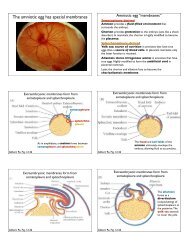
WOC 6e Guide to Microscopy
WOC 6e Guide to Microscopy
WOC 6e Guide to Microscopy

You also want an ePaper? Increase the reach of your titles
YUMPU automatically turns print PDFs into web optimized ePapers that Google loves.
Visible light<br />
Light source<br />
Glass lenses Electromagnetic lenses<br />
Human eye, pho<strong>to</strong>graphic<br />
film, or electronic detec<strong>to</strong>r<br />
(video camera)<br />
Condenser lenses<br />
Specimens<br />
Objective lenses<br />
Intermediate lenses<br />
Ocular lens<br />
50–100 kV<br />
Projec<strong>to</strong>r lens<br />
Fluorescent viewing screen<br />
or pho<strong>to</strong>graphic film<br />
A-2 Appendix Principles and Techniques of <strong>Microscopy</strong><br />
Tungsten filament<br />
Beam of electrons<br />
Anode<br />
(a) The light microscope (b) The electron microscope<br />
Despite these differences in illumination source and<br />
instrument design, both types of microscopes depend on the<br />
same principles of optics and form images in a similar manner.<br />
When a specimen is placed in the path of a light or electron<br />
beam, physical characteristics of the beam are changed<br />
in a way that creates an image that can be interpreted by the<br />
human eye or recorded on a pho<strong>to</strong>graphic detec<strong>to</strong>r. To<br />
understand this interaction between the illumination source<br />
and the specimen, we need <strong>to</strong> understand the concept of<br />
wavelength, which is illustrated in Figure A-2 using the following<br />
simple analogy.<br />
If two people hold on<strong>to</strong> opposite ends of a slack rope<br />
and wave the rope with a rhythmic up-and-down motion,<br />
they will generate a long, regular pattern of movement in<br />
the rope called a wave form (Figure A-2a). The distance from<br />
the crest of one wave <strong>to</strong> the crest of the next is called the<br />
wavelength. If someone standing <strong>to</strong> one side of the rope<br />
<strong>to</strong>sses a large object such as a beach ball <strong>to</strong>ward the rope, the<br />
ball may interfere with, or perturb, the wave form of the<br />
rope’s motion (Figure A-2b). However, if a small object such<br />
as a softball is <strong>to</strong>ssed <strong>to</strong>ward the rope, the movement of the<br />
rope will probably not be affected at all (Figure A-2c). If<br />
the rope holders move the rope more rapidly, the motion of<br />
the rope will still have a wave form, but the wavelength will<br />
be shorter (Figure A-2d). In this case, a softball <strong>to</strong>ssed <strong>to</strong>ward<br />
−<br />
+<br />
Figure A-1 The Optical Systems of the Light<br />
Microscope and the Electron Microscope.<br />
(a) The light microscope uses visible light<br />
and glass lenses <strong>to</strong> form an image of the<br />
specimen that can be seen by the eye,<br />
focused on pho<strong>to</strong>graphic film, or received by<br />
an electronic detec<strong>to</strong>r such as a video camera.<br />
(b) The electron microscope uses a<br />
beam of electrons emitted by a tungsten filament<br />
and focused by electromagnetic lenses<br />
<strong>to</strong> form an image of the specimen on a fluorescent<br />
screen, a digital detec<strong>to</strong>r, or pho<strong>to</strong>graphic<br />
film. (These diagrams have been<br />
drawn <strong>to</strong> emphasize the similarities in overall<br />
design between the two types of microscope.<br />
In reality, a light microscope is<br />
designed with the light source at the bot<strong>to</strong>m<br />
and the ocular lens at the <strong>to</strong>p, as shown in<br />
Figure 5b.)<br />
the rope is quite likely <strong>to</strong> perturb the rope’s movement<br />
(Figure A-2e).<br />
This simple analogy illustrates an important principle:<br />
The ability of an object <strong>to</strong> perturb a wave motion depends<br />
crucially on the size of the object in relation <strong>to</strong> the wavelength<br />
of the motion. This principle is of great importance in<br />
microscopy, because it means that the wavelength of the illumination<br />
source sets a limit on how small an object can be<br />
seen. To understand this relationship, we need <strong>to</strong> recognize<br />
that the moving rope of Figure A-2 is analogous <strong>to</strong> the beam<br />
of light (pho<strong>to</strong>ns) or electrons that is used as an illumination<br />
source in a light or electron microscope, respectively—in<br />
other words, both light and electrons behave as waves. When<br />
a beam of light or electrons encounters a specimen, the specimen<br />
alters the physical characteristics of the illuminating<br />
beam, just as the beach ball or softball alters the motion of<br />
the rope. And because an object can be detected only by its<br />
effect on the wave, the wavelength must be comparable in<br />
size <strong>to</strong> the object that is <strong>to</strong> be detected.<br />
Once we understand this relationship between wavelength<br />
and object size, we can readily appreciate why very<br />
small objects can be seen only by electron microscopy: The<br />
wavelengths of electrons are very much shorter than those of<br />
pho<strong>to</strong>ns. Thus, objects such as viruses and ribosomes are <strong>to</strong>o<br />
small <strong>to</strong> perturb a wave of pho<strong>to</strong>ns, but they can readily