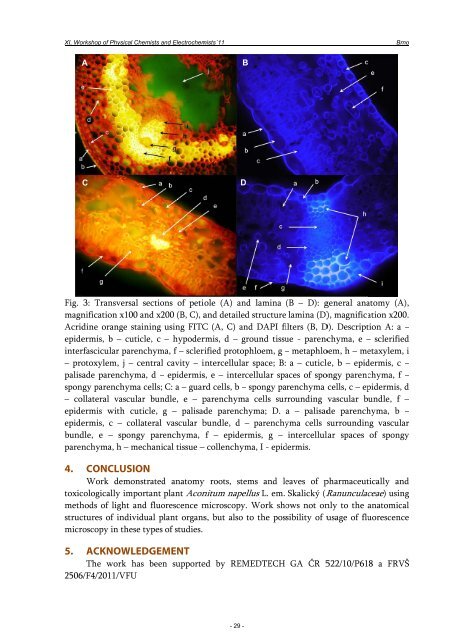
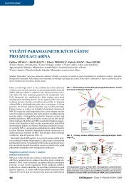
XI. Woorkshop of Physsical Chemists and a Electrochemmists´11 A C Fig. 3: Transveersal sectio ons of petiole (A) an nd lamina (B – D): ggeneral ana atomy (A), , magnnification xx100 and x2 200 (B, C), aand detailed structure lamina (D) ), magnification x200. . Acriddine orangge staining using FITCC (A, C) an nd DAPI fi ilters (B, DD). Descript tion A: a – epideermis, b – cuticle, c – hypoderrmis, d – ground g tissu ue - parencchyma, e – sclerifiedd interrfascicular pparenchym ma, f – scleri rified protop phloem, g – metaphlooem, h – me etaxylem, i – prootoxylem, j – central cavity – iintercellula ar space; B: a – cuticlle, b – epid dermis, c – palissade parencchyma, d – epidermiss, e – inter rcellular spa aces of spoongy parenchyma, f – sponngy parenchhyma cells; C: a – guarrd cells, b – spongy pa arenchyma cells, c – ep pidermis, d – coollateral vaascular bun ndle, e – pparenchym ma cells sur rrounding vascular bundle, b f – epideermis withh cuticle, g – palisadde parench hyma; D. a – palisadde parench hyma, b – epideermis, c – collateral vascular bbundle, d – parenchy yma cells surroundin ng vascularr bunddle, e – sppongy pare enchyma, f – epider rmis, g – intercellullar spaces of spongyy parennchyma, h – mechanical tissue – collenchym ma, I - epid dermis. 4. CONCLUUSION Work deemonstrated d anatomyy roots, ste ems and le eaves of ppharmaceut tically andd toxiccologically important plant Aconnitum nape ellus L. em. Skalický (RRanunculac aceae) usingg methhods of lighht and fluo orescence mmicroscopy y. Work sho ows not onnly to the anatomicall strucctures of inndividual plant organss, but also to the possibility of uusage of flu uorescencee micrroscopy in tthese types of studies. 5. ACKNOWWLEDGEM MENT The workk has been n supporteed by REM MEDTECH GA ČR 5522/10/P61 18 a FRVŠŠ 25066/F4/2011/VVFU - 29 - B D Brnoo
XI. Workshop of Physical Chemists and Electrochemists´11 Brno 6. REFERENCES 1. Feldkamp, A.; Koster, B.; Weber, H. P., Fatal Poisoning by Monks-Hood (Aconitum-Napellus). Mon.schr. Kinderheilkd. 1991, 139, (6), 366-367. 2. Haas, C., Monk's hood, Aconitum napellus, poison and medecine. Ann. Med. Interne 1999, 150, (5), 446-447. 3. Pullela, R.; Young, L.; Gallagher, B.; Avis, S. P.; Randell, E. W., A case of fatal aconitine poisoning by monkshood ingestion. J. Forensic Sci. 2008, 53, (2), 491-494. 4. Mitka, J., Phenetic and geographic pattern of Aconitum sect. napellus (Ranunculaceae) in the Eastern Carpathians - A numerical approach. Acta Soc. Bot. Pol. 2002, 71, (1), 35-48. 5. Le Cadre, S.; Boisselier-Dubayle, M. C.; Lambourdiere, J.; Machon, N.; Moret, J.; Samadi, S., Polymorphic microsatellites for the study of Aconitum napellus L. (Ranunculaceae), a rare species in France. Mol. Ecol. Notes 2005, 5, (2), 358-360. 6. Chen, Y.; Koelliker, S.; Oehme, M.; Katz, A., Isolation of diterpenoid alkaloids from herb and flowers of Aconitum napellus ssp vulgare and electrospray ion trap multiple MS study of these alkaloids. J. Nat. Prod. 1999, 62, (5), 701-704. 7. Csupor, D.; Forgo, P.; Csedo, K.; Hohmann, J., C-19 and C-20 diterpene alkaloids from Aconitum toxicum RCHB. Helv. Chim. Acta 2006, 89, (12), 2981-2986. 8. Forgo, P.; Borcsa, B.; Csupor, D.; Fodor, L.; Robert, B.; Molnar, A. V.; Hohmann, J., Diterpene Alkaloids from Aconitum anthora and Assessment of the hERG-Inhibiting Ability of Aconitum Alkaloids. Planta Med. 2011, 77, (4), 368-373. 9. Weijters, B. J.; Verbunt, R.; Hoogsteen, J.; Visser, R. F., Salade malade: malignant ventricular arrhythmias due to an accidental intoxication with Aconitum napellus. Neth. Heart J. 2008, 16, (3), 96-+. 10. Chan, T. Y. K., Aconite poisoning. Clin. Toxicol. 2009, 47, (4), 279-285. 11. Strzelecki, A.; Pichon, N.; Gaulier, J. M.; Amiel, J. B.; Champy, P.; Clavel, M., Acute Toxic Herbal Intake in a Suicide Attempt and Fatal Refractory Ventricular Arrhythmia. Basic Clin. Pharmacol. Toxicol. 2010, 107, (2), 698-699. 12. Braca, A.; Fico, G.; Morelli, I.; De Simone, F.; Tome, F.; De Tommasi, N., Antioxidant and free radical scavenging activity of flavonol glycosides from different Aconitum species. J. Ethnopharmacol. 2003, 86, (1), 63-67. 13. Diaz, J. G.; Ruiz, J. G.; Dias, B. R.; Sazatornil, J. A. G.; Herz, W., Flavonol 3,7-glycosides and dihydroxyphenethyl glycosides from Aconitum napellus subsp lusitanicum. Biochem. Syst. Ecol. 2005, 33, (2), 201-205. 14. Luis, J. C.; Valdes, F.; Martin, R.; Carmona, A. J.; Diaz, J. G., DPPH radical scavenging activity of two flavonol glycosides from Aconitum napellus sp lusitanicum. Fitoterapia 2006, 77, (6), 469-471. 15. Fico, G.; Braca, A.; De Tommasi, N.; Tome, F.; Morelli, I., Flavonoids from Aconitum napellus subsp neomontanum. Phytochemistry 2001, 57, (4), 543-546. 16. Fico, G.; Braca, A.; Bilia, A. R.; Tome, F.; Morelli, I., New flavonol glycosides from the flowers of Aconitum napellus ssp tauricum. Planta Med. 2001, 67, (3), 287-290. 17. Vitalini, S.; Braca, A.; Passarella, D.; Fico, G., New flavonol glycosides from Aconitum burnatii Gayer and Aconitum variegatum L. Fitoterapia 2010, 81, (7), 940-947. 18. Singhuber, J.; Zhu, M.; Prinz, S.; Kopp, B., Aconitum in Traditional Chinese Medicine-A valuable drug or an unpredictable risk? J. Ethnopharmacol. 2009, 126, (1), 18-30. 19. Piltan, D.; Rist, L.; Simoes-Wust, A. P.; Saller, R., Test of a Homeopathic Dilution of Aconitum napellus A Clinical, Randomized, Double-Blind, Controlled Crossover Study in Healthy Volunteers. Forsch. Komplement.med. 2009, 16, (3), 168-173. 20. Thurneysen, A., On: Piltan D, Rist L, Simoes-Wust A, Saller R: Test of a homeopathic dilution of Aconitum napellus. A clinical, randomized, double-blind, controlled crossover study in healthy volunteers. Forsch Komplementmed 2009;16:168-173. Forsch. Komplement.med. 2009, 16, (5), 349- 349. - 30 -
- Page 2 and 3: XI. Woorkshop of Physsical Chemists
- Page 4 and 5: XI. Woorkshop of Physsical Chemists
- Page 6 and 7: XI. Workshop of Physical Chemists a
- Page 8 and 9: XI. Workshop of Physical Chemists a
- Page 10 and 11: XI. Workshop of Physical Chemists a
- Page 12 and 13: XI. Workshop of Physical Chemists a
- Page 14 and 15: XI. Workshop of Physical Chemists a
- Page 16 and 17: XI. Workshop of Physical Chemists a
- Page 18 and 19: XI. Workshop of Physical Chemists a
- Page 20 and 21: XI. Workshop of Physical Chemists a
- Page 22 and 23: XI. Woorkshop of Physsical Chemists
- Page 24 and 25: XI. Workshop of Physical Chemists a
- Page 26 and 27: XI. Woorkshop of Physsical Chemists
- Page 28 and 29: XI. Woorkshop of Physsical Chemists
- Page 32 and 33: XI. Workshop of Physical Chemists a
- Page 34 and 35: XI. Workshop of Physical Chemists a
- Page 36 and 37: XI. Woorkshop of Physsical Chemists
- Page 38 and 39: XI. Workshop of Physical Chemists a
- Page 40 and 41: XI. Workshop of Physical Chemists a
- Page 42 and 43: XI. Workshop of Physical Chemists a
- Page 44 and 45: XI. Workshop of Physical Chemists a
- Page 46 and 47: XI. Workshop of Physical Chemists a
- Page 48 and 49: XI. Woorkshop of Physsical Chemists
- Page 50 and 51: XI. Workshop of Physical Chemists a
- Page 52 and 53: XI. Workshop of Physical Chemists a
- Page 54 and 55: XI. Workshop of Physical Chemists a
- Page 56 and 57: XI. Workshop of Physical Chemists a
- Page 58 and 59: XI. Workshop of Physical Chemists a
- Page 60 and 61: XI. Workshop of Physical Chemists a
- Page 62 and 63: XI. Workshop of Physical Chemists a
- Page 64 and 65: XI. Workshop of Physical Chemists a
- Page 66 and 67: XI. Workshop of Physical Chemists a
- Page 68 and 69: XI. Workshop of Physical Chemists a
- Page 70 and 71: XI. Workshop of Physical Chemists a
- Page 72 and 73: XI. Workshop of Physical Chemists a
- Page 74 and 75: XI. Workshop of Physical Chemists a
- Page 76 and 77: XI. Workshop of Physical Chemists a
- Page 78 and 79: XI. Workshop of Physical Chemists a
- Page 80 and 81:
XI. Woorkshop of Physsical Chemists
- Page 82 and 83:
XI. Workshop of Physical Chemists a
- Page 84 and 85:
XI. Workshop of Physical Chemists a
- Page 86 and 87:
XI. Workshop of Physical Chemists a
- Page 88 and 89:
XI. Workshop of Physical Chemists a
- Page 90 and 91:
XI. Woorkshop of Physsical Chemists
- Page 92 and 93:
XI. Woorkshop of Physsical Chemists
- Page 94 and 95:
XI. Workshop of Physical Chemists a
- Page 96 and 97:
XI. Woorkshop of Physsical Chemists
- Page 98 and 99:
XI. Woorkshop of Physsical Chemists
- Page 100 and 101:
XI. Woorkshop of Physsical Chemists
- Page 102 and 103:
XI. Workshop of Physical Chemists a
- Page 104 and 105:
XI. Woorkshop of Physsical Chemists
- Page 106 and 107:
XI. Woorkshop of Physsical Chemists
- Page 108 and 109:
XI. Woorkshop of Physsical Chemists
- Page 110 and 111:
XI. Woorkshop of Physsical Chemists
- Page 112 and 113:
XI. Workshop of Physical Chemists a
- Page 114 and 115:
XI. Woorkshop of Physsical Chemists
- Page 116 and 117:
XI. Workshop of Physical Chemists a
- Page 118 and 119:
XI. Woorkshop of Physsical Chemists
- Page 120 and 121:
XI. Workshop of Physical Chemists a
- Page 122 and 123:
XI. Workshop of Physical Chemists a
- Page 124 and 125:
XI. Workshop of Physical Chemists a
- Page 126 and 127:
XI. Workshop of Physical Chemists a
- Page 128 and 129:
XI. Woorkshop of Physsical Chemists
- Page 130 and 131:
XI. Workshop of Physical Chemists a
- Page 132 and 133:
XI. Woorkshop of Physsical Chemists
- Page 134 and 135:
XI. Workshop of Physical Chemists a
- Page 136 and 137:
XI. Woorkshop of Physsical Chemists
- Page 138 and 139:
XI. Workshop of Physical Chemists a
- Page 140 and 141:
XI. Woorkshop of Physsical Chemists
- Page 142 and 143:
XI. Woorkshop of Physsical Chemists
- Page 144 and 145:
XI. Workshop of Physical Chemists a
- Page 146 and 147:
XI. Woorkshop of Physsical Chemists
- Page 148 and 149:
XI. Workshop of Physical Chemists a
- Page 150 and 151:
XI. Workshop of Physical Chemists a
- Page 152 and 153:
XI. Workshop of Physical Chemists a
- Page 154 and 155:
XI. Workshop of Physical Chemists a
- Page 156 and 157:
XI. Workshop of Physical Chemists a
- Page 158 and 159:
XI. WWorkshop of Phyysical Chemists
- Page 160 and 161:
XI. Workshop of Physical Chemists a
- Page 162 and 163:
XI. Workshop of Physical Chemists a
- Page 164 and 165:
XI. Workshop of Physical Chemists a
- Page 166 and 167:
XI. Workshop of Physical Chemists a
- Page 168 and 169:
XI. Workshop of Physical Chemists a
- Page 170 and 171:
XI. WWorkshop of Phyysical Chemists
- Page 172 and 173:
XI. Workshop of Physical Chemists a
- Page 174 and 175:
XI. Workshop of Physical Chemists a
- Page 176 and 177:
XI. Workshop of Physical Chemists a
- Page 178 and 179:
XI. Workshop of Physical Chemists a
- Page 180 and 181:
XI. Workshop of Physical Chemists a
- Page 182 and 183:
XI. Workshop of Physical Chemists a
- Page 184 and 185:
XI. WWorkshop of Phyysical Chemists
- Page 186 and 187:
XI. Workshop of Physical Chemists a
- Page 188 and 189:
XI. Workshop of Physical Chemists a
- Page 190 and 191:
XI. Workshop of Physical Chemists a
- Page 192 and 193:
XI. WWorkshop of Phyysical Chemists
- Page 194 and 195:
XI. Workshop of Physical Chemists a
- Page 196 and 197:
XI. WWorkshop of Phyysical Chemists
- Page 198 and 199:
XI. Workshop of Physical Chemists a
- Page 200 and 201:
XI. Workshop of Physical Chemists a
- Page 202 and 203:
XI. Workshop of Physical Chemists a
- Page 204 and 205:
XI. Workshop of Physical Chemists a
- Page 206 and 207:
XI. Workshop of Physical Chemists a
- Page 208 and 209:
XI. Workshop of Physical Chemists a
- Page 210 and 211:
XI. WWorkshop of Phyysical Chemists
- Page 212 and 213:
XI. Workshop of Physical Chemists a
- Page 214 and 215:
XI. Workshop of Physical Chemists a
- Page 216 and 217:
XI. WWorkshop of Phyysical Chemists
- Page 218 and 219:
XI. Workshop of Physical Chemists a
- Page 220 and 221:
XI. WWorkshop of Phyysical Chemists
- Page 222 and 223:
XI. Workshop of Physical Chemists a
- Page 224 and 225:
XI. WWorkshop of Phyysical Chemists
- Page 226 and 227:
XI. Workshop of Physical Chemists a
- Page 228 and 229:
XI. Workshop of Physical Chemists a
- Page 230 and 231:
XI. Workshop of Physical Chemists a
- Page 232 and 233:
XI. WWorkshop of Phyysical Chemists
- Page 234 and 235:
XI. Workshop of Physical Chemists a
- Page 236 and 237:
XI. Workshop of Physical Chemists a
- Page 238 and 239:
XI. Workshop of Physical Chemists a
- Page 240 and 241:
XI. Workshop of Physical Chemists a
- Page 242 and 243:
XI. WWorkshop of Phyysical Chemists
- Page 244 and 245:
XI. WWorkshop of Phyysical Chemists
- Page 246 and 247:
XI. WWorkshop of Phyysical Chemists
- Page 248 and 249:
XI. Workshop of Physical Chemists a
- Page 250 and 251:
XI. Workshop of Physical Chemists a
- Page 252 and 253:
XI. WWorkshop of Phyysical Chemists
- Page 254 and 255:
XI. WWorkshop of Phyysical Chemists
- Page 256 and 257:
XI. Workshop of Physical Chemists a
- Page 258 and 259:
XI. WWorkshop of Phyysical Chemists
- Page 260 and 261:
XI. Workshop of Physical Chemists a
- Page 262 and 263:
XI. WWorkshop of Phyysical Chemists
- Page 264 and 265:
XI. Workshop of Physical Chemists a
- Page 266 and 267:
XI. WWorkshop of Phyysical Chemists
- Page 268 and 269:
XI. Workshop of Physical Chemists a
- Page 270 and 271:
XI. Workshop of Physical Chemists a
- Page 272 and 273:
XI. Workshop of Physical Chemists a
- Page 274 and 275:
XI. Workshop of Physical Chemists a
- Page 276 and 277:
XI. Workshop of Physical Chemists a
- Page 278 and 279:
XI. WWorkshop of Phyysical Chemists
- Page 280 and 281:
XI. Workshop of Physical Chemists a
- Page 282 and 283:
XI. Workshop of Physical Chemists a
- Page 284 and 285:
XI. WWorkshop of Phyysical Chemists
- Page 286 and 287:
XI. Workshop of Physical Chemists a
- Page 288 and 289:
XI. Workshop of Physical Chemists a
- Page 290 and 291:
XI. WWorkshop of Phyysical Chemists
- Page 292 and 293:
XI. Workshop of Physical Chemists a
- Page 294 and 295:
XI. Workshop of Physical Chemists a
- Page 296 and 297:
XI. Workshop of Physical Chemists a
- Page 298 and 299:
XI. Workshop of Physical Chemists a
- Page 300 and 301:
XI. Workshop of Physical Chemists a
- Page 302 and 303:
XI. Workshop of Physical Chemists a
- Page 304 and 305:
XI. Workshop of Physical Chemists a
- Page 306 and 307:
XI. Workshop of Physical Chemists a
- Page 308 and 309:
XI. Workshop of Physical Chemists a
- Page 310 and 311:
XI. Workshop of Physical Chemists a
- Page 312 and 313:
XI. Workshop of Physical Chemists a
- Page 314 and 315:
XI. Workshop of Physical Chemists a
- Page 316 and 317:
XI. WWorkshop of Phyysical Chemists
- Page 318 and 319:
XI. Workshop of Physical Chemists a
- Page 320 and 321:
XI. Workshop of Physical Chemists a
- Page 322 and 323:
XI. Workshop of Physical Chemists a
- Page 324 and 325:
XI. Workshop of Physical Chemists a
- Page 326 and 327:
XI. Workshop of Physical Chemists a
- Page 328 and 329:
XI. Workshop of Physical Chemists a
- Page 330 and 331:
XI. Workshop of Physical Chemists a
- Page 332 and 333:
XI. Workshop of Physical Chemists a
- Page 334 and 335:
XI. Workshop of Physical Chemists a
- Page 336 and 337:
XI. Workshop of Physical Chemists a
- Page 338 and 339:
XI. Workshop of Physical Chemists a
- Page 340 and 341:
XI. Workshop of Physical Chemists a
- Page 342 and 343:
XI. WWorkshop of Phyysical Chemists
- Page 344 and 345:
XI. Workshop of Physical Chemists a
- Page 346 and 347:
XI. Workshop of Physical Chemists a
- Page 348 and 349:
XI. Workshop of Physical Chemists a
- Page 350 and 351:
XI. Workshop of Physical Chemists a
- Page 352 and 353:
XI. Workshop of Physical Chemists a
- Page 354 and 355:
XI. WWorkshop of Phyysical Chemists
- Page 356 and 357:
XI. Workshop of Physical Chemists a
- Page 358 and 359:
XI. WWorkshop of Phyysical Chemists
- Page 360 and 361:
XI. Workshop of Physical Chemists a
- Page 362 and 363:
XI. Workshop of Physical Chemists a
- Page 364 and 365:
XI. Workshop of Physical Chemists a
- Page 366 and 367:
XI. Workshop of Physical Chemists a
- Page 368 and 369:
XI. Workshop of Physical Chemists a
- Page 370 and 371:
XI. Workshop of Physical Chemists a
- Page 372 and 373:
XI. Workshop of Physical Chemists a
- Page 374 and 375:
Pr ˇ ístroje a reagencie pro klin
- Page 376 and 377:
Electrochemical tools Pump Accessor
- Page 380 and 381:
Eppendorf Xplorer ® ‡ Přehledn
- Page 382 and 383:
110415_chromservis_letak_Dani_SHS_C
- Page 384 and 385:
• • • • • • AKCE KINETE
- Page 386:
NABÍDKA SPEKTROFOTOMETRŮ Dvoupapr
- Page 390:
Metrohm Česká republika s.r.o. Na
- Page 394 and 395:
VÝROBKY NEJVYŠŠÍ KVALITY Mrazí