IABP Therapie - Die Basics
IABP Therapie - Die Basics
IABP Therapie - Die Basics

You also want an ePaper? Increase the reach of your titles
YUMPU automatically turns print PDFs into web optimized ePapers that Google loves.
Donnerstag, 6. August 2009<br />
Curriculum Medizinische Intensivtherapie<br />
<strong>IABP</strong> <strong>Therapie</strong> - <strong>Die</strong> <strong>Basics</strong><br />
Robert M. Radke<br />
Medizinische Klinik C<br />
Universitätsklinikum Münster
Donnerstag, 6. August 2009<br />
Themen<br />
• Grundprinzip der <strong>IABP</strong><br />
• Indikationen, Kontraindikationen<br />
• Einführung in das Gerät<br />
• Katheteranlage und optimale<br />
Kreislaufunterstützung<br />
• Komplikationen<br />
• Evidenz
Donnerstag, 6. August 2009<br />
<strong>IABP</strong> System<br />
<strong>IABP</strong>-Katheter<br />
Pumpe
Donnerstag, 6. August 2009<br />
Katheterposition und Funktion
Donnerstag, 6. August 2009<br />
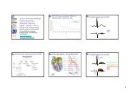
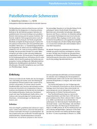
Physiologischer Herzzyklus<br />
Vorhofsystole<br />
Aortendruck<br />
Druck in der<br />
linken Kammer<br />
Druck im<br />
linken Vorhof<br />
zentraler Venendruck<br />
(ZVD)<br />
Volumen des<br />
linken Ventrikels<br />
1mV<br />
120<br />
mmHg<br />
0<br />
2<br />
mmHg<br />
0<br />
120<br />
ml<br />
40<br />
0<br />
a<br />
IVc I IIa IIb III IVa IVb<br />
1<br />
2<br />
3<br />
4<br />
Q<br />
R<br />
S<br />
Kammersystole Kammerdiastole<br />
c<br />
enddiastolisches<br />
Volumen<br />
(EDV)<br />
x<br />
T<br />
Inzisur<br />
v<br />
Schlagvolumen<br />
(SV)<br />
EKG<br />
Restvolumen (ESV)<br />
y<br />
P<br />
IVc<br />
Q<br />
a<br />
Vorhofkontraktion
Donnerstag, 6. August 2009<br />
Myokardiale Sauerstoffversorgung<br />
Angebot<br />
•Koronaranatomie<br />
•Diastolischer Druck<br />
•Diastolendauer<br />
•O2 Transport<br />
(Hb, PaO2)<br />
Verbrauch<br />
•Herzfrequenz<br />
•Afterload<br />
•Preload<br />
•Kontraktilität
Donnerstag, 6. August 2009<br />
Myokardiale Sauerstoffversorgung<br />
DPTI / TTI = Angebot / Nachfrage
EKG<br />
<strong>IABP</strong> - Prinzip<br />
Aortendruck<br />
<strong>IABP</strong><br />
Donnerstag, 6. August 2009<br />
nicht<br />
assistierte Systole<br />
nicht assistierter<br />
enddiastolischer Druck<br />
diastolische<br />
nicht<br />
Augmentation<br />
assistierte Systole<br />
assistierte<br />
Systole<br />
assistierter<br />
enddiastolischer Druck
Donnerstag, 6. August 2009<br />
<strong>IABP</strong> - Prinzip
Donnerstag, 6. August 2009<br />
<strong>IABP</strong> - Physiologische Effekte<br />
Aortaler<br />
Druck<br />
Kardiale<br />
Last<br />
! Systolisch ! Nachlast<br />
" Diastolisch ! Vorlast<br />
Blutfluss LV Drücke LV<br />
" Koronarer<br />
Blutfluss<br />
" Cardiac<br />
Output<br />
" Renaler<br />
Blutfluss<br />
! Systolisch ! Volumen<br />
! Enddiastolisch<br />
! Arbeit<br />
! Wandspannung<br />
Maccioli GA et al. 1988
Donnerstag, 6. August 2009<br />
Indikationen - Kardiologie<br />
• Hypotension oder Low Output nach<br />
akutem Myokardinfarkt<br />
(AHA : I, B ESC: I, B)<br />
• Infarkt-VSD, Papillarmuskelabriss<br />
• Polymorphe VTs nach MI<br />
• Patienten vor operativer Notfall<br />
Myokardrevaskularisation<br />
(AHA : IIa, B)<br />
• Supportiv bei Thrombolyse<br />
• ggf. Überbrückung zur Transplantation<br />
bei Herzinsuffizienz
Donnerstag, 6. August 2009<br />
Indikationen - Anästhesiologie<br />
• Notfall-Myokardrevaskularisationen<br />
• Prophylaktischer Einsatz bei<br />
Hochrisikopatienten mit reduzierter<br />
linksventrikulärer Pumpfunktion<br />
• Low Output beim Weaning von der<br />
HLM
Donnerstag, 6. August 2009<br />
Indikationen - Zukunft?<br />
• Septischer Schock mit schlechter<br />
Pumpfunktion und gutem peripheren<br />
Wiederstand?<br />
Solomon SB, Critical Care Medicine, 2009
Donnerstag, 6. August 2009<br />
Kontraindikationen<br />
• Schwere Aorteninsuffizienz<br />
• Aortenaneurysma<br />
• Gefäßprothese der A. femoralis<br />
• schwerste pAVK
Donnerstag, 6. August 2009<br />
Anlage des <strong>IABP</strong> Katheters<br />
• Anlage Schleuse im Seldingerverfahren<br />
• Vorschieben des Führungsdrahtes unter<br />
Sicht bis zum Erreichen des Aortenbogens
Donnerstag, 6. August 2009<br />
Anlage des <strong>IABP</strong> Katheters<br />
• <strong>IABP</strong> Katheterspitze kurz unterhalb des<br />
Bogens positionieren. Sollte am unteren<br />
Rand des Aortenknopfes sein.<br />
• Fixieren. Bein gestreckt lassen.<br />
• Röntgenthoraxkontrolle. Alternativ im OP<br />
per TEE.<br />
• Vollheparinisierung.<br />
• Keine Pausen über 30 min!
Donnerstag, 6. August 2009
Donnerstag, 6. August 2009
Donnerstag, 6. August 2009<br />
Unsere <strong>IABP</strong>s (Datascope CS100)<br />
• Automatische Analyse der<br />
optimalen Triggerquelle<br />
und Ableitung<br />
• Automatisches Timing<br />
• Druckmonitoring über das<br />
Katheterlumen
Donnerstag, 6. August 2009<br />
Aufbau
Donnerstag, 6. August 2009<br />
Anschlüsse<br />
• Helium Anschluss<br />
• EKG Buchse<br />
• Druck Buchse
Donnerstag, 6. August 2009<br />
Überblick Display
Donnerstag, 6. August 2009<br />
Überblick Bedienfeld
Donnerstag, 6. August 2009<br />
<strong>Therapie</strong> Starten - I<br />
• Alles konnektieren<br />
• <strong>IABP</strong> Einschalten: „Mains On“ und „<strong>IABP</strong><br />
On“<br />
• Heliumflasche aufdrehen, Druck<br />
kontrollieren<br />
• Transducer nullen: 3W-Hahn auf Luft<br />
drehen. „Zero Pressure“ für 2 Sekunden<br />
drücken. 3W-Hahn zurückdrehen.
Donnerstag, 6. August 2009<br />
<strong>Therapie</strong> Starten II<br />
• Operation Mode „Auto“ einstellen.<br />
• 1:2 Modus Einstellen.<br />
• „Start“ drücken. (Füllung und Pumpen beginnt)<br />
• Kontrolle der Druckkurven und Finetuning des<br />
Entlüftungstiming (Inflationstiming im Auto<br />
Modus nicht beeinflussbar)<br />
• 1:1 Modus auswählen<br />
• Setzten des Augmentationsalarms auf ca.<br />
10mmHg unter der Augmentierten astolischen<br />
Druck.
Donnerstag, 6. August 2009<br />
Aspekte des Automatik Modus<br />
• Trigger wird automatisch ausgesucht. Wenn<br />
EKG, dann automatisch die beste Ableitung.<br />
• Timing wird automatisch eingestellt,<br />
Deflation veränderbar<br />
• Timing wird an HF und Rhythmus angepasst
•<br />
•<br />
•<br />
•<br />
•<br />
•<br />
Donnerstag, 6. August 2009<br />
!"#$%&%'(#()%*+##,-,-.,##/0/1#23##24(+#5<br />
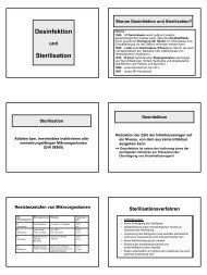
Optimierung der Druckkurven - Idealbild<br />
Beurteilung in 1:2 Modus<br />
Scharfes A = One V complete sicher nach dem<br />
cardiac cycle<br />
Klappenschluss (A)<br />
B = Unassisted aortic<br />
end diastolic pressure<br />
Diastolische C = Unassisted Augmentation, die<br />
systolic pressure<br />
idelaerweise über der<br />
D = Diastolic<br />
normalen augmentation Systole liegt (D)<br />
E = Reduced aortic<br />
end diastolic pressure<br />
Verminderter enddiastolischer<br />
F = Reduced<br />
Druck (max. systolic pressure um 10mmHg) (E)<br />
Verminderung des darauf<br />
folgenden systolischen Drucks<br />
(F)<br />
Anstieg der MAP<br />
P<br />
Q<br />
B<br />
R<br />
S<br />
T<br />
Correct <strong>IABP</strong> Timing<br />
C<br />
A<br />
D<br />
E<br />
F
Unassisted<br />
systole<br />
Zu frühe Inflation<br />
•<br />
•<br />
Donnerstag, 6. August 2009<br />
Incorrect <strong>IABP</strong> Timing<br />
Assisted Aortic End<br />
Diastolic Pressure<br />
Effekte:<br />
Diastolic<br />
Augmentation<br />
Assisted Systole<br />
•<br />
•<br />
Inflation vor dem<br />
Aortenklappenschluss<br />
Verschmelzung von<br />
diastolischer Augmentation<br />
und Systole<br />
Verfrühter Aortenklappenschluss. Ggf. Anstieg von LVEDP.<br />
Anstieg des Afterloads, Erhöhter O2 Bedarf
Zu späte Inflation<br />
LATE INFLATION<br />
tion of the IAB markedly after<br />
re of the aortic valve.<br />
form Characteristics:<br />
• Inflation<br />
deutlich nach<br />
lation of IAB after the dicrotic notch.<br />
sence of sharp V.<br />
iologic Effects:<br />
der Inzisur<br />
b-optimal coronary artery perfusion.<br />
Donnerstag, 6. August 2009<br />
• Kein scharfes<br />
V!<br />
Effekt:<br />
Dicrotic Notch<br />
Diastolic Augmentation<br />
Assisted Aortic End<br />
Diastolic Pressure<br />
• Sub-optimale Koronarperfusion<br />
Assisted Systole
eform Characteristics:<br />
Zu späte Deflation<br />
LATE DEFLATION<br />
sisted aortic end diastolic pressure<br />
ay be equal to the unassisted<br />
rtic end diastolic pressure.<br />
• Assistierter<br />
enddiastolischer Druck<br />
ate of rise of assisted systole<br />
prolonged.<br />
nicht tiefer als normal<br />
iastolic augmentation may appear<br />
idened.<br />
• Druckanstieg der<br />
assistierten Systole,<br />
siologic Effects:<br />
terload reduction is essentially<br />
sent<br />
flacher als normal.<br />
creased MV02 consumption due to<br />
e left ventricle ejecting against a<br />
eater resistance and a prolonged<br />
ovolumetric contraction phase<br />
• Diastolische<br />
Augmentation<br />
B may impede<br />
verbreitert.<br />
left ventricular<br />
ection and increase the afterload<br />
Effekt:<br />
Donnerstag, 6. August 2009<br />
Unassisted Systole<br />
Widened<br />
Appearance<br />
Prolonged Rate<br />
of rise of Assisted<br />
Systole<br />
Assisted Aortic End<br />
Diastolic Pressure<br />
• LV pumpt z.T. gegen geschlossenen Ballon. Erhöhte<br />
Nachlast und O2 Bedarf.
Zu frühe Deflation<br />
Diastolic<br />
Augmentation<br />
Assisted Aortic End<br />
Diastolic Pressure Unassisted Aortic<br />
End Diastolic Pressure<br />
Effekt:<br />
Donnerstag, 6. August 2009<br />
Incorrect <strong>IABP</strong> Timing<br />
Assisted Systole<br />
• Sub-optimale Koronarperfusion und<br />
Nachlastminderung<br />
• Deflation führt zu sehr<br />
scharfem Abfall des<br />
diastolischen Drucks<br />
• Suboptimale<br />
diastolische<br />
Augmentation<br />
• Assistierte systolischer<br />
Druck zu hoch.
Donnerstag, 6. August 2009<br />
Keine supersystolische Diastole - Andere<br />
Gründe<br />
• Patient<br />
•<br />
•<br />
•<br />
• Katheter<br />
•<br />
•<br />
•<br />
•<br />
•<br />
Tachykardie. Zu wenig Zeit zum Aufblasen.<br />
Hypovolämie. Zu geringes Schlagvolumen.<br />
Hypotonie. Diastole unter 40mmHg<br />
Ballon noch in der Schleuse?<br />
Katheter abgeknickt?<br />
Falsche Katheter Größe?<br />
Falsche Katheterlage<br />
Helium Leck
<strong>IABP</strong> - <strong>Die</strong> Risiken<br />
Komplikation Inzidenz (%)<br />
Beinischämie 5-18<br />
Schwerwiegende vaskuläre Komplikation 1-4<br />
Mesenteriale Ischämie 1<br />
Fehlplatzierung 1-5<br />
Ballonleck 1-5<br />
Aortenperforation
Donnerstag, 6. August 2009<br />
Überwachung der <strong>Therapie</strong><br />
• Antikoagulation sicherstellen.<br />
• Klinische Untersuchung (Extremität<br />
durchblutet? Pulse? Farbe? Blutung?)<br />
• Röntgen Thorax zur Lagekontrolle<br />
• CK-Kontrolle (Ischämie der Extremität?)<br />
• Überwachung von Urinausscheidung und<br />
Retentionsparametern.
Donnerstag, 6. August 2009<br />
Weaning<br />
• Keine Richtlinien für die <strong>Therapie</strong>-Dauer.<br />
• Abhängig von Klinik. Typisch 3-5 Tage.<br />
• Weaning durch stufenweises reduzieren in<br />
Abhängigkeit vom Katecholaminbedarf über<br />
Stunden oder Tage.
Donnerstag, 6. August 2009<br />
Leitlinien - <strong>IABP</strong> und STEMI<br />
• Kardiogener Schock, nicht unmittelbar<br />
ansprechend auf Volumen und<br />
Pharmakotherapie (AHA : I, B ESC: I, B)<br />
• Low Output (AHA: I, B ESC IIa, B)<br />
• Evidenz?
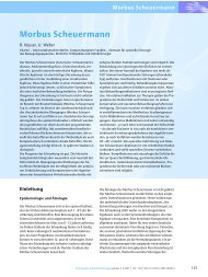
<strong>IABP</strong> bei high-risk STEMI<br />
ized clinical trials of <strong>IABP</strong> therapy in STEMI. All meta-analyses show effect estimates for the individual trials,<br />
and for the overall analysis. The size of each square is proportional to the weight of the individual trial. (A)<br />
lity. (B) The mean differences in left ventricular ejection fraction (LVEF). (C and D) The risk differences in<br />
P, intra-aortic balloon counterpulsation; PCI, percutaneous coronary intervention.<br />
difference for three trials that<br />
. Overall, <strong>IABP</strong> support in the<br />
d with a change in LVEF at<br />
95% CI, 22.2 to 2.0%; P ¼<br />
absolute numbers of stroke<br />
, together with the respective<br />
ial. Overall, the use of <strong>IABP</strong><br />
stroke rate of 2% (95% CI,<br />
leeding rate of 6% (95% CI,<br />
ype of reperfusion therapy<br />
the comprehensive analyses.<br />
neity across the seven trials.<br />
ewed distributions, suggesting<br />
.<br />
Donnerstag, 6. August 2009<br />
K.D. Sjauw et al.<br />
464<br />
Meta-analysis of cohort studies of<br />
intra-aortic balloon pump therapy in<br />
STEMI patients with cardiogenic shock<br />
Nine cohort studies of <strong>IABP</strong> therapy in STEMI patients with cardiogenic<br />
shock included a total of 10 529 patients. 20 – 29 Table 4 shows<br />
the study characteristics. Patients in the <strong>IABP</strong> group were younger<br />
(66 vs. 73 years) and more often male (63 vs. 53%). Figure 3A<br />
shows the absolute numbers of deaths in each treatment group,<br />
with the absolute risk difference for each cohort study. The thrombolysis<br />
studies showed adjunctive <strong>IABP</strong> therapy to be associated<br />
with an absolute decrease in 30 day mortality of 18% (95% CI,<br />
16–20%; P , 0.0001). Contrariwise, the primary PCI studies<br />
showed <strong>IABP</strong> therapy to be associated with an absolute increase<br />
464<br />
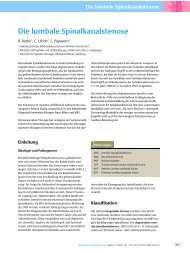
Figure 2 Meta-analysis of randomized clinical trials of <strong>IABP</strong> therapy in STEMI. All meta-analyses show effect estimates for the individual trials,<br />
for each type of reperfusion therapy and for the overall analysis. The size of each square is proportional to the weight of the individual trial. (A)<br />
The risk differences in 30 day mortality. (B) The mean differences in left ventricular ejection fraction (LVEF). (C and D) The risk differences in<br />
stroke and major bleeding rate. <strong>IABP</strong>, intra-aortic balloon counterpulsation; PCI, percutaneous coronary intervention.<br />
each treatment group, with the mean difference for three trials that<br />
reported on left ventricular function. Overall, <strong>IABP</strong> support in the<br />
setting of STEMI was not associated with a change in LVEF at<br />
follow up (mean difference 20.1%; 95% CI, 22.2 to 2.0%; P ¼<br />
0.93). Figure 2C and D shows the absolute numbers of stroke<br />
and bleeding in each treatment group, together with the respective<br />
absolute risk differences for each trial. Overall, the use of <strong>IABP</strong><br />
was associated with an increased stroke rate of 2% (95% CI,<br />
0–4%; P ¼ 0.03) and an increased bleeding rate of 6% (95% CI,<br />
1–11%; P ¼ 0.02). Analyses by type of reperfusion therapy<br />
yielded similar results to those of the comprehensive analyses.<br />
There was no evidence of heterogeneity across the seven trials.<br />
None of the funnel plots showed skewed distributions, suggesting<br />
that no publication bias was involved.<br />
each treatment group, with the mean difference for three trials that<br />
reported on left ventricular function. Overall, <strong>IABP</strong> support in the<br />
setting of STEMI was not associated with a change in LVEF at<br />
follow up (mean difference 20.1%; 95% CI, 22.2 to 2.0%; P ¼<br />
0.93). Figure 2C and D shows the absolute numbers of stroke<br />
and bleeding in each treatment group, together with the respective<br />
absolute risk differences for each trial. Overall, the use of <strong>IABP</strong><br />
was associated with an increased stroke rate of 2% (95% CI,<br />
0–4%; P ¼ 0.03) and an increased bleeding rate of 6% (95% CI,<br />
1–11%; P ¼ 0.02). Analyses by type of reperfusion therapy<br />
yielded similar results to those of the comprehensive analyses.<br />
Meta-analysis of cohort studies of<br />
intra-aortic balloon pump therapy in<br />
STEMI patients with cardiogenic shock<br />
Nine cohort studies of <strong>IABP</strong> therapy in STEMI patients with cardiogenic<br />
shock included a total of 10 529 patients. 20 – 29 Table 4 shows<br />
the study characteristics. Patients in the <strong>IABP</strong> group were younger<br />
(66 vs. 73 years) and more often male (63 vs. 53%). Figure 3A<br />
shows the absolute numbers of deaths in each treatment group,<br />
with the absolute risk difference for each cohort study. The thrombolysis<br />
studies showed adjunctive <strong>IABP</strong> therapy to be associated<br />
with an absolute decrease in 30 day mortality of 18% (95% CI,<br />
16–20%; P , 0.0001). Contrariwise, the primary PCI studies<br />
showed <strong>IABP</strong> therapy to be associated with an absolute increase<br />
K.D. Sjauw et al.<br />
Figure 2 Meta-analysis of randomized clinical trials of <strong>IABP</strong> therapy in STEMI. All meta-analyses show effect estimates for the individual trials,<br />
for each type of reperfusion therapy and for the overall analysis. The size of each square is proportional to the weight of the individual trial. (A)<br />
The risk differences in 30 day mortality. (B) The mean differences in left ventricular ejection fraction (LVEF). (C and D) The risk differences in<br />
stroke and major bleeding rate. <strong>IABP</strong>, intra-aortic balloon counterpulsation; PCI, percutaneous coronary intervention.<br />
Meta-analysis of cohort studies of<br />
intra-aortic balloon pump therapy in<br />
STEMI patients with cardiogenic shock<br />
Sjauw, EHJ 2009<br />
Nine cohort studies of <strong>IABP</strong> therapy in STEMI patients with cardiogenic<br />
shock included a total of 10 529 patients. 20 – 29 Table 4 shows<br />
the study characteristics. Patients in the <strong>IABP</strong> group were younger<br />
(66 vs. 73 years) and more often male (63 vs. 53%). Figure 3A<br />
shows the absolute numbers of deaths in each treatment group,<br />
with the absolute risk difference for each cohort study. The thrombolysis<br />
studies showed adjunctive <strong>IABP</strong> therapy to be associated
Donnerstag, 6. August 2009<br />
466<br />
STEMI mit Kardiogenem Schock<br />
Keine randomisierten Studien!<br />
the hypothesis that IA<br />
therapy in STEMI pat<br />
coronary perfusion. 32<br />
explanations for the o<br />
in this setting. First, th<br />
years younger and th<br />
known from the curre<br />
by 49–60% for every<br />
a lower clinical risk pr<br />
of cardiogenic shock<br />
co-treatment with c<br />
more frequent in pa<br />
patients who did no<br />
clearly showed that r<br />
in cardiogenic shock p<br />
SHOCK trial in the em<br />
vative medical treatme<br />
tive risk 3.4), whereas<br />
groups. In comparison<br />
thrombolysis studies f<br />
<strong>IABP</strong> group were 39 a<br />
in the thrombolysis stu<br />
sidered too ill to bene<br />
Sjauw, EHJ 2009
Donnerstag, 6. August 2009<br />
Vielen Dank
Donnerstag, 6. August 2009<br />
Wenn nichts mehr hilft...<br />
• RTFM
hythm changes.<br />
• With sustained unpredictable rhythms, R-wave deflation is automatically selected.<br />
Semi-Automatik Modus<br />
The message “Auto R-Wave Deflate” will be displayed.<br />
The system will return to predictive timing once the rhythm becomes<br />
predictable.<br />
SEMI AUTO Operation Mode<br />
• Operator selects the most appropriate trigger source.<br />
• Operator establishes initial timing and thereafter, software algorithms<br />
automatically track changes in patient heart rate or rhythm and adjusts<br />
timing accordingly.<br />
• Changing the trigger source will cause the pump to go to Standby. Pumping will<br />
resume when the START key is pressed.<br />
• Leads I, II, III, AVR, AVL, AVF, V or External ECG source can be selected.<br />
• With sustained unpredictable rhythms and a valid ECG Trigger, R-wave<br />
deflation is automatically selected. The message “Auto R-Wave Deflate” will be<br />
displayed. The system will return to predictive timing once the rhythm becomes<br />
predictable.<br />
• Loss of trigger causes an alarm and pumping will stop.<br />
Manual Operation Mode<br />
• Trigger source and timing of IAB inflation and deflation is determined by<br />
the operator.<br />
Donnerstag, 6. August 2009
esume when the START key is pressed.<br />
• Leads I, II, III, AVR, AVL, AVF, V or External ECG source can be selected.<br />
Manueller Modus<br />
• With sustained unpredictable rhythms and a valid ECG Trigger, R-wave<br />
deflation is automatically selected. The message “Auto R-Wave Deflate” will be<br />
displayed. The system will return to predictive timing once the rhythm becomes<br />
predictable.<br />
• Loss of trigger causes an alarm and pumping will stop.<br />
Manual Operation Mode<br />
• Trigger source and timing of IAB inflation and deflation is determined by<br />
the operator.<br />
• Changing the trigger source will cause the pump to go to Standby. Pumping will<br />
resume when the START key is pressed.<br />
• Leads I, II, III, AVR, AVL, AVF, V or External ECG source can be selected.<br />
• Timing must be readjusted by the operator if heart rate or rhythm changes.<br />
• Loss of trigger causes an alarm and pumping will stop.<br />
• Manual timing is typically used for pediatric <strong>IABP</strong> patients.<br />
WARNING: The CS100 must be in Semi-Auto Operational mode whenever no aortic pulse is<br />
present, and IAB assist is desired. For example, whenever circulatory bypass or a laminar flow,<br />
left ventricular assist device is in use.<br />
Donnerstag, 6. August 2009