Damage And repair In Human
Damage And repair In Human
Damage And repair In Human

You also want an ePaper? Increase the reach of your titles
YUMPU automatically turns print PDFs into web optimized ePapers that Google loves.
DAVID TAYLOR 1 *, JAN G. HAZENBERG 1,2<br />
AND T. CLIVE LEE 1,2<br />
1Trinity Centre for Bioengineering, Mechanical Engineering Department,<br />
Trinity College, Dublin 2, Ireland<br />
2Department of Anatomy, Royal College of Surgeons in Ireland, St. Stephen’s<br />
Green, Dublin 2, Ireland<br />
e-mail: dtaylor@tcd.ie<br />
Bone has evolved to provide us with structural support: it needs to<br />
be stiff and strong whilst being as light as possible. Th ese days we can<br />
make materials with excellent structural properties, such as metals<br />
and fi bre composites, but, if off ered a set of replacement bones, I<br />
would still choose to keep the ones I’ve got. Bone has a property,<br />
which, up to now, we have not been able to build into artifi cial<br />
materials: it can <strong>repair</strong> itself. Th is is very useful — it means that our<br />
bones can operate under conditions of high loading, subjected to<br />
stresses and strains that cause damage, which, if not <strong>repair</strong>ed, would<br />
lead to failure in a relatively short time. Engineers make use of the<br />
same philosophy in fi nite-life structures such as the fuselage of an<br />
aircraft — they know that cracks will form and grow as a result of<br />
the cyclic stresses during take off , landing, and other manoeuvres;<br />
these are known as fatigue cracks. Th ey specify regular maintenance<br />
schedules that involve inspections to detect cracks so that material<br />
can be <strong>repair</strong>ed or replaced.<br />
We have suspected for some time that bone is doing the<br />
same thing, continuously checking for the presence of cracks and<br />
other types of damage to its structure, and deliberately replacing<br />
damaged regions with new material 1 . Living dangerously, but<br />
thereby obtaining an evolutionary advantage for the organism in<br />
the form of bones that are lighter, allowing ease of movement, and<br />
less massive, reducing the drain on the body’s energy resources 2 .<br />
Evidence for this includes the behaviour of bones in which the<br />
<strong>repair</strong> process has been suppressed, by the use of drugs 3 or by genetic<br />
manipulation 4 ; in these cases, although bones form and grow, they<br />
fail spontaneously due to accumulation of un<strong>repair</strong>ed damage. Even<br />
now there are many intriguing aspects of the process that are not<br />
understood. Th is is a truly interdisciplinary fi eld, requiring three<br />
diff erent types of expertise: biologists to investigate the cellular and<br />
PROGRESS ARTICLE<br />
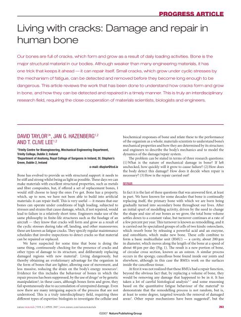
Living with cracks: <strong>Damage</strong> and <strong>repair</strong> in<br />
human bone<br />
Our bones are full of cracks, which form and grow as a result of daily loading activities. Bone is the<br />
major structural material in our bodies. Although weaker than many engineering materials, it has<br />
one trick that keeps it ahead — it can <strong>repair</strong> itself. Small cracks, which grow under cyclic stresses by<br />
the mechanism of fatigue, can be detected and removed before they become long enough to be<br />
dangerous. This article reviews the work that has been done to understand how cracks form and grow<br />
in bone, and how they can be detected and <strong>repair</strong>ed in a timely manner. This is truly an interdisciplinary<br />
research fi eld, requiring the close cooperation of materials scientists, biologists and engineers.<br />
biochemical responses of bone and relate these to the performance<br />
of the organism as a whole; materials scientists to understand bone’s<br />
mechanical properties and how they are determined by its structure;<br />
and engineers to describe the body’s mechanics and to model the<br />
dynamics of the damage/<strong>repair</strong> system.<br />
Th e problem can be stated in terms of three research questions:<br />
(1) What is the nature of mechanical damage in bone? If left<br />
unchecked, how quickly will it grow to cause failure? (2) How does<br />
the body detect this damage? How does it decide when <strong>repair</strong> is<br />
necessary? (3) How is the <strong>repair</strong> carried out?<br />
nature materials | VOL 6 | APRIL 2007 | www.nature.com/naturematerials 263<br />
REPAIR<br />
<strong>In</strong> fact it is the last of these questions that was answered fi rst, at least<br />
in part. We have known for some decades that bone is continually<br />
replacing itself, the primary bone with which we are born being<br />
gradually turned into secondary bone throughout our lives. Aft er<br />
an initial spurt of modelling activity, driven by the need to change<br />
the shape and size of our bones as we grow, the total bone volume<br />
settles down to a constant value, but turnover continues at a rate of<br />
a few percent per year. Th is turnover is known as remodelling, and it<br />
is carried out by specialized groups of cells of two kinds: osteoclasts,<br />
which resorb bone by releasing a powerful acid and an enzyme,<br />
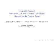
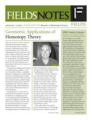
and osteoblasts, which make new bone. Th ese cells combine to<br />
form a basic multicellular unit (BMU) — a cavity, about 200 μm<br />
in diameter, which moves along the length of the bone at a speed of<br />
about 40 μm per day (Fig. 1). Th e result is a new portion of bone,<br />
of circular cross section, known as an osteon. A similar process<br />
occurs in the spongy, cancellous bone found inside our joints and<br />
elsewhere, although in this case the BMUs work on the surfaces<br />
inside the cancellous tissue.<br />
At fi rst it was not realized that these BMUs had a <strong>repair</strong> function,<br />
beyond the obvious fact that, by replacing a volume of bone, they<br />
would be removing any damage that happened to be in it. It has<br />
taken a lot of careful histological analysis 5–7 and some reasoning<br />
based on the quantitative fatigue behaviour of the material 8 to<br />
demonstrate that the remodelling process is not random, but is,<br />
at least to some degree, targeted towards the removal of damaged<br />
areas 3 . Other <strong>repair</strong> mechanisms have been suggested 9 , but the
PROGRESS ARTICLE<br />
200 μm<br />
current paradigm holds that BMUs are the primary way by which<br />
bone maintains its structural integrity.<br />
DAMAGE<br />
New bone Osteoblasts Osteoclasts<br />
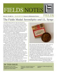
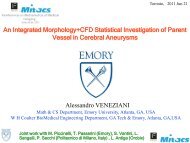
To discuss the nature of damage in bone we fi rst need to say<br />
something of its structure, which is summarized in Fig. 2. At the<br />
macroscopic scale bone comes in two forms. Compact (cortical)<br />
bone is essentially solid material, although spaces for blood<br />
vessels and living cells give it a porosity of about 5%; it makes up<br />
the majority of our long bones. Spongy cancellous bone is found<br />
inside bony structures, especially close to joints. It is essentially the<br />
0 50<br />
Spongy bone<br />
<strong>In</strong>terstitial<br />
lamellae<br />
Lymphatic<br />
vessel<br />
40 μm per day<br />
Figure 1 Bone’s <strong>repair</strong> mechanism. A BMU in which osteoclasts and osteoblasts work<br />
in sequence to eat away old and generate new bone.<br />
Compact bone<br />
same material as compact bone, but arranged in an open network<br />
to create a foam-like structure. At the microscopic scale both<br />
materials are made rather like plywood, from sheets of alternating<br />
lamellae that can be laid fl at, or curved around in circles to<br />
protect blood vessels, forming osteons. <strong>In</strong>side each lamella, at the<br />
ultrastructural level we fi nd fi bres of collagen, a soft , polymeric<br />
material made from long-chain molecules arranged in a triple<br />
helix, and crystals of hydroxyapatite (HA), a hard, brittle mineral<br />
material based on calcium. Th e ultrastructure is highly oriented,<br />
creating a strong anisotropy.<br />
Mechanical loading creates damage; at the microscopic scale<br />
one can see small cracks that take the form of planar ellipses,<br />
typically having a major axis of length 2c = 400 μm oriented<br />
approximately parallel to the bone’s longitudinal axis, and a minor<br />
axis of length 2a = 100 μm. Th e observation of these cracks is<br />
greatly aided by the use of coloured dyes that are able to diff use into<br />
the bone and chemically bind to exposed calcium 10 . Th ese dyes have<br />
been improved in recent years and are now available in diff erent<br />
colours 11 , allowing cracks to be labelled at diff erent times during<br />
an experiment 12 . Th is has yielded a great deal of information about<br />
how damage develops. Microcracks predominate in regions of<br />
compression — which constitutes the principal type of loading in our<br />
long bones — where they experience shear due to their orientation.<br />
<strong>In</strong> addition to these individual, linear cracks, areas called ‘diff use<br />
damage’ 13 are also observed, which contain many small cracks, each<br />
Outer<br />
circumferential<br />
lamellae<br />
Concentric lamellae<br />
Periosteum<br />
264 nature materials | VOL 6 | APRIL 2007 | www.nature.com/naturematerials<br />
Osteon<br />
Blood vessel<br />
Lymphatic vessel<br />
Lacuna<br />
Canaliculi<br />
Spongy<br />
bone<br />
Osteocyte<br />
Periosteal vein<br />
Periosteal artery<br />
Periosteum:<br />
Outer fibrous layer<br />
<strong>In</strong>ner osteogenic layer<br />
Central canal<br />
Perforating canal<br />
Compact bone<br />
Medullary cavity<br />
Figure 2 The structure of bone. Colour schematic reprinted from ref. 66. Copyright (2002) Wiley. Black and white microscope image (scale bar in μm) showing osteocytes<br />
(white dots) linked by processes (fi ne white lines) forming the syncytium. Image reprinted from ref. 54. Copyright (2004) with permission from Elsevier.
of the order of a micrometre in size. Multiple cracks of intermediate<br />
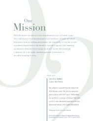
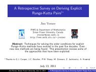
size also occur, forming cross-hatched patterns 14 . Figure 3 shows<br />
examples of these various types of damage.<br />
Histological studies have told us a lot about how this damage<br />
develops: chelating dyes can be administered in vitro and in vivo,<br />
allowing us to monitor the amount and type of damage in a<br />
laboratory specimen of dead bone, or in living animals while <strong>repair</strong><br />
processes are operating. Other techniques are being developed, such<br />
as positron emission tomography 15,16 , and new dyes may allow realtime<br />
imaging of microcracks in the body via scanning by computed<br />
tomography (CT) or magnetic resonance imaging (MRI) 17 .<br />
Currently the study of damage is a very active fi eld; many<br />
experimental studies have been undertaken in recent years, showing,<br />
for example, that damage can be linked to reduced stiff ness 18 , to<br />
regions low in bone cells 19 , to ageing in humans and animals 20–24<br />
and to osteoporosis and its treatment 25 . Microcracking can also be<br />
found in the more calcifi ed regions of cartilage and may be linked<br />
to osteoarthritis 26 . A lot of data is now appearing, along with some<br />
useful computer simulations 27,28 , but currently we lack a clear<br />
theoretical perspective from which to view all this information.<br />
TOUGHNESS AND FATIGUE<br />
Some cracks will be benign whilst others will threaten the<br />
integrity of the structure and so must be <strong>repair</strong>ed: how can we tell<br />
which is which? Th is is the province of fracture mechanics, the<br />
science that studies cracks and how they grow in materials. We<br />
defi ne a material property known as the fracture toughness, K c,<br />
which tells us how tolerant a material is to the presence of cracks<br />
or, to put it another way, by how much a given crack will weaken<br />
the structure.<br />
Like other composite materials, bone achieves a level of<br />
toughness that is much greater than that of its constituents — HA<br />
and collagen — and we have only recently begun to understand<br />
the mechanisms by which this toughness is achieved. Nalla and<br />
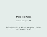
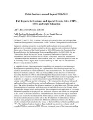
co-workers 29,30 and others 31 showed that K c is not constant but<br />
increases with crack length for cracks of the order of millimetres<br />
(Fig. 4a) — an important fi nding because the cracks in our bones<br />
rarely exceed this length. Nalla et al. 32 postulated that toughness in<br />
bone is achieved through bridging of the crack faces by unbroken<br />
ligaments of material (Fig. 4c), inhibiting further crack growth<br />
in the same way that reinforcing rods protect concrete. However,<br />
there are other possible mechanisms that may contribute, such as<br />
microcracking 32,33 bridging by collagen 34 , and plasticity (which is<br />
the dominant source of toughness in metals). Currey 35 has shown<br />
how toughness and strength are juggled to allow bones to have<br />
special functions, such as the antlers of deer, which require high<br />
toughness and can tolerate low strength in order to get it.<br />
Cracks can also grow very slowly, over long periods of time,<br />
even when their loading conditions are unchanged. Researchers<br />
studying the growth of microcracks 36,37 showed that, although<br />
a crack may initially grow quite quickly, its growth rate oft en<br />
declines to a minimum value, and indeed the crack may stop<br />
growing altogether (Fig. 5). Th is occurs when the crack meets<br />
up with features in the microstructure such as osteons or tubular<br />
canals. An example is shown in Fig. 3b where the left end of the<br />
crack has hit the outside of an osteon. Examination of microcracks<br />
formed in vivo or in vitro shows that most of them never grow<br />
beyond their fi rst osteon 38 .<br />
Th ese investigations have emphasized the importance of<br />
microstructure in preventing crack growth and thereby improving<br />
toughness and fatigue resistance, but ultrastructure is important<br />
too, as it determines the underlying mechanical properties of<br />
the material 39 . High strength, achieved via the HA crystals and<br />
their intimate bonding to the collagen around them, prevents the<br />
PROGRESS ARTICLE<br />
highly stressed material near the crack from failing. High stiff ness<br />
is important too, because it reduces the amount of elastic strain<br />
energy, which is what a crack feeds on when it grows. A full<br />
picture on the role of ultrastructure has yet to emerge, but there<br />
have been several interesting observations published in recent<br />
years, in relation to the eff ects of ageing 29,40,41 , gamma radiation 42<br />
and varying levels of HA 43,44 and water 45 . What all this tells us is<br />
that bone cracking is a multiscale phenomenon that requires a<br />
hierarchical perspective, ranging from the molecular level up to<br />
the scale of the entire bone 46,47 .<br />
COMBINING DAMAGE AND REPAIR<br />
We can think of living bones as a system of continual damage and<br />
<strong>repair</strong>, and this has inspired two very diff erent lines of research.<br />
One line looks at the big picture, seeing bone as an example of a<br />
control system requiring feedback and stability — we will return to<br />
this research below. Th e other aims to understand the mechanisms<br />
by which bone detects the presence of cracks and decides whether<br />
or not to <strong>repair</strong> them. Th is is possibly the most challenging area and<br />
the one in which, despite some important recent work, there is still<br />
a lot to be done. Th e challenge is to understand the link between the<br />
mechanics of cracks and the biology of cellular behaviour. Attention<br />
has focused on the osteocytes — cells that live inside bone, contained<br />
nature materials | VOL 6 | APRIL 2007 | www.nature.com/naturematerials 265<br />
a<br />
b<br />
Figure 3 <strong>Damage</strong> in bone. a, Cancellous bone showing examples of microdamage<br />
revealed by staining with basic fuschin (pink). The arrows indicate (from left to right):<br />
a microcrack (approximately 200 μm long), cross-hatching and diffuse damage<br />
respectively. Reprinted from ref. 23. Copyright (1998) with permission from Elsevier.<br />
b, A microcrack ‘C’ encounters an osteon ‘O’, and begins to grow around its cement<br />
line (dashed line). The microcrack is approximately 100 μm long. Reprinted from<br />
ref. 12. Copyright (2003) with permission from Elsevier.
PROGRESS ARTICLE<br />
a b c<br />
6<br />
<strong>Human</strong> cortical bone, 25 ºC<br />
K (MPa m ½ )<br />
4<br />
2<br />
34–41<br />
years<br />
61–69<br />
years<br />
85–99<br />
years<br />
0<br />
0 2 4<br />
Δa (mm)<br />
6 8<br />
in small cavities and linked to their neighbours by extensions to<br />
the cell known as cellular processes. Th ese processes meet at gap<br />
junctions, forming a network between osteocytes (Fig. 2) and bone<br />
lining cells that is reminiscent of that of the network of neurons<br />
in the brain 48 . Th is network, or ‘syncytium’ seems to be a means<br />
of intercommunication that can be used to detect and control the<br />
amount of damage in the surrounding bone.<br />
Klein-Nulend and co-workers 49,50 have proposed a means<br />
by which BMUs could orient themselves to grow along a bone,<br />
or more specifi cally, parallel to the direction of principal stress.<br />
Fluid is continually fl owing through the network of fi ne channels<br />
that contain the osteocytes, bringing essential nutrients. Using a<br />
computer simulation, they showed that fl ow rates are low in the<br />
region of bone immediately ahead of the BMU cavity 49,50 . Cells<br />
die in this stagnant area, possibly because they don’t receive<br />
enough nutrients, but in the Klein–Nulend model, cell death is<br />
c<br />
ct<br />
deliberate (apoptosis), triggered by a change in the level of nitric<br />
oxide released by cells, which acts as a signal indicating the rate of<br />
fl uid fl ow. Osteoclasts are attracted to the apoptotic cells and so<br />
preferentially eat away the bone in this region.<br />
Th is is a complex and elegant model, with elements of solid<br />
mechanics, fl uid mechanics and biochemistry, and most of its<br />
steps have been demonstrated experimentally. However, it does<br />
not explain how BMUs can detect cracks. Cracks create regions of<br />
both high stress (near their tips) and low stress (along their sides)<br />
that cancel out, so that from a distance, a crack would be invisible<br />
to Klein–Nulend’s BMU. However, cells do become apoptotic near<br />
cracks and regions of diff use damage 51–53 , where the osteocyte<br />
network becomes disrupted 54 , possibly due to local changes in<br />
fl uid fl ow. Other researchers have suggested various mechanisms<br />
by which dead or apoptotic cells could attract osteoclasts and thus<br />
initiate <strong>repair</strong> 55 .<br />
It has also been suggested that a crack could be detected as a<br />
result of the damage it causes to the network of cellular processes<br />
in canaliculi (small channels) 10 . We have recently investigated<br />
this mechanism in detail, using fracture-mechanics theory to<br />
show that cellular processes can be cut by the shear motions that<br />
occur across crack faces; the action is similar to that of a pair of<br />
scissors. We showed that the number of damaged processes varied<br />
signifi cantly with both crack size and applied stress 56 . Microscopy<br />
demonstrated that these processes are indeed cut if the crack-face<br />
displacements are high enough 57 ; current work is directed towards<br />
identifying the substances that are released by the broken processes<br />
and understanding how they are detected by osteoclast precursors,<br />
stimulating the formation of a BMU.<br />
Other researchers don’t worry about the mechanisms, but try<br />
to make models of the entire system, assuming that the amount<br />
of damage in a region of bone increases with time at a rate that<br />
depends on the local stress or strain: strain-energy density is<br />
oft en used as a parameter 27,58 because it is a simple scalar quantity.<br />
Repair is modelled as a decrease in the amount of damage, and<br />
a stable equilibrium is characterized by a balance between the<br />
rates of damage and <strong>repair</strong> 59 . Th is so-called ‘damage mechanics’<br />
approach can be used in several ways. First, it can demonstrate<br />
the evolutionary advantages of living in a state of constant <strong>repair</strong>,<br />
allowing lighter, more delicate, gracile bones to be used than<br />
would be possible if damage never occurred 2 . Second, it can be<br />
266 nature materials | VOL 6 | APRIL 2007 | www.nature.com/naturematerials<br />
mc<br />
Haversian canals<br />
20 μm<br />
Uncracked<br />
ligaments<br />
Figure 4 <strong>In</strong>vestigations of crack behaviour. a, Results from Nalla et al. 29 showing that crack extension (Δa) in the millimetre range requires increasing stress intensity (K ),<br />
especially in bone from younger people. Copyright (2004) with permission from Elsevier. Possible toughening mechanisms include: b, microcracks ‘mc’ near the tip ‘ct’ of<br />
the crack ‘c’ (optical microscope image of bone stained with chelating dye; scale bar = 50 μm. From Zarrinkalam et al. 67 , www.tandf.co.uk/journals); c, uncracked ligaments<br />
behind the crack tip (image obtained by Nalla et al. 30 using microtomography. Copyright (2005) with permission from Elsevier.<br />
dc/dN (mm per cycle)<br />
5 × 10 –5<br />
1 × 10 –5<br />
5 × 10 –6<br />
1 × 10 –6<br />
5 × 10 –7<br />
40<br />
B2<br />
C1<br />
B1<br />
A1<br />
B3<br />
D1<br />
D2<br />
60 80 100 200<br />
2c (μm)<br />
Figure 5 Data from Akkus and Rimnac 37 showing the growth characteristics of various<br />
individual cracks (labelled A1, A2, B1, B2, B3, C1, D1, D2). The cyclic growth rate dc/dN<br />
(change in crack half-length c with number of cycles N) decelerates with crack length<br />
(2c) initially, often passing through a minimum value as the crack encounters a barrier<br />
to growth such as an osteon, as illustrated in Fig. 3b. Both scales are logarithmic.<br />
Reprinted from ref. 37. Copyright (2001), with permission from Elsevier.<br />
A2<br />
Crack
implemented in the form of a computer simulation, interfacing<br />
with other soft ware such as fi nite element analysis: an entire bone<br />
can be modelled in all the complexity of its geometry and loading<br />
patterns. Surgical interventions such as the introduction of a hipjoint<br />
implant can be investigated 58 , as can the eff ects of diseases<br />
such as osteoporosis. An advantage of this top-down approach is<br />
that we can include another important aspect of living bone, which<br />
is called functional adaptation. If bones are subjected to high levels<br />
of stress, or high numbers of cycles, to compensate they become<br />
thicker and stronger. Likewise, bones that are underused, due to<br />
periods of incapacity, become thin, porous and weak: this is known<br />
as ‘disuse osteoporosis’ and is one of the causes of fractures in<br />
elderly patients. Th e mechanisms by which bone recognizes these<br />
changes in its mechanical environment, and initiates adaptations,<br />
are just as poorly understood as the mechanisms by which it<br />
achieves <strong>repair</strong>, but it is very tempting to think that the two<br />
phenomena must be connected. For an engineer, the easiest way<br />
to optimise a design is to make the structure, use it, and see what<br />
happens, modifying your design in the light of experience. Perhaps<br />
bone is continually monitoring its state of damage and adjusting<br />
its architecture accordingly, the ideal state being not to have no<br />
damage, but to have just enough damage that can be <strong>repair</strong>ed as<br />
you go along, ensuring a fi nite but acceptable risk of failure.<br />
Two theoretical models have attempted to bridge the gap<br />
between the systems approach and the mechanistic approach.<br />
Martin has created computer simulations in which the number<br />
density of microcracks, and their <strong>repair</strong> by BMUs, are included 60,61 ;<br />
this model has been used to investigate many interesting cases,<br />
such as the eff ects of anti-osteoporosis drugs that work by<br />
suppressing osteoclast activity 62 . It takes account of the fact that<br />
BMUs contribute to a bone’s porosity, tending to increase stress<br />
and, with it, the rate of damage accumulation, creating a potential<br />
instability. Th is negative eff ect of the <strong>repair</strong>/remodelling process has<br />
been highlighted by some workers who believe that osteoporosis is<br />
caused by unnecessarily high levels of remodelling 63 . We developed<br />
a simulation that includes the growth-rate characteristics of short<br />
cracks (as shown in Fig. 5) as well as BMU behaviour, modelling<br />
every individual crack and BMU in a volume of bone using<br />
stochastic variables 28,64 .<br />
Th ese models are capable of realistic predictions of the stable,<br />
equilibrium state of damage and <strong>repair</strong>, and of the instabilities that<br />
would lead to adaptation (for example, extra bone deposition) and,<br />
in extreme cases, to stress fractures. Th ey can also be used to study<br />
other phenomena, such as the eff ect of bone size in diff erent animals 65<br />
and evolutionary development 2 . Large, multivariable simulations of<br />
this kind are diffi cult to set up and run stably, but they are probably<br />
an essential tool if we want to understand the behaviour of natural<br />
systems in all their complexity.<br />
CONCLUDING REMARKS<br />
Th e fascinating study of bone damage and <strong>repair</strong> is clearly an activity<br />
that requires a multidisciplinary approach on many diff erent fronts.<br />
<strong>In</strong> recent years we have seen some excellent progress. For example,<br />
we are now able to describe and quantify mechanical damage<br />
and to elucidate the mechanisms of cracking and toughness. A<br />
deeper understanding of these issues should not be too far away.<br />
Some more diffi cult parts of the puzzle are those that concern<br />
the responses of the living system. Although some pathways have<br />
been described whereby cells can detect strain and damage and<br />
initiate biochemical responses, the whole area of cell signalling<br />
is both complex and fascinating. Phenomena such as <strong>repair</strong> and<br />
remodelling are controlled not by a single biological system but by<br />
a variety of systems, working in parallel and probably interacting<br />
with each other. A major practical problem, in this as in other areas<br />
PROGRESS ARTICLE<br />
of bioengineering, is the provision of accurate experimental data.<br />
<strong>In</strong> many cases there is no alternative to the animal experiment, but<br />
great progress is being made in the development of cell cultures,<br />
which allow aspects of the living system to be studied in vitro.<br />
Th e act of assembling the various pieces of this jigsaw is the<br />
business of those who make simulations of the whole system. With<br />
increasing computer power this activity is becoming more feasible:<br />
control theory could contribute greatly here. <strong>In</strong> the past we tended<br />
to view the human body as a complex engine — nowadays we<br />
can describe the workings of many parts of this engine, but we<br />
don’t know how it is controlled. <strong>In</strong> the past few decades we have<br />
seen many examples of the life sciences and the physical sciences<br />
coming together to solve particular problems. <strong>In</strong> studying how<br />
bone becomes damaged and how it <strong>repair</strong>s itself, we are required<br />
to ask some very big questions; the answers to these questions have<br />
repercussions far beyond the particular topic that we are studying.<br />
doi:10.1038/nmat1866<br />
References<br />
1. Martin, R. B. & Burr, D. B. A hypothetical mechanism for ths timulation of osteonal remodelling by<br />
fatigue damage. J. Biomech. 15, 137–139 (1982).<br />
2. Martin, R. B. Fatigue damage, remodeling, and the minimization of skeletal weight. J. Th eor. Biol.<br />
220, 271–276 (2003).<br />
3. Burr, D. B. Targeted and nontargeted remodeling. Bone 30, 2–4 (2002).<br />
4. Tsuji, K. et al. BMP2 activity, although dispensible for bone formation, is required for the initiation<br />
of fracture healing. Nature Genet. 38, 1424–1429 (2006).<br />
5. Burr, D. B. & Martin, B. R. Calculating the probability that microcracks initiate resorption spaces.<br />
J. Biomech. 26, 613–616 (1993).<br />
6. Mori, S. & Burr, D. B. <strong>In</strong>creased intracortical remodeling following fatigue damage. Bone<br />
14, 103–109 (1993).<br />
7. Lee, T. C., Staines, A. & Taylor, D. Bone adaptation to load: Microdamage as a stimulus for bone<br />
remodelling. J. Anat. 201, 437–446 (2002).<br />
8. Taylor, D. Fatigue of bone and bones: An analysis based on stressed volume. J. Orthop. Res.<br />
16, 163–169 (1998).<br />
9. Boyde, A. Th e real response of bone to exercise. J. Anat. 203, 173–189 (2003).<br />
10. Frost, H. M. Presence of microscopic cracks in vivo in bone. Henry Ford Hosp. Med. Bull.<br />
8, 25–35 (1960).<br />
11. O’Brien, F., Taylor, D. & Lee, T. C. An improved labelling technique for monitoring microcrack<br />
growth in bone. J. Biomech. 35, 523–526 (2002).<br />
12. O’Brien, F. J., Taylor, D. & Lee, T. C. Microcrack accumulation at diff erent intervals during fatigue<br />
testing of compact bone. J. Biomech. 36, 973–980 (2003).<br />
13. Vashishth, D. et al. <strong>In</strong> vivo diff use damage in human vertebral trabecular bone. Bone<br />
26, 147–152 (2000).<br />
14. Choi, K. & Goldstein, S. A. A comparison of the fatigue behaviour of human trabecular and cortical<br />
bone tissue. J. Biomech. 25, 1371–1381 (1992).<br />
15. Li, J., Miller, M. A., Hutchins, G. D. & Burr, D. B. Imaging bone microdamage in vivo with positron<br />
emission tomography. Bone 37, 819–824 (2005).<br />
16. Silva, M. J. et al. <strong>In</strong> vivo skeletal imaging of 18F-fl uoride with positron emission tomography reveals<br />
damage- and time-dependent responses to fatigue loading in the rat ulna. Bone 39, 229–236 (2006).<br />
17. Lee, T. C. et al. Detecting microdamage in bone. J. Anat. 203, 161–172 (2003).<br />
18. Kawahara, D. & Murakami, T. Evaluation for mechanism of diff use damage in cortical bone.<br />
Trans. Jpn Soc. Mech. Eng. A 71, 936–943 (2005).<br />
19. Qiu, S., Rao, D. S., Fyhrie, D. P., Palnitkar, S. & Parfi tt, A. M. Th e morphological association between<br />
microcracks and osteocyte lacunae in human cortical bone. Bone 37, 10–15 (2005).<br />
20. Sobelman, O. S. et al. Do microcracks decrease or increase fatigue resistance in cortical bone?<br />
J. Biomech. 37, 1295–1303 (2004).<br />
21. Frank, J. D. et al. Aging and accumulation of microdamage in canine bone. Bone 30, 201–206 (2002).<br />
22. Schaffl er, M. B., Choi, K. & Milgrom, C. Aging and matrix microdamage accumulation in human<br />
compact bone. Bone 17, 521–525 (1995).<br />
23. Fazzalari, N. L., Forwood, M. R., Smith, K., Manthey, B. A. & Herreen, P. Assessment of cancellous<br />
bone quality in severe osteoarthrosis: Bone mineral density, mechanics, and microdamage. Bone<br />
22, 381–388 (1998).<br />
24. Mori, S., Harruff , R., Ambrosius, W. & Burr, D. B. Trabecular bone volume and microdamage<br />
accumulation in the femoral heads of women with and without femoral neck fractures. Bone<br />
21, 521–526 (1997).<br />
25. Dai, R. C., Liao, E. Y., Yang, C., Wu, X. P. & Jiang, Y. Microcracks: An alternative index for evaluating<br />
bone biomechanical quality. J. Bone Miner. Metab. 22, 215–223 (2004).<br />
26. Muir, P. et al. Role of endochondral ossifi cation of articular cartilage and functional adaptation of the<br />
subchondral plate in the development of fatigue microcracking of joints. Bone 38, 342–349 (2006).<br />
27. Huiskes, R., Rulmerman, R., Van Lenthe, G. H. & Janssen, J. D. Eff ects of mechanical forces on<br />
maintenance and adaptation of form in trabecular bone. Nature 405, 704–706 (2000).<br />
28. Taylor, D. & Lee, T. C. Microdamage and mechanical behaviour: Predicting failure and remodelling<br />
in compact bone. J. Anat. 203, 203–211 (2003).<br />
29. Nalla, R. K., Kruzic, J. J., Kinney, J. H. & Ritchie, R. O. Eff ect of aging on the toughness of human<br />
cortical bone: Evaluation by R-curves. Bone 35, 1240–1246 (2004).<br />
30. Nalla, R. K., Kruzic, J. J., Kinney, J. H. & Ritchie, R. O. Mechanistic aspects of fracture and R-curve<br />
behavior in human cortical bone. Biomaterials 26, 217–231 (2005).<br />
nature materials | VOL 6 | APRIL 2007 | www.nature.com/naturematerials 267
PROGRESS ARTICLE<br />
31. Malik, C. L., Stover, S. M., Martin, R. B. & Gibeling, J. C. Equine cortical bone exhibits rising<br />
R-curve fracture mechanics. J. Biomech. 36, 191–198 (2003).<br />
32. Nalla, R. K., lken, J. S., Kinney, J. H. & Ritchie, R. O. Fracture in human cortical bone: Local fracture<br />
criteria and toughening mechanisms. J. Biomech. 38, 1517–1525 (2005).<br />
33. Vashishth, D., Tanner, K. E. & Bonfi eld, W. Experimental validation of a microcracking-based<br />
toughening mechanism for cortical bone. J. Biomech. 36, 121–124 (2003).<br />
34. Yeni, Y. N. & Fyhrie, D. P. A rate-dependent microcrack-bridging model that can explain the strain<br />
rate dependency of cortical bone apparent yield strength. J. Biomech. 36, 1343–1353 (2003).<br />
35. Currey, J. D. Mechanical properties of bone tissues with greatly diff ering functions. J. Biomech.<br />
12, 313–319 (1979).<br />
36. Hazenberg, J. G., Taylor, D. & Lee, T. C. Mechanisms of short crack growth at constant stress in<br />
bone. Biomaterials 27, 2114–2122 (2006).<br />
37. Akkus, O. & Rimnac, C. M. Cortical bone tissue resists fatigue fracture by deceleration and arrest of<br />
microcrack growth. J. Biomech. 34, 757–764 (2001).<br />
38. O’Brien, F. J., Taylor, D. & Lee, T. C. Th e eff ect of bone microstructure on the initiation and growth<br />
of microcracks. J. Orthop. Res. 23, 475–480 (2005).<br />
39. Nyman, J. S., Reyes, M. & Wang, X. Eff ect of ultrastructural changes on the toughness of bone.<br />
Micron 36, 566–582 (2005).<br />
40. Wang, X., Xiaoe, L. I., Shen, X. & Agrawal, C. M. Age-related changes of noncalcifi ed collagen in<br />
human cortical bone. Ann. Biomed. Eng. 31, 1365–1371 (2003).<br />
41. Zioupos, P., Currey, J. D. & Hamer, A. J. Th e role of collagen in the declining mechanical properties<br />
of aging human cortical bone. J. Biomed. Mater. Res. 45, 108–116 (1999).<br />
42. Mitchell, E. J., Stawarz, A. M., Kayacan, R. & Rimnac, C. M. Th e eff ect of gamma radiation<br />
sterilization on the fatigue crack propagation resistance of human cortical bone. J. Bone Joint Surg. A<br />
86, 2648–2657 (2004).<br />
43. Currey, J. D. Eff ects of diff erences in mineralization on the mechanical properties of bone.<br />
Phil. Trans. R. Soc. Lond. B 304, 509–518 (1984).<br />
44. Wasserman, N., Yerramshetty, J. & Akkus, O. Microcracks colocalize within highly mineralized<br />
regions of cortical bone tissue. Eur. J. Morphol. 42, 43–51 (2005).<br />
45. Nyman, J. S. et al. Th e infl uence of water removal on the strength and toughness of cortical bone.<br />
J. Biomech. 39, 931–938 (2006).<br />
46. Akkus, O., Yeni, Y. N. & Wasserman, N. Fracture mechanics of cortical bone tissue: A hierarchical<br />
perspective. Crit. Rev. Biomed. Eng. 32, 379–425 (2004).<br />
47. Vashishth, D. Age-dependent biomechanical modifi cations in bone. Crit. Rev. Eukar. Gene<br />
15, 343–357 (2005).<br />
48. Turner, C. H., Robling, A. G., Duncan, R. L. & Burr, D. B. Do bone cells behave like a neuronal<br />
network? Calcifi ed Tissue <strong>In</strong>t. 70, 435–442 (2002).<br />
49. Burger, E. H., Klein-Nulend, J. & Smit, T. H. Strain-derived canalicular fl uid fl ow regulates<br />
osteoclast activity in a remodelling osteon — a proposal. J. Biomech. 36, 1453–1459 (2003).<br />
50. Klein-Nulend, J., Bacabac, R. G. & Mullender, M. G. Mechanobiology of bone tissue. Pathol. Biol.<br />
53, 576–580 (2005).<br />
51. Bentolila, V. et al. <strong>In</strong>tracortical remodeling in adult rat long bones aft er fatigue loading. Bone<br />
23, 275–281 (1998).<br />
52. Noble, B., Alini, M. & Richards, R. G. Bone microdamage and cell apoptosis. Eur. Cells Mater.<br />
6, 46–55 (2003).<br />
53. Noble, B. S. et al. Mechanical loading: Biphasic osteocyte survival and targeting of osteoclasts for<br />
bone destruction in rat cortical bone. Am. J. Physiol. Cell Ph. 284, C934–C943 (2003).<br />
54. Colopy, S. A. et al. Response of the osteocyte syncytium adjacent to and distant from linear<br />
microcracks during adaptation to cyclic fatigue loading. Bone 35, 881–891 (2004).<br />
55. Verborgt, O., Gibson, G. J. & Schaffl er, M. B. Loss of osteocyte integrity in association with<br />
microdamage and bone remodeling aft er fatigue in vivo. J. Bone Min. Res. 15, 60–67 (2000).<br />
56. Taylor, D., Hazenberg, J. G. & Lee, T. C. Th e cellular transducer in damage-stimulated bone<br />
remodelling: A theoretical investigation using fracture mechanics. J. Th eor. Biol.<br />
225, 65–75 (2003).<br />
57. Hazenberg, J. G., Freeley, M., Foran, E., Lee, T. C. & Taylor, D. Microdamage: a cell transducing<br />
mechanism based on ruptured osteocyte processes. J. Biomech. 39, 2096–2103 (2006).<br />
58. Huiskes, R. in Non-cemented Total Hip Arthroplasty (ed. Fitzgerald, R.) 283–302<br />
(Raven, New York, 1988).<br />
59. Prendergast, P. J. & Taylor, D. Prediction of bone adaptation using damage accumulation. J. Biomech.<br />
27, 1067–1076 (1994).<br />
60. Martin, B. Mathematical model for <strong>repair</strong> of fatigue damage and stress fracture in osteonal bone.<br />
J. Orthop. Res. 13, 309–316 (1995).<br />
61. Hazelwood, S. J., Martin, R. B., Rashid, M. M. & Rodrigo, J. J. A mechanistic model for internal bone<br />
remodeling exhibits diff erent dynamic responses in disuse and overload. J. Biomech. 34, 299–308 (2001).<br />
62. Nyman, J. S., Yeh, O. C., Hazelwood, S. J. & Martin, R. B. A theoretical analysis of long-term<br />
bisphosphonate eff ects on trabecular bone volume and microdamage. Bone 35, 296–305 (2004).<br />
63. Heaney, R. P. Is the paradigm shift ing? Bone 33, 457–465 (2003).<br />
64. Taylor, D. & Lee, T. C. A crack growth model for the simulation of fatigue in bone. <strong>In</strong>t. J. Fatigue<br />
25, 387–395 (2003).<br />
65. Taylor, D. Scaling eff ects in the fatigue strength of bones from diff erent animals. J. Th eor. Biol.<br />
206, 299–306 (2000).<br />
66. Tortora, G. J. Principles of <strong>Human</strong> Anatomy (Wiley, New York, 2002).<br />
67. Zarrinkalam, K. H., Kuliwaba, J. S., Martin, R. B., Wallwork, M. A. B. & Fazzalari, N. L. New insights<br />
into the propagation of fatigue damage in cortical bone using confocal microscopy and chelating<br />
fl uorochromes. Eur. J. Morph. 42, 81–90 (2005).<br />
Competing fi nancial interests<br />
Th e authors declare no competing fi nancial interests.<br />
268 nature materials | VOL 6 | APRIL 2007 | www.nature.com/naturematerials