
Create successful ePaper yourself
Turn your PDF publications into a flip-book with our unique Google optimized e-Paper software.
This sample chapter is for review purposes only. Copyright © The <strong>Goodheart</strong>-<strong>Willcox</strong> Co., Inc. All rights reserved.<br />
164 Introduction to Anatomy and Physiology<br />
Vesicles containing<br />
acetylcholine<br />
Axon terminal<br />
Acetylcholine receptor sites<br />
Muscle fiber<br />
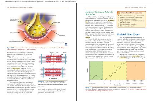
Figure 5.6 The neuromuscular junction, the site at which nerve impulses are transmitted to muscle. Which<br />
chemical labeled in this drawing is a neurotransmitter?<br />
the actin fi laments over the myosin fi laments,<br />
resulting in a contraction of the sarcomere<br />
(Figure 5.7).<br />
What causes the actin fi laments to slide<br />
over the myosin fi laments? Notice in Figure 5.7<br />
that the myosin fi laments are encircled by small<br />
protrusions called heads. When the sarcomere<br />
is activated by an action potential, these heads<br />
attach to receptor sites on the actin fi laments,<br />
forming cross bridges. The cross bridges<br />
contract, pulling the actin fi laments toward the<br />
center of the sarcomere. During the process of<br />
sarcomere contraction, these cross bridges attach,<br />
pull, and release multiple times. The Ca++ ions<br />
released with the arrival of the action potential<br />
enable the attachments of the myosin heads to<br />
the actin fi laments.<br />
The neuromuscular system has the ability<br />
to produce slow, gentle movements as well as<br />
fast, forceful movements. This ability to produce<br />
different kinds of movements and force variance<br />
is accomplished by regulating the number and<br />
frequency of action potentials. Only a small<br />
number of action potentials are needed for<br />
Axon<br />
Myosin Actin Heads<br />
A. Relaxed<br />
Synaptic cleft<br />
B. Contracted<br />
Sarcolemma<br />
Diffusing acetylcholine<br />
Figure 5.7 The sarcomere is the contractile unit of<br />
muscle. When the muscle is stimulated, the actin<br />
filaments slide together, producing contraction of the<br />
sarcomere.<br />
slow, gentle movements, while fast or forceful<br />
movements require a large number of action<br />
potentials, released rapidly.<br />
Label Art<br />
Maximum Tension and Return to<br />
Relaxation<br />
When receiving an action potential, a given<br />
motor unit always develops maximum tension, a<br />
physiological principle known as the all-or-none<br />
law. But because each whole muscle includes<br />
multiple motor units, simultaneous activation<br />
of many motor units is required for the muscle<br />
to develop maximum tension. The diagram in<br />
Figure 5.8 displays the relationship between<br />
number and frequency of action potentials and<br />
the development of tension in the muscle. With<br />
high-frequency stimulation, the muscle develops<br />
a sustained, maximal level of tension called<br />
tetanus.<br />
Almost all skeletal motor units develop<br />
tension in a twitch-like fashion, generating<br />
maximum tension very briefl y and then<br />
immediately relaxing. After the action potential<br />
has traveled the length of the muscle fi ber,<br />
chemical processes return the fi ber to its resting<br />
state. Sodium ions diffuse back out of the cell<br />
into the interstitial fl uid, and calcium ions<br />
return to storage sites within the cell. The actin<br />
fi laments slide back to their original positions as<br />
the cross bridges release them, and the muscle<br />
fi ber returns to a state of relaxation.<br />
Tension<br />
0<br />
<strong>Chapter</strong> 5 The Muscular System 165<br />
Check Your Understanding<br />
1. What structures make up a motor unit?<br />
2. Describe the neuromuscular junction.<br />
3. What is an action potential?<br />
4. Why do you want a small motor unit for<br />
fi ne motor skills?<br />
5. Explain the all-or-none law.<br />
6. How do muscles relax?<br />
Skeletal Fiber Types<br />
Why are some athletes especially good at<br />
events or tasks that require endurance, whereas<br />
others excel at activities that require explosive<br />
strength or speed? The answer may have<br />
something to do with the ways in which these<br />
individuals train, but that is only a small part of<br />
the explanation. In fact, a big part of why certain<br />
people are better at particular activities and<br />
sports may relate to the characteristics of their<br />
skeletal muscle fi bers.<br />
Skeletal muscle fi bers may be divided into<br />
two umbrella categories—slow-twitch (Type I)<br />
and fast-twitch (Type II). As the names suggest,<br />
A B C<br />
S = Stimulus S S S S S S S S S S S S S S S S S S S S S<br />
Time (msec)<br />
Figure 5.8 Tension developed in a muscle in response to a single stimulus (A), in response to repetitive<br />
stimulation (B), and in response to high-frequency stimulation, or tetanus (C). Do you think this graph represents<br />
the activation of one or many motor units? Explain.