A Molecular Analysis Of Prion Protein Expression In Alzheimer's ...
A Molecular Analysis Of Prion Protein Expression In Alzheimer's ...
A Molecular Analysis Of Prion Protein Expression In Alzheimer's ...

Create successful ePaper yourself
Turn your PDF publications into a flip-book with our unique Google optimized e-Paper software.
Copyright © 2004 by MJM<br />
MJM 2004 8: 7-14 7<br />
ORIGINAL ARTICLE<br />
A <strong>Molecular</strong> <strong>Analysis</strong> <strong>Of</strong> <strong>Prion</strong> <strong>Protein</strong> <strong>Expression</strong><br />
<strong>In</strong> <strong>Alzheimer's</strong> Disease<br />
Alisdair McNeill*, BSc, MedSci<br />
ABSTRACT: <strong>In</strong> <strong>Prion</strong> Diseases, misfolding of neuronal prion protein (PrPC) to a pathogenic isomer<br />
(PrPSC) is associated with neuronal death. Previous pathological studies have demonstrated increased<br />
i m m u n o r e a c t i v i t y o f P r P C a t Ab plaques in <strong>Alzheimer's</strong> Disease, and it has been suggested that this<br />
either reflects a role for PrPC in the neuronal response to stress or is a feature of the neuropathogenesis<br />
of atypical subtypes of <strong>Alzheimer's</strong> disease. <strong>In</strong> this paper we utilised western blotting to examine the<br />
molecular characteristics of PrP in frozen Hippocampal tissue from 7 cases of <strong>Alzheimer's</strong> Disease in<br />
which prion protein expression was demonstrated by immunohistochemistry, before using Restriction<br />
Fragment Length Polymorphism (RFLP) methodology to define the genotype of the codon 129<br />
polymorphism of PRNP in each case. We observed PrP accumulating as globular structures at A<br />
plaques, and within ependymal cells lining the lateral ventricle. Immunohistochemistry also showed<br />
that PrPC and Superoxide dismutase-1 where deposited in a similar pattern at Ab plaques. Western<br />
blotting revealed that PrP in <strong>Alzheimer's</strong> disease is composed of the same 208-residue peptide expressed<br />
in non-diseased brain. Quantitative western blot analysis demonstrated increased levels of PrPC in a<br />
short duration case of <strong>Alzheimer's</strong> Disease, while, in the remaining cases, levels of PrPC decreased in<br />
parallel with increasing disease duration and decreasing brain mass. RFLP genotyping revealed that<br />
all codon 129 genotypes (M/M, M/V, V/V) were represented in our study cohort. Our data suggest that<br />
increased levels of PrPC may account for PrP immunoreactivity at plaques in <strong>Alzheimer's</strong> disease, and<br />
that PrP deposition is not restricted to certain atypical subtypes of <strong>Alzheimer's</strong> disease.<br />
KEY WORDS: <strong>Alzheimer's</strong>, <strong>Prion</strong> protein, Immunohistochemistry, Western Blotting<br />
I N T R O D U C T I O N<br />
<strong>In</strong> the <strong>Prion</strong> diseases (Creutzfeldt-Jakob Disease,<br />
Kuru, Gerstmann-Straussler-Scheinker Syndrome and<br />
Fatal Familial <strong>In</strong>somnia) a post-translational<br />
conformational change in neuronal prion protein<br />
(PrPC) to a β-sheet rich isomer termed PrPSC is<br />
associated with neuronal death (1). It is proposed that<br />
this process induces neurodegeneration either via<br />
production of neurotoxic PrPSC, or by ablation of the<br />
physiological function of PrPC (1).<br />
The physiological role of PrPC remains unresolved,<br />
however, studies of PrP null mice have shown that it<br />
* To whom correspondence should be addressed: 11 Broomieknowe<br />
Park, Bonnyrigg, Midlothian, EH19 2JB<br />
email: s9809172@sms.ed.ac.uk<br />
is not essential for viability and various investigators<br />
have suggested that it may have a function in sleep<br />
regulation (2), cell adhesion (3) or Purkinje cell<br />
viability (4). More recently, in vitro studies showing<br />
that PrP-/- neurons are excessively vulnerable to<br />
oxidative stress, and that PrPC has a superoxide<br />
dismutase-1 (SOD-1) like activity (5,6), have lead to<br />
the proposal that PrPC might have a role in the<br />
cellular response to oxidative stress. It is suggested<br />
that ablation of this anti-oxidant function of PrPC<br />
might be associated with neurodegeneration in <strong>Prion</strong><br />
disease (6).<br />
<strong>In</strong> <strong>Alzheimer's</strong> disease (AD) the neuronal β-<br />
Amyloid Precursor <strong>Protein</strong> (β-APP) is aberrantly<br />
processed, causing extracellular deposition of its
8 McGill Journal of Medicine<br />
2004<br />
Table 1. Autopsy Material Used For Immunohistochemistry and Western Blotting<br />
Case Number Age/Sex Diagnosis Disease Duration (y) Cause of Death<br />
1 67/F AD N/A Pneumonia<br />
2 69/M AD 15 Pneumonia<br />
3 66/F AD 3 Pneumonia<br />
4 73/F AD 4 Pneumonia<br />
5 42/F AD 4 Pneumonia<br />
6 80/F AD 3 Pneumonia<br />
7 60/M AD 2 Pneumonia<br />
8 82/F AD N/A Pneumonia<br />
9 64/M AD 11 Pneumonia<br />
10 90/F AD N/A Pneumonia<br />
11 25/M TBI N/A TBI<br />
12 66/F Old <strong>In</strong>farcts,SAH N/A Haemorrhage<br />
13 32/M Cardiomyopathy N/A Cardiomyopathy<br />
14 22/F Myocarditis N/A Myocarditis<br />
transmembrane domain. This peptide is designated<br />
Aβ and aggregates into neurotoxic Aβ plaques.<br />
Previous pathological studies have demonstrated that<br />
PrPC deposition occurs at Aβ plaques in <strong>Alzheimer's</strong><br />
disease (7,8). Voigtlander and colleagues (7)<br />
postulated that this phenomenon might reflect a role<br />
for PrPC in the neuronal response to oxidative stress<br />
induced by reactive oxygen species (ROS) produced<br />
by the Aβ-peptide. However, as PRNP mutations<br />
have been reported in familial AD kindreds (9), and<br />
PRNP codon 129 valine homozygosity increases risk<br />
of early onset AD in patients with a family history of<br />
AD (10), it is also possible PrPC immunostaining is a<br />
feature of atypical neuropathogenesis in certain<br />
subtypes of AD (11).<br />
<strong>In</strong> this study we sought to investigate whether PrP<br />
deposition at Aβ plaques in <strong>Alzheimer's</strong> disease is a<br />
general phenomenon, possibly representing an antioxidant<br />
function of PrPC, or restricted to certain<br />
atypical cases. To this end, we utilised<br />
immunohistochemistry to analyse PrP deposition in<br />
paraffin embedded Hippocampal tissue from 9<br />
sporadic and 1 familial case of <strong>Alzheimer's</strong> disease.<br />
We hypothesised that if PrP deposition at Aβ plaques<br />
was restricted to subtypes of <strong>Alzheimer's</strong> disease then<br />
immunostaining would only be seen in some of the<br />
<strong>Alzheimer's</strong> cases. Furthermore, the influence of the<br />
codon 129 polymorphism of PRNP was examined by<br />
restriction fragment length polymorphism (RFLP)<br />
genotyping of 7 cases of AD in which PrPC<br />
expression had been demonstrated. We then<br />
performed immunohistochemistry to examine the<br />
expression of the anti-oxidant enzyme SOD-1, which<br />
is known to be induced by oxidative stress in AD,<br />
hypothesising that an anti-oxidant PrPC would be<br />
deposited in a similar pattern to SOD-1 in AD brain.<br />
Western blotting was then utilised to investigate the<br />
hypotheses that PrP immunostaining of Aβ plaques<br />
reflects either increased levels of PrP or the presence<br />
of a bioactive PrP which is structurally distinct from,<br />
and more immunoreactive than PrPC.<br />
AD is the most common cause of dementia in the<br />
Western World (around 20% of those age 65 or over<br />
are affected) (12), and is the fourth leading cause of<br />
death (12). With our ageing population, AD is set to<br />
cause an increasing burden of ill health and healthcare<br />
costs. Current treatments for AD are only partly<br />
effective, and there is no cure or proven preventative<br />
strategy (12). Studying the neuropathological<br />
changes associated with Aβ plaque formation will<br />
increase our understanding of the processes leading to<br />
neuronal death, and hopefully suggest new<br />
therapeutic targets.<br />
M E T H O D S<br />
C a s e s f o r s t u d y<br />
Formalin-fixed, paraffin-embedded Hippocampal<br />
tissue from 10 cases of <strong>Alzheimer's</strong> disease (age range<br />
42 - 90 years, mean 69.3 years, SD 13 years) and 3<br />
neuropsychologically normal controls (age range 25 -<br />
66 years, mean 40 years old, SD 23 years) were<br />
selected for study (Table 1). The cases were from the<br />
pathology archives of the Western General Hospital,<br />
Edinburgh. Cases were randomly selected from lists<br />
of cases with the desired diagnosis.<br />
I m m u n o h i s t o c h e m i s t r y ( I H C )<br />
For IHC analysis 5 µM sections were cut from fixed<br />
brain tissue blocks in all cases. Sections were<br />
mounted on glass slides (Superfrost Plus; BDH,<br />
Poole, UK), dewaxed, rehydrated and endogenous<br />
peroxidases quenched in 3% hydrogen peroxide in<br />
methanol for 30 minutes. The primary antibodies,<br />
dilutions and antigen retrieval methods used in this
Vol. 8 No. 1<br />
<strong>Prion</strong> <strong>Expression</strong> in Alzheimer’s Disease 9<br />
Table 2. Primary Antibodies Used For Immuhistochemistry<br />
Antibody Antigen Source <strong>In</strong>cubation Antigen<br />
Period<br />
Retrieval<br />
[conc.]<br />
6F/3D (Aß) DAKO 30 min FA. MW.<br />
[1:100]<br />
AT8 (Tau) Autogen 30 min None<br />
[1:3000]<br />
Ab1 (Cu/Zn Bioquots Overnight FA. MW<br />
SOD) [1:1000]<br />
3F4 (PrP) DAKO 60 min FA. M<br />
FA = 5 minutes formic acid,<br />
MW = 15 min microwaving in citric acid.<br />
study are summarised in Table 2. For PrP IHC,<br />
antigen retrieval was performed by microwaving in<br />
citrate buffer (7mM (pH 6.0)) for 30 minutes at 450<br />
W. Sections were then washed in Tris-buffered saline<br />
(TBS; 20 mM Tris-HCl (pH 7.6)) before incubation in<br />
20% normal rabbit serum in TBS for 30 minutes.<br />
Sections of hippocampus were then exposed to the<br />
anti-PrP monoclonal antibody 3F4 (Dako, Glostrup,<br />
Denmark), diluted in 20% normal rabbit serum in<br />
TBS, for one hour. After primary antibody<br />
incubation, sections were repeatedly rinsed in TBS<br />
before being incubated with biotinylated rabbit antimouse<br />
antibody (SAPU, Edinburgh, UK) diluted<br />
1:200 in normal rabbit serum in TBS, for 30 minutes<br />
at room temperature. The staining pattern was then<br />
visualised using the Catalysed Signal Amplification<br />
system (CSA, Dako). Sections were then<br />
counterstained in haematoxylin before mounting in<br />
DPX (BDH, Poole, UK).<br />
Adjacent tissue sections were exposed to antibodies<br />
raised against the Aβ peptide (6F/3D, Dako, Glostrup,<br />
Denmark), tau (AT8, Autogen) and SOD-1 (AB1,<br />
Bioquote). Sections were prepared as for PrP IHC.<br />
No antigen retrieval was performed for tau. For the<br />
Aβ and tau antibodies the staining pattern was<br />
visualised using a standard avidin-biotin method<br />
(ABC Kit, Vector), with 3,3'-diaminobenzidine<br />
tetrahydro-chloride (Vector, CA, USA) as the<br />
chromogen. For SOD-1 the CSA system (DAKO)<br />
was used. For all antibodies controls were performed<br />
where the primary antibody was omitted. The stained<br />
sections were examined microscopically, and images<br />
captured using a Roper Scientific Photometrics<br />
camera and Image Pro 4.5 Cool Snap Software.<br />
W e s t e r n B l o t t i n g<br />
For Western blotting, frozen tissue was available<br />
for cases 1 - 7 of <strong>Alzheimer's</strong> disease. Hippocampal<br />
tissue (50 - 100 mg) was dissected out from each case<br />
and homogenised in 10 volumes (w/v) of protein<br />
extraction buffer. The homogenate was then<br />
centrifuged at 3,000 RPM at 40C for 10 minutes, and<br />
the supernatant and pellet stored at -700C. The<br />
homogenised protein was normalised using a Bio-Rad<br />
protein assay with bovine serum albumin as a<br />
standard, to ensure each sample contained the same<br />
quantity of protein. Glycosylation of PrP was<br />
examined by incubating homogenate with PNGase-F,<br />
following manufacturer's protocols, and <strong>Protein</strong>ase K<br />
sensitivity determined by treating homogenate with<br />
<strong>Protein</strong>ase K.<br />
<strong>Protein</strong> was separated by SDS-PAGE, using 12%<br />
Resolving Gel and 4% Stacking Gel, on a Hoeffer<br />
Scientific <strong>In</strong>struments "Mighty Small" gel running<br />
apparatus. Gels were stained with Coomasie blue to<br />
allow visualisation of protein expression profiles, or<br />
transferred to a Hybond-P (Amersham) membrane.<br />
The transfer was performed at 100V, for 60 minutes<br />
in a Bio-Rad transfer apparatus. The membrane was<br />
first soaked in methanol for one minute, and then<br />
rinsed in distilled water. Both gel and membrane<br />
were then immersed in transfer buffer for 5 minutes<br />
before being sandwiched between sheets of blotting<br />
paper, and placed in the transfer apparatus filled with<br />
transfer buffer along with a cooling block.<br />
After transfer, the membrane was blocked in 5%<br />
fat-free milk solution overnight at 40C. After<br />
blocking, the membrane was rinsed in TBS-T and<br />
incubated with primary antibody (3F4, Dako,<br />
Glostrup, Denmark) for 60 minutes. The membrane<br />
was then rinsed in TBS-T, before incubation with<br />
anti-mouse-HRP antibody. Detection was then<br />
performed using an Amersham ECL-plus<br />
Chemiluminescence kit, following manufacturer's<br />
instructions.<br />
R e s t r i c t i o n F r a g m e n t L e n g t h P o l y m o r p h i s m<br />
G e n o t y p i n g O f C o d o n 1 2 9 O f P R N P<br />
DNA was extracted from the seven AD cases where<br />
frozen tissue was available, using the GenElute DNA<br />
extraction kit (Sigma). The eluted DNA was used as<br />
a template in a polymerase chain reaction (PCR)<br />
performed to amplify a 932 bp fragment of PRNP,<br />
which included the open reading frame (ORF). The<br />
ORF was amplified from 2 µL of eluted DNA using<br />
TAQ polymerase (Sigma), following manufacturer's<br />
instructions, with a reaction cycle consisting of 35<br />
cycles of one minute at 940C followed by one cycle<br />
of one minute at 500C and one cycle of one minute at<br />
720C on a Qiagen Omnigene PCR machine.<br />
Codon 129 may be either the sequence (C)AUG(G),
10 McGill Journal of Medicine<br />
2004<br />
A<br />
B<br />
*<br />
*<br />
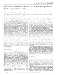
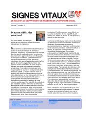
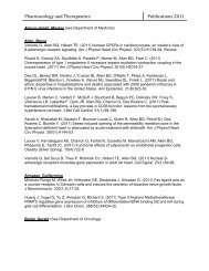
Figures 1. Pattern of PrP Deposition in AD and DS. A. Diffuse Aß plaque (*) colocalising with: B. Globular PrP deposits (*), C. PrP<br />
immunoreactive ependyma (arrowed).<br />
C<br />
spatial pattern to the amyloid delineated by the anti-<br />
Aβ antibody. No neurofibrillary tangles were<br />
observed to immunostain for PrP. No neuronal<br />
somatic, microglial or astroglial immunoreactivity for<br />
PrP was detected. However, ependymal cells<br />
(morphologically identified) lining the lateral<br />
ventricle were observed to accumulate PrP (Fig.1).<br />
No PrP immunostaining was demonstrated in the<br />
young control cases, though PrP immunoreactive<br />
plaques were seen in an aged control (66 years).<br />
encoding methionine, or (C)GUG(G), encoding<br />
valine. Restriction endonuclease Bsa AI cleaves at<br />
CG, and thus does not cleave a 932 bp PCR product<br />
coding methionine at position 129, but cleaves a PCR<br />
product coding valine into a 465 bp and a 467 bp<br />
fragment. Bsa AI was thus incubated with the<br />
purified PCR product, and the cleavage products<br />
separated on an agarose gel (0.8 g agarose in 50 ml<br />
TAE) in order to differentiate PCR products which<br />
had / had not been cleaved, and hence genotype each<br />
individual.<br />
R E S U L T S<br />
I m m u n o h i s t o c h e m i s t r y<br />
<strong>In</strong> each case of <strong>Alzheimer's</strong> disease (n=10) PrP<br />
immunohistochemistry demonstrated PrP being<br />
expressed as globular deposits, which delineated<br />
structures resembling the Aβ plaques of <strong>Alzheimer's</strong><br />
disease (Fig.1). Immunostaining also showed<br />
deposition of SOD-1 at Aβ plaques. Examining<br />
adjacent sections revealed that the PrP and SOD-1<br />
immunostained plaques were distributed in a similar<br />
W e s t e r n B l o t t i n g<br />
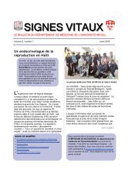
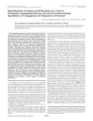
SDS-PAGE produced two bands labelled by 3F4 on<br />
Western blot, corresponding to the 34 kDa<br />
diglycosylated and 28 kDa monoglycosylated PrP<br />
glycoforms found in non-diseased brain (Fig.2).<br />
Treatment with <strong>Protein</strong>ase K (1 µg/ml) digested PrP<br />
present in the <strong>Alzheimer's</strong> disease samples completely<br />
(data not shown). Thus, the PrP expressed in<br />
<strong>Alzheimer's</strong> disease is not PrPSC. Deglycosylation<br />
with PNGase-F produced a single species of PrP with<br />
a molecular weight corresponding to the<br />
unglycosylated glycoform of PrP, indicating that PrP<br />
expressed in <strong>Alzheimer's</strong> disease is composed of the<br />
208 residue peptide expressed in non-diseased brain<br />
tissue (data not shown).<br />
S e m i - Q u a n t i t a t i v e W e s t e r n B l o t t i n g<br />
The optical density of the PrP bands produced by<br />
Western blotting on an autorad film differed between<br />
each case of <strong>Alzheimer's</strong> disease and normal brain<br />
(Fig.2). Optical density reflects the amount of protein<br />
detected by primary antibody in a sample, and, as<br />
total protein in each sample had been normalised, the<br />
differing optical densities indicate different levels of<br />
PrP in each case. With cases one and two displaying<br />
absent PrP, cases three - six reduced levels of PrP
Vol. 8 No. 1<br />
<strong>Prion</strong> <strong>Expression</strong> in Alzheimer’s Disease 11<br />
1 2 3 4 5 6 7 Normal<br />
15 y 3 y 4 y 4y 3 y 2y N/A<br />
8 14 1132 1320 1091 1306 1594 1400-1500<br />
Figure 2. Quantitative Western Blot showing differential expression of PrP in AD and normal hippocampus. Lanes 1 - 7 contain AD tissue with<br />
normal tissue in lane 8. The case number, in bold, and disease duration, in italics, are tabulated above each lane with brain mass (g) below each<br />
lane. Blank = data unavailable. (The lanes in this image were rearranged from the original blot, so as to be in numerical order by case number.)<br />
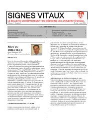
1 2 3 4 5 6 7<br />
M/V V/V M/M M/M M/M M/V M/M<br />
Figure 3. RFLP analysis of codon 129 polymorphism. Lanes 1 - 3 contain control DNA, Lane 1 shows the banding pattern of methionine<br />
homozygotes, lane 2 a valine homozygote, and 3 a heterozygote. The lanes to the right of the markers contain AD DNA, the case number is<br />
above each lane, and genotype below. (The lanes in this image were rearranged from the original blot, so as to be in numerical order by case<br />
number.)<br />
compared with normal brain, and case seven elevated<br />
levels of PrP compared with control (Table 3).<br />
Normal brain consisted of hippocampal tissue from a<br />
cerebrum that did not manifest histopathological<br />
abnormalities.<br />
C o d o n 1 2 9 G e n o t y p i n g<br />
Restriction Fragment Length Polymorphism<br />
genotyping demonstrated that all codon 129 (MM,<br />
MV, VV) genotypes were represented in the study<br />
cohort (Fig.3). Cases one and six being heterozygous,<br />
cases three - five and seven being methionine<br />
homozygotes, and case two a valine homozygote.<br />
The ratio of genotypes in the study cohort is<br />
comparable with that in the general population (1).<br />
D I S C U S S I O N<br />
The biological function of PrPC remains<br />
incompletely understood. However, in vitro studies<br />
demonstrating a SOD-like anti-oxidant activity of<br />
PrPC have lead to the hypothesis that cellular prion<br />
protein may help to protect neurons from oxidative<br />
damage (5,6). <strong>In</strong> this study we sought in vivo<br />
evidence of a SOD-activity of PrPC by investigating<br />
the proposal that PrP deposition at Aβ plaques in AD<br />
reflects an anti-oxidant function of PrPC. For PrP
12 McGill Journal of Medicine 2004<br />
Table3. Optical Densities <strong>Of</strong> <strong>Prion</strong> <strong>Protein</strong> Bands<br />
C a s e Diglycosylated Monoglycosylated Unglycosylated<br />
1 0 0 0<br />
2 0 0 0<br />
3 0.68 0.18 0<br />
4 0.67 0.11 0<br />
5 0.34 0.08 0<br />
6 0.42 0.5 0<br />
7 1.92 1.71 0<br />
Normal 1.29 0.61 0<br />
expression in AD to give in vivo evidence supporting<br />
an anti-oxidant function of PrPC we believed that the<br />
following conditions should be met: 1. PrP at Aβ<br />
plaques must not be PrPSC (the PrP isoform found in<br />
<strong>Prion</strong> disease), 2. the pattern of PrPC deposition<br />
should resemble that of proteins (SOD-1) known to be<br />
upregulated by neuronal stress in AD, and 3. PrP<br />
expression at A β plaques must not be restricted to rare<br />
subtypes of AD. Our Western blot analysis fulfils the<br />
first criterion, as it demonstrated PrP in AD to have<br />
the characteristics of PrPC (mono- and diglycosylated<br />
glycoforms present, 208 amino acids long, proteinase<br />
K sensitive) (1). Evidence relating to points 2 and 3<br />
is discussed below.<br />
The topology of PrP deposition demonstrated by<br />
our immunohistochemical study is compatible with an<br />
anti-oxidant function for PrPC. Firstly, assuming<br />
PrPC at A plaques is accumulating within neuronal<br />
processes, PrPC is probably being expressed within<br />
neurons exposed to oxidative stress, since Aβ plaques<br />
are associated with oxidative damage to adjacent<br />
neuronal processes (13). Moreover, the fact that the<br />
pattern of PrPC expression resembled that of SOD-1<br />
supports the contention that PrPC is upregulated by<br />
neuronal stress in AD, since neuronal SOD-1 has been<br />
shown to be induced by ROS generated by the Aβpeptide<br />
(14,15). The significance of ependymal<br />
accumulation of PrP is uncertain. However, it<br />
correlates with in situ hybridisation studies showing<br />
PrP mRNA expression in ependymal cells (16), and<br />
may reflect a response to ROS within cerebrospinal<br />
fluid in AD brain.<br />
An alternative explanation for PrP immunostaining<br />
of A plaques in AD is that it is a feature of atypical<br />
neuropathogenesis in a rare subtype of AD (11,17,18).<br />
Evidence for this proposal comes from a pathological<br />
study that demonstrated PrP deposition at Aβ plaques<br />
using an antibody which was claimed to be specific<br />
for PrPSC (11). Furthermore, research demonstrating<br />
that valine homozygosity at codon 129 of PRNP<br />
increases risk of early onset AD (10), combined with<br />
reports of PRNP mutations in an FAD kindred (9),<br />
lends credence to the proposal that there may be a<br />
subtype of AD in which prion protein plays a<br />
pathogenic role. However, several factors argue<br />
against the PrP immunostaining in our study<br />
representing atypical neuropathogenesis. Firstly, our<br />
study contained sporadic (n=9) and familial (n=1)<br />
cases with a wide range of ages (42 - 90 years old),<br />
making it likely our cohort represented a spectrum of<br />
AD cases. Furthermore, though it was not possible to<br />
screen for PRNP mutations in our study, RFLP<br />
genotyping demonstrated that all PRNP codon 129<br />
genotypes were represented in our cohort - suggesting<br />
that this polymorphism does not influence PrPC<br />
upregulation in AD. Taken together, our results argue<br />
that PrPC immunostaining in our study is not part of<br />
the primary pathologic processes in AD, but<br />
secondary to deposition of Aβ-peptide, possibly<br />
reflecting an anti-oxidant action of PrPC.<br />
<strong>In</strong> this study we hypothesised that immunohistochemical<br />
detection of PrP in <strong>Alzheimer's</strong> disease<br />
might reflect either elevated levels of PrP<br />
accumulating at amyloid plaques, or the synthesis of a<br />
putative, bioactive PrP which was structurally distinct<br />
from, and more immunoreactive than, PrPC. Western<br />
blotting provided no evidence of a novel PrP<br />
expressed at Aβ plaques, as the molecular<br />
characteristics of PrP in <strong>Alzheimer's</strong> disease are<br />
indistinguishable from PrPC present in normal brain.<br />
However, semi-quantitative Western blot analysis<br />
showing differential levels of PrP expression in each<br />
case of <strong>Alzheimer's</strong> disease compared with nondiseased<br />
brain (Figure 2); suggestive of increased<br />
levels of PrPC in <strong>Alzheimer's</strong> disease. With reference<br />
to Figure 2, it can be seen that levels of PrP detected<br />
on Western blot decrease in parallel with increasing<br />
disease duration and neuronal loss (evidenced by<br />
decreasing brain mass). Thus, case 7 (normal brain<br />
mass) has supra-normal levels of PrP on densitometry,<br />
while cases 1 - 6 (brain mass: 814 - 1306g) have<br />
decreased levels of PrP compared with nonpathological<br />
brain. This suggests that there may be<br />
increased levels of PrP within individual cells, but<br />
with advancing disease duration, and increasing<br />
neuronal loss (12), there is a subnormal quantity of<br />
PrP at the tissue level. A caveat must be attached to<br />
this conclusion. Western blotting measures protein<br />
levels in a volume of tissue, but does not enable<br />
identification of what cells/structures contain the<br />
protein. Though, as PrPC is considered to be<br />
predominantly expressed in neurons (1), it is<br />
reasonable to suggest that the increased PrPC levels<br />
reflect neuronal PrPC. Even if elevated levels of<br />
PrPC in AD tissue were due to upregulation in nonneuronal<br />
cells, it would not invalidate the proposed
Vol. 8 No. 1<br />
<strong>Prion</strong> <strong>Expression</strong> in Alzheimer’s Disease 13<br />
SOD-like function of PrPC as the protein may also<br />
play an anti-oxidant role in glia and other nonneuronal<br />
elements. Though our study methodology<br />
had limitations, other techniques would not be able to<br />
provide proof of increased PrP protein or mRNA. For<br />
example, in situ hybridisation would not be helpful as<br />
a cell can express mRNA without protein, while<br />
Northern blotting would not permit examination of<br />
mRNA expression at the level of single cells.<br />
Attempting to demonstrate increased levels of PrPC<br />
protein in AD by counting cells has been performed<br />
previously, without definitive conclusions (7).<br />
This apparent increase in levels of PrPC observed<br />
in AD tissue could be accounted for by two nonmutually<br />
exclusive mechanisms. Firstly, increased<br />
production of PrPC, via either elevated mRNA<br />
transcription/translation or reduced PrPC degradation,<br />
could result in accumulation of PrPC within neuronal<br />
processes at Aβ plaques. Alternatively, the PrP<br />
visualised at Aβ plaques may represent extracellular<br />
protein that has "nucleated" around fibrillar Aβpeptides<br />
within amyloid plaques (18). The study of<br />
Voigtlander and colleagues (7), which demonstrated<br />
significantly increased numbers of PrPC<br />
immunostained neurons in hippocampal tissue from<br />
AD brains, lends support to the former hypothesis, as<br />
it suggests increased synthesis of PrPC in AD.<br />
Nevertheless, before PrP nucleation at Aβ-amyloid<br />
can be discounted, appropriate immunoprecipitation<br />
studies must be performed.<br />
It is important to note the limitations that the small<br />
sample size places on our study. Firstly, if there is a<br />
subgroup of AD that does not express PrPC then our<br />
study may have been too small to detect this. <strong>In</strong><br />
corollary, our small sample of PrPC expressing AD<br />
cases may represent a minority of cases which<br />
immunostain for PrPC. The factors noted above<br />
militate against these suggestions. Moreover, in our<br />
small study it was not possible to examine if the<br />
codon 129 genotype had any influence on factors such<br />
as extent of Aβ plaque deposition or any influence on<br />
the clinical course of AD (disease duration etc). The<br />
relationship between disease duration and PrPC levels<br />
on semi-quantitative western blot also needs to be<br />
confirmed by examining a large cohort of cases with<br />
well defined short, medium and long disease<br />
durations.<br />
<strong>In</strong> summary, this study has expanded upon previous<br />
reports of PrP expression in AD by showing that PrPC<br />
deposition at A plaques parallels deposition of SOD-<br />
1, by demonstrating that PrP expression is not limited<br />
to rare subtypes of AD and by providing evidence to<br />
suggest that levels of PrPC are elevated in AD.<br />
However, in concluding, certain factors, which might<br />
potentially confound our results, must be<br />
acknowledged. For example, since deposition of<br />
certain synaptic vesicle proteins at Aβ plaques occurs<br />
due to cytoskeletal disruption (20), PrPC<br />
immunostaining in <strong>Alzheimer's</strong> disease might merely<br />
represent a passive result of disrupted protein<br />
trafficking and endocytosis rather than an induced<br />
response to stress. This deranged protein trafficking<br />
and metabolism in moribund <strong>Alzheimer's</strong> neurons<br />
might, in theory, also account for the differential<br />
levels of PrP detected on Western blot in the cases<br />
examined. Nonetheless, it seems reasonable to<br />
conclude that accumulation of PrPC at Aβ plaques in<br />
AD reflects a physiological function of PrPC. The<br />
precise biological activity of PrPC in AD brain cannot<br />
be elucidated from our results. However, given in<br />
vitro evidence of an anti-oxidant activity of PrPC<br />
(5,6), and the similar immunostaining patterns of<br />
PrPC and SOD-1, an anti-oxidant enzyme which is<br />
induced by ROS produced by A -peptide (14,15), it is<br />
possible PrPC expression in AD represents a role for<br />
the protein in the neuronal response to oxidative<br />
stress. <strong>In</strong> this respect, it is interesting to note that<br />
some in vitro studies have suggested PrPC might act<br />
as a co-factor for SOD-1 (6). If PrPC is a<br />
neuroprotective anti-oxidant protein which is<br />
upregulated by oxidative stress then it could have an<br />
important role in multiple neurodegenerative<br />
disorders, possibly acting to slow neuronal death and<br />
clinical deterioration. This hypothesis is supported by<br />
the study of Kovacs and associates (21), which<br />
showed increased neuronal immunoreactivity for<br />
PrPC in Multiple Systems Atrophy, Progressive<br />
Supranuclear Palsy and Motor Neuron Disease, as<br />
well as by research indicating that the codon 129<br />
polymorphism predisposes to early onset AD and<br />
modulates rate of cognitive decline in established AD<br />
(10). The hypothesis that PrPC has a neuroprotective<br />
function has obvious implications for <strong>Prion</strong> disease,<br />
as oxidative stress induced by PrPSC will upregulate<br />
PrPC and provide more substrate for toxic PrPSC<br />
production, which, in turn, leaves neurons vulnerable<br />
to damage by depriving them of a protective protein.<br />
Should the foregoing proposals prove correct then<br />
anti-oxidant therapy may provide a valuable<br />
neuroprotective strategy for <strong>Prion</strong> disease.<br />
R E F E R E N C E S<br />
1. Ironside JW. <strong>Prion</strong> Diseases <strong>In</strong> Man. Journal of Pathology<br />
186(3):227-234; 1998.<br />
2. Collinge J, Whittington MA, Sidle KC, et al. <strong>Prion</strong> <strong>Protein</strong> Is<br />
Necessary For Normal Synaptic Function. Nature<br />
370(6487):295-297; 1994.<br />
3. Herms J, Tings T, Gall S, et al. Evidence <strong>Of</strong> Presynaptic<br />
Location And Function <strong>Of</strong> The <strong>Prion</strong> <strong>Protein</strong>. Journal of<br />
Neuroscience 19(20): 8866-8875; 1999.
14 McGill Journal of Medicine 2004<br />
4. Moore RC, Lee IY, Silverman GL, et al. Ataxia <strong>In</strong> <strong>Prion</strong><br />
<strong>Protein</strong> Deficient Mice Is Associated With Upregulation <strong>Of</strong><br />
The Novel PrP - Like <strong>Protein</strong> Doppel. Journal of <strong>Molecular</strong><br />
Biology 292(4):797-817; 1999.<br />
5. Brown DR, Besinger A. <strong>Prion</strong> <strong>Protein</strong> And Superoxide<br />
Dismutase Activity. Biochemical Journal 334:423-429; 1998.<br />
6. Brown DR, Schulz - Schaeffer WJ, Schmidt B, Kretzschmar<br />
HA. <strong>Prion</strong> <strong>Protein</strong> Deficient Cells Show Altered Response To<br />
Oxidative Stress Due To Decreased SOD -1 Activity.<br />
Experimental Neurology 146(1):104-112; 1997.<br />
7. Voigtlander T, Kloppel S, Birner P, et al. Marked <strong>In</strong>crease <strong>Of</strong><br />
Neuronal <strong>Prion</strong> <strong>Protein</strong> Immunoreactivity <strong>In</strong> <strong>Alzheimer's</strong><br />
Disease And Human <strong>Prion</strong> Diseases. Acta Neuropathologica<br />
101(5):417-423; 2001.<br />
8. Ferrer I, Blanco R, Carmona M, et al. <strong>Prion</strong> <strong>Protein</strong><br />
<strong>Expression</strong> <strong>In</strong> Senile Plaques <strong>In</strong> <strong>Alzheimer's</strong> Disease. Acta<br />
Neuropathologica 101(1):49-56; 2001.<br />
9. Perry RT, Go RCP, Harrell LE, et al. SSCP <strong>Analysis</strong> And<br />
Sequencing <strong>Of</strong> The <strong>Prion</strong> <strong>Protein</strong> Gene Detects Two 24 bp<br />
Deletions <strong>In</strong> An Atypical <strong>Alzheimer's</strong> Disease Family.<br />
American Journal of Medical Genetics 60(1):12-18; 1995.<br />
10. Casadei VM, Ferri C, Calabrese E, et al. <strong>Prion</strong> <strong>Protein</strong> Gene<br />
Polymorphism And <strong>Alzheimer's</strong> Disease: One Modulatory<br />
Trait of Cognitive Decline? Journal of Neurology,<br />
Neurosurgery and Psychiatry 71(2):279-280; 2001<br />
11. Leuba G, Saini K, Savioz A, Charnay Y. Early Onset Familial<br />
Alzheimer Disease With Co-existing -Amyloid And <strong>Prion</strong><br />
Pathology. Journal of the American Medical Association<br />
283(13):756; 2000.<br />
12. Graham DI, Lantos PL. Ageing And Dementia. <strong>In</strong>: Graham<br />
DI, Lantos PL, eds. Greenfield's Neuropathology. London:<br />
Arnold, 1997.<br />
13. Lyras L, Cairns NJ, Jenner A, Jenner P, Halliwell B. An<br />
Assessment <strong>Of</strong> Oxidative Damage To <strong>Protein</strong>s, Lipids, And<br />
DNA <strong>In</strong> Brain From Patients With <strong>Alzheimer's</strong> Disease.<br />
Journal of Neurochemistry 68(5):2061-2069; 1997.<br />
14. Yatin SM, Aksenova M, Aksenov M, et al. Temporal relations<br />
among amyloid beta-peptide-induced free-radical oxidative<br />
stress, neuronal toxicity, and neuronal defensive responses.<br />
Journal of <strong>Molecular</strong> Neuroscience 11(3):183-97; 1998.<br />
15. Celsi F, Ferri A, Casciati A, et al. Overexpression of SOD-1<br />
Protects Against -Amyloid Peptide Toxicity: Effect of<br />
Oestrogen and Copper Chelators. Neurochemistry<br />
<strong>In</strong>ternational 44(1):25-33; 2004.<br />
16. McLennan NF, Rennison KA, Bell JE, Ironside JW. <strong>In</strong> Situ<br />
Hybridisation <strong>Analysis</strong> <strong>Of</strong> PrP mRNA <strong>In</strong> Human CNS Tissue.<br />
Neuropath. Applied Neurobiology 27(5):373-383; 2001.<br />
17. DeArmond SJ. <strong>Alzheimer's</strong> Disease and Creutzfeldt-Jakob<br />
Disease: Overlap <strong>Of</strong> Pathogenic Mechanisms. Currents<br />
Opinions in Neurology 6(6):872-881; 1993.<br />
18. Hainfellner JA, Wanschitz J, Jellinger K, Liberski PP,<br />
Gullotta F, Budka H. Coexistance <strong>Of</strong> Alzheimer-Type<br />
Neuropathology <strong>In</strong> Creutzfeldt Jakob Disease. Acta<br />
Neuropathologica 96(2):116-122; 1998.<br />
19. Forloni G, Tagliavini F, Bugiani O, Salmona M. Amyloid<br />
in <strong>Alzheimer's</strong> Disease And <strong>Prion</strong> Related Encephalopathies:<br />
Studies With Synthetic Peptides. Progress in Neurobiology<br />
49(4):287-315; 1996.<br />
20. Dickson DW. The Pathogenesis of Senile Plaques. Journal of<br />
Neuropathology and Experimental Neurology 56(4):321-339;<br />
1997.<br />
21. Kovacs GG, Zerbi P, Voigtlander T, et al. The <strong>Prion</strong> <strong>Protein</strong> <strong>In</strong><br />
Human Neurodegenerative Disorders. Neuroscience Letters<br />
329(3):269-272; 2002.<br />
A l i s d a i r M c N e i l l is currently a House <strong>Of</strong>ficer at Gartnavel General Hospital and the Western <strong>In</strong>firmary Glasgow. His<br />
research interests are stroke, prion disease and neurologic education. He hopes to work as a Clinical Academic in<br />
Neurosciences