Basics of Wound Care - GHDonline
Basics of Wound Care - GHDonline
Basics of Wound Care - GHDonline

Create successful ePaper yourself
Turn your PDF publications into a flip-book with our unique Google optimized e-Paper software.
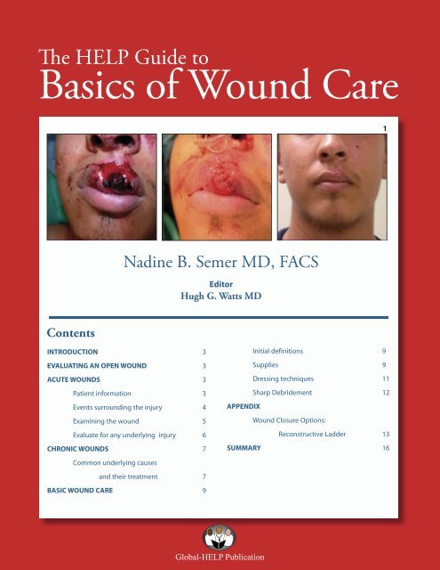
The HELP Guide to<br />
<strong>Basics</strong> <strong>of</strong> <strong>Wound</strong> <strong>Care</strong><br />
1<br />
Nadine B. Semer MD, FACS<br />
Editor<br />
Hugh G. Watts MD<br />
Contents<br />
INTRODUCTION 3<br />
EVALUATING AN OPEN WOUND 3<br />
ACUTE WOUNDS 3<br />
Patient information 3<br />
Events surrounding the injury 4<br />
Examining the wound 5<br />
Evaluate for any underlying injury 6<br />
CHRONIC WOUNDS 7<br />
Common underlying causes<br />
and their treatment 7<br />
BASIC WOUND CARE 9<br />
Initial definitions 9<br />
Supplies 9<br />
Dressing techniques 11<br />
Sharp Debridement 12<br />
APPENDIX<br />
<strong>Wound</strong> Closure Options:<br />
Reconstructive Ladder 13<br />
SUMMARY 16<br />
Global-HELP Publication
2<br />
Author: Nadine B. Semer MD, FACS<br />
Nadine is an experienced plastic and reconstructive<br />
surgeon in Los Angeles. She has volunteered her<br />
skills performing reconstructive surgery and teaching<br />
wound care techniques in rural Africa. She is the<br />
author <strong>of</strong> Practical Plastic Surgery for Nonsurgeons- a<br />
book targeted to health care providers working in the<br />
developing world.<br />
Editor: Hugh G. Watts MD<br />
Dr. Watts is a pediatric orthopedic surgeon with a keen<br />
interest in health problems from a global perspective.<br />
Born in Japan, educated in Canada and the USA, he<br />
worked for two years in Afghanistan, and five years in<br />
Saudi Arabia. He has lectured extensively in the U.S.A.,<br />
Europe, the Middle East, Central and South America,<br />
He is on the staff <strong>of</strong> the Shriner’s Hosp for Children in<br />
Los Angeles and is Clinical Pr<strong>of</strong>essor <strong>of</strong> Orthopedic<br />
Surgery at UCLA.<br />
Publisher’s Information<br />
Published by<br />
Global-HELP Organization<br />
Copyright<br />
Copyright, Global-HELP.Organization, 2003<br />
This is a Global-HELP publication<br />
Visit our web site at global-help.org
3<br />
Introduction<br />
A common treatment provided by rural health care<br />
providers is wound care. Whether it is a fresh acute<br />
wound or a chronic longstanding wound the basic<br />
treatment is the same, only your initial approach to<br />
the wound changes.<br />
This HELP publication will present the basic information<br />
for evaluating both acute and chronic wounds<br />
and then providing the appropriate care.<br />
This publication does NOT cover Life-Threatening<br />
Injuries.<br />
Evaluating an open wound<br />
First Question: Is it Life-Threatening? A life-threatening<br />
wound would be, for example, a chest wound- where<br />
the underlying lung could be injured, an abdominal<br />
wound that could involve the contents <strong>of</strong> the abdominal<br />
cavity, a wound with very active bleeding, or a<br />
neck wound, which could compromise the patient’s<br />
airway.<br />
This publication does not cover Life-Threatening<br />
<strong>Wound</strong>s (refer to publications on Major Trauma <strong>Care</strong><br />
for this information).<br />
Second question: Is it a fresh (acute) or longstanding<br />
(chronic) wound?<br />
For the purposes <strong>of</strong> this HELP guide, an acute wound<br />
is one that is less than a few days old, whereas a<br />
chronic wound is one that has been present more<br />
than a week.<br />
Acute wounds<br />
When evaluating a patient that comes to you with an<br />
acute wound, the first step is to control blood loss and<br />
evaluate the need for other emergency procedures.<br />
This information is beyond this HELP guide. This HELP<br />
guide describes treatment for a basic, non-life threatening<br />
wound- one without any chance for significant<br />
internal injury (i.e., pneumothorax, intra-abdominal,<br />
etc.).<br />
Start by obtaining a thorough history- both pertaining<br />
to the patient and the events surrounding the injury.<br />
Patient information<br />
A. Tetanus immunization status and what to do:<br />
(see chart next page).<br />
B. Bleeding at time <strong>of</strong> injury:<br />
Even if the patient is not actively bleeding at the time<br />
<strong>of</strong> evaluation, the history <strong>of</strong> bright red, pulsatile bleeding<br />
at the time <strong>of</strong> injury should alert you to the possibility<br />
<strong>of</strong> underlying arterial injury. Check pulses at<br />
and distal to the injury to be sure circulation is intact.<br />
Formal exploration in the operating room by a qualified<br />
clinician is usually warranted if you suspect an<br />
artery has been injured.<br />
Acute <strong>Wound</strong><br />
Chronic <strong>Wound</strong><br />
C. Medical illnesses:<br />
Malnutrition, diabetes, HIV are a few common medical<br />
illnesses that can make a patient more prone to infection<br />
and warrant closer follow-up care. Encourage<br />
patients with diabetes to keep their blood sugar well<br />
controlled. Encourage adequate protein/vitamin<br />
intake vital for normal healing.<br />
D. Smoking history:<br />
The use <strong>of</strong> tobacco products dramatically slows<br />
the`healing process. Strongly encourage your patients<br />
to quit smoking immediately.
4<br />
Tetanus immunization status and what to do:<br />
Years since<br />
immunization<br />
<strong>Wound</strong>*<br />
< 5 Clean or Tetanus<br />
prone<br />
no further immunization needed<br />
>5 5 Tetanus prone Tetanus toxoid 0.5ml IM<br />
>10 Clean or tetanus<br />
prone<br />
Tetanus toxoid 0.5ml IM<br />
Never immunized clean Start full tetanus toxoid immunization regimen (0.5ml IM; repeat in 4<br />
wks and 6-12 mo after second injection).<br />
Never immunized Tetanus prone Start full tetanus toxoid immunization regimen (0.5ml IM;<br />
repeat in 4 wks and 6-12 mo after second injection).<br />
Human tetanus immunoglobulin 250 U, deep IM- not in the same area<br />
as the toxoid shot.<br />
* see tetanus-prone wounds page 5<br />
** a thorough cleansing <strong>of</strong> the wound is indicated for all wounds<br />
Events surrounding the injury<br />
A. Timing <strong>of</strong> injury: when did the injury occur?<br />
If less than 6 hours between injury and evaluation,<br />
the wound can usually be sutured closed. If more<br />
than 6 hours have passed, the wound should not be<br />
closed due to high infection risk. EXCEPTION: due to<br />
cosmetic concerns and because the face has an excellent<br />
blood supply, face wounds may be closed even 24<br />
hours after injury.<br />
B. Nature <strong>of</strong> Injury:<br />
Nature <strong>of</strong> Injury<br />
Animal bite<br />
Human bite<br />
Crush injury- example leg<br />
rolled over by a car tire,<br />
hand caught in a press<br />
Dirty wounds- covered<br />
with grass, dirt, etc.<br />
Notes<br />
Cat bites penetrate deeper<br />
than other animals (dogs for<br />
example) and especially on<br />
the hand <strong>of</strong>ten enter deep<br />
joints- associated with a high<br />
infection rate. Be aggressive in<br />
cleaning the wound and treating<br />
with antibiotics.<br />
Especially to hand, high risk<br />
for infection. Be aggressive in<br />
cleaning the wound and treating<br />
with antibiotics. Use antibiotics<br />
that will treat anerobic<br />
bacteria present in the human<br />
mouth.<br />
There is <strong>of</strong>ten more underlying<br />
damage than you may initially<br />
think. Don’t be fooled if the<br />
skin looks uninjured- the<br />
muscle may be severely<br />
damaged.<br />
Will need thorough debridement<br />
and removal <strong>of</strong> foreign<br />
material.
C. Tetanus-prone wound- definitions:<br />
<strong>Wound</strong> information Is tetanus-prone Is not tetanus<br />
prone<br />
Time since injury > 6 hours 1cm < 1cm<br />
Mechanism <strong>of</strong> injury Crush, burn, gunshot,<br />
Sharp cut<br />
frostbite, pen-<br />
etration through<br />
clothing<br />
Dead tissue present yes no<br />
Foreign material<br />
(grass, dirt, etc.)<br />
contamination<br />
D. Rabies concerns:<br />
yes<br />
Be aware <strong>of</strong> the rabies virus risk in the area where<br />
you are working. Some countries (England) have no<br />
rabies, but in most other countries rabies is a concern.<br />
Livestock (pigs, cows, goats), rodents (mice, squirrels,<br />
rats), rabbits are usually not associated with transmission<br />
<strong>of</strong> rabies. Bats, skunks, dogs, cats, raccoons, jackals,<br />
wolves are just a few animals that can harbor the<br />
rabies virus.<br />
If you feel a patient is at risk for rabies:<br />
1. Thoroughly clean the wound- irrigate it with saline,<br />
wash it with soap and water, and then apply alcohol or<br />
povidone iodine solution.<br />
3. Rabies vaccine 1.0ml IM in the deltoid area adult/<br />
older children, outer thigh (NOT gluteal area) in<br />
younger children. Repeat on days 3, 7, 14, and 28.<br />
Other regimens have been described.<br />
2. Administer human rabies immunoglobulin (20 IU/<br />
kg). Half <strong>of</strong> this should be injected in and around the<br />
wound. The rest should be given IM at the deltoid or<br />
outer thigh area (at a spot not used for vaccine injection).<br />
<br />
4. Unless the wound is over a critical area, don’t suture<br />
the wound closed.<br />
5. Remember to control for other infections - give<br />
appropriate tetanus treatment and antibiotics.<br />
no<br />
Examining the wound<br />
A. Need for debridement:<br />
Foreign material for example grass, dirt, wood, clothing,<br />
must be removed from all wounds as they are<br />
sources for infection.<br />
An exception to this rule is a needle or bullet deeply<br />
embedded in the tissues. In the absence <strong>of</strong> underlying<br />
injury or other need to formally explore the wound in<br />
the operating room, these foreign bodies can <strong>of</strong>ten<br />
be left in place- attempts at removal may cause more<br />
injury. They are also surprisingly difficult to locate<br />
without the assistance <strong>of</strong> x-ray equipment. Usually<br />
what happens is that the body will wall <strong>of</strong>f these foreign<br />
materials and they will either stay in place without<br />
problem or may work their way to the surface or<br />
will become locally infected. When their presence is<br />
more noticeable, then removal is warranted.<br />
Obviously dead tissue: Loose fat, skin purple in color, or<br />
tissue embedded with dirt should be sharply debrided<br />
(see “Sharp debridement” section page 12 for description).<br />
B. Cleansing the wound<br />
All wounds should be thoroughly cleansed to allow<br />
full examination and subsequent closure. This will<br />
remove all loose particulate matter and decrease<br />
bacterial content. Remember, this can be painful, so<br />
whenever possible start by injecting local anesthetic<br />
around the wound.<br />
The patient in Photo A fell <strong>of</strong>f his bicycle a few hours<br />
ago. On first appearance, it looks as if the tissue in the<br />
center <strong>of</strong> his lip is dead. However, after giving local<br />
anesthesia and washing the wound, the black area<br />
was actually a blood clot. Photo B shows that no tissue<br />
was dead.<br />
A<br />
B<br />
5
6<br />
Irrigate the wound with several hundred cc <strong>of</strong> sterile<br />
saline. For puncture wounds- bites, etc., you may need<br />
to cut into the skin to enlarge the opening to thoroughly<br />
wash out the wound. When you have irrigated<br />
until no further particulate matter comes out and the<br />
wound looks clean, irrigate with 50-100 cc more just to<br />
be sure.<br />
How to do it:<br />
Don’t just pour saline on the wound. To fully clean<br />
the wound there must be some pressure behind<br />
the flow <strong>of</strong> water. The simplest method is to create<br />
an irrigating device using a syringe (any size but<br />
20-50 cc is easiest) with a blunt tipped needle or<br />
IV catheter on the end. Photo A. A 20 gauge is<br />
best, it may take longer for the fluid to come out<br />
compared with an 18 gauge, but it creates a higher<br />
force for better cleansing. A needle can be used,<br />
but be careful not to stick yourself or your patient.<br />
A<br />
After the wound has been thoroughly irrigated, gently<br />
apply povidone or other antiseptic solution. Although<br />
these solutions can be harsh to the tissues, it is useful<br />
to gently wipe the wound and the surrounding skin<br />
with the solution to further clean the wound. The<br />
wound is now ready for further treatment.<br />
Evaluate for any underlying<br />
injury- vascular, bone, nerve, etc.<br />
A. Vascular injury<br />
If the injury is near a pulse point- for example, above<br />
the volar (palmar or anterior surface) wrist, check to<br />
see if you can feel the radial and ulnar pulses. Also<br />
check the circulation distal to the injury- in this example<br />
check that the fingers are pink with good capillary<br />
refill. Look for pulsatile bleeding from the wound<br />
(arterial injury) or dark red oozing (venous injury), or<br />
ask if there was pulsatile bleeding at the time <strong>of</strong> injury<br />
which has now stopped.<br />
Any evidence <strong>of</strong> arterial injury- even if the wound is<br />
not actively bleeding at the time <strong>of</strong> evaluation warrants<br />
urgent formal surgical exploration. An arteriogram,<br />
if available, may be indicated even if there is no<br />
definite sign <strong>of</strong> arterial injury if the wound is in proximity<br />
to an important vessel.<br />
One shot arteriogram: Inject IV contrast into<br />
nearby vessel. Ex. Suspect injury to superficial femoral<br />
artery in the thigh, inject into femoral artery and take<br />
an Xray as you inject. This is a very crude way to evaluate<br />
the vessel, if no formal arteriography equipment is<br />
available.<br />
B. Nerve injury<br />
If an injury runs along the course <strong>of</strong> an important<br />
nerve, evaluate for nerve function. For example,<br />
an injury in the forearm warrants checking sensation<br />
distal to the injury and checking the function <strong>of</strong><br />
muscles outside the zone <strong>of</strong> injury (example, forearm<br />
laceration, check intrinsic hand muscles to rule out an<br />
ulnar nerve injury). A nerve injury does not necessarily<br />
require immediate exploration- the wound can be<br />
closed in the short term, but formal exploration/repair<br />
should be done by a specialist as soon as reasonably<br />
possible.<br />
C. Tendon injury<br />
If an injury occurs over the course <strong>of</strong> a tendon, evaluate<br />
its action to be sure it is intact. Weakness/pain<br />
may be a sign <strong>of</strong> partial laceration. Again, a tendon<br />
injury does not require immediate repair- clean the<br />
wound and close the wound initially. Formal exploration<br />
can be done as soon as reasonable.<br />
D. Fracture or joint dislocation<br />
In patients with obvious bony deformity- x-rays are<br />
warranted. A wound over a fracture or dislocation,<br />
makes it an “open” or “compound” injury, (Photo B). An<br />
open fracture has a much higher chance for infection<br />
than a closed fracture (no open wound). Particularly<br />
if an orthopedic surgeon is not readily available, it is<br />
very important to thoroughly clean the wound, immobilize<br />
the fracture (reduce it if possible) and start the<br />
patient on intravenous antibiotics (a cephalosporin is<br />
usually good +/- gentamicin). If you can loosely close<br />
the skin, do so or just apply a sterile moist dressing<br />
until definitive care can be completed.<br />
B
Chronic wounds<br />
Chronic wounds are wounds that for some reason<br />
just will not heal. They may be present for weeks or<br />
months or even years. You must evaluate the patient<br />
and the wound to try to determine why the wound<br />
won’t heal. Once the cause is identified and appropriately<br />
treated, basic wound care (see “Basic wound<br />
care” section) should be instituted and healing should<br />
result.<br />
The wound pictured in Photo A has been present for<br />
many months. There is a base <strong>of</strong> granulation tissue<br />
(the bright red tissue) which is covered by a layer <strong>of</strong><br />
pale yellowish, protein rich material. The bright red<br />
ring around the wound does NOT represent infection.<br />
We know this because the skin just outside the ring<br />
is healthy- it’s not warm or swollen. The red ring is an<br />
area <strong>of</strong> skin which has started to heal in around the<br />
wound. With proper care, this wound will eventually<br />
heal, but it may take a long time. Covering the wound<br />
with a split thickness skin graft will allow it to heal<br />
faster.<br />
A<br />
B<br />
The patient in Photo B has a chronic wound on his<br />
thumb. This x-ray shows a piece <strong>of</strong> metal in the tissues,<br />
probably from a previous work injury .<br />
Infection:<br />
An infected wound will not heal. If the skin around<br />
the wound is red/warm/swollen/tender start the<br />
patient on antibiotics. If these signs <strong>of</strong> infection are<br />
not present, antibiotic treatment is usually not warranted.<br />
See Photo B, page 9.<br />
7<br />
B. Chronic osteomyelitis<br />
Common underlying causes and<br />
their treatment<br />
A. Neglected wound/ poor basic care<br />
Many wounds do not heal simply because they are<br />
inadequately cared for. All necrotic tissue must be<br />
removed, surrounding infection treated appropriately<br />
with antibiotics, and good basic wound care instituted.<br />
Foreign material in the wound:<br />
Foreign material (wood, glass, pebbles, metal) may<br />
cause a reaction in the tissues that prevents wound<br />
healing. Ask the patient about the events that caused<br />
the wound and this may point you in the direction <strong>of</strong><br />
looking for foreign bodies. An x-ray may be helpful,<br />
but many materials are not seen on x-ray. The foreign<br />
material must be removed before the chronic wound<br />
will heal.<br />
Consider infection <strong>of</strong> the underlying bone (called<br />
chronic osteomyelitis), particularly if there is a history<br />
<strong>of</strong> trauma or an open fracture. Chronic osteomyelitis<br />
is a real problem in the developing world. Because<br />
the infection in the bone prevents both the s<strong>of</strong>t tissue<br />
and the injured bone from healing, it is a major cause<br />
<strong>of</strong> morbidity for patients who have sustained an open<br />
fracture. The patient usually requires 6 weeks <strong>of</strong> antibiotics<br />
and the bone must be debrided for healing to<br />
occur.<br />
C
8<br />
The patient in Photo C (previous page) has a chronic<br />
wound on the side <strong>of</strong> her knee. Several years earlier,<br />
she was in a car accident and had an open fracture<br />
<strong>of</strong> her tibia. The wound never healed properly. The<br />
underlying bone is infected and exposed. The entire<br />
area (infected bone and s<strong>of</strong>t tissue) must be debrided<br />
before healing will occur.<br />
E. Malnutrition<br />
Malnutrition is a particularly difficult problem in rural<br />
areas. Adequate protein and calories are needed<br />
to promote wound healing. Vitamin C, A, iron, and<br />
zinc are also important nutrients for wound healing.<br />
If available, nutritional supplements for depleted<br />
patients are necessary.<br />
C. Tobacco use<br />
Many people are unaware on tobacco’s ill effects on<br />
wound healing. Nicotine decreases blood flow by<br />
clamping down on smaller blood vessels. Oxygen<br />
delivering capacity is also diminished due to carbon<br />
monoxide. This is particularly damaging to traumatized<br />
tissue and relatively hypoxic tissues such as<br />
bone. Encourage your patient to stop the use <strong>of</strong> all<br />
tobacco products.<br />
D. Cancer<br />
A longstanding wound (present for months or years)<br />
that looks shiny and will not heal may be a cancer.<br />
Usually these wounds look a bit different than<br />
the usual open wound- edges are raised and more<br />
irregular, surrounding skin may be thicker. See Photo<br />
Below. Be aware that chronic wounds in a burn scar<br />
can turn into a virulent skin cancer- when in doubt,<br />
take a small biopsy <strong>of</strong> the tissue and have it evaluated<br />
by a pathologist. The cancer must be completely<br />
excised for healing to occur.<br />
F. Diabetes<br />
Patients with diabetes can be notoriously slow healers.<br />
Keeping good blood glucose control will promote<br />
healing.<br />
G. Medications<br />
Look over your patient’s medication list. Steroids<br />
and NSAID’s can interfere with healing. Vitamin A<br />
25,000IU/day orally or 200,000 IU/8 hours topically for<br />
1-2 weeks may counter the effects <strong>of</strong> steroids.<br />
H. Radiation Therapy (XRT)<br />
A wound in a previously irradiated field may take a<br />
very long time to heal. A short course (1-2 weeks) <strong>of</strong><br />
oral Vitamin E supplementation (100-400 IU/day) may<br />
be useful.<br />
I. Poor circulation<br />
For wounds on the lower extremities, feel for the pulses<br />
around the ankle and foot. If no palpable pulses are<br />
present, the patient has insufficient blood flow to the<br />
extremity and the wound may not heal.
Basic <strong>Wound</strong> <strong>Care</strong><br />
B<br />
9<br />
Initial Definitions<br />
A) A Clean <strong>Wound</strong>:<br />
The skin surrounding the wound looks relaively normal<br />
as in Photo A.The skin is not tender to touch and not<br />
warm or swollen. If the wound is acute the exposed<br />
flesh will look normal. If it is an older wound, there may<br />
be a bed <strong>of</strong> granulation tissue (bright red tissue that<br />
bleeds if you try to wipe it <strong>of</strong>f) over the wound. There<br />
should be no necrotic tissue overtop <strong>of</strong> the wound.<br />
There may be some fibrinous/proteinaceous material<br />
(exudate, see below) on the wound- but it is not<br />
creamy, like pus. Systemic antibiotics are not required<br />
for these wounds.<br />
B) An Infected <strong>Wound</strong>:<br />
In an infected wound, the surrounding skin is <strong>of</strong>ten<br />
red and warm and swollen Photo B. There may be pus<br />
or other necrotic tissue on the wound. In general, an<br />
infected wound is more painful than a clean wound.<br />
Systemic antibiotics and debridement are required if<br />
the wound is infected.<br />
It is important to distinguish between a clean wound and<br />
an infected one so as to know when systemic antibiotics<br />
are required. Just because someone has an open wound<br />
does not mean that antibiotics are necessary. Antibiotics<br />
are only required if the wound is infected.<br />
A<br />
C) Exudate:<br />
the material that naturally builds up on wounds. It<br />
is made up <strong>of</strong> proteins, fluid, and cellular debris that<br />
gets to the wound from the surrounding tissue as<br />
a result <strong>of</strong> the healing process. This is not pus, see<br />
Photo A, page 7.<br />
Supplies<br />
A. Dressing materials<br />
The best material for dressings is simple cotton<br />
gauze. You only need enough to lightly cover the<br />
wound. Be sure to open the gauze completely to<br />
prevent unnecessary waste <strong>of</strong> supplies.<br />
Remember, there is nothing sterile about an open<br />
wound. Bacteria will always colonize the wound.<br />
Unless there is an important underlying structure (a<br />
prosthetic joint), clean technique is usually sufficient.<br />
Sterile technique vs. Clean technique<br />
Sterile technique uses instruments and supplies that<br />
have been specifically treated so that no bacterial<br />
or viral particles are present on their surfaces.<br />
Instruments autoclaved for use in the operating room<br />
or gauze/gloves individually packaged at the factory<br />
are examples <strong>of</strong> sterile equipment.<br />
Clean technique uses instruments and supplies that<br />
are not as thoroughly treated. Nonsterile gloves or<br />
gauze usually come with many in a single box. Clean<br />
supplies are much less expensive and easier to store<br />
than sterile ones and save valuable resources when<br />
appropriately used.<br />
D. New wound care products<br />
There are many very good new wound care products<br />
available, but they are very expensive and not readily<br />
available throughout the world. These will not be<br />
discussed.
10<br />
B. Solutions<br />
Various solutions are appropriate for wound care.<br />
These same solutions can be used to cleanse the<br />
wounds at the time <strong>of</strong> dressing change.<br />
Solution Preparation Notes<br />
Povidone iodine<br />
Saline<br />
Sterile water<br />
Dakin’s solution<br />
Comes pre-made in containers.<br />
Best diluted for dressings:<br />
1 part povidone iodine to at<br />
least 3 or 4 parts saline or sterile<br />
water.<br />
Comes pre-made, but easy<br />
to make yourself. To 1 liter <strong>of</strong><br />
water add 1 tsp salt. Boil the<br />
solution for at least 60 seconds<br />
and allow to cool. Store in a<br />
closed, sterile container and<br />
refrigerate if possible. Good<br />
for several days.<br />
Boil a liter <strong>of</strong> water for at least<br />
60 seconds and allow to cool.<br />
Store in a closed, sterile container<br />
and refrigerate if possible.<br />
Good for several days.<br />
Some pharmacies keep Dakin’s<br />
solution in stock, but it is easy<br />
to make. To 1 liter <strong>of</strong> saline<br />
solution, add 5-10 cc <strong>of</strong> liquid<br />
bleach. Store in a closed, sterile<br />
container and refrigerate<br />
if possible. If your pharmacy<br />
carries Dakin’s solution, it’s<br />
best used diluted: 1 part<br />
Dakin’s solution mixed with 3-<br />
4 parts saline.<br />
Toxic to healthy tissues; best<br />
used in diluted form for only<br />
a few days- then change to a<br />
milder solution. Safe on the<br />
face and around the eyes.<br />
Safe anywhere on the body.<br />
Safe anywhere on the body.<br />
Better antibacterial agent<br />
than saline- so a little harsher<br />
on normal tissue. Do not<br />
use around the eyes. Makes<br />
wounds smell better.<br />
C. Antibiotic ointments<br />
Some wounds, for example a burn wound, are best<br />
treated with a topical antibiotic ointment. The ointment<br />
keeps the wound moist and helps decrease the<br />
pain associated with a wound that has dried out. Also,<br />
the antibiotics can penetrate the wound and prevent<br />
infection.
Dressing techniques<br />
The following dressing techniques are easy to do and<br />
require no sophisticated equipment. Clean technique<br />
is usually sufficient. Pain medication may be required<br />
as dressing changes can be painful. Gently cleanse the<br />
wound at the time <strong>of</strong> dressing change.<br />
A. Wet-to-dry<br />
Indication: to clean a dirty or infected wound.<br />
Technique: Moisten a piece <strong>of</strong> gauze with solution and<br />
squeeze out the excess fluid. The gauze should be<br />
damp, not soaking wet. Open the gauze Photo A and<br />
place it overtop <strong>of</strong> the wound to cover it Photo B. You<br />
do not need many layers <strong>of</strong> wet gauze. Place a dry<br />
dressing overtop. The dressing is allowed to dry out<br />
and when it is removed it pulls <strong>of</strong>f the debris. It’s ok to<br />
moisten the dressing if it is too stuck.<br />
How <strong>of</strong>ten: Ideally, 3-4 times per day. More <strong>of</strong>ten on a<br />
wound in need <strong>of</strong> debridement, less <strong>of</strong>ten on a cleaner<br />
wound. When the wound is clean, change to a wetto-wet<br />
dressing or an antibiotic ointment.<br />
A<br />
B. Wet-to-wet<br />
Indication: to keep a clean wound clean and prevent<br />
build-up <strong>of</strong> exudates.<br />
11<br />
Technique: Moisten a piece <strong>of</strong> gauze with solution<br />
and just barely squeeze out the excess fluid so it’s not<br />
soaking wet. Open the gauze and place it overtop <strong>of</strong><br />
the wound to cover it. Place a dry dressing overtop.<br />
The gauze should not be allowed to dry or stick to the<br />
wound.<br />
How <strong>of</strong>ten: Ideally, 2-3 times a day. If the dressing gets<br />
too dry, poor saline over the gauze to keep it moist.<br />
C. Antibiotic ointment<br />
Indication: Antibiotic ointment is used to keep a clean<br />
wound clean and promote healing.<br />
Technique: apply ointment to the wound- not a thick<br />
layer, just a thin layer is enough. Cover with dry gauze.<br />
How <strong>of</strong>ten: 1-2 times per day.<br />
D. When to do which dressing<br />
Remember, the goal is to promote healing. We know that<br />
a moist environment facilitates healing.<br />
• For a clean wound, it is best to use a wet-to-wet or ointment<br />
based dressing<br />
• For a wound in need <strong>of</strong> debridement the wet-to-dry<br />
technique should be done until the wound is clean and<br />
then change to a different dressing regimen.<br />
• For a wound covered with necrotic tissue, dressings<br />
cannot take the place <strong>of</strong> mechanical debridement.<br />
When present, necrotic tissue must be sharply debrided<br />
(although there are some preparations than work to<br />
dissolve necrotic tissue, they are very expensive and not<br />
readily available in rural settings) and then the wound<br />
treated with appropriate dressings.<br />
B
12<br />
Sharp Debridement<br />
When a wound is covered with black, dead tissue or<br />
thick gray/green debris, dressings alone may be inadequate.<br />
Surgical removal- sharp debridement– is necessary<br />
to remove the dead tissue to allow healing.<br />
Technique<br />
• Sedation or general anesthesia may be required.<br />
However, usually the dead tissue has no sensation, so<br />
debridement may be done at the bedside or in the<br />
outpatient setting.<br />
• Photos A & B: Using a forceps, grasp the edge <strong>of</strong> the<br />
dead tissue and use a knife or sharp scissors to cut it<br />
<strong>of</strong>f <strong>of</strong> the underlying wound.<br />
Bleeding tissue is healthy, so cut away the dead stuff<br />
until you get to a bleeding base.<br />
• The patient may only tolerate this for a short period<br />
<strong>of</strong> time. Additionally, you don’t want to cut <strong>of</strong>f tissue<br />
that may be viable. So, you may have to do this a little<br />
at a time, and repeat this procedure as needed until all<br />
<strong>of</strong> the necrotic tissue has been removed.<br />
• Photo C shows the wound after three weeks <strong>of</strong> wetto-dry<br />
dressings.<br />
A<br />
B<br />
C
Appendix<br />
<strong>Wound</strong> closure optionsreconstructive<br />
ladder<br />
Plastic surgeons have organized wound closure<br />
options into a reconstructive ladder. The beginning<br />
ones are the simplest and require least amount<br />
<strong>of</strong> expertise. If the first steps don’t work, proceed<br />
up the ladder to more complicated techniques.<br />
Unfortunately, they <strong>of</strong>ten require expertise that is<br />
beyond the basics <strong>of</strong> this guide to explain.<br />
A<br />
13<br />
1. secondary closure- leave the wound open and<br />
do local wound care. The wound heals on its own.<br />
Photo A shows the initial wound. Photo B after two<br />
weeks <strong>of</strong> antibiotic ointment dressings.<br />
Photo C shows the final healed wound.<br />
B<br />
C
14<br />
2. primary wound closure- suture the wound<br />
closed.<br />
A B C<br />
3. delayed primary closure - a good option for a<br />
wound that is too swollen to suture together at the<br />
time <strong>of</strong> injury or for a wound that you worry may<br />
become infected. Initially the wound is thoroughly<br />
cleaned and covered with saline moistened gauze.<br />
The dressing is left in place for 24-48 hours and then<br />
the dressing is removed. Usually within this timeframe,<br />
the swelling has subsided and you can tell<br />
whether there is infection. If the wound is clean and<br />
the skin can be brought together without it being<br />
too tight, the wound is sutured closed. (Photo D) It<br />
is <strong>of</strong>ten useful to put a drain in the wound (place a<br />
penrose drain or a piece <strong>of</strong> sterile glove in the wound<br />
and have one end come out through the suture line,<br />
Photos E and F). This drain will prevent fluid from collecting<br />
under your repair. Remove the drain in 24-48<br />
hours. Orthopedic surgeons commonly use this technique.<br />
D<br />
F<br />
E
15<br />
4. skin graft- harvest the top layers <strong>of</strong> skin from<br />
a distant sight (usually the thigh) to cover a wound.<br />
Split thickness skin grafts (STSG) takes just a portion <strong>of</strong><br />
the dermis; full thickness skin grafts (FTSG) takes full<br />
thickness skin. Usually in a traumatic wound a STSG<br />
works better, since it is thinner and “takes” more easily.<br />
Neither type <strong>of</strong> skin graft will take over exposed tendon<br />
or bone if its thin layer <strong>of</strong> connective tissue covering<br />
is not present. Photo A shows an open wound<br />
on the foot. Photo B shows an STSG sewn in place.<br />
The suture ends are left long to tie the dressing into<br />
place, see Photo C. Photo D shows the final result two<br />
months later.<br />
A<br />
D<br />
5. local flap- tissue (skin or muscle) near the wound<br />
is moved over to provide coverage for the wound. The<br />
donor site is usually closed primarily, but sometimes<br />
requires STSG or secondary closure.<br />
6. distant flap- if there is no local tissue available<br />
to cover a wound, tissue can be taken from a distant<br />
sight. Example- burying a hand with a wound into the<br />
groin and detaching it later, or taking tissue from the<br />
abdomen and completely removing it from the body<br />
and moving it to the leg to cover an open fracture (this<br />
is a free flap- the vessels to the tissue must be reconnected<br />
to vessels in the leg).<br />
B<br />
C<br />
The method chosen for wound closure <strong>of</strong>ten is<br />
determined by the characteristics <strong>of</strong> the wound. A<br />
wound greater than 6 hours old should usually not<br />
be sutured closed, unless it is on the face. Just treat<br />
it with dressings. A wound with exposed tendons,<br />
bone, or other vital structure will need closure- primary<br />
closure is best. Sometimes delayed primary<br />
closure can be tried. If this is not possible due to<br />
the nature <strong>of</strong> the injury a skin graft or some type <strong>of</strong><br />
flap will be required to prevent loss <strong>of</strong> the important<br />
structures. If you cannot provide tissue coverage for<br />
the wound, the best thing is to thoroughly clean the<br />
wound, cover with a sterile dressing and try to get<br />
the patient to the appropriate provider in a timely<br />
fashion.
Global-HELP Publication<br />
Summary<br />
<strong>Wound</strong>s are common problems for people throughout the world. Without<br />
proper treatment, significant disability can result. A good understanding<br />
<strong>of</strong> basic wound care principles will help your patients to heal as quickly as<br />
possible with the best outcome.<br />
This HELP publication provides practical information for evaluating<br />
patients with wounds. Treatments using techniques and supplies<br />
accessible to rural health care providers are discussed. By understanding<br />
the principles described in this HELP publication, a patient such as the<br />
one shown here- who accidentally cut <strong>of</strong>f his fingertips with a saw can be<br />
successfully treated.<br />
Health<br />
Education<br />
Low-cost<br />
Publications<br />
Global-HELP (GHO) is a not-for-pr<strong>of</strong>it, non-political,<br />
humanitarian organization that creates low-cost<br />
publications to improve the quality <strong>of</strong> health care<br />
in transitional and developing countries.<br />
Global-HELP’s objective is to create and distribute<br />
publications using desktop computer technology,<br />
digital imaging, and electronic media. This<br />
new technology makes possible the production<br />
<strong>of</strong> low-cost books, brochures, pamphlets, and CDs<br />
that are affordable to health care providers in countries<br />
with limited resources.<br />
Other Global-HELP Publications<br />
English:<br />
Clubfoot: Ponsetti Management<br />
What Parents Should Know<br />
Bibliography <strong>of</strong> Orthopaedic Problems<br />
in Developing Countries<br />
Cerebral Palsy<br />
Spina Bifida<br />
Hip Ultrasonography<br />
Turkish:<br />
Parent’s Guide to Cerebral Palsy<br />
Parent’s Guide to Spina Bifida<br />
Human Gait<br />
Publications in Development:<br />
Management <strong>of</strong> Tuberculosis<br />
Management <strong>of</strong> Poliomyelitis<br />
Krukenberg’s Operation in Children<br />
Managing Limb Deficiencies in Children<br />
For more information about Global-HELP and other publications,<br />
visit our web site at www.global-HELP.org