

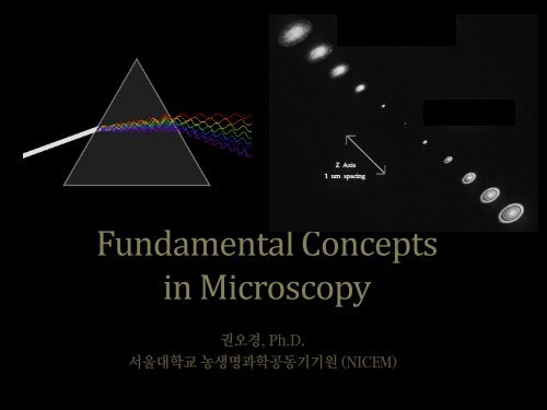
Fundamental Concepts in Microscopy

Create successful ePaper yourself
Turn your PDF publications into a flip-book with our unique Google optimized e-Paper software.
<strong>Fundamental</strong> <strong>Concepts</strong><br />
<strong>in</strong> <strong>Microscopy</strong><br />
권오경, Ph.D.<br />
서울대학교 농생명과학공동기기원 (NICEM)
서울대학교 농생명과학공동기기원 (NICEM)<br />
<strong>Microscopy</strong><br />
• Image Formation<br />
• Resolution<br />
• Structure of Microscope<br />
• Fluorescence <strong>Microscopy</strong><br />
• Confocal <strong>Microscopy</strong><br />
• Super-resolution <strong>Microscopy</strong>
서울대학교 농생명과학공동기기원 (NICEM)<br />
<strong>Microscopy</strong><br />
• Image Formation<br />
• Resolution<br />
• Structure of Microscope<br />
• Fluorescence <strong>Microscopy</strong><br />
• Confocal <strong>Microscopy</strong><br />
• Super-resolution <strong>Microscopy</strong>
E<br />
Object<br />
Lens<br />
Image
Lens<br />
PSF
E<br />
Convolution<br />
PSF
Po<strong>in</strong>t Spread Function (PSF)
PSF <strong>in</strong> 3D
서울대학교 농생명과학공동기기원 (NICEM)<br />
<strong>Microscopy</strong><br />
• Image Formation<br />
• Resolution<br />
• Def<strong>in</strong>ition<br />
• PSF and Resolution<br />
• Aperture and PSF<br />
• Aperture and Resolution<br />
• Degradation of Resolution<br />
• Structure of Microscope<br />
• Fluorescence <strong>Microscopy</strong><br />
• Confocal <strong>Microscopy</strong><br />
• Super-resolution <strong>Microscopy</strong>
RESOLUTION
PSF and Resolution
d = l<br />
2NA<br />
Diffraction Limited Resolution
High NA Objective<br />
Low NA Objective
Aperture and Resolution<br />
Objective<br />
Tube lens<br />
Intermediate<br />
image plane<br />
Diffraction spot<br />
on image plane<br />
= Po<strong>in</strong>t Spread Function<br />
Sample<br />
Back focal plane aperture
Aperture and Resolution<br />
Objective<br />
Tube lens<br />
Intermediate<br />
image plane<br />
Diffraction spot<br />
on image plane<br />
= Po<strong>in</strong>t Spread Function<br />
Sample<br />
Back focal plane aperture
Aperture and Resolution<br />
Objective<br />
Tube lens<br />
Intermediate<br />
image plane<br />
Diffraction spot<br />
on image plane<br />
= Po<strong>in</strong>t Spread Function<br />
Sample<br />
Back focal plane aperture
Aperture and Resolution<br />
Objective<br />
Tube lens<br />
Intermediate<br />
image plane<br />
Diffraction spot<br />
on image plane<br />
= Po<strong>in</strong>t Spread Function<br />
Sample<br />
<br />
Back focal plane aperture<br />
• Image resolution improves with Numerical Aperture (NA)<br />
NA = n s<strong>in</strong>()<br />
where:<br />
= light gather<strong>in</strong>g angle<br />
n = refractive <strong>in</strong>dex of sample
서울대학교 농생명과학공동기기원 (NICEM)<br />
What does degrade Resolution?<br />
• Chromatic aberration<br />
• Spherical aberration<br />
• Astigmatism<br />
32
서울대학교 농생명과학공동기기원 (NICEM)<br />
Chromatic Aberration<br />
33
서울대학교 농생명과학공동기기원 (NICEM)<br />
Spherical Aberration<br />
34
서울대학교 농생명과학공동기기원 (NICEM)<br />
Astigmatism<br />
35
서울대학교 농생명과학공동기기원 (NICEM)<br />
What does degrade Resolution?<br />
• Misalignment<br />
• Cleanness of optics<br />
• Improper aperture size<br />
• Quality of record<strong>in</strong>g media<br />
• Improper acquisition parameters<br />
36
서울대학교 농생명과학공동기기원 (NICEM)<br />
<strong>Microscopy</strong><br />
• Image Formation<br />
• Resolution<br />
• Structure of Microscope<br />
• Transmitted Light Microscope<br />
• Objective lens<br />
• Fluorescence <strong>Microscopy</strong><br />
• Confocal <strong>Microscopy</strong><br />
• Super-resolution <strong>Microscopy</strong>
서울대학교 농생명과학공동기기원 (NICEM)<br />
<strong>Microscopy</strong><br />
• Image Formation<br />
• Resolution<br />
• Structure of Microscope<br />
• Fluorescence <strong>Microscopy</strong><br />
• Excitation and Emission<br />
• Filters<br />
• Confocal <strong>Microscopy</strong><br />
• Super-resolution <strong>Microscopy</strong>
Notch
Band Pass Filters<br />
630 nm BandPass Filter<br />
White Light Source<br />
Transmitted Light<br />
Long Pass Filters<br />
620 -640 nm Light<br />
Light Source<br />
520 nm Long Pass Filter<br />
Transmitted Light<br />
Short Pass Filters<br />
Light Source<br />
>520 nm Light<br />
575 nm Short Pass Filter<br />
Transmitted Light<br />
510 LP dichroic Mirror<br />
Dichroic Filter/Mirror at 45 deg<br />
Light Source<br />
Transmitted Light<br />
Reflected light
서울대학교 농생명과학공동기기원 (NICEM)<br />
<strong>Microscopy</strong><br />
• Image Formation<br />
• Resolution<br />
• Structure of Microscope<br />
• Fluorescence <strong>Microscopy</strong><br />
• Confocal <strong>Microscopy</strong><br />
• Fluorescence vs Confocal microscopy<br />
• Confocal <strong>Microscopy</strong> +<br />
• How it works<br />
• Super-resolution <strong>Microscopy</strong>
Tissue culture cell with 60x / 1.4NA objective
Fluorescence vs Confocal <strong>Microscopy</strong><br />
• Common<br />
• Fluorescence dyes<br />
• Filters<br />
• Difference<br />
• Light source: Laser<br />
• Po<strong>in</strong>t Detection: PMT, APD, HyD<br />
• Optical section: P<strong>in</strong>hole 3D image reconstruction<br />
The term “confocal” means “hav<strong>in</strong>g the same focus” This is<br />
accomplished by focus<strong>in</strong>g the condenser lens to the same focal<br />
plane as the objective lens.
Confocal <strong>Microscopy</strong> +<br />
• Reduced blurr<strong>in</strong>g of the image from light scatter<strong>in</strong>g<br />
• Increased effective resolution<br />
• Improved signal to noise ratio<br />
• Clear exam<strong>in</strong>ation of thick specimen<br />
• Z-axis scann<strong>in</strong>g<br />
• Depth perception <strong>in</strong> Z-sectioned images<br />
• Magnification can be adjusted electronically
Scann<strong>in</strong>g<br />
Chang<strong>in</strong>g entrance<br />
angle of illum<strong>in</strong>ation<br />
moves illum<strong>in</strong>ation spot<br />
on sample<br />
Objective lens<br />
Sample<br />
The emission spot<br />
moves, so we have to<br />
make sure p<strong>in</strong>hole is<br />
co<strong>in</strong>cident with it
Select<strong>in</strong>g Proper Dyes and Filters
서울대학교 농생명과학공동기기원 (NICEM)<br />
<strong>Microscopy</strong><br />
• Image Formation<br />
• Resolution<br />
• Structure of Microscope<br />
• Fluorescence <strong>Microscopy</strong><br />
• Confocal <strong>Microscopy</strong><br />
• Super-resolution <strong>Microscopy</strong><br />
• Super-resolution methods<br />
• Confocal vs Super-resolution microscopy<br />
• How it works: Make PSF smaller
Selected Super-resolution Techniques<br />
STED (Stimulation Emission Depletion)<br />
SIM (Structured Illum<strong>in</strong>ation <strong>Microscopy</strong>)<br />
PALM (Photo-Activated Localization <strong>Microscopy</strong>)<br />
STORM (Stochastic Optical Reconstruction <strong>Microscopy</strong>)
Smaller PSF<br />
Better Resolution
Thank you very much<br />
for your attention!