Obtryx⢠Sling System - Boston Scientific
Obtryx⢠Sling System - Boston Scientific
Obtryx⢠Sling System - Boston Scientific

Create successful ePaper yourself
Turn your PDF publications into a flip-book with our unique Google optimized e-Paper software.
Gary E. Leach, MD<br />
Director, Tower Urology Institute for Continence<br />
Los Angeles, CA<br />
® Obtryx <strong>Sling</strong> <strong>System</strong><br />
T ECHNIQUE S POTLIGHT<br />
Transobturator Mid-Urethral <strong>Sling</strong>
Obtryx®<br />
Transobturator Mid-Urethral<br />
<strong>Sling</strong> <strong>System</strong><br />
P OTENTIAL B ENEFITS OF U SING<br />
T RANSOBTURATOR A PPROACH:<br />
Various techniques of sling procedures have continued to evolve over the last 30<br />
years. Early techniques utilizing large segments of autologous abdominal fascia or<br />
fascia lata evolved into sling procedures with shorter segments of fascia and smaller<br />
incisions with less retropubic dissection. Over the last 7 years, new types of synthetic<br />
sling materials have gained popularity (TVT) along with the concept of placing the sling in<br />
the area of the mid-urethra. Needle passage through the retropubic space was still required with the<br />
associated risks of bladder injury, injury to adjacent bowel, and bleeding in an area of difficult exposure.<br />
These risks have been minimized with the introduction of the transobturator approach, which requires<br />
no entry into the retropubic space. The combination of an improved synthetic mid-urethral sling with<br />
introduction instruments designed to allow safe passage of the sling through the obturator foramen has<br />
been a significant advance in the treatment of female stress incontinence. Following is the technique<br />
that I use when performing a transobturator sling with the Obtryx Transobturator <strong>Sling</strong>.<br />
P A TIENT S ELECTION:<br />
The author’s surgical procedure of choice for female stress incontinence is the transobturator sling using<br />
the Obtryx <strong>Sling</strong> <strong>System</strong>. All candidates for this procedure must have urodynamically documented<br />
genuine stress incontinence with or without urethral hypermobility. Other options discussed with<br />
potential surgical candidates include biofeedback/ pelvic floor training and injection therapy with a<br />
bulking agent in those patients with minimal to no urethral hypermobility. Those women with the<br />
combined problem of stress incontinence with significant cystocele are offered the combination of<br />
transobturator sling with repair of the cystocele defect utilizing non-frozen cadaveric fascia lata.<br />
P ROCEDURE L OCATION:<br />
The procedure is performed in the hospital OR either as an outpatient or inpatient procedure<br />
depending on the individual patient circumstances.<br />
P RE-OPERATIVE P REPARATION:<br />
Patient Preparation:<br />
Gary E. Leach, MD<br />
Director, Tower Urology Institute for Continence<br />
Los Angeles, CA<br />
All patients are taught the technique of clean self-intermittent catheterization pre-operatively. In the<br />
event that the patient is unable to void after the transobturator sling, the patient is then reinstructed in<br />
the self-catheterization technique that she learned before the surgery. The urine is confirmed to be sterile<br />
pre-operatively and patients perform a provo-iodine douche the night before and the morning of surgery<br />
with parenteral antibiotics administered pre-operatively. When the procedure is planned as an outpatient<br />
procedure, the patient is advised to arrange transportation home after discharge.
I NTRA-OPERATIVE:<br />
Anesthesia:<br />
T ECHNIQUE S POTLIGHT<br />
The transobturator sling procedure is performed under either a general or spinal anesthetic. The choice<br />
of anesthesia type is made based upon patient preference and clinical factors considered by the<br />
anesthesiologist.<br />
Patient Preparation and Positioning:<br />
After anesthesia is induced, the patient’s legs are placed into Allen stirrups and the adductor longus<br />
tendon is palpated on each side as a “landmark“ since the needle to transfer the sling is passed just<br />
below this tendon at the level of the clitoris. For obese patients, a slightly more exaggerated lithotomy<br />
position may be required to facilitate palpation of this tendon landmark. The patient is shaved and<br />
prepared with a lower abdominal and vaginal provoiodine scrub. A Foley catheter is inserted before<br />
starting the procedure.<br />
Technique of <strong>Sling</strong> Placement:<br />
After the bladder neck is identified by palpation of the Foley catheter balloon, either a<br />
vertical midline incision is made from the mid-urethra to the bladder neck or an inverted<br />
“U” shaped incision is made with the apex of the “U” at the junction of the mid and<br />
distal 1/3 of the urethra. If the patient has had previous sling surgery, I prefer the inverted<br />
“U” incision to maximize exposure and allow direct control of any bleeding in the area.<br />
An anterior vaginal wall flap is then reflected to the bladder neck. Regardless of the type<br />
of incision used, the next step is sharp lateral dissection to the pubic ramis. The ramis is<br />
palpated on each side and dissection continues laterally to the medial aspect of the<br />
obturator membrane.<br />
Utilizing the landmarks of the adductor longus tendon and the clitoris, a 22 g spinal<br />
needle is passed through the skin in the area of the groin crease towards the vaginal finger<br />
on the inside obturator membrane. This needle serves to define the direction of the larger<br />
needle used to pass the sling from the vagina out to the skin level. Once the needle is felt<br />
with the fingertip, 1% marcaine (which is optional) is injected as the needle is withdrawn.<br />
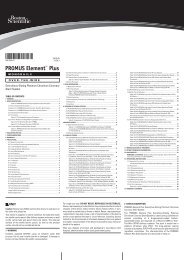
A small skin incision is then made on each side and either the Obtryx ® Halo Needle or<br />
the Obtryx Curved Needle is chosen to use for the procedure. I prefer the Curved Needle,<br />
(FIGURE A ), which is easier to pass in the “large” patient. The needle is passed through the<br />
skin and muscle layers directly onto the fingertip on the inside of the obturator<br />
membrane, (FIGURE B). Care is taken to pass the needle “above” the vagina with avoidance<br />
of perforation into the vagina at the junction of the anterior and lateral vaginal walls.<br />
After the needle is passed, the association loop at the end of the sling is fed over the tip of<br />
the needle and the needle is withdrawn out to the skin level, (FIGURE C). This process is<br />
repeated on the contra-lateral side with the sling being positioned in the area of the midurethra,<br />
(FIGURE D). The centering tab should be positioned out towards the surgeon.<br />
Should the vaginal wall be perforated and recognized after the mesh is attached and pulled<br />
through the tissue, the Obtryx Needle has a removable mesh assembly connection that<br />
can be removed from the needle, pulled back out and placed again without having to<br />
open another device. At this point, if there is any question regarding possible bladder or<br />
urethral injury, cystoscopy is performed.<br />
FIGURE A<br />
FIGURE B<br />
FIGURE C<br />
FIGURE D
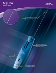
FIGURE E<br />
FIGURE F<br />
<strong>Sling</strong> Tensioning/ Sleeve Removal:<br />
S UMMARY:<br />
The Transobturator <strong>Sling</strong> Procedure with the Obtryx <strong>Sling</strong> <strong>System</strong> is a<br />
significant advance in the surgical treatment of female stress incontinence.<br />
The unique aspects of the Advantage R Synthetic <strong>Sling</strong> material coupled<br />
with the transobturator approach is a significant advance in the surgical<br />
treatment of female stress incontinence. Our experience thus far in over<br />
75 cases has been very promising without significant complications.<br />
T ECHNIQUE S POTLIGHT<br />
After both needles have been withdrawn and the sling is seen to lay flat against the area of the mid-urethra, a<br />
Metzenbaum scissors is placed between the sling and the urethra. Both needles are equally withdrawn to bring<br />
the sling in contact with the scissors. The blue plastic tab is then cut at the midline of the<br />
sling beneath the urethra, (FIGURE E). Next, the plastic sleeves are simultaneously removed<br />
from each end of the sling leaving the synthetic sling material in contact with the scissors held<br />
up against the urethra. Given the special characteristics of the “de-tanged” suburethral<br />
segment of the Advantage ® sling, (FIGURE F), the sling material assumes a very flat<br />
configuration beneath the mid-urethra. The vaginal incision is closed with a 2-0 Vicryl ®<br />
tanged<br />
edges<br />
de-tanged<br />
edges<br />
P OST-OPERATIVE C ARE:<br />
suture. The excess sling material above the skin level is trimmed and the too small groin<br />
incisions are closed with 4-0 Monocryl ® suture. When the procedure is performed on an<br />
outpatient basis, the patient is sent to the recovery room without a Foley catheter or vaginal<br />
packing. Should the patient be held “overnight”, the Foley catheter is inserted and placed to a<br />
sterile drainage bag. A vaginal pack may be utilized at the discretion of the surgeon.<br />
When the sling is performed with a cystocele repair, a single vertical incision is made from the<br />
mid-urethra to the cervix or vaginal cuff and the cystourethrocele is mobilized off the anterior<br />
vaginal wall. The sling is then placed and the cystocele repair is completed with a “patch” of<br />
cadaveric fascia tissue to repair the cystocele defect. The tension on the sling is adjusted after<br />
the cystocele repair as noted above.<br />
The post-void residual urine volume is checked after surgery. When the patient is unable to void or if the postvoid<br />
residual volume is > 100cc, the patient is reinstructed in the technique of self-catheterization that she<br />
learned before the sling procedure. The patient is discharged on oral antibiotics for 1 week and she is instructed<br />
to limit her physical activity. Pain is usually minimal and treated with oral Tylenol ® . Post-operative follow-up<br />
appointments occur at 1 and 6 weeks after surgery.<br />
P O TENTIAL P OST-OP ERATIVE C OMPLICATIONS:<br />
In general, complications have been rare following the Obtryx ® Transobturator <strong>Sling</strong> procedure. We have seen<br />
few reports of significant bleeding, injuries to the bladder or urethra, and permanent urinary retention. In my<br />
experience, 80% of patients void immediately after surgery and the remainder usually require self-catheterization<br />
for less than one week. As of the date of this publication, we have seen no complications related to the synthetic<br />
sling material (ie. no urethral erosions, infections, or vaginal extrusions). Post operative pain has generally been<br />
minimal.<br />
The opinions and recommendations expressed in this Technique Spotlight are those of<br />
the author alone. <strong>Boston</strong> <strong>Scientific</strong> makes no representations or warranties as to the<br />
accuracy or completeness of the information set forth herein.<br />
Vicryl and Monocryl are trademarks of Johnson & Johnson Corporation.<br />
Caution: Federal Law (USA) restricts devices to sale by or on the order of a physician.<br />
Refer to Directions for Use provided with these products for complete instructions,<br />
warnings and precautions prior to using these products.<br />
<strong>Boston</strong> <strong>Scientific</strong> Corporation<br />
One <strong>Boston</strong> <strong>Scientific</strong> Place<br />
Natick, MA 01760-1537<br />
www.bostonscientific.com<br />
Ordering Information<br />
1.888.272.1001<br />
© 2006 <strong>Boston</strong> <strong>Scientific</strong> Corporation<br />
or it’s affiliates. All rights reserved.<br />
MVU5710 12/05–12/07