

Eble JN, Sauter G., Epstein JI, Sesterhenn IA - iarc
Eble JN, Sauter G., Epstein JI, Sesterhenn IA - iarc
Eble JN, Sauter G., Epstein JI, Sesterhenn IA - iarc

Create successful ePaper yourself
Turn your PDF publications into a flip-book with our unique Google optimized e-Paper software.
mal or erythematous which sometimes<br />
represents the microscopic areas of carcinoma<br />
in situ.<br />
Histopathology<br />
The histology of infiltrating urothelial carcinomas<br />
is variable {80,293,944}. Most of<br />
pT1 cancers are papillary, low or high<br />
grade, whereas most pT2-T4 carcinomas<br />
are non-papillary and high grade.<br />
These carcinomas are graded as low<br />
grade and high grade depending upon<br />
the degree of nuclear anaplasia and<br />
some architectural abnormalities {706,<br />
1548,1798}. Some cases may show relatively<br />
bland cytology {2896}.<br />
The most important element in pathologic<br />
evaluation of urothelial cancer is<br />
recognition of the presence and extent of<br />
invasion {293}. In early invasive urothelial<br />
carcinomas (pT1), foci of invasion are<br />
characterized by nests, clusters, or single<br />
cells within the papillary cores and/or<br />
lamina propria. It is recommended that<br />
the extent of lamina propria invasion in<br />
pT1 tumours should be stated {706}. The<br />
depth of lamina propria invasion is<br />
regarded as a prognostic parameter in<br />
pT1 cancer. Morphologic criteria useful<br />
in assessing of lamina propria invasion<br />
include the presence of desmoplastic<br />
stromal response, tumour cells within the<br />
retraction spaces, and paradoxical differentiation<br />
(invasive nests of cells with<br />
abundant eosinophilic cytoplasm at the<br />
advancing edge of infiltration {2117}).<br />
Recognition of invasion may be problematic<br />
because of tangential sectioning,<br />
thermal and mechanical injury, marked<br />
inflammatory infiltrate obscuring neoplastic<br />
cells and inverted or broad front<br />
growth {78}. Thermal artefact can also<br />
hamper the interpretation of muscularis<br />
propria invasion.<br />
The histology of infiltrative urothelial carcinoma<br />
has no specific features and<br />
shows infiltrating cohesive nests of cells<br />
with moderate to abundant amphophilic<br />
cytoplasm and large hyperchromatic<br />
nuclei. In larger nests, palisading of<br />
nuclei may be seen at the edges of the<br />
nests. The nucleus is typically pleomorphic<br />
and often has irregular contours<br />
with angular profiles. Nucleoli are highly<br />
variable in number and appearance with<br />
some cells containing single or multiple<br />
small nucleoli and others having large<br />
eosinophilic nucleoli. Foci of marked<br />
pleomorphism may be seen, with bizarre<br />
and multinuclear tumour cells {293}.<br />
Mitotic figures are common, with numerous<br />
abnormal forms. The invasive nests<br />
usually induce a desmoplastic stromal<br />
reaction which is occasionally pronounced<br />
and may mimic a malignant<br />
spindle cell component, a feature known<br />
as pseudosarcomatous stromal reaction<br />
{1555}. In most cases, the stroma contains<br />
a lymphocytic infiltrate with a variable<br />
number of plasma cells. The inflammation<br />
is usually mild to moderate and<br />
focal, although it may be severe, dense,<br />
and widespread. Neutrophils and<br />
eosinophils are rarely prominent.<br />
Retraction clefts are often present<br />
around the nests of carcinoma cells,<br />
mimicking vascular invasion. It is important<br />
to be aware of this feature in order to<br />
avoid misinterpretation as vascular invasion.<br />
Foci of squamous and glandular<br />
differentiation are common, and should<br />
be reported {1554,2177,2276}. Intraepithelial<br />
neoplasia including carcinoma<br />
in situ is common in the adjacent urothelium<br />
{1547,1552}. Occasionally, mucoid<br />
cytoplasmic inclusions may be present.<br />
Histologic variants<br />
Urothelial carcinoma has a propensity for<br />
divergent differentiation with the most<br />
common being squamous followed by<br />
glandular. Virtually the whole spectrum of<br />
bladder cancer variants may be seen in<br />
variable proportions accompanying otherwise<br />
typical urothelial carcinoma.<br />
Divergent differentiation frequently parallels<br />
high grade and high stage urothelial<br />
cancer. When small cell differentiation is<br />
present, even focally, it portends a poor<br />
prognosis and has different therapeutic<br />
ramifications, and hence should be diagnosed<br />
as small cell carcinoma.<br />
Infiltrating urothelial carcinoma with<br />
squamous differentiation<br />
Squamous differentiation, defined by the<br />
presence of intercellular bridges or keratinization,<br />
occurs in 21% of urothelial carcinomas<br />
of the bladder, and in 44% of<br />
tumours of the renal pelvis {1554,1637}.<br />
Its frequency increases with grade and<br />
stage {1554}. Detailed histologic maps of<br />
urothelial carcinoma with squamous differentiation<br />
have shown that the proportion<br />
of the squamous component may<br />
vary considerably, with some cases having<br />
urothelial carcinoma in situ as the<br />
only urothelial component {2276}. The<br />
diagnosis of squamous cell carcinoma is<br />
reserved for pure lesions without any<br />
associated urothelial component, including<br />
urothelial carcinoma in situ {2177}.<br />
Tumours with any identifiable urothelial<br />
element are classified as urothelial carcinoma<br />
with squamous differentiation<br />
{1554,2177} and an estimate of the percentage<br />
of squamous component should<br />
be provided. Squamous differentiation<br />
may show basaloid or clear cell features.<br />
Cytokeratin 14 and L1 antigen have been<br />
reported as immunohistochemical markers<br />
of squamous differentiation {1025,<br />
2655}. Uroplakins, are expressed in<br />
urothelial carcinoma and not in squamous<br />
differentiation {2848}.<br />
The clinical significance of squamous<br />
differentiation remains uncertain, but<br />
seems to be an unfavourable prognostic<br />
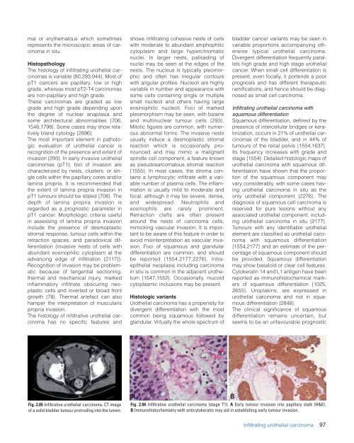
Fig. 2.05 Infiltrative urothelial carcinoma. CT image<br />
of a solid bladder tumour protruding into the lumen.<br />
A<br />
Fig. 2.06 Infiltrative urothelial carcinoma (stage T1). A Early tumour invasion into papillary stalk (H&E).<br />
B Immunohistochemistry with anticytokeratin may aid in establishing early tumour invasion.<br />
B<br />
Infiltrating urothelial carcinoma<br />
97


