A scanning and transmission electron microscopic study of ... - Digitum
A scanning and transmission electron microscopic study of ... - Digitum
A scanning and transmission electron microscopic study of ... - Digitum

Create successful ePaper yourself
Turn your PDF publications into a flip-book with our unique Google optimized e-Paper software.
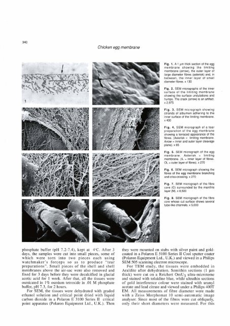
Chicken egg membrane<br />
Flg. 3. SEM micrograph showing<br />
str<strong>and</strong>s <strong>of</strong> albumen adherlng to the<br />
lnner su- <strong>of</strong> the limiting membrane.<br />
x400<br />
Fig. 4. SEM micrograph <strong>of</strong> a tear<br />
preparation <strong>of</strong> the egg membrane<br />
showing a terraced appearance <strong>of</strong> the<br />
fibres. (Asterisk<br />
limitlng membrane.<br />
Amm = lnner <strong>and</strong> outer layer deavage<br />
m). x 65<br />
Flg. 5. SEM micrograph <strong>of</strong> the egg<br />
membrane. Asterisk = limiting<br />
mmbtme. (IL = inner layer <strong>of</strong> fibres.<br />
OL = outer layer <strong>of</strong> fibres). x 270<br />
Flg. 6. SEM micrograph showing the<br />
fibra <strong>of</strong> the egg membrane bmching<br />
<strong>and</strong> cfiss-crossing. x 270<br />
Fig. 7. SEM micrograph <strong>of</strong> the fibre<br />
core (C) surrounded by the manthle<br />
layer (M). x 8,500<br />
Fig. 8. SEM micrograph <strong>of</strong> the flbre<br />
phosphate buffer (pH 7.2-7.4), kept at 4OC. After 3<br />
daya, the samples were cut into small pieces, some <strong>of</strong><br />
which were torn into two pieces each using<br />
watchmaker's forceps so as to produce "tear<br />
preparations", Small pieces <strong>of</strong> the shell <strong>and</strong> shell<br />
membranes above the air-sac were also removed <strong>and</strong><br />
fixed for 3 days before they were decalcified in glacial<br />
acetic acid for 1 week. After that, al1 the tissues were<br />
osmicated in 1% osrnium tetroxide in .O1 M phosphate<br />
buffer, pH 7.3, for 2 hours.<br />
For SEM, the tissues were dehydrated with graded<br />
ethanol solution <strong>and</strong> critical point dried with liquid<br />
carbon dioxide in a Polaron E 3100 Series 11 critical<br />
point apparatus (Polaron Equipment Ltd., U.K.). Then<br />
they were mounted on stubs with silver paint <strong>and</strong> goldcoated<br />
in a Polaron E 5100 Series II Cool sputter coater<br />
(Polaron Equipment Ltd., U.K.) <strong>and</strong> viewed in a Philips<br />
SEM 505 <strong>scanning</strong> <strong>electron</strong> microscope.<br />
For TEM <strong>study</strong>, the tissues were embedded in<br />
Araldite after dehydration. Semithin sections (1 pm<br />
thick) were cut on a Reichert OmU3 ultra-microtome<br />
<strong>and</strong> stained with toluidine blue, while ultrathin sections<br />
<strong>of</strong> gold interfererice colour were stained with uranyl<br />
acetate <strong>and</strong> lead citrate <strong>and</strong> viewed under a Philips W<br />
EM. Al1 measurements <strong>of</strong> fibre diameter were made<br />
with a Zeiss Morphomat 10 semi-automatic image<br />
analyser. Since most <strong>of</strong> the fibres were cut obliquely,<br />
only their short diameters were measured. For this