Post Graduate Diploma in Diagnostic Ultrasound (OBGYN) in ...
Post Graduate Diploma in Diagnostic Ultrasound (OBGYN) in ...
Post Graduate Diploma in Diagnostic Ultrasound (OBGYN) in ...
Create successful ePaper yourself
Turn your PDF publications into a flip-book with our unique Google optimized e-Paper software.
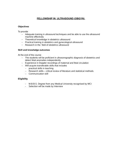
FELLOWSHIP IN ULTRASOUND (<strong>OBGYN</strong>)<br />
Objectives<br />
To provide<br />
− Adequate tra<strong>in</strong><strong>in</strong>g <strong>in</strong> ultrasound techniques and be able to use the ultrasound<br />
mach<strong>in</strong>e effectively<br />
− Theoretical knowledge <strong>in</strong> obstetrics ultrasound<br />
− Practical tra<strong>in</strong><strong>in</strong>g <strong>in</strong> obstetrics and gynecological ultrasound<br />
− Research <strong>in</strong> the field of obstetrics ultrasound<br />
Skill and knowledge outcomes<br />
At the end of the course<br />
− The students will be proficient <strong>in</strong> ultrasonographic diagnosis of obstetrics and<br />
detect fetal anomalies <strong>in</strong>dependently<br />
− Experience <strong>in</strong> Doppler record<strong>in</strong>gs of maternal and fetal circulation<br />
− Will acquire transferable skills that <strong>in</strong>cludes<br />
− practical skills <strong>in</strong> teach<strong>in</strong>g<br />
− Research skills – critical review of literature and statistical methods<br />
− Communication skill<br />
Eligibility<br />
- M.B.B.S. Degree from any Medical University recognised by MCI<br />
- Selection will be made by Interview
Syllabus<br />
The students will go through the follow<strong>in</strong>g modules dur<strong>in</strong>g the 2 year course.<br />
First year<br />
Module – 1 – Basics of <strong>Ultrasound</strong><br />
• Physics of medical ultrasound<br />
• Biostatistics<br />
Module – 2 – Basic sciences related to ultrasound and cl<strong>in</strong>ical <strong>OBGYN</strong><br />
• Embryology<br />
• Anatomy & Physiology of reproductive System<br />
• Pathology<br />
• Cl<strong>in</strong>ical aspects of Obstetrics and Gynaecology<br />
Module – 3 - Gynaecology<br />
• <strong>Ultrasound</strong> Anatomy of Pelvis and USG related pathology of pelvis<br />
Module 4<br />
• Obstetrics <strong>Ultrasound</strong> – Part 1<br />
Second Year<br />
Obstetrics <strong>Ultrasound</strong> – Part II<br />
Module 5 - Fetal Environment & Doppler<br />
Module 6 - Fetal anomalies & 3D <strong>Ultrasound</strong><br />
Module 7- PROJECT
Annexure – 1<br />
Syllabus for the Ist Year Students<br />
PAPER – 1 – BASICS OF ULTRASOUND<br />
Physics of ultrasound & Biostatistics<br />
<br />
<br />
<br />
<br />
<br />
<br />
Introduction of ultrasound<br />
Physics & Bioeffects of ultrasound<br />
Instrumentation & Knobology<br />
Pr<strong>in</strong>ciples of Imag<strong>in</strong>g techniques, Equipment ma<strong>in</strong>tenance, basics of power<br />
supply and Network<strong>in</strong>g<br />
Basic ultrasound appearances like cysts, solids and complex masses<br />
Bedside manners, medicolegal issue<br />
Biostatistics<br />
<br />
<br />
Mean, Median, Mode and Standard Deviation<br />
Correlation and Regression<br />
PAPER – II<br />
EMBRYOLOGY, ANATOMY, PHYSIOLOGY OF REPRODUCTIVE SYSTEM,<br />
PATHOLOGY & CLINICAL ASPECTS OF OBSTETRICS AND GYNAECOLOGY<br />
Embryology<br />
• General embryology<br />
• Development of ma<strong>in</strong> organ systems CNS - sp<strong>in</strong>al cord and bra<strong>in</strong>, CVS, ur<strong>in</strong>ary<br />
system, digestive tube and its derivatives<br />
• Development of placenta and membrane<br />
• Basic pr<strong>in</strong>ciples and mechanism of teratogenesis<br />
Anatomy & Physiology of reproductive System<br />
• Sectional anatomy and physiology related to obstetrics and gynaecology<br />
Pathology<br />
• Gynaec Pathology<br />
• Fetal and placental pathology
Cl<strong>in</strong>ical aspects Obstetrics and Gynaecology<br />
Pregnancy<br />
• Diagnosis<br />
o<br />
Cl<strong>in</strong>ical / US/ HCG<br />
• Early preg problems<br />
o<br />
o<br />
Bleed<strong>in</strong>g <strong>in</strong> early preg<br />
Pa<strong>in</strong><br />
• Ectopic & Pregnancy of unknown location<br />
• Second / third trim bleed<strong>in</strong>g<br />
o<br />
o<br />
o<br />
Placenta previa<br />
Abruption<br />
others<br />
• Maternal diseases <strong>in</strong> pregnancy<br />
o<br />
o<br />
o<br />
o<br />
HTN/ PIH/Preeclampsia<br />
Diabetes<br />
Autoimmune<br />
Haematological<br />
• Fetal well be<strong>in</strong>g - per<strong>in</strong>atal hypoxia<br />
o<br />
Orientation to BPP with basic CTG<br />
• Labour<br />
Gyn<br />
• Menstrual cycle and ovulation<br />
o<br />
Normal physiology<br />
• Anovulation <strong>in</strong>clud<strong>in</strong>g pcos<br />
o<br />
Why anovulation – what do we expect on ultrasound<br />
• Uter<strong>in</strong>e malformations<br />
• Dub<br />
o patterns what to look for
o<br />
How ultrasound helps management<br />
• <strong>Post</strong>menopausal bleed<strong>in</strong>g<br />
o<br />
o<br />
Causes<br />
Management<br />
• Fibroids<br />
• Endometriosis and adenomyosis<br />
• Ov cysts<br />
• Infertility<br />
PAPER - III – GYNAECOLOGICAL ULTRASOUND<br />
<strong>Ultrasound</strong> Anatomy of Pelvis and USG related pathology of pelvis<br />
<br />
<br />
<br />
<br />
<br />
<br />
<br />
<br />
<br />
Uter<strong>in</strong>e and ovarian sonography<br />
Transvag<strong>in</strong>al ultrasound and follicular monitor<strong>in</strong>g<br />
An understand<strong>in</strong>g of the menstural cycle<br />
Pelvic <strong>in</strong>flammatory disease<br />
Poly cystic ovarian syndrome<br />
Endometriosis<br />
Uter<strong>in</strong>e fibroids, Adenomyosis & polyps<br />
Endometrial pathology<br />
Adnexal lesions<br />
PAPER - IV - OBSTETRICS ULTRASOUND – PART I<br />
<br />
<br />
<br />
<br />
<strong>Ultrasound</strong> evaluation <strong>in</strong> Ist trimester – <strong>in</strong>clud<strong>in</strong>g 11-14 weeks<br />
<strong>Ultrasound</strong> evaluation <strong>in</strong> IInd trimester – Target Scan & ultrasound evaluation of<br />
cervix<br />
<strong>Ultrasound</strong> evaluation <strong>in</strong> IIIrd trimester – Growth Scan<br />
Biochemical screen<strong>in</strong>g<br />
PRACTICALS<br />
<br />
<br />
Hands on tra<strong>in</strong><strong>in</strong>g <strong>in</strong> pelvis, USG related pathology normal and abnormal.<br />
Hands on tra<strong>in</strong><strong>in</strong>g <strong>in</strong> obstetrics, Normal and Abnormal
Obstetrics <strong>Ultrasound</strong> – Part II<br />
Syllabus for the 2 nd Year Student<br />
PAPER – V - FETAL ENVIRONMENT & DOPPLER<br />
• Biometry and biophysical profile<br />
• USG evaluation of Placenta<br />
• USG evaluation of Cord<br />
• USG evaluation of Amniotic fluid<br />
• Role of Doppler <strong>in</strong> <strong>OBGYN</strong><br />
PAPER VI - FETAL ANOMALIES & 3D ULTRASOUND<br />
<strong>Ultrasound</strong> evaluation of the follow<strong>in</strong>g fetal anomalies<br />
• CNS anomalies<br />
• Cardiac anomalies<br />
• Genitour<strong>in</strong>ary anomalies<br />
• Gastro <strong>in</strong>test<strong>in</strong>al tract anomalies<br />
• Abdom<strong>in</strong>al wall<br />
• Neck and Face<br />
• Fetal hydrops<br />
• Multiple pregnancies<br />
3D ultrasound<br />
• Role of 3D ultrasound and <strong>OBGYN</strong> imag<strong>in</strong>g<br />
PROJECT<br />
PRACTICALS<br />
<br />
<br />
<br />
Hands on tra<strong>in</strong><strong>in</strong>g <strong>in</strong> obstetrics ultrasound, fetal environment and fetal anomalies.<br />
Observation of <strong>in</strong>terventional ultrasound and therapy project<br />
Case based discussion obstetrics ultrasound<br />
Books recommended for reference<br />
1. Cl<strong>in</strong>ically oriented Anatomy – By Keith Moore Arthur F. Dalley<br />
2. Sear's Anatomy for Nurses – R.S.W<strong>in</strong>wood and J.L.Smith<br />
3. Develop<strong>in</strong>g Human cl<strong>in</strong>ically oriented Embryology - Keith Moore<br />
4. Abdom<strong>in</strong>al ultrasound and <strong>OBGYN</strong> ultrasound – Roger Sanders, Peter Callen,<br />
Carol M.Rumuck, Vol II obstetrics.<br />
5. <strong>Diagnostic</strong> ultrasound of fetal anomalies by David A. Nyberg et. al.<br />
6. Medic<strong>in</strong>e of the fetus and mother by Reece and Hobb<strong>in</strong>s.
Exam<strong>in</strong>ation<br />
At the end of every year there will be theory and practical exam<strong>in</strong>ations as follows<br />
IST YEAR<br />
S.L.<br />
No<br />
Subject Theory IA Practical Viva<br />
I Basics of <strong>Ultrasound</strong> 100 50<br />
II Basics sciences<br />
related to ultrasound<br />
(Embryology,<br />
Anatomy,Physiology<br />
of reproductive<br />
system, Pathology<br />
and Cl<strong>in</strong>ical aspects<br />
of Obstetrics and<br />
Gynaecology )<br />
III<br />
IV<br />
IInd Year<br />
Gynaecological<br />
<strong>Ultrasound</strong><br />
Obstetrics <strong>Ultrasound</strong><br />
– Part I<br />
Max M<strong>in</strong> Max M<strong>in</strong> Max M<strong>in</strong> Max M<strong>in</strong><br />
100 50<br />
100 50<br />
100 50<br />
50 25 100 50 50 25<br />
S.L.No Subject Theory IA Practical Viva<br />
V<br />
VI<br />
Fetal Environment<br />
& Doppler<br />
Fetal anomalies &<br />
3D ultrasound<br />
Max M<strong>in</strong> Max M<strong>in</strong> Max M<strong>in</strong> Max M<strong>in</strong><br />
100 50<br />
100 50<br />
50 25 100 50 50 25<br />
EVALUATION OF DISSERTATION<br />
Evaluation of Dissertation 200<br />
Viva/Presentation 50<br />
IA 50<br />
Total 300<br />
Pass<strong>in</strong>g M<strong>in</strong>imum 150


