Armed Forces Hospital Hamburg, Germany customer story - Philips ...
Armed Forces Hospital Hamburg, Germany customer story - Philips ...
Armed Forces Hospital Hamburg, Germany customer story - Philips ...

You also want an ePaper? Increase the reach of your titles
YUMPU automatically turns print PDFs into web optimized ePapers that Google loves.
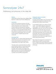
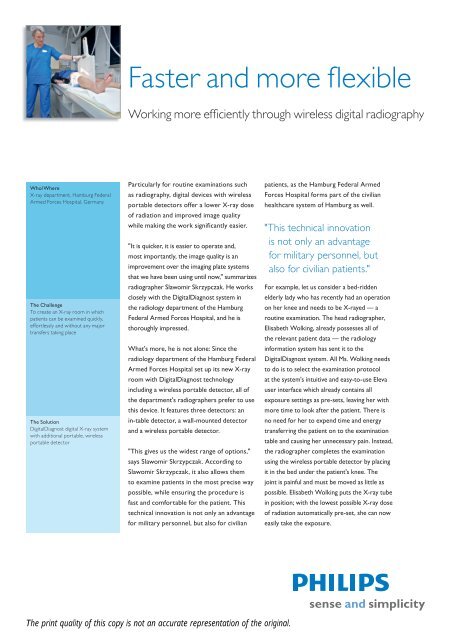
Faster and more flexible<br />
Working more efficiently through wireless digital radiography<br />
Who/Where<br />
X-ray department, <strong>Hamburg</strong> Federal<br />
<strong>Armed</strong> <strong>Forces</strong> <strong>Hospital</strong>, <strong>Germany</strong><br />
The Challenge<br />
To create an X-ray room in which<br />
patients can be examined quickly,<br />
effortlessly and without any major<br />
transfers taking place<br />
The Solution<br />
DigitalDiagnost digital X-ray system<br />
with additional portable, wireless<br />
portable detector<br />
Particularly for routine examinations such<br />
as radiography, digital devices with wireless<br />
portable detectors offer a lower X-ray dose<br />
of radiation and improved image quality<br />
while making the work significantly easier.<br />
"It is quicker, it is easier to operate and,<br />
most importantly, the image quality is an<br />
improvement over the imaging plate systems<br />
that we have been using until now," summarizes<br />
radiographer Slawomir Skrzypczak. He works<br />
closely with the DigitalDiagnost system in<br />
the radiology department of the <strong>Hamburg</strong><br />
Federal <strong>Armed</strong> <strong>Forces</strong> <strong>Hospital</strong>, and he is<br />
thoroughly impressed.<br />
What's more, he is not alone: Since the<br />
radiology department of the <strong>Hamburg</strong> Federal<br />
<strong>Armed</strong> <strong>Forces</strong> <strong>Hospital</strong> set up its new X-ray<br />
room with DigitalDiagnost technology<br />
including a wireless portable detector, all of<br />
the department's radiographers prefer to use<br />
this device. It features three detectors: an<br />
in-table detector, a wall-mounted detector<br />
and a wireless portable detector.<br />
"This gives us the widest range of options,"<br />
says Slawomir Skrzypczak. According to<br />
Slawomir Skrzypczak, it also allows them<br />
to examine patients in the most precise way<br />
possible, while ensuring the procedure is<br />
fast and comfortable for the patient. This<br />
technical innovation is not only an advantage<br />
for military personnel, but also for civilian<br />
patients, as the <strong>Hamburg</strong> Federal <strong>Armed</strong><br />
<strong>Forces</strong> <strong>Hospital</strong> forms part of the civilian<br />
healthcare system of <strong>Hamburg</strong> as well.<br />
"This technical innovation<br />
is not only an advantage<br />
for military personnel, but<br />
also for civilian patients."<br />
For example, let us consider a bed-ridden<br />
elderly lady who has recently had an operation<br />
on her knee and needs to be X-rayed — a<br />
routine examination. The head radiographer,<br />
Elisabeth Wolking, already possesses all of<br />
the relevant patient data — the radiology<br />
information system has sent it to the<br />
DigitalDiagnost system. All Ms. Wolking needs<br />
to do is to select the examination protocol<br />
at the system's intuitive and easy-to-use Eleva<br />
user interface which already contains all<br />
exposure settings as pre-sets, leaving her with<br />
more time to look after the patient. There is<br />
no need for her to expend time and energy<br />
transferring the patient on to the examination<br />
table and causing her unnecessary pain. Instead,<br />
the radiographer completes the examination<br />
using the wireless portable detector by placing<br />
it in the bed under the patient's knee. The<br />
joint is painful and must be moved as little as<br />
possible. Elisabeth Wolking puts the X-ray tube<br />
in position; with the lowest possible X-ray dose<br />
of radiation automatically pre-set, she can now<br />
easily take the exposure.<br />
The print quality of this copy is not an accurate representation of the original.
She has to wait just five seconds before<br />
the image she has taken appears on the<br />
application workstation monitor in front of<br />
her. After ensuring the quality of the image<br />
is sufficient, she can now conclude this part<br />
of the examination. The image data is sent<br />
directly to the internal PACS image archiving<br />
system and both Dr. Hans-Michael Schlegel<br />
COL MC. and the surgeon operating on the<br />
knee can access the X-rays immediately.<br />
"Working wirelessly improves<br />
hygiene around the<br />
hospital bed."<br />
"I am grateful we have the wireless portable<br />
detector, particularly when examining<br />
patients who must be monitored," explains<br />
Slawomir Skrzypczak. "They are already<br />
connected to so many cables and tubes, so<br />
it's good that I don't have to keep my eye on<br />
a bulky device cable." It's also inevitable that<br />
cables come into contact with the ground,<br />
which may bring pathogens into the patient's<br />
bed. Working wirelessly therefore improves<br />
hygiene around the hospital bed.<br />
The wireless portable detector also comes<br />
into its own when examining the increasing<br />
numbers of heavily overweight patients.<br />
If they are so severely injured that they<br />
cannot lie or sit on the examination table<br />
under their own power, they must be lifted<br />
onto it by the radiographers. If the X-ray is<br />
taken using the wireless portable detector,<br />
the patients can remain in their wheelchair<br />
or on their stretcher and the radiographers<br />
do not need to do any heavy lifting.<br />
This is especially useful for X-rays of the<br />
extremity and thorax. "Of course, we could<br />
take X-rays like these using a cassette-based<br />
X-ray system, but it would be nowhere<br />
near as easy and, above all, would not offer<br />
this level of quality and speed," affirms<br />
Slawomir Skrzypczak.<br />
In this regard, the wireless portable<br />
detector not only offers the greatest degree<br />
of flexibility — the combination of wallmounted<br />
and in-table detectors allows<br />
superior X-rays to be taken, e.g. X-rays of<br />
the pelvis in the event of a neck of femur<br />
fracture. "On one level, we can take X-rays<br />
using the in-table detector as before, but on<br />
a second level, we can also take axial images<br />
without any trouble," explains Skrzypczak.<br />
The vertical stand is so flexible that it can<br />
be moved closer to the patient along with<br />
the mobile examination table, so that images<br />
can be taken at a 90-degree angle. "During<br />
this time, the patient lies on the examination<br />
table and must not be moved. This means we<br />
do not cause the patient unnecessary pain."<br />
The image quality of devices that were<br />
previously used in the department was<br />
not so good, particularly when it involved<br />
radiography of large objects, for example<br />
pelvis X-rays. "We used to find X-rays like<br />
these rather difficult," confirms Slawomir<br />
Skrzypczak. Digital processing using the<br />
image processing software UNIQUE<br />
(UNified Image QUality Enhancement) has<br />
made the system less sensitive to exposure<br />
problems. This is an important factor for<br />
radiographers to consider. "After all, we<br />
want to take X-rays at the lowest possible<br />
X-ray dose of radiation so that the patient<br />
is protected, but we also want to get the<br />
best possible images."<br />
"We do not cause the patient<br />
unnecessary pain."<br />
Slawomir Skrzypczak can now scarcely<br />
imagine working without the digital, wireless<br />
system — and he is not the only one. In<br />
fact, if the radiographers had their way, all<br />
of the X-ray systems workstations would<br />
be equipped with a wireless portable<br />
detector, as Dr. Hans-Michael Schlegel<br />
COL MC. confirms: "I have never seen my<br />
radiographers be so enthusiastic about<br />
a device after such a short time."<br />
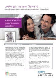
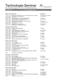
Overtable X-ray of<br />
the lower extremity<br />
The print quality of this copy is not an accurate representation of the original.
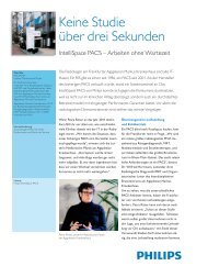
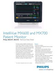
Dr. Hans-Michael Schlegel COL MC.<br />
with the wireless portable detector<br />
What makes you stand out from other<br />
hospitals or specialist clinics?<br />
One of our definite plus points is that we<br />
use state-of-the-art equipment. This puts<br />
patients at an advantage as they are subjected<br />
to a lower X-ray dose of radiation during<br />
examinations. As a Federal <strong>Armed</strong> <strong>Forces</strong><br />
<strong>Hospital</strong>, we operate certified quality<br />
management procedures. I am convinced that<br />
we give more of our time to patients and can<br />
use the full range of medical diagnostic and<br />
treatment options available. This hospital<br />
brings together a host of different disciplines.<br />
There is a flat hierarchy and the level of<br />
interdisciplinary cooperation is good. The<br />
result is that our patients are satisfied. We<br />
often get the feedback that we are friendlier<br />
towards them than other hospitals.<br />
Interview<br />
Dr. Hans-Michael Schlegel COL MC., Chief<br />
Physician of the radiology department at the<br />
<strong>Hamburg</strong> Federal <strong>Armed</strong> <strong>Forces</strong> <strong>Hospital</strong><br />
Dr. Schlegel, <strong>Hamburg</strong> Federal <strong>Armed</strong><br />
<strong>Forces</strong> <strong>Hospital</strong> has been treating<br />
civilian patients since 1969. Since<br />
2006, it has been part of <strong>Hamburg</strong>'s<br />
emergency service network. To what<br />
extent do emergencies determine<br />
the course of your working day?<br />
127 of our 307 beds are currently available<br />
for civilian patients. Particularly at night,<br />
emergencies determine the day-to-day<br />
operation of our radiology department.<br />
They make up around 80% of the cases we<br />
deal with. They are primarily older people<br />
who have fallen. We then provide further<br />
treatment for 90% of these emergencies.<br />
How important is treatment of civilian<br />
patients in your department?<br />
Up to now, the majority of our patients<br />
are military personnel, as patients are<br />
transferred to us from all military clinics in<br />
the region. This will probably change when<br />
the military is restructured and the relative<br />
proportion of civilian patients increases.<br />
Unfortunately, there is some bad news for<br />
patients covered by public health insurance:<br />
We can only diagnose them as an emergency<br />
case or if they are in-patients. Out-patients<br />
must be covered by private health insurance.<br />
However, this does not make us any different<br />
from other radiological departments that do<br />
not have an X-ray clinic, as statutory health<br />
insurance patients are the exception here.<br />
Are there any particularly tricky cases<br />
that are difficult to examine with<br />
conventional X-ray equipment?<br />
Definitely traumas with older people.<br />
Very often, they are not overly mobile<br />
and sometimes not as mentally alert<br />
as they once were. This used to make<br />
examinations quite difficult.<br />
But now that we have the wireless digital<br />
detector, it has become much easier:<br />
The patients can simply sit or lie down,<br />
The print quality of this copy is not an accurate representation of the original.
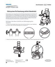
Bed X-ray of the lower leg using the wireless portable detector<br />
and the examinations are much quicker,<br />
which reduces patient waiting times. There is<br />
no read-out required as with cassette-based<br />
systems, meaning we save two to three<br />
minutes per X-ray. As the data is transferred<br />
digitally, I can see straight away whether an<br />
image has been successful. This saves a lot of<br />
time, particularly during routine X-rays.<br />
What advantages do the new digital<br />
X-ray system and the wireless portable<br />
detector offer you from a diagnostic<br />
point of view?<br />
The exposure is simply better, meaning the<br />
image quality is better too. X-rays that used<br />
to be complicated or virtually impossible,<br />
such as axial neck of femur X-rays, are now<br />
no trouble at all. This means we can give<br />
a much clearer diagnosis.<br />
In what circumstances do you still use<br />
the cassette-based X-ray device?<br />
When the digital detector is unavailable.<br />
Until recently, we were still using the<br />
cassette-based X-ray system in the intensive<br />
care unit, but only because the wireless<br />
connection for the digital device did not<br />
reach that far.<br />
How have organizational processes<br />
been changed by the X-ray system?<br />
We streamlined the radiology department<br />
some time ago. Now things are more likely<br />
to run to schedule, as procedures such<br />
as the read-out process of cassettes are<br />
omitted. The time we save can be spent<br />
on patients.<br />
Do you save on costs compared with<br />
the old system?<br />
If we can replace the imaging plate system<br />
completely, then yes. However, we need to<br />
safeguard against failure. But our first task is<br />
to check whether we can get rid of imaging<br />
plate technology completely from our dayto-day<br />
working processes. We hope this will<br />
happen in the not-too-distant future.<br />
What features would you like a future<br />
wireless portable detector to have?<br />
The detector could be flatter. But above all,<br />
it would be nice if it were suitable for use in<br />
extreme environments such as Afghanistan.<br />
To do this, it would need to be lighter and<br />
smaller, and the entire X-ray system would<br />
have to be waterproof, dust-proof and<br />
able to withstand temperatures of –20 to<br />
+50 degrees Celsius. Then there would be<br />
no need for us to read out the cassettes,<br />
as we have been doing until now.<br />
In addition, the data could then be<br />
transferred quickly to a larger medical<br />
facility. In difficult cases, for example,<br />
specialists can then be consulted<br />
at this facility.<br />
The print quality of this copy is not an accurate representation of the original.
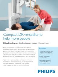
DigitalDiagnost TH/VM in the<br />
Federal <strong>Armed</strong> <strong>Forces</strong> <strong>Hospital</strong><br />
Clinic Profile<br />
Radiology Department, <strong>Hamburg</strong><br />
Federal <strong>Armed</strong> <strong>Forces</strong> <strong>Hospital</strong><br />
Offering 307 beds, <strong>Hamburg</strong> Federal<br />
<strong>Armed</strong> <strong>Forces</strong> <strong>Hospital</strong> is one of the five<br />
largest military hospitals in <strong>Germany</strong> and<br />
is located in <strong>Hamburg</strong>'s Wandsbek district.<br />
It is the largest military treatment facility<br />
in northern <strong>Germany</strong> and is a key part of<br />
<strong>Hamburg</strong>'s civilian healthcare network.<br />
Since January 2007, it has been an academic<br />
teaching hospital affiliated with the University<br />
Medical Centre <strong>Hamburg</strong>-Eppendorf (UKE).<br />
Department VIII — Radiology sees itself<br />
as a service provider for all other in-house<br />
departments as well as for patients who<br />
require immediate out-patient treatment.<br />
As well as patient safety and accurate<br />
diagnoses, the department aims to provide<br />
patient comfort and rapid availability<br />
of diagnostic findings. Naturally, the<br />
department offers an on-call radiology<br />
service for urgent examinations. It also<br />
carries out X-ray rounds that not only<br />
strengthen support for other departments,<br />
but are also used to provide training. What's<br />
more, it organizes lectures that are certified<br />
by the German Medical Association. The<br />
hospital is entirely "film-less," meaning<br />
image data is created, distributed and saved<br />
digitally. The report that corresponds to<br />
each image can also be loaded quickly and<br />
easily and saved on CD-ROM if necessary.<br />
The range of services offered by<br />
Department VIII — Radiology includes all<br />
conventional X-ray procedures (excluding<br />
mammography and angiography):<br />
• Computed tomography (MS spiral<br />
CT scan)<br />
• Digital radiography and digital archiving<br />
and distribution system (PACS)<br />
• Magnetic resonance tomography (MRT)<br />
including the possibility of examining<br />
patients on mechanical ventilation<br />
• Sonography<br />
• Needle aspiration biopsies and punch<br />
biopsies<br />
• Facet joint infiltrations<br />
• Periradicular therapy<br />
Its diagnostic focus is on:<br />
• Stage diagnosis of germ cell tumors<br />
of the testicles and of tumors in the<br />
head and throat<br />
• Medical imaging of diseases of the<br />
temporomandibular joint<br />
• Imaging of injuries to the<br />
musculoskeletal system<br />
The print quality of this copy is not an accurate representation of the original.
<strong>Philips</strong> Healthcare is a Royal <strong>Philips</strong><br />
Electronics Company<br />
You can contact us at:<br />
www.philips.com/healthcare<br />
healthcare@philips.com<br />
Asia<br />
+49 7031 463 2254<br />
Europe, Middle East, Africa<br />
+49 7031 463 2254<br />
Latin America<br />
+55 11 2125 0744<br />
North America<br />
+1 425 487 7000<br />
800 285 5585 (toll free, US only)<br />
Please visit www.philips.com/Digital_Radiography<br />
© 2011 Koninklijke <strong>Philips</strong> Electronics N.V.<br />
All rights are reserved.<br />
<strong>Philips</strong> Healthcare reserves the right to make changes in specifications and/or to discontinue any product at any time without notice or<br />
obligation and will not be liable for any consequences resulting from the use of this publication.<br />
Printed in The Netherlands.<br />
4522 962 71281 * MAR 2011<br />
The print quality of this copy is not an accurate representation of the original.