Head posture and dental wear evaluation of bruxist children ... - IBMC
Head posture and dental wear evaluation of bruxist children ... - IBMC
Head posture and dental wear evaluation of bruxist children ... - IBMC
Create successful ePaper yourself
Turn your PDF publications into a flip-book with our unique Google optimized e-Paper software.
Journal <strong>of</strong> Oral Rehabilitation 2007<br />
1<br />
2<br />
3<br />
4<br />
5<br />
6<br />
7<br />
8<br />
9<br />
10<br />
11<br />
12<br />
13<br />
14<br />
15<br />
16<br />
17<br />
18<br />
19<br />
20<br />
21<br />
22<br />
23<br />
24<br />
25<br />
26<br />
27<br />
28<br />
29<br />
30<br />
31<br />
32<br />
33<br />
34<br />
35<br />
36<br />
37<br />
38<br />
39<br />
40<br />
41<br />
42<br />
43<br />
44<br />
45<br />
46<br />
47<br />
48<br />
49<br />
2<br />
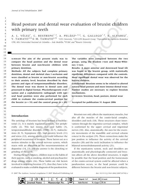
<strong>Head</strong> <strong>posture</strong> <strong>and</strong> <strong>dental</strong> <strong>wear</strong> <strong>evaluation</strong> <strong>of</strong> <strong>bruxist</strong> <strong>children</strong><br />
with primary teeth<br />
A. L. VÉLEZ * , C. RESTREPO *,† , A. PELÁEZ* ,†,‡ , G. GALLEGO *,† , E. ALVAREZ * ,<br />
V. TAMAYO †, § &M.TAMAYO †, *CES University, † CES-LPH Research Group, ‡ Especialista en Ingeniería Biomédica<br />
UPB, MSc Universidad Nacional de Colombia – Sede Medellín, § FUMC <strong>and</strong> Rosario University<br />
SUMMARY The aim <strong>of</strong> the present study was to<br />
compare the head position <strong>and</strong> the <strong>dental</strong> <strong>wear</strong><br />
between <strong>bruxist</strong> <strong>and</strong> non-<strong>bruxist</strong> <strong>children</strong> with<br />
primary dentition.<br />
Methods: All the subjects had complete primary<br />
dentition, <strong>dental</strong> <strong>and</strong> skeletal class I occlusion <strong>and</strong><br />
were classified as <strong>bruxist</strong> or non-<strong>bruxist</strong> according<br />
to their anxiety level, bruxism described by their<br />
parents <strong>and</strong> signs <strong>of</strong> temporom<strong>and</strong>ibular disorders.<br />
The <strong>dental</strong> <strong>wear</strong> was drawn in <strong>dental</strong> casts <strong>and</strong><br />
processed in digital format. Physiotherapeutic <strong>evaluation</strong><br />
<strong>and</strong> a cephalometric radiograph with natural<br />
head position were also performed for each<br />
child to evaluate the cranio-cervical position for<br />
the <strong>bruxist</strong> (n = 33) <strong>and</strong> the control group (n = 20).<br />
Introduction<br />
The aetiology <strong>of</strong> bruxism has been defined as multifactorial<br />
(1). It is mainly regulated centrally, not peripherally<br />
(2). This fact means that oral habits (3),<br />
temporom<strong>and</strong>ibular disorders (TMD) (4–7), malocclusions<br />
(8, 9), hypopnoea (10), high-anxiety levels (11)<br />
<strong>and</strong> stress (12), among others (13) could influence the<br />
peripheral occurrence <strong>of</strong> bruxism. These factors act as a<br />
motion stimulus to the central nervous system, which<br />
reacts with an alteration in the neurotransmission <strong>of</strong><br />
dopamine (14, 15) <strong>and</strong> the answer is the clenching or<br />
grinding <strong>of</strong> the teeth.<br />
During the early infancy, <strong>children</strong> react to the habits <strong>of</strong><br />
their parents, such as smoking, alcohol <strong>and</strong> psychoactive<br />
drugs among others (16). These habits are risk factors<br />
involved in acquiring bruxism (17), thus they have to be<br />
evaluated to make a reliable diagnosis <strong>of</strong> the parafunction.<br />
The variables were compared between the two<br />
groups, using the Student t-test <strong>and</strong> Mann–Whitney<br />
U-test.<br />
Results: A more anterior <strong>and</strong> downward head tilt<br />
was found in the <strong>bruxist</strong> group, with statistically<br />
significant differences compared with the controls.<br />
More significant <strong>dental</strong> <strong>wear</strong> was observed for the<br />
<strong>bruxist</strong> <strong>children</strong>.<br />
Conclusions: Bruxism seems to be related to altered<br />
natural head <strong>posture</strong> <strong>and</strong> more intense <strong>dental</strong> <strong>wear</strong>.<br />
Further studies are necessary to explore bruxism<br />
mechanisms.<br />
KEYWORDS: bruxism, head, <strong>posture</strong>, <strong>dental</strong> <strong>wear</strong><br />
Accepted for publication 31 August 2006<br />
Bruxism not only affects the masticatory muscles, but<br />
also all the muscles <strong>of</strong> the cranio-facial complex,<br />
shoulders <strong>and</strong> neck (18). These structures share innervations<br />
through the trigemino-cervical complex, which<br />
is conformed by the upper cervical <strong>and</strong> trigeminal<br />
nerves (18). Also, anatomically, the axes for the excentric<br />
movements <strong>of</strong> the m<strong>and</strong>ible <strong>and</strong> cervical column<br />
concur in the occiput (19). These connections make the<br />
jaw position to influence the activity <strong>of</strong> the cervical<br />
muscles (20) <strong>and</strong> the neck inclination to influence the<br />
bilateral sternocleidomastoid activity (21).<br />
If the masticatory system, neck <strong>and</strong> shoulders are<br />
anatomically <strong>and</strong> physiologically connected <strong>and</strong> bruxism<br />
affects all the above described structures, it might<br />
be possible that the head position <strong>and</strong> the homeostasis<br />
<strong>of</strong> the cranio-cervical system could be affected when a<br />
parafunction occurs, so the head <strong>posture</strong> could be<br />
different between the <strong>bruxist</strong> <strong>and</strong> non-<strong>bruxist</strong> subjects.<br />
ª 2007 The Authors. Journal compilation ª 2007 Blackwell Publishing Ltd doi: 10.1111/j.1365-2842.2007.01742.x<br />
J O R 1 7 4 2<br />
B<br />
Dispatch: 7.6.07 Journal: JOR CE: Senthil<br />
Journal Name Manuscript No. Author Received: No. <strong>of</strong> pages: 8 PE: Sri
2<br />
A. L. VÉLEZ et al.<br />
1<br />
2<br />
3<br />
4<br />
5<br />
6<br />
7<br />
8<br />
9<br />
10<br />
11<br />
12<br />
13<br />
14<br />
15<br />
16<br />
17<br />
18<br />
19<br />
20<br />
21<br />
22<br />
23<br />
24<br />
25<br />
26<br />
27<br />
28<br />
29<br />
30<br />
31<br />
32<br />
33<br />
34<br />
35<br />
36<br />
37<br />
38<br />
39<br />
40<br />
41<br />
42<br />
43<br />
44<br />
45<br />
46<br />
47<br />
48<br />
49<br />
The head <strong>posture</strong> could be affected by the skeletal<br />
(22) <strong>and</strong> <strong>dental</strong> occlusion (23, 24). During the mixed<br />
dentition, the <strong>dental</strong> occlusion changes (25), so the<br />
head <strong>posture</strong> could be affected (26). In the primary<br />
teeth period, the arch dimensions seem to be stable (27,<br />
28), so if there are changes in the head <strong>and</strong> cervical<br />
column <strong>posture</strong>, these might be due to other factors<br />
besides changes in occlusion, for example, the occurrence<br />
<strong>of</strong> oral parafunctions.<br />
The available evidence based dentistry is still not<br />
enough to support the multifactorial diagnosis <strong>of</strong> bruxism,<br />
especially in <strong>children</strong>. Historically, the background<br />
<strong>of</strong> bruxism has been confined to the visual examination<br />
<strong>of</strong> <strong>dental</strong> <strong>wear</strong> (1, 4) <strong>and</strong> the reports <strong>of</strong> grinding by the<br />
parents.<br />
The difference between normal <strong>and</strong> pathological<br />
<strong>dental</strong> <strong>wear</strong> has been previously described in the mixed<br />
dentition with digital analysis (29). Dental <strong>wear</strong> produced<br />
by bruxism is characterized by a plane surface<br />
with a central zone that sometimes reaches the dentine,<br />
surrounded by enamel zones (30). Waltimo et al. (31)<br />
found that the most common <strong>dental</strong> facets in adults are<br />
the ones with horizontal shape that indicates the<br />
occurrence <strong>of</strong> a grinding pattern rather than a clenching<br />
pattern <strong>of</strong> bruxism.<br />
There are sophisticated methods to measure the<br />
<strong>dental</strong> <strong>wear</strong> related to bruxism (29, 32–36), but other<br />
factors contributing to parafunction, such as body<br />
<strong>posture</strong>, have not been measured together with the<br />
<strong>dental</strong> <strong>wear</strong>, to find a better underst<strong>and</strong>ing <strong>of</strong> the<br />
peripheral multifactorial aetiology <strong>of</strong> bruxism.<br />
The aim <strong>of</strong> the present study was to compare head<br />
<strong>posture</strong> <strong>and</strong> <strong>dental</strong> <strong>wear</strong> between <strong>bruxist</strong> <strong>and</strong> non<strong>bruxist</strong><br />
<strong>children</strong> with primary dentition.<br />
Materials <strong>and</strong> methods<br />
A case–control study was performed. The procedures,<br />
possible discomforts or risks, as well as possible benefits<br />
were fully explained to the participant patients <strong>and</strong><br />
their parents, <strong>and</strong> the informed consent from their<br />
parents was obtained prior to the investigation.<br />
Subjects<br />
Participating <strong>children</strong> were selected from Susalud<br />
(A Clinic <strong>of</strong> the Colombian Private Health Service)<br />
<strong>and</strong> CES Sabaneta (Clinic <strong>of</strong> the CES University Dental<br />
School). All the subjects (<strong>bruxist</strong> <strong>and</strong> non-<strong>bruxist</strong>) were<br />
required to be healthy, with normal facial morphology,<br />
complete primary teeth, absence <strong>of</strong> other types <strong>of</strong> oral<br />
habits, presence <strong>of</strong> <strong>dental</strong> <strong>wear</strong> <strong>and</strong> with no history <strong>of</strong><br />
trauma.<br />
The sample size was calculated with a confidence <strong>of</strong><br />
95% <strong>and</strong> a statistical power <strong>of</strong> 80%. The number <strong>of</strong><br />
subjects required in each group to make the comparisons<br />
was 20.<br />
Inclusion <strong>and</strong> exclusion criteria<br />
The exclusion criteria were skeletal malocclusions<br />
confirmed with cephalometric X-rays (37, 38) <strong>and</strong><br />
<strong>dental</strong> malocclusions confirmed with <strong>dental</strong> casts. The<br />
reports <strong>of</strong> respiratory diseases, presence <strong>of</strong> mouth<br />
breathing <strong>and</strong> functional alterations in the body <strong>posture</strong><br />
were also reasons to exclude patients from the<br />
study. The asymmetry in the <strong>children</strong>’s legs or any<br />
other mobility alteration that could generate changes in<br />
head <strong>posture</strong> due to anatomically detectable reasons<br />
were exclusion criteria as well.<br />
An <strong>evaluation</strong> <strong>of</strong> the temporom<strong>and</strong>ibular joint (TMJ)<br />
was performed on all the <strong>children</strong> together with a<br />
questionnaire <strong>and</strong> a clinical examination, according to<br />
Bernal <strong>and</strong> Tsamtsouris (39).<br />
Children’s anxiety was measured using the Conners’<br />
Parents Rating Scales (40) (CPRS). Both instruments,<br />
Tsamtsouris <strong>and</strong> Bernal <strong>and</strong> CPRS had been previously<br />
used to diagnose bruxism in <strong>children</strong> (16).<br />
Children were included in the <strong>bruxist</strong> group (n = 33)<br />
when their anxiety level was above 0Æ75% according to<br />
the CPRS, presented two or more signs <strong>of</strong> TMD<br />
according to Bernal <strong>and</strong> Tsamtsouris <strong>and</strong> accomplished<br />
the American Sleep Disorders Association (41) criteria<br />
for sleep bruxism:<br />
1 The <strong>children</strong> parents indicated the occurrence <strong>of</strong><br />
tooth-grinding or tooth-clenching during sleep.<br />
2 No other medical or mental disorders (e.g., sleeprelated<br />
epilepsy, accounts for the abnormal movements<br />
during sleep).<br />
3 Other sleep disorders (e.g., obstructive sleep apnoea<br />
syndrome) were absent.<br />
All <strong>children</strong> not accomplishing the above criteria,<br />
were included in the control group.<br />
Seventy-two <strong>children</strong> were initially evaluated <strong>and</strong> 53<br />
were finally included in the study, 33 in the <strong>bruxist</strong><br />
group <strong>and</strong> 20 in the control group. Eleven subjects were<br />
excluded because they presented exfoliating movements<br />
<strong>of</strong> the primary teeth or appearance <strong>of</strong> eruption <strong>of</strong><br />
ª 2007 The Authors. Journal compilation ª 2007 Blackwell Publishing Ltd
1 XXX 3<br />
1<br />
2<br />
3<br />
4<br />
5<br />
6<br />
7<br />
8<br />
9<br />
10<br />
11<br />
12<br />
13<br />
14<br />
15<br />
16<br />
17<br />
18<br />
19<br />
20<br />
21<br />
22<br />
23<br />
24<br />
25<br />
26<br />
27<br />
28<br />
29<br />
30<br />
31<br />
32<br />
33<br />
34<br />
35<br />
36<br />
37<br />
38<br />
39<br />
40<br />
41<br />
42<br />
43<br />
44<br />
45<br />
46<br />
47<br />
48<br />
49<br />
any permanent molar. Three presented skeletal malocclusions<br />
in the cephalogram, three were excluded by<br />
difficulties in their behaviour during any <strong>of</strong> the<br />
required initial procedures, one presented functional<br />
scoliosis, one showed legs asymmetry <strong>and</strong> one presented<br />
cerebral palsy.<br />
All the <strong>children</strong> were 3–6 years. For the <strong>bruxist</strong><br />
group, the mean age was 56Æ70 7Æ22 months,<br />
while for the non-<strong>bruxist</strong> <strong>children</strong> it was 55Æ20 <br />
7Æ89 months. As it can be observed, with regard to<br />
their chronological age the groups were homogeneous.<br />
Techniques<br />
The risk factors to acquire TMD were assessed by<br />
questioning those in charge <strong>of</strong> the <strong>children</strong>, using the<br />
questionnaire <strong>of</strong> the Bernal <strong>and</strong> Tsamtsouris test. The<br />
clinical <strong>evaluation</strong> <strong>of</strong> TMD included the auscultation <strong>of</strong><br />
TMJ sounds, the palpation <strong>of</strong> discontinuous condylar<br />
movement, measurement <strong>of</strong> the maximum opening <strong>of</strong><br />
the mouth <strong>and</strong> deviation <strong>of</strong> the m<strong>and</strong>ible during<br />
opening.<br />
The physiotherapeutic <strong>evaluation</strong> (42) was performed<br />
to exclude any possible anatomical disturbance<br />
<strong>of</strong> the cervical column that could affect the head<br />
<strong>posture</strong> or the crani<strong>of</strong>acial growth <strong>of</strong> the studied<br />
<strong>children</strong>. The test included a questionnaire to ask the<br />
parents about family history that could indicate possible<br />
alterations in the body <strong>posture</strong> <strong>of</strong> the subjects.<br />
Then, the real <strong>and</strong> apparent measurements <strong>of</strong> the legs<br />
were taken with the subjects in supine position with a<br />
st<strong>and</strong>ardized technique. The examination also included<br />
impression <strong>of</strong> the plantar foot with the child in<br />
bipedestation over a non-sliding surface. With this<br />
procedure, the feet track <strong>of</strong> the subject was copied.<br />
Additionally, photographs <strong>of</strong> the front, back <strong>and</strong> both<br />
sides’ views <strong>of</strong> each child were taken. The data<br />
obtained in the physiotherapeutic examination were<br />
analysed separately by two different physiotherapists at<br />
different moments to detect abnormalities or asymmetries.<br />
The upper <strong>and</strong> lower <strong>dental</strong> arches <strong>of</strong> all subjects<br />
were reproduced from alginate impressions cast in<br />
<strong>dental</strong> stone with a st<strong>and</strong>ardized technique.<br />
The <strong>dental</strong> <strong>wear</strong> <strong>of</strong> all the casts was drawn, acquired<br />
in digital format <strong>and</strong> processed automatically. The<br />
technique to analyse it was previously reported (29).<br />
The size <strong>and</strong> shape <strong>of</strong> the <strong>dental</strong> <strong>wear</strong> were calculated<br />
for each <strong>dental</strong> cast.<br />
The size <strong>of</strong> the <strong>dental</strong> <strong>wear</strong> was quantified through its<br />
area (mm 2 ) <strong>and</strong> perimeter (mm) <strong>and</strong> the shape by the<br />
roundness <strong>and</strong> the form factor (D factor) (29), which<br />
are non-dimensional. The last two measurements were<br />
used to calculate the format <strong>of</strong> objects without geometrical<br />
shapes (29).<br />
For D factor, the following ratio was used:<br />
pffiffiffi<br />
a<br />
D factor ¼<br />
p<br />
where a is the area (mm 2 ) <strong>and</strong> p the perimeter (mm).<br />
Each X-ray was taken with an Orthophos Plus<br />
Ceph( * ) for lateral cephalograms. The machine was<br />
vertically adjustable; it had a st<strong>and</strong>ardized focus – film<br />
distance <strong>of</strong> 190 cm <strong>and</strong> a distance from the film to the<br />
medial plane <strong>of</strong> 10 cm. The subject stood up without<br />
fixation in orthoposition after balancing forward <strong>and</strong><br />
backward three times, with the teeth together <strong>and</strong> the<br />
lips in rest, looking to a light in a mirror, located<br />
perpendicular to the eyes <strong>of</strong> the child. This position<br />
made sure that the head <strong>and</strong> the neck were in natural<br />
position. The exposures were taken at 60–80 kv <strong>and</strong><br />
32 mAs. A vertical 0Æ5 mm wide wire was put in front<br />
<strong>of</strong> the cassette to register the perfect vertical line (VV).<br />
The technique used to take the lateral cephalogram<br />
was the natural head <strong>posture</strong>, described previously by<br />
different authors (43). It is a reproducible technique<br />
(44–46) <strong>and</strong> allows the clinician to evaluate the natural<br />
position <strong>of</strong> the cervical vertebras <strong>and</strong> the inclination <strong>of</strong><br />
the cervical column <strong>and</strong> head <strong>posture</strong>.<br />
Afterwards, the lateral cephalograms were scanned<br />
<strong>and</strong> traced digitally according to Solow <strong>and</strong> Tallgren (43),<br />
in a dark room, using a MATLAB 5Æ3( † MathWorks, Inc.,<br />
4 MA, USA) program. Based on the vertical reference,<br />
a horizontal line was traced perpendicular to the vertical<br />
one (HOR). These two lines were the references to<br />
calculate the angles between head <strong>and</strong> neck in the<br />
cephalogram. All the measurements to evaluate the head<br />
<strong>and</strong> cervical column <strong>posture</strong> are described in Fig. 1.<br />
The following angles were measured to analyse the<br />
head <strong>and</strong> cervical column <strong>posture</strong>:<br />
- Angle between tangent (CVT) to the cervical vertebra<br />
(cv4ip) <strong>and</strong> VV: the wider the angle, the more relevant<br />
the kyphosis <strong>of</strong> the cervical column.<br />
- Angle between CVT <strong>and</strong> HOR: the narrower the<br />
angle, the more significant the anterior tilt <strong>of</strong> the head.<br />
3 *Sirona Co, Alemania<br />
†<br />
ª 2007 The Authors. Journal compilation ª 2007 Blackwell Publishing Ltd
4<br />
A. L. VÉLEZ et al.<br />
1<br />
2<br />
3<br />
4<br />
5<br />
6<br />
7<br />
8<br />
9<br />
10<br />
11<br />
12<br />
13<br />
14<br />
15<br />
16<br />
17<br />
18<br />
19<br />
20<br />
21<br />
22<br />
23<br />
24<br />
25<br />
26<br />
27<br />
28<br />
29<br />
30<br />
31<br />
32<br />
33<br />
34<br />
35<br />
36<br />
37<br />
38<br />
39<br />
40<br />
41<br />
42<br />
43<br />
44<br />
45<br />
46<br />
47<br />
48<br />
49<br />
LOW RESOLUTION FIG<br />
HOR<br />
CV2ip<br />
CV4ip<br />
CVT OPT<br />
7;10 Fig. 1. xxxxxxxxxxxxxx.<br />
- Angle between the tangent (OPT) to odontoides<br />
(cv2ip) <strong>and</strong> VV: the wider the angle, the more relevant<br />
the kyphosis <strong>of</strong> the cervical column.<br />
- Angle between OPT <strong>and</strong> HOR: the narrower the<br />
angle, the more relevant the anterior tilt <strong>of</strong> the head.<br />
The examiners evaluating the condition <strong>of</strong> bruxism<br />
⁄ non-bruxism were not aware as regards who<br />
performed the physiotherapeutic <strong>evaluation</strong> <strong>and</strong> who<br />
analysed the <strong>dental</strong> <strong>wear</strong> <strong>and</strong> the X-ray images.<br />
Error <strong>of</strong> method<br />
St<strong>and</strong>ardizations <strong>of</strong> the examiners <strong>and</strong> calibration <strong>of</strong><br />
all the techniques to evaluate the <strong>children</strong> regarding<br />
the clinical examination, TMD <strong>and</strong> anxiety were<br />
made on 12 subjects different from the ones included<br />
in the investigation. The Intra-tester <strong>and</strong> intertester<br />
error was not statistically significant (ICC >0Æ9 <strong>and</strong><br />
Kappa >0Æ7).<br />
A calibration <strong>of</strong> the X-ray technique <strong>and</strong> a st<strong>and</strong>ardization<br />
<strong>of</strong> the digital tracing <strong>of</strong> the cephalogram were<br />
also performed. The tracing <strong>of</strong> the cephalogram was<br />
st<strong>and</strong>ardized between two investigators with 10 X-rays,<br />
scanned <strong>and</strong> traced three times each by each <strong>of</strong> the two<br />
investigators. To determine the Intra-tester <strong>and</strong> intertester<br />
reliability, the intra-class correlation coefficient<br />
was applied (ICC >0Æ6).<br />
The <strong>dental</strong> <strong>wear</strong> was traced only by one investigator<br />
which intra-class error was not statistically significant<br />
(ICC >0Æ7).<br />
Statistical analysis<br />
VV<br />
Univariant <strong>and</strong> bivariant analysis were performed for<br />
each variable, using frequencies <strong>and</strong> mean values. The<br />
bivariant analysis was carried out using Student’s<br />
S-N<br />
Real horizontal line (HOR)<br />
Real vertical line (VV)<br />
Tangent to odontoides (cv2ip)<br />
Tangent to cervical vertebras (cv4ip)<br />
t-test, or Mann–Whitney U-tests, depending on the<br />
normality <strong>of</strong> the variables distribution. Distributions<br />
were tested using the Shapiro–Wilk test.<br />
Results<br />
The non-<strong>bruxist</strong> group was composed <strong>of</strong> nine girls <strong>and</strong><br />
11 boys, while in the <strong>bruxist</strong> group there were 14 girls<br />
<strong>and</strong> 19 boys. In both groups there were more boys 55%<br />
<strong>and</strong> 58% respectively for each group (Table 1).<br />
The four outcome parameters <strong>of</strong> <strong>dental</strong> <strong>wear</strong> were<br />
compared between the <strong>bruxist</strong> <strong>and</strong> non-<strong>bruxist</strong> groups<br />
(Table 2). There was no statistically significant difference<br />
for the shape (Roundness <strong>and</strong> D factor) <strong>of</strong> the<br />
<strong>dental</strong> <strong>wear</strong> between the <strong>bruxist</strong> <strong>and</strong> the non-<strong>bruxist</strong><br />
<strong>children</strong> (P >0Æ05). The size <strong>of</strong> <strong>dental</strong> <strong>wear</strong> (area <strong>and</strong><br />
perimeter) showed higher values in the case <strong>of</strong> <strong>bruxist</strong><br />
<strong>children</strong> (P
1 XXX 5<br />
1<br />
2<br />
3<br />
4<br />
5<br />
6<br />
7<br />
8<br />
9<br />
10<br />
11<br />
12<br />
13<br />
14<br />
15<br />
16<br />
17<br />
18<br />
19<br />
20<br />
21<br />
22<br />
23<br />
24<br />
25<br />
26<br />
27<br />
28<br />
29<br />
30<br />
31<br />
32<br />
33<br />
34<br />
35<br />
36<br />
37<br />
38<br />
39<br />
40<br />
41<br />
42<br />
43<br />
44<br />
45<br />
46<br />
47<br />
48<br />
49<br />
Table 2. Comparison between<br />
<strong>dental</strong> <strong>wear</strong> measurements in <strong>bruxist</strong><br />
<strong>and</strong> non-<strong>bruxist</strong> <strong>children</strong><br />
<strong>bruxist</strong> group. However, it must not be taken as the<br />
only sign to diagnose this parafunctional activity.<br />
Some studies in the literature have left aside factors<br />
related to the parafunction that give important information<br />
regarding the aetiology <strong>of</strong> bruxism in the<br />
central <strong>and</strong> peripheral nervous system (1), such as<br />
body <strong>and</strong> cervical column positions (51). There are no<br />
specific measurement methods or criteria to diagnose<br />
bruxism (52), but as it has a multifactorial aetiology<br />
(53), the study <strong>of</strong> its associated factors (54) could lead to<br />
an accurate diagnosis <strong>of</strong> the parafunction. The diagnosis<br />
<strong>of</strong> bruxism, as it was performed in this study, should be<br />
multifactorial <strong>and</strong> include the associated peripherally<br />
factors, such as the analysis <strong>of</strong> the <strong>dental</strong> <strong>wear</strong> digitally,<br />
<strong>evaluation</strong> <strong>of</strong> the TMD <strong>and</strong> alterations in anxiety.<br />
In this study, <strong>dental</strong> <strong>wear</strong> present in <strong>bruxist</strong> <strong>and</strong> non<strong>bruxist</strong><br />
<strong>children</strong> was used to compare the size <strong>and</strong> shape<br />
differences <strong>of</strong> <strong>dental</strong> <strong>wear</strong> between the two groups. It<br />
Criteria Diagnosis n Mean s.d. P-value<br />
Upper arch roundness <strong>of</strong> <strong>dental</strong> <strong>wear</strong> Bruxist 33 0Æ12 0Æ07 0Æ341<br />
Non-<strong>bruxist</strong> 20 0Æ14 0Æ13<br />
Lower arch roundness <strong>of</strong> <strong>dental</strong> <strong>wear</strong> Bruxist 33 0Æ17 0Æ26 0Æ788<br />
Non-<strong>bruxist</strong> 20 0Æ15 0Æ09<br />
Upper arch D factor <strong>of</strong> <strong>dental</strong> <strong>wear</strong> Bruxist 33 11Æ18 1Æ98 0Æ310<br />
Non-<strong>bruxist</strong> 20 10Æ59 2Æ18<br />
Lower arch D factor <strong>of</strong> <strong>dental</strong> <strong>wear</strong> Bruxist 33 10Æ52 2Æ60 0Æ086<br />
Non-<strong>bruxist</strong> 20 9Æ15 3Æ00<br />
Upper arch area <strong>of</strong> <strong>dental</strong> <strong>wear</strong> (mm 2 ) Bruxist 33 33Æ24 20Æ52 0Æ004<br />
Non-<strong>bruxist</strong> 20 20Æ26 10Æ45<br />
Lower arch area <strong>of</strong> <strong>dental</strong> <strong>wear</strong> (mm 2 ) Bruxist 33 20Æ23 13Æ92 0Æ003<br />
Non-<strong>bruxist</strong> 20 11Æ22 6Æ82<br />
Upper arch perimeter <strong>of</strong> <strong>dental</strong> <strong>wear</strong> (mm) Bruxist 33 59Æ35 30Æ54 0Æ058<br />
Non-<strong>bruxist</strong> 20 44Æ47 19Æ86<br />
Lower arch perimeter <strong>of</strong> <strong>dental</strong> <strong>wear</strong> (mm) Bruxist 33 45Æ42 27Æ01 0Æ008<br />
Non-<strong>bruxist</strong> 20 29Æ42 15Æ23<br />
8 Significance P
6<br />
A. L. VÉLEZ et al.<br />
1<br />
2<br />
3<br />
4<br />
5<br />
6<br />
7<br />
8<br />
9<br />
10<br />
11<br />
12<br />
13<br />
14<br />
15<br />
16<br />
17<br />
18<br />
19<br />
20<br />
21<br />
22<br />
23<br />
24<br />
25<br />
26<br />
27<br />
28<br />
29<br />
30<br />
31<br />
32<br />
33<br />
34<br />
35<br />
36<br />
37<br />
38<br />
39<br />
40<br />
41<br />
42<br />
43<br />
44<br />
45<br />
46<br />
47<br />
48<br />
49<br />
to search the association <strong>of</strong> the TMD with the head<br />
<strong>posture</strong>. However, controversy does exist regarding the<br />
relationship between TMD <strong>and</strong> head <strong>posture</strong>. Some<br />
authors support it (62, 63), but their methodology is<br />
not good enough to establish the relationship between<br />
the TMD <strong>and</strong> the anterior head <strong>posture</strong> in <strong>children</strong>.<br />
Some <strong>of</strong> them used a stethoscope to detect only TMJ<br />
sounds (62), leaving aside other TMD that are not<br />
audible. Others used the Helkimo’s index (64), whose<br />
measurements <strong>of</strong> the muscle tenderness <strong>and</strong> pain are<br />
not reliable in <strong>children</strong> (63). However, other authors<br />
have better evidence to conclude about the poor<br />
relationship between TMD <strong>and</strong> head <strong>posture</strong> (65, 66).<br />
Bruxism is mainly centrally regulated, not peripherally<br />
(2, 4). Alterations in body positions have been<br />
identified <strong>and</strong> described in the literature, as one <strong>of</strong> the<br />
peripherical factors that could initiate the parafunction<br />
(67, 68), while in the central nervous system, the<br />
partial hypoxia (69) has been defined as one <strong>of</strong> the<br />
factors that could generate the failure in the neurotransmission<br />
<strong>of</strong> dopamine (15).<br />
The oral airway resistance increases with modest<br />
degrees <strong>of</strong> head <strong>and</strong> neck flexions in healthy adult<br />
humans (70), while in healthy infants, hyperflexion<br />
<strong>of</strong> the head has been shown to affect the airflow,<br />
airway patency <strong>and</strong> pulmonary mechanisms (71, 72).<br />
Additionally, sleep bruxism has been correlated to<br />
hypopnoea (10) <strong>and</strong> increasing airway patency (69).<br />
In this work, more anterior <strong>and</strong> downward head<br />
<strong>posture</strong>s <strong>and</strong> kyphotic necks were found in the<br />
<strong>bruxist</strong> group, with hyperflexion <strong>of</strong> the head <strong>posture</strong>.<br />
These characteristics could affect the airflow in the<br />
<strong>bruxist</strong> <strong>children</strong> <strong>and</strong> could be part <strong>of</strong> the aetiology <strong>of</strong><br />
their parafunction. Now, the question is whether<br />
physiotherapeutic therapies applied aiming at changing<br />
head <strong>posture</strong> could work as a therapeutic option<br />
for <strong>bruxist</strong> <strong>children</strong>.<br />
The anterior <strong>and</strong> downward head <strong>posture</strong>s, like the<br />
ones found in the <strong>bruxist</strong> <strong>children</strong> in this study, make<br />
the masticatory muscles to be more hypertonic (73).<br />
This finding coincides with the muscular signs found<br />
by other authors (74), when the parafunction is<br />
exacerbated.<br />
This study did not explore the relationship between<br />
the m<strong>and</strong>ibular rotation <strong>and</strong> the head <strong>posture</strong>, because<br />
we would need a bigger sample to match the <strong>children</strong> for<br />
malocclusions class I, II <strong>and</strong> III. However, observations <strong>of</strong><br />
past studies (51, 74) indicate that anterior <strong>and</strong> downward<br />
head <strong>posture</strong>s affect the m<strong>and</strong>ibular position.<br />
The results <strong>of</strong> the present research showed that if<br />
the parafunction has a multifactorial aetiology, then<br />
the diagnosis <strong>and</strong> the treatment has to be multifactorial<br />
as well.<br />
Conclusions<br />
The head <strong>posture</strong>s found in the <strong>bruxist</strong> group were<br />
more anterior <strong>and</strong> downward than the ones found in<br />
the control group.<br />
It is always important to make a multifactorial<br />
diagnosis <strong>of</strong> the parafunction to establish the individual<br />
causes <strong>of</strong> bruxism in each case <strong>and</strong> determine the best<br />
therapeutic alternative for each subject.<br />
Further work is required to underst<strong>and</strong> whether head<br />
<strong>posture</strong>s are causes or consequences <strong>of</strong> bruxism.<br />
Acknowledgment<br />
The authors acknowledge the financial support to this<br />
study from Susalud EPS <strong>and</strong> CES University.<br />
References<br />
1. Negoro T, Briggs J, Plesh O, Nielsen I, McNeill C, Miller AJ.<br />
Bruxing patterns in <strong>children</strong> compared to intercuspal clenching<br />
<strong>and</strong> chewing as assessed with <strong>dental</strong> models, electromyography<br />
<strong>and</strong> incisor jaw tracing: preliminary study. ASDC<br />
J Dent Child. 1998;65:449–458.<br />
2. Lobbezzo F, Naeije , M . Bruxism is mainly regulated centrally,<br />
not peripherally. J Oral Rehabil. 2001;28:1085–1091.<br />
3. Castelo PM, Gaviao MB, Pereira LJ, Bonjardim LR. Relationship<br />
between oral parafunctional ⁄ nutritive sucking habits <strong>and</strong><br />
temporom<strong>and</strong>ibular joint dysfunction in primary dentition.<br />
Int J Paediatr Dent. 2005;15:29–36.<br />
4. Camparis CM, Siqueira JT. Sleep bruxism: clinical aspects <strong>and</strong><br />
characteristics in patients with <strong>and</strong> without chronic or<strong>of</strong>acial<br />
pain. Oral Surg Oral Med Oral Pathol Oral Radiol Endod.<br />
2006;101:188–193.<br />
5. Bonjardim LR, Gaviao MB, Pereira LJ, Castelo PM, Garcia RC.<br />
Signs <strong>and</strong> symptoms <strong>of</strong> temporom<strong>and</strong>ibular disorders in<br />
adolescents. Pesqui Odontol Bras. 2005;19:93–98.<br />
6. Magnusson T, Egermarki I, Carlsson GE. A prospective<br />
investigation over two decades on signs <strong>and</strong> symptoms <strong>of</strong><br />
temporom<strong>and</strong>ibular disorders <strong>and</strong> associated variables. A final<br />
summary. Acta Odontol Sc<strong>and</strong>. 2005;63:99–109.<br />
7. Molina OF, dos Santos J, Mazzetto M, Nelson S, Nowlin T,<br />
Mainieri ET. Oral jaw behaviors in TMD <strong>and</strong> bruxism:<br />
a comparison study by severity <strong>of</strong> bruxism. Cranio.<br />
2001;19:114–122.<br />
8. Demir A, Uysal T, Guray E, Basciftci FA. The relationship<br />
between bruxism <strong>and</strong> occlusal factors among seven- to 19-<br />
year-old Turkish <strong>children</strong>. Angle Orthod. 2004;74:672–676.<br />
ª 2007 The Authors. Journal compilation ª 2007 Blackwell Publishing Ltd
1 XXX 7<br />
1<br />
2<br />
3<br />
4<br />
5<br />
6<br />
7<br />
8<br />
9<br />
10<br />
11<br />
12<br />
13<br />
14<br />
15<br />
16<br />
17<br />
18<br />
19<br />
20<br />
21<br />
22<br />
23<br />
24<br />
25<br />
26<br />
27<br />
28<br />
29<br />
30<br />
31<br />
32<br />
33<br />
34<br />
35<br />
36<br />
37<br />
38<br />
39<br />
40<br />
41<br />
42<br />
43<br />
44<br />
45<br />
46<br />
47<br />
48<br />
49<br />
9. Sari S, Sonmez H. The relationship between occlusal factors<br />
<strong>and</strong> bruxism in permanent <strong>and</strong> mixed dentition in Turkish<br />
<strong>children</strong>. J Clin Pediatr Dent. 2001;25:191–194.<br />
10. Oksenberg A, Arons E. Sleep bruxism related to obstructive<br />
sleep apnea: the effect <strong>of</strong> continuous positive airway pressure.<br />
5 Sleep Med. 2002;3:513–515.<br />
11. Manfredini D, L<strong>and</strong>i N, Fantoni F, Segù M, Bosco M. Anxiety<br />
symptoms in clinically diagnosed bruxers. J Oral Rehabil.<br />
2005;32:584–588.<br />
12. Tsai CM, Chou SL, Gale EN, Mccall JR. Human masticatory<br />
muscle activity <strong>and</strong> jaw position under experimental stress.<br />
J Oral Rehabil. 2002;29:44–51.<br />
13. Bayardo RE, Mejia JJ, Orozco S, Montoya K. Etiology <strong>of</strong> oral<br />
habits. ASDC J Dent Child. 1996;63:350–353.<br />
14. Lobbezoo F, Soucy JP, Montplaisir JY, Lavigne GJ. Striatal d2<br />
receptor binding in sleep bruxism: a controlled study with<br />
iodine-123-iodobenzamide <strong>and</strong> single-photon-emission computed<br />
tomography. J Dent Res. 1996;75:1804–1810.<br />
15. Lobbezoo F, Soucy JP, Hartman NG, Montplaisir JY, Lavigne<br />
GJ. Effects <strong>of</strong> the d2 receptor agonist bromocriptine on sleep<br />
bruxism <strong>of</strong> two single-patient clinical trials. J Dent Res.<br />
1997;76:1610–1614.<br />
16. Restrepo CC, Alvarez E, Jaramillo C, Velez C, Valencia I.<br />
Effects <strong>of</strong> psychological techniques on bruxism in <strong>children</strong><br />
with primary teeth. J Oral Rehabil. 2001;28:354–360.<br />
17. Ohayon MM, Li KK, Guilleminault C. Risk factors for sleep<br />
bruxism in the general population. Chest. 2001;119:<br />
53–61.<br />
18. Friedman MH, Weisberg J. The craniocervical connection:<br />
a retrospective analysis <strong>of</strong> 300 whiplash patients with<br />
cervical <strong>and</strong> temporom<strong>and</strong>ibular disorders. Cranio. 2000;18:<br />
163–167.<br />
19. Austin DG. Special considerations in or<strong>of</strong>acial pain <strong>and</strong><br />
headache. Dent Clin North Am. 1997;41:325–339.<br />
20. Zuñiga C, Miralles R, Mena B, Montt R, Moran D, Sant<strong>and</strong>er<br />
H, Moya H. Influence <strong>of</strong> variation in jaw <strong>posture</strong> on<br />
sternocleidomastoid <strong>and</strong> trapezius eletromyographic activity.<br />
Cranio. 1995;13:157–162.<br />
21. Sant<strong>and</strong>er H, Miralles R, Perez J, Valenzuela S, Ravera MJ,<br />
Ormeno G, Villegas R. Effects <strong>of</strong> head <strong>and</strong> neck inclination on<br />
bilateral sternocleidomastoid EMG activity in healthy subjects<br />
<strong>and</strong> in patients with myogenic cranio-cervical-m<strong>and</strong>ibular<br />
dysfunction. Cranio. 2000;18:181–191.<br />
22. D’Attilio M, Caputi S, Epifania E, Festa F, Tecco S. Evaluation<br />
<strong>of</strong> cervical <strong>posture</strong> <strong>of</strong> <strong>children</strong> in skeletal class I, II, <strong>and</strong> III.<br />
Cranio. 2005;23:219–228.<br />
23. Cesar GM, Tosato Jde P, Biasotto-Gonzalez DA. Correlation<br />
between occlusion <strong>and</strong> cervical <strong>posture</strong> in patients with<br />
9 bruxism. Compend Contin Educ Dent. 2006;27:463–468.<br />
24. Gadotti IC, Berzin F, Biasotto-Gonzalez DA. Preliminary<br />
rapport on head <strong>posture</strong> <strong>and</strong> muscle activity in subjects with<br />
class I <strong>and</strong> II. J Oral Rehabil. 2005;32:794–799.<br />
25. Slaj M, Jezina MA, Lauc T, Rajic-Mestrovic S, Miksic M.<br />
Longitudinal <strong>dental</strong> arch changes in the mixed dentition.<br />
Angle Orthod. 2003;73:509–514.<br />
26. Solow B, Sonnesen L. <strong>Head</strong> <strong>posture</strong> <strong>and</strong> malocclusions. Eur<br />
J Orthod. 1998;20:685–693.<br />
27. Bishara SE, Jakobsen JR, Treder J, Nowak A. Arch length<br />
changes from 6 weeks to 45 years. Angle Orthod. 1998;<br />
68:69–74.<br />
28. Moorrees CF, Reed RB. Changes in <strong>dental</strong> arch dimensions<br />
expressed on the basis <strong>of</strong> tooth eruption as a measure <strong>of</strong><br />
6 biologic age. J Dent Res. 1965;???:44–129.<br />
29. Restrepo C, Peláez A, Alvarez E, Paucar C, Abad P. Digital<br />
imaging <strong>of</strong> patterns <strong>of</strong> <strong>dental</strong> <strong>wear</strong> to diagnose bruxism in<br />
<strong>children</strong>. Int J Pediatric Dent. 2006;16:278–285.<br />
30. Mehl A, Gloger W, Kunzelmann KH, Hickel R. A new optical<br />
3-D device for the detection <strong>of</strong> <strong>wear</strong>. J Dent Res.<br />
1997;76:1799–1807.<br />
31. Waltimo A, Nystrom M, Kononen M. Bite force <strong>and</strong> dent<strong>of</strong>acial<br />
morphology in men with severe <strong>dental</strong> attrition. Sc<strong>and</strong><br />
J Dent Res. 1994;102:92–96.<br />
32. Ramfjord SP. Bruxism, a clinical <strong>and</strong> electromyographic study.<br />
J Am Dent Assoc. 1961;62:22–44.<br />
33. Delong R, Douglas WH, Sakaguchi RL, Pintado MR. The <strong>wear</strong><br />
on <strong>dental</strong> porcelain in an artificial mouth. Dent Mater J.<br />
1986;2:214–219.<br />
34. Adams LP, Wilding RJ. Tooth <strong>wear</strong> measurements using a<br />
reflex microscope. J Oral Rehabil. 1988;15:605–613.<br />
35. Adams LP, Jooste CH, Thomas CJ, Harris AM. Biostereometric<br />
quantification <strong>of</strong> clinical denture tooth <strong>wear</strong>. J Oral Rehabil.<br />
1996;23:667–674.<br />
36. Ungar PS, Brown CA, Bergstrom TS, Walkers A. Quantification<br />
<strong>of</strong> <strong>dental</strong> micro<strong>wear</strong> by t<strong>and</strong>em scanning confocal<br />
microscopy <strong>and</strong> scale-sensitive fractal analysis. Scanning.<br />
2003;25:185–193.<br />
37. Vann WF Jr, Dilley GJ, Nelson RM. A cepalometric analysis<br />
for the child in the primary dentition. ASDC J Dent Child.<br />
1978;45:45–52.<br />
38. Kocadereli I, Telli AE. Evaluation <strong>of</strong> Ricketts’ long-range<br />
growth prediction in Turkish <strong>children</strong>. Am J Orthod Dent<strong>of</strong>acial<br />
Orthop. 1999;115:515–520.<br />
39. Bernal M, Tsamtsouris A. Signs <strong>and</strong> symptoms <strong>of</strong> temporom<strong>and</strong>ibular<br />
joint dysfunction in 3 to 5 year old <strong>children</strong>.<br />
J Pedod. 1986;10:127–140.<br />
40. Conners CK, Sitarenios G, Parker JD, Epstein JN. The revised<br />
Conners’ Parent Rating Scale (CPRS-R): factor structure,<br />
reliability <strong>and</strong> criterion validity. J Abnorm Child Psychol.<br />
1998;26:257–268.<br />
41. Buysse DJ, Young T, Edinger JD, Carroll J, Kotagal S.<br />
Clinicians’ use <strong>of</strong> the International Classification <strong>of</strong> Sleep<br />
Disorders: results <strong>of</strong> a national survey. Sleep. 2003;26:48–51.<br />
42. Lin CH, Lee HY, Chen JJ, Lee HM, Kuo MD. Development <strong>of</strong> a<br />
quantitative assessment system for correlation analysis <strong>of</strong><br />
footprint parameters to postural control in <strong>children</strong>. Physiol<br />
Meas. 2006;27:119–130.<br />
43. Solow B, Tallgren A. <strong>Head</strong> <strong>posture</strong> <strong>and</strong> crani<strong>of</strong>acial morphology.<br />
Am J Phys Anthropol. 1976;44:417–435.<br />
44. Vig PS, Showfety KJ, Phillips C. Experimental manipulation <strong>of</strong><br />
head <strong>posture</strong>. Am J Orthod Dent<strong>of</strong>acial Orthop 1980;<br />
77:258–268.<br />
45. Cooke M, Wei SH. The reproducibility <strong>of</strong> natural head<br />
<strong>posture</strong>: a methodological study. Am J Orthod Dent<strong>of</strong>acial<br />
Orthop. 1988;93:280–288.<br />
ª 2007 The Authors. Journal compilation ª 2007 Blackwell Publishing Ltd
8<br />
A. L. VÉLEZ et al.<br />
1<br />
2<br />
3<br />
4<br />
5<br />
6<br />
7<br />
8<br />
9<br />
10<br />
11<br />
12<br />
13<br />
14<br />
15<br />
16<br />
17<br />
18<br />
19<br />
20<br />
21<br />
22<br />
23<br />
24<br />
25<br />
26<br />
27<br />
28<br />
29<br />
30<br />
31<br />
32<br />
33<br />
34<br />
35<br />
36<br />
37<br />
38<br />
39<br />
40<br />
41<br />
42<br />
43<br />
44<br />
45<br />
46<br />
47<br />
48<br />
49<br />
46. Siersbaek-Nielsen S, Solow B. Intra <strong>and</strong> interexaminer variability<br />
in head <strong>posture</strong> recorded by <strong>dental</strong> auxiliaries. Am<br />
J Orthod Dent<strong>of</strong>acial Orthop. 1982;82:50–57.<br />
47. Kato T, Thie NM, Huynh N, Miyawaky S, Lavigne GJ. Topical<br />
review: sleep bruxism <strong>and</strong> the role <strong>of</strong> peripheral sensory<br />
influences. J Or<strong>of</strong>ac Pain. 2003;17:191–213.<br />
48. Lobbezoo F, Lavigne GJ. Do bruxism <strong>and</strong> temporom<strong>and</strong>ibular<br />
disorders have a cause-<strong>and</strong>-effect relationship? J Or<strong>of</strong>ac Pain.<br />
1997;11:15–23.<br />
49. Attanasio R. An overview <strong>of</strong> bruxism <strong>and</strong> its management.<br />
Dent Clin North Am. 1997;41:229–241.<br />
50. Bader G, Lavigne G. Sleep bruxism; an overview <strong>of</strong> an<br />
orom<strong>and</strong>ibular sleep movement disorder. Review Article.<br />
Sleep Med Rev. 2000;4:27–43.<br />
51. Solow B, SiersbaeK-Nielsen S. Growth changes in head<br />
<strong>posture</strong> related to crani<strong>of</strong>acial development. Am J Orthod<br />
Dent<strong>of</strong>acial Orthop. 1986;89:132–140.<br />
52. Marbach JJ, Raphael KG, Janal MN, Hirschkorn-Roth R.<br />
Reliability <strong>of</strong> clinician judgements <strong>of</strong> bruxism. J Oral Rehabil.<br />
2003;30:113–118.<br />
53. Manfredini D, L<strong>and</strong>i N, Romagnoli M, Cantini E, Bosco M.<br />
Etiopathogenesis <strong>of</strong> parafunctional habits <strong>of</strong> the stomatognathic<br />
system. Minerva Stomatol. 2003;52:339–345.<br />
54. Alamoudi N. Correlation between oral parafunction <strong>and</strong><br />
temporom<strong>and</strong>ibular disorders <strong>and</strong> emotional status among<br />
saudi <strong>children</strong>. J Clin Pediatr Dent. 2001;26:71–80.<br />
55. Hirsch C, John MT, Lobbezoo F, Setz JM, Schaller HG. Incisal<br />
tooth <strong>wear</strong> <strong>and</strong> self-reported TMD pain in <strong>children</strong> <strong>and</strong><br />
adolescents. Int J Prosthodont. 2004;17:205–210.<br />
56. Mahoney E, Holt A, Swain M, Kilpatrick N. The hardness <strong>and</strong><br />
modulus <strong>of</strong> elasticity <strong>of</strong> primary molar teeth: an ultra-microindentation<br />
study. J Dent. 2000;28:589–594.<br />
57. Mahoney EK, Rohanizadeh R, Ismail FS, Kilpatrick NM,<br />
Swain MV. Mechanical properties <strong>and</strong> microstructure <strong>of</strong><br />
hypomineralised enamel <strong>of</strong> permanent teeth. Biomaterials.<br />
2004;25:5091–5100.<br />
58. Monaco A, Ciammella NM, Marci MC, Pirro R, Giannoni M.<br />
The anxiety in bruxer child. A case-control study. Minerva<br />
Stomatol. 2002;51:247–50.<br />
59. Romanelli F, Adler DA, Bungay KM. Possible paroxetineinduced<br />
bruxism. Ann Pharmacother. 1996;30:1246–1248.<br />
60. Ohno H, Wada M, Saitoh J, Sunaga N, Nagai M. The effect <strong>of</strong><br />
anxiety on postural control in humans depends on visual<br />
information processing. Neurosci Lett. 2004;364:37–39.<br />
61. Casanova-Rosado JF, Medina-Solis CE, Vallejos-Sanchez AA,<br />
Casanova-Rosado AJ, Hern<strong>and</strong>ez-Prado B, Avila-Burgos L.<br />
Prevalence <strong>and</strong> associated factors for temporom<strong>and</strong>ibular<br />
disorders in a group <strong>of</strong> Mexican adolescents <strong>and</strong> youth adults.<br />
Clin Oral Investig. 2006;10:42–9.<br />
62. Sonnesen L, Bakke M, Solow B. Temporom<strong>and</strong>ibular disorder<br />
in relation to crani<strong>of</strong>acial dimensions, head <strong>posture</strong> <strong>and</strong> bite<br />
force in <strong>children</strong> selected for orthodontic treatment. Eur<br />
J Orthod. 2001;23:179–193.<br />
63. Kritsineli M, Shim YS. Malocclusion, body <strong>posture</strong>, <strong>and</strong><br />
temporom<strong>and</strong>ibular disorder in <strong>children</strong> with primary <strong>and</strong><br />
mixed dentition. J Clin Pediatr Dent. 1992;16:86–93.<br />
64. Van der Weele LT, Dibbets JM. Helkimo’s index: a scale or just<br />
a set <strong>of</strong> symptoms? J Oral Rehabil. 1987;14:229–237.<br />
65. de Wijer A, Steenks MH, Leeuw JR, Bosman F, Helders PJ.<br />
Symptoms <strong>of</strong> the cervical spine in temporom<strong>and</strong>ibular<br />
<strong>and</strong> cervical column disorders. J Oral Rehabil. 1996;23:742–<br />
750.<br />
66. Olivo SA, Bravo J, Magee DJ, Thie NM, Major PW, Flores-Mir<br />
C. The association between head <strong>and</strong> cervical <strong>posture</strong> <strong>and</strong><br />
temporom<strong>and</strong>ibular disorders: a systematic review. J Or<strong>of</strong>ac<br />
Pain. 2006;20:9–23.<br />
67. Kibana Y, Ishijima T, Hirai T. Occlusal support <strong>and</strong> head<br />
<strong>posture</strong>. J Oral Rehabil. 2002;29:58–63.<br />
68. DiFrancesco RC, Junqueira PA, Trezza PM, de Faria ME,<br />
Frizzarini R, Zerati FE. Improvement <strong>of</strong> bruxism after T & A<br />
surgery. Int J Pediatr Otorhinolaryngol. 2004;68:441–445.<br />
69. Lavigne GJ, Kato T, Kolta A, Sessle BJ. Neurobiological<br />
mechanisms involved in sleep bruxism. Crit Rev Oral Biol<br />
Med. 2003;14:30–46.<br />
70. Amis TC, O’Neill N, Wheatley JR. Oral airway flow dynamics<br />
in healthy humans. J Physiol. 1999;515:293–298.<br />
71. Reiterer F, Abbasi S, Bhutani VK. Influence <strong>of</strong> head-neck<br />
<strong>posture</strong> on airflow <strong>and</strong> pulmonary mechanics in preterm<br />
neonates. Pediatr Pulmonol. 1994;17:149–154.<br />
72. Carlo WA, Beoglos A, Siner BS, Martin RJ. Neck <strong>and</strong> body<br />
position on pulmonary mechanics in infants. Pediatrics.<br />
1989;84:670–674.<br />
73. Young DV, Rinchuse DJ, Pierce CJ, Zullo T. The crani<strong>of</strong>acial<br />
morphology <strong>of</strong> bruxers versus non bruxers. Angle Orthod.<br />
1999;69:14–18.<br />
74. Bazzotti L. M<strong>and</strong>ible position <strong>and</strong> head <strong>posture</strong>: electromyography<br />
<strong>of</strong> sternocleidomastoids. Cranio. 1998;16:100–<br />
108.<br />
Correspondence: Claudia Restrepo, Calle 3 No. 43 B. 48 Apto 203.<br />
Medellín, Colombia.<br />
E-mail: martinezrestrepo@une.net.co<br />
ª 2007 The Authors. Journal compilation ª 2007 Blackwell Publishing Ltd
Author Query Form<br />
Journal:<br />
JOR<br />
Article: 1742<br />
Dear Author,<br />
During the copy-editing <strong>of</strong> your paper, the following queries arose. Please respond to these by marking up your<br />
pro<strong>of</strong>s with the necessary changes/additions. Please write your answers on the query sheet if there is insufficient<br />
space on the page pro<strong>of</strong>s. Please write clearly <strong>and</strong> follow the conventions shown on the attached corrections sheet.<br />
If returning the pro<strong>of</strong> by fax do not write too close to the paper’s edge. Please remember that illegible mark-ups<br />
may delay publication.<br />
Many thanks for your assistance.<br />
Query<br />
reference<br />
Query<br />
Remarks<br />
Q1<br />
Au: Please supply a short title <strong>of</strong> up to 40 characters.<br />
Q2 Au: Please provide complete affiliation details for all authors -<br />
unit ⁄ section ⁄ division ⁄ department <strong>and</strong> location (city, country).<br />
Q3<br />
Q4<br />
Q5<br />
Q6<br />
Au: Please provide the city name.<br />
Au: Please provide the city name.<br />
Au: References [10.] <strong>and</strong> [73.] are identical. Hence, reference<br />
[73.] is deleted <strong>and</strong> rest <strong>of</strong> the references is renumbered. Please<br />
check.<br />
Au: Please provide the volume number.<br />
Q7 Au: Please provide a brief legend for figure 1.<br />
Q8 Au: Please indicate the significance <strong>of</strong> bold figures in table 2.<br />
Q9 Au: Please check page range in ref. [23].<br />
Q10<br />
Au: This figure is low resolution <strong>and</strong> not <strong>of</strong> sufficient quality to<br />
print. Please e-mail a high resolution, high quality figure file to<br />
McLeod Fiona [Fiona.McLeod@edn.blackwellpublishing.com]<br />
along with all other pro<strong>of</strong> corrections. Please access the<br />
following link for the Blackwell Publishing guidelines to<br />
produce electronic artwork: http://<br />
www.blackwellpublishing.com/bauthor/illustration.asp.
MARKED PROOF<br />
Please correct <strong>and</strong> return this set<br />
Please use the pro<strong>of</strong> correction marks shown below for all alterations <strong>and</strong> corrections. If you<br />
wish to return your pro<strong>of</strong> by fax you should ensure that all amendments are written clearly<br />
in dark ink <strong>and</strong> are made well within the page margins.<br />
Instruction to printer<br />
Leave unchanged<br />
Insert in text the matter<br />
indicated in the margin<br />
Delete<br />
Substitute character or<br />
substitute part <strong>of</strong> one or<br />
more word(s)<br />
Change to italics<br />
Change to capitals<br />
Change to small capitals<br />
Change to bold type<br />
Change to bold italic<br />
Change to lower case<br />
Change italic to upright type<br />
Change bold to non-bold type<br />
Insert ‘superior’ character<br />
Insert ‘inferior’ character<br />
Insert full stop<br />
Insert comma<br />
Insert single quotation marks<br />
Textual mark<br />
under matter to remain<br />
through single character, rule or underline<br />
or<br />
through all characters to be deleted<br />
through letter or<br />
through characters<br />
under matter to be changed<br />
under matter to be changed<br />
under matter to be changed<br />
under matter to be changed<br />
under matter to be changed<br />
Encircle matter to be changed<br />
(As above)<br />
(As above)<br />
through character or<br />
where required<br />
(As above)<br />
(As above)<br />
(As above)<br />
(As above)<br />
Marginal mark<br />
New matter followed by<br />
or<br />
or<br />
new character<br />
new characters<br />
or<br />
under character<br />
e.g.<br />
or<br />
or<br />
or<br />
over character<br />
e.g.<br />
<strong>and</strong>/or<br />
or<br />
Insert double quotation marks<br />
(As above)<br />
or<br />
or<br />
<strong>and</strong>/or<br />
Insert hyphen<br />
Start new paragraph<br />
No new paragraph<br />
(As above)<br />
Transpose<br />
Close up<br />
linking<br />
characters<br />
Insert or substitute space<br />
between characters or words<br />
through character or<br />
where required<br />
Reduce space between<br />
characters or words<br />
between characters or<br />
words affected




![registration form - download [pdf] - IBMC](https://img.yumpu.com/22442689/1/184x260/registration-form-download-pdf-ibmc.jpg?quality=85)
